· Web view(I) HL-7702 hepatocytes were transfected with GSK3β siRNA for 48 h, after the...
106
Title page Title: Inhibition of HSP90β Improves Lipid Homeostasis Disorder by Promoting Mature SREBPs Ubiquitin-proteasome Degradation Zu-Guo Zheng 1 , Xin Zhang 1 , Xiao-Xiao Liu 1,3 , Xiu-Xiu Jin 4 , Lunzhi Dai 4 , Hui-Min Cheng 1 , Dan Jing 1 , Pyone Myat Thu 1 , Mu Zhang 1 , Hongyang Li 5 , Jing Zhu 6 , Chang Liu 1,2 , Bin Xue 7 , Yu Li 8 , Ligong Chen 9 , Cheng Peng 10 , Weiliang Zhu 10 , Lin Wang 11 , Junli Liu 3 * , Hui-Jun Li 1 * , Ping Li 1 * , and Xiaojun Xu 1&2 * Affiliations: 1
Transcript of · Web view(I) HL-7702 hepatocytes were transfected with GSK3β siRNA for 48 h, after the...

Title page
Title: Inhibition of HSP90β Improves Lipid Homeostasis Disorder by Promoting
Mature SREBPs Ubiquitin-proteasome Degradation
Zu-Guo Zheng1, Xin Zhang1, Xiao-Xiao Liu1,3, Xiu-Xiu Jin4, Lunzhi Dai4, Hui-Min
Cheng1, Dan Jing1, Pyone Myat Thu1, Mu Zhang1, Hongyang Li5, Jing Zhu6, Chang
Liu1,2, Bin Xue7, Yu Li8, Ligong Chen9, Cheng Peng10, Weiliang Zhu10, Lin Wang11,
Junli Liu3 *, Hui-Jun Li1 *, Ping Li1 *, and Xiaojun Xu1&2 *
Affiliations:
1 State Key Laboratory of Natural Medicines, China Pharmaceutical University,
210009, Nanjing, Jiangsu, China.
2 Jiangsu Key Laboratory of Drug Discovery for Metabolic Diseases, China
Pharmaceutical University, 210009, Nanjing, Jiangsu, China.
3 Shanghai Diabetes Institute, Department of Endocrinology and Metabolism,
Shanghai Jiao Tong University Affiliated Sixth People’s Hospital, 200233, Shanghai,
China.
4 Department of General Practice and Lab of PTM, State Key Laboratory of
Biotherapy, West China Hospital, Sichuan University, and Collaborative Innovation
Center of Biotherapy, Chengdu 610041, China.
5 Institute of Dermatology, Chinese Academy of Medical Sciences, Peking Union
Medical College. 210009, Nanjing, Jiangsu, China.
6 Department of Infection Diseases, Hangzhou First People’s Hospital, 310006,
Zhejiang, China.
7 State Key Laboratory of Pharmaceutical Biotechnology and Jiangsu Key Laboratory
1
of Molecular Medicine and School of Medicine, Nanjing University, 210093,
Nanjing, Jiangsu, China.
8 Key Laboratory of Nutrition and Metabolism, Institute for Nutritional Sciences,
Shanghai Institutes for Biological Sciences, Chinese Academy of Sciences, University
of Chinese Academy of Sciences, 200031, Shanghai, China.
9 School of Pharmaceutical Sciences, Tsinghua University, Beijing, 100084.
10 Drug Discovery and Design Center, CAS Key Laboratory of Receptor Research,
Shanghai Institute of Materia Medica, Chinese Academy of Sciences, 555 Zuchongzhi
Road, Shanghai, 201203, China.
11 Department of Hepatobiliary Surgery, Xi-Jing Hospital, Fourth Military Medical
University, Xi'an, 710032, China.
*Corresponding authors:
Xiaojun Xu, State Key Laboratory of Natural Medicines, China Pharmaceutical
University, 210009, Nanjing, Jiangsu, China. Jiangsu Key Laboratory of Drug
Discovery for Metabolic Diseases, China Pharmaceutical University, 210009,
Nanjing, Jiangsu, China. Email: [email protected]. Tel: 0086-025-83271205,
Fax: 0086-025-83271382
Ping Li, State Key Laboratory of Natural Medicines, China Pharmaceutical
University, 210009, Nanjing, Jiangsu, China. Email: [email protected]
Hui-Jun Li, State Key Laboratory of Natural Medicines, China Pharmaceutical
University, 210009, Nanjing, Jiangsu, China. Email: [email protected]
Junli Liu, Shanghai Diabetes Institute, Department of Endocrinology and Metabolism,
2
Shanghai Jiao Tong University Affiliated Sixth People’s Hospital, 200233, Shanghai,
China. Email: [email protected]
Running title: Paralog-specific inhibition of HSP90 ameliorates metabolic
diseases
3
Abstract
Rationale: Heat shock protein 9 (HSP90) are a family of the most highly expressed
cellular proteins and attractive drug targets against cancer, neurodegeneration
diseases, etc. HSP90 proteins have also been suggested to be linked to lipid
metabolism. However, the specific function of HSP90 paralogs, as well as the
underlying molecular cascades remains largely unknown. This study aims to unravel
the paralog-specific role of HSP90 in lipid metabolism and try to discover paralog-
specific HSP90 inhibitors.
Methods: In non-alcohol fatty liver disease (NAFLD) patients, as well as in diet
induced obese (DIO) mice, expression of HSP90 paralogs were analyzed by
immunohistochemistry and western blot. In hepatocytes and in DIO mice, HSP90
proteins were knockdown by siRNAs/shRNAs, metabolic parameters, as well as
downstream signaling were then investigated. By virtue screening, corylin was
discovered bind specifically to HSP90β. Using photo-affinity labeling and mass
spectrum, corylin binding proteins were identified. After oral administration of
corylin, its lipid lowering effects in different metabolic disease mice models were
evaluated.
Results: We showed that hepatic HSP90β, rather than HSP90α, was overexpressed in
NAFLD patients and obese mice. Hepatic HSP90β was also clinical relevant to serum
lipid level. Depletion of HSP90β promoted mature sterol regulatory element-binding
proteins (mSREBPs) degradation through Akt-GSK3β-FBW7 pathway, thereby
dramatically decreased the content of neutral lipids and cholesterol. We discovered an
4
HSP90β-selective inhibitor (corylin) that only bound to its middle domain. We found
that corylin treatment partially suppressed Akt activity only at Thr308 site and
specifically promoted mSREBPs ubiquitination and proteasomal degradation. Corylin
treatment significantly reduced lipid content in both liver cell lines and human
primary hepatocytes. In animal studies, we showed that corylin ameliorated obesity-
induced fatty liver disease, type 2 diabetes and atherosclerosis.
Principle conclusions: HSP90β plays a parolog-specific role in regulating lipid
homeostasis. Compound that selectively inhibits HSP90β could be useful in the clinic
for the treatment for metabolic diseases.
Keywords: HSP90β, lipid metabolism, corylin, SREBPs, proteasomal degradation
5
Introduction
Heat shock protein 90 (HSP90) is a molecular chaperone family member that is
crucial for the correct folding of many newly synthesized peptides and rematuration
of denatured/misfolded client proteins. The client proteins of HSP90 are intensively
involved in physiological, pathological and evolutionary processes [1]. Inhibition of
HSP90 activity by small-molecule inhibitors has been demonstrated to cause potent
reversal of diseases in the model of cancer, neurodegenerative disorders, epilepsy,
infections and inflammatory diseases [2]. Although the role of HSP90 in metabolic
diseases has gradually aroused people’s concern [3-5], the underlying detailed
mechanism, as well as the paralog specific roles remain elusive. The HSP90 family
consists of four paralogs: HSP90AA1, HSP90AB1, HSP90B1 (encoding glucose-
regulated protein 94), and TRAP1 (encoding TNF receptor-associated protein-1) [6].
Genetic evidences already suggest functional difference between HSP90α and
HSP90β [7]. However, due to common location [8] and the high sequence identity
between these two paralogs (85.8% identity and 93.4% similarity of amino acid
sequence), it is rather difficult to distinguish their functional specialty through
biological separation. Most of the pan-HSP90 inhibitors bind to the highly conserved
N-terminal ATP binding domain (NTD). As a consequence, these pan-HSP90
inhibitors bind both HSP90α and HSP90β [9]. To interrogate the different functions of
HSP90α and HSP90β, there is pressing need for the discovery of paralog-specific
inhibitors.
Lipids are essential for energy homeostasis, reproductive and organ physiology and
6
numerous aspects of cellular biology [10]. Dysregulation of lipid homeostasis leads to
metabolic diseases, including obesity, type 2 diabetes, non-alcoholic fatty liver
disease (NAFLD) and cardiovascular diseases [11, 12]. Elevated peripheral non-
esterified fatty acid (NEFA) pool and hepatic de novo lipogenesis (DNL) are key
factors that contribute the lipid accumulation in the liver. Notably, in patients with
non-alcoholic fatty liver disease (NAFLD), triglyceride turnover rates were not
increased [13], while DNL accounts more than 20% of total liver triacylglycerol, 3-5
times higher than healthy populations [14, 15]. Thus, targeting DNL is a potent
strategy to develop medications treating metabolic diseases [16, 17]. Sterol regulatory
element-binding proteins (SREBPs) are major regulators of DNL that control the
expression of genes involved in the biosynthesis and uptake of fatty acids,
triglycerides, cholesterol and phospholipids [18, 19]. Even though a previous report
has already shown that inhibition of HSP90 by 17-AAG modulates lipid homeostasis
via SREBP pathway [20], the clinical and mechanical relevance between HSP90
paralogs and lipid disorders remains largely elusive. Moreover, 17-AAG, as a pan-
inhibitor of HSP90, has a wide range of effects on their clients and shows some
serious side effects, such as primarily diarrhea, fatigue and hepatotoxicity [21]. This
makes 17-AAG unsuitable for long-term treatment of chronic diseases, such as
metabolic diseases. Therefore, the search for HSP90 inhibitors with lower toxicity has
become a new strategy for the treatment of metabolic disorders.
In this study, we found that HSP90β, rather than HSP90α, is clinical relevant to lipid
dysregulation in NAFLD patients and animal models. It was demonstrated that
7
HSP90β is vital in regulating fatty acid and cholesterol metabolism. Mechanistically,
HSP90β promotes mSREBPs ubiquitination and, thus, increasing proteasomal
degradation depending on Akt-GSK3β-FBW7 pathway. Corylin, a natural product
from the fruits of Psoralea corylifolia Linn., was identified as a novel selective
HSP90β inhibitor. In vivo, corylin treatment reduced body weight gain and
ameliorated hyperlipidemia, insulin resistance and atherosclerosis. Overall, these
results suggest that inhibiting HSP90β may be a potential therapeutic treatment of
lipid disorders and corylin may become a promising lead compound to treat obesity-
induced fatty liver disease, type 2 diabetes and atherosclerosis.
Materials and Methods
Cell culture. HEK293T cell lines (293T cells) were obtained from the American Type
Culture Collection. HL-7702 cells were purchased from Keygen Biotech (Nanjing,
China). Both of the cell lines were grown in medium B under 37 °C, 5% CO2. HH
(Human Hepatocytes) cells were purchased from ScienCell Research Laboratories
(San Diego, CA, USA) and were cultured in 10 cm2 flask coated with poly-L-lysine in
medium E.
Animal experiments. The laboratory animal facility in the animal experimental
center has been accredited by Association for Assessment and Accreditation of
Laboratory Animal Care International. All experiments and animal care in this study
were conducted in accordance with the national and international directives (the
8
Provision and General Recommendation of Chinese Experimental Animals
Administration Legislation and Guide for the Care and Use of Laboratory Animals,
United States National Research Council, 2011) and approved by the Science and
Technology Department of Jiangsu Province (SYXK (SU) 2016-0011). The C57BJ/6L
and ApoE-/- mice (SPF grade, six weeks old, 20~24 g) were purchased from Nanjing
University (Nanjing, China). The animals were kept under a consistent temperature
(24 °C) with 12 h light/dark cycle and fed standard food pellets with access to sterile
water ad libitum. HFD was contained 60% fat, 20.6% carbohydrate and 19.4% protein
w/w. WD for lipid disorder was contained 20% fat, 1.25% cholesterol, 0.5% cholic
acid w/w. WD for atherosclerosis contained 60% fat, 20.6% carbohydrate, 1.25%
cholesterol and 19.4% protein w/w.
Human nonalcoholic fatty liver and normal healthy liver samples. Human liver
samples from patients with NAFLD who underwent liver biopsy, resection.
Nonalcoholic fatty liver was diagnosed by abdominal ultrasound and was verified
according to liver histology. The liver samples were from treatment naïve patients
following bariatric surgery to rule out the possibility that medication could complicate
the experimental results. Normal human liver tissue was the non-involved
surrounding tissue, obtained from NAFLD-free donors undergoing partial
hepatectomy for hepatocarcinoma. The information of NAFLD-free donors was listed
in Supplementary Table 1. An informed consent in writing was obtained from each
patient and the study protocol was conformed to the ethical guidelines of the 1975
9
Declaration of Helsinki as reflected in a priori approval by the IRB.
Statistical analysis. All data are expressed as the means standard error (SE).
Statistical significance was calculated by student’s t-test, one-way ANOVA or two-
way ANOVA. When ANOVA indicated a significant difference among the groups, the
statistical difference between two groups was compared using a stricter criterion for
statistical significance. Differences with p < 0.05 were considered to be statistically
significant (p* < 0.05, p** < 0.01, p*** < 0.001, NS = Non-significance).
Additional methods. Detailed materials and methods are available in the
supplemental data.
Results
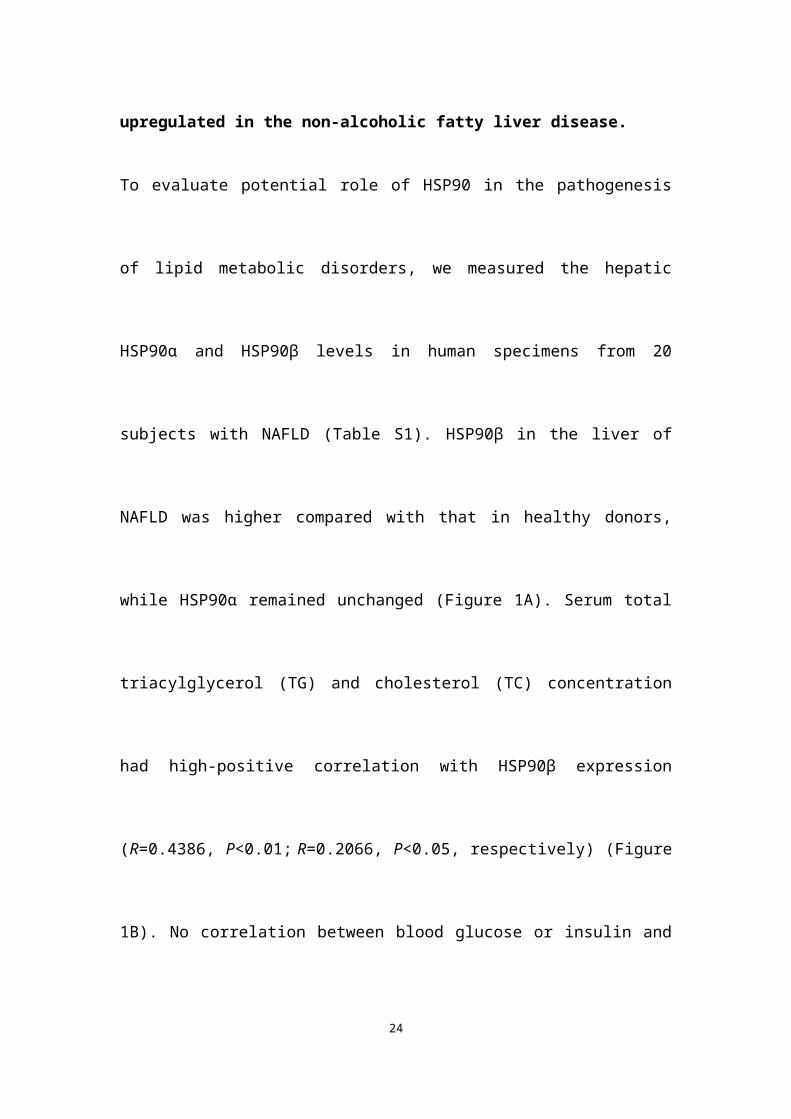
The expression of HSP90β, rather than HSP90α, was upregulated in the non-
alcoholic fatty liver disease.
To evaluate potential role of HSP90 in the pathogenesis of lipid metabolic disorders,
we measured the hepatic HSP90α and HSP90β levels in human specimens from 20
subjects with NAFLD (Table S1). HSP90β in the liver of NAFLD was higher
compared with that in healthy donors, while HSP90α remained unchanged (Figure
1A). Serum total triacylglycerol (TG) and cholesterol (TC) concentration had high-
positive correlation with HSP90β expression (R=0.4386, P<0.01; R=0.2066, P<0.05,
respectively) (Figure 1B). No correlation between blood glucose or insulin and
10
HSP90β was observed (Figure 1B). In contrast, there were no correlations between
HSP90α level and these clinical parameters (Figure 1B). Next, the expression of these
two HSP90s in diet induced obese mice and ob/ob mice was checked. We observed
that the hepatic mRNA and protein level of HSP90AB1, but not HSP90AA1, was
largely increased in high fat diet (HFD)-fed mouse liver (Figure 1C-F), as well as in
ob/ob mice (Figure 1G-J). All together, these results suggest that HSP90β, rather than
HSP90α, is correlated with lipid dysregulation in NAFLD.
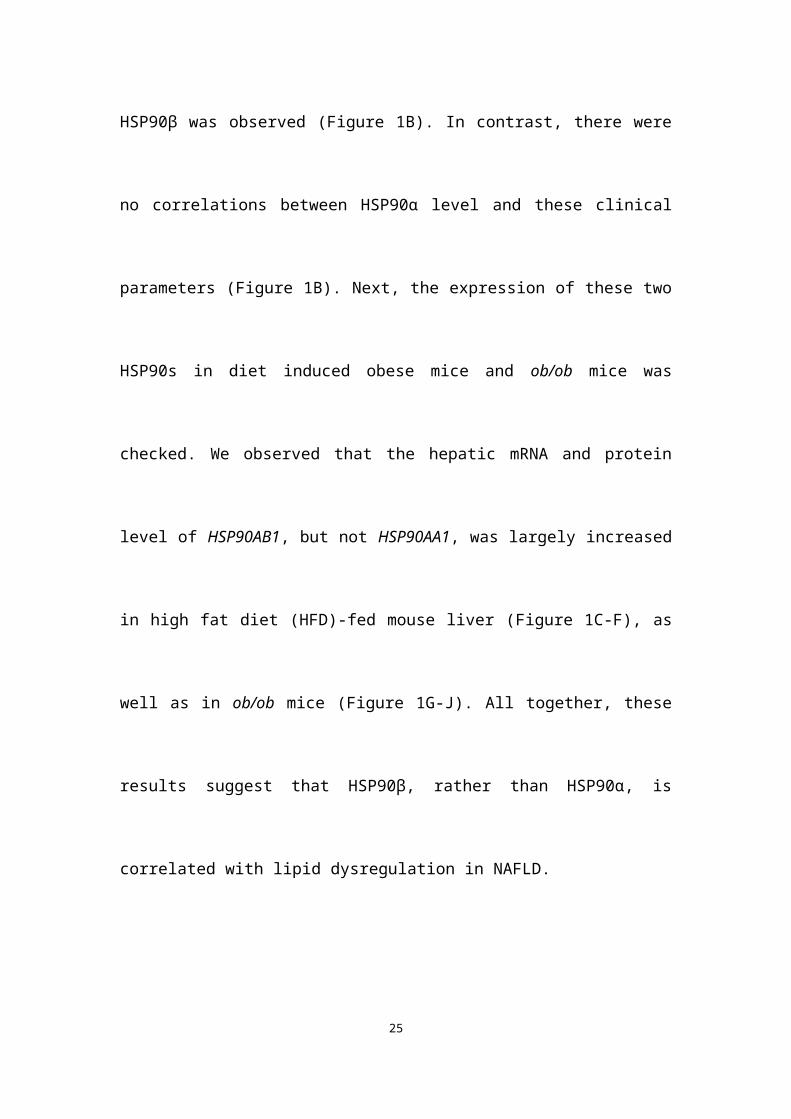
Knockdown of HSP90β improves lipid homeostasis through inhibiting de novo
lipid biogenesis
To check whether modulation of HSP90 paralogs indeed regulates lipid homeostasis,
HL-7702 hepatocytes were transfected with HSP90 siRNAs (Figure 2A) or
overexpression plasmids (Figure 2B). Knockdown of HSP90β reduced 21% of
cellular TG and 33% of TC (Figure 2A). On the contrary, overexpression of HSP90β
increased 24% of TG and 22% of TC (Figure 2B). By contrast, modulating HSP90α
did not affect lipid contents (Figure 2A-B). Nile-Red and filipin staining gave the
same results (Figure 2C). Moreover, knockdown of HSP90β significantly inhibited
the de novo synthesis of both cholesterol and fatty acid in hepatic cells (Figure 2D).
To verify the in vivo role of HSP90β in regulating lipid homeostasis, adenoviruses
carrying HSP90β-shRNA were injected into HFD-fed mice via the tail vein, leading to
an 80% decrease of hepatic HSP90β (Figure 2E). Simultaneously, the expression of
HSP90α was not affected (Figure 2E). In the absence of significant differences in
11
dietary intake (Figure 2F), the body weight was about 16% lower than that in the
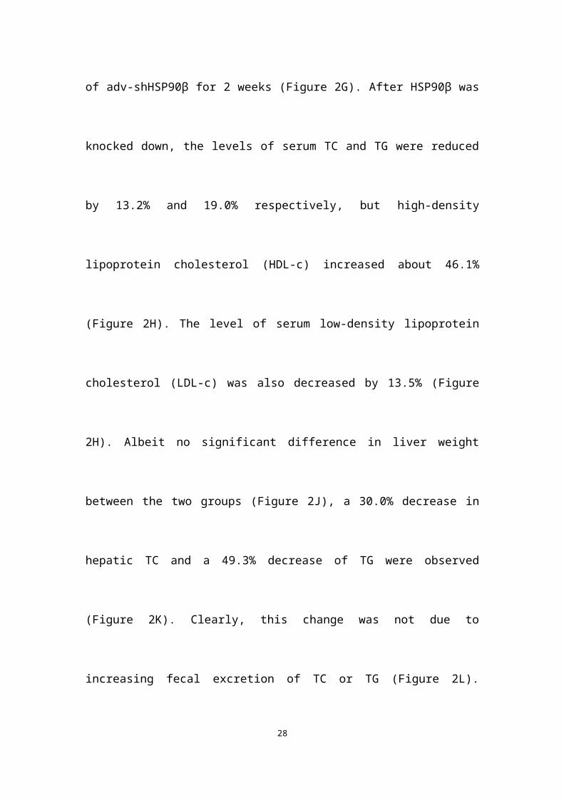
LacZ-shRNA group, after injection of adv-shHSP90β for 2 weeks (Figure 2G). After
HSP90β was knocked down, the levels of serum TC and TG were reduced by 13.2%
and 19.0% respectively, but high-density lipoprotein cholesterol (HDL-c) increased
about 46.1% (Figure 2H). The level of serum low-density lipoprotein cholesterol
(LDL-c) was also decreased by 13.5% (Figure 2H). Albeit no significant difference in
liver weight between the two groups (Figure 2J), a 30.0% decrease in hepatic TC and
a 49.3% decrease of TG were observed (Figure 2K). Clearly, this change was not due
to increasing fecal excretion of TC or TG (Figure 2L). Consistently, the size of
hepatic lipid droplets dramatically decreased in HSP90β knockdown mice (Figure 2I).
The expression of genes involved in the biosynthesis of cholesterol, fatty acid and
triglycerides were lower in HSP90β knockdown mice (Figure 2M). While the
expression of genes involved in VLDL secretion, cholesterol efflux and fatty acid
oxidation was not significantly changed (Figure 2M). Therefore, we reasoned that the
knockdown of HSP90β improves lipid homeostasis, most probably through inhibiting
DNL.
HSP90 have a plethora of client proteins [22]. For example, in adipocytes, HSP90
chaperones PPARγ to facilitate adipocytes survival and differentiation [4]. Indeed, we
observed a reduction of cell size in adipose tissues in adv-shHSP90β treated mice
(Figure S1A). TNF-α and IL-6 expression did not change, suggesting there is no
inflammation (Figure S1B). We did not observe changes of browning and lipid
metabolism related genes in BAT and WAT (Figure S1B). It is possible that HSP90α
12
and HSP90β may recognize and selective bind to different client proteins. Through
Hsp90Int.DB website database query and alignment, we found that GZMA and
H2AFX are specific client proteins for HSP90α, but not regulated by HSP90β [23,
24]. Thus, only when HSP90α was knocked down, the levels of GZMA and H2AFX
proteins were decreased (Figure S2). The functional difference of HSP90 paralogs
might attribute to the difference of their client proteins.
HSP90β improves mSREBPs stability
Since SREBPs are the major transcription factors regulating DNL, we next examined
the effect of HSP90β on SREBPs [18, 25]. Knockdown of HSP90β significantly
inhibited SRE-luciferase activity (Figure 3A) and effectively decreased the expression
of mature SREBP-1/2 in hepatocytes (Figure 3B). By contrast, no significant
influence was observed when HSP90α was knocked down (Figure 3A-B). HSP90β
depletion also decreased the expression of target genes of SREBP (Figure 3C). In
HSP90AB1 knockout cells (HL-7702/KO), mSREBP-1/2 was significantly reduced
(Figure 3D). Furthermore, overexpression of HSP90β obviously increased the
mSREBP-1/2 levels and their target genes expression (Figure 3E-F). These results
provided strong evidences that HSP90β affects the abundance and activity of
SREBPs.
Similar to endogenous mSREBPs, overexpressed exogenous mSREBP-1 and -2 levels
were also reduced in hepatocytes silencing HSP90β (Figure 3G-H), suggesting that
HSP90β may regulate mSREBPs stability. The reduction of mature SREBPs was
13
caused by the increased turnover of SREBP-1 and -2 (Figure 3I-J) since MG-132, a
proteasome inhibitor, abolished the effect of HSP90β depletion (Figure 3K-L). FBW7,
a substrate recognition component of E3 ubiquitin ligase, is responsible for the
degradation of mSREBPs [26]. In the absence of HSP90β, the ubiquitination of
mSREBP-1 was obviously increased (Figure 3M), while knockdown of FBW7
blunted the increased ubiquitination of mSREBP-1 (Figure 3M) and reversed the
stability of mSREBP-1 (Figure 3N). Unexpectedly, neither exogenous nor
endogenous SREBPs were observed to bind HSP90β (Figure S3A, B and D).
Similarly, there was no interaction between HSP90β and FBW7 (Figure S3C-D).
Based on previous study, HSP90β binds to Akt and protects the Thr308 of Akt from
protein phosphatase 2A (PP2A)-mediated dephosphorylation in BALB/3T3 and 293T
cells [27]. In our studies, a direct binding of HSP90β to Akt was also detected (Figure
S3E). When the middle domain of HSP90β consisting amino acids 327-340 was
deleted, the binding of HSP90β to Akt was lost (Figure S3E), suggesting that Akt
interacts with HSP90β in the middle domain. Knockdown of HSP90β dramatically
decreased Akt phosphorylation at Thr308 (Figure S3F). Interestingly, Ser473
phosphorylation was slightly increased, possibly due to feedback reactions. Akt
phosphorylates GSK3β at Ser9, leading to the inactivation of GSK activity. In turn,
GSK3β loses the capacity to phosphorylate SREBPs, thus to stabilize SREBPs by
preventing FBW7 mediated ubiquitination [28]. CHIR9902 and SB216763, specific
inhibitors of GSK3β, reversed the reduction of mSREBPs by HSP90β knockdown
(Figure S3G). Similar results were observed when GSK3β was knocked down (Figure
14
S3H), suggesting that GSK3β activity is required for mSREBPs degradation by
HSP90β inhibition. In HSP90β knockdown mice, the changes of SREBPs and
AKT/GSK3β were in line with in vitro data (Figure S3I). In contrast, knockdown of
HSP90α did not affect Akt-GSK3β signalling pathway (Figure S3F). Collectively,
these results demonstrated that HSP90β prevents the SREBPs ubiquitination and
proteasomal degradation depending on Akt-GSK3β-FBW7 pathway.
Corylin is a specific inhibitor of HSP90β
The above results suggested that selectively inhibiting HSP90β may be a potential
therapeutic strategy for lipid disorders. As the discovery of HSP90β specific inhibitors
is not satisfying [29], we decided to use new tactics. We performed virtue screening to
search compounds that have higher binding affinity to HSP90β (PDB ID code 3PRY)
than to HSP90α (3Q6M). From a structure database of around 3000 in-house natural
compound library, we found 12 compounds with the big difference in the binding
energy between HSP90β and HSP90α. The ability to inhibit SREBP activity of these
compounds were further evaluated (Table S2). It turned out that 9 out of 12 of the
compounds exhibited SREBPs inhibitory effects, suggesting paralog specific
inhibition of HSP90β might particularly affect SREBP activity. Among these
compounds, corylin exhibited highest SREBP inhibitory effects (Table S2). Next, we
used photo-affinity labelling and pull-down experiments to identify the interaction
proteins to corylin [30]. According to the experiment reproducibility (p<0.05) and the
enrichment of proteins (>1.2 fold enrichment) by corylin-agarose beads, we finally
15
focused on three proteins (HSP90β, HSP90α and ENO1) (Table S3). The binding
affinity of corylin to these three proteins was then analyzed using microscale
thermophoresis (MST) assay [31]. The KD between corylin and HSP90β was 24.65 ±
10.16 nM, much lower than that of 17-AAG (Figure 4A and Figure S4A-B). 17-AAG,
but not corylin bound with HSP90α (Figure S4C-D). AP-III-a4 (a specific inhibitor of
ENO1), but not corylin bound with ENO1 (Figure S4E-F). Collectively, the above
results clearly indicate that the binding of corylin to HSP90β is specific. Consistently,
the inhibitory effects of corylin on SREBPs activity were reversed by overexpression
of HSP90β (Figure S5A-C), but not HSP90α or ENO1 (Figure S5D-E). 17-AAG
binds to the NTD of HSP90, while, corylin did not bind to the NTD (1-217), the
charged linker domain (218-276) and the C-terminal domain (CTD) (Figure 4A and
Figure S6A-F). Residues between 276 and 602 are crucial for corylin binding (Figure
4A and Figure S6G-H). 17-AAG and XL-888, but not corylin, significantly inhibited
the ATPase activity of HSP90β (Figure S7A), consistent with the binding data (Figure
4A). Next, we identified a single potential binding pocket located around residues 312
to 439 in the MD of HSP90β using Autodock (Figure 4B), a pocket that is
distinguishable from HSP90α [32]. The binding energy between HSP90β and corylin
is -10.5 KJ/mol. In contrast, the binding of HSP90α and corylin is poor (-5.0 KJ/mol),
which is similar to the results using Glide 6.9 (Table S2). More specifically, we found
three hydrogen bonds formed between the amino acid residues of HSP90β MD and
corylin based on surface topography and sequence of HSP90β. Two hydrogen bonds
are predicted to form between the phenolic hydroxyl group and amino acids N375 and
16
N436. Another hydrogen bond is formed between carbonyl group and W312 (Figure
4B). The residues W312, N375 and N436 were then mutated to alanine to prevent
hydrogen bond formation, consequently, the mutated HSP90β no longer bound to
corylin (Figure 4A and Figure S6I). By contrast, the point mutations did not influence
the interaction between HSP90β and 17-AAG (Figure 4A and Figure S6J).
Consistently, either mutation in these three amino acids, or corylin treatment, did not
affect HSP90β ATPase activity (Figure S7B). When corylin was modified to
neobavaisoflavone, the binding affinity was only slightly reduced (Figure 4C and
Figure S6K). When phenolic hydroxyl group was converted to methoxy group
(compound A, Figure 4C), the binding to HSP90β was significantly weakened (Figure
4C and Figure S6L). Consistently, the effect on SREBP activity of neobavaisoflavone
was similar as corylin, while compound A no longer affected the abundance and
transcriptional activity of SREBPs (Figure 4M and N). Thus, we concluded that the
residues W312, N375 and N436 of HSP90β are essential for corylin binding.
Corylin inhibits de novo lipid synthesis by promoting mSREBPs ubiquitination
and proteasomal degradation
Corylin did not cause obvious hepatic toxicity (Figure 5A). In contrast, a series of
pan-HSP90 inhibitors exhibited severe hepatic toxicity (Figure 5A). This is probably
due to inhibition of HSP90α, since knockdown of HSP90α led to cell death (Figure
S8A). Corylin treatment impaired the levels of mSREBP-1 and -2 in time- and dose-
dependent manners (Figure 5B). Nevertheless, ATF6, a transcription factor with
17
similar processing route as SREBPs, was not affected by corylin (Figure S8B),
suggesting that the effect of corylin on SREBPs is specific. As corylin reduced the
mSREBPs in the nucleus (Figure S8C), the expression of their target genes was
significantly reduced (Figure 5C-D). These results were reproducible in human
primary hepatocytes (Figure S9A-D). Consistently, DNL was significantly attenuated
by corylin (Figure 5E). Furthermore, corylin largely reduced cellular cholesterol and
neutral lipids level (Figure S8D).
In HSP90β knockout hepatocytes (KO cells), corylin no longer influenced mSREBPs
(Figure S10A). KO cells got sensitive to corylin when wild-type HSP90β, but not
triple mutant (W312A/N375A/N436A) HSP90β was put back (Figure S10B). Corylin
decreased endogenous and exogenous mSREBPs (Figure S10C) through increased
ubiquitination (Figure S10D) and accelerated degradation (Figure S10E and F). These
effects were blocked by the proteasome inhibitor MG-132 (Figure S10G) or by FBW7
siRNAs (Figure S10H-I). Corylin disrupted the binding of HSP90β with Akt (Figure
5F) and significantly reduced Thr308 phosphorylation of Akt and Ser9
phosphorylation of GSK3β in a time- and dose-dependent manner (Figure S10K-L).
Notably, in HSP90β KO cells, insulin no longer increased T308 phosphorylation, but
still increased S473 phosphorylation. T308 dephosphorylation by corylin decreased
~55% of Akt activity, a much weaker effect than 17-AAG that inhibited ~80% of Akt
activity (Figure S11). GSK3β S9 phosphorylation and mature form of SREBP-1c
were downregulated in insulin treated HSP90β KO cells (Figure S10J). These data
suggest that HSP90β is involved in insulin stimulated Akt activation, subsequently
18
affect GSK3β-SREBPs and lipid biosynthesis in hepatocytes. Meanwhile, the
decreased levels of mSREBPs was reversed by the GSK3β inhibitors SB216763
(Figure 5G), CHIR9902 (Figure 5H), or GSK3β siRNAs (Figure 5I and Figure S12A).
Overexpression of Akt also blocked the effect of corylin on mSREBPs (Figure 5J and
Figure S12B-D). Taken together, these data demonstrated that corylin, as a specific
inhibitor of HSP90β, promotes mSREBPs ubiquitination and proteasomal degradation
through the Akt-GSK3β-FWB7 pathway.
Corylin improves lipid homeostasis and insulin resistance
Next, we investigated the effect of corylin on obesity in vivo. The bodyweight of mice
fed with western-type diet (WD) plus corylin (15 mg/kg/day and 30 mg/kg/day,
respectively) or lovastatin (30 mg/kg/day) was lighter than that of vehicle treated WD
mice while the food intake remained almost unchanged (Figure 6A). Meanwhile, the
fat/lean or fat/whole-body ratio dropped more than 50% in the same dosage (30
mg/kg/day) of corylin- or lovastatin-treated mice (Figure 6B). Corylin or lovastatin
significantly reduced the level of serum TC, TG and LDL-c, and increased the level of
HDL-c (Figure 6C). The liver weight, hepatic TC and TG in corylin-treated mice were
notably lower than those of the vehicle-treated mice (Figure 6D and E). Corylin
significantly decreased the level of serum AST in WD-fed mice. No significant
difference but reduced ALT level was detected in WD-fed mice with corylin (Figure
6F). Corylin at 30 mg/kg/day dramatically reduced the cell sizes of both white adipose
tissue (WAT) and brown adipose tissue (BAT) in WD-fed mice. The contents of lipids
19
were decreased in the livers of corylin-treated mice (Figure 6G). Similar body weight
lowering effects were observed in HFD-fed mice (Figure S13A). Notably, there was
no significant difference in food intake (Figure S13B), faecal cholesterol or
triglyceride content (Figure S13C-D). Corylin treatment did not increase cold
resistance in HFD-fed mice when exposed to cold (Figure S13E). The energy
expenditure was then measured for 24 h under a 12 h light-day cycle. Interestingly,
although no significant difference was observed at night between the HFD-fed mice
and HFD plus corylin-fed mice, the O2 consumption and energy expenditure (EE)
were significantly increased during the day (Figure S13F-H). The EE increase also
contributed, at least partially, to the body weight loss in HFD-fed mice. Weight
reduction is considered to be beneficial for improving insulin resistance. Next, we
investigated whether corylin improves insulin resistance in vivo. Corylin significantly
blunted the elevated fasting blood glucose and insulin level (Figure S14A-B) and
improves the glucose and insulin intolerance (Figure S14C-D) in WD-fed mice.
Consistent with the in vitro results, corylin reduced the level of mSREBPs in the liver
of WD-fed mice (Figure S14E-F). Most of SREBP-1 and SREBP-2 target genes were
significantly downregulated in the liver of WD-fed mice by corylin (Figure 14G-I). In
hepatocytes, as well as in mice livers, corylin neither increases LXR target gene
expression, nor activates ER stress genes expression (Figure S15). We also found that
the expression of VLDL secretion, cholesterol efflux, fatty acid oxidation, adipocyte
browning, and glucose uptake were not significantly changed (Figure S16).
To address the question whether corylin improves liver metabolism through inhibiting
20
HSP90β, we treated HFD-fed mice with corylin for 4 weeks, then adenoviral virus
expressing HSP90β or control were injected. Overexpression of HSP90β reversed the
corylin effects on SREBPs and their target gene expression (Figure S17A-H). HSP90β
also reversed lipid parameters that were improved by corylin (Figure S17I-M). In
summary, corylin inhibits SREBPs and improves lipid homeostasis in vivo through
inhibiting HSP90β.
Corylin decreases atherosclerosis development
The effect of corylin on atherosclerotic lesion formation was further explored in
ApoE-/- mice. Corylin treatment did not cause obvious toxicity and difference in food
intake (Figure 7A), but reduced the weight gain at high dose (Figure 7B). Both corylin
and lovastatin treatment made the plasma transparent, indicating reduced lipid content
(Figure 7C). Moreover, corylin or lovastatin obviously decreased TC, TG and LDL-c
levels and elevate HDL-c levels in plasma (Figure 7D). Corylin reduced the number
and size of aortic plaques in WD-fed ApoE-/- (Figure 7E and F). Particularly, the areas
of the lesion in the thoracic aorta and abdominal aorta were significantly smaller
(Figure 7G-I). The lesion areas in cross-sections of the aortic sinus in the corylin-
treated mice were reduced dramatically (Figure S18A). The activated endothelial cells
marker VCAM-1 was reduced in corylin-treated mice (Figure S18B). Upon corylin
treatment, the expression of HSP90AB1 and HSP90AA1 remained unchanged, while
SREBPs and their target genes involved in lipid synthesis were downregulated (Figure
7J). In contrast, HMGCR, HMGCS and FASN were dramatically upregulated by
21
lovastatin (Figure 7J). These results demonstrated that corylin ameliorates
atherosclerosis lesion formation of plaques in the aortic root in WD-fed ApoE-/- mice
without increasing fatty acid synthesis, a feature superior to lovastatin.
Discussion
In this report, we have demonstrated the clinical significance of the expression of
HSP90β in NAFLD patients. Further, a novel paralog-specific HSP90 inhibitor,
corylin has been identified. Genetic targeting or pharmacologic inhibition of HSP90β
leads to degradation of SREBPs via Akt-GSK3β-FBW7 pathway, thus improves lipid
homeostasis.
To date, the paralog-specific role of HSP90 in the liver is little known. In this study,
we observed HSP90β, instead of HSP90α, obviously increases in the liver of human
NAFLD patients and obesity-induced fatty liver mice (Figure 1). Further studies
indicate that HSP90β plays a paralog-specific role in regulating lipid homeostasis.
Our results help researchers to understand the role of different paralogs of HSP90 in
lipid metabolism, and promote HSP90β as a drug target for the treatment of lipid
disorders. Chemical compounds that specifically inhibit HSP90β certainly satisfy the
demand of precise medical treatment for lipid disorders. The NTD with ATP pocket-
and CTD with novobiocin-binding site of HSP90 are under intense study and
therefore attract most interest in drug discovery. Small compounds such as 17-
allylaminogeldamamycin (17-AAG), ganetespib (STA-9090), SNX-2112 and
novobiocin have been developed accordingly [33]. The NTD is not only homology to
22
HSP90 chaperone family members, but also to members of the gyrase-Hsp90-
histidine kinase-MutL (GHKL) group within the ATPase/kinase superfamily [34].
Similarly, the CTD contains a second ATP binding site, which can be competed with
novobiocin, cisplatin, epilgallocatechin-3-gallate (EGCG) and taxol [35]. These two
binding domains are closely related to cell proliferation and tumor progression [36].
Similar as other drugs targeting ATP/ADP binding pocket, the off-target effects
hampered the development of the first generation geldanamycins that exerted
significant toxicities [37, 38]. Indeed, we observed cytotoxicity of these compounds in
hepatocytes (Figure 5A). Corylin binds to HSP90β in a unique pocket located in the
middle domain formed by amino acids W312/N375/N436, notably, either mutation in
these three amino acids, or corylin treatment, did not affect HSP90β ATPase activity
(Figure S7B). The KD of corylin with HSP90β was 24.65 ± 10.16 nM, the highest
binding affinity ever reported. Therefore, corylin is the small molecule that
distinguishes HSP90α and HSP90β. With this paralog-specific inhibitor, less
cytotoxicity was observed (Figure 5A) and further understanding of the biologic
function of this chaperone family is warranted.
In this study, we found genetic deletion or pharmacological inhibition of HSP90β
caused the Thr308 dephosphorylation of AKT (Figure S2F, Figure S10J-L). Akt full
activation needs both T308 in the catalytic domain and S473 in the carboxy-terminal
noncatalytic region [39]. It is reported that either T308 or S473 mutation leads to
diminished Akt activity, although T308 mutation seems to cause more severe effect
23
[40]. The downstream Akt targets are affected differently by Akt mutations. For
example, it has been shown that genetic ablation of mTORC2 components (rictor,
mLST8 or sin1) abolished S473 phosphorylation and only affected a subset of Akt
targets in vivo, including FoxO1/3a, while other Akt targets, TSC2, GSK3, S6K and
4E-BP1 were unaffected [41]. As in our case, we found that genetic or pharmacologic
inhibition of HSP90β caused T308 dephosphorylation. T308 dephosphorylation by
corylin decreased ~55% of Akt activity, a much weaker effect than 17-AAG that
inhibited ~80 % of Akt activity (Figure S16). Subsequently, GSK3β was then
activated and stimulated SREBP degradation (Figure S2F and Figure S10). The effect
could rather be a result of changes in overall Akt activity change than S473
phosphorylation, since T308 and S473 phosphorylation could be separated regulated
[41]. As corylin decreased T308 without affecting S473 very much, it could be a good
tool to study mTORC2 independent Akt regulation.
Previous reports used pan-HSP90 inhibitor 17-AAG or its analogues to study the
function role of HSP90 in different disease models [42]. As 17-AAG binds both
HSP90α and HSP90β, meanwhile, there is no HSP90β-flox mice available (HSP90β
KO mice are lethal), the HSP90β specific role in physiological and pathological
conditions remain largely unknown. With HSP90β specific inhibitor corylin, HSP90β
specific roles in these diseases could be re-examined. It should also be noted that
although alcoholic fatty liver disease (AFLD) and NAFLD share similar pathological
spectra, these two diseases differ from each other not only in terms of clinical features
and patient outcomes [43], but also in terms of underlying mechanisms [44]. Notably,
24
chemicals that protects against ethanol induced liver injury, such as silymarin [45],
not necessarily alleviated NAFLD and vice versa [46]. Whether inhibition of HSP90β
in AFLD models also decreased alcohol mediated oxidative stress, reduced serum
endotoxin, decreased inflammatory responses in hepatic macrophages, deserve further
study.
In the last decades, the lab of Brown and Goldstein has discovered that SREBPs are
master regulators of de novo lipogenesis [18, 47]. These studies suggest that selective
inhibition of SREBPs might constitute a new class of drugs against lipid disorders, as
well as type 2 diabetes. However, the inhibitors of SREBPs processing, such as 25-
HC, activate LXR and induce ER stress, leading to hepatic steatosis and insulin
resistance [48, 49]. Corylin suppressed SREBPs activity in a different mechanism. It
selectively inhibits HSP90β and promotes SREBPs degradation depending on Akt-
GSK3β-FBW7 pathway (Figure 4 and Figure S2 and Figure S10), without affect LXR
target genes and ER stress gene expression. In animal experiments, corylin alleviates
hepatic steatosis and hyperglycemia (Figure 6G), insulin resistance (Figure S14A-D).
These data suggested that corylin has an incomparable advantage in the treatment of
lipid disorders. In a previous study [50], it shows that corylin was subjected to
massive first-pass metabolism in liver and intestine. Therefore, we supposed that liver
is the major organ that responsible for the beneficial effects of corylin.
We believe our findings pave the way for future investigation on the paralog-specific
roles of HSP90α and HSP90β in different physiological and pathological conditions.
In the obese animals, both mRNA and protein level of HSP90AB1 is increased.
25
Therefore, unraveling the different molecular mechanisms that activate HSP90
paralogs is very important to advance the understanding of HSP90 functions, thus to
improve the therapeutic effects in metabolic disease, cancer, neurodegeneration
diseases, eplipsy, etc.
26
Reference1. Rohl A, Rohrberg J, Buchner J. The chaperone Hsp90: changing partners for demanding clients. Trends Biochem Sci. 2013; 38: 253-62.2. Sha L, Wang X, Li J, Shi X, Wu L, Shen Y, et al. Pharmacologic inhibition of Hsp90 to prevent GLT-1 degradation as an effective therapy for epilepsy. J Exp Med. 2017; 214: 547-63.3. Ambade A, Catalano D, Lim A, Kopoyan A, Shaffer SA, Mandrekar P. Inhibition of heat shock protein 90 alleviates steatosis and macrophage activation in murine alcoholic liver injury. J Hepatol. 2014; 61: 903-11.4. Nguyen MT, Csermely P, Soti C. Hsp90 chaperones PPARgamma and regulates differentiation and survival of 3T3-L1 adipocytes. Cell Death Differ. 2013; 20: 1654-63.5. Wheeler MC, Gekakis N. Hsp90 modulates PPAR gamma activity in a mouse model of nonalcoholic fatty liver disease. J Lipid Res. 2014; 55: 1702-10.6. Seo YH. Organelle-specific Hsp90 inhibitors. Arch Pharm Res. 2015; 38: 1582-90.7. Chen B, Piel WH, Gui L, Bruford E, Monteiro A. The HSP90 family of genes in the human genome: insights into their divergence and evolution. Genomics. 2005; 86: 627-37.8. Tsutsumi S, Neckers L. Extracellular heat shock protein 90: A role for a molecular chaperone in cell motility and cancer metastasis. Cancer Sci. 2007; 98: 1536-9.9. Shrestha L, Patel HJ, Chiosis G. Chemical Tools to Investigate Mechanisms Associated with HSP90 and HSP70 in Disease. Cell Chem Biol. 2016; 23: 158-72.10. Wymann MP, Schneiter R. Lipid signalling in disease. Nat Rev Mol Cell Bio. 2008; 9: 162-76.11. Abrass CK. Cellular lipid metabolism and the role of lipids in progressive renal disease. Am J Nephrol. 2004; 24: 46-53.12. Kawakami A, Yoshida M. Apolipoprotein CIII Links Dyslipidemia with Atherosclerosis. J Atheroscler Thromb. 2009; 16: 6-11.13. Diraison F, Moulin P, Beylot M. Contribution of hepatic de novo lipogenesis and reesterification of plasma non esterified fatty acids to plasma triglyceride synthesis during non-alcoholic fatty liver disease. Diabetes Metab. 2003; 29: 478-85.14. Sundqvist A, Bengoechea-Alonso MT, Ye X, Lukiyanchuk V, Jin JP, Harper JW, et al. Control of lipid metabolism by phosphorylation-dependent degradation of the SREBP family of transcription factors by SCFFbw7. Cell Metab. 2005; 1: 379-91.15. Lambert JE, Ramos-Roman MA, Browning JD, Parks EJ. Increased De Novo Lipogenesis Is a Distinct Characteristic of Individuals With Nonalcoholic Fatty Liver Disease. Gastroenterology. 2014; 146: 726-35.16. Rotman Y, Sanyal AJ. Current and upcoming pharmacotherapy for non-alcoholic fatty liver disease. Gut. 2017; 66: 180-90.17. Stiede K, Miao W, Blanchette HS, Beysen C, Harriman G, Harwood HJ, Jr., et al. Acetyl-coenzyme A carboxylase inhibition reduces de novo lipogenesis in overweight male subjects: A randomized, double-blind, crossover study. Hepatology. 2017; 66: 324-34.18. Horton JD, Goldstein JL, Brown MS. SREBPs: activators of the complete program of cholesterol and fatty acid synthesis in the liver. J Clin Invest. 2002; 109: 1125-31.19. Brown MS, Goldstein JL. The SREBP pathway: regulation of cholesterol metabolism by proteolysis of a membrane-bound transcription factor. Cell. 1997; 89: 331-40.20. Kuan YC, Hashidume T, Shibata T, Uchida K, Shimizu M, Inoue J, et al. Heat Shock Protein 90 Modulates Lipid Homeostasis by Regulating the Stability and Function of Sterol Regulatory Element-
27
binding Protein (SREBP) and SREBP Cleavage-activating Protein. J Biol Chem. 2017; 292: 3016-28.21. Jhaveri K, Taldone T, Modi S, Chiosis G. Advances in the clinical development of heat shock protein 90 (Hsp90) inhibitors in cancers. Biochim Biophys Acta. 2012; 1823: 742-55.22. Taipale M, Krykbaeva I, Koeva M, Kayatekin C, Westover KD, Karras GI, et al. Quantitative analysis of HSP90-client interactions reveals principles of substrate recognition. Cell. 2012; 150: 987-1001.23. Martinvalet D, Dykxhoorn DM, Ferrini R, Lieberman J. Granzyme A cleaves a mitochondrial complex I protein to initiate caspase-independent cell death. Cell. 2008; 133: 681-92.24. Yang XL, Zou P, Yao J, Yun D, Bao HM, Du RY, et al. Proteomic Dissection of Cell Type-Specific H2AX-Interacting Protein Complex Associated with Hepatocellular Carcinoma. J Proteome Res. 2010; 9: 1402-15.25. Eberle D, Hegarty B, Bossard P, Ferre P, Foufelle F. SREBP transcription factors: master regulators of lipid homeostasis. Biochimie. 2004; 86: 839-48.26. Sundqvist A, Bengoechea-Alonso MT, Ye X, Lukiyanchuk V, Jin J, Harper JW, et al. Control of lipid metabolism by phosphorylation-dependent degradation of the SREBP family of transcription factors by SCF(Fbw7). Cell Metab. 2005; 1: 379-91.27. Sato S, Fujita N, Tsuruo T. Modulation of Akt kinase activity by binding to Hsp90. Proc Natl Acad Sci U S A. 2000; 97: 10832-7.28. Xiao X, Song BL. SREBP: a novel therapeutic target. Acta Biochim Biophys Sin (Shanghai). 2013; 45: 2-10.29. Sidera K, Patsavoudi E. HSP90 Inhibitors: Current Development and Potential in Cancer Therapy. Recent Pat Anti-Canc. 2014; 9: 1-20.30. Kanoh N, Honda K, Simizu S, Muroi M, Osada H. Photo-cross-linked small-molecule affinity matrix for facilitating forward and reverse chemical genetics. Angewandte Chemie. 2005; 44: 3559-62.31. Jerabek-Willemsen M, Wienken CJ, Braun D, Baaske P, Duhr S. Molecular Interaction Studies Using Microscale Thermophoresis. Assay Drug Dev Techn. 2011; 9: 342-53.32. Yim KH, Prince TL, Qu S, Bai F, Jennings PA, Onuchic JN, et al. Gambogic acid identifies an isoform-specific druggable pocket in the middle domain of Hsp90beta. Proc Natl Acad Sci U S A. 2016; 113: E4801-9.33. Donnelly A, Blagg BSJ. Novobiocin and Additional Inhibitors of the Hsp90 C-Terminal Nucleotide-binding Pocket. Curr Med Chem. 2008; 15: 2702-17.34. Prodromou C, Pearl LH. Structure and functional relationships of Hsp90. Curr Cancer Drug Targets. 2003; 3: 301-23.35. Marcu MG, Chadli A, Bouhouche I, Catelli M, Neckers LM. The heat shock protein 90 antagonist novobiocin interacts with a previously unrecognized ATP-binding domain in the carboxyl terminus of the chaperone. J Biol Chem. 2000; 275: 37181-6.36. Whitesell L, Lindquist SL. HSP90 and the chaperoning of cancer. Nat Rev Cancer. 2005; 5: 761-72.37. Butler LM, Ferraldeschi R, Armstrong HK, Centenera MM, Workman P. Maximizing the Therapeutic Potential of HSP90 Inhibitors. Mol Cancer Res. 2015; 13: 1445-51.38. Hawle P, Siepmann M, Harst A, Siderius M, Reusch HP, Obermann WM. The middle domain of Hsp90 acts as a discriminator between different types of client proteins. Mol Cell Biol. 2006; 26: 8385-95.39. Bhaskar PT, Hay N. The two TORCs and Akt. Dev Cell. 2007; 12: 487-502.
28
40. Alessi DR, Andjelkovic M, Caudwell B, Cron P, Morrice N, Cohen P, et al. Mechanism of activation of protein kinase B by insulin and IGF-1. EMBO J. 1996; 15: 6541-51.41. Guertin DA, Stevens DM, Thoreen CC, Burds AA, Kalaany NY, Moffat J, et al. Ablation in mice of the mTORC components raptor, rictor, or mLST8 reveals that mTORC2 is required for signaling to Akt-FOXO and PKC alpha but not S6K1. Dev Cell. 2006; 11: 859-71.42. Taipale M, Jarosz DF, Lindquist S. HSP90 at the hub of protein homeostasis: emerging mechanistic insights. Nat Rev Mol Cell Bio. 2010; 11: 515-28.43. Toshikuni N, Tsutsumi M, Arisawa T. Clinical differences between alcoholic liver disease and nonalcoholic fatty liver disease. World J Gastroenterol. 2014; 20: 8393-406.44. Gao B, Bataller R. Alcoholic liver disease: pathogenesis and new therapeutic targets. Gastroenterology. 2011; 141: 1572-85.45. Song Z, Deaciuc I, Song M, Lee DY, Liu Y, Ji X, et al. Silymarin protects against acute ethanol-induced hepatotoxicity in mice. Alcohol Clin Exp Res. 2006; 30: 407-13.46. Cacciapuoti F, Scognamiglio A, Palumbo R, Forte R, Cacciapuoti F. Silymarin in non alcoholic fatty liver disease. World J Hepatol. 2013; 5: 109-13.47. Goldstein JL, DeBose-Boyd RA, Brown MS. Protein sensors for membrane sterols. Cell. 2006; 124: 35-46.48. Repa JJ, Mangelsdorf DJ. The role of orphan nuclear receptors in the regulation of cholesterol homeostasis. Annu Rev Cell Dev Biol. 2000; 16: 459-81.49. Schultz JR, Tu H, Luk A, Repa JJ, Medina JC, Li L, et al. Role of LXRs in control of lipogenesis. Genes Dev. 2000; 14: 2831-8.50. Qin Z, Li S, Yao Z, Hong X, Xu J, Lin P, et al. Metabolic profiling of corylin in vivo and in vitro. J Pharm Biomed Anal. 2018; 155: 157-68.
Article Information
Acknowledgments. The authors acknowledge technical support from Prof. Hongbin
Sun (China Pharmaceutical University) for the synthesis of compound A.
Duality of Interest. There are two pending patent applications of corylin for the
treatment of metabolic diseases.
Funding. This work was supported by the National Science Foundation of China
(81773957, 81421005, 81322051, 31722028), National Science and Technology
Major Projects for "Major New Drugs Innovation and Development"
(2018ZX09201001-001-001), the 111 Project (B16046). This project was also
supported by the Project Program of State Key Laboratory of Natural Medicines,
29
China Pharmaceutical University (SKLNMZZCX201820), and the Open Project of
State Key Laboratory of Natural Medicines (SKLNMKF201706), the "Double First-
Class" University Project (CPU2018GF04). China Postdoctoral Science Foundation
(2018M642380).
Author contributions. X. X., HJ.L, P. L, J. L. and ZG.Z. conceived and designed the
work; X.X. and ZG.Z. wrote the manuscript. ZG.Z., X.Z., D.J., HM.C., XX. L., XX.
J. , L. D, PM.T., performed experiments. Y.L., B.X., C.L., H.L., J.Z., L.D., L.C., C.P.,
W.Z. and L.W. provided technical and material support. X.X. is the guarantor of this
work and, as such, had full access to all the data in the study and takes responsibility
for the integrity of the data and the accuracy of the data analysis.
Data and Resource Availability. The datasets generated during and/or analyzed
during the current study are available from the corresponding author upon reasonable
request.
30
31
Figures and legends
Figure 1. The Level of HSP90 paralogs in the Liver of Obese Mice and NAFLD Patients(A) The immunohistochemistry staining of HSP90β or HSP90α in the liver of healthy
donors (n=5) and NAFLD patients (n=20). (B) The correlation between
immunostaining signal of HSP90s and the clinical parameters including serum level
of glucose, insulin, TG and TC (n=20). (C, D, G, H) Liver sections of HFD-induced
obese mice (C, D) or ob/ob mice (G, H) were stained with HSP90 paralog specific
antibodies. (D and H) The mRNA level of HSP90AB1 or HSP90AA1 in the liver was 32
analyzed in HFD- induced obese mice (E) and ob/ob mice (I), respectively. n = 9 for
chow group or HFD group; n = 14 for WT mice group, n = 8 for ob/ob mice group. (F
and J) The protein level of HSP90α or HSP90β in the liver was analyzed by western
blot in HFD- induced obese mice (E) and ob/ob mice (I), respectively. Error bars are
represented as mean ± SEM. The Pearson correlation coefficient is used to test the
correlation between HSP90 paralogs expression and clinical parameters (B). Student
t-test was used to analyze significance between two sets of data (D and F). ***p <
0.001 vs chow diet mice or WT mice.
33
Figure 2. Knockdown of HSP90β Improves Lipid Homeostasis
(A and B) The cellular TG and TC levels were measured in human liver HL-7702
34
hepatocytes transfected with siRNA targeting HSP90β or HSP90α (A) or
overexpression (OE) of HSP90β (3.6 times) or HSP90α (4.2 times) (B), the effect of
interference or overexpression was verified by western blot. (C) The siRNA treated
HL-7702 hepatocytes incubated in medium B for 48 h, and then switched to medium
D for another 24 h. The cells then were stained with Nile-Red, which specifically
recognizes neutral lipids or filipin, which definitely binds free cholesterol. (D) HL-
7702 hepatocytes were incubated in medium B treating with indicated concentrations
of siRNA HSP90β for 48 h, and then switched to medium D for another 24 h. Acetic
acid sodium salt 1-14C was directly added into the medium and incubated for
additional 2 h. The radioactive products were identified by comparison with unlabeled
standards and visualized with iodine vapor. (E-L) Male C57BL/6J mice (6 weeks)
were randomly grouped (10 mice each group). Mice were allowed ad libitum access
to water and high fat diet (HFD). After four weeks, mice were intravenously injected
with titer of 5×10⁹ adenovirus expressing the shRNA targeting HSP90β or the shRNA
targeting LacZ. HFD was still administrated to mice for additional 14 days. Then,
mice were sacrificed and subjected to various analysis. (E) The total protein from
mice liver were prepared, and subjected to immunoblot analysis. Statistical analysis of
hepatic HSP90β protein level in panel E normalized to Actin. (F) Food intake during
the six week treatment. (G) Body weight measurement during the six weeks
treatment. (H) The effect of HSP90β knockdown on serum TG, TC, HDL-c and LDL-
c levels. (I) Oil red staining of liver sections. (J) The weight of liver. (K) The TC and
TG level in the liver. (L) The TC and TG in the feces were analyzed by GC-MS. (M)
35
Lipid mebabolism related genes expression was detected by q-RT-PCR. Mouse
GAPDH was used as the internal control. Error bars are represented as mean ± SEM.
Statistical analysis was done with one-way ANOVA (Dunnett’s post test) (A, B and
C), two-way ANOVA (Bonferroni’s test) (F and G), or student’s t-test (E, H, J, K, and
L). *p < 0.05,**p < 0.01,***p < 0.001 vs control vector, control adenovirus or
control siRNA.
36
Figure 3. HSP90β Regulates de novo Lipid Synthesis through Promoting the
mSREBPs Ubiquitination and Proteasomal degradation
(A) HL-7702/SRE-Luc cells were incubated in medium B and treated with indicated
37
siRNA for 48 h, the cells were then switched to medium D or medium D containing
25-HC (2 μg/ml) for 4 h. After the treatment, cells were lysed and luciferase activity
was measured. (B and C) HL-7702 hepatocytes were incubated in medium B and
treated with indicated siRNA for 48 h, the cells were then switched to medium D or
medium D containing 25-HC (2 μg/ml) for 4 h. After the treatment, (B) the whole cell
extracts underwent immunoblotting with indicated antibodies. P represents precursor
SREBP and m represents mSREBP. (C) The expression of various genes was analyzed
by qRT-PCR. Human GAPDH was used as the control. (D) The HL-7702/WT and
HL-7702/KO HSP90β cells were depleted of sterols by incubating in medium D for
24 h, the total protein of cells were prepared and subjected to immunoblotting with
indicated antibodies. (E and F) HL-7702 hepatocytes were transfected with myc-
HSP90β (3.4 times) for 24 h, and then depleted of sterols by incubating in medium D
for another 24 h, (E) the whole cell extracts underwent immunoblotting with indicated
antibodies. (F) The expression of various genes was analyzed by qRT-PCR. Human
GAPDH was used as the internal control. (G and H) HL-7702 hepatocytes were
transfected with flag-mSREBP-1 (G) or flag-mSREBP-2 (H) plasmids and cultured
for 24 h. The cells were switched to medium D for another 24 h, the whole cell
extracts underwent immunoblotting with indicated antibodies. (I and J) HL-7702
hepatocytes were transfected with flag-mSREBP-1 plasmid and siRNA against
HSP90β for 48 h (I). HL-7702 hepatocytes were transfected with flag-mSREBP-2
plasmid and siRNA HSP90β for 48 h (J). After adding 50 µM cycloheximide for 1 h,
the whole cell extracts were harvested after incubation for indicated periods of time,
38
mSREBPs were detected by immunoblotting with indicated antibodies. The graph
depicted the averaged ratio of the autoradiographic signals of flag to actin levels, the
dotted lines show the half-life of flag-mSREBP-1/-2. (K) HL-7702 hepatocytes were
transfected with indicated siRNA for 48 h. The cells were switched to medium D
containing MG-132 for 12 h, the whole cell extracts underwent immunoblotting with
indicated antibodies. (L) HL-7702/SRE-Luc cells were transfected with HSP90β
siRNAs for 48 h, luciferase activity was then measured. (M) 293T cells were
transfected with flag-mSREBP-1 and HA-ubiquitin and with or without siRNA
HSP90β and FBW7, ubiquitylated SREBP-1 was detected by immunoblot. (N) HL-
7702 hepatocytes were transfected with the indicated siRNA for 48 h, the whole cell
extracts underwent immunoblotting with indicated antibodies. Error bars are
represented as mean ± SEM. Statistical analyses were done with one-way ANOVA
(Dunnett’s post test) (A, C, and E). *p < 0.05,**p < 0.01,***p < 0.001 vs control
siRNA or control vector.
39
Figure 4. Corylin Binds specifically to HSP90β
(A) Various EGFP-HSP90β truncation mutant plasmids were constructed and the
recombination mutant proteins of EGFP-HSP90β were purified. The interaction
between different truncated or a triple mutant (W312A, N375A, N436A) form of
HSP90β and 17-AAG or corylin was detected by microscale thermophoresis (MST).
(B) Molecular docking model of corylin binding to HSP90β. Molecular modeling
suggested that corylin binds to a pocket within MD composed of residues 312 to 439.
Hydrogen bonds are formed between corylin and W312, N375, N436 of EGFP-
HSP90β. (C) Schematic diagram of key structural difference among corylin,
40
compound A and neobavaisoflavone. The interaction between recombination proteins
EGFP-HSP90β and corylin, compound A or neobavaisoflavone was detected by
microscale thermophoresis (MST).
Figure 5. Corylin Inhibits de novo Lipids Synthesis by Regulating SREBPs
41
Activity
(A) HL-7702 cells were treated with HSP90 inhibitors with different structure
backbone. Only corylin did not cause cytotoxicity. (B) HL7702 cells were depleted of
sterols by incubating in medium D for 24 h, and then switched to medium D
containing indicated concentration of corylin for 4 h, or corylin (3 μg/ml) for different
time, the whole cell extracts underwent immunoblotting with indicated antibodies. (C-
D) HL7702 cells were depleted of sterols by incubating in medium D for 24 h, and
then switched to medium D containing 3 µg/ml corylin for 4 h. The expression of
various genes was analyzed by qRT-PCR. (E) HL7702 cells were depleted of sterols
by incubating in medium D for 24 h, and then switched to medium D containing 3
μg/ml corylin or 2 μM lovastatin for 16 h. Acetic acid sodium salt 1-14C was directly
added into the medium and incubated for additional 2 h. (F) 293T cells were
transfected with Myc-HSP90β and HA-Akt for 24 h, after the treatment, the cells
were incubated with medium D containing corylin for another 4 h. Cells were lysed
and pulled down by Myc antibody. HL-7702 hepatocytes were treated with GSK3β
inhibitor SB216763 (10 µM) (G) or CHIR-9902 (10 µM) (H) for 1 h, the cells were
switched to medium D supplemented with inhibitors plus vehicle, or 3 µg/ml corylin
for 4 h. The whole cell extracts underwent immunoblotting with indicated antibodies.
(I) HL-7702 hepatocytes were transfected with GSK3β siRNA for 48 h, after the
treatment, the cells were switched to medium D treated with 3 µg/ml corylin for 4 h.
(J) HL-7702 hepatocytes were transfected with HA-Akt for 24 h, after the treatment,
the cells were switched to medium D treated with 3 µg/ml corylin for 4 h. Whole cell
42
extracts underwent immunoblotting with indicated antibodies. Error bars are
represented as mean ± SEM. Statistical analyses were done with one-way ANOVA
(Dunnett’s post test) (A and C). *p < 0.05, **p < 0.01, ***p < 0.001 vs DMSO.
43
Figure 6. Corylin Improves Lipid Homeostasis in WD-fed mice.
Male C57BL/6J mice at 6 weeks of age were randomly grouped (n=6). Mice were
allowed ad libitum access to water and different types of diets (WD, western-type
diet). Vehicle (0.5% CMC-Na), corylin (15 or 30 mg/kg), or lovastatin (30 mg/kg)
was administrated to mice by gastric irrigation every day. After 6 weeks treatment, the
mice were sacrificed and subjected to a series of analysis as indicated below. (A)
44
Food intake and body weight. (B) The ratio of fat and body weight or lean. (C) Effect
of corylin on serum TG, TC, LDL-c and HDL-c levels. (D) The weight of liver. (E)
Effect of corylin on TG and TC levels in the liver. (F) Effect of corylin on Alanine
aminotransferase (ALT) and aspartate transaminase (AST) in the serum. (G) Oil red
staining in liver and histological analysis of liver, WAT and BAT. Error bars are
represented as mean ± SEM. Statistical analyses were done with two-way ANOVA
(Bonferroni’s test) (A) or one-way ANOVA (Dunnett’s post test) (B-G). *p < 0.05,
**p < 0.01, ***p < 0.001 vs WD.
45
Figure 7. Corylin Decreases the Atherosclerosis Development in ApoE-/- Mice
Six-week-old male ApoE-/- mice were randomly grouped (n=12) and fed with WD
supplemented with vehicle (0.5% CMC-Na), corylin (30mg/kg) or lovastatin (30
mg/kg) for 16 weeks. After the treatment, the mice were sacrificed and subjected to
various analyses as described below. (A) Food intake during the 16 weeks experiment
period. (B) Body weight of each group during the 16 weeks experiment period. (C)
46
Representative photographs of plasma from each group. (D) The levels of TC, TG,
LDL-c and HDL-c in plasma after 16 weeks treatment. (E) Representative
photographs from en face analysis of aortas from different groups after 16 weeks
treatment. (F-I) Quantification of the lesion areas in (E). (J) qRT-PCR analysis of gene
expression in ApoE-/- mice liver after 16 weeks. For each group, equal amounts of
total RNA from three mice were subjected to qRT-PCR quantification. Error bars are
represented as mean±SEM. Statistical analyses were done with two-way ANOVA
(Bonferroni’s test) (A and B) or one-way ANOVA (Dunnett’s post test) (D, and F-J).
*p < 0.05, **p < 0.01, ***p < 0.001 vs WD.
47
Graphical abstract
48
AbbreviationsABCA1, ATP-binding cassette transporter 1; ACC, acetyl-CoA carboxylase; ACO,
acyl-CoA oxidase; Akt, protein kinase B; ApoB, apolipoprotein B; ApoE,
apolipoprotein E; BAT, brown adipose tissue; CPT1, carntine palmitoyltransferase 1;
CYP7A1, cholesterol α-hydroxylase; Dio2, type 2 iodothyronine deiodinase; FASN,
fatty acid synthase; FBW7, F-Box and WD40 Domain Protein-7; FDPS, farnesyl
diphosphate synthase; Glut1, glucose transporter 1; Glut4, glucose transporter 4;
GSK3β, glycogen synthase kinase 3 beta; HDL-c, serum high-density cholesterol;
HFD, high fat diet; HMGCR, 3-hydroxy-3-methylglutary-CoA reductase; HMGCS, 3-
hydroxy-3-methylglutary-CoA synthase; HL, hepatic lipase; HSP90AB1, heat shock
protein 90 alpha family class B member 1; HSP90AA1, heat shock protein 90 alpha
family class A member 1; Insig, insulin induced gene; LDL-c, serum low-density
cholesterol; LDLR, low density lipoprotein receptor; LPDS, lipoprotein deficient
serum; LPL, lipoprotein lipase; NAFLD, non-alcoholic fatty liver disease; PGC-1α,
PPARγ-coactivator-1α; PPARγ, peroxisome proliferator-activated receptor γ; RQ,
respiratory quotient; SCAP, SREBP cleavage activated protein; SCD, stearoyl-CoA
desaturase; SREBPs, sterol regulatory element-binding proteins; SR-BI, scavenger
receptor class B member 1; TC, total cholesterol; TG, triacylglycerol; TNF-α, tumor
necrosis factor α; UCP-1, uncoupling protein 1;UCP-2, uncoupling protein 2.
49