
Transactivation of MCP-1/CCL2 by β-catenin/TCF-4 in … M IJC 2006...cadherinF...
13
Published in: International Journal of Cancer (2006), vol. 118, iss.1, pp. 35-42 Status: Postprint (Author’s version) Transactivation of MCP-1/CCL2 by β-catenin/TCF-4 in human breast cancer cells Melanie Mestdagt 1* , Myriam Polette 2 , Giovanna Buttice 1,3 , Agnes Noël 1 , Atsuhisa Ueda 4 , Jean-Michel Foidart 1 and Christine Gilles 1* 1 Laboratory of Developmental and Tumour Biology, University of Liège, Liège, Belgium 2 Unité I. N. S. E. R. M. U.514, Laboratoire Pol Bouin, Reims, France 3 Department of Biochemistry, Boston University School of Medicine, Boston, MA, USA 4 Yokohama City University School of Medecine, Fukuura, Kanazawa-ku, Japan ABSTRACT The loss of E-cadherin expression and the translocation of β-cate-nin to the nucleus are frequently associated with the metastatic conversion of epithelial cells. In the nucleus, β-catenin binds to the TCF/LEF-1 (T-cell factor/ lymphoid enhancer factor) transcription factor family resulting in the activation of several genes, some of them having important implications in tumour progression. In our study, we investigated the potential regulation of monocyte chemotactic protein-1 (MCP-1/CCL2) expression by the β-cate-nin/TCF pathway. This CC- chemokine has been implicated in tumour progression events such as angiogenesis or tumour associated macrophage (TAM) infiltration. We thus demonstrated that MCP-1 expression correlates with the reorganization of the E-cad-herin/β-catenin complexes. Indeed, MCP-1 was expressed by invasive breast cancer cells (MDA- MB-231, BT549 and Hs578T), which do not express E-cadherin but was not produced by noninvasive breast cancer cell lines (MCF7 and T47D) expressing high level of E-cadherin. In addition, the MCP-1 promoter was activated in BT549 breast cancer cells transfected with β-catenin and TCF-4 cDNAs. The MCP-1 mRNA level was similarly upregu-lated. Moreover, we showed that MCP-1 mRNA was downregu-lated after transfection with a siRNA against β-catenin in both BT549 and Hs578T cells. Our results therefore identify MCP-1 as a target of the β-catenin/TCF/LEF pathway in breast tumour cells, a regulation which could play a key role in breast tumour progression. Key words: MCP-1; β-catenin; breast cancer Chemokines constitute a superfamily of proinflammatory cytokines that are implicated in tumour progression, modulating not only host tumour-specific immunological response but also angiogenesis or tumour cell invasion and proliferation. 1-3 Monocyte chemotactic protein-1 (MCP-1) or CCL2 is a CC-chemokine that specifically attracts monocytes and memory T cells. It has been particularly implicated in processes characterized by mononuclear cell infiltration including breast cancer. 4,5 In some models, MCP-1 has been shown to display an antitumoral effect. 6,7 Nevertheless, a large number of studies demonstrate that MCP-1 facilitates tumour progression through its ability to recruit stromal cells including tumour associated macrophages (TAMs) and endothelial cells. A pro-angiogenic role of MCP-1 has been demonstrated in many systems, facilitating tumour growth and dissemination. 8,9,11,12 Moreover, a direct effect of MCP-1 on tumour cell migration in vitro has also been described. 13 Recently, Lebrecht et al. 14 have correlated an increased MCP-1 serum level with an advanced breast tumour stage and the presence of lymph node metastasis. In the same way, Amann et al. 15 reported a significant association between MCP-1 urinary levels and tumour stage and grade. They suggested a use of its urinary level as prognosis marker in bladder cancer. The correlation between MCP-1 expression and a poor prognosis was also demonstrated for the squamous cell carcinoma of the oesophagus. 16 In tumours, MCP-1 is produced both by peri-tumoral stromal cells and by tumour cells themselves, but an over-expression of MCP-1 in invasive tumour cells has been documented in several human carcinomas including breast, gastric and oesophageal carcinomas. 10,17,18 If the functional implication of MCP-1 in tumour progression is therefore largely documented, the mechanisms regulating MCP-1 expression in tumour cells are still poorly understood. A major mechanism involved in the regulation of tumour-promoting genes by tumour cells is the loss of E-cadherin mediated cell-cell adhesion. 19,20 E-cadherin is a transmembrane glycoprotein found in adherens junctions implicated in the maintenance of a polarized and cohesive epithelium. In normal conditions, the * M.M. is a Research Fellow from the FNRS (Belgium). C.G. is a Research Associate from the FNRS (Belgium).
Transcript of Transactivation of MCP-1/CCL2 by β-catenin/TCF-4 in … M IJC 2006...cadherinF...
Published in: International Journal of Cancer (2006), vol. 118, iss.1, pp. 35-42 Status: Postprint (Author’s version)
Transactivation of MCP-1/CCL2 by β-catenin/TCF-4 in human breast cancer cells
Melanie Mestdagt1*, Myriam Polette2, Giovanna Buttice1,3, Agnes Noël1, Atsuhisa Ueda4, Jean-Michel Foidart1
and Christine Gilles1*
1Laboratory of Developmental and Tumour Biology, University of Liège, Liège, Belgium 2Unité I. N. S. E. R. M. U.514, Laboratoire Pol Bouin, Reims, France 3Department of Biochemistry, Boston University School of Medicine, Boston, MA, USA 4Yokohama City University School of Medecine, Fukuura, Kanazawa-ku, Japan
ABSTRACT
The loss of E-cadherin expression and the translocation of β-cate-nin to the nucleus are frequently associated with the metastatic conversion of epithelial cells. In the nucleus, β-catenin binds to the TCF/LEF-1 (T-cell factor/ lymphoid enhancer factor) transcription factor family resulting in the activation of several genes, some of them having important implications in tumour progression. In our study, we investigated the potential regulation of monocyte chemotactic protein-1 (MCP-1/CCL2) expression by the β-cate-nin/TCF pathway. This CC-chemokine has been implicated in tumour progression events such as angiogenesis or tumour associated macrophage (TAM) infiltration. We thus demonstrated that MCP-1 expression correlates with the reorganization of the E-cad-herin/β-catenin complexes. Indeed, MCP-1 was expressed by invasive breast cancer cells (MDA-MB-231, BT549 and Hs578T), which do not express E-cadherin but was not produced by noninvasive breast cancer cell lines (MCF7 and T47D) expressing high level of E-cadherin. In addition, the MCP-1 promoter was activated in BT549 breast cancer cells transfected with β-catenin and TCF-4 cDNAs. The MCP-1 mRNA level was similarly upregu-lated. Moreover, we showed that MCP-1 mRNA was downregu-lated after transfection with a siRNA against β-catenin in both BT549 and Hs578T cells. Our results therefore identify MCP-1 as a target of the β-catenin/TCF/LEF pathway in breast tumour cells, a regulation which could play a key role in breast tumour progression.
Key words: MCP-1; β-catenin; breast cancer
Chemokines constitute a superfamily of proinflammatory cytokines that are implicated in tumour progression, modulating not only host tumour-specific immunological response but also angiogenesis or tumour cell invasion and proliferation.1-3 Monocyte chemotactic protein-1 (MCP-1) or CCL2 is a CC-chemokine that specifically attracts monocytes and memory T cells. It has been particularly implicated in processes characterized by mononuclear cell infiltration including breast cancer.4,5 In some models, MCP-1 has been shown to display an antitumoral effect. 6,7Nevertheless, a large number of studies demonstrate that MCP-1 facilitates tumour progression through its ability to recruit stromal cells including tumour associated macrophages (TAMs) and endothelial cells. A pro-angiogenic role of MCP-1 has been demonstrated in many systems, facilitating tumour growth and dissemination. 8,9,11,12 Moreover, a direct effect of MCP-1 on tumour cell migration in vitro has also been described. 13Recently, Lebrecht et al. 14have correlated an increased MCP-1 serum level with an advanced breast tumour stage and the presence of lymph node metastasis. In the same way, Amann et al.15 reported a significant association between MCP-1 urinary levels and tumour stage and grade. They suggested a use of its urinary level as prognosis marker in bladder cancer. The correlation between MCP-1 expression and a poor prognosis was also demonstrated for the squamous cell carcinoma of the oesophagus. 16In tumours, MCP-1 is produced both by peri-tumoral stromal cells and by tumour cells themselves, but an over-expression of MCP-1 in invasive tumour cells has been documented in several human carcinomas including breast, gastric and oesophageal carcinomas.10,17,18If the functional implication of MCP-1 in tumour progression is therefore largely documented, the mechanisms regulating MCP-1 expression in tumour cells are still poorly understood.
A major mechanism involved in the regulation of tumour-promoting genes by tumour cells is the loss of E-cadherin mediated cell-cell adhesion. 19,20E-cadherin is a transmembrane glycoprotein found in adherens junctions implicated in the maintenance of a polarized and cohesive epithelium. In normal conditions, the
* M.M. is a Research Fellow from the FNRS (Belgium). C.G. is a Research Associate from the FNRS (Belgium).
Published in: International Journal of Cancer (2006), vol. 118, iss.1, pp. 35-42 Status: Postprint (Author’s version)
intracellular domain of the E-cadherin is linked to the actin cyto-skeleton via cytoplasmic linkers: the catenins (α-, β- and γ-catenin). In pathological processes including cancer progression, β-catenin can accumulate in the cytoplasm and then translocate to the nucleus where it binds transcription factors of the TCF/LEF family (T Cell Factor/Lymphoïd Enhanced Factor), leading to the activation of specific genes.21,23 The accumulation of cytoplasmic β-catenin and its translocation to the nucleus has been associated with epithelial cell migration and invasion in various physiological and pathological processes including tumour progression. 21,23-26Several β-catenin/TCF target genes have now been identified. They pertain to various families of proteins including matrix metalloproteases, cytoskeletal proteins or angiogenic cytokines and chemokines (IL-8, MMP-7, MMP-14, CD44, cyclin D1, c-myc, claudin-1, vimentin, MMP26, VEGF...), some of them having important implications in cancer progression.27-36
These independent data from the literature therefore suggest that a reorganisation of E-cadherin/β-catenin and an overexpres-sion of MCP-1 in tumour cells are 2 events associated with cancer progression. This prompted us to investigate the potential regulation of MCP-1 by the β-catenin/TCF pathway. We thus here document the overexpression of MCP-1 mRNA and the transactivation of the MCP-1 promoter following the activation of the β-catenin/ TCF pathway.
Material and methods
Cell lines and culture conditions
Human breast carcinoma cell lines (MCF7, T47D, MDA-MB-231, BT549 and Hs578T) were obtained from the American Type Culture Collection (Rockville, MD) and grown in Dulbecco's Modified Eagle's Medium (Life Technologies, Grand Island, NY) supplemented with 10% FCS, 2 mM of L-glutamine and 100 Ul/ml of penicillin and streptomycin (Life Technologies).
RT-PCR analysis
Total RNA was extracted using the High Pure RNA isolation kit (Roche Diagnostics, Mannheim, Germany). Ten nanograms of RNA was then reverse transcribed into cDNA and amplified using the GeneAmp Thermostable rTth enzyme (Perkin Elmer Life Sciences, Boston, MA). The following reverse (R) and forward (F) primers were used: MCP-1R 5'-AATGGTCTTGAAGATCAC-AGCTTC-3', MCP-1F 5'-TAGCAGCCACCTTCATTCCCC-AAG-3', 28SR 5'-GATTCTGACTTAGAGGCGTTCAGT-3', 28SF 5'-GTTCACCCACTAATAGGGAACGTGA-3', E-cadherinR 5'-CTGTCACCTTCAGCCATCCTGTTT-3', E-cadherinF 5'-CCC-ATCAGCTGCCCAGAAAATGAA-3'. Amplification cycles were as follow : 30 cycles at 94°C 15 sec, 60°C 20 sec, 72°C 10 sec for MCP-1 detection, 16 cycles at 94°C 15 sec, 68°C 20 sec, 72°C 10 sec for 28S detection and 35 cycles at 94°C 15 sec, 68°C 20 sec and 72°C 10 sec for E-cadherin detection. Each experiment was performed at least 3 times.
Detection of MCP-1 by ELISA
MCP-1 secretion was measured by ELISA in conditioned media of the different breast cancer cell lines. 3 ×10⁵ cells of each cell lines were seeded in 6-well plates containing 2 ml of DMEM 10% FCS per well. The MCP-1 ELISA was performed on 48 hr conditioned media according to the manufacturer's instructions (DuoSet human MCP-1 kit, R&D Systems, Minneapolis, MN). Results are expressed in pg/ml of conditioned medium as means ± SEM of 3 independent experiments.
Western blot analysis
Total protein extracts from the different cell lines were made in Tris 50 mM, pH 7.4, NaCl 150 mM, Nonidet P-40 1%, Triton X-100 1%, Deoxycholic acid 1%, SDS 0,1% and Iodoacetamid 5 niM supplemented with a cocktail of proteases inhibitors (Roche Diagnostics, Mannheim, Germany). Ten micrograms of protein samples were separated by electrophoresis on 12% SDS-polyacry-lamide gels and then transferred on a PVDF membrane (Perkin-Elmer Life Sciences). Immunoblotting was performed with a monoclonal anti E-cadherin or β-catenin antibody (Transduction Laboratories, BD Bioscience, San Diego, CA) followed by an incubation with a horseradish peroxydase-conjugated goat anti-mouse secondary antibody (Dako, Glostrup, Denmark). Signals were visualized using the Western Lightning Chemiluminescence Reagent kit (Perkin-Elmer Life Sciences). In order to normalize the signals, the blots were then incubated, after extensive washing, with a rabbit anti-actin antibody (Sigma Chemical Co., St. Louis, MO) followed by a horseradish peroxydase-conjugated swine anti-rabbit secondary antibody (Dako). Actin signals were also visualized using the Western Lightning
Published in: International Journal of Cancer (2006), vol. 118, iss.1, pp. 35-42 Status: Postprint (Author’s version)
Chemiluminescence Reagent kit (Perkin-Elmer Life Sciences).
Immunofluorescence
Confluent cell monolayers on glass coverslips were fixed in 2.5% paraformaldehyde in PBS for 30 min and then permeabilized with 0,2% Triton X-100 for 15 min at room temperature. The coverslips were then saturated for 30 min with 3% bovine serum albumin in PBS. After intermediate washes in PBS, monolayers were incubated for 1 hr with a monoclonal antibody to β-catenin (Transduction Laboratories, BD Bioscience, San Diego, CA). Cells were then exposed to a tetramethylrhodamine isothiocya-nate-conjugated antibody (Dako). The coverslips were then mounted with Aquapolymount antifading solution (Polysciences, Warrington, PA) onto glass slides and observed under a MRC 600 confocal laser scanning microscope (Bio-Rad, Richmond, CA).
Plasmids
The human MCP-1 promoter construct was generated by sub-cloning a 3.8 kb fragment of the 5' promoter region (GI access number: 516772) into the firefly luciferase reporter plasmid pGL-3 (Promega, Madison, WI). This fragment was isolated from the pCAT∆3.8k plasmid previously described. This human MCP-1 promoter contains a consensus β-catenin/TCF binding site (5'-CTTTGTA-3') located 1,399 bp upstream of the transcription initiation start. We also constructed a reporter vector containing a mutated β-catenin/TCF binding site (5'-CTTTGGC-3') using the Quick Change site directed mutagenesis kit (Stratagene, LaJolla, CA) according to the manufacturer's instructions. The mutation was verified by sequencing. Such a two nucleotide substitution in the β-catenin/TCF binding site has previously been described to dramatically reduce β-catenin/TCF signal transduction. 38,39The TCF-4 cDNA cloned into pCDNA3zeo expression vector and the ∆NTCF-4 expression vector (which lacks the β-catenin binding domain) were kindly provided by Dr. H. Clevers (University Hospital, Utrecht, The Netherlands). The β-catenin expression vector (in pCDNA3) expresses a mutated form of β-catenin less susceptible to degradation and was kindly provided by Dr. K. Orford and Dr. S. Byers (Lombardi Cancer Center, Georgetown University, Washington DC).
Transient transfections of β-catenin and TCF-4
Transient transfections were performed with Lipofectamine 2000 transfection reagent (Invitrogen, Carlsbad, CA) on 2 × 10⁵ BT549 cells plated in 6-well-plates. Twenty-four hours after plating, transfection was carried on as recommended by the manufacturer by adding in each well a mixture containing 500 µl of serum-free medium, 3 µl of Lipofectamine 2000 and 1 µg of the β-catenin expression vector + 1 µg of pCDNA3zeo vector or 1 µg of the TCF-4 expression vector + 1 µg of pCDNA3 vector or 1 µg of both β-catenin and TCF-4 expression vectors. Controls were generated by transfecting the cells with 1 µg of each corresponding empty vectors. RNA extraction was performed for MCP-1 RT-PCR analysis.
Luciferase reporter assays
Transient transfection experiments were performed using Fugene-6 transfection reagent (Roche Diagnostics) on 5 × 10⁴ MCF7, BT549 or Hs578T cells seeded in a 24-well plate half an hour before the addition of the DNA/Fugene mixture. Each well was incubated with a mixture containing 20 µl of serum-free DMEM, 0.6 µl of Fugene, 0.15 µg of the firefly promoter-lucifer-ase reporter plasmid containing either the wild-type MCP-1 human promoter (WT MCP-1) or the MCP-1 promoter in which the putative β-catenin/TCF binding site was mutated (mut MCP-1), 0.15 µg of the β-catenin expression vector (or the corresponding empty vector), 0.15 µg of the TCF-4 expression vector (or the corresponding empty vector) and 0.8 ng of the renilla luciferase expression vector phRG-TK (Promega). To compare the effect of wild-type TCF-4 vs. ∆NTCF-4 on the activation of the MCP-1 promoter, each well was incubated with a mixture containing 20 µl of serum-free DMEM, 0.6 µl of Fugene, 0.05 µg of the wild-type MCP-1 promoter and 0.4 µg of TCF-4 or ∆NTCF-4 expression vector (or the corresponding empty vector as control) and 0.8 ng of the renifla luciferase vector phRG-TK (Promega). Twenty-four hours after transfection, the cells were lysed in 100 µl of passive lysis buffer and the luciferase activity was determined with a luminometer using the Dual Luciferase Assay System (Promega) on 20 µl of lysate. For each experiment, the firefly luciferase activity was normalized to the activity of the renifla luciferase used as internal control. For β-catenin/TCF-4 induction experiments, results were expressed as fold induction determined by normalizing each firefly luciferase value to the renilla luciferase internal control value and by dividing these normalized values with the mean normalized value of the corresponding reporter construct transfected with the empty expression vectors. Each experiment was performed at least 3 times in triplicate. Data are expressed as means ± SEM.
Published in: International Journal of Cancer (2006), vol. 118, iss.1, pp. 35-42 Status: Postprint (Author’s version)
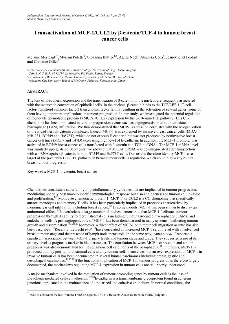
FIGURE 1 - MCP-1 expression is restricted to invasive, E-cadherin-negative breast tumour cell lines, (a) RT-PCR analysis of MCP-1 and E-cadherin in different human breast cancer cell lines (MCF7, T47D. MDA-MB-231, BT549 and Hs578T). The RT-PCR of the 28S mRNA is shown as a control. Each experiment was performed at least 3 times and 1 representative experiment is shown. (b) ELISA analysis of MCP-1 in 48 hr conditioned media obtained from the different breast cancer cell lines.
Transfection of small interfering RNA
A 19-nt specific sequence was selected in the coding sequence of β-catenin (Genebank accession number: X87838) to generate 21-nt sense and 21-nt antisense strands of the type (19N)TT (N, any nucleotide). The sense and antisense strands were then annealed to obtain duplexes with identical 3' overhangs. The sequence was submitted to a BLAST search against the human genome to ensure the specificity of the siRNA to the targeted sequence. A corresponding scramble duplexe which does not recognize any sequence in the human genome was used as control. The 19-nt specific sequence for the β-catenin siRNA is as follow: 5'-GAAACGGCTTTCAGTTGAG-3'. The 19-nt specific sequence for the control siRNA is as follow: 5'-GCTTATGGTGACAGA-TGTT-3'. For transfection of the siRNA duplexes, 7 × 10³ BT549 cells and 1 × 10s Hs578T cells were plated in 6-well plates in 2 ml per well of culture medium. Twenty-four hours after plating, the cells were transfected by phosphate calcium precipitation by adding in each well 200 µl of a mixture containing the siRNA duplexes (50 nM), 140 mM NaCl, 0.75 mM Na2HPO4, 6 mM glucose, 5 mM KC1, 25 mM HEPES and 125 mM CaCl2. Twenty-four hours after transfection, the cells were extensively washed with PBS and incubated for 48 hr in culture medium before they were harvested for RT-PCR, Western blotting or Elisa analyses.

Published in: International Journal of Cancer (2006), vol. 118, iss.1, pp. 35-42 Status: Postprint (Author’s version)
FIGURE 2 - β-catenin localization in the different breast cancer cell lines. (a) Western blot analysis of E-cadherin and β-catenin expression in human breast cancer cell lines (MCF7, T47D, MDA-MB-231, BT549 and Hs578T). β-actin level is shown as a control. (b) Confocal analysis of β-catenin localization in noninvasive, E-cadherin-positive, MCP-1-negative MCF7 breast cell line and in invasive, E-cadherin-negative, MCP-1-positive BT549 breast cancer cells. Bar = 8µM.
Results
MCP-1 is expressed by invasive, E-cadherin-negative breast cancer cell lines
In order to establish a potential relationship between a reorganization of E-cadherin/β-catenin complexes and MCP-1 expression, we first examined the expression of MCP-1 in a set of breast tumour cell lines previously characterized for their in vitro invasive properties and for their expression of E-cadherin.41-43 Examining MCP-1 mRNA expression by RT-PCR analysis, we observed that invasive, E-cadherin-negative cell lines (MDA-MB-231, BT549 and Hs578T) expressed high levels of MCP-1, whereas noninvasive, E-cadherin-positive cell lines (MCF7 and T47D) did not express MCP-1 mRNA (Fig. 1a). This selective expression of MCP-1 by invasive, E-cadherin-negative breast cancer cell lines was confirmed at the protein level by ELISA (Fig. 1b). Looking at β-catenin expression by Western blotting, we found it expressed in all cell lines regardless of E-cadherin expression (Fig. 2a). However, the subcellular localization of β-catenin was different among these cell lines. Indeed, as we previously showed, the nuclear activity of β-catenin revealed by the TOP-FLASH/FOP-FLASH reporter system, correlating with a nuclear localization of β-catenin, was higher in E-cadherin-negative cell lines than in E-cadherin-positive cell lines which displayed a membrane-associated staining for β-catenin34. Such a different localization of β-catenin in E-cadherin-negative, MCP-1-positive cell lines is also illustrated here (Fig. 2b). Indeed, β-cat-enin was mainly localized in the cytolplasm and in the nucleus of E-cadherin-negative, MCP-1-positive cells, whereas it was mainly found at the cell membrane in E-cadherin-positive, MCP-1-negative cell lines. These observations prompted us to examine the potential regulation of MCP-1 by the β-catenin/TCF pathway.
MCP-1 is upregulated by the β-catenin/TCF pathway
In order to show a direct effect of the β-catenin/TCF pathway on MCP-1 expression, we used a luciferase reporter plasmid containing the human MCP-1 promoter or the same promoter in which the consensus β-catenin/TCF binding site was mutated (CTTTGTA—>CTTTGGC). Cotransfection experiments were performed in MCF7, BT549 and Hs578T cells using the wild-type or the mutated human MCP-1 promoter cotransfected with expression vectors encoding β-catenin and/or a member of the TCF/LEF family, TCF-4. In the E-cadherin-
Published in: International Journal of Cancer (2006), vol. 118, iss.1, pp. 35-42 Status: Postprint (Author’s version)
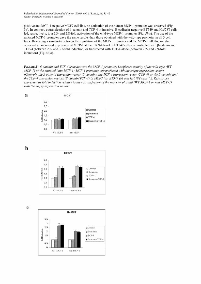
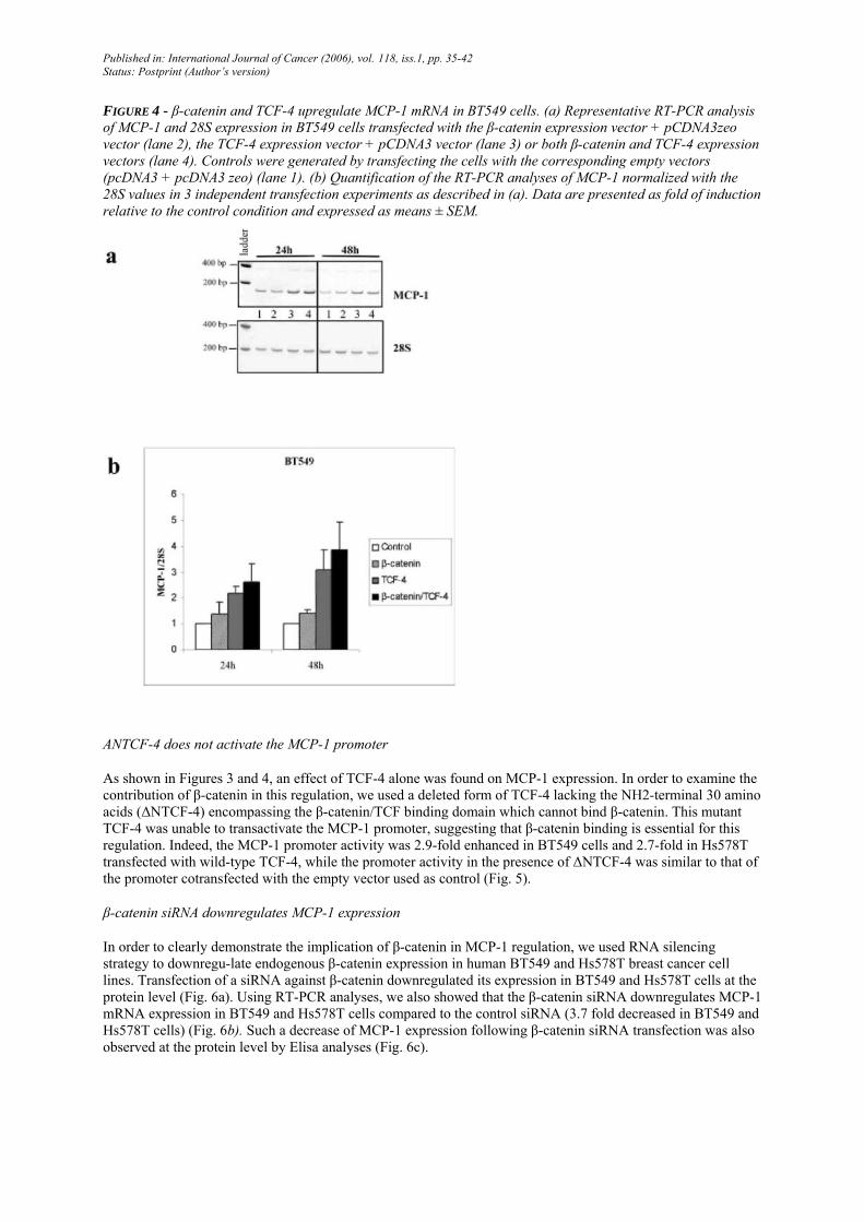
positive and MCP-1-negative MCF7 cell line, no activation of the human MCP-1 promoter was observed (Fig. 3a). In contrast, cotransfection of β-catenin and TCF-4 in invasive, E-cadherin-negative BT549 and Hs578T cells led, respectively, to a 2.3- and 2.8-fold activation of the wild-type MCP-1 promoter (Fig. 3b,c). The use of the mutated MCP-1 promoter gave the same results than those obtained with the wild-type promoter in all 3 cell lines. Revealing a similarity between the regulation of the MCP-1 promoter and the MCP-1 mRNA, we also observed an increased expression of MCP-1 at the mRNA level in BT549 cells cotransfected with β-catenin and TCF-4 (between 2.3- and 3.5-fold induction) or transfected with TCF-4 alone (between 2.2- and 2.9-fold induction) (Fig. 4a,b).
FIGURE 3 - β-catenin and TCF-4 transactivate the MCP-1 promoter. Luciferase activity of the wild-type (WT MCP-1) or the mutated (mut MCP-1) MCP-1 promoter cotransfected with the empty expression vectors (Control), the β-catenin expression vector (β-catenin), the TCF-4 expression vector (TCF-4) or the β-catenin and the TCF-4 expression vectors (β-catenin/TCF-4) in MCF7 (a), BT549 (b) and Hs578T cells (c). Results are expressed as fold induction relative to the cotransfection of the reporter plasmid (WT MCP-1 or mut MCP-1) with the empty expression vectors.
Published in: International Journal of Cancer (2006), vol. 118, iss.1, pp. 35-42 Status: Postprint (Author’s version)
FIGURE 4 - β-catenin and TCF-4 upregulate MCP-1 mRNA in BT549 cells. (a) Representative RT-PCR analysis of MCP-1 and 28S expression in BT549 cells transfected with the β-catenin expression vector + pCDNA3zeo vector (lane 2), the TCF-4 expression vector + pCDNA3 vector (lane 3) or both β-catenin and TCF-4 expression vectors (lane 4). Controls were generated by transfecting the cells with the corresponding empty vectors (pcDNA3 + pcDNA3 zeo) (lane 1). (b) Quantification of the RT-PCR analyses of MCP-1 normalized with the 28S values in 3 independent transfection experiments as described in (a). Data are presented as fold of induction relative to the control condition and expressed as means ± SEM.
ANTCF-4 does not activate the MCP-1 promoter
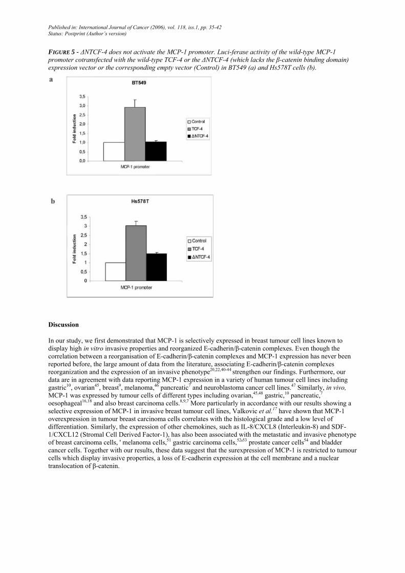
As shown in Figures 3 and 4, an effect of TCF-4 alone was found on MCP-1 expression. In order to examine the contribution of β-catenin in this regulation, we used a deleted form of TCF-4 lacking the NH2-terminal 30 amino acids (∆NTCF-4) encompassing the β-catenin/TCF binding domain which cannot bind β-catenin. This mutant TCF-4 was unable to transactivate the MCP-1 promoter, suggesting that β-catenin binding is essential for this regulation. Indeed, the MCP-1 promoter activity was 2.9-fold enhanced in BT549 cells and 2.7-fold in Hs578T transfected with wild-type TCF-4, while the promoter activity in the presence of ∆NTCF-4 was similar to that of the promoter cotransfected with the empty vector used as control (Fig. 5).
β-catenin siRNA downregulates MCP-1 expression
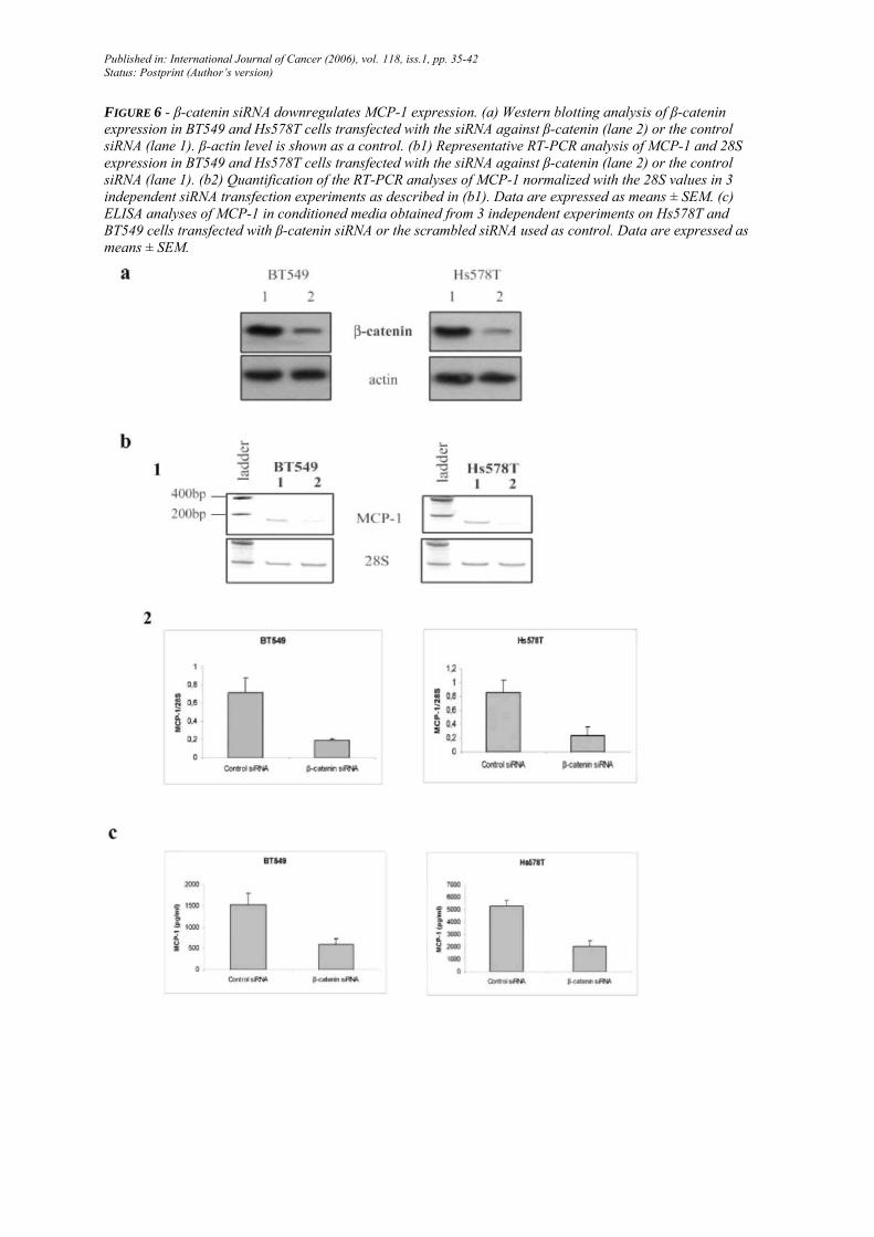
In order to clearly demonstrate the implication of β-catenin in MCP-1 regulation, we used RNA silencing strategy to downregu-late endogenous β-catenin expression in human BT549 and Hs578T breast cancer cell lines. Transfection of a siRNA against β-catenin downregulated its expression in BT549 and Hs578T cells at the protein level (Fig. 6a). Using RT-PCR analyses, we also showed that the β-catenin siRNA downregulates MCP-1 mRNA expression in BT549 and Hs578T cells compared to the control siRNA (3.7 fold decreased in BT549 and Hs578T cells) (Fig. 6b). Such a decrease of MCP-1 expression following β-catenin siRNA transfection was also observed at the protein level by Elisa analyses (Fig. 6c).
Published in: International Journal of Cancer (2006), vol. 118, iss.1, pp. 35-42 Status: Postprint (Author’s version)
FIGURE 5 - ∆NTCF-4 does not activate the MCP-1 promoter. Luci-ferase activity of the wild-type MCP-1 promoter cotransfected with the wild-type TCF-4 or the ∆NTCF-4 (which lacks the β-catenin binding domain) expression vector or the corresponding empty vector (Control) in BT549 (a) and Hs578T cells (b).
Discussion
In our study, we first demonstrated that MCP-1 is selectively expressed in breast tumour cell lines known to display high in vitro invasive properties and reorganized E-cadherin/β-catenin complexes. Even though the correlation between a reorganisation of E-cadherin/β-catenin complexes and MCP-1 expression has never been reported before, the large amount of data from the literature, associating E-cadherin/β-catenin complexes reorganization and the expression of an invasive phenotype20,22,40-44 strengthen our findings. Furthermore, our data are in agreement with data reporting MCP-1 expression in a variety of human tumour cell lines including gastric10, ovarian45, breast9, melanoma,46 pancreatic7 and neuroblastoma cancer cell lines.47 Similarly, in vivo, MCP-1 was expressed by tumour cells of different types including ovarian,45,48 gastric,10 pancreatic,7 oesophageal16,18 and also breast carcinoma cells.8,9,7 More particularly in accordance with our results showing a selective expression of MCP-1 in invasive breast tumour cell lines, Valkovic et al.17 have shown that MCP-1 overexpression in tumour breast carcinoma cells correlates with the histological grade and a low level of differentiation. Similarly, the expression of other chemokines, such as IL-8/CXCL8 (Interleukin-8) and SDF-1/CXCL12 (Stromal Cell Derived Factor-1), has also been associated with the metastatic and invasive phenotype of breast carcinoma cells, ' melanoma cells,51 gastric carcinoma cells,52'53 prostate cancer cells54 and bladder cancer cells. Together with our results, these data suggest that the surexpression of MCP-1 is restricted to tumour cells which display invasive properties, a loss of E-cadherin expression at the cell membrane and a nuclear translocation of β-catenin.
Published in: International Journal of Cancer (2006), vol. 118, iss.1, pp. 35-42 Status: Postprint (Author’s version)
FIGURE 6 - β-catenin siRNA downregulates MCP-1 expression. (a) Western blotting analysis of β-catenin expression in BT549 and Hs578T cells transfected with the siRNA against β-catenin (lane 2) or the control siRNA (lane 1). β-actin level is shown as a control. (b1) Representative RT-PCR analysis of MCP-1 and 28S expression in BT549 and Hs578T cells transfected with the siRNA against β-catenin (lane 2) or the control siRNA (lane 1). (b2) Quantification of the RT-PCR analyses of MCP-1 normalized with the 28S values in 3 independent siRNA transfection experiments as described in (b1). Data are expressed as means ± SEM. (c) ELISA analyses of MCP-1 in conditioned media obtained from 3 independent experiments on Hs578T and BT549 cells transfected with β-catenin siRNA or the scrambled siRNA used as control. Data are expressed as means ± SEM.
Published in: International Journal of Cancer (2006), vol. 118, iss.1, pp. 35-42 Status: Postprint (Author’s version)
In addition to the correlation between E-cadherin/β-catenin redistribution and MCP-1 expression, we also showed that MCP-1 promoter activity and MCP-1 mRNA expression were upregulated after cotransfection of β-catenin and TCF-4. Even though the sequence of the MCP-1 promoter revealed a putative β-catenin/ TCF binding site, mutation of this site did not abrogate the regulation of the MCP-1 promoter. The implication of other β-catenin/ TCF binding sites similar in sequence cannot be excluded. Alternatively, other β-catenin/TCF target genes could activate the MCP-1 promoter. Furthermore, the fact that MCP-1 regulation following β-catenin/TCF-4 transfections was observed only in invasive, E-cadherin-negative BT549 and Hs578T cells but not in MCF7 cells underlines the influence of the cellular context on the
β-catenin/TCF transcriptional regulation. This indeed emphasizes the importance of the levels of endogenous β-catenin and TCF/LEF factors but also of other transcriptional activators or repressors that can directly or indirectly interact with this complex. Indeed, an increasing number of TCF-bound compressors (Groucho, CtBP,...) have been identified.21,56-58 Also, a synergy between β-catenin-TCF/LEF complexes and other transcription factors (including AP-1 or PEA-3) have been described in the promoter regulation of several β-catenin/TCF targets genes including MMP-7 or the γ2 chain of laminin-5.35,59 The human MCP-1 promoter has accordingly been shown to be activated by several transcription factors including NFKB, Sp1 or AP-1.37,60-62 Also emphasizing the importance of the cellular context, we observed that the transfection of TCF-4 alone led to an upregulation of the MCP-1 promoter activity in the BT549 and Hs578T cells in which β-catenin is abundantly found in the nucleus.34 This endogenous level of nuclear β-catenin might thus be sufficient to activate the MCP-1 promoter in the presence of TCF-4. In agreement with ours results, Levy et al.32 have shown that, in hepatoma cells containing a constitutively activated β-catenin allele, exogenous wild-type TCF-4 is sufficient to activate the IL-8 promoter. In contrast, the deleted form of TCF-4 which lacks the β-catenin binding domain (∆NTCF-4) was unable to activate this promoter. Accordingly, our findings showing that ∆NTCF-4 does not transactivate the MCP-1 promoter clearly implicate β-catenin in this regulation. The decreased expression of MCP-1 mRNA and protein observed following transfection of a β-catenin siRNA in BT549 and Hs578T also emphasizes the implication of the β-catenin/TCF pathway in MCP-1 regulation. Therefore, if the cellular context obviously modulates the β-catenin/TCF regulation of the human MCP-1 promoter, our data clearly indicate that MCP-1 can be a target of the β-catenin/TCF pathway. Because of the major roles of MCP-1 in tumour progression, such a positive regulation of MCP-1 by the β-catenin/TCF pathway have obvious implications for tumour progression. Accordingly, many targets of the β-catenin pathway identified so far have been shown to play a key role in tumour progression by affecting tumour cell proliferation, tumour cell invasive properties or tumour cell survival. More particularly in agreement with our observations, other chemokines, including IL-8/CXCL8, another angiogenic chemokine, and MCP-3/CCL7, have been shown to be regulated by the β-catenin/TCF pathway.32,63 The proangiogenic vascular endothelial growth factor (VEGF) has also been shown to be regulated by the β-catenin/TCF pathway.
In conclusion, our results demonstrate that MCP-1 expression is restricted to invasive breast cancer cell lines and correlates with the reorganization of the E-cadherin/β-catenin complexes in these cells. We also show that MCP-1 is a target gene of the β-catenin/TCF pathway. Considering the various stimulatory roles of MCP-1 on tumour progression, such a regulation of MCP-1 by the β-catenin/TCF pathway might play an important role in breast cancer progression.
Acknowledgements
We thank Dr. H. Clevers for the TCF-4 and ∆NTCF-4 expression vectors and Dr. K. Orford and Dr. S. Byers for the β-catenin expression vector.
Grant sponsor
Communauté française de Belgique (Actions de Recherches Concert2es); Commission of European Communities (FP6 Brecosm); Fonds de la Recherche Scientifique Médicale; Fonds National de la Recherche Scientifique (FNRS, Belgium); Fédération Belge Contre le Cancer; C.G.R.I.-F.N.R.S.-INSERM Coopération; Fonds spéciaux de la Recherche (University of Liège ; Centre Anticancéreux près l’Université de Liège; Fortis Banque Assurances; Fondation Léon Frédéricq (University of Liège); D.G.T.R.E. from the ‘‘Région Wallonne’’; Fonds d’Investissements de la Recherche Scientifique (CHU, Liège, Belgium); Interuniversity Attraction Poles Programme-Belgian Science Policy (Brussels, Belgium).

Published in: International Journal of Cancer (2006), vol. 118, iss.1, pp. 35-42 Status: Postprint (Author’s version)
References
1. Baggiolini M, Dewald B, Moser B. Human chemokines: an update. Annu Rev Immunol 1997;15:675-705.
2. Rossi D, Zlotnik A. The biology of chemokines and their receptors. Annu Rev Immunol 2000; 18:217-42.
3. Muller A, Homey B, Soto H, Ge N, Catron D, Buchanan ME, McCla-nahan T, Murphy E, Yuan W, Wagner SN, Barrera JL, Mohar A et al. Involvement of chemokine receptors in breast cancer metastasis. Nature 2001;410:50-6.
4. Daly C, Rollins BJ. Monocyte chemoattractant protein-1 (CCL2) in inflammatory disease and adaptive immunity: therapeutic opportunities and controversies. Microcirculation 2003;10:247-57.
5. Yu JL, Rak JW. Host microenvironment in breast cancer development: inflammatory and immune cells in tumour angiogenesis and arteriogenesis. Breast Cancer Res 2003;5:83-8.
6. Huang S, Singh RK, Xie K, Gutman M, Berry KK, Bucana CD, Fidler IJ, Bar-Eli M. Expression of the JE/MCP-1 gene suppresses metastatic potential in murine colon carcinoma cells. Cancer Immunol Immun-other 1994;39:231-8.
7. Monti P, Leone BE, Marchesi F, Balzano G, Zerbi A, Scaltrini F, Pas-quali C, Calori G, Pessi F, Sperti C, Di CV, Allavena Pv et al. The CC chemokine MCP-1/CCL2 in pancreatic cancer progression: regulation of expression and potential mechanisms of antimalignant activity. Cancer Res 2003;63:7451-61.
8. Ueno T, Toi M, Saji H, Muta M, Bando H, Kuroi K, Koike M, Inadera H, Matsushima K. Significance of macrophage chemoattractant protein-1 in macrophage recruitment, angiogenesis, and survival in human breast cancer. Clin Cancer Res 2000;6:3282-9.
9. Saji H, Koike M, Yamori T, Saji S, Seiki M, Matsushima K, Toi M. Significant correlation of monocyte chemoattractant protein-1 expression with neovascularization and progression of breast carcinoma. Cancer 2001;92:1085-91.
10. Ohta M, Kitadai Y, Tanaka S, Yoshihara M, Yasui W, Mukaida N, Haruma K, Chayama K. Monocyte chemoattractant protein-1 expression correlates with macrophage infiltration and tumor vascularity in human gastric carcinomas. Int J Oncol 2003;22:773-8.
11. Nakashima E, Mukaida N, Kubota Y, Kuno K, Yasumoto K, Ichimura F, Nakanishi I, Miyasaka M, Matsushima K. Human MCAF gene transfer enhances the metastatic capacity of a mouse cachectic adenocarcinoma cell line in vivo. Pharm Res 1995;12:1598-604.
12. Salcedo R, Ponce ML, Young HA, Wasserman K, Ward JM, Klein-man HK, Oppenheim JJ, Murphy WJ. Human endothelial cells express CCR2 and respond to MCP-1: direct role of MCP-1 in angiogenesis and tumor progression. Blood 2000;96:34-40.
13. Youngs SJ, Ali SA, Taub DD, Rees RC. Chemokines induce migra-tional responses in human breast carcinoma cell lines. Int J Cancer 1997;71:257-66.
14. Lebrecht A, Grimm C, Lantzsch T, Ludwig E, Hefler L, Ulbrich E, Koelbl H. Monocyte chemoattractant protein-1 serum levels in patients with breast cancer. Tumour Biol 2004;25:14-7.
15. Amann B, Perabo FG, Wirger A, Hugenschmidt H, Schultze-Seemann W. Urinary levels of monocyte chemo-attractant protein-1 correlate with tumour stage and grade in patients with bladder cancer. Br J Urol 1998;82:118-21.
16. Koide N, Nishio A, Sato T, Sugiyama A, Miyagawa S. Significance of macrophage chemoattractant protein-1 expression and macrophage infiltration in squamous cell carcinoma of the esophagusa. Am J Gastroenterol 2004;99:1667-74.
17. Valkovic T, Lucin K, Krstulja M, Dobi-Babic R, Jonjic N. Expression of monocyte chemotactic protein-1 in human invasive ductal breast cancer. Pathol Res Pract 1998;194:335-40.
18. Ohta M, Kitadai Y, Tanaka S, Yoshihara M, Yasui W, Mukaida N, Haruma K, Chayama K. Monocyte chemoattractant protein-1 expression correlates with macrophage infiltration and tumor vascularity in human esophageal squamous cell carcinomas. Int J Cancer 2002;102:220-4.
19. Behrens J. Cadherins and catenins: role in signal transduction and tumor progression. Cancer Metastasis Rev 1999;18:15-30.
20. Van Aken E, De Wever O, Correia da Rocha AS, Mareel M. Defective E-cadherin/catenin complexes in human cancer. Virchows Arch 2001;439:725-51.
21. Hecht A, Kemler R. Curbing the nuclear activities of beta-catenin: control over Wnt target gene expression. EMBO Rep 2000;1:24-8.
22. Wijnhoven BP, Dinjens WN, Pignatelli M. E-cadherin-catenin cell-cell adhesion complex and human cancer. Br J Surg 2000;87:992-1005.
23. Gottardi CJ, Gumbiner BM. Adhesion signaling: how beta-catenin interacts with its partners. Curr Biol 2001; 11 :R792-94.

Published in: International Journal of Cancer (2006), vol. 118, iss.1, pp. 35-42 Status: Postprint (Author’s version)
24. Conacci-Sorrell M, Zhurinsky J, Ben Ze'ev A. The cadherin-catenin adhesion system in signaling and cancer. J Clin Invest 2002;109:987-91.
25. Giles RH, van Es JH, Clevers H. Caught up in a Wnt storm: Wnt signaling in cancer. Biochim Biophys Acta 2003;1653:1-24.
26. Jechlinger M, Grunert S, Tamir IH, Janda E, Ludemann S, Waerner T, Seither P, Weith A, Beug H, Kraut N. Expression profiling of epithelial plasticity in tumor progression. Oncogene 2003; 22: 7155-69.
27. He TC, Sparks AB, Rago C, Hermeking H, Zawel L, da Costa LT, Morin PJ, Vogelstein B, Kinzler KW. Identification of c-MYC as a target of the APC pathway. Science 1998;281:1509-12.
28. Brabletz T, Jung A, Dag S, Hlubek F, Kirchner T. beta-catenin regulates the expression of the matrix metalloproteinase-7 in human colorectal cancer. Am J Pathol 1999;155:1033-8.
29. Wielenga VJ, Smits R, Korinek V, Smit L, Kielman M, Fodde R, Clevers H, Pals ST. Expression of CD44 in Apc and Tcf mutant mice implies regulation by the WNT pathway. Am J Pathol 1999;154:515-23.
30. Miwa N, Furuse M, Tsukita S, Niikawa N, Nakamura Y, Furukawa Y. Involvement of claudin-1 in the beta-catenin/Tcf signaling pathway and its frequent upregulation in human colorectal cancers. Oncol Res 2000;12:469-76.
31. Marchenko GN, Ratnikov BI, Rozanov DV, Godzik A, Deryugina EI, Strongin AY. Characterization of matrix metalloproteinase-26, a novel metalloproteinase widely expressed in cancer cells of epithelial origin. Biochem J 2001;356:705-18.
32. Levy L, Neuveut C, Renard CA, Chameau P, Branchereau S, Gauthier F, Van Nhieu JT, Cherqui D, Petit-Bertron AF, Mathieu D, Buendia MA. Transcriptional activation of interleukin-8 by Beta-catenin-Tcf4. J Biol Chem 2002;277:42386-93.
33. Easwaran V, Lee SH, Inge L, Guo L, Goldbeck C, Garrett E, Wies-mann M, Garcia PD, Fuller JH, Chan V, Randazzo F, Gundel R et al. Beta-Catenin regulates vascular endothelial growth factor expression in colon cancer. Cancer Res 2003;63:3145-53.
34. Gilles C, Polette M, Mestdagt M, Nawrocki-Raby B, Ruggeri P, Bire-mbaut P, Foidart JM. Transactivation of vimentin by beta-catenin in human breast cancer cells. Cancer Res 2003;63:2658-64.
35. Hlubek F, Spaderna S, Jung A, Kirchner T, Brabletz T. Beta-catenin activates a coordinated expression of the preinvasive factors laminin-5 gamma2 chain and MT1-MMP in colorectal carcinomas. Int J Cancer 2004;108:321-6.
36. Marchenko ND, Marchenko GN, Weinreb RN, Lindsey JD, Kyshtoo-bayeva A, Crawford HC, Strongin AY. Beta-catenin regulates the gene of MMP-26, a novel metalloproteinase expressed both in carcinomas and normal epithelial cells. Int J Biochem Cell Biol 2004; 36:942-56.
37. Ueda A, Okuda K, Ohno S, Shirai A, Igarashi T, Matsunaga K, Fukushima J, Kawamoto S, Ishigatsubo Y, Okubo T. NF-kappa B and Spl regulate transcription of the human monocyte chemoattractant protein-1 gene. J Immunol 1994;153:2052-63.
38. Korinek V, Barker N, Morin PJ, van Wichen D, de Weger R, Kinzler KW, Vogelstein B, Clevers H. Constitutive transcriptional activation by a beta-catenin-Tcf complex in APC—/— colon carcinoma. Science 1997;275:1784-7.
39. van de WM, Cavallo R, Dooijes D, van Beest M, van Es J, Loureiro J, Ypma A, Hursh D, Jones T, Bejsovec A, Peifer M, Mortin M et al. Armadillo coactivates transcription driven by the product of the Dro-sophila segment polarity gene dTCF. Cell 1997;88:789-99.
40. Orford K, Crockett C, Jensen JP, Weissman AM, Byers SW. Serine phosphorylation-regulated ubiquitination and degradation of beta-catenin. J Biol Chem 1997;272:24735-8.
41. Thompson EW, Paik S, Brunner N, Sommers CL, Zugmaier G, Clarke R, Shima TB, Torri J, Donahue S, Lippman ME, . Association of increased basement membrane invasiveness with absence of estrogen receptor and expression of vimentin in human breast cancer cell lines. J Cell Physiol 1992;150:534-44.
42. Sommers CL, Byers SW, Thompson EW, Torri JA, Gelmann EP. Differentiation state and invasiveness of human breast cancer cell lines. Breast Cancer Res Treat 1994;31:325-35.
43. Nawrocki RB, Polette M, Gilles C, Clavel C, Strumane K, Matos M, Zahm JM, Van Roy F, Bonnet N, Birembaut P. Quantitative cell dispersion analysis: new test to measure tumor cell aggressiveness. Int J Cancer 2001;93:644-52.
44. Sommers CL, Thompson EW, Torri JA, Kemler R, Gelmann EP, Byers SW. Cell adhesion molecule uvomorulin expression in human breast cancer cell lines: relationship to morphology and invasive capacities. Cell Growth Diff 1991;2:365-72.
45. Negus RP, Stamp GW, Relf MG, Burke F, Malik ST, Bernasconi S, Allavena P, Sozzani S, Mantovani A, Balkwill FR. The detection and localization of monocyte chemoattractant protein-1 (MCP-1) in human ovarian cancer. J Clin Invest 1995;95:2391-6.
46. Nesbit M, Schaider H, Miller TH, Herlyn M. Low-level monocyte chemoattractant protein-1 stimulation of monocytes leads to tumor formation in nontumorigenic melanoma cells. J Immunol 2001; 166:6483-90.

Published in: International Journal of Cancer (2006), vol. 118, iss.1, pp. 35-42 Status: Postprint (Author’s version)
47. Metelitsa LS, Wu HW, Wang H, Yang Y, Warsi Z, Asgharzadeh S, Groshen S, Wilson SB, Seeger RC. Natural killer T cells infiltrate neuroblastomas expressing the chemokine CCL2. J Exp Med 2004; 199:1213-21.
48. Negus RP, Stamp GW, Hadley J, Balkwill FR. Quantitative assessment of the leukocyte infiltrate in ovarian cancer and its relationship to the expression of C-C chemokines. Am J Pathol 1997;150:1723-34.
49. De Larco JE, Wuertz BR, Rosner KA, Erickson SA, Gamache DE, Man-ivel JC, Furent LT. A potential role for interleukin-8 in the metastatic phenotype of breast carcinoma cells. Am J Pathol 2001; 158:639-46.
50. Fernandis AZ, Prasad A, Band H, Klosel R, Ganju RK. Regulation of CXCR4-mediated chemotaxis and chemoinvasion of breast cancer cells. Oncogene 2004;23:157-67.
51. Singh RK, Gutman M, Radinsky R, Bucana CD, Fidler IJ. Expression of interleukin 8 correlates with the metastatic potential of human melanoma cells in nude mice. Cancer Res 1994;54:3242-7.
52. Kitadai Y, Haruma K, Mukaida N, Ohmoto Y, Matsutani N, Yasui W, Yamamoto S, Sumii K, Kajiyama G, Fidler IJ, Tahara E. Regulation of disease-progression genes in human gastric carcinoma cells by interleukin 8. Clin Cancer Res 2000;6:2735-40.
53. Saito S, Kitayama J, Jin ZX, Tsuno N, Kaisaki S, Seto Y, Nagawa H. Beta-chemokine, macrophage inflammatory protein-lbeta (MlP-lbeta), is highly expressed in diffuse type human gastric cancers. J Exp Clin Cancer Res 2003;22:453-9.
54. Inoue K, Slaton JW, Eve BY, Kim SJ, Perrotte P, Balbay MD, Yano S, Bar-Eli M, Radinsky R, Pettaway CA, Dinney CP. Interleukin 8 expression regulates tumorigenicity and metastases in androgen-inde-pendent prostate cancer. Clin Cancer Res 2000;6:2104-19.
55. Inoue K, Slaton JW, Kim SJ, Perrotte P, Eve BY, Bar-Eli M, Radinsky R, Dinney CP. Interleukin 8 expression regulates tumorigenicity and metastasis in human bladder cancer. Cancer Res 2000;60:2290-9.
56. Cavallo RA, Cox RT, Moline MM, Roose J, Polevoy GA, Clevers H, Peifer M, Bejsovec A. Drosophila Tcf and Groucho interact to repress Wingless signalling activity. Nature 1998;395:604-8.
57. Levanon D, Goldstein RE, Bernstein Y, Tang H, Goldenberg D, Sti-fani S, Paroush Z, Groner Y. Transcriptional repression by AML1 and LEF-1 is mediated by the TLE/Groucho corepressors. Proc Natl Acad SciUSA 1998;95:11590-5.
58. Barolo S, Stone T, Bang AG, Posakony JW. Default repression and Notch signaling: Hairless acts as an adaptor to recruit the corepressors Groucho and dCtBP to Suppressor of Hairless. Genes Dev 2002; 16:1964-76.
59. Crawford HC, Fingleton BM, Rudolph-Owen LA, Goss KJ, Rubinfeld B, Polakis P, Matrisian LM. The metalloproteinase matrilysin is a target of beta-catenin transactivation in intestinal tumors. Oncogene 1999;18:2883-91.
60. Ueda A, Ishigatsubo Y, Okubo T, Yoshimura T. Transcriptional regulation of the human monocyte chemoattractant protein-1 gene. Cooperation of two NF-kappaB sites and NF-kappaB/Rel subunit specificity. J Biol Chem 1997;272:31092-9.
61. Wang N, Verna L, Hardy S, Forsayeth J, Zhu Y, Stemerman MB. Adenovirus-mediated overexpression of c-Jun and c-Fos induces intercellular adhesion molecule-1 and monocyte chemoattractant protein-1 in human endothelial cells. Arterioscler Thromb Vasc Biol 1999;19:2078-84.
62. Kanda N, Watanabe S. 17Beta-estradiol inhibits MCP-1 production in human keratinocytes. J Invest Dermatol 2003;120:1058-66.
63. Fujita M, Furukawa Y, Nagasawa Y, Ogawa M, Nakamura Y. Down-regulation of monocyte chemotactic protein-3 by activated beta-catenin. Cancer Res 2000;60:6683-7.


![Introduction Abstract - Neurology...medulla oblongata were dissected [27] and were stored in RNA later solution for RNA isolation. Whole brain (n = 5 per group) weighing 80-90mg) and](https://static.fdocument.org/doc/165x107/5f7aaac355c0bb44193d6438/introduction-abstract-neurology-medulla-oblongata-were-dissected-27-and.jpg)