217-225, 2008 Hepatocytes, rather than leukocytes reverse ...
10
Biol Res 41: 217-225, 2008 BR Hepatocytes, rather than leukocytes reverse DNA damage in vivo induced by whole body γ-irradiation of mice, as shown by the alkaline comet assay JUANA PINCHEIRA 1 , PILAR CARRERA 2 , KATHERINE MARCELAIN 1 and CONSUELO DE LA TORRE 3 1 Programa de Genética Humana, ICBM, Facultad de Medicina, Santiago 7, Chile 2 Departamento de Biología, Universidad Autónoma de Madrid, Cantoblanco, Madrid, Spain 3 Centro de Investigaciones Biológicas, CSIC, Madrid, Spain ABSTRACT DNA damage repair was assessed in quiescent (G 0 ) leukocytes and in hepatocytes of mice, after 1 and 2 hours recovery from a single whole body γ-irradiation with 0.5, 1 or 2 Gy. Evaluation of single-strand breaks (SSB) and alkali-labile sites together were carried out by a single-cell electrophoresis at pH>13.0 (alkaline comet assay). In non-irradiated (control) mice, the constitutive, endogenous DNA damage (basal) was around 1.5 times higher in leukocytes than in hepatocytes. Irradiation immediately increased SSB frequency in both cell types, in a dose-dependent manner. Two sequential phases took place during the in vivo repair of the radio- induced DNA lesions. The earliest one, present in both hepatocytes and leukocytes, further increased the SSB frequency, making evident the processing of some primary lesions in DNA bases into the SSB repair intermediates. In a second phase, SSB frequency decreased because of their removal. In hepatocytes, such a frequency regressed to the constitutive basal level after 2 hours recovery from either 0.5 or1 Gy. On the other hand, the SSB repair phase was specifically abrogated in leukocytes, at the doses and recovery times analyzed. Thus, the efficiency of in vivo repair of radio-induced DNA damage in dormant cells (lymphocytes) is quite different from that in hepatocytes whose low proliferation activity accounts only for cell renewal. Key terms: alkaline comet assay, ionizing radiation, hepatocytes, leukocytes, in vivo DNA repair. Corresponding Author: Consuelo de la Torre. Centro de Investigaciones Biológicas, CSIC. Ramiro de Maeztu, 9. Madrid 28040, Spain. Fax: +34-915-360-432; E-mail: [email protected] Received: December 21, 2007. In Revised form: June 5 14, 2008. Accepted: July 2, 2008 INTRODUCTION In proliferating cells, repair of DNA damage is under constraints imposed by the autonomous running of the cyclin- dependent kinases (CDK) cycle. Partial unlinking of DNA repair for such constraints is obtained through checkpoint pathways that, in the presence of DNA damage, transiently block the overcoming of irreversible transitions to a subsequent cycle phase by preventing CDK activation (López-Sáez et al. 1998). In this way, active checkpoints provide additional time for the completion of DNA repair before specific transitions between subsequent cycle phases are allowed (Zhou and Elledge, 2000). In mammals and plant cells, the DNA damage response is proportional to the proliferative activity (Essers et al, 2006; Hefner et al. 2006). However, it is unknown whether dormant mammalian cells (with no time constraint to repair their DNA) may still display some tissue specificity in this function. For instance, some authors postulate that DNA repair takes place in both hepatocytes (Ono and Okada, 1978) and lymphocytes (Ori et al., 2004), while other authors conclude that DNA damage accumulates in both cell types (Sharma and Yamamoto, 1980; Fenech and Morley, 1985, respectively). To investigate the DNA repair response in quiescent cells, we have studied both the mouse circulating
Transcript of 217-225, 2008 Hepatocytes, rather than leukocytes reverse ...
217PINCHEIRA ET AL. Biol Res 41, 2008, 217-225Biol Res 41: 217-225, 2008 BRHepatocytes, rather than leukocytes reverse DNAdamage in vivo induced by whole body γ-irradiation ofmice, as shown by the alkaline comet assay
JUANA PINCHEIRA1, PILAR CARRERA2, KATHERINE MARCELAIN1
and CONSUELO DE LA TORRE3
1 Programa de Genética Humana, ICBM, Facultad de Medicina, Santiago 7, Chile2 Departamento de Biología, Universidad Autónoma de Madrid, Cantoblanco, Madrid, Spain3 Centro de Investigaciones Biológicas, CSIC, Madrid, Spain
ABSTRACT
DNA damage repair was assessed in quiescent (G0) leukocytes and in hepatocytes of mice, after 1 and 2 hoursrecovery from a single whole body γ-irradiation with 0.5, 1 or 2 Gy. Evaluation of single-strand breaks (SSB)and alkali-labile sites together were carried out by a single-cell electrophoresis at pH>13.0 (alkaline cometassay). In non-irradiated (control) mice, the constitutive, endogenous DNA damage (basal) was around 1.5times higher in leukocytes than in hepatocytes. Irradiation immediately increased SSB frequency in both celltypes, in a dose-dependent manner. Two sequential phases took place during the in vivo repair of the radio-induced DNA lesions. The earliest one, present in both hepatocytes and leukocytes, further increased the SSBfrequency, making evident the processing of some primary lesions in DNA bases into the SSB repairintermediates. In a second phase, SSB frequency decreased because of their removal. In hepatocytes, such afrequency regressed to the constitutive basal level after 2 hours recovery from either 0.5 or1 Gy. On the otherhand, the SSB repair phase was specifically abrogated in leukocytes, at the doses and recovery timesanalyzed. Thus, the efficiency of in vivo repair of radio-induced DNA damage in dormant cells (lymphocytes)is quite different from that in hepatocytes whose low proliferation activity accounts only for cell renewal.
Key terms: alkaline comet assay, ionizing radiation, hepatocytes, leukocytes, in vivo DNA repair.
Corresponding Author: Consuelo de la Torre. Centro de Investigaciones Biológicas, CSIC. Ramiro de Maeztu, 9. Madrid28040, Spain. Fax: +34-915-360-432; E-mail: [email protected]
Received: December 21, 2007. In Revised form: June 5 14, 2008. Accepted: July 2, 2008
INTRODUCTION
In proliferating cells, repair of DNAdamage is under constraints imposed by theautonomous running of the cyclin-dependent kinases (CDK) cycle. Partialunlinking of DNA repair for suchconstraints is obtained through checkpointpathways that, in the presence of DNAdamage, transiently block the overcomingof irreversible transitions to a subsequentcycle phase by preventing CDK activation(López-Sáez et al. 1998). In this way, activecheckpoints provide additional time for thecompletion of DNA repair before specifictransitions between subsequent cycle phasesare allowed (Zhou and Elledge, 2000).
In mammals and plant cells, the DNAdamage response is proportional to theproliferative activity (Essers et al, 2006;Hefner et al. 2006). However, it is unknownwhether dormant mammalian cells (with notime constraint to repair their DNA) maystill display some tissue specificity in thisfunction. For instance, some authorspostulate that DNA repair takes place inboth hepatocytes (Ono and Okada, 1978)and lymphocytes (Ori et al., 2004), whileother authors conclude that DNA damageaccumulates in both cell types (Sharma andYamamoto, 1980; Fenech and Morley,1985, respectively). To investigate theDNA repair response in quiescent cells, wehave studied both the mouse circulating
PINCHEIRA ET AL. Biol Res 41, 2008, 217-225218
leukocytes and hepatocytes. To comparetheir DNA repair capacities, single cellelectrophoresis (comet assay) at a pH>13was applied at different times after wholemouse γ-irradiation. This alkaline variant ofthe comet assay allows the estimation ofendogenous and radio-induced single-strandbreaks (SSBs), as well as those associatedwith incomplete excision repair sites. Theamount of fragmented DNA migrating tothe comet tail is a function of the SSB levelpresent in the nucleus. This technique alsodetects the fixed set of genomic constitutivealkali-labile sites and, if present, DNA-DNA and DNA-protein cross-linkings (Ticeet al., 2000). The alkaline comet assay isthe most sensitive technique for analyzingthe kinetics of in vivo DNA repair in non-proliferating cells.
The mechanisms involved in theproduction of both endogenous and radio-induced DNA fragments are mediated bythe formation of SSB. A few of these DNAlesions are directly formed in response tolow doses of ionizing radiation. However,most SSB are intermediate lesions producedthrough the early processing of damagedDNA bases, by the sequential action ofenzymes involved in the base or nucleotideexcision repair pathways (Christman et al.,2003; Huffman et al., 2005).
In the present work, we found that whilehepatocytes were able to repair during 2hours of recovery, most if not all DNAdamage induced by γ-rays at 0.5 and 1 Gy,leukocytes were unable to. Thus, both celltypes differ in kinetics of appearance of theradio-induced DNA lesions, in themaximum level they achieve and, overall,in the rate of their DNA repair.
METHODS
Two-month-old female mice were used,because nuclear ploidy in liver cellsbecomes stabilized at this age (Brodsky andUryvaeva, 1985). Thirty six mice from theNSA CFI strain (2807919A registerednumber of the UAM Biotherium) were usedfor the present work. Mice were kept in a12 h light/12 h dark regime, in the fixedconditions earlier described (Carrera et al.,1998).
Irradiation was carried out with a 137Cssource of 1,700 Ci of activity, at a dose rateof 3.7 Gy min-1. For each experiment, sets ofthree mice were irradiated together with 0.5,1 or 2 Gy, while another single unirradiatedmouse was processed similarly and used as acontrol. Whole experiments were repeatedthree times. In each experiment, themanipulation to obtain hepatocytes andleukocytes for each single animal wascarried out in parallel by two differentpersons. It took about 10 min from the timewhen the liver and blood samples wereobtained from each mouse until cell nucleiwere embedded in the second agarose layer(from low melting temperature agarose,InCert, FMC BioProducts Europe,Denmark).
Hepatocytes were obtained according tothe procedure previously described. Itmainly consists of exposing a fresh sectionof the liver to a drop of alkalinedenaturation solution, and its immediateinclusion in agarose to carry out themicroelectrophoresis in alkaline (pH>13)conditions (Carrera et al. 1998).
Leukocytes were isolated from wholeblood (around 250 μl) extracted from theheart. Blood aliquots (10 μl) were diluted toone ml with Hank’s balanced salt solution(Gibco, USA), 20 mM EDTA pH 7.2, andcentrifuged at 15,000 g for 5 min at roomtemperature to collect the resulting pellets.Leukocyte or hepatocyte suspensions (120μl) in 0.5% low melting point agarose wereextended at 35ºC on the slides covered firstby a normal melting point agarose layer(Boehringer-Mannheim, Barcelona, Spain).This second agarose layer was kept at 0ºCfor 10 min. After removing the cover slide, alast 0.5 % normal melting point agaroselayer (85 μl) was placed on top and flattenedby a cover slide. This procedure, as well aslysis, pre-electrophoresis, and staining ofDNA was carried out by 4 mg ml-1ethidiumbromide, according to previously describedprotocols (Tice and Andrews, 1993;Navarrete et al. 1997; Carrera et al., 1998),except that the electrophoresis was carriedout at 0.8 V cm-1, at 11 ± 0.5ºC.
An epifluorescence microscope equippedwith a 40X objective, a 515-560 nmexcitation filter, and a 590 nm barrier filter
219PINCHEIRA ET AL. Biol Res 41, 2008, 217-225
was used. Comet images were taken by avideo camera (Hammamatsu, Photonics k.k.,Sunayama-Cho, Japan) that transmitted theimages to a computer. In each animal, aminimum of 20 comet images perexperimental condition were analyzed by thePC-image VGA program, version 2.10(Forster-Findlay Ass. Ltd.). The number ofDNA lesions or SSB was expressed as afunction of the percentage of fragmentedDNA present in the tails of the differentcomet types. The levels for significance ofdifferences among mean values wereestimated by applying the Student’s t test.The minimal significance was considered ata maximum P value equal to 0.05.
RESULTS
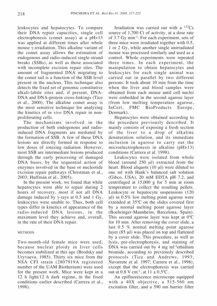
In control non-irradiated animals (Fig. 1),the basal DNA damage was around 1.5 timesgreater in leukocytes than in hepatocytes (P< 0.01, Student’s t test). DNA damage wasrecorded as the frequency of DNAfragmented into pieces, as a consequence of
the SSB presence, that migrated to the comettail during electrophoresis. These DNAlesions were endogenous, constitutive ones,produced by aerobic metabolism. Theirfrequency is considered to be tissue-specific.The recorded DNA lesions also include afixed number of alkaline-labile sites thatcharacterizes the genome of each species.Both sets of lesions were made evident bythe lysis step at pH>13, followed byelectrophoresis, under the alkaline cometassay.
The immediate response to radiation (norecovery)
The number of DNA lesions (SSB) wasevaluated in hepatocytes and leukocytes ofmice exposed to 0.5, 1 or 2 Gy of irradiationdoses. The lesions were first recorded at theend of the irradiation period, actually afterthe 10 min required for the abbreviatedpreparatory steps of a fast variant of thealkaline comet assay up to the time thenuclei were included in the second agaroselayer (Navarrete et al. 1997). The frequency
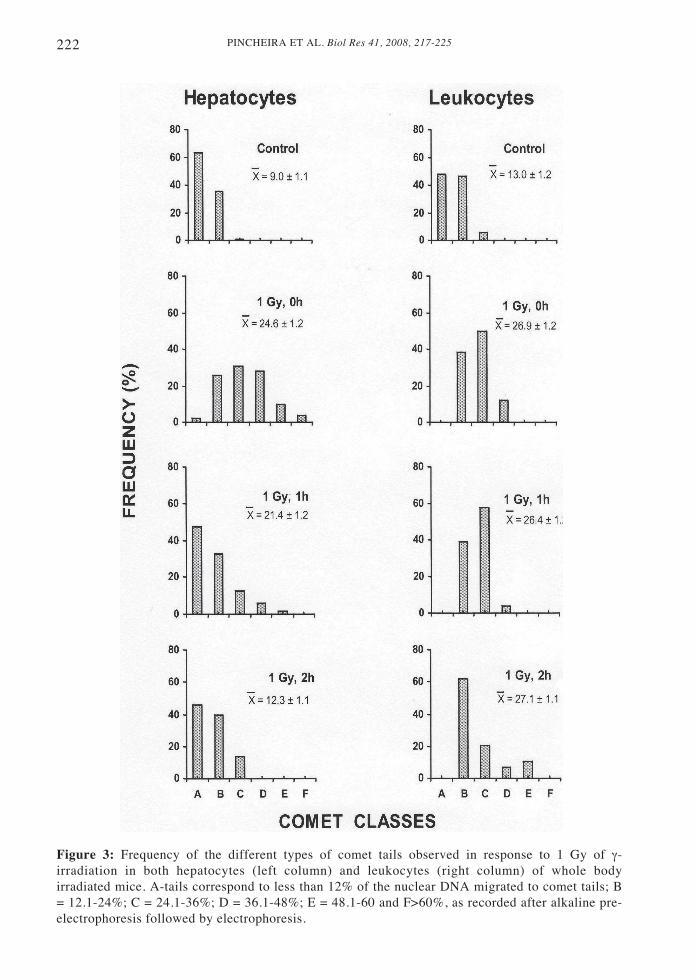
Figure 1: Effect of γ-rays dose on the frequency of single-stranded breaks (SSB) recorded at theend of irradiation in both hepatocytes and leukocytes by single cell electrophoresis at pH>13.Horizontal lines in each bar represent the endogenous, constitutive SSB level found in the non-irradiated conditions.
PINCHEIRA ET AL. Biol Res 41, 2008, 217-225220
of the SSB responsible for DNAfragmentation, and the migration of thenuclear DNA fragments to the comet tail,after single cell electrophoresis, increasedimmediately in both cell types afterirradiation, in relation to their constitutivedamage present before irradiation (Fig. 1).The frequency of SSB recorded immediatelyafter irradiation was a direct function of theradiation dose. Strikingly, for eachirradiation condition, the frequencies of SSBachieved at the very end of irradiation weresimilar in both hepatocytes and leukocytes.When substracting the constitutive DNAdamage present in non-irradiated conditions(corresponding to the physiologicaloxidative damage produced by metabolismplus the alkaline-labile sites unmasked atpH>13) from that recorded after irradiation,it was found that the radio-induced SSBwere always higher in hepatocytes than inleukocytes (Fig. 1).
In short, the earliest response toirradiation is apparently producedsimultaneously to irradiation itself. Thisfast response is characterized by an increasein SSB, making evident the processing ofradio-induced primary DNA lesions thatresult in a transient accumulation of SSBrepair intermediates. Hepatocytes weremore efficient than leukocytes in this earlyprocessing step.
Effect of recovery time on the frequency ofradio-induced SSB
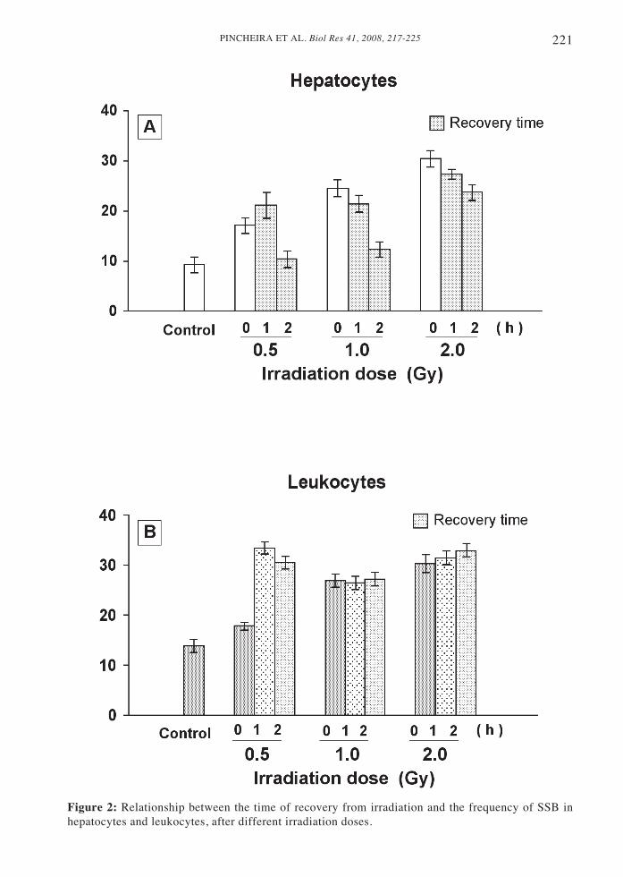
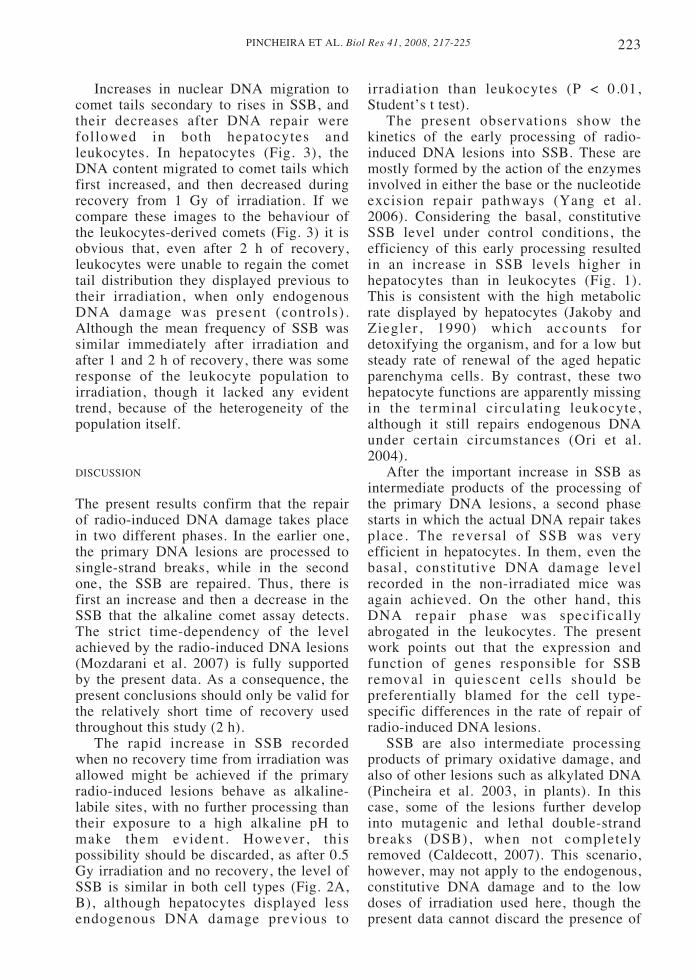
SSB levels increased not only duringirradiation, but also during recovery, bothin hepatocytes and leukocytes (Fig. 2). Celltype specificity was determinant for theresponse to radio-induced DNA damageafter 0, 1 and 2 hours of recovery. At thelowest irradiation dose (0.5 Gy) themaximum frequency of DNA lesions tookplace after 1h of recovery, both inhepatocytes and leukocytes. Moreover,though after 0h recovery time the SSB levelwas similar in both cell types, the radiation-induced increase in DNA lesions inleukocytes was higher than in hepatocytes(Fig. 2A, B). At 1 or 2 Gy irradiation doses,the maximum SSB recorded in this latercell type was achieved immediately after
irradiation, when no recovery (0h) wasallowed (Fig. 2A). Conversely, inleukocytes, no variation at the SSB levelwas recorded in response to 1 or 2 Gy ofirradiation doses, after 1 or 2 h of recovery(Fig. 2B).
The most obvious divergence betweenhepatocytes and leukocytes was the differencein their capability to repair radio-inducedDNA damage, even though both cell typeswere quiescent, non-cycling cells. Thus,hepatocytes were able to repair their radio-induced SSB, while leukocytes were unableto repair in the experimental conditions hereassayed. In fact, after 2 h of recovery from the0.5 or 1 Gy of irradiation doses, the frequencyof SSB regressed to the SSB constitutive levelthe hepatocytes had before irradiation (Fig.2A). After 2 Gy irradiation, however, therewas only a small but still significant DNArepair activity in the hepatocytes (P < 0.02,Student’s t test). The corresponding repairrate remained similar throughout the first twohours of recovery.
In open contrast to the situation inhepatocytes, no significant reversal of DNAdamage took place in leukocytes (Fig. 2B),at least during the first two h of recoveryanalyzed, with the exception of a smalldecrease in the SSB presence that tookplace 2 h after irradiation with 0.5 Gy.
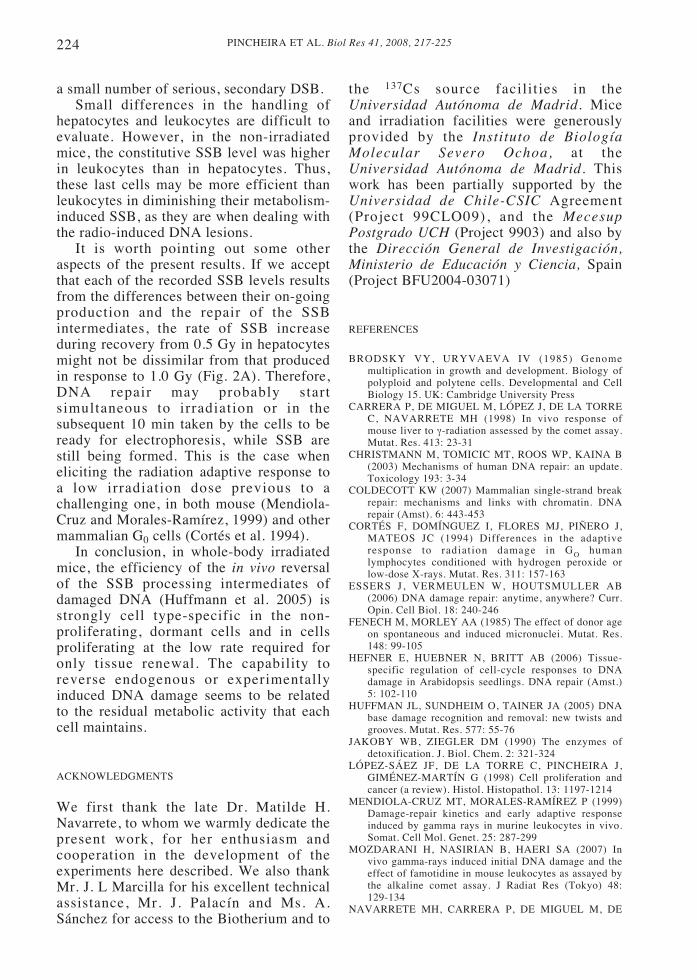
Analysis of the population of cometsobtained under the different experimentalconditions was then carried out. In order tovisualize the changes taking place in bothcell types -because of the migration offragmented DNA to their tails as aconsequence of the SSB presence- cometswere distributed in six different A to Fclasses (Fig. 3). A-type comets possessedtails with less than 12% of the total nuclearDNA, while the F-type comets with morethan 60% of its nuclear DNA migrated totails should correspond to apoptotic cells.The other comet classes displayedfrequencies of DNA migration to tailsbetween these two values (see legend ofFig. 3). Though the A-E comet types shouldalso include some few apoptotic cellsdeveloping earlier steps of the apoptoticprogramme, they usually remain unknown,due to the technical difficulty in applyingspecific apoptotic assays.
221PINCHEIRA ET AL. Biol Res 41, 2008, 217-225
Figure 2: Relationship between the time of recovery from irradiation and the frequency of SSB inhepatocytes and leukocytes, after different irradiation doses.
PINCHEIRA ET AL. Biol Res 41, 2008, 217-225222
Figure 3: Frequency of the different types of comet tails observed in response to 1 Gy of γ-irradiation in both hepatocytes (left column) and leukocytes (right column) of whole bodyirradiated mice. A-tails correspond to less than 12% of the nuclear DNA migrated to comet tails; B= 12.1-24%; C = 24.1-36%; D = 36.1-48%; E = 48.1-60 and F>60%, as recorded after alkaline pre-electrophoresis followed by electrophoresis.
223PINCHEIRA ET AL. Biol Res 41, 2008, 217-225
Increases in nuclear DNA migration tocomet tails secondary to rises in SSB, andtheir decreases after DNA repair werefollowed in both hepatocytes andleukocytes. In hepatocytes (Fig. 3), theDNA content migrated to comet tails whichfirst increased, and then decreased duringrecovery from 1 Gy of irradiation. If wecompare these images to the behaviour ofthe leukocytes-derived comets (Fig. 3) it isobvious that, even after 2 h of recovery,leukocytes were unable to regain the comettail distribution they displayed previous totheir irradiation, when only endogenousDNA damage was present (controls).Although the mean frequency of SSB wassimilar immediately after irradiation andafter 1 and 2 h of recovery, there was someresponse of the leukocyte population toirradiation, though it lacked any evidenttrend, because of the heterogeneity of thepopulation itself.
DISCUSSION
The present results confirm that the repairof radio-induced DNA damage takes placein two different phases. In the earlier one,the primary DNA lesions are processed tosingle-strand breaks, while in the secondone, the SSB are repaired. Thus, there isfirst an increase and then a decrease in theSSB that the alkaline comet assay detects.The strict time-dependency of the levelachieved by the radio-induced DNA lesions(Mozdarani et al. 2007) is fully supportedby the present data. As a consequence, thepresent conclusions should only be valid forthe relatively short time of recovery usedthroughout this study (2 h).
The rapid increase in SSB recordedwhen no recovery time from irradiation wasallowed might be achieved if the primaryradio-induced lesions behave as alkaline-labile sites, with no further processing thantheir exposure to a high alkaline pH tomake them evident. However, thispossibility should be discarded, as after 0.5Gy irradiation and no recovery, the level ofSSB is similar in both cell types (Fig. 2A,B), although hepatocytes displayed lessendogenous DNA damage previous to
irradiation than leukocytes (P < 0.01,Student’s t test).
The present observations show thekinetics of the early processing of radio-induced DNA lesions into SSB. These aremostly formed by the action of the enzymesinvolved in either the base or the nucleotideexcision repair pathways (Yang et al.2006). Considering the basal, constitutiveSSB level under control conditions, theefficiency of this early processing resultedin an increase in SSB levels higher inhepatocytes than in leukocytes (Fig. 1).This is consistent with the high metabolicrate displayed by hepatocytes (Jakoby andZiegler, 1990) which accounts fordetoxifying the organism, and for a low butsteady rate of renewal of the aged hepaticparenchyma cells. By contrast, these twohepatocyte functions are apparently missingin the terminal circulating leukocyte,although it still repairs endogenous DNAunder certain circumstances (Ori et al.2004).
After the important increase in SSB asintermediate products of the processing ofthe primary DNA lesions, a second phasestarts in which the actual DNA repair takesplace. The reversal of SSB was veryefficient in hepatocytes. In them, even thebasal, constitutive DNA damage levelrecorded in the non-irradiated mice wasagain achieved. On the other hand, thisDNA repair phase was specificallyabrogated in the leukocytes. The presentwork points out that the expression andfunction of genes responsible for SSBremoval in quiescent cells should bepreferentially blamed for the cell type-specific differences in the rate of repair ofradio-induced DNA lesions.
SSB are also intermediate processingproducts of primary oxidative damage, andalso of other lesions such as alkylated DNA(Pincheira et al. 2003, in plants). In thiscase, some of the lesions further developinto mutagenic and lethal double-strandbreaks (DSB), when not completelyremoved (Caldecott, 2007). This scenario,however, may not apply to the endogenous,constitutive DNA damage and to the lowdoses of irradiation used here, though thepresent data cannot discard the presence of
PINCHEIRA ET AL. Biol Res 41, 2008, 217-225224
a small number of serious, secondary DSB.Small differences in the handling of
hepatocytes and leukocytes are difficult toevaluate. However, in the non-irradiatedmice, the constitutive SSB level was higherin leukocytes than in hepatocytes. Thus,these last cells may be more efficient thanleukocytes in diminishing their metabolism-induced SSB, as they are when dealing withthe radio-induced DNA lesions.
It is worth pointing out some otheraspects of the present results. If we acceptthat each of the recorded SSB levels resultsfrom the differences between their on-goingproduction and the repair of the SSBintermediates, the rate of SSB increaseduring recovery from 0.5 Gy in hepatocytesmight not be dissimilar from that producedin response to 1.0 Gy (Fig. 2A). Therefore,DNA repair may probably startsimultaneous to irradiation or in thesubsequent 10 min taken by the cells to beready for electrophoresis, while SSB arestill being formed. This is the case wheneliciting the radiation adaptive response toa low irradiation dose previous to achallenging one, in both mouse (Mendiola-Cruz and Morales-Ramírez, 1999) and othermammalian G0 cells (Cortés et al. 1994).
In conclusion, in whole-body irradiatedmice, the efficiency of the in vivo reversalof the SSB processing intermediates ofdamaged DNA (Huffmann et al. 2005) isstrongly cell type-specific in the non-proliferating, dormant cells and in cellsproliferating at the low rate required foronly tissue renewal. The capability toreverse endogenous or experimentallyinduced DNA damage seems to be relatedto the residual metabolic activity that eachcell maintains.
ACKNOWLEDGMENTS
We first thank the late Dr. Matilde H.Navarrete, to whom we warmly dedicate thepresent work, for her enthusiasm andcooperation in the development of theexperiments here described. We also thankMr. J. L Marcilla for his excellent technicalassistance, Mr. J. Palacín and Ms. A.Sánchez for access to the Biotherium and to
the 137Cs source facilit ies in theUniversidad Autónoma de Madrid. Miceand irradiation facilities were generouslyprovided by the Instituto de BiologíaMolecular Severo Ochoa, at theUniversidad Autónoma de Madrid. Thiswork has been partially supported by theUniversidad de Chile-CSIC Agreement(Project 99CLO09), and the MecesupPostgrado UCH (Project 9903) and also bythe Dirección General de Investigación,Ministerio de Educación y Ciencia, Spain(Project BFU2004-03071)
REFERENCES
BRODSKY VY, URYVAEVA IV (1985) Genomemultiplication in growth and development. Biology ofpolyploid and polytene cells. Developmental and CellBiology 15. UK: Cambridge University Press
CARRERA P, DE MIGUEL M, LÓPEZ J, DE LA TORREC, NAVARRETE MH (1998) In vivo response ofmouse liver to γ-radiation assessed by the comet assay.Mutat. Res. 413: 23-31
CHRISTMANN M, TOMICIC MT, ROOS WP, KAINA B(2003) Mechanisms of human DNA repair: an update.Toxicology 193: 3-34
COLDECOTT KW (2007) Mammalian single-strand breakrepair: mechanisms and links with chromatin. DNArepair (Amst). 6: 443-453
CORTÉS F, DOMÍNGUEZ I, FLORES MJ, PIÑERO J,MATEOS JC (1994) Differences in the adaptiveresponse to radiation damage in GO humanlymphocytes conditioned with hydrogen peroxide orlow-dose X-rays. Mutat. Res. 311: 157-163
ESSERS J, VERMEULEN W, HOUTSMULLER AB(2006) DNA damage repair: anytime, anywhere? Curr.Opin. Cell Biol. 18: 240-246
FENECH M, MORLEY AA (1985) The effect of donor ageon spontaneous and induced micronuclei. Mutat. Res.148: 99-105
HEFNER E, HUEBNER N, BRITT AB (2006) Tissue-specific regulation of cell-cycle responses to DNAdamage in Arabidopsis seedlings. DNA repair (Amst.)5: 102-110
HUFFMAN JL, SUNDHEIM O, TAINER JA (2005) DNAbase damage recognition and removal: new twists andgrooves. Mutat. Res. 577: 55-76
JAKOBY WB, ZIEGLER DM (1990) The enzymes ofdetoxification. J. Biol. Chem. 2: 321-324
LÓPEZ-SÁEZ JF, DE LA TORRE C, PINCHEIRA J,GIMÉNEZ-MARTÍN G (1998) Cell proliferation andcancer (a review). Histol. Histopathol. 13: 1197-1214
MENDIOLA-CRUZ MT, MORALES-RAMÍREZ P (1999)Damage-repair kinetics and early adaptive responseinduced by gamma rays in murine leukocytes in vivo.Somat. Cell Mol. Genet. 25: 287-299
MOZDARANI H, NASIRIAN B, HAERI SA (2007) Invivo gamma-rays induced initial DNA damage and theeffect of famotidine in mouse leukocytes as assayed bythe alkaline comet assay. J Radiat Res (Tokyo) 48:129-134
NAVARRETE MH, CARRERA P, DE MIGUEL M, DE
225PINCHEIRA ET AL. Biol Res 41, 2008, 217-225
LA TORRE C (1997) A fast comet assay variant forsolid tissue cells. Mutat Res 389: 271-277
ONO T, OKADA S (1978) Does the capacity to rejoinradiation-induced DNA breaks decline in senescentmice? Int. J. Radiat. Biol. 33: 403-407
ORI Y, HERMAN M, WEINSTEIN T, CHAGNAC A,ZEVIN D, MILO G, GAFTER U, MALACHI T (2004)Spontaneous DNA repair in human peripheral bloodmammalian cells. Biochem. Biophys. Res. Commun.320: 578-586
PINCHEIRA J, LÓPEZ-SÁEZ JF, CARRERA P,NAVARRETE MH, DE LA TORRE C (2003) Effect ofcaffeine on “in vivo” processing of alkylated bases inproliferating plant cells. Cell Biol. Int. 27: 837-843
SHARMA RC, YAMAMOTO 0 (1980) Base modificationsin adult animal liver DNA and the similarity toradiation-induced base modification. Biochem.Biophys. Res. Commun. 96: 662-671
TICE RR, ANDREWS PW (1993) Protocol for theapplication of the alkaline single cell gel (SCG) assayto the detection of DNA damage in mammalian cells.Integrated Research Triangle Park, North Carolina,document # 27709)
TICE RR, AGURELL E, ANDERSON D, BURLINSON B,HARTMAN A, KOBAYASHI H, MIYAMAE Y,ROJAS E, KYU JC, SASAKI YF (2000) Single cellgel/comet assay: guidelines for in vitro and in vivogenetic toxicology testing. Environ. Mol. Mutagen. 35:206-221
YANG N, CHAUDHRY MA, WALLACE SS (2006) Baseexcision repair by hNTHI and hOGGI: a two edgedsword in the processing of DNA damage in gamma-irradiated human cells. DNA Repair (Amst) 5: 43 -51
ZHOU BBS, ELLEDGE SJ (2000) The DNA damageresponse: putting checkpoints in perspective. Nature408: 433-439

PINCHEIRA ET AL. Biol Res 41, 2008, 217-225226