Metabolic Mechanism of Mannan in a Ruminal Bacterium ... · MP from B. fragilis (BfMP) is...
23
Instructions for use Title Metabolic Mechanism of Mannan in a Ruminal Bacterium, Ruminococcus albus, Involving Two Mannoside Phosphorylases and Cellobiose 2-Epimerase : Discovery of a New Carbohydrate Phosphorylase, β-1,4- Mannooligosaccharide Phosphorylase Author(s) Kawahara, Ryosuke; Saburi, Wataru; Odaka, Rei; Taguchi, Hidenori; Ito, Shigeaki; Mori, Haruhide; Matsui, Hirokazu Citation Journal of Biological Chemistry, 287(50), 42389-42399 https://doi.org/10.1074/jbc.M112.390336 Issue Date 2012-12-07 Doc URL http://hdl.handle.net/2115/51063 Rights This research was originally published in Journal of Biological Chemistry. Ryosuke Kawahara, Wataru Saburi, Rei Odaka, Hidenori Taguchi, Shigeaki Ito, Haruhide Mori and Hirokazu Matsui. Metabolic Mechanism of Mannan in a Ruminal Bacterium, Ruminococcus albus, Involving Two Mannoside Phosphorylases and Cellobiose 2-Epimerase : Discovery of a New Carbohydrate Phosphorylase, β-1,4-Mannooligosaccharide Phosphorylase. Journal of Biological Chemistry. 2012; Vol:42389-42399. © the American Society for Biochemistry and Molecular Biology. Type article (author version) Additional Information There are other files related to this item in HUSCAP. Check the above URL. File Information JBC287-50_42389-42399.pdf Hokkaido University Collection of Scholarly and Academic Papers : HUSCAP
Transcript of Metabolic Mechanism of Mannan in a Ruminal Bacterium ... · MP from B. fragilis (BfMP) is...
Instructions for use
TitleMetabolic Mechanism of Mannan in a Ruminal Bacterium, Ruminococcus albus, Involving Two MannosidePhosphorylases and Cellobiose 2-Epimerase : Discovery of a New Carbohydrate Phosphorylase, β-1,4-Mannooligosaccharide Phosphorylase
Author(s) Kawahara, Ryosuke; Saburi, Wataru; Odaka, Rei; Taguchi, Hidenori; Ito, Shigeaki; Mori, Haruhide; Matsui, Hirokazu
Citation Journal of Biological Chemistry, 287(50), 42389-42399https://doi.org/10.1074/jbc.M112.390336
Issue Date 2012-12-07
Doc URL http://hdl.handle.net/2115/51063
Rights
This research was originally published in Journal of Biological Chemistry. Ryosuke Kawahara, Wataru Saburi, ReiOdaka, Hidenori Taguchi, Shigeaki Ito, Haruhide Mori and Hirokazu Matsui. Metabolic Mechanism of Mannan in aRuminal Bacterium, Ruminococcus albus, Involving Two Mannoside Phosphorylases and Cellobiose 2-Epimerase :Discovery of a New Carbohydrate Phosphorylase, β-1,4-Mannooligosaccharide Phosphorylase. Journal of BiologicalChemistry. 2012; Vol:42389-42399. © the American Society for Biochemistry and Molecular Biology.
Type article (author version)
Additional Information There are other files related to this item in HUSCAP. Check the above URL.
File Information JBC287-50_42389-42399.pdf
Hokkaido University Collection of Scholarly and Academic Papers : HUSCAP
Metabolic Mechanism of Mannan in Ruminococcus albus
1
Metabolic Mechanism of Mannan in a Ruminal Bacterium, Ruminococcus albus, involving two
Mannoside Phosphorylases and Cellobiose 2-Epimerase: Discovery of a New Carbohydrate
Phosphorylase, β-1,4-Mannooligosaccharide Phosphorylase*
Ryosuke Kawahara**, Wataru Saburi**, Rei Odaka, Hidenori Taguchi, Shigeaki Ito, Haruhide
Mori, and Hirokazu Matsui
From Research Faculty of Agriculture, Hokkaido University, N-9, W-9, Sapporo 060-8589, Japan
*Running title: Metabolic Mechanism of Mannan in Ruminococcus albus
To whom correspondence should be addressed: Wataru Saburi, Research Faculty of Agriculture,
Hokkaido University, N-9, W-9, Sapporo 060-8589, Japan. Tel/Fax: +81-11-706-2508; E-mail:
Keywords: mannooligosaccharide phosphorylase; mannan; glycoside hydrolase family 130;
phosphorolysis; Ruminococcus albus
Background: Characteristics of two
4-O-β-D-mannosyl-D-glucose phosphorylases from
Ruminococcus albus were investigated.
Results: One enzyme was specific for
4-O-β-D-mannosyl-D-glucose, as observed for the
Bacteroides fragilis enzyme, but the other showed
high activity towards mannooligosaccharides longer
than β-1,4-mannobiose.
Conclusion: Two phosphorylases play distinct roles
in the metabolism of mannan.
Significance: A new enzyme catalyzing the
phosphorolysis of β-1,4-mannooligosaccharides was
identified.
SUMMARY
Ruminococcus albus is a typical ruminal
bacterium digesting cellulose and hemicellulose.
Cellobiose 2-epimerase (EC 5.1.3.11, CE), which
converts cellobiose to
4-O-β-D-glucosyl-D-mannose, is a particularly
unique enzyme in R. albus, but its physiological
function is unclear. Recently, a new metabolic
pathway of mannan involving CE was postulated
for another CE producing bacterium, Bacteroides
fragilis. In this pathway, β-1,4-mannobiose is
epimerized to 4-O-β-D-mannosyl-D-glucose
(Man-Glc) by CE, and Man-Glc is
phosphorolyzed to α-D-mannosyl 1-phosphate
(Man1P) and D-glucose by Man-Glc
phosphorylase (EC 2.4.1.281, MP).
Ruminococcus albus NE1 showed intracellular
MP activity, and two MP isozymes, RaMP1 and
RaMP2, were obtained from the cell-free extract.
These enzymes were highly specific for the
mannosyl residue at the non-reducing end of the
substrate, and catalyzed the phosphorolysis and
synthesis of Man-Glc through a sequential bi bi
mechanism. In a synthetic reaction, RaMP1
showed high activity only towards D-glucose and
6-deoxy-D-glucose in the presence of Man1P,
while RaMP2 showed acceptor specificity
significantly different from RaMP1. RaMP2
acted on D-glucose derivatives at the C2- and
C3-positions including deoxy- and
deoxyfluoro-analogues and epimers, but not on
those substituted at the C6-position. Furthermore,
Metabolic Mechanism of Mannan in Ruminococcus albus
2
RaMP2 had high synthetic activity towards the
following oligosaccharides: β-linked glucobioses,
maltose, N,N’-diacetylchitobiose, and
β-1,4-mannooligosaccharides. Particularly,
β-1,4-mannooligosaccharides served as
significantly better acceptor substrates for
RaMP2 than D-glucose. In the phosphorolytic
reactions, RaMP2 had weak activity towards
β-1,4-mannobiose, but efficiently degraded
β-1,4-mannooligosaccharides longer than
β-1,4-mannobiose. Consequently, RaMP2 is
thought to catalyze the phosphorolysis of
β-1,4-mannooligosaccharides longer than
β-1,4-mannobiose to produce Man1P and
β-1,4-mannobiose.
Ruminococcus albus is a typical ruminal
bacterium producing various cellulolytic enzymes
including cellulase (EC 3.2.1.4) (1), β-glucosidase
(EC 3.2.1.21) (2), and cellobiose phosphorylase (EC
2.4.1.20) (3). Cellobiose 2-epimerase (EC 5.1.3.11,
CE), which converts cellobiose to
4-O-β-D-glucosyl-D-mannose, is a unique enzyme
acting on cellulose-related carbohydrates in R. albus
(4), but the physiological meaning of its
epimerization of cello-oligosaccharides is unclear.
Once the CE gene of R. albus NE1 was cloned (5),
the CE genes were identified based on the amino
acid sequence similarities in various bacteria
including a non-cellulolytic bacterium, Bacteroides
fragilis NCTC9343 (6). In B. fragilis, the CE gene
comprises the operon along with the genes encoding
β-mannanase and 4-O-β-D-mannosyl-D-glucose
phosphorylase (EC 2.4.1.281, MP), which
specifically catalyzes the phosphorolysis of
4-O-β-D-mannosyl-D-glucose (Man-Glc) to
α-D-mannosyl phosphate (Man1P) and D-glucose
(7), implying that CE is involved in the metabolism
of mannan, in which it converts β-1,4-mannobiose to
Man-Glc for further phosphorolysis.
Known inverting carbohydrate phosphorylases
catalyzing the phosphorolysis of a glycoside with
anomeric inversion have catalytic domains that are
structurally similar to inverting glycoside hydrolases
(8, 9). Thus the inverting phosphorylases are
classified into glycoside hydrolase (GH) families on
the basis of the similarity of their amino acid
sequences (10). Only limited information concerning
MP from B. fragilis (BfMP) is available, but this
enzyme was recently categorized into GH family
130 together with the putative proteins from the
genome sequence of known bacterial species, the
amino acid sequences of which are similar to that of
BfMP. In this family, three-dimensional structures of
Thermotoga maritima MSB8 TM1225 protein
(PDB code, 1vkd), Bacteroides thetaiotaomicron
VPI-5482 BT_4094 protein (3R67), and
Parabacteroides distansonis ATCC 8503 BDI_3141
protein (3TAW) were elucidated, although their
biochemical properties have not been examined thus
far. These proteins are formed by five-bladed
β-propeller folds, which are observed in the
glycoside hydrolases belonging to GH families 32
(11, 12), 43 (13-18), and 68 (19, 20).
R. albus 7 has several putative
β-1,4-mannanase genes and two MP-like genes
(Rumal_0099 and Rumal_0852) in the genome,
indicating that this bacterium also has components
necessary to metabolize mannan via epimerization
and phosphorolysis as observed in B. fragilis.
Rumal_0852 and Rumal_0099 encode proteins with
59% and 27% sequence identity to BfMP,
respectively. In this study, two MP isozymes from R.
albus NE1, which is phylogenetically close to R.
albus 7 (5), were characterized in detail, and the
physiological functions of these enzymes in the
metabolism of mannan are discussed.
EXPERIMENTAL PROCEDURES
Preparation of Man-Glc – For the enzyme assay,
highly-pure Man-Glc was prepared. A reaction
mixture of 210 mL containing 100 mg/mL locust
bean gum (Wako Pure Chemical Industries, Osaka,
Japan), 1 U/mL β-mannanase from Aspergillus niger
(Shin Nihon Chemical, Anzyo, Japan), and 10 mM
sodium acetate buffer (pH 4.0) was incubated at
37°C for 24 h. The β-1,4-mannobiose produced was
purified by carbon-celite column chromatography, as
previously reported (21), and epimerized by CE as
follows: a reaction mixture consisting of 50 mg/mL
mannobiose, 0.1 U/mL CE from R. albus NE1 (22),
and 20 mM sodium phosphate buffer (pH 7.5) was
incubated at 37°C for 24 h. Man-Glc was purified,
desalted, and freeze-dried, as described previously
(21).
Identification of MP isozymes in the cell-free extract
of R. albus NE1 – MP activity of the cell-free extract
of R. albus NE1, obtained from 1.5 L of the culture

Metabolic Mechanism of Mannan in Ruminococcus albus
3
fluid (5), was detected by thin layer chromatography
(TLC). Six μL of cell-free extract of R. albus NE1
was mixed with 2 μL of 100 mM Man-Glc, and 2 μL
of 50 mM reaction buffer (pH 7.0), and incubated at
37°C for 3 h. Sodium phosphate buffer or
MES-NaOH buffer was used as reaction buffer. The
reaction mixture of 1 μl was analyzed by TLC, in
which a developing solvent of
acetonitrile/ethylacetate/1-propanol/water
(85:20:50:30, v/v) was used. The chromatogram was
visualized by spraying a detection reagent (acetic
acid/sulfuric acid/anisaldehyde, 100:2:1, v/v) and
heating. The cell-free extract was subjected to
DEAE Sepharose CL-6B column chromatography
(⌀1.5 × 9.0 cm, GE Healthcare, Uppsala, Sweden),
in which adsorbed protein was eluted by a linear
gradient of 0–0.5 M NaCl in 20 mM MES-NaOH
buffer (pH 6.5). Enzyme activity was checked by
TLC analysis as described above. Two active peaks
(the enzymes eluted by high and low concentrations
of NaCl were designated as RaMP1 and RaMP2
respectively) were further purified by Butyl
Sepharose CL-6B (⌀1.5 × 9.0 cm, GE Healthcare),
hydroxyapatite (⌀1.5 × 12 cm, Seikagaku, Tokyo),
and Superdex 200 (⌀1.6 × 60 cm, GE Healthcare)
column chromatography. Finally, the sample was
separated by SDS-PAGE. The masses of the tryptic
peptides derived from the approximately 40 kDa
protein were measured and assigned to the
theoretical values for Rumal_0099 and Rumal_0852
proteins, as described elsewhere (23).
Production and purification of recombinant RaMP1
and RaMP2 – The genomic DNA of R. albus NE1
as the template, Primestar HS DNA polymerase
(Takara Bio, Otsu, Japan), and the primers listed in
Table 1 were used in the PCR. The RaMP1 and
RaMP2 genes were amplified by PCR and cloned
into the EcoRV site of pBluescript II SK (+) vector
(Stratagene, La Jolla, CA). The DNA sequences of
the cloned genes were analyzed with the ABI Prism
310 Genetic Analyzer (Applied Biosystems, Foster
City, CA). These plasmids were used as the
templates in the PCR to construct the expression
plasmids. NdeI and XhoI sites were introduced to the
5’- and 3’-termini of the target gene respectively by
PCR (primers listed in Table 1), and cloned into the
NdeI and XhoI sites of the pET-23a vector (Novagen,
Darmstadt, Germany).
The transformants of Escherichia coli BL21
(DE3) harboring the expression plasmid of RaMP1
or RaMP2 were cultured in 0.5 L and 1.5 L of LB
broth containing 100 μg/mL ampicillin respectively
at 37°C until the A600 reached 0.6 The protein
production was induced by the addition of isopropyl
β-D-thiogalactoside at the final concentration of 0.1
mM, and the incubation was continued at 18°C for
16 h. The recombinant enzymes were purified from
the E. coli cell-extract. The concentrations of the
purified enzymes were determined based on the
concentration of each amino acid after acid
hydrolysis (24).
Enzyme assay – A reaction mixture of 50 μL
consisting of the appropriate concentration of
enzyme diluted with 20 mM MES-NaOH buffer (pH
6.5) containing 1 mg/mL bovine serum albumin, 100
mM sodium phosphate buffer (pH 6.5), and 2 mM
Man-Glc was incubated at 37°C for 10 min. The
enzyme reaction was terminated by boiling the
mixture for 3 min after the addition of 25 μL of 4 M
Tris-HCl buffer (pH 7.0). The D-glucose produced
was measured by the glucose oxidase-peroxidase
method (25). One U of enzyme activity was defined
as the amount of enzyme producing 1 μmol of
D-glucose in 1 min under these conditions.
To determine the optimum pH, the reaction
buffer was changed to 100 mM sodium citrate buffer
(pH 3.0–6.0), MES-NaOH buffer (pH 6.0–7.0),
HEPES-NaOH buffer (pH 7.0–8.5), and
glycine-NaOH buffer (pH 8.5–10.5). Sodium
phosphate buffer (pH 6.5) was added as the substrate
to the reaction mixture at the final concentration of
10 mM.
Temperature and pH stabilities were evaluated
by the residual activity after temperature treatment
(incubation at various temperatures for 20 min at pH
6.5) and pH treatment (incubation at various pH
values for 24 h at 4°C) respectively. The values over
which the enzymes retained more than 90% of their
original activities were considered to be stable
ranges.
The kinetic parameters for phosphorolysis and
synthesis of Man-Glc were calculated from the
initial velocities towards various concentrations of
Man-Glc and inorganic phosphate, and Man1P and
D-glucose, respectively by fitting to the equation for
a sequential bi bi mechanism (26). Non-linear
Metabolic Mechanism of Mannan in Ruminococcus albus
4
regression was performed with Grafit version 7.0.2
(Erithacus Software, West Sussex, UK).
Product inhibition analysis was carried out to
determine the order of substrate binding and product
release. First, the rate of phosphorolysis catalyzed by
rRaMP1 and rRaMP2 toward Man-Glc in the
presence of 0–2 mM Man1P were measured at
varying concentrations of Man-Glc and 10 mM
sodium phosphate buffer (pH 6.5) or at varying
concentrations of sodium phosphate buffer (pH 6.5)
and 2 mM Man-Glc. Then, in the case of rRaMP2,
the phosphorolysis of Man-Glc was measured in the
presence of 0–100 mM cellobiose at varying
concentrations of Man-Glc and 10 mM sodium
phosphate buffer (pH 6.5) or at varying
concentrations of sodium phosphate buffer (pH 6.5)
and 2 mM Man-Glc. In the case of rRaMP1, initial
velocities for the synthesis of Man-Glc were
measured in the presence of 0–20 mM Man-Glc at
varying concentrations of Man1P and 50 mM
D-glucose or at varying concentrations of D-glucose
and 5 mM Man1P.
The rates for the synthesis of Man-Glc was
measured as follows: a reaction mixture of 50 μL
consisting of an appropriate concentration of
enzyme, 50 mM MES-NaOH buffer (pH 6.5),
0.25–10 mM Man1P (sodium salt hydrate, Sigma, St.
Louis, MO), and 10–200 mM D-glucose (Wako Pure
Chemical Industries) were incubated at 37°C for 10
min, and heated at 80°C for 5 min. The released
inorganic phosphate was measured following Lowry
and Lopez (27). The apparent kinetic parameters for
the synthetic reactions towards various acceptor
substrates were determined by fitting the initial rates
at various concentrations of acceptors in the
presence of 10 mM Man1P to the Michaelis-Menten
equation. The acceptor substrates tested were from
the following suppliers: D-mannose, D-allose,
3-O-methyl-D-glucose, D-xylose,
1,5-anhydro-D-glucitol, methyl α-D-glucoside, and
methyl β-D-glucoside were from Wako Pure
Chemical Industries; 2-deoxy-D-glucose and
D-glucosamine were from Tokyo Chemical Industry,
Tokyo, Japan; 2-deoxy-2-fluoro-D-glucose,
sophorose, and 6-deoxy-D-glucose were from
Sigma; N-acetyl-D-glucosamine, maltose, cellobiose,
and gentiobiose were from Nacalai Tesque, Kyoto,
Japan; 3-deoxy-D-glucose,
3-deoxy-3-fluoro-D-glucose, and
6-deoxy-6-fluoro-D-glucose were from Carbosynth,
Berkshire, UK; the series of mannooligosaccharides
was from Megazyme; N,N’-diacetyl chitobiose and
laminaribiose were from Seikagaku.
The phosphorolytic velocity of rRaMP2
towards β-1,4-mannooligosaccharides was
determined based on the amount of
β-1,4-mannooligosaccharides with reduced
chain-length (D-mannose was measured in the
reaction towards β-1,4-mannobiose) using HPLC. A
reaction mixture of 200 μL consisting of an
appropriate concentration of enzyme, 10 mM sodium
phosphate buffer (pH 6.5), and various
concentrations of β-1,4-mannooligosaccharides was
incubated at 37°C for 10 min, and heated at 80°C for
5 min. The reaction mixture was applied to HPLC
under the following conditions: injection volume, 10
μL; column, Sugar D (⌀4.6 × 250 mm, Nacalai
Tesque); eluent, 65% acetonitrile; flow rate, 0.8
mL/min; detection, pulsed amperometry.
Analysis of reaction products towards
β-1,4-mannooligosaccharides by rRaMP2 –
Phosphorolytic activities of rRaMP2 towards
β-1,4-mannotriose and β-1,4-mannotetraose were
detected by TLC. A reaction mixture of 10 μL
containing 0.24 μM rRaMP2, 20 mM
β-1,4-mannotriose or β-1,4-mannotetraose, and 10
mM sodium phosphate buffer (pH 6.5) was
incubated at 37°C. One μL of the reaction mixture
taken at 0, 30, 60, and 120 min was spotted on a
TLC plate and dried immediately to stop the reaction.
Developing solvent and detection conditions were as
described above.
Structural analysis of the oligosaccharides produced
by the synthetic reaction – The oligosaccharides
produced by recombinant RaMP1 (rRaMP1) were
prepared as follows: a reaction mixture of 0.2 mL
consisting of 493 nM rRaMP1, 50 mM MES-NaOH
buffer (pH 6.5), 250 mM D-glucose or D-xylose, and
250 mM Man1P was incubated at 37°C for 3 h. The
reaction products were purified by HPLC as
described previously (28). The synthetic reaction of
recombinant RaMP2 (rRaMP2) was performed in a
reaction mixture of 1 mL, consisting of 79 nM
rRaMP2, 50 mM MES-NaOH buffer (pH 6.5), 100
mM Man1P, and 100 mM acceptor substrate. The
reaction was carried out at 37°C for 12 h. Cellobiose,

Metabolic Mechanism of Mannan in Ruminococcus albus
5
β-1,4-mannobiose, and N,N’-diacetylchitobiose were
used as acceptors. The reaction products were
purified by gel filtration column chromatography
under the following conditions: column, Bio-Gel P-2
(⌀1.8 × 100 cm, Bio-Rad, Hercules, CA); eluent,
water; flow rate, 2.5 mL/h; fraction volume, 3 mL.
Electrospray ionization-mass spectrometry
(ESI-MS) of the oligosaccharides produced was
carried out using an Exactive (Thermo Scientific,
San Jose, CA). The samples were introduced by
flow injection. Methanol was used as a mobile phase
solvent. The positive ion was detected under the
following conditions: spray voltage, 3.00 kV;
capillary temperature, 300°C. Nuclear magnetic
resonance (NMR) spectra were recorded in D2O
(Wako Pure Chemical Industries) at 300 K using an
ECP-400 (400 MHz, Jeol, Tokyo, Japan). Sodium
3-(trimethylsilyl)-1-propanesulfonate was used as
the standard. A series of two-dimensional homo- and
hetero-nuclear correlated spectra (COSY, HSQC,
HSQC-TOCSY, and HMBC) were obtained.
RESULTS
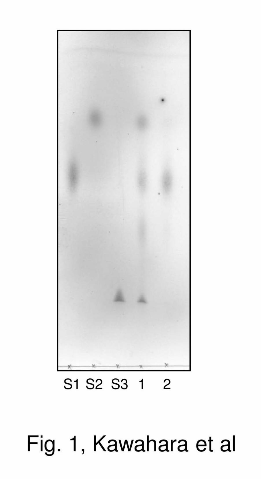
Identification of RaMP1 and RaMP2 in the
cell-free extract of R. albus NE1 – MP activity in the
cell-free extract of R. albus NE1 was analyzed by
TLC (Fig. 1). Man-Glc was degraded to D-glucose
and Man1P only in the presence of inorganic
phosphate, indicating that R. albus NE1 produces
intracellular MP. R. albus MP was purified to
identify the gene encoding it. MP activity was
identified in two peaks, RaMP1 and RaMP2, eluted
by high and low concentrations of NaCl respectively,
via anion-exchange column chromatography using a
DEAE Sepharose CL-6B column (data not shown).
Two MPs were further purified, and the masses of
the tryptic peptides were analyzed by matrix-assisted
laser desorption ionization time-of-flight mass
spectrometry (Supplementary Fig. S1). The obtained
masses of the digests of RaMP1 and RaMP2
matched with the theoretical values of the proteins
encoded by Rumal_0852 and Rumal_0099 from R.
albus 7, respectively. The analyzed tryptic fragments
derived from RaMP1 and RaMP2 covered 27% and
36% of their entire sequences, respectively.
Production, purification, and general
properties of recombinant RaMP1 and RaMP2 –
The genes encoding RaMP1 and RaMP2 were
obtained from the genomic DNA of R. albus NE1 by
PCR. The sequences of the amplified genes were
completely identical to the corresponding genes of R.
albus 7. These genes were overexpressed in E. coli,
and the recombinant enzymes were purified to
homogeneity. From 0.5 and 1.5 L of the culture
broths of the E. coli transformants, 6.8 mg and 11.4
mg of the purified rRaMP1 and rRaMP2 (the
specific activities were 55.2 U/mg and 2.03 U/mg)
were obtained. RaMP1 and RaMP2 were 45 kDa
and 38 kDa on SDS-PAGE respectively (data not
shown), which coincided well with the theoretical
masses from their amino acid sequences, but were
80 kDa and 209 kDa on a gel filtration column
respectively. These results indicate that RaMP1 and
RaMP2 exist as a homodimer and homohexamer in
solution, respectively. Both RaMP1 and RaMP2
showed highest activity at pH 6.5, and their
optimum temperatures were 50°C and 45°C
respectively. RaMP1 retained original activity in a
pH range of 4.5–10.5 and below 45°C, whereas
RaMP2 was stable at pH 3.5–9.5 and below 40°C.
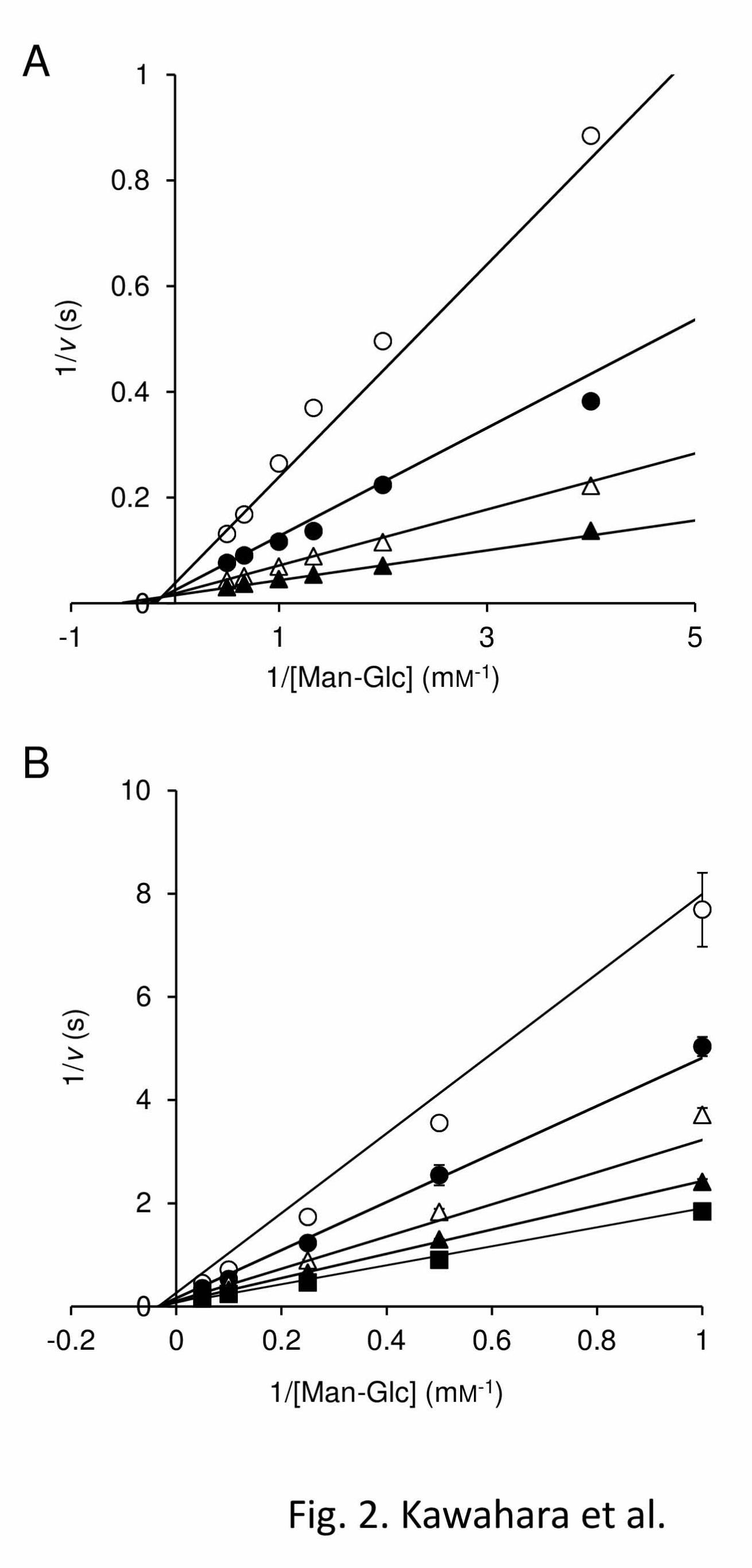
Kinetic mechanisms for phosphorolysis and
synthesis of Man-Glc by RaMP1 and RaMP2 –
Initial reaction velocities for phosphorolysis at
various concentrations of Man-Glc and inorganic
phosphate were measured to investigate the kinetic
mechanisms of RaMP1 and RaMP2. The lines
obtained for both enzymes from double reciprocals
plots of 1/v versus 1/[Man-Glc] at various
concentrations of inorganic phosphate were linear
and crossed at a certain point (Fig. 2). This indicates
that these enzymes catalyze the phosphorolysis of
Man-Glc through a sequential bi bi mechanism
involving the formation of a ternary complex. To
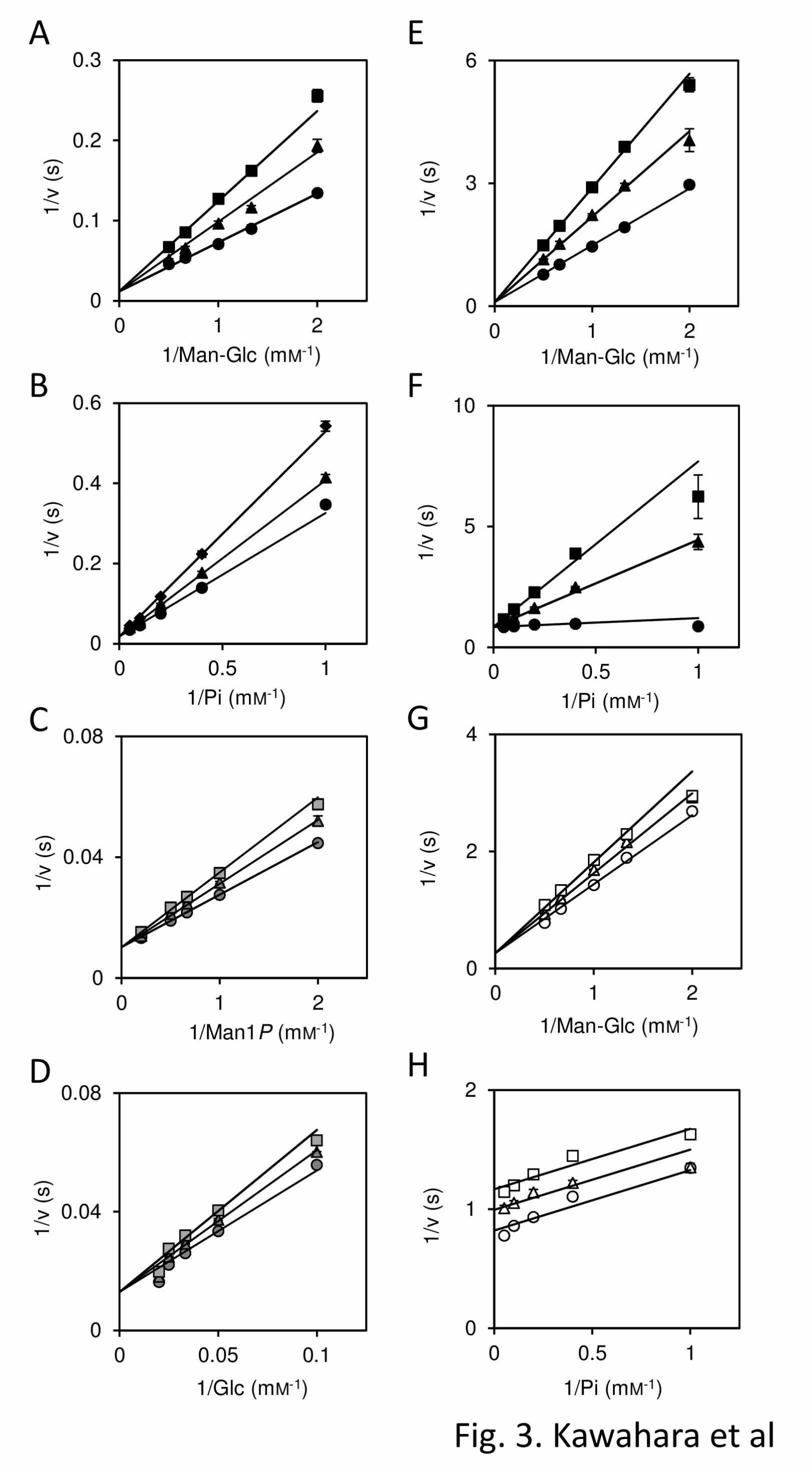
investigate the orders of substrate binding and
product release, product inhibition analysis was
carried out (Fig. 3). Man1P acted as a competitive
inhibitor of rRaMP1 and rRaMP2 against both
Man-Glc and inorganic phosphate as observed for
several cellobiose phosphorylases (3, 29), indicating
that these substrates bind to the enzymes in random
order, and Man1P can be the second product (the
first substrate in the reverse reaction). Therefore, a
possible kinetic mechanism for these enzymes is a
random order bi bi mechanism or a random-ordered
bi bi mechanism (3, 29). In the reverse reaction of
rRaMP1, Man-Glc served as a competitive inhibitor
Metabolic Mechanism of Mannan in Ruminococcus albus
6
against both Man1P and D-glucose, indicating that
rRaMP1 catalyzes phosphorolysis of Man-Glc
through a random order bi bi mechanism. In the case
of rRaMP2, inhibition of phosphorolysis towards
Man-Glc by cellobiose which is not phosphorolyzed
by rRaMP2 but acts as an acceptor substrate of this
enzyme (described later) was analyzed, because very
high concentration of Man-Glc is required for the
inhibition analysis of the synthetic reaction.
Uncompetitive inhibition by cellobiose was
observed, when the concentration of inorganic
phosphate was varied, presumably because of the
formation of an enzyme-phosphate-cellobiose
complex, which does not react or reacts very slowly.
On the other hand, cellobiose competitively
inhibited the phosphorolysis of Man-Glc at various
concentrations of Man-Glc. This inhibition occurred
by binding of cellobiose to +1 and +2 subsites or to
-1 and +1 subsites. The former case indicates
D-glucose (cellobiose) also can be the second
product, and the kinetic mechanism of the
phosphorolysis of Man-Glc by rRaMP2 is a random
order bi bi mechanism as rRaMP1. The possibility
of the latter case makes order of release (binding) of
Man1P and D-glucose (cellobiose) ambiguous.
However in the kinetic analysis of the synthetic
reaction towards cellobiose and Man1P, no substrate
inhibition by cellobiose was observed, indicating
that cellobiose acts only as an acceptor substrate, and
does not bind to -1 and +1 subsites. The kinetic
parameters for the phosphorolysis and synthesis of
Man-Glc are summarized in Table 2. In the
phosphorolytic reaction of Man-Glc, rRaMP1 had a
4.8-fold higher kcat and 91-fold lower KmMG
compared with rRaMP2. The KmPi of rRaMP2 was
considerably lower (12-fold) than that of rRaMP1.
Kinetic parameters for the synthesis of
Man-Glc with rRaMP1 and rRaMP2 were
determined from the initial reaction rates for various
concentrations of Man1P and D-glucose (Table 2).
rRaMP1 had a 6.6-fold higher kcat value for the
synthetic reaction than rRaMP2 as it did for the
phosphorolytic reaction. The KmGlc value of rRaMP1
was 3.7-fold lower than that of rRaMP2, indicating
that the +1 subsite of RaMP1 is more suitable for
binding the D-glucose moiety than RaMP2,
consistent with the higher affinity of rRaMP1
towards Man-Glc than that of rRaMP2. The
rRaMP2 KmMan1P value was 18-fold lower than that
of rRaMP1, corresponding with the observation that
RaMP2 has higher affinity toward inorganic
phosphate than RaMP1 during the phosphorolysis of
Man-Glc.
The internal consistency of the kinetic
parameters of rRaMP1 and rRaMP2 was confirmed
with the Haldane relationship for a sequential bi bi
mechanism (K = (kcatpKsMan1PKmGlc)/(kcat
sKsMan-GlcKmPi),
kcatp and kcat
s are kcat for phosphorolytic and synthetic
reactions, respectively) (30). Values of 0.435 ± 0.062
and 0.658 ± 0.137 were obtained for K of rRaMP1
and rRaMP2, respectively, from the kinetic
parameters in Table 2. These values were similar
in magnitude to the value 0.272 ± 0.021
experimentally obtained with rRaMP1 at pH 6.5 at
37°C for the thermodynamic equilibrium constant
(K) calculated from following equation:
K = ([Man1P][Glc])/([Man-Glc][phosphate])
Substrate specificity of RaMP1 and RaMP2 in
phosphorolytic reactions – D-Glucose was not
detected in the reaction carried out in the presence of
rRaMP1 or rRaMP2 and cellobiose or lactose even
at a 10-fold higher enzyme concentration than used
for the enzyme assay towards Man-Glc, indicating
that these enzymes are highly specific to the
mannosyl residue at the non-reducing end of the
substrate.
Acceptor specificity of RaMP1 in synthetic
reaction – The acceptor specificity of rRaMP1 was
investigated based on the initial reaction rates
towards Man1P and various acceptor substrates.
rRaMP1 showed a very narrow acceptor specificity.
Almost all the oligosaccharides tested were not
recognized as acceptor substrates. Only
laminaribiose served as a poor acceptor substrate for
rRaMP1. The apparent kinetic parameters, kcatapp and
Kmapp, for laminaribiose, determined in the presence
of 10 mM Man1P, were 7.6-fold lower and 6.0-fold
higher than those for D-glucose respectively (Table
3).
rRaMP1 was not active towards D-glucose
derivatives with changes at the C2-position, and it
was less active towards derivatives with changes at
the C3-position compared with D-glucose. The
monodeoxygenation at the 6-OH position was less
effective for RaMP1 than at the other positions. The
kcatapp and Kmapp values of rRaMP1 for
Metabolic Mechanism of Mannan in Ruminococcus albus
7
6-deoxy-D-glucose were 1.4- and 4.6-fold higher
than those for D-glucose, respectively. On the other
hand, 6-deoxy-6-fluoro-D-glucose and D-xylose
were considerably less favorable acceptor substrates
than 6-deoxy-D-glucose. This enzyme had a 3.3-fold
lower kcatapp and 6.0-fold higher Kmapp for
6-deoxy-6-fluoro-D-glucose than those for D-glucose,
and a saturation curve was not obtained for D-xylose
because of its very high Kmapp. These results indicate
that the 6-OH partially contributes to the
enzyme-substrate interaction as a hydrogen bond
donor, and that the methylene group of D-glucose is
also involved in stabilization of the transition state
presumably through the hydrophobic interaction.
Structures of oligosaccharides produced by the
synthetic reaction of rRaMP1 with D-glucose and
D-xylose as acceptors were investigated. A single
oligosaccharide was produced in each reaction, and
2.0 and 4.9 mg of product was obtained by HPLC,
respectively. The structures of these oligosaccharides
were elucidated by ESI-MS and NMR. The
oligosaccharides produced from the reactions with
D-glucose and D-xylose gave signals at 365.11 and
335.09 m/z [M + Na]+, respectively. Each
oligosaccharide had a correlation peak between the
4C of the acceptor and the 1H of the D-mannosyl
residue at the non-reducing end. The 1JCH values for
the D-mannosyl residues at the non-reducing ends of
these oligosaccharides in 13
C-NMR (insensitive
nuclei enhanced by polarization transfer) were 161
Hz, indicating that D-mannosyl residues were bound
to the acceptor substrates through a β-linkage (31).
On the basis of these results, the oligosaccharides
produced in the synthetic reactions in the presence of
D-glucose and D-xylose were identified as Man-Glc
and 4-O-β-D-mannosyl-D-xylose, respectively. The
chemical shifts of 1H- and
13C-NMR analyses are
summarized in supplementary Table S1.
Acceptor specificity of RaMP2 in synthetic
reaction – rRaMP2 was not active when D-glucose
was derivatized at the C6-position, but showed very
broad acceptor specificity (Table 3). rRaMP2
showed the highest kcatapp/Kmapp towards
N-acetyl-D-glucosamine among the 2-OH
derivatives of D-glucose, even though
N-acetyl-D-glucosamine has a bulky acetamide
group at the C2 position. All the D-glucose
derivatives with changes at the C3-position acted as
acceptor substrates of rRaMP2. The kcatapp and Kmapp
values for these derivatives were considerably lower
than for D-glucose. The Kmapp value for
3-deoxy-D-glucose was too low to determine the
value.
rRaMP2 had kcatapp/Kmapp values for
1,5-anhydro-D-glucitol and methyl α-D-glucoside
similar to that for D-glucose, and the kcatapp/Kmapp for
methyl β-D-glucoside was 2.1-fold higher than for
D-glucose. All the oligosaccharides acted as acceptor
substrates of rRaMP2. The kcatapp/Kmapp values for
β-linked glucobioses were 1.4–4.5-fold higher than
for D-glucose due to low Kmapp values for these
oligosaccharides. Cellobiose was the best acceptor
substrate for rRaMP2 among the β-linked
glucobioses in terms of its kcatapp/Kmapp value,
followed by gentiobiose. rRaMP2 also had a
3.4-fold lower Kmapp for N,N’-diacetylchitobiose.
The kcatapp/Kmapp for maltose was 50% of that for
D-glucose, thus the acceptor site of rRaMP1
efficiently accommodates even an α-linked
oligosaccharide. D-Mannose was a poor acceptor
substrate of rRaMP2 compared with D-glucose, but
rRaMP2 showed 7.3–20-fold higher kcatapp/Kmapp
values for β-1,4-mannooligosaccharides than for
D-glucose. The kcatapp/Kmapp values for
β-1,4-mannooligosaccharides increased depending
on the degree of polymerization (DP) up to
β-1,4-mannotetraose, while those for substrates
longer than β-1,4-mannotetraose slightly decreased
with increasing substrate chain-length, although a
lower Kmapp was observed for longer
β-1,4-mannooligosaccharides.
Structural analysis of oligosaccharides
produced in the synthetic reactions of rRaMP2
towards cellobiose, β-1,4-mannobiose, and
N,N’-diacetylchitobiose were performed. In contrast
to rRaMP1, rRaMP2 produced several
oligosaccharides with different DPs. The resulting
reaction mixtures were subjected to gel filtration
column chromatography (data not shown). The
yields of the oligosaccharides purified were as
follows: from cellobiose, DP3, 9.5 mg; DP4, 2.7 mg;
DP5, 0.8 mg; and DP6, 0.4 mg; from
β-1,4-mannobiose, DP3, 4.5 mg; DP4, 2.8 mg; DP5,
0.8 mg; and DP6, 0.3 mg; from
N,N’-diacetylchitobiose, DP3, 2.4 mg; DP4, 1.3 mg;
DP5, 0.8 mg; and DP6, 0.6 mg. Molecular masses of
the obtained oligosaccharides were analyzed by
Metabolic Mechanism of Mannan in Ruminococcus albus
8
ESI-MS. The masses were as follows: the products
from cellobiose, 527.16, 689.26, 851.27, and
1013.32 m/z [M + Na]+; from β-1,4-mannobiose,
527.16, 689.21, 851.27, and 1013.32 m/z [M + Na]+;
from N,N’-diacetylchitobiose, 609.21, 771.27,
933.36, and 1095.37 m/z [M + Na]+. These results
indicate that rRaMP2 successively transferred
mannosyl residues to the synthesized
oligosaccharides. The chemical structures of the
trisaccharides synthesized in the reactions in the
presence of cellobiose and N,N’-diacetylchitobiose
by rRaMP2 were analyzed as described above, and
determined to be 4-O-β-D-mannosyl-cellobiose and
4-O-β-D-mannosyl-N,N’-diacetylchitobiose,
respectively (supplementary Table S1). 13
C- and 1H-NMR spectra of the trisaccharides produced
from β-1,4-mannobiose were completely identical to
those of β-1,4-mannotriose.
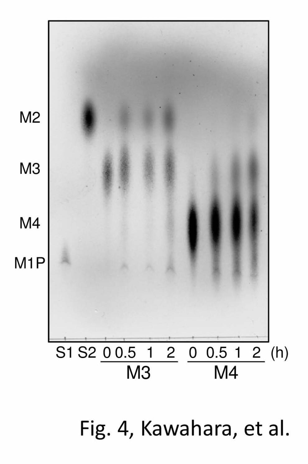
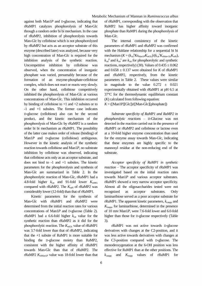
Phosphorolysis of β-1,4-mannooligosaccharides by
rRaMP2 – The kinetic analysis of the synthetic
reaction of rRaMP2 indicated that this enzyme
mainly catalyzes the phosphorolysis of
β-1,4-mannooligosaccharides rather than that of
Man-Glc. In the phosphorolytic reaction, rRaMP2
degraded β-1,4-mannotriose and
β-1,4-mannotetraose to Man1P and shorter
β-1,4-mannooligosaccharides (Fig. 4).
β-1,4-Mannobiose was accumulated in the late stage
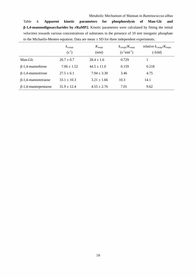
of the reaction with β-1,4-mannotriose. The apparent
kinetic parameters of rRaMP2 for Man-Glc and
β-1,4-mannooligosaccharides, which was measured
in the presence of 10 mM sodium phosphate buffer
(pH 6.5), were compared (Table 4). The kcatapp values
for β-1,4-mannooligosaccharides longer than β-1,4-
mannobiose were similar to that for Man-Glc, but
the Kmapp values were 3.6–8.8-fold lower, consistent
with the acceptor specificity for the synthetic
reactions. On the other hand, β-1,4-mannobiose was
a very poor substrate for rRaMP2. The kcatapp/Kmapp
value was 22–65-fold lower than those for the longer
β-1,4-mannooligosaccharides.
DISCUSSION
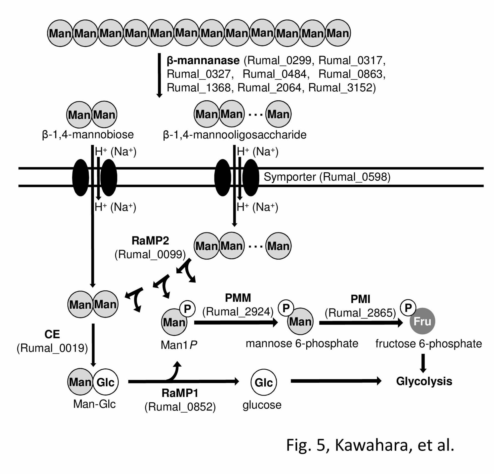
In B. fragilis, the metabolic pathway of mannan
has been postulated as follows: i) hydrolysis of
mannan by extracellular β-1,4-mannanase, ii)
epimerization of the resulting β-1,4-mannobiose to
Man-Glc by CE, iii) phosphorolysis of Man-Glc to
D-glucose and Man1P by MP (7). Phosphomannose
mutase and mannose-6-phosphate isomerase convert
Man1P to fructose 6-phosphate, which is then
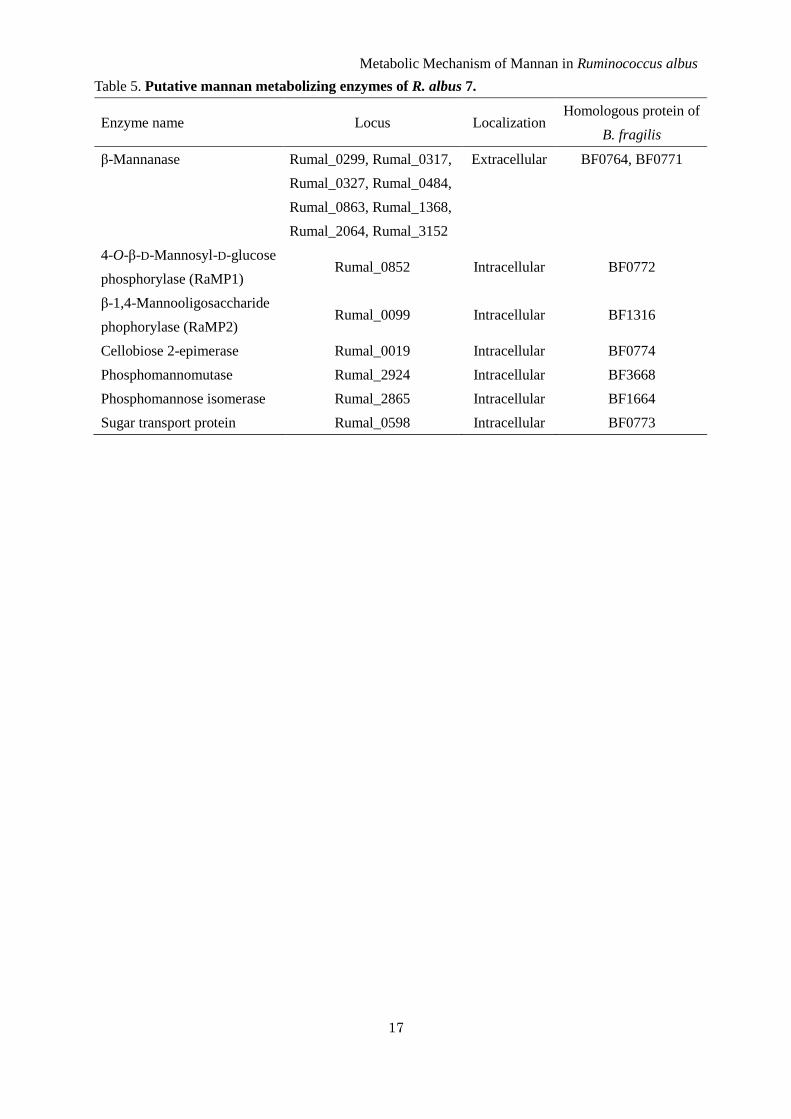
metabolized through glycolysis. R. albus NE1 is also
a CE producer (5), and two MP homologous genes
were encoded in the genome of R. albus 7. This
bacterium also has putative proteins involved in the
metabolism of mannan, eight extracellular
β-1,4-mannanases belonging to GH family 26, a
phosphomannomutase, phosphomannose isomerase,
and sugar transport protein similar to sugar/cation
symporter (Table 5). MP activity was detected in the
cell-free extract of R. albus NE1, and two MP
isozymes, the deduced amino acid sequences of
which are completely identical to the corresponding
genes of R. albus 7, were found. Consequently, these
MP isozymes are predicted to be physiologically
functional and R. albus NE1 (R. albus 7) is thought
to degrades mannan through a similar pathway as
postulated for B. fragilis (Fig. 5).
Both rRaMP1 and rRaMP2 catalyzed
phosphorolytic and synthetic reactions of Man-Glc
through a random bi bi mechanism in contrast to
other known inverting carbohydrate phosphorylases
(3, 32-34). Both rRaMP1 and rRaMP2 specifically
catalyzed the phosphorolysis of a β-1,4-mannosidic
linkage at the non-reducing end of a substrate, but
they had acceptor specificities which were clearly
different from each other. Consistent with the
acceptor specificity of BfMP, which shows high
sequence similarity to RaMP1 (7), rRaMP1 was
highly active only towards D-glucose and
6-deoxy-D-glucose, but rRaMP2 recognized a
significantly wider variety of sugars including
oligosaccharides as the acceptor substrates. rRaMP2
was more active towards
β-1,4-mannooligosaccharides than D-glucose, and
successive formations of β-1,4-mannosidic linkages
were observed in the synthetic reactions. In the
phosphorolytic reactions,
β-1,4-mannooligosaccharides were degraded to
Man1P and shorter oligosaccharides (Fig. 4).
β-1,4-Mannooligosaccharides longer than β-1,4-
mannobiose were considerably better substrates for
rRaMP2 than Man-Glc (Table 4). Hence the
physiological role of RaMP2 is not phosphorolysis
of Man-Glc, but is phosphorolysis of
β-1,4-mannooligosaccharides of ≥DP3 to
β-1,4-mannobiose. On the basis of the substrate
Metabolic Mechanism of Mannan in Ruminococcus albus
9
preference, RaMP2 should be named
β-1,4-mannooligosaccharide phosphorylase rather
than 4-O-β-D-mannosyl-D-glucose phosphorylase.
The low activity of rRaMP2 towards
β-1,4-mannobiose clearly indicates the importance
of the processes catalyzed by CE and RaMP1 as
described above. B. fragilis also has a RaMP2-like
protein, BF1316, (NCBI reference sequence,
YP_210978.1), which has 64.6% sequence identity
to RaMP2. It is possible that this bacterium also
degrades mannooligosaccharides longer than
β-1,4-mannobiose by the RaMP2-type
phosphorylase. Furthermore, the aerobic
CE-producing bacterium, Rhodothermus murinus
(28), also has both RaMP1- and RaMP2-type
proteins (YP_003291706 and YP_003291875
respectively), and hence, the metabolic pathway of
mannan postulated here is not likely limited to
anaerobic bacteria.
β-1,4-Mannotetraose was the best acceptor
substrate for RaMP2 among the
β-1,4-mannooligosacccharides tested in terms of the
kcatapp/Kmapp (Table 3), while the kcatapp/Kmapp for longer
β-1,4-mannooligosacccharides slightly decreased
with an increase of DP. The acceptor site of RaMP2
is likely composed of at least four subsites. Hence
RaMP2 presumably has a cleft-type substrate
binding site to accommodate a long-chain acceptor
substrate unlike RaMP1, which recognizes only
monosaccharides as the acceptors. In the case of
RaMP2, a β-1,4-glycosidic linkage is preferred in
subsites +1 and +2, but the specificity for the
acceptor substrates is very loose and even maltose
and N,N’-diacetylchitobiose were acceptable.
Herein, we described in detail the biochemical
properties of two MP isozymes from R. albus NE1
belonging to GH family 130. Both enzymes were
specific to the β-1,4-mannosidic linkage at the
non-reducing end, but RaMP1 and RaMP2 showed
narrow and broad acceptor specificities in synthetic
reactions. RaMP2 is the first enzyme known to
efficiently participate in the phosphorolysis and
synthesis of β-1,4-mannooligosacchairdes longer
than β-1,4-mannobiose. Structural analysis of an
enzyme-substrate complex is required for further
understanding of the structure-function relationship
of GH family 130 enzymes.
REFERENCES
1. Leatherwood, J. M. (1965) Appl. Microbiol. 13, 771-775
2. Ohmiya, K., Shirai, M., Kurachi, Y., and Shimizu, S. (1985) J. Bacteriol. 161, 432-434
3. Hamura, K., Saburi, W., Abe, S., Morimoto, N., Taguchi, H., Mori, H., and Matsui, H. (2012)
Biosci. Biotechnol. Biochem. 76, 812-818
4. Tyler, T. and Leatherwood, J.M. (1967) Arch. Biochem. Biophys. 119, 363-367
5. Ito, S., Hamada, S., Yamaguchi, K., Umene, S., Ito, H., Matsui, H., Ozawa, T., Taguchi, H.,
Watanabe, J., Wasaki, J., and Ito, S. (2007) Biochem. Biophys. Res. Commun. 360, 640-645
6. Senoura, T., Taguchi, H., Ito, S., Hamada, S., Matsui, H., Fukiya, S., Yokota, A., Watanabe, J.,
Wasaki, J., and Ito, S. (2009) Biosci Biotechnol Biochem 73, 400-406
7. Senoura, T., Ito, S., Taguchi, H., Higa, M., Hamada, S., Matsui, H., Ozawa, T., Jin, S., Watanabe,
J., Wasaki, J., and Ito, S. (2011) Biochem. Biophys. Res. Commun. 408, 701-706
8. Hidaka, M., Kitaoka, M., Hayashi, K., Wakagi, T., Shoun, H., and Fushinobu, S. (2006) Biochem.
J 398, 37-43
9. Cantarel, B. L., Coutinho, P. M., Rancurel, C., Bernard, T., Lombard, V., and Henrissat, B. (2009)
Nucleic Acids Res. 37, D233-D238
10. Egloff, M., Uppenberg, J., Haalck, L., and van Tibeurgh, H. (2001) Structure 9, 689-697
11. Nagem, R. A. P., Rojas, A. L., Golubev, A. M., Korneeva, O. S., Eneyskaya, E. V., Kulminskaya, A.
A., Neustroev, K. N., and Polikarpov, I. (2004) J. Mol. Biol. 344, 471-480
Metabolic Mechanism of Mannan in Ruminococcus albus
10
12. Alberto, F., Bignon, C., Sulzenbacher, G., Henrissat, B., and Czjzek, M. (2004) J. Biol. Chem. 279,
18903-18910
13. Nurizzo, D., Turkenburg, J. P., Chrnock, S. J., Roberts, S. M., Dodson, E. J., McKie, V. A., Taylor,
E. J., Gilbert, H. J., and Davies, G. J. (2002) Nat. Struct. Biol. 9, 665-675
14. Brüx, C., Ben-David, A., Shallom-Shezifi, D., Leon, M., Niefind, K., Shoham, G., Shoham, Y., and
Schomburg, D. (2006) J. Mol. Biol. 359, 97-109
15. Fujimoto, Z., Ichinose, H., Maehara, T., Honda, M., Kitaoka, M, and Kaneko, S. (2010) J. Biol.
Chem. 285, 34134-34143
16. Cartmell, A., McKee, L.S., Peña, M.J., Larsbrink, J., Brumer, H., Kaneko, S., Ichinose, H., Lewis,
R.J., Viksø-Nielsen, A., Gilbert, H.J., and Marles-Wright, J. (2011) J. Biol. Chem. 286,
15483-15495
17. Alhassid, A., Ben-David, A., Tabachnikov, O., Libster, D., Naveh, E., Zolotnitsky, G., Shoham, Y.,
and Shoham, G. (2009) Biochem. J. 422, 73-82
18. Yamaguchi, A., Tada, T., Wada, K., Nakaniwa, T., Kitatani, T., Sogabe, Y., Takao, M., Sakai, T.,
and Nishimura, K. (2005) J. Biochem. 137, 587-592
19. Meng, G. and Fütterer, K. (2003) Nat. Struct. Biol. 10, 935-941
20. Martínez-Fleites, C., Ortíz-Lombardía, M., Pons, T., Tarbouriech, N., Taylor, E.J., Arrieta, J.G.,
Nernández, L., and Davies, G.J. (2005) Biochem. J. 390, 19-27
21. Sato, H., Saburi, W., Ojima, T., Taguchi, H., Mori, H., and Matsui, H. (2012) Biosci. Biotechnol.
Biochem. 76, 1584-1587
22. Ito, S., Taguchi, H., Hamada, S., Kawauchi, S., Ito, H., Senoura, T., Watanabe, J., Nishimukai, M.,
Ito, S., and Matsui, H. (2008) Appl. Microbiol. Biotechnol. 79, 433-441
23. Wakuta, S., Hamada, S., Ito, H., Matsuura, H., Nabeta, K., and Matsui, H. (2010) Phytochemistry
71, 1280-1288
24. Moore, S. and Stein W.H. (1948) J. Biol. Chem. 176, 367-388
25. Miwa, I., Okudo, J., Maeda, K., and Okuda, G. (1972) Clin. Chim. Acta 37, 538-540
26. Cleland, W.W. (1963) Biochim. Biophys. Acta 67, 104-137
27. Lowry, O.H. and Lopez, J.A. (1946) J. Biol. Chem. 162, 421-428
28. Ojima, T., Saburi, W., Sato, H., Yamamoto, T., Mori, H., and Matsui, H. (2011) Biosci. Biotechnol.
Biochem. 75, 2162-2168
29. Rajashekhara, E., Kitaoka, M., Kim, Y-K, and Hayashi, K. (2002) Biosci. Biotechnol. Biochem. 66,
2578-2586
30. Dixon, M. and Webb, E. C. (1979) Enzymes, 3rd Ed., Longman Group Limited, London
31. Bock, K. and Pedersen, C. (1974) J. Chem. Soc., Perkin Trans. 2, 293-297
32. Kitaoka, M., Sasaki, T., and Taniguchi, H. (1992) Biosci. Biotechnol. Biochem. 56, 652-655
33. Nihira, T., Nakai, H., Chiku, K., and Kitaoka, M. (2011) Appl. Microbiol. Biotechnol. 93,
1513-1522
34. Kitaoka, M., Matsuoka, Y., Mori, K., Nishimoto, M., and Hayashi, K. (2012) Biosci. Biotechnol.
Biochem. 76, 343-348
Metabolic Mechanism of Mannan in Ruminococcus albus
11
FOOTNOTES
*This work was partially supported by Kakenhi, the Grants-in-Aid for Young Scientists (B)
(24780091) of the Ministry of Education, Culture, Sports, Science and Technology of Japan.
**First authors, who contributed equally.
The abbreviations used are: CE, cellobiose 2-epimerase; MP, 4-O-β-D-mannosyl-D-glucose
phosphorylase; Man-Glc, 4-O-β-D-mannosyl-D-glucose; Man1P, α-D-mannosyl phosphate; BfMP,
Bacteroides fragilis MP; GH, glycoside hydrolase; TLC, thin layer chromatography; RaMP1,
Ruminococcus albus MP1 (Rumal_0852); RaMP2, R. albus MP2 (Rumal_0099); rRaMP1,
recombinant RaMP1; rRaMP2, recombinant RaMP2; ESI-MS, electrospray ionization-mass
spectrometry; NMR, nuclear magnetic resonance
FIGURE LEGENDS
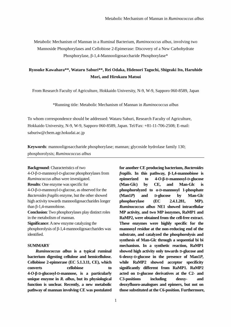
FIGURE 1. Detection of MP activity in the cell-free extract of R. albus NE1. Phosphorolytic
activity of the cell-free extract of R. albus NE1 towards Man-Glc in the presence or absence of
inorganic phosphate was analyzed by TLC. S1, Man-Glc; S2, glucose; S3, Man1P. Man-Glc was
degraded only in sodium phosphate buffer.
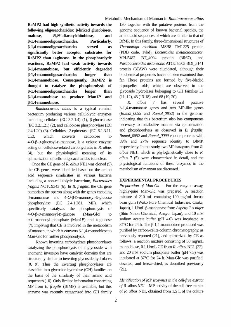
FIGURE 2. Double reciprocal plot for the phosphorolysis of Man-Glc by rRaMP1 and rRaMP2.
The initial velocities for phosphorolysis of Man-Glc at various concentrations of Man-Glc and
inorganic phosphate were measured. (A) rRaMP1. Concentrations of inorganic phosphate were 2.5
mM (open circle), 5.0 mM (filled circle), 10 mM (open triangle), and 20 mM (filled triangle). (B)
rRaMP2. Concentrations of inorganic phosphate were 0.125 mM (open circle), 0.25 mM (filled circle),
0.5 mM (open triangle), 1 mM (filled triangle), and 3 mM (filled square). Data are the mean ± SD for
three independent experiments.
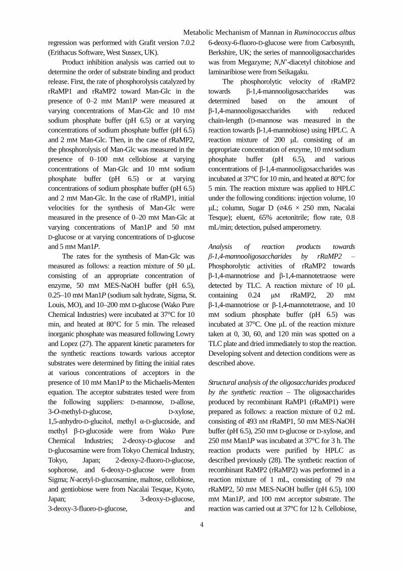
FIGURE 3. Product inhibition analyses of rRaMP1 and rRaMP2.
Product inhibition analyses of rRaMP1 and rRaMP2 are shown in panel A-D and E-H, respectively. A
and B (E and F for rRaMP2) are inhibition of the phosphorolysis of Man-Glc by Man1P against
Man-Glc and inorganic phosphate, respectively. C and D are inhibition of the synthesis of Man-Glc by
Man-Glc against Man1P and D-glucose, respectively. G and H are inhibition of the phosphorolysis of
Man-Glc by cellobiose against Man-Glc and inorganic phosphate, respectively. Black circle, triangle
square, and diamond indicate 0, 1, 2, and 2.5 mM, respectively. Gray circle, triangle, and square
indicate 0, 10, and 20 mM, respectively. Open circle, triangle, and square are 0, 50, and 100 mM,
respectively. Data are the mean ± SD for three independent experiments.
FIGURE 4. Phosphorolysis of β-1,4-mannooligosaccharides by rRaMP2. Phosphorolytic reactions
of rRaMP2 towards β-1,4-mannotriose and β-1,4-mannotetraose were analyzed by TLC. Reaction time
Metabolic Mechanism of Mannan in Ruminococcus albus
12
is indicated below the figure. S1 and S2 are standards of Man1P and β-1,4-mannobiose, respectively.
M2, β-1,4-mannobiose; M3, β-1,4-mannotriose; M4, β-1,4-mannotetraose; M1P, Man1P.
FIGURE 5. Metabolic pathway of mannan in R. albus NE1. Metabolic pathway of mannan
involving two MPs and CE is illustrated. CE, cellobiose 2-epimerase; PMM, phosphomannose mutase;
PMI mannose 6-phosphate isomerase.
Metabolic Mechanism of Mannan in Ruminococcus albus
13
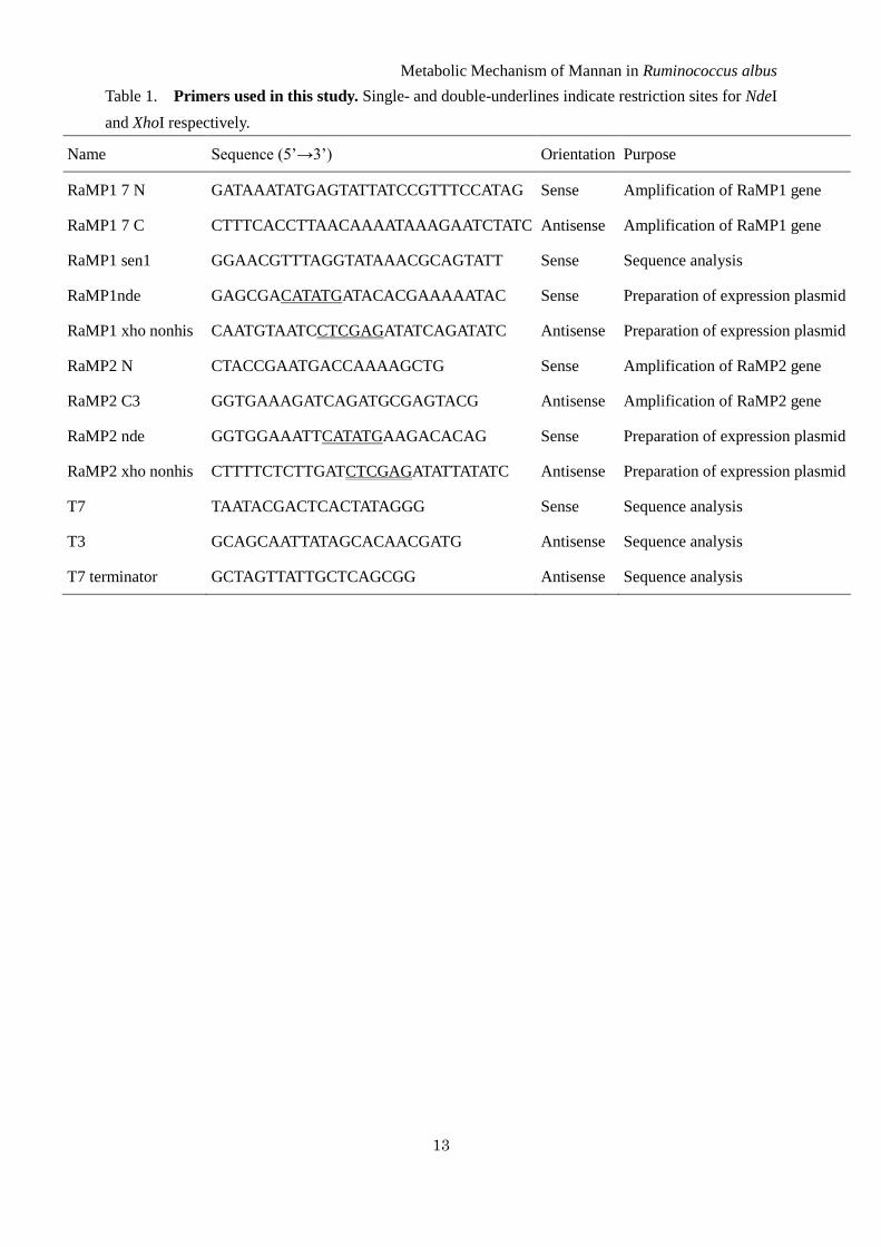
Table 1. Primers used in this study. Single- and double-underlines indicate restriction sites for NdeI
and XhoI respectively.
Name Sequence (5’→3’) Orientation Purpose
RaMP1 7 N GATAAATATGAGTATTATCCGTTTCCATAG Sense Amplification of RaMP1 gene
RaMP1 7 C CTTTCACCTTAACAAAATAAAGAATCTATC Antisense Amplification of RaMP1 gene
RaMP1 sen1 GGAACGTTTAGGTATAAACGCAGTATT Sense Sequence analysis
RaMP1nde GAGCGACATATGATACACGAAAAATAC Sense Preparation of expression plasmid
RaMP1 xho nonhis CAATGTAATCCTCGAGATATCAGATATC Antisense Preparation of expression plasmid
RaMP2 N CTACCGAATGACCAAAAGCTG Sense Amplification of RaMP2 gene
RaMP2 C3 GGTGAAAGATCAGATGCGAGTACG Antisense Amplification of RaMP2 gene
RaMP2 nde GGTGGAAATTCATATGAAGACACAG Sense Preparation of expression plasmid
RaMP2 xho nonhis CTTTTCTCTTGATCTCGAGATATTATATC Antisense Preparation of expression plasmid
T7 TAATACGACTCACTATAGGG Sense Sequence analysis
T3 GCAGCAATTATAGCACAACGATG Antisense Sequence analysis
T7 terminator GCTAGTTATTGCTCAGCGG Antisense Sequence analysis
Metabolic Mechanism of Mannan in Ruminococcus albus
14
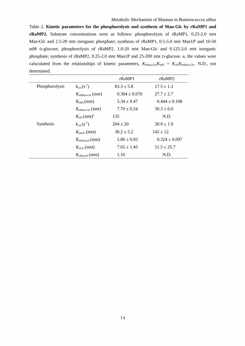
Table 2. Kinetic parameters for the phosphorolysis and synthesis of Man-Glc by rRaMP1 and
rRaMP2. Substrate concentrations were as follows: phosphorolysis of rRaMP1, 0.25-2.0 mM
Man-Glc and 2.5-20 mM inorganic phosphate; synthesis of rRaMP1, 0.5-5.0 mM Man1P and 10-50
mM D-glucose; phosphorolysis of rRaMP2, 1.0-20 mM Man-Glc and 0.125-3.0 mM inorganic
phosphate; synthesis of rRaMP2, 0.25-2.0 mM Man1P and 25-200 mM D-glucose. a, the values were
caluculated from the relationships of kinetic parameters, KsMan-GlcKmPi = KsPiKmMan-Glc. N.D., not
determined.
rRaMP1 rRaMP2
Phosphorolysis kcat (s-1
) 083.3 ± 5.8 017.5 ± 1.3
KmMan-Glc (mM) 000.304 ± 0.070 027.7 ± 2.7
KmPi (mM) 005.34 ± 0.47 000.444 ± 0.108
KsMan-Glc (mM) 007.70 ± 0.24 030.3 ± 6.0
KsPi (mM)a 135 N.D.
Synthesis kcat (s-1
) 204 ± 20 030.9 ± 1.9
KmGlc (mM) 038.2 ± 5.2 142 ± 12
KmMan1P (mM) 005.80 ± 0.93 000.324 ± 0.097
KsGlc (mM) 007.65 ± 1.45 051.5 ± 25.7
KsMan1P (mM) 001.16 N.D.
Metabolic Mechanism of Mannan in Ruminococcus albus
15
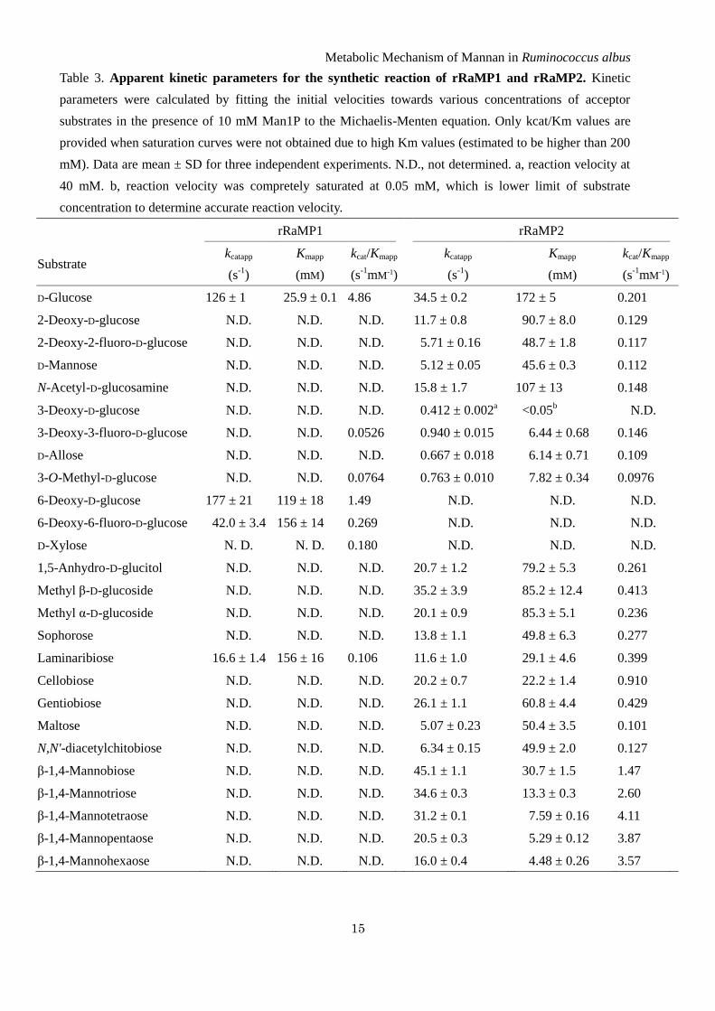
Table 3. Apparent kinetic parameters for the synthetic reaction of rRaMP1 and rRaMP2. Kinetic
parameters were calculated by fitting the initial velocities towards various concentrations of acceptor
substrates in the presence of 10 mM Man1P to the Michaelis-Menten equation. Only kcat/Km values are
provided when saturation curves were not obtained due to high Km values (estimated to be higher than 200
mM). Data are mean ± SD for three independent experiments. N.D., not determined. a, reaction velocity at
40 mM. b, reaction velocity was compretely saturated at 0.05 mM, which is lower limit of substrate
concentration to determine accurate reaction velocity.
rRaMP1 rRaMP2
Substrate kcatapp
(s-1
)
Kmapp
(mM)
kcat/Kmapp
(s-1
mM-1)
kcatapp
(s-1
)
Kmapp
(mM)
kcat/Kmapp
(s-1
mM-1)
D-Glucose 126 ± 1 025.9 ± 0.1 4.86 34.5 ± 0.2 172 ± 5 0.201
2-Deoxy-D-glucose N.D. N.D. N.D. 11.7 ± 0.8 090.7 ± 8.0 0.129
2-Deoxy-2-fluoro-D-glucose N.D. N.D. N.D. 05.71 ± 0.16 048.7 ± 1.8 0.117
D-Mannose N.D. N.D. N.D. 05.12 ± 0.05 045.6 ± 0.3 0.112
N-Acetyl-D-glucosamine N.D. N.D. N.D. 15.8 ± 1.7 107 ± 13 0.148
3-Deoxy-D-glucose N.D. N.D. N.D. 00.412 ± 0.002a 0<0.05
b N.D.
3-Deoxy-3-fluoro-D-glucose N.D. N.D. 0.0526 00.940 ± 0.015 006.44 ± 0.68 0.146
D-Allose N.D. N.D. N.D. 00.667 ± 0.018 006.14 ± 0.71 0.109
3-O-Methyl-D-glucose N.D. N.D. 0.0764 00.763 ± 0.010 007.82 ± 0.34 0.0976
6-Deoxy-D-glucose 177 ± 21 119 ± 18 1.49 N.D. N.D. N.D.
6-Deoxy-6-fluoro-D-glucose 042.0 ± 3.4 156 ± 14 0.269 N.D. N.D. N.D.
D-Xylose N. D. N. D. 0.180 N.D. N.D. N.D.
1,5-Anhydro-D-glucitol N.D. N.D. N.D. 20.7 ± 1.2 079.2 ± 5.3 0.261
Methyl β-D-glucoside N.D. N.D. N.D. 35.2 ± 3.9 085.2 ± 12.4 0.413
Methyl α-D-glucoside N.D. N.D. N.D. 20.1 ± 0.9 085.3 ± 5.1 0.236
Sophorose N.D. N.D. N.D. 13.8 ± 1.1 049.8 ± 6.3 0.277
Laminaribiose 016.6 ± 1.4 156 ± 16 0.106 11.6 ± 1.0 029.1 ± 4.6 0.399
Cellobiose N.D. N.D. N.D. 20.2 ± 0.7 022.2 ± 1.4 0.910
Gentiobiose N.D. N.D. N.D. 26.1 ± 1.1 060.8 ± 4.4 0.429
Maltose N.D. N.D. N.D. 05.07 ± 0.23 050.4 ± 3.5 0.101
N,N'-diacetylchitobiose N.D. N.D. N.D. 06.34 ± 0.15 049.9 ± 2.0 0.127
β-1,4-Mannobiose N.D. N.D. N.D. 45.1 ± 1.1 030.7 ± 1.5 1.47
β-1,4-Mannotriose N.D. N.D. N.D. 34.6 ± 0.3 013.3 ± 0.3 2.60
β-1,4-Mannotetraose N.D. N.D. N.D. 31.2 ± 0.1 007.59 ± 0.16 4.11
β-1,4-Mannopentaose N.D. N.D. N.D. 20.5 ± 0.3 005.29 ± 0.12 3.87
β-1,4-Mannohexaose N.D. N.D. N.D. 16.0 ± 0.4 004.48 ± 0.26 3.57
Metabolic Mechanism of Mannan in Ruminococcus albus
16
Table 4. Apparent kinetic parameters for phosphorolysis of Man-Glc and
β-1,4-mannooligosaccharides by rRaMP2. Kinetic parameters were calculated by fitting the initial
velocities towards various concentrations of substrates in the presence of 10 mM inorganic phosphate
to the Michaelis-Menten equation. Data are mean ± SD for three independent experiments.
kcatapp
(s-1
)
Kmapp
(mM)
kcatapp/Kmapp
(s-1
mM-1
)
relative kcatapp/Kmapp
(-fold)
Man-Glc 20.7 ± 0.7 28.4 ± 1.6 00.729 01
β-1,4-mannobiose 07.06 ± 1.52 44.5 ± 11.0 00.159 00.218
β-1,4-mannotriose 27.5 ± 6.1 07.94 ± 3.30 03.46 04.75
β-1,4-mannotetraose 33.1 ± 10.3 03.21 ± 1.66 10.3 14.1
β-1,4-mannopentaose 31.9 ± 12.4 04.55 ± 2.76 07.01 09.62
Metabolic Mechanism of Mannan in Ruminococcus albus
17
Table 5. Putative mannan metabolizing enzymes of R. albus 7.
Enzyme name Locus Localization Homologous protein of
B. fragilis
β-Mannanase Rumal_0299, Rumal_0317,
Rumal_0327, Rumal_0484,
Rumal_0863, Rumal_1368,
Rumal_2064, Rumal_3152
Extracellular BF0764, BF0771
4-O-β-D-Mannosyl-D-glucose
phosphorylase (RaMP1) Rumal_0852 Intracellular BF0772
β-1,4-Mannooligosaccharide
phophorylase (RaMP2) Rumal_0099 Intracellular BF1316
Cellobiose 2-epimerase Rumal_0019 Intracellular BF0774
Phosphomannomutase Rumal_2924 Intracellular BF3668
Phosphomannose isomerase Rumal_2865 Intracellular BF1664
Sugar transport protein Rumal_0598 Intracellular BF0773