-Mannan degradation by gut bacteria - …lup.lub.lu.se/search/ws/files/5494621/5277658.pdf1...
66
-Mannan degradation by gut bacteria - Characterization of -mannanases from families GH5 and GH26 Kulcinskaja, Evelina Published: 2015-01-01 Document Version Publisher's PDF, also known as Version of record Link to publication Citation for published version (APA): Kulcinskaja, E. (2015). β-Mannan degradation by gut bacteria - Characterization of β-mannanases from families GH5 and GH26 Department of Chemistry, Lund University General rights Copyright and moral rights for the publications made accessible in the public portal are retained by the authors and/or other copyright owners and it is a condition of accessing publications that users recognise and abide by the legal requirements associated with these rights. • Users may download and print one copy of any publication from the public portal for the purpose of private study or research. • You may not further distribute the material or use it for any profit-making activity or commercial gain • You may freely distribute the URL identifying the publication in the public portal
Transcript of -Mannan degradation by gut bacteria - …lup.lub.lu.se/search/ws/files/5494621/5277658.pdf1...

LUND UNIVERSITY
PO Box 117221 00 Lund+46 46-222 00 00
-Mannan degradation by gut bacteria - Characterization of -mannanases from familiesGH5 and GH26
Kulcinskaja, Evelina
Published: 2015-01-01
Document VersionPublisher's PDF, also known as Version of record
Link to publication
Citation for published version (APA):Kulcinskaja, E. (2015). β-Mannan degradation by gut bacteria - Characterization of β-mannanases from familiesGH5 and GH26 Department of Chemistry, Lund University
General rightsCopyright and moral rights for the publications made accessible in the public portal are retained by the authorsand/or other copyright owners and it is a condition of accessing publications that users recognise and abide by thelegal requirements associated with these rights.
• Users may download and print one copy of any publication from the public portal for the purpose of privatestudy or research. • You may not further distribute the material or use it for any profit-making activity or commercial gain • You may freely distribute the URL identifying the publication in the public portal

Take down policyIf you believe that this document breaches copyright please contact us providing details, and we will removeaccess to the work immediately and investigate your claim.
Download date: 29. Jun. 2018

1
(Spikblad)
-Mannan degradation
by gut bacteria Characterization of -mannanases from families GH5
and GH26
Evelina Kulcinskaja
DOCTORAL DISSERTATION by due permission of the Faculty of Science, Lund University, Sweden.
To be defended on Friday the 22nd of May 2015 at 10.15 in Hall B, Kemicentrum, Lund.
Faculty opponent
Professor Anne Meyer,
Chemical Engineering, DTU, Lyngby, Denmark

2015-04-14
164
2
Organization
LUND UNIVERSITY
Document name
Doctoral Dissertation
Date of issue
May 22nd 2015
Author(s) Evelina Kulcinskaja Sponsoring organization
Title and subtitle
-Mannan degradation by gut bacteria, Characterization of -mannanases from families GH5 and GH26
The human gut flora is important for our well-being. The gut bacteria are able to degrade and metabolize complex carbohydrates. Examples of such carbohydrates are -mannans. -Mannans consist of a backbone of -1,4-linked mannose units and are present in e.g. the endosperm of legumes such as guar or carob. The composition of the -mannan varies with the plant source. Carob and guar -mannans are substituted with
-1,6-linked galactose units and are soluble in water. They make a viscous solution that is used e.g. as food thickener. It is known that guar gum galactomannan can be fermented by the human gut flora. The main enzymes that hydrolyze -mannan backbones are called -mannanases. -Mannanases have been studied and characterized from a range of environments. However, -mannanases have yet not been studied from the gut flora to any significant extent. This thesis focuses on -mannanases from families GH5 and GH26 from the gut bacteria Bacteroides and Bifidobacterium.
In Paper I, we have studied the effect of galactomannan on metabolic markers and four bacterial genera in rats fed with guar gum. Guar gum of different viscosities was tested. We found that Bifidobacterium counts were increased when the rats were fed guar gum, regardless of viscosity, while the number of Bacteroides was not different from the control. In Papers II-IV we characterized four enzymes from these genera possibly involved in the degradation of guar gum and other mannans. We characterized BaMan26A, a GH26 -mannanase from Bifidobacterium adolescentis in Paper II and a GH5 -mannanase, BlMan5_8, from Bifidobacterium animalis subsp. lactis in Paper IV. In Paper III we characterized two GH26 -mannanases, BoMan26A and BoMan26B, from Bacteroides ovatus that are encoded by a polysaccharide utilization locus. A crystal structure of BoMan26A displayed the ( / )8 fold of clan GH-A enzymes and a substrate binding cleft. The four -mannanases were found to vary in product profile and fine-tuned substrate specificity within the group of -mannans. While BlMan5_8 produced oligosaccharides of varying length from -mannan, BoMan26A produced almost exclusively mannobiose from -mannan. BoMan26B produced mainly mannobiose, while BaMan26A produced mannotriose as a major product. BlMan5_8 is predicted to be secreted, while BoMan26B and BaMan26A appear to be anchored to the cell. BaMan26A is predicted to be located in the periplasmic space.
This work gives further insight in the molecular details of -mannan catabolism in the gut.
Key words: glycoside hydrolases, gut microflora, mannan, enzymes, GH26, GH5, -mannanase
Classification system and/or index terms (if any)
Supplementary bibliographical information Language English
ISSN and key title ISBN
978-91-7422-397-2
Recipient’s notes Number of pages Price
Security classification
I, the undersigned, being the copyright owner of the abstract of the above-mentioned dissertation, hereby grant to all reference sourcespermission to publish and disseminate the abstract of the above-mentioned dissertation.
Signature Date
3
-Mannan degradation by gut bacteria
Characterization of -mannanases from families GH5 and GH26
Evelina Kulcinskaja

4
Copyright Evelina Kulcinskaja
Faculty of Science, Department of Chemistry Division of Biochemistry and Structural Biology Center for Molecular Protein Science ISBN 978-91-7422-397-2 Printed in Sweden by Media-Tryck, Lund University Lund 2015

5
Contents
Contents 5
Populärvetenskaplig sammanfattning 7
List of papers 9My contributions to the papers 10
Abbreviations 11
Introduction 13Human gut bacteria 14
-Mannans 15-Mannan conversion by gut bacteria 17-Mannan degrading enzymes 18
Glycoside hydrolases 18GH5 and GH26 -mannanases 19Carbohydrate binding modules 22Cellular location and cell anchoring 23Gene organization for -mannan utilization 24
Methods 27Anaerobic cultivation 27Mass spectrometry 27
Results and discussion 29Paper I: Effect of galactomannan on metabolic markers and selected genera of gut bacteria 30
-Mannanases in the human gut 31Paper II: B. adolescentis has a mannanase with two CBM23s 31Paper III: B. ovatus has a galactomannan utilization locus containing two -mannanases 33Paper IV: B. animalis subsp. lactis has a -mannanase with a high catalytic efficiency 35
General discussion 39Cellular location and organization of mannanases 39Substrate specificity, products and mode of attack 40

6
Carbohydrate binding modules 42Mannan utilization by gut bacteria 42
Conclusions and future perspectives 45
Acknowledgements 47
References 51

7
Populärvetenskaplig sammanfattning
Människan lever inte bara själv, utan tillsammans med en mängd bakterier som lever i kroppen. Större delen av dessa lever i tjocktarmen. Dessa organismer är viktiga för vår hälsa. De stimulerar immunförsvaret, sänker pH i tarmen och gör det ogynnsamt för sjukdomsframkallande bakterier att växa till. Bakterierna lever i symbios med oss och med varandra. Förändringar i tarmfloran har kunnat kopplas till fetma, diabetes typ 1 och 2 och allergier.
För att få energi bryter bakterierna ner kolhydrater och fibrer. För att bryta ner kolhydrater behövs enzymer. Kolhydraterna som når tarmen är ofta sådanna som vi inte kan bryta ner för att vi inte har enzymer som kan bryta ner dem. Bakterierna har olika enzymer som bryter ner kolhydraterna, men många av dem har hittills inte studerats i detalj och man vet inte hur nedbrytningen av vissa fibrer fungerar i tarmen.
Eftersom bakterierna konkurrerar om maten med varandra måste de specialisera sig på vissa typer av kolhydrater som de bryter ner och får energi utav. Olika bakterier kan därför ha enzymer som bryter ner kolhydraterna på olika sätt och till olika produkter. Eftersom kolhydraterna är stora molekyler behöver bakterierna utsöndra enzymerna i omgivningen för att kunna bryta ner dem.
I vår forskargrupp studerar vi en typ av fibrer som kallas mannaner, mer specifikt -mannaner. Mannaner består av långa kedjor av ihopkopplade sockermolekyler,
precis som cellulosa, fast med ett socker som heter mannos i stället för glukos. Mannaner finns i kokos, kaffe och bajlväxter. De finns också i fruktkärnmjöl (E410), guarkärnmjöl (E412) och glukomannan (E425). Dessa växtbaserade sockermolekyler används som förtjockningsmedel i livsmedel, bl.a. glass. I min avhandling har jag studerat enzymer som kan bryta ner -mannan till kortare kedjor av mannos. De kallas -mannanaser.
Vi har studerat fyra olika mannanaser från tarmbakterier. Alla bakterier har inte mannanaser, så det första vi gör är att ta reda på vilka bakterier har dessa enzymer. Det finns flera sätt att göra det på. Vi har använt databaser, där forskare lägger upp DNA-sekvensen för bakterier för att se om där finns DNA-sekvenser som liknar de som kodar för kända mannanaser. När vi har hittat dem klipper vi ut genen och sätter in dem i en laboratoriestam av E. coli. Denna teknik kallas kloning. Sedan får E. coli tillverka mannanaset åt oss. När vi har framställt mannanaserna studerar vi hur de fungerar, vilken molekylstruktur de har, vilka nedbrytningsprodukter de

8
ger, om de är känsliga för temperatur och surhet. Genom att studera enzymerna får vi även svar på vilka socker den relevanta bakterien troligen kan växa på, och på så sätt förutse vilka socker som skulle kunna gagna deras tillväxt.
Mannaner kan se olika ut beroende på vilken växt de kommer ifrån. Oftast består de inte bara av mannos, utan också av andra enkla sockerarter, t.ex. galaktos och glukos. P.g.a. att mannan består av olika sockerarter gör det svårare att bryta ner det och det behövs flera olika enzymer för att bryta ner mannan till mannos. Vi har funnit att mannanaserna från tarmfloran inte kan bryta ner alla typer av mannan. Ett mannanas från Bifidobacterium adolescentis kan bryta ner guarkärnmjöl (Paper II), som innehåller mycket galaktos, medan mannanaset från Bifidobacterium animalis lactis inte kan det (Paper IV).
Eftersom mannanerna är stora molekyler kan bakterierna inte ta upp dem som de är, utan behöver utsöndra enzymer som bryter ner dem för att sedan kunna ta upp de mindre bitarna. Det kan ske på flera sätt. Bifidobacterium adolescentis har sitt mannanas ankrat utanpå cellen (Paper II), medan Bifidobacterium animalis subsp. lactis sänder ut sitt mannanas i omgivningen (Paper IV). Bacteroides ovatus har två mannanaser. Ett av dessa är ankrat på cellväggen, medan det andra sitter innanför bakteriens cellmembran. Dessa två mannanaser får hjälp av ett till enzym i Bacteroides ovatus som knoppar av galaktoserna från mannanet (Paper III).
I ett av mina projekt studerar vi hälsoeffekten av olika långa mannan-molekyler från guarkärnmjöl. För att få molekylerna olika långa använde jag ett mannanas för att bryta ner dem på ett kontrollerat sätt. Vi har sedan sett att det spelar inget roll hur långa mannanerna är för att antalet bifidobakterier ska öka. Däremot såg vi skillnader i halter av vissa ämnen i blodet och levern, vilket tyder på skillnader i förjäsningen av mannan i tarmen (Paper I).
I mitt arbete har jag bidragit med kunskap om hur vissa tarmbakterier bryter mer mannan och vilka bakteriegrupper som ökar om man äter mannan. Genom dessa studier kan vi få reda på vilka fibrer vi ska äta för att ändra tarmfloran. Denna kunskapen är viktig för att kunna skapa balans i tarmfloran hos en sjuk person. Men detaljkunskap om enzymerna kan också användas för att se vilka enzymer som skulle passa att användas i industrin för att göra olika produkter som är baserade på förnyelsebara kolhydrater.

9
List of papers
I. Fåk, F.*, Jakobsdottir, G.*, Kulcinskaja, E., Marungruang, N., Matziouridou, C., Nilsson, U., Stålbrand, H. and Nyman, M. The physico-chemical properties of dietary fiber determine metabolic responses, short-chain fatty acid profiles and gut microbiota composition in rats fed low- and high-fat diets. Accepted for publication in PLoS ONE *these authors contributed equally to this work
II. Kulcinskaja, E., Rosengren, A., Ibrahim, R., Kolenová, K. and Stålbrand, H. Expression and Characterization of a Bifidobacterium adolescentis Beta-Mannanase Carrying Mannan-Binding and Cell Association Motifs. Applied and Environmental Microbiology, 2013. 79(1): p. 133-140
III. Bågenholm, V., Reddy S.K., Bouraoui H., Sjöberg J. S., Kulcinskaja, E., Bahr, C. M., Aurelius, O., Logan, D., Martens, E., Koropatkin, N and Stålbrand, H. A periplasmic GH26 beta-mannanase from a alpha-mannan utilisation locus of Bacteroides ovatus: crystal structure and galactomannan catabolic pathway. Manuscript
IV. Sjöberg, J.S., Kulcinskaja, E., Sulewska, A.M., Lahtinen, S., Stålbrand, H., Svensson, B., and Abou Hachem, M. The GH5 endo-1,4- -mannanase from Bifidobacterium animalis subsp. lactis Bl-04 possesses a low-affinity mannan-binding module and highlights the diversity of bifidobacterial mannolytic enzymes. Manuscript
10
My contributions to the papers
I. I took part in planning of the preparation of one of the fibers and
performed and analyzed the hydrolysis of it. I analyzed the guar gums in this study, took part in interpretation of data and discussions of the study and wrote parts of the manuscript.
II. I took part in planning the study, completed the cloning of the constructs, expressed and purified the proteins, planned and performed the majority of the experiments and took major part in writing the manuscript.
III. I did the bioinformatic studies, took part in designing the constructs for recombinant expression, took part in planning of the study, did some of the HPLC analysis and data analysis.
IV. I planned and carried out experiments and analysis of LBG kinetics, as well as determination of initial hydrolysis products and data interpretation. I also participated in writing the manuscript.
11
Abbreviations
BaMan26A -mannanase from Bifidobacterium adolescentis ATCC 15703
BlMan5_8 -mannanase from Bifidobacterium animalis subsp. lactis Bl-04
BoMan26A -mannanase A from Bacteroides ovatus ATCC 8483
BoMan26B -mannanase B from Bacteroides ovatus ATCC 8483
CAZy carbohydrate active enzymes database
CfMan26A -mannanase from Cellulomonas fimi ATCC 484
CjMan5A -mannanase GH5 A from Cellvibrio japonicus Ueda107
CjMan5B -mannanase GH5 B from Cellvibrio japonicus Ueda107
CjMan5C -mannanase GH5 C from Cellvibrio japonicus Ueda107
CjMan26A -mannanase GH26 A from Cellvibrio japonicus Ueda107
CjMan26B -mannanase GH26 B from Cellvibrio japonicus Ueda107
CjMan26C -mannanase GH26 C from Cellvibrio japonicus Ueda107
CBM carbohydrate binding module
DP degree of polymerization
GH glycoside hydrolase
GH5 glycoside hydrolase family 5
GH5_8 glycoside hydrolase family 5, subfamily 8
GH26 glycoside hydrolase family 26
HTCS hybrid two-component system
HPAEC-PAD high performance anion exchange chromatography with pulsed amperometric detection
Ig-like immunoglobulin-like
LBG locust bean gum galactomannan
M2 -1,4-mannobiose

12
M3 -1,4-mannotriose
M4 -1,4-mannotetraose
M5 -1,4-mannopentaose
M6 -1,4-mannohexaose
PHGG partially hydrolyzed guar gum
PUL polysaccharide utilization locus
SCFA short chain fatty acids
SLH S-layer homology module
SUS starch utilization system

13
Introduction
Polysaccharides constitute a major source of renewable material in the world. Plants use these polysaccharides to give structure and strength in the cell wall and also for energy storage in seeds (Scheller and Ulvskov, 2010). The polysaccharides, including -mannans, have a potential to be utilized industrially for various applications such as biofuel production, in paper and textile making, as food thickeners or to make packaging materials (Mudgil et al., 2014, Mikkonen et al., 2012, Kisonen et al., 2012). Polysaccharides are also an important part of the human diet, but not all polysaccharides are digestible by human enzymes. Some gut bacteria have the enzymes needed to degrade certain dietary fibers and are able to feed on them (Martens et al., 2011, Larsbrink et al., 2014, El Kaoutari et al., 2013). Human gut flora has gained more interest recently, since the gut flora is important for our health and alterations of the bacterial composition have been linked to various conditions such as type I and II diabetes and obesity (Qin et al., 2012, Sekirov et al., 2010, Bleau et al., 2014, Bäckhed et al., 2005). Therefore, it is beneficial to be able to modulate the gut flora. It is interesting to study which polysaccharides selectively stimulate growth of beneficial bacteria (Figueroa-Gonzalez et al., 2011, Ohashi et al., 2012). The term used to describe such compounds is prebiotics (Koropatkin et al., 2012).
Polysaccharides differ in sugar composition and linkages. -Mannans are mannose-based polysaccharides present in e.g. cell walls of softwoods, seeds of legumes, coconut and coffee beans. -Mannans have been shown to stimulate growth of health promoting bacteria (Okubo et al., 1994), relieve symptoms of irritable bowel syndrome (Giannini et al., 2006) and have been suggested for use in drug delivery (Gliko-Kabir et al., 2000, Andersson Roos et al., 2008).
To use a polysaccharide for an application, such as prebiotics or for packaging purposes, polysaccharides often need to be modified with respect to molecular weight and degree of substitution to give them new physico-chemical properties, such as changed viscosity and solubility needed for the application (Andersson Roos et al., 2008, Dominiak et al., 2014). It is difficult to modify these properties chemically in a controlled way. Instead, enzymes are powerful tools that can potentially be used to tailor the polysaccharides to the form needed for the application. Only a fraction of naturally occurring enzymes has been explored so far in this respect. Digestive enzymes are usually specific for certain polysaccharides, and can produce different products from them, depending on the

14
enzyme used. The enzymes also vary in the rate of the reaction, pH optimum and temperature sensitivity. It is beneficial to study enzymes from novel niches, such as the gut, to be able to select suitable candidate enzymes for the specific applications.
-Mannan polysaccharides have various characteristics based on their sugar composition, length, and substitution, giving them different properties. Several enzymes are needed to break them down to monomeric sugars. To break down galactoglucomannan, present in soft wood cell walls, six main enzymes are needed: -mannanase, that cleaves the internal bonds between the mannose units and produces oligosaccharides; -mannosidase, that cleaves of terminal mannose from the oligosaccharides; -glucosidase, that removes glucose from oligosaccharides, -galactosidase that removes the galactose side chains and acetyl esterase that removes the acetyl substitutions (Gilbert et al., 2008).
To be able to utilize the polysaccharides, bacteria need to have enzymes to degrade them. The aim of this thesis is to bring new knowledge about the molecular details of the enzymatic -mannan conversion in the gut. To achieve this, -mannanases from different gut bacteria were studied (Papers II-IV). Also the effect of mannan on metabolic markers in rats was studied (Paper I). This thesis gives new insight into the -mannan metabolism of the gut bacteria.
Human gut bacteria
There are more bacterial cells in the human body than human cells. Most of these cells populate the intestines, where they reach a cell density of 1011-1012 colony forming units per mg of intestinal content in the colon (Sekirov et al., 2010). The human gut flora has co-evolved with its host and is important for its well-being. It consists mostly of bacteria, but also archaea and eukaryotic organisms are present (Bäckhed et al., 2005). The microorganisms have adapted to feed on our diet and also endogenous human glycans (Koropatkin et al., 2012). The bacteria stimulate the immune system, produce short chain fatty acids (SCFA), which lower the pH, forming a possibly unfavorable environment for pathogenic bacteria (Bäckhed et al., 2005). Changes in the microbiota have been linked to inflammatory bowel syndrome, type 1 and 2 diabetes, allergy and obesity (Karlsson et al., 2013, Sekirov et al., 2010, Everard et al., 2013).
The gut microbiota accompanies us throughout life and changes in its composition with age (Karlsson et al., 2013). Common phyla in the adult human gut are Firmicutes (such as Clostridia and Lactobacillus) and Bacteroidetes (such as Bacteroides), while Actinobacteria (such as Bifidobacterium) have relatively lower abundancy. The bacterial composition varies with diet and environmental exposure, which results in great differences between individuals when sampled.
15
The bacterial diversity decreases with aging, resulting in lower levels of SCFA (Eckburg et al., 2005, Karlsson et al., 2013, Koropatkin et al., 2012).
The bacteria feed of the polysaccharides that pass through our digestive system and on host-derived mucins. The gut flora composition can change with the diet composition. Hence, diet can be used to modulate the gut flora (Koropatkin et al., 2012, Okubo et al., 1994, McNulty et al., 2013).
-Mannans
-Mannans are -1,4-linked mannose based polymers that are found in diverse sources, such as plant and algae cell walls, as storage polysaccharides in endosperms of seeds and corms and even as anti-freeze molecules in different species (Walters et al., 2011, Scheller and Ulvskov, 2010, Rodriguez-Gacio del Carmen et al., 2012, Lundqvist et al., 2003). The structure of the mannans varies with the source. Some yeasts, such as Saccharomyces cerevisiae and Candida albicans contain -1,6 and -1,2-linked mannans in their cell walls. A different set of enzymes is needed to degrade -mannans (Cuskin et al., 2015, Kobayashi et al., 1997) and they will not be discussed in this thesis. Hereafter, -1,4-mannans will be referred to as mannans.
The structures of -mannans used in this thesis are presented in Table 1. Linear chains of -1,4-polymannose are found in ivory nut and date kernels. This polysaccharide has an approximate degree of polymerization (DP) of 20 and is insoluble in water (Meier, 1958). In other sources, such as endosperms of carob and guar seeds, the mannan is substituted in a random fashion with -1,6-linked galactose, which makes it water soluble, see Figure 1. In carob seed galactomannan (also called locust bean gum, LBG), the galactose substitution ratio is about one galactose unit per 3-4 mannose units (Lazaridou et al., 2001, McCleary et al., 1985), while guar seeds contain galactomannan where every 1.5-2 mannose units are substituted with a galactose unit (McCleary et al., 1985), see Table 1.
-Mannans can also have glucose in the backbone. The corm of Amorphophallus konjac contains glucomannan, consisting of a backbone of -1,4-liked mannose and glucose (ratio 1.6:2). Konjac glucomannan is acetylated, which makes it water soluble (Albrecht et al., 2011). Both galactomannans and konjac glucomannan are used as thickeners in the food industry (Mudgil et al., 2014) and can be fermented by the gut flora (Matsuura, 1998, Tomlin et al., 1986, Albrecht et al., 2011). Secondary cell walls of soft woods contain up to 25% dry weight galactoglucomannan, which has a backbone of glucose and mannose and is substituted with -1,6-linked galactose units and is also acetylated (Lundqvist et al., 2003).

16
The preparations of guar gum are sometimes partially hydrolyzed to reduce viscosity for use in e.g. the food industry. Partially hydrolyzed guar gum (PHGG) is available in different molecular weight depending on the preparation. Studies on human PHGG consumption have shown that PHGG decreases symptoms of irritable bowel syndrome, such as diarrhea and constipation (Giannini et al., 2006). PHGG of different molecular weight has been shown to have different effect on SCFA profiles when fed to rats (Stewart and Slavin, 2006).
Figure 1. Structural representation of a galactomannan.
Table 1. Structure of -mannan substrates used in this work (Albrecht et al., 2011, McCleary et al., 1985, Lazaridou et al., 2001, Meier, 1958).
Type Plant source Backbone Side groups Solubility in water
galactomannan locust bean (carob) -1,4-Mannosyl -1,6-Galactosyl
(Gal:Man ratio 1:3-4) Soluble
galactomannan guar -1,4-Mannosyl -1,6-Galactosyl (Gal:Man ratio 1:1.5-2) Soluble
mannan ivory nut -1,4-Mannosyl – Insoluble
glucomannan Amorphophallus konjac
-1,4-Mannosyl, -1,4-glucosyl Acetyl Soluble
17
-Mannan conversion by gut bacteria
Both konjac glucomannan and guar gum galactomannan can be fermented by the human gut flora (Matsuura, 1998, Tomlin et al., 1986). Tomlin et al. suggested that the degradation is accomplished both by secreted (non-cell attached) and cell associated enzymes, as both filtered and non-filtered feces decreased the viscosity of guar gum. Partially hydrolyzed guar gum (PHGG) of molecular weight of about 20 kDa has been shown to be degraded by human gut bacteria and increase the number of bifidobacteria in vivo in human volunteers (Okubo et al., 1994). Bifidobacteria were also found to increase in numbers in human fecal inoculum when grown on galactoglucomannan from spruce (Rivas et al., 2012). The metabolism of PHGG in the human gut is associated with increase in short chain fatty acids (SCFA) (Ohashi et al., 2012).
Bifidobacterium adolescentis, B. longum and several other intestinal bacteria tested were unable to grow on PHGG in vitro (Okubo et al., 1994). These results are also in line with earlier in vitro studies by Crociani et al. (1994) and Salyers et al. (1977) where most strains tested of B. adolescentis (incl. ATCC 15703) did not grow on guar gum or locust bean gum galactomannan. There are differences between different strains of B. adolescentis in the ability to grow on mannose, where type strain ATCC 15703 has been found not to grow on mannose (Turroni et al., 2012, Degnan and Macfarlane, 1991).
As suggested by Ohashi et al. (2012), PHGG does not stimulate the growth of Bifidobacterium directly, but may be a substrate for other bacteria. The oligosaccharides or other molecules produced by these other bacteria could then potentially stimulate the growth of Bifidobacterium. Bacteroides ovatus, Clostridium coccoides and Peptostreptococcus productus did grow on PHGG in vitro (Okubo et al., 1994), suggesting that they could potentially have enzymes that perform the initial hydrolysis of guar gum to oligosaccharides. A study by Hartemink and Rombouts also showed that some strains of B. ovatus, as well as Clostridium butyricum were able to reduce the viscosity of guar gum (Hartemink and Rombouts, 1999), but did not identify the enzymes responsible for this effect.
The chain length of PHGG had an effect on the SCFA production by the human gut bacteria, as studied by Stewart and Slavin (2006). In this study, human fecal inoculum was supplemented with guar gum of 15, 20, 400 and 1100 kDa molecular mass. Although SCFA were produced on all masses of guar gum, the highest production rate was achieved on 400 kDa guar gum. However, the microbial species responsible for the production of SCFA were not identified in the paper.

18
-Mannan degrading enzymes
Glycoside hydrolases
Enzymes that hydrolyze the glycosidic bond are called glycoside hydrolases (GHs). Lyases, phosphorylases and oxidases can also be involved in degradation of polysaccharides (Agger et al., 2014, Scheller and Ulvskov, 2010, Puchart, 2015), but the focus of this thesis is the glycoside hydrolases. Glycoside hydrolases are grouped in families based on sequence similarity in the Carbohydrate Active enZYmes (CAZY) database (www.CAZy.org) (Lombard et al., 2014). The families are further grouped in clans of families that share the same fold and catalytic mechanism (Gilbert et al., 2008). Enzymes in the same family do not necessarily act on the same substrate.
Endo-acting enzymes cleave internal bonds in the polysaccharide, whereas exo-acting enzymes cleave terminal bonds (Gilbert et al., 2008). Some endo-acting enzymes can hydrolyze the polysaccharide chain in a random fashion, while other enzymes do the subsequent cuts near the first cut in a processive fashion (Robyt and French, 1967, Irwin et al., 1998). Endo-mannanases usually cleave the mannan to fragments of mannobiose or larger, and can usually not cleave mannobiose. Enzymes with a biochemically determined activity are given EC numbers based on the reaction they catalyze. Endo- -1,4-mannanases have EC number 3.2.1.78.
Polysaccharide binding to the active site often includes interaction with aromatic residues. Each sugar unit is bound in one subsite. The subsites are numbered with negative integers in the glycon part and positive integers in the aglycon part (towards the reducing end). The cleavage occurs between subsites -1 and +1 by definition. The number of subsites differs in different enzymes (Davies et al., 1997). Commonly, mannanases have four or more subsites (Gilbert et al., 2008, Couturier et al., 2013).
Substrate specificity is mainly determined by substrate interaction in the -1 subsite, which, in the case for mannanase activity, is selective for mannose (Davies et al., 2012). Enzymes that are able to productively accommodate various sugars in the -1 subsite can be said to display dual activity. Several GH5 enzymes are predicted to display mannan and glucan dual activity (Chen et al., 2012) An example of this is GH5 enzyme from Caldanaerobius polysaccharolyticus, CpMan5B, which displays activity on both mannooligosaccharides and cellooligosaccharides. However, the activity on cellooligosaccharides is about 200-fold lower than on mannooligosaccharides (Oyama et al., 2013). Other -mannanase subsites than -1 can display tolerance for a glucosyl unit, which would be present in e.g. konjac glucomannan (Tailford et al., 2009, Tsukagoshi et al.,

19
2014b). Some glycoside hydrolases have two catalytic modules with a different catalytic activities (Aspeborg et al., 2012), a biochemically characterized example being an CbCel9B/Man5A from Caldicellulosiruptor bescii that contains both a GH9 and GH5 catalytic modules (Su et al., 2012).
-Mannanases (endo- -1,4-mannanases) that have been characterized to date belong to GH families 5, 26 and 113 that all belong to clan GH-A. The GH-A enzymes have ( / )8 barrel fold and a retaining double displacement mechanism involving two glutamates: a nucleophile and a proton donor (acid/base). The nucleophile makes a nucleophilic attack on the C1 of the sugar bound in the -1 subsite and forms a covalent intermediate. The rest of the chain, the aglycon, leaves, taking the proton from the proton donor glutamate. Then water molecule, attacks the covalent intermediate, see Figure 2 (Davies et al., 2012).
Figure 2. Retaining double displacement mechanism . R2 = H in hydrolysis reaction.
Some glycoside hydrolases can accept a hydroxylated molecule other than water as acceptor for the covalent intermediate in a transglycosylation reaction. So far, several -mannanases capable of transglycosylation have been identified in GH5 (Rosengren et al., 2014, Schroder et al., 2006, Rosengren et al., 2012). However, no wild type GH26 -mannanase has yet been shown to perform transglycosylation.
GH5 and GH26 -mannanases
Family GH5 contains many characterized enzymes with various substrate specificities. The enzymes in this large family were further divided into subfamilies by Aspeborg et al. (2012). GH5 subfamily 8 (designated GH5_8) contains to date bacterial -mannanases, one of which was characterized in Paper IV.

20
Several mannanases belonging to GH5_8 have been structurally determined (Hilge et al., 1998, Tailford et al., 2009, Akita et al., 2004). As expected for enzymes in clan GH-A, the catalytic modules have a ( / )8 barrel fold. The glutamate that serves as acid-base is located at the end of -strand 4, while the glutamate that acts as nucleophile is located at the end of -strand 7 in the active site, which generally is situated in a cleft (Tailford et al., 2009). Commonly, aromatic residues provide hydrophobic platforms that bind the sugar through hydrophobic interaction (Hilge et al., 1998). The first mannanase structure was solved for TfMan5_8 from the soil bacterium Thermobifida fusca (previously Thermomonospora fusca) by Hilge et al (1998). The mannanase has -3 to +3 subsites and possibly also a -4 subsite with a weaker substrate interaction (Hilge et al., 1998).
Bacillus agaradhaerens BaMan5A (family GH5_8) probably has at least six subsites, judging from the oligosaccharide hydrolysis rate, which increased 10-fold on M6 compared to M5. In the crystal structure, in complex with M3, the M3 spans from subsites -4 to -2. BaMan5A is active on LBG and glucomannan and has been shown to tolerate glucose at subsites -2 and +1 (Tailford et al., 2009).
Several plants and filamentous fungi have -mannanases from GH5_7 for which 3D structures have been determined, e.g. the structure for the mannanass from the industrially important fungus Trichoderma reesei (Sabini et al., 2000) and tomato (Bourgault et al., 2005). The filamentous fungus Podospora anserina has both a GH5_7 (PaMan5A) and a GH26 mannanase (PaMan26A). Structures of both these enzymes were determined by Couturier et al (2013). The structure of the GH26 enzyme was determined together with the carbohydrate binding module (CBM). The surface of the CBM has three aromatic amino acids (Phe and two Trp) facing the active site cleft. These residues are possibly involved in polysaccharide binding. PaMan5A and PaMan26A display differences in product profile: while PaMan5A produces mainly M3 from M6, PaMan26A produces mainly M2 and M4 from M6 (Couturier et al., 2013).
Family GH26 mainly contains enzymes that act on -1,4-mannans, although some enzymes show -1,3-1,4-glucan or -1,3-xylan activity. Most of the enzymes listed in family GH26 to date are from bacteria. Several bacterial GH26 mannanases have been predicted to be cell attached. CjMan26A from the soil living Gram-negative bacterium Cellvibrio japonicus has a single catalytic module that is predicted to be cell attached via a lipid anchor. A crystal structure is available and displays four subsites: -2 to +2 (Hogg et al., 2001).
A close homolog of CjMan26A is the -mannanase CfMan26A from Cellulomonas fimi. C. fimi is a member of Actinobacteria and lives in the soil. CfMan26A has five modules: catalytic, Ig-like, CBM23, S-layer homology module and one more module of unknown function (Stoll et al., 1999). A crystal structure was solved for the truncated variant CfMan26A-50K, which comprises the catalytic and Ig-like modules (Le Nours et al., 2005). The catalytic module
21
displays the typical ( / )8 barrel and subsites at least from -4 to +1 and has a mannotriose bound in subsites -4 to -2 in the crystal structure. The -2 subsite has been studied as it seems to provide little interaction with the substrate (Hekmat et al., 2010). Two mutations of A323R and F325A promoted the affinity in -2 subsite and reduced the affinity in -3 subsite, resulting in a changed product profile of CfMan26A-50K. While the wild-type CfMan26A-50K produces M2 and M4 from M6, the mutant had M3 as a dominant product (Hekmat et al., 2010).
The Ig-like module of CfMan26A is comprised of seven -sheets. The function of the Ig-like module is not clear, but it appears to be needed for the production of a stable protein (Le Nours et al., 2005, Stoll et al., 1999). Several bacterial GHs have been found to have Ig-like modules, (Paper II, Ozdemir et al., 2012, Mackenzie et al., 2015) in some cases required for the catalytic module to have activity (Kataeva et al., 2004).
A close homolog to CfMan26A and CjMan26A is the GH26 -mannanase BaMan26A from the human gut bacterium Bifidobacterium adolescentis, studied in Paper II. Mannanases from the gut microbiota have not been extensively studied. There are five cloned and characterized -mannanases from human gut bacteria to date, four of which are presented in this work (Papers II-IV) and BfMan26 from Bacteroides fragilis (Kawaguchi et al., 2013). The latter releases mannobiose.
Two -mannanases from gut symbionts of other species have been reported to have mannobiohydrolase activity: RsMan26H from a protist from a termite gut (Tsukagoshi et al., 2014a) and GH26_i from a Bacteroidetes from the gut of the Svalbard reindeer (Mackenzie et al., 2015).
The structure of another -mannanase, RsMan26C, from a protist living in the gut of a termite was recently reported. The mannanase displays higher activity on glucomannan than on LBG. The active site of this enzyme contains an unusual oxidized methionine that interacts with the OH of C2 on the glucose the -3 subsite (Tsukagoshi et al., 2014b).
-Mannanases are most commonly endo-acting. However, some GH26 and one GH5 -mannanases were found to display exo-activity (Dias et al., 2004, Kawaguchi et al., 2013, Cartmell et al., 2008). The GH26 -mannanase CjMan26C from the soil bacterium Cellvibrio japonicus (previously Pseudomonas fluorescens subsp. cellulosa) was found to have an exo-activity, releasing mannobiose from -mannans. The structure of this enzyme was solved and a loop was found in the glycone side of the active site cleft. The loop, closes off the active site, allowing only -2 and -1 as glycone subsites, giving rise to the exo-activity (Cartmell et al., 2008). Interestingly, its homolog BfMan26 from Bacteroides fragilis is also reported as an exo-acting mannobiohydrolase (Kawaguchi et al., 2013).

22
In GH5, an exo-mannosidase, CmMan5A, was described from Cellvibrio mixtus (Dias et al., 2004). This enzyme displays exo-activity on -mannan, which is novel in the GH5 family. The glycone subsites are closed off by a loop in CmMan5A, leaving room only for a single glycone subsite -1. The enzyme also has subsites +1 and +2. It can cleave off mannose from the non-reducing end of mannooligosaccharides and ivory nut mannan (Dias et al., 2004). CmMan5A belongs to subfamily 7 of GH5.
Carbohydrate binding modules
-Mannanases may contain other modules besides the catalytic one. Some -mannanases have carbohydrate binding modules (CBMs). CBMs are separately folded modules, appended to the catalytic one, that bind, but do not hydrolyze the substrate. Like glycoside hydrolases, CBMs are classified into families based on sequence similarity in the CAZy database (Lombard et al., 2014).
The function of CBMs has been proposed to be to increase the local concentration of substrate near the enzyme, target the enzyme to cell walls or contribute to processivity (Irwin et al., 1998, Boraston et al., 2004a, Zhang et al., 2014, Li et al., 2007) and may not necessarily have the same substrate preferences as the catalytic module (Hägglund et al., 2003). In some mannanases, CBMs improve activity towards insoluble or complex substrates (Bolam et al., 2004), while other enzymes become less thermostable when the CBM is removed (dos Santos et al., 2012).
Many CBMs use aromatic platforms for sugar binding with subtle changes in twisting of these platforms for substrate fine-tuning. Residues typically involved in the binding are Trp and Tyr and, less commonly Phe (Boraston et al., 2004a). CBMs can be divided into three types depending on the way they bind to the polysaccharide (Boraston et al., 2004b). Type A CBMs bind crystalline surfaces of polysaccharides, such as cellulose. The binding surface consists of a planar platform with little or no affinity for soluble polysaccharides. Type B CBMs, on the other hand, bind polysaccharides present as single chains into their groove-shaped binding site. The binding occurs internally in the polysaccharide chain (endo-type). Type C CBMs bind small sugars, or bind near the termini of glycan chains (exo-type) (Boraston et al., 2004a, Gilbert et al., 2013).
Several structures of CBMs have been determined. In many glycoside hydrolases, the CBM and the catalytic modules are separated by a long linker, which makes it hard to form a crystal. Therefore, many structures are of CBMs cloned as separate modules. However, the PaMan26A from the fungus Podospora anserina has a CBM35 close to the catalytic modules and the structure was determined for these two modules together. The structure reveals the aromatic Phe87, Trp117 and Trp119 facing the cleft and possibly involved in the binding. PaMan26A was not successfully expressed without the CBM35 (Couturier et al., 2013).

23
One structure is available for a CBM from family 10, the CBM10 of Cellvibrio japonicus xylanase CjXyn10A (Ponyi et al., 2000). C. japonicus also has mannanases with appended CBM10: CjMan5A (has two tandem CBM10), CjMan5B and CjMan5C. The CBM10 of CjMan5A has been shown to bind to crystalline Avicel cellulose and ivory nut mannan (Hogg et al., 2003). The CBM10 have so far shown to bind crystalline or insoluble substrates, however, CBM10 of BlMan5_8, studied in Paper IV shows a new specificity in this family.
CBMs in family 23 have not been extensively studied and no structures have been solved for this family to date. C. fimi mannanase CfMan26A contains a CBM23 that binds soluble galactomannan but not insoluble mannan (Stoll et al., 2000). In Paper II we study the CBM23s of BaMan26A.
Cellular location and cell anchoring
Bacteria and fungi often secrete their -mannanases to hydrolyze the polysaccharide substrate. Many GH5 mannanases seem to be secreted to the environment, while many GH26 mannanases appear cell attached by various mechanisms, two of which are described below.
Secreted enzymes typically have a secretion signal peptide, that can be predicted by bioinformatics (Nordahl Petersen et al., 2011). In Gram-negative bacteria, which have two cell membranes, enzymes that carry a signal peptide might be periplasmic. Bacteria have several ways of anchoring proteins to the cell. Lipid anchoring mechanisms are present in both Gram-positive and Gram-negative bacteria and can also be predicted using the LipoP server (Rahman et al., 2008). The proteins are then after proteolysis attached to a lipid molecule via an N-terminal cysteine. CjMan26A-C from the Gram-negative bacterium Cellvibrio japonicus are predicted to be anchored in this way to the cell membrane (Cartmell et al., 2008)
At least some Gram-positive bacteria (which have a single cell membrane) anchor some of their proteins to the cell wall peptidoglycan by a mechanism involving an enzyme called sortase. The sortase recognizes an LPXTG amino acid motif at the C-terminus of the protein and anchors the protein covalently to the cell wall peptidoglycan (Ton-That et al., 2004). Although some Gram-positive bacteria anchor virulence factors by the sortase pathway, some glycoside hydrolases have also been predicted to be attached via the sortase mediated pathway and shown to be cell wall associated (Mead et al., 2013).

24
Gene organization for -mannan utilization
Many proteins are needed for gut bacteria to utilize complex carbohydrates. Besides the enzymes that cleave the polysaccharides, also sugar transporters are needed. Many carbohydrate utilization systems are transcriptionally upregulated when the substrate is added to the medium (Martens et al., 2009). The human gut bacterium Bacteroides thetaiotaomicron was found to have the genes needed for starch utilization in one genetic locus, termed Starch utilization system (Sus) as reviewed by Bolam and Koropatkin (Bolam and Koropatkin, 2012). Since then, other homologous utilization systems for different carbohydrates have been found in the Bacteroidetes phylum and termed Polysaccharide utilization loci (PULs). A database, PULDB; with predicted and characterized PULs has been established within the CAZy database (http://www.CAZy.org/PULDB/) (Terrapon et al., 2015).
The characteristic feature of a PUL is the presence of homologs to SusC and SusD, as the proteins were named in the starch utilization system (Sus) in B. thetaiotaomicron. SusD binds the substrate in the starch utilization system, which is then degraded by a hydrolase and the oligosaccharides are delivered into the cell via SusC, which is a porin (Bolam and Koropatkin, 2012, Martens et al., 2009).
Bacteroides fragilis mannan catabolic pathway involves an operon encoding four proteins: the secreted mannobiohydrolase BfMan26 that cleaves mannooligosaccharides to mannobiose, a sugar transporter, mannobiose epimerase, and a GH130 mannosylglucose phosphorylase. The mannobiose epimerase catalyzes the reaction to convert Man-Man to Man-Glc. Then the mannosyl-glucose is converted to monosaccharides by the phosphorylase encoded in this operon. Homologous proteins were found among several bacteria, including B. ovatus and C. japonicus (Kawaguchi et al., 2013). The B. ovatus PUL contains two -mannanases that were cloned and characterized in Paper III.
Bacteria have been shown to, in many cases, utilize ATP-binding cassette (ABC)-type transporters for transport of various oligosaccharides. The ABC-transporter consists of solute binding protein on the cell surface, which binds to the substrate. The solute binding protein is coupled to a transporter that transports the oligosaccharides over the membrane, consuming ATP. ABC transporters are present in many organisms and are used to transport a variety of compounds (Ejby et al., 2013, Koropatkin et al., 2012, Gilad et al., 2010).
In Caldanaerobius polysaccharolyticus, a solute binding protein that binds mannotriose was recently characterized. The Gram-positive C. polysaccharolyticus (previuosly Thermoanaerobacterium polysaccharolyticum) was isolated from the waste pile of a corn canning factory. Interestingly, this solute binding protein can bind both unsubstituted mannooligosaccharides and galactosylated mannooligosaccharides. The solute binding protein is part of a gene
25
cluster with genes involved in utilization of mannooligosaccharides in C. polysaccharolyticus. The cluster contains genes that code for a GH130 phosphorylase and a GH5 -mannanase, CpMan5B. C. polysaccharolyticus also encodes an extracellular -mannanase, CpMan5A, which is coded for by a different gene cluster (Chekan et al., 2014).

26

27
Methods
In Paper II, the gene coding for -mannanase BaMan26A from Bifidobacterium adolescents was cloned and the enzyme characterized. Its potential expression in B. adolescentis was studied by protein mass spectrometry, but is not part of any of the manuscripts in this thesis. In this chapter, the procedures used to find the enzyme in the B. adolescentis culture are described. The results are described in the Results and Discussion section.
Anaerobic cultivation
Bifidobacterium adolescentis ATCC 15703 was grown anaerobically on a medium decribed by Degnan and Macfarlane (1991), that was slightly modified. The medium was prepared by dissolving (g/l) yeast extract 2.5, Tween 80 1.0, NaCI 0.2, KCl, 0.25; NH4Cl, 0.40 in water.The mixture was heated in a microwave oven and then cooled under flow of N2. Resazurin was added to a concentration of 1 mg/l. The medium was distributed in Hungate tubes, which were then sealed under N2 atmosphere and autoclaved. Prior to use, a mixure of 0.15 g/l MgCl2 6·H20, 6 g/l KH2PO4, 4 g/l Na2HPO4 and 0.5 g/l cyctein-HCl (final concentrations) was sterile filtered and added to the medium. Locust bean gum (Sigma) was autoclaved separately and added to the medium to a final concentration of 2 g/l. The pre-culture was done in de Man, Rogosa and Sharpe (MRS) medium (Scharlau) containing 0.5 g/l cyctein HCl. 0.1 ml of the pre-culture was inoculated anaerobically to 10 ml medium. The cells were grown at 37°C with no shaking.
Mass spectrometry
To investigate if BaMan26A is expressed, mass spectrometry was used. An aliquote sampled after 10 h of growth was spun down to separate the cells from medium and the proteins in the medium were concentrated by aceton precipitation. The cells were lysed in 100 μl B-PER (Pierce) with lysozyme added, so that a pellet and a supernatant was formed. All fractions were analyzed by sodium dodecyl sulphate – polyacrylamide electrophoresis (SDS-PAGE) and stained by
28
silver staining as described by Sorensen et al. (2002). The samples were then prepared for mass spectrometry according to Shevchenko et al. (2006) and analyzed by LTQ-Orbitrap Velos Pro mass spectrometer (Thermo Fisher Scientific) as described by Don-Doncow et al. (2014).

29
Results and discussion
In this chapter, I will first describe our findings on the effect of galactomannan feed on selected genera of gut bacteria in rats (Paper I). To increase the knowledge of the molecular basis for galactomannan degradation by gut bacteria, four -mannanases from three different human gut bacteria were cloned and biochemically characterized in Papers II-IV: two -mannanases from the Gram-negative Bacteroides ovatus ATCC 8483 (Paper III ) and one -mannanase each from the Gram-positive Bifidobacterium adolescentis ATCC 15703 (Paper II) and B. animalis subsp. lactis Bl-04 (Paper IV). The mannanases were found to display differences in modular organization (Figure 3), cellular localization, substrate specificity (Table 2 in the thesis), and product profile.
Figure 3. Modular organization of the -mannanases characterized in this work. Numbers in the panel above represent the amino acid number. Catalytic modules, with family classification, in green; Ig-like module in blue; carbohydrate binding modules (CBMs) in grey; predicted secretion signal peptides as black boxes. BoMan26A and BoMan26B are predicted to have lipid anchors. LSRTG motif is a potential variation of the sortase motif and the white box is the transmembrane helix.
LSRTG
200 400 600 800
BaMan26A
BlMan5_8
BoMan26A
BoMan26B
Ig CBM23a CBM23bGH26
CBM10GH5_8
GH26
GH26

30
Paper I: Effect of galactomannan on metabolic markers and selected genera of gut bacteria
In Paper I, rats were fed diets containing guar gum galactomannan or citrus pectin. The effect of these dietary fibers on the metabolic responses and bacterial composition in caecum was studied.
The bacterial abundance was chosen to be measured in caecum, as there can be differences in which bacterial species are present in feces compared to bacteria present in mucus (Eckburg et al., 2005, Durban et al., 2011). Three bacterial genera and one species were quantified in caecum by RT-PCR: Bacteroides, Bifidobacterium, Lactobacillus and Akkermansia muciniphila. These genera have previously been shown to be affected by dietary fiber and linked to effects on health (Everard et al., 2013, Bleau et al., 2014) and represent four different phyla. Guar gum and pectin are dietary fibers, consumption of which has been linked to health effects (den Besten et al., 2014, Ekström et al., 2013, Hamaker and Tuncil, 2014). Previous studies (Schroeder et al., 2013, Brockman et al., 2014) have shown that both the chemical composition and the viscosity of dietary fiber give rise to different metabolic effects in rats. To distinguish the effect of the sugar composition of guar gum from its physico-chemical properties, such as viscosity, guar gum of three different viscosities was tested. Also, another soluble dietary fiber was used: citrus pectin of low or high degree of methoxylation, since some studies have shown different effects from different methoxylation degrees in humans (Brouns et al., 2012).
The guar gums used were two commercial preparations of 2000-3000 kDa and 900 kDa, as well as one low viscosity guar galactomannan (Paper I). The latter was produced for the study by enzymatic hydrolysis the 900 kDa guar gum with the commercial and well-characterized -mannanase from Aspergillus niger (Ademark et al., 1998). This enzyme can be deactivated at pH 8, which was used together with boiling to stop further hydrolysis of guar gum. The weight average molecular weight, Mw, of the guar gum decreased to 315 kDa, and resulted in a considerable decrease in viscosity (from 10 Pa·s to 0.02 Pa·s at shear stress of 10 Pa). As desired, no increase in mono- or oligosaccharides was observed after the hydrolysis.
The rats were divided into groups that were fed either low fat or high fat diet. The diets were supplemented by either one of the five fiber preparations or wheat starch (control). Some of the metabolic responses varied in rats fed the different viscosities of guar gum, e.g. the triglyceride levels in blood were lowest with low viscosity guar gum, while rats fed medium and high viscosity guar gum showed lower liver fat and liver cholesterol as compared to control high-fat diet (see Paper I, Table 5). Concentration of total SCFA increased in the caecum and serum of rats

31
fed guar gum, compared to control (Paper I, Figures 2 and 3). Butyric acid formation was especially increased on medium viscosity guar gum.
No effect of pectin was observed on the levels of Bacteroides or Bifidobacterium compared to the control (Paper I, Figure 4). Akkermansia muciniphila counts increased on high fat diet compared to low fat diet. Levels of Bifidobacterium sp. were higher in rats fed guar gum regardless of viscosity compared to control. Levels of Bacteroides sp. were not higher than in rats that were fed guar gum than in the control, although Bacteroides were found to be increased in rats fed guar gum, compared to control, in a study by Jakobsdottir et al. (2013). Also, it has been shown that Bacteroides ovatus is specialized in hemicellulose utilization and can grow on galactomannan in vitro. B. ovatus growth on galactomannan is followed by an up-regulation of a PUL containing a set of genes encoding proteins potentially involved in galactomannan utilization (Martens et al., 2011). Two of these genes were predicted to encode -mannanases, which were cloned and the gene products were characterized in Paper III. Interestingly, Bifidobacterium sp. increased when rats were fed guar gum (Paper I), while in a study by Crociani et al. (1994) many species of Bifidobacterium are unable to grow on guar gum in vitro. Two -mannanases possibly involved in the galactomannan utilization of Bifidobacterium were characterized in Papers II and IV.
The findings in Paper I show that some of the effects of high fat diets, such as plasma cholesterol, can be counteracted by addition of guar gum to the diet in rats.
-Mannanases in the human gut
Paper II: B. adolescentis has a mannanase with two CBM23s
In Paper II, we cloned the gene encoding BaMan26A, a -mannanase from B. adolescentis. BaMan26A is modular and contains, besides the catalytic GH26 module, also a module with immunoglobulin-like (Ig-like) fold and two carbohydrate binding modules (CBMs) of family 23 (Figure 3 in the thesis). The mannanase gene was cloned to produce a full length protein variant (BaMan26A-101K) and as a variant without CBMs, containing only the catalytic and Ig-like module (BaMan26A-53K).
With these two variants, the function of the CBMs could be studied. BaMan26A lost half of its specific activity towards LBG when the CBMs were deleted. Kinetic measurements were done on BaMan26A-53K and the Km was found to be 21.3 ± 2.9 g/l for LBG, which is high compared to homologous mannanases. The close homolog from the soil living bacteria Cellulomonas fimi CfMan26A-50K (catalytic and Ig-like module of CfMan26A) has a much lower Km of 2.3 g/l (Le
32
Nours et al., 2005), while another homolog, CjMan26A, which lacks CBMs, shows a Km 8.5 g/l towards LBG (Hogg et al., 2001).
BaMan26A-101K, containing the two CBMs, binds to LBG with a Kd of 8.8 mg/l. The CBMs could possibly increase the local concentration of substrate near the catalytic module by binding to it. The full length CfMan26A (that contains one CBM23) shows a Kd of 4.4 mg/l towards the same substrate (Stoll et al., 2000), which is in the same range as BaMan26A. BaMan26A shows affinity for insoluble ivory nut mannan, but since both BaMan26A-101K and BaMan26A-53K show this affinity, it is probably not provided by the CBMs.
BaMan26A produces mannotriose and galactosylated mannotriose as main products from LBG, initially (figure 5b in the thesis) but also produces M2, M4 and other oligosaccharides as incubation proceeds, see Figure 4b in Paper II. From ivory nut mannan the main product for BaMan26A is mannotriose (Paper II, Table 1; Figure 5a in the thesis). Other mannanases from human gut bacteria characterized so far show either a broad variety of products, like BlMan5_8 (Paper IV, Figure 1a) or produce mannobiose, like BfMan26 from B. fragilis (Kawaguchi et al., 2013) and BoMan26A or mainly mannobiose as BoMan26B (Paper III, Figure 3a and 3c). However, an extracellular galactanase from the human gut bacterium Bifidobacterium longum has been found to release mainly galactotriose from arabinogalactan (Hinz et al., 2005). Also a solute binding protein that binds mannotriose and galactosylated mannotriose has been studied from Caldanaerobius polysaccharolyticus (Chekan et al., 2014).
BaMan26A is active on guar gum, but has lower specific activity and produces larger fragments than from LBG (Paper II, Figure 4c), probably due to steric hindrance in the active site from the galactose substitutions. Lower specific activity on the highly galactose substituted guar gum is observed for several mannanases such as BoMan26B (Paper III) and CfMan26A (Le Nours et al., 2005) compared to their activity on the less substituted LBG. BaMan26A can also act on insoluble ivory nut mannan, to which its homolog CjMan26B does not show activity (Hogg et al., 2003).
BaMan26A is the only predicted -mannanase in B. adolescentis. BaMan26A is predicted to be secreted and probably attached to the cell wall peptidoglycan via a sortase mediated mechanism (Paper II). The expression of BaMan26A by B. adolescentis was confirmed by tandem mass spectrometry (as described in the methods section) when grown on LBG. However, mannotriose or galactosylated mannotriose (dominant products of BaMan26A from LBG) were not seen to be formed in the medium containing polymeric LBG.
To break down galactomannan to monomeric sugars, several enzymes are needed. No other genes coding for enzymes involved in galactomannan degradation were found adjacent to the BaMan26A coding gene in the B. adolescentis genome by in silico analysis. However, B. adolescentis has genes coding for two GH36 -

33
galactosidases and one -galactosidase from GH27, according to CAZy. At least one of the B. adolescentis -galactosidases appears to be constitutively expressed as - galactosidase activity could be detected in B. adolescentis grown on a variety of sugars (Amaretti et al., 2006). One of the GH36 -galactosidases is characterized (Leder et al., 1999, van Laere et al., 1999, van den Broek et al., 1999), but probably not responsible for galactomannan degradation, since it is not active on galactomannooligosaccharides (but is active on e.g. raffinose) (van Laere et al., 1999). Several GH27 -galactosidases, including one from Aspergillus niger, has been shown to have activity on galactomannan (Ademark et al., 2001), so the yet uncharacterized GH27 -galactosidase could possibly be involved in galactomannan degradation by B. adolescentis.
Paper III: B. ovatus has a galactomannan utilization locus containing two -mannanases
Members of the phylum Bacteroidetes have been found to have genes coding for proteins involved in polysaccharide degradation organized in polysaccharide utilization loci (PUL). B. ovatus has several PULs for utilization of different polysaccharides, among them a PUL encoding putative galactomannan degrading enzymes (Martens et al., 2011). The PUL contains genes putatively coding for a GH130 phosphorylase, a GH36 -galactosidase BoGal36A (Reddy et al., submitted) two GH26 -mannanases, and genes coding for proteins homologous to SusC and SusD. When B. ovatus is grown on galactomannan and glucomannan, the genes in this locus are transcriptionally upregulated (Martens et al., 2011). In Paper III, we cloned and characterized the two GH26 -mannanases encoded by this PUL. BoMan26A is most likely located in the periplasm, while BoMan26B appears to be extracellular and attached to the outer membrane via a predicted lipid anchor.
BoMan26A and BoMan26B share 28% identity to each other and show some differences in the biochemical properties. While they share a pH optimum of around 6.5, BoMan26B has overall lower activity on LBG (10 kat/mol), as well as on other mannan substrates, see Table 2 in the thesis. However, BoMan26B has higher specific activity on guar gum than does BaMan26A. BoMan26A and BoMan26B were able to liberate galactosylated oligosaccharides from guar gum, Paper III, Figure 3e and 3f. BoMan26A seems sensitive to galactose substitutions, as judged by the dramatic decrease in specific activity on guar gum compared to LBG. BoMan26A can however act in synergy with -galactosidase BoGal36A from the same PUL, which both are suggested to be located in the periplasm. These results suggest that the substrate is first de-galactosylated by BoGal36A and then further hydrolyzed by BoMan26A. Both BoMan26A and BoMan26B show very little activity towards the insoluble ivory nut mannan, 2% of that on LBG.

34
BoMan26A has a relatively high Km on polymeric LBG as saturation could not be reached during kinetic measurements. The high Km, combined with its suggested location in the periplasm, could indicate that LBG might not be the optimal substrate of this enzyme. Rather, oligosaccharides of at least four units could be the optimal substrate, judging from the much higher catalytic efficiency on M4 compared to M3. While the dominant product of both enzymes is mannobiose, BoMan26A produces almost exclusively mannobiose from ivory nut mannan, while BoMan26B also produces higher products, see Paper III, Figure 3a and 3c and Figure 5a in the thesis.
BoMan26A appears to have four subsites: -2 to +2, judging from the crystal structure (Figure 4 in the thesis and Paper III, Figure 6 and 7) and from the catalytic efficiency on oligosaccharides, where there is a 1000 fold increase in catalytic efficiency from hydrolysis of M3 to M4. The structure shows that BoMan26A has an extended loop, similar to the one in CjMan26C. In CjMan26C, this extended loop is suggested to confer the exo-activity of this enzyme (Cartmell et al., 2008). However, the area around the active site is more open in BoMan26A than in CjMan26C, potentially allowing it to have an endo-activity.
Figure 4. Crystal structure of BoMan26A with the catalytic glutamates Glu164 and Glu268 marked in green.

35
Homologous -mannanases with high sequence identity to BoMan26A have been characterized from Cellvibrio japonicus and Bacteroides fragilis (Cartmell et al., 2008, Kawaguchi et al., 2013). Both these mannanases are described as exo-acting mannobiohydrolases optimized for oligosaccharide hydrolysis. In Bacteroides fragilis, the -mannanase BfMan26 is part of a gene cluster, encoding a mannobiose epimerase that forms gluco-mannose from the mannobiose that is produced by BfMan26 (Kawaguchi et al., 2013). The gluco-mannose is then converted to monomers by a phosphorylase (Senoura et al., 2011). The B. ovatus PUL is also predicted to have a phosphorylase, so it is possible that mannobiose is converted through a similar mechanism . CjMan26C from the Gram-negative soil bacterium Cellvibrio japonicus was the first described mannobiohydrolase (Cartmell et al., 2008).
Paper IV: B. animalis subsp. lactis has a -mannanase with a high catalytic efficiency
In Paper IV, the GH5 -mannanase BlMan5_8 from Bifidobacterium animalis subsp. lactis was cloned and studied. BlMan5_8 appears to be a typical endo-acting mannanase and releases oligosaccharides of various lengths from locust bean gum, see Paper IV, Figure 1b. The hydrolysis profile is similar to CjMan5A from the soil bacterium C. japonicus (Hogg et al., 2003), with which it shares 44% identity on amino acid level. BlMan5_8 shows a very high kcat of 1828 s-1 and low Km of 1.58 g/l on LBG, which give a high catalytic efficiency compared to many other mannanases (Figure S3, Paper IV). However, activity on guar gum by BlMan5_8 was not detected. BlMan5_8 does not seem to produce low DP galactosylated oligosaccharides from locust bean gum (Paper IV, Figure 1b), which could indicate that the active site does not tolerate galactose substitutions. BlMan5_8 shows higher specific activity on glucomannan than on LBG, possibly further supporting this indication.
BlMan5_8 has much lower kcat on M5 compared to LBG, and seems optimized for polysaccharides. When incubating with M5, BlMan5_8 can produce products that are larger than M5, indicating ability to perform transglycosylation, Paper IV: Figure 4. BlMan5_8 shows activity towards ivory nut mannan, but this activity is 16-fold lower that on LBG, indicating that the enzyme is optimized for soluble substrates.
BlMan5_8 was predicted to be secreted, which is also observed for at least some other enzymes in GH5 subfamily 8 (Aspeborg et al., 2012). BlMan5_8 has a CBM10 at the C-terminal side of the catalytic module. The CBM does not seem to alter the apparent kinetic constants or product profile but has affinity towards LBG and glucomannan.

36
Neither the truncated enzyme, nor the full length version bound insoluble ivory nut mannan or Avicel cellulose, which some other members of the CBM10 family have been found to do (Hogg et al., 2003, Ponyi et al., 2000). Instead, BlMan5_8 (containing the CBM10) showed affinity towards glucomannan and LBG, but not soluble hydroxyethyl cellulose. For LBG a Kd was determined to 310 mg/l for BlMan5_8, which is much higher compared to BaMan26A, that contains two CBM23s (Paper II). BlMan5_8 also shows affinity for mannooligosaccharides, with a high Kd in the mM range. In the hyperthermophilic bacterium Thermotoga petrophila a CBM27 gives better thermal stability to a GH5 mannanase (dos Santos et al., 2012). However, the environment that BlMan5_8 is acting in is stable at 37°C, while the Tm is 55.9°C for the full length enzyme.
In BlMan5_8 the CBM10 has a low mannan affinity and is part of a distinct phylogenetic cluster of CBM10s (Paper IV). This is a novel discovery for mannanases as a whole. The CBM may confer more dynamic binding due to the low affinity (e.g. as compared to BaMan26A, Paper II), in accordance with what has been discussed for other low affinity CBMs, eg. a CBM20 showing starch affinity (Christiansen et al., 2009).

37
1
Tab
le 2
. Bio
chem
ical
ly c
hara
cter
ized
-m
anna
nase
s fro
m h
uman
gut
bac
teri
a.
* ki
netic
mea
sure
men
ts w
ere
done
on
BaM
an26
A-5
3K,
trunc
ated
var
iant
of
BaM
an26
A,
cont
aini
ng c
atal
ytic
and
Ig-
like
mod
ules
** th
is e
nzym
e ha
s muc
h hi
gher
act
ivity
on
man
nool
igos
acch
arid
es th
an o
n LB
G
***
bioi
nfor
mat
ic a
naly
sis
for B
fMan
26, B
aMan
26A
and
BlM
an5_
8. B
ioin
form
atic
and
exp
erim
enta
l dat
a fo
r BoM
an26
A a
nd
BoM
an26
B
ND
not
det
erm
ined
, NR
not
repo
rted,
MO
S m
anno
olig
osac
char
ides
M
ain
prod
ucts
from
LB
G
k cat, K
m o
n LB
G
Act
ivity
on
guar
gum
A
ctiv
ity o
n gl
ucom
anna
n G
ene
orga
niza
tion
Cel
lula
r lo
catio
n***
R
efer
ence
BaM
an26
A
M3,
gal
acto
syla
ted
M3
44
4 s-1
, 21
*g/l
110
kat/m
ol
Act
ive
not p
art o
f cl
uste
r ex
trace
llula
r, at
tach
ed
Pape
r II
BlM
an5_
8 di
ffer
ent M
OS
1828
s-1, 1
.58
g/l
not d
etec
ted
1592
kat
/mol
no
t par
t of
clus
ter
secr
eted
Pa
per I
V
BoM
an26
A
M2
N
D, S
peci
fic a
ctiv
ity
75 k
at/m
ol, K
m >
21
g/l
0.5
kat/m
ol
115
kat/m
ol
PUL
perip
lasm
ic,
lipid
anc
hor
Pape
r III
BoM
an26
B
mai
nly
M2,
MO
S D
P >5
N
D, S
peci
fic a
ctiv
ity
10 k
at/m
ol
8.7
kat/m
ol
6.5
kat/m
ol
PUL
extra
cellu
lar,
lipid
anc
hor
Pape
r III
BfM
an26
M
2
NR
, Spe
cific
act
ivity
18
kat
/mol
**
not d
etec
ted
3 ka
t/mol
pa
rt of
cl
uste
r se
cret
ed,
Lipo
P: li
pid
anch
or
Kaw
aguc
hi
et a
l 201
3,
Seno
ura
et
al 2
011
38
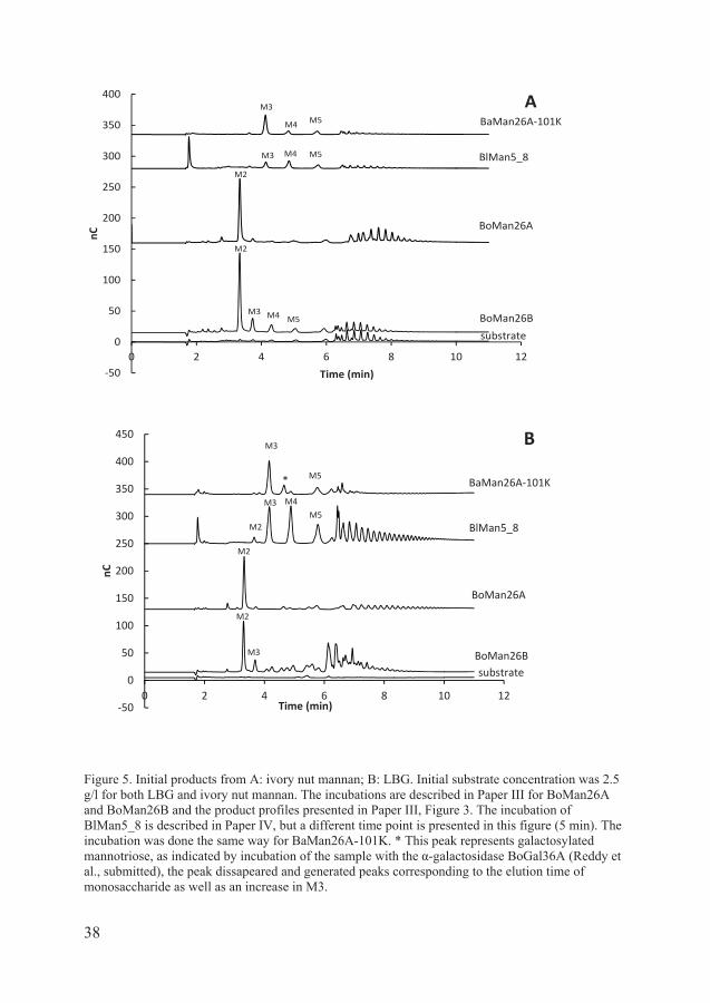
Figure 5. Initial products from A: ivory nut mannan; B: LBG. Initial substrate concentration was 2.5 g/l for both LBG and ivory nut mannan. The incubations are described in Paper III for BoMan26A and BoMan26B and the product profiles presented in Paper III, Figure 3. The incubation of BlMan5_8 is described in Paper IV, but a different time point is presented in this figure (5 min). The incubation was done the same way for BaMan26A-101K. * This peak represents galactosylated mannotriose, as indicated by incubation of the sample with the -galactosidase BoGal36A (Reddy et al., submitted), the peak dissapeared and generated peaks corresponding to the elution time of monosaccharide as well as an increase in M3.
50
0
50
100
150
200
250
300
350
400
0 2 4 6 8 10 12
nC
Time (min)
BaMan26A 101K
BlMan5_8
BoMan26B
BoMan26A
M3
M5
M3 M4 M5
M2
M2
M4
substrate
M3
A
M4 M5
50
0
50
100
150
200
250
300
350
400
450
0 2 4 6 8 10 12
nC
Time (min)
B
BaMan26A 101K
BlMan5_8
BoMan26B
BoMan26A
substrate
M3
* M5
M3 M4
M5
M2
M2
M3
M2

39
General discussion
Several species of the human gut bacteria Bifidobacterium and Bacteroides are predicted to harbor -mannanases, the main responsible enzyme for -mannan degradation. In this work, we have studied four previously uncharacterized -mannanases from both genera (Papers II-IV). The -mannanases were found to differ in substrate specificity, product profile and modularity. In this chapter, the
-mannanases will be further compared and their potential roles in the gut discussed. In addition, the in vivo effect of galactomannan on Bifidobacterium and Bacteroides levels in caecum of rats will be discussed (Paper I).
Cellular location and organization of mannanases
Bifidobacterium animalis subsp. lactis is predicted to secrete BlMan5_8, while B. adolescentis and Bacteroides ovatus anchor their GH26 mannanases to the cell (Papers II-IV). The Gram-negative B. ovatus expresses at least two -mannanases (Gherardini and Salyers, 1987a, Gherardini and Salyers, 1987b) and has been shown to grow on galactomannan in vitro (Martens et al., 2011). Two mannanases (Paper III) are encoded by a PUL that is transcriptionally up-regulated during growth of B. ovatus on galactomannan (Martens et al., 2011). The two -mannanase genes were cloned and the mannanases studied in Paper III: the periplasmic BoMan26A and extracellular outer membrane anchored BoMan26B. PULs for utilization of various polysaccharides have been discovered in several Bacteroides sp. The first PUL to be discovered was a starch utilization system in the human gut bacterium Bacteroides thetaiotaomicron (Shipman et al., 2000). Also, a B. ovatus xyloglucan PUL was characterized (Larsbrink et al., 2014).
In Paper III, the following model was proposed for galactomannan catabolism conferred by the B. ovatus mannan PUL: the extracellular cell attached BoMan26B does the initial hydrolysis of galactomannan to mannobiose and galactosylated mannooligosaccharides. These products would be taken up into the periplasm by the putative transporter encoded by the PUL. In the periplasm, the -galactosidase BoGal36A could remove the galactose substitutions from the galactosylated mannooligosaccharides, which are further broken down by BoMan26A to mannobiose. The mannobiose produced by BoMan26A and BoMan26B could potentially be further converted by e.g. the putative GH130
40
phosphorylase encoded by the same PUL to monosaccharides (Paper III), in a similar manner as proposed for B. fragilis (Senoura et al., 2011).
Unlike BoMan26A and BoMan26B, that are coded by genes found in the same PUL in B. ovatus, genes coding for BaMan26A and BlMan5_8 do not have adjacent genes in the genome coding for other proteins involved in galactomannan degradation or transport.
To fully degrade galactomannan to monosaccharides, additional enzymes are needed to degrade the oligosaccharides produced by the mannanases. BoMan26B produces mannobiose as a main product, but also other oligosaccharides, while BoMan26A produces almost exclusively mannobiose from linear ivory nut mannan (Paper III, Figure 3a). Also from LBG the main product is mannobiose, see Figure 5b in the thesis. The mannobiose could possibly be further converted to monosugars by a phosphorylase predicted in the same PUL, as is the case in B. fragilis (Senoura et al., 2011). Although BoMan26A only has low activity against guar gum, it can act in synergy with the periplasmic -galactosidase from the same PUL to be able to degrade galactomannooligosaccharides (Paper III).
As it stands, while B. ovatus produces an -galactosidase (Paper III, Reddy et al., submitted ) which is implicated in galactomannan digestion, there is not an obviuos connection between mannan digestion and -galactosidase activity neither for B. adolescentis, nor for B. animalis subsp. lactis. Both B. adolescentis and B. animalis subsp. lactis are predicted to encode at least three -galactosidases each, however, the genes coding for these -galactosidases are not adjacent to the genes coding for the -mannanases characterized in this work. Furthermore no -galactosidase is present in the mannan catabolic operon in B. fragilis (Senoura et al., 2011)
Substrate specificity, products and mode of attack
The secreted BlMan5_8 has a high catalytic efficiency and produced a variety of oligosaccharides from ivory nut mannan, see Paper IV, Figure 1a. BoMan26A, BoMan26B and BaMan26A differ from BlMan5_8 in the product profile, by producing dominant products both early in the incubation and as hydrolysis proceeds (Paper III, Figure 3; Paper II, Table 1). BaMan26A from Bifidobacterium adolescentis produced initially mainly mannotriose from ivory nut mannan (Paper II, Table 1). BoMan26A and BoMan26B clearly display mannobiose as the main product from ivory nut mannan. BoMan26B, however, also produces minor amounts of M3, M4 and M5. These different product profiles are also visualized in Figure 5 in the thesis.
BlMan5_8 produces a variety of oligosaccharides from LBG, indicating endo-activity (Paper IV, Figure 1b). BoMan26B and BaMan26A show, in addition to

41
their respective dominant product, also minor amounts of other, longer products, (Paper III, Figure 3; Paper II, Figure 4; thesis Figure 5), also indicating endo-activity. Enzymes with exo-activity usually produce a defined product. Product profiles with dominant products could also arise from processive enzymes even for endo-attacking enzymes, an example being endoglucanase Cel9A from T. fusca (Li et al., 2007). Two exo-acting mannobiohydrolases with high sequence identity to BoMan26A have been characterized: BfMan26 from the human gut bacterium Bacteroides fragilis (Kawaguchi et al., 2013), and CjMan26C from the soil bacterium Cellvibrio japonicus (Cartmell et al., 2008). However, BoMan26A seems to be endo-acting, as it, in addition to mannobiose, produces some longer products from LBG early on in the incubation, see Figure 5b in the thesis. The potential processivity of the four mannanases studied in this thesis has not yet been addressed. An example of a glycoside hydrolase from a human gut bacterium with proposed processive endo-activity is the GH53 galactanase, GalA, from Bifidobacterium longum. After the initial cleavage within the galactan chain (endo-activity), GalA continues cleaving off galactotriose units from the newly generated chain terminus (Hinz et al., 2005).
The -mannanases display differences in the ability to break down guar gum, the highly galactosylated mannan from guar seeds. While BaMan26A and BoMan26B show activity on guar gum, although lower than that for LBG, BaMan5_8 and BoMan26A do not seem to be able to hydrolyze guar gum under the conditions tested (Table 2 in the thesis ). The inability to act on guar gum is also shared with BfMan26 (Kawaguchi et al., 2013). In B. ovatus this inability is potentially compensated for by the periplasmic -galactosidase BoGal36A that can remove the galactoses from the galactomannooligosaccharides before BoMan26A acts on it, potentially contributing to the ability of the bacterium to utilize galactomannan (Paper III). It remains to be investigated if any of the -galactosidases predicted to be encoded in the genomes of B. adolescentis and B. animalis subsp. lactis are able to act on galactomannan or galactomannooligosaccharides.
Several mannanases in both families GH5 and GH26, including BoMan26A, BlMan5_8, T. petrophila Man5, Aspergillus niger Man5, RsMan26C, and Man5A and Man5B from C. polysaccharolyticus (Paper III, Paper IV, dos Santos et al., 2012, Do et al., 2009, Tsukagoshi et al., 2014b, Han et al., 2010) show higher specific activity towards konjac glucomannan than LBG, possibly indicating difficulties of accommodating the galactose unit in the active site of these enzymes. However not all -mannanases prefer glucomannan over LBG: e.g. BoMan26B has a 65% activity on glucomannan compared to LBG (Paper III, Table 3; Table 2 in thesis), and CjMan5A has a kcat of 1025 s-1 of glucomannan, which is about 40% of the kcat on LBG for this enzyme (Hogg et al., 2003). Also BfMan26 shows very little activity towards glucomannan (Kawaguchi et al., 2013), see Table 2 in the thesis.

42
In a different niche, Cellvibrio japonicus, feeds on dead organic matter in the soil. To accomplish this task it produces several GH5 and GH26 mannanases that break down the mannan present in plant cell walls. The enzymes appear to have specialized functions in regards to substrate specificity and cellular location. The GH5A-C that all have CBMs are predicted to be secreted and target insoluble cell wall components, while the GH26A-C (lacking CBMs) are predicted to be anchored to the outer membrane via lipid anchors and are suggested to act on the mannan fragments produced by the GH5 mannanases (Cartmell et al., 2008).
Carbohydrate binding modules
CjMan5C is the only of the characterized C. japonicus mannanases that is active on insoluble ivory nut mannan (Hogg et al., 2003), while all four -mannanases studied in this thesis show activity towards ivory nut mannan, regardless of presence or absence of CBMs. However, the activity on ivory nut mannan is much lower for all four enzymes studied in this thesis compared to activity on LBG. Interestingly, BaMan5A from Bacillus agaradhaerens has a kcat in the same range for glucomannan, LBG and ivory nut mannan (500-750 s-1) (Tailford et al., 2009).
The presence of the two CBM23s in BaMan26A seems to increase the specific activity towards LBG and guar gum. In BlMan5_8, no effect was observed on the kinetic parameters in presence or absence of the CBM10. The CBM10 of BlMan5_8 seems to be a low affinity CBM, as the affinity of BlMan5_8 towards LBG was 35-fold lower compared to that of BaMan26A.
The homolog of BlMan5_8, CjMan5A, contains two tandem CBMs of family 10, that show affinity towards cellulose and ivory nut mannan. However, CjMan5A shows no activity towards ivory nut mannan. In BlMan5_8, the situation is reverse: the enzyme shows no detected affinity towards ivory nut mannan, but can act on it, releasing oligosaccharides of mainly M3, M4 and M5.The activity on ivory nut mannan, is however much lower than that on LBG.
In BaMan26A, which otherwise has a high Km, the function of the CBM could be to increase the local concentration of the substrate in the vicinity of the enzyme. It is interesting to observe that the mannanases from B. adolescentis and B. animalis subsp. lactis have CBMs with different affinities for mannan. In this case, the enzyme with the higher affinity CBM is potentially cell anchored and could, hypothetically, bind the substrate to the cell.
Mannan utilization by gut bacteria
Although studies have found that only a few strains of B. adolescentis can grow on guar or locust bean gum (Crociani et al., 1994, Salyers et al., 1977),
43
Bifidobacterium species have been shown to increase when the host includes guar gum in the diet (Paper I, Ohashi et al., 2012, Okubo et al., 1994).
Also, many Bacteroides species do not grow on guar gum in vitro (Hartemink and Rombouts, 1999, Okubo et al., 1994, Martens et al., 2011), and the group Bacteroidaceae, to which they belong, did not increase in cell counts in the study in the human subjects that received PHGG (Okubo et al., 1994). This is in line with our findings in rats (Paper I), where Bacteroides cell counts were not significantly increased when rats were fed different molecular weight of guar gum (315, 900 and 2000-3000 kDa) compared to control. However, it is perhaps unlikely that abundance of a genus would significantly increase if only a few species are able to metabolize the fiber. In addition, B. ovatus can grow on galactomannan in vitro (Martens et al., 2011) and mannanase activity in B. ovatus was detected when grown on guar gum, but not on mannose or galactose (Gherardini and Salyers, 1987a).
The difference in the effect on the cell counts in the caecum between Bifidobacterium and Bacteroides could possibly be due to that not all species of the genus are able to grow on guar gum. Furthermore, bacteria live in consortium where cross-feeding plays an important role (De Vuyst and Leroy, 2011) and the presence of other bacteria induce altered gene expression compared to single culture (Ruiz et al., 2009).
The extracellular mannanases present in a bacterium do not necessarily have the function of providing mannooligosaccharides for that specific bacterium, as gut bacteria live in consortium and cross-feed (De Vuyst and Leroy, 2011). Studies of individual bacteria and their enzymes give valuable information on the enzymatic mechanism and interplay of the enzymes. This is a valuable step towards also, further on, to understand the potential interplay between different species in the gut.

44
45
Conclusions and future perspectives
The mannan utilization in the gut has not been extensively studied in molecular detail. The findings in this thesis add to the knowledge of galactomannan utilization by the gut flora, both on organism level and in molecular detail. Guar gum of different viscosities gave rise to different metabolic effects is rats, indicating differences in the fermentation process. However, the effect of galactomannan on the levels Bacteroides sp and Bifidobacterium sp. was similar regardless of viscosity, indicating that the sugar composition gives rise effect, rather than viscosity (Paper I). It would be interesting to study the effect of galactomannan on species or even strain level, while in consortium.
In addition, four -mannanases from the human gut bacteria were characterized (Papers II-IV). Together with the recently characterized -mannanase BfMan26 from B. fragilis (Kawaguchi et al., 2013), this work gives new insight into the molecular details of -mannan utilization by the gut bacteria. Although guar gum is known to be fermented in the gut, some mannanases are unable to hydrolyze it, probably due to the high degree of galactose substitution. Some of the gut bacteria have, however, -galactosidases that can aid in the mannan hydrolysis by cleaving off the galactose substitutions. In B. ovatus an -galactosidase is part of the same PUL as the mannanases, and can act in synergy with the mannanases to break down galactomannooligosaccharides (Paper III). It would be interesting to study the potential interplay between enzymes, such as mannanases and -galactosidases, from the gut flora and elucidate any potential synergistic actions between enzymes from different organisms, since they live in consortium and share the same niche.
Three of the -mannanases were found to mainly produce products of a defined length. The dominant product of BaMan26A was mannotriose, while BoMan26A and BoMan26B produced mannobiose or mainly mannobiose, respectively. All four enzymes have an endo-mode of action. BoMan26A seems to be an endo-acting mannanase, in contrast to its homolog CjMan26C, which is described as exo-acting. It would be interesting to study the mechanism behind the product profiles displaying such distinct dominant products like BoMan26A and address whether some of the mannanases are processive or not. In contrast, BlMan5_8 produces a variety of oligosaccharides from LBG and ivory nut mannan. It is interesting to note that of the four mannanases studied in this thesis, the anchored

46
GH26 mannanases were the ones to have a product profile showing distinct products, while the secreted BlMan5_8 produced products of different length.
The CBM10 in BlMan5_8 probably has a different function to the CBM23s of BaMan26A. While CBM23s in BaMan26A have a high affinity for LBG and increase the specific activity of BaMan26A, the CBM10 in BlMan5_8 does not seem to have this function.
This work adds to the insight into the molecular details of mannan utilization by gut bacteria. The study gives fundamental knowledge on the molecular mechanisms, including molecular structure knowledge and, in the case of B. ovatus, the interplay between enzymes, accomplishing digestion of a complex polysaccharide, in this case galactomannan. The study gives detailed knowledge on individual enzymes, which also potentially could be useful in applications for polysaccharide conversion or tailoring directed towards food applications or biomass processing.

47
Acknowledgements
This thesis would never have been written without the support from some people. I always thought that a statement like this is exaggerated, but today I know that it is not. I want to thank my supervisor Henrik Stålbrand for introducing me to the interesting topic of glycoside hydrolases, for teaching me to work methodically and for your interest in the research. I want to that thank current and previous members of our research group for collaborations, advice and company. I want to especially thank Anna Rosengren for the company in the office during most of my PhD and all the help with practical and scientific questions, especially when I was new in the lab. Sumitha - I appreciate that you are so direct and honest. Thanks also for all the discussions we had about everything from experimental setup to career and life. You were right in so many aspects. Thank you for sharing your worldview with me! It has been really interesting to get to know you. Viktoria – thank you for the collaboration on the B. ovatus enzyme and all the advice. Peter Immerzeel- for teaching me the chromatography equipment and always being there to answer my questions. Johan - thank you for the collaboration on the interesting lactis enzyme. Anna Aronsson – it was great to have you as an office mate! Thanks for comforting me when I needed it. Nick – it seems that I have missed the good wild days when you were a PhD student at the department! Thanks for all the career advice and critical questions on my projects. Lis – I appreciate you being so open and honest. I also want thank Anette, Kourosh and Hanene for the company and contribution in the lab. I got to supervise and work with three really good diploma students during my PhD. I learned a lot about supervising, science and myself by being your supervisor and really enjoyed it. Romany - thanks for a nice collaboration on the Bifidobacterium enzyme. Ignacio – It was fun to work with you and I really enjoyed that semester when I was your and David’s supervisor. David, I admire your enthusiasm and energy! Thanks for sharing it with me, it has meant a lot to me during the last two years of my PhD when I struggled with my motivation. Thank you also all the advice on my projects, technical assistance and for keeping me company during those late nights in the lab. I learned a lot of things from you, both in life and in the lab.
48
I want to thank my co-supervisor Sofi Elmroth for providing useful career seminars and activities with FLÄK and LiL. Thanks to Ingemar André and to late Cecilia Hägerhäll for being a driving force for CMPS research school and providing all the interesting seminars and retreats. I want to thank FLÄK for financing my PhD studies, Fysiografiska sällskapet for the equipment grant, Knut and Alice Wallenberg and Lawski foundations for travel grants. I want to thank people with whom I have had interesting collaborations that I also learned a lot from: Ulrica Edlund at KTH, Maher Abou Hachem and Birte Svensson at DTU, Frida Fåk, Margareta Nyman, Greta Jakobsdottir and Nittaya Marungruang at Applied Nutrition (LTH), Leila Lo Leggio at Copenhagen University. And also Roya at Biotechnology (LTH) - I learned so much about chromatography and detectors in that project! Thanks to Folke Tjerneld for encouragement and proofreading my thesis! Special thanks to Sven Kjellström for help with mass spectrometry, Peter Falck for help with the Dionex and Rosa Arãgao Börner and Ed van Niel for help with the anaerobic cultivation technique. I also want to thank Eva Nordberg Karlsson and Patrik Adlercreutz (Biotechnology, LTH) and their groups for interesting journal clubs and discussions. I want to thank all the co-authors on the manuscripts in this thesis, especially Katarina Kolenova. I want to thank my wonderful colleagues at the department of Biochemistry and Structural Biology. I am happy that I could work in such a supportive environment. I want to thank some people in particular, who have helped and inspired me: Alak Alshiekh, you have supported me a lot in emotionally tough times during my whole PhD. Kamrul – you are always so happy! I Thank you for sharing your happiness and thoughts. Special thanks to Kamil for always having time to show me equipment and being patient with me. I am happy that we got to teach on KEMA03 together, that was a nice experience. You have so many funny stories! Erik, it was nice teaching with you! Thanks also for all the advice! I also want to thank Eva, Irem, Robert, Eva-Christina, Sebastian, Stefan, Angelica, Mariana and Andreas thanks for the company and encouragement. I want to thank Gert, Adine and Magnus for always being there to help with equipment and technical questions. The department would collapse without you! Thanks to Susanna for help with administrative questions. I want to thank all the present and former people at division of Theoretical chemistry for all the game evenings, bowling, parties (to which I was always late because I got stuck at work) and traveling that we did together! My PhD would really not be the same without you! Svante - I almost learned to enjoy dubstep with you. Thanks for all the help with the guitar and for letting me play in the living room. I would not advance as fast without your help. Thanks also for all the discussions we have had about everything from politics to why molecules have

49
potential energy. Also thanks to Anil and Korhan, Samuel and Maja, Björn Stenqvist, Steven, Alexei (we should dance salsa more often!) and Francesco. Thanks for making our home lively! I want to thank the brothers at St. Maria Magdalena’s Convent in Lund and Domino student group for being my second home during my whole stay in Lund since 2006. With you I found what I really needed and I learned a lot about my faith. I want to thank brother Björn for guiding the group and for the large amout of work you put in it. I also want to thank Magda, Anna S, Benôit, Alexander, Cassandra, Markus, Elin, Nolwenn, Anne-Laure, Verena, Stefano, Milan, Taras and Martynas and the rest of you people from all over Sweden and the world for making the group what it is. I also want to thank the sisters at St. Dominikus kloster in Rögle for providing a nice and quiet place for reflection. I especially want to thank sister Marie for your honesty, openness, and for being there for me and never judging me. I want to thank my friends. Sofia vad vore studenttiden utan dig?! Utan alla nationskvällar då vi dansade hela kvällen, spontana promenader och för att både jag och Paulius har bott hos dig och Elisabeth i olika omgångar. Kristofer WW – vad spännande att följa din utveckling och studier. Tack för alla diskussioner om vetenskap, terminologi (fulmutant ) och för din uppmuntan. Lycka till på din resa! Jag vill också tacka Raminta och Oskar, Diana, Maria, Sara och Linda. Jag vill också tacka min familj för allt stöd. ,
, . To my boyfriend Paulius – thanks for keeping me company during those nights that we both wrote our theses. I am happy that I could share and experience this crazy time called PhD with you. It was very nice to go to your office whenever I needed help or support. I am happy that we made it through together.

50

51
References
ADEMARK, P., DE VRIES, R. P., HAGGLUND, P., STALBRAND, H. & VISSER, J. 2001. Cloning and characterization of Aspergillus niger genes encoding an alpha-galactosidase and a beta-mannosidase involved in galactomannan degradation. Eur J Biochem, 268, 2982-90.
ADEMARK, P., VARGA, A., MEDVE, J., HARJUNPAA, V., DRAKENBERG, T., TJERNELD, F. & STALBRAND, H. 1998. Softwood hemicellulose-degrading enzymes from Aspergillus niger: purification and properties of a beta-mannanase. J Biotechnol, 63, 199-210.
AGGER, J. W., ISAKSEN, T., VARNAI, A., VIDAL-MELGOSA, S., WILLATS, W. G., LUDWIG, R., HORN, S. J., EIJSINK, V. G. & WESTERENG, B. 2014. Discovery of LPMO activity on hemicelluloses shows the importance of oxidative processes in plant cell wall degradation. Proc Natl Acad Sci U S A, 111, 6287-92.
AKITA, M., TAKEDA, N., HIRASAWA, K., SAKAI, H., KAWAMOTO, M., YAMAMOTO, M., GRANT, W. D., HATADA, Y., ITO, S. & HORIKOSHI, K. 2004. Crystallization and preliminary X-ray study of alkaline mannanase from an alkaliphilic Bacillus isolate. Acta Crystallogr D Biol Crystallogr, 60, 1490-2.
ALBRECHT, S., VAN MUISWINKEL, G. C., XU, J., SCHOLS, H. A., VORAGEN, A. G. & GRUPPEN, H. 2011. Enzymatic production and characterization of konjac glucomannan oligosaccharides. J Agric Food Chem, 59, 12658-66.
AMARETTI, A., TAMBURINI, E., BERNARDI, T., POMPEI, A., ZANONI, S., VACCARI, G., MATTEUZZI, D. & ROSSI, M. 2006. Substrate preference of Bifidobacterium adolescentis MB 239: compared growth on single and mixed carbohydrates. Appl Microbiol Biotechnol, 73, 654-62.
ANDERSSON ROOS, A., EDLUND, U., SJÖBERG, J., ALBERTSSON, A.-C.-. & STÅLBRAND, H. 2008. Protein Release from Galactoglucomannan Hydrogels: Influence of Substitutions and Enzymatic Hydrolysis by beta-Mannanase. Biomacromolecules, 9, 2104-2110.

52
ASPEBORG, H., COUTINHO, P. M., WANG, Y., BRUMER, H. & HENRISSAT, B. 2012. Evolution, substrate specificity and subfamily classification of glycoside hydrolase family 5 (GH5). BMC Evolutionary Biology, 12, 1-16.
BLEAU, C., KARELIS, A. D., ST-PIERRE, D. H. & LAMONTAGNE, L. 2014. Crosstalk between intestinal microbiota, adipose tissue and skeletal muscle as an early event in systemic low-grade inflammation and the development of obesity and diabetes. Diabetes Metab Res Rev.
BOLAM, D. N. & KOROPATKIN, N. M. 2012. Glycan recognition by the Bacteroidetes Sus-like systems. Curr Opin Struct Biol, 22, 563-9.
BOLAM, D. N., XIE, H., PELL, G., HOGG, D., GALBRAITH, G., HENRISSAT, B. & GILBERT, H. J. 2004. X4 modules represent a new family of carbohydrate-binding modules that display novel properties. J Biol Chem, 279, 22953-63.
BORASTON, A. B., BOLAM, D. N., GILBERT, H. J. & DAVIES, G. J. 2004a. Carbohydrate-binding modules: fine-tuning polysaccharide recognition. Biochemical Journal, 382, 769-781.
BORASTON, A. B., BOLAM, D. N., GILBERT, H. J. & DAVIES, G. J. 2004b. Carbohydrate-binding modules: fine-tuning polysaccharide recognition. Biochem J, 382, 769-781.
BOURGAULT, R., OAKLEY, A. J., BEWLEY, J. D. & WILCE, M. C. J. 2005. Three-dimensional structure of (1,4)-beta-D-mannan mannanohydrolase from tomato fruit. Protein Science, 14, 1233-1241.
BROCKMAN, D. A., CHEN, X. & GALLAHER, D. D. 2014. High-viscosity dietary fibers reduce adiposity and decrease hepatic steatosis in rats fed a high-fat diet. J Nutr, 144, 1415-22.
BROUNS, F., THEUWISSEN, E., ADAM, A., BELL, M., BERGER, A. & MENSINK, R. P. 2012. Cholesterol-lowering properties of different pectin types in mildly hyper-cholesterolemic men and women. Eur J Clin Nutr, 66, 591-9.
BÄCKHED, F., LEY, R. E., SONNENBURG, J. L., PETERSON, D. A. & GORDON, J. I. 2005. Host-bacterial mutualism in the human intestine. Science, 307, 1915-20.
CARTMELL, A., TOPAKAS, E., DUCROS, V. M. A., SUITS, M. D. L., DAVIES, G. J. & GILBERT, H. J. 2008. The Cellvibrio japonicus Mannanase CjMan26C Displays a Unique exo-Mode of Action That Is
53
Conferred by Subtle Changes to the Distal Region of the Active Site. Journal of Biological Chemistry, 283, 34403-34413.
CHEKAN, J. R., KWON, I. H., AGARWAL, V., DODD, D., REVINDRAN, V., MACKIE, R. I., CANN, I. & NAIR, S. K. 2014. Structural and biochemical basis for mannan utilization by Caldanaerobius polysaccharolyticus strain ATCC BAA-17. J Biol Chem, 289, 34965-77.
CHEN, Z., FRIEDLAND, G. D., PEREIRA, J. H., REVECO, S. A., CHAN, R., PARK, J. I., THELEN, M. P., ADAMS, P. D., ARKIN, A. P., KEASLING, J. D., BLANCH, H. W., SIMMONS, B. A., SALE, K. L., CHIVIAN, D. & CHHABRA, S. R. 2012. Tracing determinants of dual substrate specificity in glycoside hydrolase family 5. J Biol Chem, 287, 25335-43.
CHRISTIANSEN, C., HACHEM, M. A., GLARING, M. A., VIKSO-NIELSEN, A., SIGURSKJOLD, B. W., SVENSSON, B. & BLENNOW, A. 2009. A CBM20 low-affinity starch-binding domain from glucan, water dikinase. FEBS Lett, 583, 1159-63.
COUTURIER, M., ROUSSEL, A., ROSENGREN, A., LEONE, P., STALBRAND, H. & BERRIN, J. G. 2013. Structural and biochemical analyses of glycoside hydrolase families 5 and 26 beta-(1,4)-mannanases from Podospora anserina reveal differences upon manno-oligosaccharide catalysis. J Biol Chem, 288, 14624-35.
CROCIANI, F., ALLESANDRINI, A., MUCCI, M. M. B. & BIAVATI, B. 1994. Degradation of Complex Carbohydrates by Bifidobacterium Spp. Int J Food Microbiol, 24, 199-210.
CUSKIN, F., LOWE, E. C., TEMPLE, M. J., ZHU, Y., CAMERON, E. A., PUDLO, N. A., PORTER, N. T., URS, K., THOMPSON, A. J., CARTMELL, A., ROGOWSKI, A., HAMILTON, B. S., CHEN, R., TOLBERT, T. J., PIENS, K., BRACKE, D., VERVECKEN, W., HAKKI, Z., SPECIALE, G., MUNOZ-MUNOZ, J. L., DAY, A., PENA, M. J., MCLEAN, R., SUITS, M. D., BORASTON, A. B., ATHERLY, T., ZIEMER, C. J., WILLIAMS, S. J., DAVIES, G. J., ABBOTT, D. W., MARTENS, E. C. & GILBERT, H. J. 2015. Human gut Bacteroidetes can utilize yeast mannan through a selfish mechanism. Nature, 517, 165-9.
DAVIES, G. J., PLANAS, A. & ROVIRA, C. 2012. Conformational analyses of the reaction coordinate of glycosidases. Acc Chem Res, 45, 308-16.
DAVIES, G. J., WILSON, K. S. & HENRISSAT, B. 1997. Nomenclature for sugar-binding subsites in glycosyl hydrolases. Biochem J, 321 ( Pt 2), 557-9.

54
DE VUYST, L. & LEROY, F. 2011. Cross-feeding between bifidobacteria and butyrate-producing colon bacteria explains bifdobacterial competitiveness, butyrate production, and gas production. Int J Food Microbiol, 149, 73-80.
DEGNAN, B. A. & MACFARLANE, G. T. 1991. Comparison of Carbohydrate Substrate Preferences in 8 Species of Bifidobacteria. FEMS Microbiology Letters, 84, 151-156.
DEN BESTEN, G., HAVINGA, R., BLEEKER, A., RAO, S., GERDING, A., VAN EUNEN, K., GROEN, A. K., REIJNGOUD, D. J. & BAKKER, B. M. 2014. The short-chain fatty acid uptake fluxes by mice on a guar gum supplemented diet associate with amelioration of major biomarkers of the metabolic syndrome. PLoS One, 9, e107392.
DIAS, F. M., VINCENT, F., PELL, G., PRATES, J. A., CENTENO, M. S., TAILFORD, L. E., FERREIRA, L. M., FONTES, C. M., DAVIES, G. J. & GILBERT, H. J. 2004. Insights into the molecular determinants of substrate specificity in glycoside hydrolase family 5 revealed by the crystal structure and kinetics of Cellvibrio mixtus mannosidase 5A. J Biol Chem, 279, 25517-26.
DO, B. C., DANG, T. T., BERRIN, J. G., HALTRICH, D., TO, K. A., SIGOILLOT, J. C. & YAMABHAI, M. 2009. Cloning, expression in Pichia pastoris, and characterization of a thermostable GH5 mannan endo-1,4-beta-mannosidase from Aspergillus niger BK01. Microb Cell Fact, 8, 59.
DOMINIAK, M., SONDERGAARD, K. M., WICHMANN, J., VIDAL-MELGOSA, S., WILLATS, W. G. T., MEYER, A. S. & MIKKELSEN, J. D. 2014. Application of enzymes for efficient extraction, modification, and development of functional properties of lime pectin. Food Hydrocolloids, 40, 273-282.
DON-DONCOW, N., ESCOBAR, Z., JOHANSSON, M., KJELLSTROM, S., GARCIA, V., MUNOZ, E., STERNER, O., BJARTELL, A. & HELLSTEN, R. 2014. Galiellalactone is a direct inhibitor of the transcription factor STAT3 in prostate cancer cells. J Biol Chem, 289, 15969-78.
DOS SANTOS, C. R., PAIVA, J. H., MEZA, A. N., COTA, J., ALVAREZ, T. M., RULLER, R., PRADE, R. A., SQUINA, F. M. & MURAKAMI, M. T. 2012. Molecular insights into substrate specificity and thermal stability of a bacterial GH5-CBM27 endo-1,4-beta-D-mannanase. J Struct Biol, 177, 469-76.
DURBAN, A., ABELLAN, J. J., JIMENEZ-HERNANDEZ, N., PONCE, M., PONCE, J., SALA, T., D'AURIA, G., LATORRE, A. & MOYA, A. 2011.
55
Assessing gut microbial diversity from feces and rectal mucosa. Microb Ecol, 61, 123-33.
ECKBURG, P. B., BIK, E. M., BERNSTEIN, C. N., PURDOM, E., DETHLEFSEN, L., SARGENT, M., GILL, S. R., NELSON, K. E. & RELMAN, D. A. 2005. Diversity of human intestinal microbial flora. Science, 308, 1635-1638.
EJBY, M., FREDSLUND, F., VUJICIC-ZAGAR, A., SVENSSON, B., SLOTBOOM, D. J. & ABOU HACHEM, M. 2013. Structural basis for arabinoxylo-oligosaccharide capture by the probiotic Bifidobacterium animalis subsp. lactis Bl-04. Mol Microbiol, 90, 1100-12.
EKSTRÖM, L. M., BJÖRCK, I. M. & ÖSTMAN, E. M. 2013. On the possibility to affect the course of glycaemia, insulinaemia, and perceived hunger/satiety to bread meals in healthy volunteers. Food & Function, 4, 522-9.
EL KAOUTARI, A., ARMOUGOM, F., GORDON, J. I., RAOULT, D. & HENRISSAT, B. 2013. The abundance and variety of carbohydrate-active enzymes in the human gut microbiota. Nat Rev Microbiol, 11, 497-504.
EVERARD, A., BELZER, C., GEURTS, L., OUWERKERK, J. P., DRUART, C., BINDELS, L. B., GUIOT, Y., DERRIEN, M., MUCCIOLI, G. G., DELZENNE, N. M., DE VOS, W. M. & CANI, P. D. 2013. Cross-talk between Akkermansia muciniphila and intestinal epithelium controls diet-induced obesity. PNAS, 110, 9066-9071.
FIGUEROA-GONZALEZ, I., QUIJANO, G., RAMIREZ, G. & CRUZ-GUERRERO, A. 2011. Probiotics and prebiotics--perspectives and challenges. J Sci Food Agric, 91, 1341-8.
GHERARDINI, F. C. & SALYERS, A. A. 1987a. Characterization of an outer membrane mannanase from Bacteroides ovatus. J Bacteriol, 169, 2031-7.
GHERARDINI, F. C. & SALYERS, A. A. 1987b. Purification and characterization of a cell-associated, soluble mannanase from Bacteroides ovatus. J Bacteriol, 169, 2038-43.
GIANNINI, E. G., MANSI, C., DULBECCO, P. & SAVARINO, V. 2006. Role of partially hydrolyzed guar gum in the treatment of irritable bowel syndrome. Nutrition, 22, 334-42.
GILAD, O., JACOBSEN, S., STUER-LAURIDSEN, B., PEDERSEN, M. B., GARRIGUES, C. & SVENSSON, B. 2010. Combined transcriptome and proteome analysis of Bifidobacterium animalis subsp. lactis BB-12 grown

56
on xylo-oligosaccharides and a model of their utilization. Applied and Environmental Microbiology, 76, 7285-91.
GILBERT, H. J., KNOX, J. P. & BORASTON, A. B. 2013. Advances in understanding the molecular basis of plant cell wall polysaccharide recognition by carbohydrate-binding modules. Curr Opin Struct Biol, 23, 669-77.
GILBERT, H. J., STÅLBRAND, H. & BRUMER, H. 2008. How the walls come crumbling down: recent structural biochemistry of plant polysaccharide degradation. Current opinion in plant biology, 11, 338-48.
GLIKO-KABIR, I., YAGEN, B., BALUOM, M. & RUBINSTEIN, A. 2000. Phosphated crosslinked guar for colon-specific drug delivery II. In vitro and in vivo evaluation in the rat. Journal of Controlled Release, 63, 129-134.
HAMAKER, B. R. & TUNCIL, Y. E. 2014. A Perspective on the Complexity of Dietary Fiber Structures and Their Potential Effect on the Gut Microbiota. J Mol Biol. 426, 3838-50
HAN, Y., DODD, D., HESPEN, C. W., OHENE-ADJEI, S., SCHROEDER, C. M., MACKIE, R. I. & CANN, I. K. 2010. Comparative analyses of two thermophilic enzymes exhibiting both beta-1,4 mannosidic and beta-1,4 glucosidic cleavage activities from Caldanaerobius polysaccharolyticus. J Bacteriol, 192, 4111-21.
HARTEMINK, R. & ROMBOUTS, F. M. 1999. Comparison of media for the detection of bifidobacteria, lactobacilli and total anaerobes from faecal samples. J Microbiol Methods, 36, 181-92.
HEKMAT, O., LO LEGGIO, L., ROSENGREN, A., KAMARAUSKAITE, J., KOLENOVA, K. & STÅLBRAND, H. 2010. Rational engineering of mannosyl binding in the distal glycone subsites of Cellulomonas fimi endo-beta-1,4-mannanase: mannosyl binding promoted at subsite -2 and demoted at subsite -3. Biochemistry, 49, 4884-96.
HILGE, M., GLOOR, S. M., RYPNIEWSKI, W., SAUER, O., HEIGHTMAN, T. D., ZIMMERMANN, W., WINTERHALTER, K. & PIONTEK, K. 1998. High-resolution native and complex structures of thermostable beta-mannanase from Thermomonospora fusca - substrate specificity in glycosyl hydrolase family 5. Structure, 6, 1433-1444.
HINZ, S. W., PASTINK, M. I., VAN DEN BROEK, L. A., VINCKEN, J. P. & VORAGEN, A. G. 2005. Bifidobacterium longum endogalactanase liberates galactotriose from type I galactans. Appl Environ Microbiol, 71, 5501-10.
57
HOGG, D., PELL, G., DUPREE, P., GOUBET, F., MARTÍN-ORÚE, S. M., ARMAND, S. & GILBERT, H. J. 2003. The modular architecture of Cellvibrio japonicus mannanases in glycoside hydrolase families 5 and 26 points to differences in their role in mannan degradation. Biochem. J., 371, 1027-1043.
HOGG, D., WOO, E. J., BOLAM, D. N., MCKIE, V. A., GILBERT, H. J. & PICKERSGILL, R. W. 2001. Crystal structure of mannanase 26A from Pseudomonas cellulosa and analysis of residues involved in substrate binding. J. Biol. Chem., 276, 31186-92.
HÄGGLUND, P., ERIKSSON, T., COLLÉN, A., NERINCKX, W., CLAEYSSENS, M. & STÅLBRAND, H. 2003. A cellulose-binding module of the Trichoderma reesei -mannanase Man5A increases the mannan-hydrolysis of complex substrates. J Biotechnol, 101, 37-48.
IRWIN, D., SHIN, D. H., ZHANG, S., BARR, B. K., SAKON, J., KARPLUS, P. A. & WILSON, D. B. 1998. Roles of the catalytic domain and two cellulose binding domains of Thermomonospora fusca E4 in cellulose hydrolysis. J Bacteriol, 180, 1709-14.
JAKOBSDOTTIR, G., XU, J., MOLIN, G., AHRNE, S. & NYMAN, M. 2013. High-Fat Diet Reduces the Formation of Butyrate, but Increases Succinate, Inflammation, Liver Fat and Cholesterol in Rats, while Dietary Fibre Counteracts These Effects. Plos One, e80476
KARLSSON, F., TREMAROLI, V., NIELSEN, J. & BÄCKHED, F. 2013. Assessing the human gut microbiota in metabolic diseases. Diabetes, 62, 3341-9.
KATAEVA, I. A., UVERSKY, V. N., BREWER, J. M., SCHUBOT, F., ROSE, J. P., WANG, B. C. & LJUNGDAHL, L. G. 2004. Interactions between immunoglobulin-like and catalytic modules in Clostridium thermocellum cellulosomal cellobiohydrolase CbhA. Protein Eng Des Sel, 17, 759-69.
KAWAGUCHI, K., SENOURA, T., ITO, S., TAIRA, T., ITO, H. & WASAKI, J. 2013. The mannobiose-forming exo-mannanase involved in a new mannan catabolic pathway in Bacteroides fragilis. Archives of Microbiology.
KISONEN, V., EKLUND, P., AUER, M., SJOHOLM, R., PRANOVICH, A., HEMMING, J., SUNDBERG, A., ASEYEV, V. & WILLFOR, S. 2012. Hydrophobication and characterisation of O-acetyl-galactoglucomannan for papermaking and barrier applications. Carbohydrate Research, 352, 151-8.
KOBAYASHI, H., SUZUKI, J., TANAKA, S., KIUCHI, Y., OYAMADA, H., IWADATE, N., SUZUKI, H., SHIBATA, N., SUZUKI, S. & OKAWA,

58
Y. 1997. Structure of a cell wall mannan from the pathogenic yeast, Candida catenulata: assignment of 1H nuclear magnetic resonance chemical shifts of the inner alpha-1,6-linked mannose residues substituted by a side chain. Arch Biochem Biophys, 341, 70-4.
KOROPATKIN, N. M., CAMERON, E. A. & MARTENS, E. C. 2012. How glycan metabolism shapes the human gut microbiota. Nat Rev Microbiol, 10, 323-35.
LARSBRINK, J., ROGERS, T. E., HEMSWORTH, G. R., MCKEE, L. S., TAUZIN, A. S., SPADIUT, O., KLINTER, S., PUDLO, N. A., URS, K., KOROPATKIN, N. M., CREAGH, A. L., HAYNES, C. A., KELLY, A. G., CEDERHOLM, S. N., DAVIES, G. J., MARTENS, E. C. & BRUMER, H. 2014. A discrete genetic locus confers xyloglucan metabolism in select human gut Bacteroidetes. Nature, 506, 498-502.
LAZARIDOU, A., BILIADERIS, C. G. & IZYDORCZYK, M. S. 2001. Structural characteristics and rheological properties of locust bean galactomannans: a comparison of samples from different carob tree populations. Journal of the Science of Food and Agriculture, 81, 68-75.
LE NOURS, J., ANDERSON, L., STOLL, D., STÅLBRAND, H. & LO LEGGIO, L. 2005. The Structure and Characterization of a Modular Endo-beta-1,4-mannanase from Cellulomonas fimi. Biochemistry, 44, 12700-12708.
LEDER, S., HARTMEIER, W. & MARX, S. P. 1999. alpha-Galactosidase of Bifidobacterium adolescentis DSM 20083. Current Microbiology, 38, 101-106.
LI, Y., IRWIN, D. C. & WILSON, D. B. 2007. Processivity, substrate binding, and mechanism of cellulose hydrolysis by Thermobifida fusca Cel9A. Appl Environ Microbiol, 73, 3165-72.
LOMBARD, V., GOLACONDA RAMULU, H., DRULA, E., COUTINHO, P. M. & HENRISSAT, B. 2014. The carbohydrate-active enzymes database (CAZy) in 2013. Nucleic Acids Res, 42, D490-5.
LUNDQVIST, J., JACOBS, A., PALM, M., ZACCHI, G., DAHLMAN, O. & STALBRAND, H. 2003. Characterization of galactoglucomannan extracted from spruce (Picea abies) by heat-fractionation at different conditions. Carbohydrate Polymers, 51, 203-211.
MACKENZIE, A. K., NAAS, A. E., KRACUN, S. K., SCHUCKEL, J., FANGEL, J. U., AGGER, J. W., WILLATS, W. G., EIJSINK, V. G. & POPE, P. B. 2015. A polysaccharide utilization locus from an uncultured bacteroidetes phylotype suggests ecological adaptation and substrate versatility. Appl Environ Microbiol, 81, 187-95.

59
MARTENS, E. C., KOROPATKIN, N. M., SMITH, T. J. & GORDON, J. I. 2009. Complex glycan catabolism by the human gut microbiota: the Bacteroidetes Sus-like paradigm. J Biol Chem, 284, 24673-7.
MARTENS, E. C., LOWE, E. C., CHIANG, H., PUDLO, N. A., WU, M., MCNULTY, N. P., ABBOTT, D. W., HENRISSAT, B., GILBERT, H. J., BOLAM, D. N. & GORDON, J. I. 2011. Recognition and degradation of plant cell wall polysaccharides by two human gut symbionts. PLoS Biol, 9, e1001221.
MATSUURA, Y. 1998. Degradation of Konjac Glucomannan by Enzymes in Human Feces and Formation of Short-Chain Fatty Acids by Intestinal Anaerobic Bacteria. Journal of Nutritional Science and Vitaminology, 44, 423-436.
MCCLEARY, B. V., CLARK, A. H., DEA, I. C. & REES, D. A. 1985. The fine structures of carob and guar galactomannans. Carbohydrate Research, 139, 237-260.
MCNULTY, N. P., WU, M., ERICKSON, A. R., PAN, C., ERICKSON, B. K., MARTENS, E. C., PUDLO, N. A., MUEGGE, B. D., HENRISSAT, B., HETTICH, R. L. & GORDON, J. I. 2013. Effects of diet on resource utilization by a model human gut microbiota containing Bacteroides cellulosilyticus WH2, a symbiont with an extensive glycobiome. PLoS Biol, 11, e1001637.
MEAD, D., DRINKWATER, C. & BRUMM, P. J. 2013. Genomic and enzymatic results show Bacillus cellulosilyticus uses a novel set of LPXTA carbohydrases to hydrolyze polysaccharides. PLoS One, 8, e61131.
MEIER, H. 1958. On the structure of cell walls and cell wall mannans from ivory nuts and from dates. Biochim Biophys Acta, 28, 229-40.
MIKKONEN, K. S., HEIKKILÄ, M. I., WILLFÖR, S. M. & TENKANEN, M. 2012. Films from Glyoxal-Crosslinked Spruce Galactoglucomannans Plasticized with Sorbitol. International Journal of Polymer Science, 2012, 1-8.
MUDGIL, D., BARAK, S. & KHATKAR, B. S. 2014. Guar gum: processing, properties and food applications-A Review. J Food Sci Technol, 51, 409-18.
NORDAHL PETERSEN, T., BRUNAK, S., VON HEIJNE, G. & NIELSEN, H. 2011. SignalP 4.0: discriminating signal peptides from transmembrane regions. Nature Methods, 8, 785-786.

60
OHASHI, Y., HARADA, K., TOKUNAGA, M., ISHIHARA, N., OKUBO, T., OGASAWARA, Y., JUNEJA, L. R. & FUJISAWA, T. 2012. Faecal fermentation of partially hydrolyzed guar gum. Journal of Functional Foods, 4, 398-402.
OKUBO, T., ISHIHARA, N., TAKAHASHI, H., FUJISAWA, T., KIM, M., YAMAMOTO, T. & MITSUOKA, T. 1994. Effects of Partially Hydrolyzed Guar Gum Intake on Human Intestinal Microflora and Its Metabolism. Bioscience, Biotechnology, and Biochemistry, 58, 1364-1369.
OYAMA, T., SCHMITZ, G. E., DODD, D., HAN, Y., BURNETT, A., NAGASAWA, N., MACKIE, R. I., NAKAMURA, H., MORIKAWA, K. & CANN, I. 2013. Mutational and structural analyses of Caldanaerobius polysaccharolyticus Man5B reveal novel active site residues for family 5 glycoside hydrolases. PLoS One, 8, e80448.
OZDEMIR, I., BLUMER-SCHUETTE, S. E. & KELLY, R. M. 2012. S-layer homology domain proteins Csac_0678 and Csac_2722 are implicated in plant polysaccharide deconstruction by the extremely thermophilic bacterium Caldicellulosiruptor saccharolyticus. Appl Environ Microbiol, 78, 768-77.
PONYI, T., SZABO, L., NAGY, T., OROSZ, L., SIMPSON, P. J., WILLIAMSON, M. P. & GILBERT, H. J. 2000. Trp22, Trp24, and Tyr8 play a pivotal role in the binding of the family 10 cellulose-binding module from Pseudomonas xylanase A to insoluble ligands. Biochemistry, 39, 985-91.
PUCHART, V. 2015. Glycoside phosphorylases: Structure, catalytic properties and biotechnological potential. Biotechnol Adv, 33, 261-276.
QIN, J., LI, Y., CAI, Z., LI, S., ZHU, J., ZHANG, F., LIANG, S., ZHANG, W., GUAN, Y., SHEN, D., PENG, Y., ZHANG, D., JIE, Z., WU, W., QIN, Y., XUE, W., LI, J., HAN, L., LU, D., WU, P., DAI, Y., SUN, X., LI, Z., TANG, A., ZHONG, S., LI, X., CHEN, W., XU, R., WANG, M., FENG, Q., GONG, M., YU, J., ZHANG, Y., ZHANG, M., HANSEN, T., SANCHEZ, G., RAES, J., FALONY, G., OKUDA, S., ALMEIDA, M., LECHATELIER, E., RENAULT, P., PONS, N., BATTO, J. M., ZHANG, Z., CHEN, H., YANG, R., ZHENG, W., LI, S., YANG, H., WANG, J., EHRLICH, S. D., NIELSEN, R., PEDERSEN, O., KRISTIANSEN, K. & WANG, J. 2012. A metagenome-wide association study of gut microbiota in type 2 diabetes. Nature, 490, 55-60.
RAHMAN, O., CUMMINGS, S., HARRINGTON, D. & SUTCLIFFE, I. 2008. Methods for the bioinformatic identification of bacterial lipoproteins

61
encoded in the genomes of Gram-positive bacteria. World Journal of Microbiology and Biotechnology, 24, 2377-2382.
REDDY, S. K., BÅGENHOLM, V., PUDLO, N. A., BOURAOUI, H., KOROPATKIN N. M., MARTENS, E. C., STÅLBRAND, H. A -mannan utilization locus in Bacteriodes ovatus involves a GH36 -galactosidase active on galactomannans. submitted.
RIVAS, S., GULLON, B., GULLON, P., ALONSO, J. L. & PARAJO, J. C. 2012. Manufacture and properties of bifidogenic saccharides derived from wood mannan. Journal of Agricultural and Food Chemistry, 60, 4296-305.
ROBYT, J. F. & FRENCH, D. 1967. Multiple attach hypothesis of alpha-amylase action: action of porcine pancreatic, human salivary, and Aspergillus oryzae alpha-amylases. Arch Biochem Biophys, 122, 8-16.
RODRIGUEZ-GACIO DEL CARMEN, M., IGLESIAS-FERNANDEZ, R., CARBONERO, P. & MATILLA, A. J. 2012. Softening-up mannan-rich cell walls. J Exp Bot, 63, 3976-88.
ROSENGREN, A., HÄGGLUND, P., ANDERSON, L., PAVON-OROZCO, P., PETERSON WULFF, R., NERINCKX, W. & STÅLBRAND, H. 2012. The role of subsite +2 of the Trichoderma reesei -mannanase TrMan5A in hydrolysis and transglycosylation. Biocatal. Biotransformation, 30, 338 - 352.
ROSENGREN, A., REDDY, S. K., SJOBERG, J. S., AURELIUS, O., LOGAN, D. T., KOLENOVA, K. & STALBRAND, H. 2014. An Aspergillus nidulans beta-mannanase with high transglycosylation capacity revealed through comparative studies within glycosidase family 5. Appl Microbiol Biotechnol, 98, 10091-104.
RUIZ, L., SANCHEZ, B., DE LOS REYES-GAVILAN, C. G., GUEIMONDE, M. & MARGOLLES, A. 2009. Coculture of Bifidobacterium longum and Bifidobacterium breve alters their protein expression profiles and enzymatic activities. International Journal of Food Microbiology, 133, 148-53.
SABINI, E., SCHUBERT, H., MURSHUDOV, G., WILSON, K. S., SIIKA-AHO, M. & PENTTILÄ, M. 2000. The three-dimensional structure of a Trichoderma reesei beta-mannanase from glycoside hydrolase family 5. Acta Crystallogr. D Biol. Crystallogr., 56, 3-13.
SALYERS, A. A., WEST, S. E. H., VERCELLOTTI, J. R. & WILKINS, T. D. 1977. Fermentation of Mucins and Plant Polysaccharides by Anaerobic Bacteria from Human Colon. Appl Environ Microbiol, 34, 529-533.
62
SCHELLER, H. V. & ULVSKOV, P. 2010. Hemicelluloses. Annual Review of Plant Biology, 61, 263-289.
SCHRODER, R., WEGRZYN, T. F., SHARMA, N. N. & ATKINSON, R. G. 2006. LeMAN4 endo-beta-mannanase from ripe tomato fruit can act as a mannan transglycosylase or hydrolase. Planta, 224, 1091-102.
SCHROEDER, N., MARQUART, L. F. & GALLAHER, D. D. 2013. The role of viscosity and fermentability of dietary fibers on satiety- and adiposity-related hormones in rats. Nutrients, 5, 2093-113.
SEKIROV, I., RUSSELL, S. L., ANTUNES, L. C. & FINLAY, B. B. 2010. Gut microbiota in health and disease. Physiol Rev, 90, 859-904.
SENOURA, T., ITO, S., TAGUCHI, H., HIGA, M., HAMADA, S., MATSUI, H., OZAWA, T., JIN, S., WATANABE, J. & WASAKI, J. 2011. New microbial mannan catabolic pathway that involves a novel mannosylglucose phosphorylase. Biochem Biophys Res Commun, 408, 701-6.
SHEVCHENKO, A., TOMAS, H., HAVLIS, J., OLSEN, J. V. & MANN, M. 2006. In-gel digestion for mass spectrometric characterization of proteins and proteomes. Nat Protoc, 1, 2856-60.
SHIPMAN, J. A., BERLEMAN, J. E. & SALYERS, A. A. 2000. Characterization of four outer membrane proteins involved in binding starch to the cell surface of Bacteroides thetaiotaomicron. J Bacteriol, 182, 5365-72.
SORENSEN, B. K., HOJRUP, P., OSTERGARD, E., JORGENSEN, C. S., ENGHILD, J., RYDER, L. R. & HOUEN, G. 2002. Silver staining of proteins on electroblotting membranes and intensification of silver staining of proteins separated by polyacrylamide gel electrophoresis. Anal Biochem, 304, 33-41.
STEWART, M. L. & SLAVIN, J. L. 2006. Molecular weight of guar gum affects short-chain fatty acid profile in model intestinal fermentation. Mol Nutr Food Res, 50, 971-6.
STOLL, D., BORASTON, A., STÅLBRAND, H., MCLEAN, B. W., KILBURN, D. G. & WARREN, R. A. J. 2000. Mannanase Man26A from Cellulomonas fimi has a mannan-binding module. FEMS Microbiology Letters, 183, 265-269.
STOLL, D., STÅLBRAND, H. & WARREN, R. A. J. 1999. Mannan-Degrading Enzymes from Cellulomonas fimi. Applied and Environmental Microbiology, 65, 2598-2605.

63
SU, X., MACKIE, R. I. & CANN, I. K. 2012. Biochemical and mutational analyses of a multidomain cellulase/mannanase from Caldicellulosiruptor bescii. Applied and Environmental Microbiology, 78, 2230-40.
TAILFORD, L. E., DUCROS, V. M., FLINT, J. E., ROBERTS, S. M., MORLAND, C., ZECHEL, D. L., SMITH, N., BJORNVAD, M. E., BORCHERT, T. V., WILSON, K. S., DAVIES, G. J. & GILBERT, H. J. 2009. Understanding how diverse beta-mannanases recognize heterogeneous substrates. Biochemistry, 48, 7009-18.
TERRAPON, N., LOMBARD, V., GILBERT, H. J. & HENRISSAT, B. 2015. Automatic prediction of polysaccharide utilization loci in Bacteroidetes species. Bioinformatics, 31, 647-55.
TOMLIN, J., READ, N. W., EDWARDS, C. A. & DUERDEN, B. I. 1986. The Degradation of Guar Gum by a Fecal Incubation System. British Journal of Nutrition, 55, 481-486.
TON-THAT, H., MARRAFFINI, L. A. & SCHNEEWIND, O. 2004. Protein sorting to the cell wall envelope of Gram-positive bacteria. Biochim Biophys Acta, 1694, 269-78.
TSUKAGOSHI, H., NAKAMURA, A., ISHIDA, T., OTAGIRI, M., MORIYA, S., SAMEJIMA, M., IGARASHI, K., KITAMOTO, K. & ARIOKA, M. 2014a. The GH26 beta-mannanase RsMan26H from a symbiotic protist of the termite Reticulitermes speratus is an endo-processive mannobiohydrolase: heterologous expression and characterization. Biochem Biophys Res Commun, 452, 520-5.
TSUKAGOSHI, H., NAKAMURA, A., ISHIDA, T., TOUHARA, K. K., OTAGIRI, M., MORIYA, S., SAMEJIMA, M., IGARASHI, K., FUSHINOBU, S., KITAMOTO, K. & ARIOKA, M. 2014b. Structural and biochemical analyses of glycoside hydrolase family 26 beta-mannanase from a symbiotic protist of the termite Reticulitermes speratus. J Biol Chem, 289, 10843-52.
TURRONI, F., STRATI, F., FORONI, E., SERAFINI, F., DURANTI, S., VAN SINDEREN, D. & VENTURA, M. 2012. Analysis of predicted carbohydrate transport systems encoded by Bifidobacterium bifidum PRL2010. Applied and Environmental Microbiology, 78, 5002-12.
WALTERS, K. R., JR., SERIANNI, A. S., VOITURON, Y., SFORMO, T., BARNES, B. M. & DUMAN, J. G. 2011. A thermal hysteresis-producing xylomannan glycolipid antifreeze associated with cold tolerance is found in diverse taxa. J Comp Physiol B, 181, 631-40.
64
VAN DEN BROEK, L. A. M., TON, J., VERDOES, J. C., VAN LAERE, K. M. L., VORAGEN, A. G. J. & BELDMAN, G. 1999. Synthesis of -galacto-oligosaccharides by a cloned -galactosidase from Bifidobacterium adolescentis. Biotechnology Letters, 21, 441-445.
VAN LAERE, K. M. L., HARTEMINK, R., BELDMAN, G., PITSON, S., DIJKEMA, C., SCHOLS, H. A. & VORAGEN, A. G. 1999. Transglycosidase activity of Bifidobacterium adolescentis DSM 20083 alpha-galactosidase. Applied Microbiology and Biotechnology, 52, 681-688.
ZHANG, X., ROGOWSKI, A., ZHAO, L., HAHN, M. G., AVCI, U., KNOX, J. P. & GILBERT, H. J. 2014. Understanding how the complex molecular architecture of mannan-degrading hydrolases contributes to plant cell wall degradation. J Biol Chem, 289, 2002-12.