Lattice Dynamics of β-V O : Raman Spectroscopic Insight ... · Lattice Dynamics of β-V 2O 5:...
8
Lattice Dynamics of β-V 2 O 5 : Raman Spectroscopic Insight into the Atomistic Structure of a High-Pressure Vanadium Pentoxide Polymorph R. Baddour-Hadjean,* ,† M. B. Smirnov, ‡ K. S. Smirnov, § V. Yu Kazimirov, ⊥ J. M. Gallardo-Amores, ¶ U. Amador, ∥ M. E. Arroyo-de Dompablo, ▽ and J. P. Pereira-Ramos † † Institut de Chimie et Mate ́ riaux Paris-Est, GESMAT, UMR 7182 CNRS et Universite ́ Paris-Est Cre ́ teil, 2 rue Henri Dunant, 94320 Thiais, France ‡ Fock Institute of Physics, Petrodvorets, Ul’yanovskaya st., Saint Petersbourg 198904, Russia § Laboratoire de Spectrochimie Infrarouge et Raman, UMR 8516 CNRS et Universite ́ Lille1 - Sciences et Technologies, 59655 Villeneuve d’Ascq, France ⊥ Frank Laboratory of Neutron Physics, Joint Institute for Nuclear Research, Dubna 141980, Russia ¶ Laboratorio de Altas Presiones, Facultad de Ciencias Químicas, Universidad Complutense, 28040 Madrid, Spain ∥ Departamento de Química, Universidad San Pablo-CEU, 28668 Boadilla del Monte, Spain ▽ MALTA Consolider Team, Departamento de Química Inorga ́ nica, Facultad de Ciencias Químicas, Universidad Complutense, 28040 Madrid, Spain ABSTRACT: We report here the Raman spectrum and lattice dynamics study of a well-crystallized β-V 2 O 5 material prepared via a high-temperature/high-pressure (HT/HP) route, using α-V 2 O 5 as the precursor. Periodic quantum-chemical density functional theory calculations show good agreement with the experimental results and allow one to assign the observed spectral features to specific vibrational modes in the β-V 2 O 5 polymorph. Key structure−spectrum relationships are extracted from comparative analysis of the vibrational states of the β-V 2 O 5 and α- V 2 O 5 structures, and spectral patterns specific to the basic units of the two V 2 O 5 phases are proposed for the first time. Such results open the way for the use of Raman spectroscopy for the structural characterization of vanadium oxide-based host lattices of interest in the field of lithium batteries and help us to greatly understand the atomistic mechanism involved in the α-to-β phase transition of vanadium pentoxide. 1. INTRODUCTION Pentavalent vanadium-based frameworks attract much attention because of their outstanding structural flexibility and their chemical and physical properties suitable for catalytic and electrochemical applications. 1−3 The ambient-pressure form of vanadium pentoxide (α-V 2 O 5 ) has a layered structure with orthorhombic symmetry (space group Pmmn) consisting of VO 5 square pyramids sharing edges and corners. 4 The structure is well adapted to the reversible incorporation of guest Li + ions, and since the 1970s, the α-V 2 O 5 polymorph is recognized as an attractive material for applications in electrochromic thin film devices and as a cathode in lithium batteries because of its high energy density and retention capacity upon cycling. 5−11 The valence state of V atoms is known to be extremely sensitive to the chemical environment, and such chemical versatility may lead to a variety of structures containing different building units and exhibiting tunable physical properties. In this regard, high- pressure/high-temperature (HP/HT) synthesis routes are promising ways to obtain novel V 2 O 5 polymorphs, particularly for potential electrochemical applications. The first HP modification of V 2 O 5 (prepared at P =4−6 GPa and T = 650 °C) was reported by Suzuki et al. 12 Later, Volkov et al. 13 reproduced the same HP phase of V 2 O 5 , named the β phase. Making use of the results of quenching experiments at 600 °C and between 3.5 and 9 GPa, Volkov et al. 13 determined by X-ray diffraction (XRD) measurements that the structure of the β phase is tetragonal. In situ Raman spectroscopic experiments by Grzechnik 14 performed in the pressure range 7−10 GPa and at room temperature (RT) revealed qualitative changes in the Raman spectrum of the V 2 O 5 sample, especially in the V−O stretching region. The author concluded that a new phase appeared and coexisted with the α-V 2 O 5 starting material; the phase was found to return to the starting orthorhombic material upon pressure release. A few years later, Loa et al. 15 performed similar RT experiments by employing synchrotron radiation for detecting structural changes. The authors observed a pronounced structural disorder under Received: December 9, 2011 Published: February 23, 2012 Article pubs.acs.org/IC © 2012 American Chemical Society 3194 dx.doi.org/10.1021/ic202651b | Inorg. Chem. 2012, 51, 3194−3201
Transcript of Lattice Dynamics of β-V O : Raman Spectroscopic Insight ... · Lattice Dynamics of β-V 2O 5:...
Lattice Dynamics of β-V2O5: Raman Spectroscopic Insight into theAtomistic Structure of a High-Pressure Vanadium PentoxidePolymorphR. Baddour-Hadjean,*,† M. B. Smirnov,‡ K. S. Smirnov,§ V. Yu Kazimirov,⊥ J. M. Gallardo-Amores,¶
U. Amador,∥ M. E. Arroyo-de Dompablo,▽ and J. P. Pereira-Ramos†
†Institut de Chimie et Materiaux Paris-Est, GESMAT, UMR 7182 CNRS et Universite Paris-Est Creteil, 2 rue Henri Dunant, 94320Thiais, France‡Fock Institute of Physics, Petrodvorets, Ul’yanovskaya st., Saint Petersbourg 198904, Russia§Laboratoire de Spectrochimie Infrarouge et Raman, UMR 8516 CNRS et Universite Lille1 - Sciences et Technologies, 59655Villeneuve d’Ascq, France⊥Frank Laboratory of Neutron Physics, Joint Institute for Nuclear Research, Dubna 141980, Russia¶Laboratorio de Altas Presiones, Facultad de Ciencias Químicas, Universidad Complutense, 28040 Madrid, Spain∥Departamento de Química, Universidad San Pablo-CEU, 28668 Boadilla del Monte, Spain▽MALTA Consolider Team, Departamento de Química Inorganica, Facultad de Ciencias Químicas, Universidad Complutense,28040 Madrid, Spain
ABSTRACT: We report here the Raman spectrum and lattice dynamics study ofa well-crystallized β-V2O5 material prepared via a high-temperature/high-pressure(HT/HP) route, using α-V2O5 as the precursor. Periodic quantum-chemicaldensity functional theory calculations show good agreement with the experimentalresults and allow one to assign the observed spectral features to specific vibrationalmodes in the β-V2O5 polymorph. Key structure−spectrum relationships areextracted from comparative analysis of the vibrational states of the β-V2O5 and α-V2O5 structures, and spectral patterns specific to the basic units of the two V2O5phases are proposed for the first time. Such results open the way for the use ofRaman spectroscopy for the structural characterization of vanadium oxide-basedhost lattices of interest in the field of lithium batteries and help us to greatlyunderstand the atomistic mechanism involved in the α-to-β phase transition ofvanadium pentoxide.
1. INTRODUCTIONPentavalent vanadium-based frameworks attract much attentionbecause of their outstanding structural flexibility and theirchemical and physical properties suitable for catalytic andelectrochemical applications.1−3 The ambient-pressure form ofvanadium pentoxide (α-V2O5) has a layered structure withorthorhombic symmetry (space group Pmmn) consisting ofVO5 square pyramids sharing edges and corners.
4 The structureis well adapted to the reversible incorporation of guest Li+ ions,and since the 1970s, the α-V2O5 polymorph is recognized as anattractive material for applications in electrochromic thin filmdevices and as a cathode in lithium batteries because of its highenergy density and retention capacity upon cycling.5−11 Thevalence state of V atoms is known to be extremely sensitive tothe chemical environment, and such chemical versatility maylead to a variety of structures containing different building unitsand exhibiting tunable physical properties. In this regard, high-pressure/high-temperature (HP/HT) synthesis routes arepromising ways to obtain novel V2O5 polymorphs, particularlyfor potential electrochemical applications.
The first HP modification of V2O5 (prepared at P = 4−6 GPaand T = 650 °C) was reported by Suzuki et al.12 Later, Volkovet al.13 reproduced the same HP phase of V2O5, named the βphase. Making use of the results of quenching experiments at600 °C and between 3.5 and 9 GPa, Volkov et al.13 determinedby X-ray diffraction (XRD) measurements that the structure ofthe β phase is tetragonal. In situ Raman spectroscopicexperiments by Grzechnik14 performed in the pressure range7−10 GPa and at room temperature (RT) revealed qualitativechanges in the Raman spectrum of the V2O5 sample, especiallyin the V−O stretching region. The author concluded that a newphase appeared and coexisted with the α-V2O5 startingmaterial; the phase was found to return to the startingorthorhombic material upon pressure release. A few years later,Loa et al.15 performed similar RT experiments by employingsynchrotron radiation for detecting structural changes. Theauthors observed a pronounced structural disorder under
Received: December 9, 2011Published: February 23, 2012
Article
pubs.acs.org/IC
© 2012 American Chemical Society 3194 dx.doi.org/10.1021/ic202651b | Inorg. Chem. 2012, 51, 3194−3201
elevated pressure and suggested that the correct determinationof the new phase necessitates the simultaneous application ofHT and HP.In 2001, two different groups16,17 published their findings on
the V2O5 phases investigated at HT/HP, and both reported theexistence of the β-V2O5 phase with orthorhombic symmetry.Filonenko and Zibrov16 performed quenching experiments andfound the β phase up to 7.5 GPa/900 °C, which returns to theα phase at atmospheric pressure and a temperature of 400 °C.Kusaba et al.17 showed the presence of the β phase at 6 GPa/500 °C from quenching experiments and at 4.5 GPa/350 °Cfrom in situ experiments. Later, Filonenko et al.18 reexaminedthe β phase of V2O5 using XRD, neutron diffraction, and high-resolution transmission electron microscopy experiments andfound the structure to be monoclinic. The authors establishedthat the β-V2O5 structure is built up of infinite chains made ofquadruple units of edge-sharing VO6 octahedra along the b axis.The chains are linked by sharing corners of two octahedraalong the c axis, which leads to a V4O10-layered composition,with the layers parallel to the (100) plane. Weak interactions,similar to those in MoO3, hold the layers together. Preliminaryelectrochemical experiments showed that this new monoclinicβ-V2O5 polymorph behaves as a reversible lithium intercalationcompound delivering a specific capacity of 250 mAh/g at a C/3.5 rate.19 More recently, the V2O5 phase diagram was revisitedin a wide P−T range (pressures up to 29 GPa and temperaturesup to 1500 °C), providing two HP/HT modifications of V2O5(β- and δ-V2O5) having well-defined stability ranges in thephase diagram.20
Structural distinctions between the α- and β-V2O5 poly-morphs must become apparent in their vibrational spectra, andthe Raman spectroscopy provides a fast, reliable, andnondestructive means of studying these differences. Further-more, Raman spectroscopy turned out to be a very efficient toolto follow the structural changes under operation conditions fora wide range of transition-metal oxides used as electrodematerials for lithium batteries.26 The Raman spectra of α-V2O5were thoroughly studied both experimentally21−23 andtheoretically.22,24,25 In addition, Raman studies of the α-V2O5/Li system as a pure thin film and as a composite powderelectrode were carried out27−30 and were recently extended tothe β-Na0.33V2O5/Li system.31
On the other hand, much less attention was paid to the β-V2O5 structure. Up to now, the in situ recorded Ramanspectrum at ambient temperatures reported in ref 14 was theonly reliable experimental information on vibrational states ofβ-V2O5. However, Raman peaks in this spectrum are markedlylarge because of strong inhomogeneous broadening due tosize/strain effects of pressure gradients in the cells.15 In thepresent work, the thermal annealing under pressure is expectedto yield a grain coarsening and well-crystallized β-V2O5 with alow level of remaining stress, which results in a better qualityspectrum.The Raman spectra measured by Balog et al.20 on
postquench samples retrieved from HP/HT experiments didnot show any marked distinction from the spectra of thestarting α-V2O5 material, although the presence of HP/HTphases was inferred from the XRD experiments. Densityfunctional theory (DFT) calculations confirmed the stability ofthe β structure32 and provided a theoretical Raman spectrum33
of the material that generally agrees with the experimentalspectra reported in ref 14. These calculations also ascertainedthe symmetry assignment of the observed Raman peaks, but the
atomistic pattern of the vibrational modes was not presented,and as a consequence, no structure−spectrum relationship wasproposed.This paper reports the results of a combined experimental
and computational study of the β-V2O5 polymorph. A well-crystallized β-V2O5 sample was prepared at HT/HP conditions,and its structure was completely characterized by XRDmeasurements. A high-quality Raman spectrum of the samplewas obtained, and DFT calculations allowed one to assign all ofthe observed spectral peaks to the vibrational modes of thestructure. A comparison of the β-V2O5 and α-V2O5 vibrationalstates is provided and leads to the clear identification of spectralfeatures related to the presence of specific structural basic unitsin the two V2O5 polymorphs.
2. EXPERIMENTS AND COMPUTATIONS2.1. Experimental Part. A commercial (Aldrich) α polymorph of
V2O5 was subjected to 8 GPa pressure and a temperature of 800 °C for1 h in a belt-type press. After the pressure and temperature wereapplied for 1 h, the vessel was quenched to RT while pressure wasslowly released. Starting and resulting samples were examined byscanning electron microscopy (SEM) on a JEOL 6400 microscopeequipped with an EDAX Inc. energy-dispersive X-ray detector formicroanalysis.
The resulting sample was also examined by XRD performed with aBruker D8 high-resolution powder X-ray diffractometer equipped withan MBraun PSD-50 M position-sensitive detector. Monochromatic CuKα1 (λ = 1.5406 Å) radiation obtained with a germanium primarymonochromator was used. The treatment of the diffraction data wascarried out using the FullProf program.34 The structure of the β-V2O5phase reported in ref 18 was employed as a starting model; isotropicthermal parameters and constraints in V−O distances were used tokeep the number of the parameter low and to ensure stability of thefitting procedure.
The Raman spectra were measured with a LaBRAM HR 800(Jobin−Yvon−Horiba) Raman microspectrometer including edgefilters and equipped with a back-illuminated charge-coupled devicedetector (Spex CCD) cooled by the Peltier effect to 200 K. A He:Nelaser (632.8 nm wavelength) was used as the excitation source. Thespectra were registered in the backscattering geometry with a spectralresolution of 0.5 cm−1. A 100× objective was used to focus the laserbeam to a spot of 1 μm2 size on the sample surface. To avoid localheating of the sample, the power of the laser beam was adjusted to0.2−0.5 mW with neutral filters of various optical densities.
2.2. Computational Details. The vibrational states of the twoV2O5 polymorphs were computed with the CASTEP program35 usingdensity functional perturbation theory (DFPT).36 The calculationswere carried out within the local density approximation (LDA) toDFT and employed Troullier−Martins norm-conserving pseudopo-tentials.37 The plane-wave energy cutoff of 35 Ha was used, and theBrillouin zone integration was done over a 2 × 4 × 2 grid of pointschosen according to the Monhorst−Pack scheme in the irreduciblepart of the Brillouin zone. The positions of atoms in the unit cell wereoptimized with the lattice parameters fixed at their experimental valuesprior to calculation of the vibrational spectral characteristics. Thisoption was favored over complete geometry optimization because atest calculation of the α-V2O5 structure with optimization of both theatomic positions and the lattice parameters, in particular, resulted in avalue of the c parameter that was by 14% too small compared to itsexperimental value. This finding is in a line with that of Zhou and He25
and is explained by the fact that the LDA fails to correctly describeweak interactions responsible for long V−O contacts (ca. 2.7 Å)between the layers in the structure. A recent computational study byLondero and Schroder38 showed that dispersion-corrected exchange-correlation functionals need to be used to mimic the interplanespacing in the α polymorph of vanadium pentoxide.
The Raman activity of the vibrational modes was computed withinDFPT as described in ref 39. To obtain the Raman scattering
Inorganic Chemistry Article
dx.doi.org/10.1021/ic202651b | Inorg. Chem. 2012, 51, 3194−32013195
intensities, the Raman activities of the vibrational modes weremultiplied by Bose−Einstein factors corresponding to the exper-imental conditions (temperature and wavelength of exciting radiation).
3. RESULTS AND DISCUSSION3.1. Structure. SEM examination of commercial α-V2O5
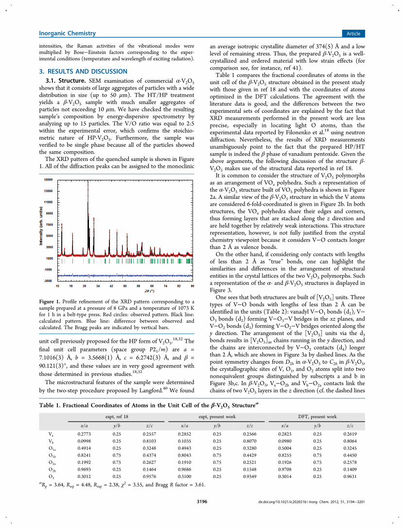
shows that it consists of large aggregates of particles with a widedistribution in size (up to 50 μm). The HT/HP treatmentyields a β-V2O5 sample with much smaller aggregates ofparticles not exceeding 10 μm. We have checked the resultingsample’s composition by energy-dispersive spectrometry byanalyzing up to 15 particles. The V/O ratio was equal to 2:5within the experimental error, which confirms the stoichio-metric nature of HP-V2O5. Furthermore, the sample wasverified to be single phase because all of the particles showedthe same composition.The XRD pattern of the quenched sample is shown in Figure
1. All of the diffraction peaks can be assigned to the monoclinic
unit cell previously proposed for the HP form of V2O5.18,32 The
final unit cell parameters (space group P21/m) are a =7.1016(3) Å, b = 3.5668(1) Å, c = 6.2742(3) Å, and β =90.121(3)°, and these values are in very good agreement withthose determined in previous studies.18,32
The microstructural features of the sample were determinedby the two-step procedure proposed by Langford.40 We found
an average isotropic crystallite diameter of 374(5) Å and a lowlevel of remaining stress. Thus, the prepared β-V2O5 is a well-crystallized and ordered material with low strain effects (forcomparison see, for instance, ref 41).Table 1 compares the fractional coordinates of atoms in the
unit cell of the β-V2O5 structure obtained in the present studywith those given in ref 18 and with the coordinates of atomsoptimized in the DFT calculations. The agreement with theliterature data is good, and the differences between the twoexperimental sets of coordinates are explained by the fact thatXRD measurements performed in the present work are lessprecise, especially in locating light O atoms, than theexperimental data reported by Filonenko et al.18 using neutrondiffraction. Nevertheless, the results of XRD measurementsunambiguously point to the fact that the prepared HP/HTsample is indeed the β phase of vanadium pentoxide. Given theabove arguments, the following discussion of the structure β-V2O5 makes use of the structural data reported in ref 18.It is common to consider the structure of V2O5 polymorphs
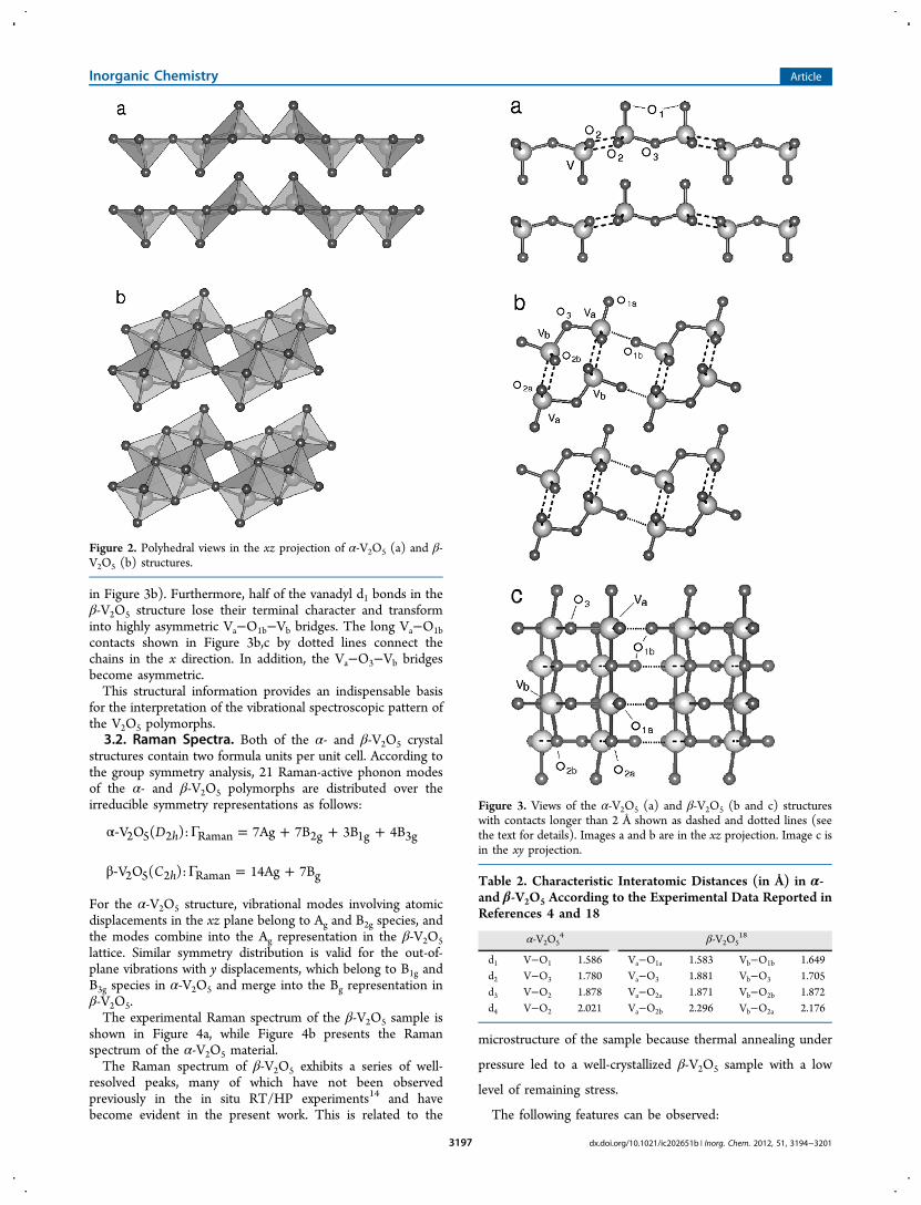
as an arrangement of VOx polyhedra. Such a representation ofthe α-V2O5 structure built of VO5 polyhedra is shown in Figure2a. A similar view of the β-V2O5 structure in which the V atomsare considered 6-fold-coordinated is given in Figure 2b. In bothstructures, the VOx polyhedra share their edges and corners,thus forming layers that are stacked along the z direction andare held together by relatively weak interactions. This structurerepresentation, however, is not fully justified from the crystalchemistry viewpoint because it considers V−O contacts longerthan 2 Å as valence bonds.On the other hand, if considering only contacts with lengths
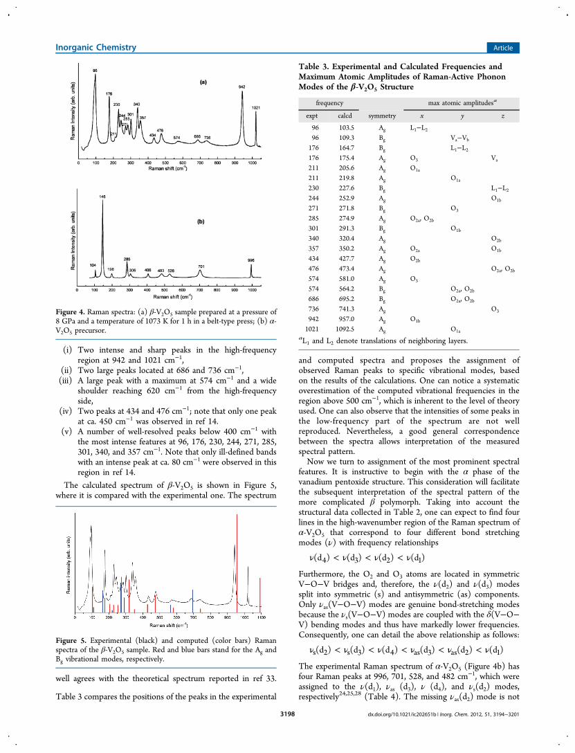
of less than 2 Å as “true” bonds, one can highlight thesimilarities and differences in the arrangement of structuralentities in the crystal lattices of the two V2O5 polymorphs. Sucha representation of the α- and β-V2O5 structures is displayed inFigure 3.One sees that both structures are built of [V2O5] units. Three
types of V−O bonds with lengths of less than 2 Å can beidentified in the units (Table 2): vanadyl V−O1 bonds (d1), V−O3 bonds (d2) forming V−O3−V bridges in the xz planes, andV−O2 bonds (d3) forming V−O2−V bridges oriented along they direction. The arrangement of the [V2O5] units via the d3bonds results in [V2O5]∞ chains running in the y direction, andthe chains are interconnected by V−O2 contacts (d4) longerthan 2 Å, which are shown in Figure 3a by dashed lines. As thepoint symmetry changes from D2h in α-V2O5 to C2h in β-V2O5,the crystallographic sites of V, O1, and O2 atoms split into twononequivalent groups distinguished by subscripts a and b inFigure 3b,c. In β-V2O5, Va−O2b and Vb−O2a contacts link thechains of two V2O5 layers in the z direction (cf. the dashed lines
Figure 1. Profile refinement of the XRD pattern corresponding to asample prepared at a pressure of 8 GPa and a temperature of 1073 Kfor 1 h in a belt-type press. Red circles: observed pattern. Black line:calculated pattern. Blue line: difference between observed andcalculated. The Bragg peaks are indicated by vertical bars.
Table 1. Fractional Coordinates of Atoms in the Unit Cell of the β-V2O5 Structurea
expt, ref 18 expt, present work DFT, present work
x/a y/b z/c x/a y/b z/c x/a y/b z/c
Va 0.2773 0.25 0.2557 0.2852 0.25 0.2566 0.2823 0.25 0.2619Vb 0.0998 0.25 0.8103 0.1035 0.25 0.8070 0.0980 0.25 0.8064O1a 0.4914 0.25 0.3248 0.4943 0.25 0.3280 0.5004 0.25 0.3245O1b 0.8241 0.75 0.4374 0.8043 0.75 0.4429 0.8255 0.75 0.4450O2a 0.1992 0.75 0.2627 0.1910 0.75 0.2521 0.1926 0.75 0.2578O2b 0.9693 0.25 0.1464 0.9686 0.25 0.1548 0.9708 0.25 0.1409O3 0.3012 0.25 0.9576 0.3100 0.25 0.9349 0.3014 0.25 0.9631
aRp = 3.64, Rwp = 4.48, Rexp = 2.38, χ2 = 3.55, and Bragg R factor = 3.61.
Inorganic Chemistry Article
dx.doi.org/10.1021/ic202651b | Inorg. Chem. 2012, 51, 3194−32013196
in Figure 3b). Furthermore, half of the vanadyl d1 bonds in theβ-V2O5 structure lose their terminal character and transforminto highly asymmetric Va−O1b−Vb bridges. The long Va−O1bcontacts shown in Figure 3b,c by dotted lines connect thechains in the x direction. In addition, the Va−O3−Vb bridgesbecome asymmetric.This structural information provides an indispensable basis
for the interpretation of the vibrational spectroscopic pattern ofthe V2O5 polymorphs.3.2. Raman Spectra. Both of the α- and β-V2O5 crystal
structures contain two formula units per unit cell. According tothe group symmetry analysis, 21 Raman-active phonon modesof the α- and β-V2O5 polymorphs are distributed over theirreducible symmetry representations as follows:
α‐ Γ = + + +DV O ( ): 7Ag 7B 3B 4Bh2 5 2 Raman 2g 1g 3g
β‐ Γ = +CV O ( ): 14Ag 7Bh2 5 2 Raman g
For the α-V2O5 structure, vibrational modes involving atomicdisplacements in the xz plane belong to Ag and B2g species, andthe modes combine into the Ag representation in the β-V2O5lattice. Similar symmetry distribution is valid for the out-of-plane vibrations with y displacements, which belong to B1g andB3g species in α-V2O5 and merge into the Bg representation inβ-V2O5.The experimental Raman spectrum of the β-V2O5 sample is
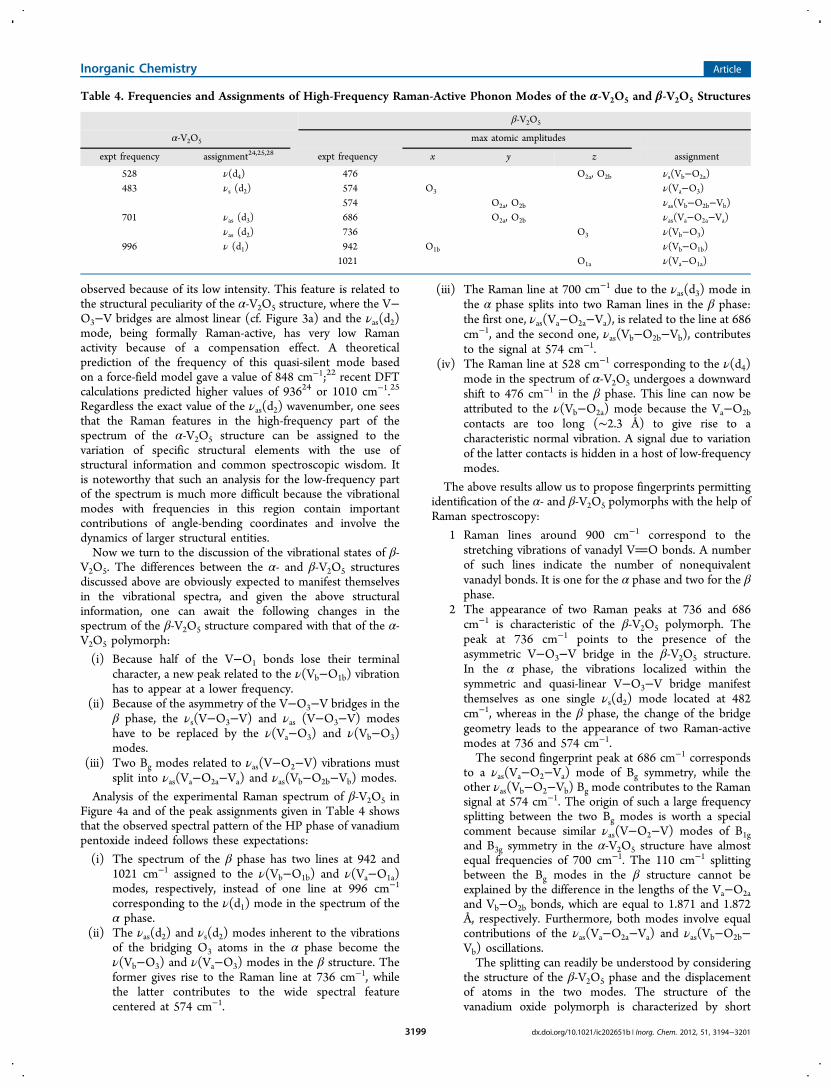
shown in Figure 4a, while Figure 4b presents the Ramanspectrum of the α-V2O5 material.The Raman spectrum of β-V2O5 exhibits a series of well-
resolved peaks, many of which have not been observedpreviously in the in situ RT/HP experiments14 and havebecome evident in the present work. This is related to the
microstructure of the sample because thermal annealing under
pressure led to a well-crystallized β-V2O5 sample with a low
level of remaining stress.
The following features can be observed:
Figure 2. Polyhedral views in the xz projection of α-V2O5 (a) and β-V2O5 (b) structures.
Figure 3. Views of the α-V2O5 (a) and β-V2O5 (b and c) structureswith contacts longer than 2 Å shown as dashed and dotted lines (seethe text for details). Images a and b are in the xz projection. Image c isin the xy projection.
Table 2. Characteristic Interatomic Distances (in Å) in α-and β-V2O5 According to the Experimental Data Reported inReferences 4 and 18
α-V2O54 β-V2O5
18
d1 V−O1 1.586 Va−O1a 1.583 Vb−O1b 1.649d2 V−O3 1.780 Va−O3 1.881 Vb−O3 1.705d3 V−O2 1.878 Va−O2a 1.871 Vb−O2b 1.872d4 V−O2 2.021 Va−O2b 2.296 Vb−O2a 2.176
Inorganic Chemistry Article
dx.doi.org/10.1021/ic202651b | Inorg. Chem. 2012, 51, 3194−32013197
(i) Two intense and sharp peaks in the high-frequencyregion at 942 and 1021 cm−1,
(ii) Two large peaks located at 686 and 736 cm−1,(iii) A large peak with a maximum at 574 cm−1 and a wide
shoulder reaching 620 cm−1 from the high-frequencyside,
(iv) Two peaks at 434 and 476 cm−1; note that only one peakat ca. 450 cm−1 was observed in ref 14.
(v) A number of well-resolved peaks below 400 cm−1 withthe most intense features at 96, 176, 230, 244, 271, 285,301, 340, and 357 cm−1. Note that only ill-defined bandswith an intense peak at ca. 80 cm−1 were observed in thisregion in ref 14.
The calculated spectrum of β-V2O5 is shown in Figure 5,where it is compared with the experimental one. The spectrum
well agrees with the theoretical spectrum reported in ref 33.
Table 3 compares the positions of the peaks in the experimental
and computed spectra and proposes the assignment ofobserved Raman peaks to specific vibrational modes, basedon the results of the calculations. One can notice a systematicoverestimation of the computed vibrational frequencies in theregion above 500 cm−1, which is inherent to the level of theoryused. One can also observe that the intensities of some peaks inthe low-frequency part of the spectrum are not wellreproduced. Nevertheless, a good general correspondencebetween the spectra allows interpretation of the measuredspectral pattern.Now we turn to assignment of the most prominent spectral
features. It is instructive to begin with the α phase of thevanadium pentoxide structure. This consideration will facilitatethe subsequent interpretation of the spectral pattern of themore complicated β polymorph. Taking into account thestructural data collected in Table 2, one can expect to find fourlines in the high-wavenumber region of the Raman spectrum ofα-V2O5 that correspond to four different bond stretchingmodes (ν) with frequency relationships
ν < ν < ν < ν(d ) (d ) (d ) (d )4 3 2 1
Furthermore, the O2 and O3 atoms are located in symmetricV−O−V bridges and, therefore, the ν(d2) and ν(d3) modessplit into symmetric (s) and antisymmetric (as) components.Only νas(V−O−V) modes are genuine bond-stretching modesbecause the νs(V−O−V) modes are coupled with the δ(V−O−V) bending modes and thus have markedly lower frequencies.Consequently, one can detail the above relationship as follows:
ν < ν < ν < ν < ν < ν(d ) (d ) (d ) (d ) (d ) (d )s 2 s 3 4 as 3 as 2 1
The experimental Raman spectrum of α-V2O5 (Figure 4b) hasfour Raman peaks at 996, 701, 528, and 482 cm−1, which wereassigned to the ν(d1), νas (d3), ν (d4), and νs(d2) modes,respectively24,25,28 (Table 4). The missing νas(d2) mode is not
Figure 4. Raman spectra: (a) β-V2O5 sample prepared at a pressure of8 GPa and a temperature of 1073 K for 1 h in a belt-type press; (b) α-V2O5 precursor.
Figure 5. Experimental (black) and computed (color bars) Ramanspectra of the β-V2O5 sample. Red and blue bars stand for the Ag andBg vibrational modes, respectively.
Table 3. Experimental and Calculated Frequencies andMaximum Atomic Amplitudes of Raman-Active PhononModes of the β-V2O5 Structure
frequency max atomic amplitudesa
expt calcd symmetry x y z
96 103.5 Ag L1−L296 109.3 Bg Va−Vb
176 164.7 Bg L1−L2176 175.4 Ag O3 Va
211 205.6 Ag O1a
211 219.8 Ag O1a
230 227.6 Bg L1−L2244 252.9 Ag O1b
271 271.8 Bg O3
285 274.9 Ag O2a, O2b
301 291.3 Bg O1b
340 320.4 Ag O2b
357 350.2 Ag O2a O1b
434 427.7 Ag O2b
476 473.4 Ag O2a, O2b
574 581.0 Ag O3
574 564.2 Bg O2a, O2b
686 695.2 Bg O2a, O2b
736 741.3 Ag O3
942 957.0 Ag O1b
1021 1092.5 Ag O1a
aL1 and L2 denote translations of neighboring layers.
Inorganic Chemistry Article
dx.doi.org/10.1021/ic202651b | Inorg. Chem. 2012, 51, 3194−32013198
observed because of its low intensity. This feature is related tothe structural peculiarity of the α-V2O5 structure, where the V−O3−V bridges are almost linear (cf. Figure 3a) and the νas(d2)mode, being formally Raman-active, has very low Ramanactivity because of a compensation effect. A theoreticalprediction of the frequency of this quasi-silent mode basedon a force-field model gave a value of 848 cm−1;22 recent DFTcalculations predicted higher values of 93624 or 1010 cm−1.25
Regardless the exact value of the νas(d2) wavenumber, one seesthat the Raman features in the high-frequency part of thespectrum of the α-V2O5 structure can be assigned to thevariation of specific structural elements with the use ofstructural information and common spectroscopic wisdom. Itis noteworthy that such an analysis for the low-frequency partof the spectrum is much more difficult because the vibrationalmodes with frequencies in this region contain importantcontributions of angle-bending coordinates and involve thedynamics of larger structural entities.Now we turn to the discussion of the vibrational states of β-
V2O5. The differences between the α- and β-V2O5 structuresdiscussed above are obviously expected to manifest themselvesin the vibrational spectra, and given the above structuralinformation, one can await the following changes in thespectrum of the β-V2O5 structure compared with that of the α-V2O5 polymorph:
(i) Because half of the V−O1 bonds lose their terminalcharacter, a new peak related to the ν(Vb−O1b) vibrationhas to appear at a lower frequency.
(ii) Because of the asymmetry of the V−O3−V bridges in theβ phase, the νs(V−O3−V) and νas (V−O3−V) modeshave to be replaced by the ν(Va−O3) and ν(Vb−O3)modes.
(iii) Two Bg modes related to νas(V−O2−V) vibrations mustsplit into νas(Va−O2a−Va) and νas(Vb−O2b−Vb) modes.
Analysis of the experimental Raman spectrum of β-V2O5 inFigure 4a and of the peak assignments given in Table 4 showsthat the observed spectral pattern of the HP phase of vanadiumpentoxide indeed follows these expectations:
(i) The spectrum of the β phase has two lines at 942 and1021 cm−1 assigned to the ν(Vb−O1b) and ν(Va−O1a)modes, respectively, instead of one line at 996 cm−1
corresponding to the ν(d1) mode in the spectrum of theα phase.
(ii) The νas(d2) and νs(d2) modes inherent to the vibrationsof the bridging O3 atoms in the α phase become theν(Vb−O3) and ν(Va−O3) modes in the β structure. Theformer gives rise to the Raman line at 736 cm−1, whilethe latter contributes to the wide spectral featurecentered at 574 cm−1.
(iii) The Raman line at 700 cm−1 due to the νas(d3) mode inthe α phase splits into two Raman lines in the β phase:the first one, νas(Va−O2a−Va), is related to the line at 686cm−1, and the second one, νas(Vb−O2b−Vb), contributesto the signal at 574 cm−1.
(iv) The Raman line at 528 cm−1 corresponding to the ν(d4)mode in the spectrum of α-V2O5 undergoes a downwardshift to 476 cm−1 in the β phase. This line can now beattributed to the ν(Vb−O2a) mode because the Va−O2bcontacts are too long (∼2.3 Å) to give rise to acharacteristic normal vibration. A signal due to variationof the latter contacts is hidden in a host of low-frequencymodes.
The above results allow us to propose fingerprints permittingidentification of the α- and β-V2O5 polymorphs with the help ofRaman spectroscopy:
1 Raman lines around 900 cm−1 correspond to thestretching vibrations of vanadyl VO bonds. A numberof such lines indicate the number of nonequivalentvanadyl bonds. It is one for the α phase and two for the βphase.
2 The appearance of two Raman peaks at 736 and 686cm−1 is characteristic of the β-V2O5 polymorph. Thepeak at 736 cm−1 points to the presence of theasymmetric V−O3−V bridge in the β-V2O5 structure.In the α phase, the vibrations localized within thesymmetric and quasi-linear V−O3−V bridge manifestthemselves as one single νs(d2) mode located at 482cm−1, whereas in the β phase, the change of the bridgegeometry leads to the appearance of two Raman-activemodes at 736 and 574 cm−1.The second fingerprint peak at 686 cm−1 corresponds
to a νas(Va−O2−Va) mode of Bg symmetry, while theother νas(Vb−O2−Vb) Bg mode contributes to the Ramansignal at 574 cm−1. The origin of such a large frequencysplitting between the two Bg modes is worth a specialcomment because similar νas(V−O2−V) modes of B1gand B3g symmetry in the α-V2O5 structure have almostequal frequencies of 700 cm−1. The 110 cm−1 splittingbetween the Bg modes in the β structure cannot beexplained by the difference in the lengths of the Va−O2aand Vb−O2b bonds, which are equal to 1.871 and 1.872Å, respectively. Furthermore, both modes involve equalcontributions of the νas(Va−O2a−Va) and νas(Vb−O2b−Vb) oscillations.The splitting can readily be understood by considering
the structure of the β-V2O5 phase and the displacementof atoms in the two modes. The structure of thevanadium oxide polymorph is characterized by short
Table 4. Frequencies and Assignments of High-Frequency Raman-Active Phonon Modes of the α-V2O5 and β-V2O5 Structures
β-V2O5
α-V2O5 max atomic amplitudes
expt frequency assignment24,25,28 expt frequency x y z assignment
528 ν(d4) 476 O2a, O2b νs(Vb−O2a)483 νs (d2) 574 O3 ν(Va−O3)
574 O2a, O2b νas(Vb−O2b−Vb)701 νas (d3) 686 O2a, O2b νas(Va−O2a−Va)
νas (d2) 736 O3 ν(Vb−O3)996 ν (d1) 942 O1b ν(Vb−O1b)
1021 O1a ν(Va−O1a)
Inorganic Chemistry Article
dx.doi.org/10.1021/ic202651b | Inorg. Chem. 2012, 51, 3194−32013199
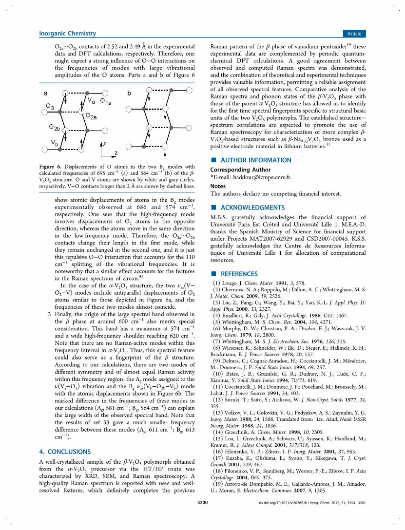
O2a···O2b contacts of 2.52 and 2.49 Å in the experimentaldata and DFT calculations, respectively. Therefore, onemight expect a strong influence of O−O interactions onthe frequencies of modes with large vibrationalamplitudes of the O atoms. Parts a and b of Figure 6
show atomic displacements of atoms in the Bg modesexperimentally observed at 686 and 574 cm−1,respectively. One sees that the high-frequency modeinvolves displacements of O2 atoms in the oppositedirection, whereas the atoms move in the same directionin the low-frequency mode. Therefore, the O2a···O2bcontacts change their length in the first mode, whilethey remain unchanged in the second one, and it is justthis repulsive O−O interaction that accounts for the 110cm−1 splitting of the vibrational frequencies. It isnoteworthy that a similar effect accounts for the featuresin the Raman spectrum of zircon.42
In the case of the α-V2O5 structure, the two νas(V−O2−V) modes include antiparallel displacements of O2atoms similar to those depicted in Figure 6a, and thefrequencies of these two modes almost coincide.
3 Finally, the origin of the large spectral band observed inthe β phase at around 600 cm−1 also merits specialconsideration. This band has a maximum at 574 cm−1
and a wide high-frequency shoulder reaching 620 cm−1.Note that there are no Raman-active modes within thisfrequency interval in α-V2O5. Thus, this spectral featurecould also serve as a fingerprint of the β structure.According to our calculations, there are two modes ofdifferent symmetry and of almost equal Raman activitywithin this frequency region: the Ag mode assigned to theν(Va−O3) vibration and the Bg νas(Vb−O2b−Vb) modewith the atomic displacements shown in Figure 6b. Themarked difference in the frequencies of these modes inour calculations (Ag, 581 cm
−1; Bg, 564 cm−1) can explain
the large width of the observed spectral band. Note thatthe results of ref 33 gave a much smaller frequencydifference between these modes (Ag, 611 cm−1; Bg, 613cm−1).
4. CONCLUSIONS
A well-crystallized sample of the β-V2O5 polymorph obtainedfrom the α-V2O5 precursor via the HT/HP route wascharacterized by XRD, SEM, and Raman spectroscopy. Ahigh-quality Raman spectrum is reported with new and well-resolved features, which definitely completes the previous
Raman pattern of the β phase of vanadium pentoxide;14 theseexperimental data are complemented by periodic quantum-chemical DFT calculations. A good agreement betweenobserved and computed Raman spectra was demonstrated,and the combination of theoretical and experimental techniquesprovides valuable information, permitting a reliable assignmentof all observed spectral features. Comparative analysis of theRaman spectra and phonon states of the β-V2O5 phase withthose of the parent α-V2O5 structure has allowed us to identifyfor the first time spectral fingerprints specific to structural basicunits of the two V2O5 polymorphs. The established structure−spectrum correlations are expected to promote the use ofRaman spectroscopy for characterization of more complex β-V2O5-based structures such as β-Na0.33V2O5 bronze used as apositive-electrode material in lithium batteries.31
■ AUTHOR INFORMATIONCorresponding Author*E-mail: [email protected].
NotesThe authors declare no competing financial interest.
■ ACKNOWLEDGMENTSM.B.S. gratefully acknowledges the financial support ofUniversite Paris Est Creteil and Universite Lille 1. M.E.A.-D.thanks the Spanish Ministry of Science for financial supportunder Projects MAT2007-62929 and CSD2007-00045. K.S.S.gratefully acknowledges the Centre de Ressources Informa-tiques of Universite Lille 1 for allocation of computationalresources.
■ REFERENCES(1) Livage, J. Chem. Mater. 1991, 3, 578.(2) Chernova, N. A.; Roppolo, M.; Dillon, A. C.; Whittingham, M. S.J. Mater. Chem. 2009, 19, 2526.(3) Liu, Z.; Fang, G.; Wang, Y.; Bai, Y.; Yao, K.-L. J. Appl. Phys. D:Appl. Phys. 2000, 33, 2327.(4) Enjalbert, R.; Galy, J. Acta Crystallogr. 1986, C42, 1467.(5) Whittingham, M. S. Chem. Rev. 2004, 104, 4271.(6) Murphy, D. W.; Christian, P. A.; Disalvo, F. J.; Waszczak, J. V.Inorg. Chem. 1979, 18, 2800.(7) Whittingham, M. S. J. Electrochem. Soc. 1976, 126, 315.(8) Wiesener, K.; Schneider, W.; Ilic, D.; Steger, E.; Hallmeir, K. H.;Brackmann, E. J. Power Sources 1978, 20, 157.(9) Delmas, C.; Cognac-Auradou, H.; Cocciantelli, J. M.; Menetrier,M.; Doumerc, J. P. Solid State Ionics 1994, 69, 257.(10) Bates, J. B.; Gruzalski, G. R.; Dudney, N. J.; Luck, C. F.;Xiaohua, Y. Solid State Ionics 1994, 70/71, 619.(11) Cocciantelli, J. M.; Doumerc, J. P.; Pouchard, M.; Broussely, M.;Labat, J. J. Power Sources 1991, 34, 103.(12) Suzuki, T.; Saito, S.; Arakawa, W. J. Non-Cryst. Solids 1977, 24,355.(13) Volkov, V. L.; Golovkin, V. G.; Fedyukov, A. S.; Zaynulin, Y. G.Inorg. Mater. 1988, 24, 1568. Translated from: Izv. Akad. Nauk USSRNeorg. Mater. 1988, 24, 1836.(14) Grzechnik, A. Chem. Mater. 1998, 10, 2505.(15) Loa, I.; Grzechnik, A.; Schwarz, U.; Syassen, K.; Hanfland, M.;Kremer, R. J. Alloys Compd. 2001, 317/318, 103.(16) Filonenko, V. P.; Zibrov, I. P. Inorg. Mater. 2001, 37, 953.(17) Kusaba, K.; Ohshima, E.; Syono, Y.; Kikegawa, T. J. Cryst.Growth 2001, 229, 467.(18) Filonenko, V. P.; Sundberg, M.; Werner, P.-E.; Zibrov, I. P. ActaCrystallogr. 2004, B60, 375.(19) Arroyo-de Dompablo, M. E.; Gallardo-Amores, J. M.; Amador,U.; Moran, E. Electrochem. Commun. 2007, 9, 1305.
Figure 6. Displacements of O atoms in the two Bg modes withcalculated frequencies of 695 cm−1 (a) and 564 cm−1 (b) of the β-V2O5 structure. O and V atoms are shown by white and gray circles,respectively. V−O contacts longer than 2 Å are shown by dashed lines.
Inorganic Chemistry Article
dx.doi.org/10.1021/ic202651b | Inorg. Chem. 2012, 51, 3194−32013200
(20) Balog, P.; Orosel, D.; Cancarevic, Z.; Schon, C.; Jansen, M. J.Alloys Compd. 2007, 429, 87.(21) Clauws, P.; Vennik, J. Phys. Status Solidi B 1976, 76, 707.(22) Abello, L.; Husson, E.; Repelin, Y.; Lucazeau, G. Spectrochim.Acta 1983, 39, 641.(23) Clauws, P.; Broeckx, J.; Vennik, J. Phys. Status Solidi B 1985,131, 459.(24) Brazdova, V.; Ganduglia-Pirovano, M. V.; Sauer, J. Phys. Rev. B2004, 69, 165420.(25) Zhou, B.; He, D. J. Raman Spectrosc. 2008, 39, 1475.(26) Baddour-Hadjean, R.; Pereira-Ramos, J. P. Chem. Rev. 2010,110, 1278.(27) Baddour-Hadjean, R.; Rackelboom, E.; Pereira-Ramos, J. P.Chem. Mater. 2006, 18, 3548.(28) Baddour-Hadjean, R.; Pereira-Ramos, J. P.; Navone, C.;Smirnov, M. Chem. Mater. 2008, 20, 1916.(29) Baddour-Hadjean, R.; Navone, C.; Pereira-Ramos, J. P.Electrochim. Acta 2009, 54, 6674.(30) Baddour-Hadjean, R.; Marzouk, A.; Pereira-Ramos, J. P. J.Raman Spectrosc. 2011, DOI: 10.1002/jrs.2984.(31) Baddour-Hadjean, R.; Bach, S.; Emery, N.; Pereira-Ramos, J. P.J. Mater. Chem. 2011, 21, 11296.(32) Gallardo-Amores, J. M.; Biskup, N.; Amador, U.; Persson, K.;Ceder, G.; Moran, E.; Arroyo-de Dompablo, M. E. Chem. Mater. 2007,19, 5262.(33) Zhou, B.; Su, Q.; He, D. Chin. Phys. B 2009, 18, 4988.(34) FullProf: Rodriguez-Carjaval, J. Satellite Meeting on PowderDiffraction of the XY Congress of the IUCr, Toulouse, France, 1990.(35) Clark, S. J.; Segall, M. D.; Pickard, C. J.; Hasnip, P. J.; Probert,M. J.; Refson, K.; Payne, M. C. Z. Kristallogr. 2005, 220, 567.(36) Refson, K.; Clark, S. J.; Tulip, P. R. Phys. Rev. B 2006, 73,155114.(37) Troullier, N.; Martins, J. L. Phys. Rev. B 1991, 43, 1993.(38) Londero, E.; Schroder, E. Comput. Phys. Commun. 2011, 182,1805.(39) Porezag, D.; Pederson, M. R. Phys. Rev. B 1996, 54, 7830.(40) Langford, J. I. NIST Special Publication 846. Proceedings of theInternational Conference on Accuracy in Powder Diffraction II,Gaithersburg, MD, 1992(41) Fuentes, A. F.; Boulahya, K.; Maczka, M.; Hanuza, J.; Amador,U. Solid State Sci. 2005, 7, 343.(42) Lazarev, A. N., Mirgorodsky, A. P., Mazhenov, H. A. InVibrations of Oxide Lattices; Lazarev, A., Ed.; Nauka Publishers:Leningrad, Russia, 1980; pp 72−99.
Inorganic Chemistry Article
dx.doi.org/10.1021/ic202651b | Inorg. Chem. 2012, 51, 3194−32013201