involved suppressing the S100A9/NF-κ
29
Page 1/29 Anemoside B4 ameliorates TNBS-induced colitis involved suppressing the S100A9/NF- κ B signaling pathway Yong Zhang Soochow University Zhengxia Zha Soochow University Wenhua Shen Soochow University Dan Li Soochow University Naixin Kang Soochow University Zhong Chen Soochow University Yanli Liu Soochow University Guoqiang Xu Soochow University Qiongming Xu ( [email protected] ) Soochow University Research Keywords: Anemoside B4, Colitis, Quantitative proteomics, S100A9, NF- κB Posted Date: September 3rd, 2020 DOI: https://doi.org/10.21203/rs.3.rs-70028/v1 License: This work is licensed under a Creative Commons Attribution 4.0 International License. Read Full License Version of Record: A version of this preprint was published on January 18th, 2021. See the published version at https://doi.org/10.1186/s13020-020-00410-1.
Transcript of involved suppressing the S100A9/NF-κ
Page 1/29
Anemoside B4 ameliorates TNBS-induced colitisinvolved suppressing the S100A9/NF-κB signalingpathwayYong Zhang
Soochow UniversityZhengxia Zha
Soochow UniversityWenhua Shen
Soochow UniversityDan Li
Soochow UniversityNaixin Kang
Soochow UniversityZhong Chen
Soochow UniversityYanli Liu
Soochow UniversityGuoqiang Xu
Soochow UniversityQiongming Xu ( [email protected] )
Soochow University
Research
Keywords: Anemoside B4, Colitis, Quantitative proteomics, S100A9, NF-κB
Posted Date: September 3rd, 2020
DOI: https://doi.org/10.21203/rs.3.rs-70028/v1
License: This work is licensed under a Creative Commons Attribution 4.0 International License. Read Full License
Version of Record: A version of this preprint was published on January 18th, 2021. See the publishedversion at https://doi.org/10.1186/s13020-020-00410-1.
Page 2/29
Abstract
BackgroundDespite the increased morbidity of ulcerative colitis (UC) in the developing countries, available treatmentsremain unsatisfactory. Therefore, it is urgent to discover more effective therapeutic strategies. Pulsatillachinensis was widely used for the treatment of in�amed intestinal diseases including UC for thousandsof years in China. However, it is unclear which compound in P. chinensis is responsible for the therapeuticeffect. Our previous study reported that anemoside B4, the most abundant triterpenoid saponin isolatedfrom P. chinensis, exerts anti-in�ammatory and antioxidant effects.
MethodsHere, we used the 2, 4, 6-trinitrobenzene sulfonic acid (TNBS)-induced colitis rat model to evaluate thetherapeutic effect of anemoside B4. Blood samples of colitis rat were collected for hematology analysis.The effects of anemoside B4 on in�ammation-associated mediators were investigated by Enzyme-linkedimmunosorbent assay (ELISA) and hematoxylin and eosin staining (HE) staining. Cell proliferation orapoptosis was measured by immuno�uorescence technique. The mechanisms of anemoside B4 wasinvestigated using label-free quantitative proteomics. The level of proteins was quanti�ed by westernblotting. mRNA expression was quanti�ed by quantitative real-time RT-PCR.
ResultsThe results showed that anemoside B4 ameliorated TNBS-induced colitis symptoms, including tissuedamage, in�ammatory cell in�ltration, and pro-in�ammatory cytokine production, apoptosis and slowedproliferation in the colon. Quantitative proteomic analyses discovered that 56 proteins were signi�cantlyaltered by anemoside B4 in the TNBS-induced rats. These proteins were mainly clustered in tricarboxylicacid cycle (TCA) cycle and respiratory electron transport chain. Among the altered proteins, S100A9 isone of the most signi�cantly downregulated proteins and associated with NF-κB and MAPK signalingpathways in the pathogenesis of UC. Further experiments revealed that anemoside B4 suppresses theexpression of S100A9 and its downstream genes including TLR4, NF-κB, and p-JNK in colon. In vitro,S100A9 protein could active NF-κB signaling pathway in human intestinal epithelial Caco-2 cells.However, anemoside B4 could inhibit the NF-κB signaling pathway induced by S100A9 protein. Besides, italso inhibited active of NF-κB signaling pathway stimulated by LPS or IL-6.
ConclusionsOur results demonstrate that anemoside B4 prevents TNBS-induced colitis involved inhibiting the NF-κBsignaling pathway through inactivating S100A9 suggesting that anemoside B4 is a promising therapeutic
Page 3/29
candidate for colitis.
BackgroundIn�ammatory bowel disease (IBD) is a prevailing worldwide disease, especially in developing countriesand its incidence has increased signi�cantly in the past two decades [1]. The two main forms of IBD areulcerative colitis (UC) and Crohn’s disease (CD), which are characterized by abdominal pain, diarrhea,bowel obstruction, weight loss, and related immune disorders. Both of them are incurable and are usuallydiagnosed at a young age with a signi�cant morbidity [2].
It is currently accepted that multifactor, such as microbial �ora, dysregulation of the immune response,genetic susceptibility, and environmental factors, involve in IBD, although it’s precise etiology remainselusive. In particular, a pronounced imbalance in the activation of pro-in�ammatory and anti-in�ammatory signaling pathways in the gut is thought to be an important contributor to the developmentand progression of IBD. S100A9 is an intracellular calcium-binding protein belonging to the S100 family,which was originally discovered as an immunogenic protein expressed and secreted by neutrophils [3].Besides neutrophils and monocytes, S100A8 and S100A9 can also be induced in keratinocytes andepithelial cells under in�ammatory conditions [4, 5]. Growing evidence shows that S100A9 has a dual rolein the in�ammation response which is strongly upregulated in trauma, infections, heat, stress, and manyother in�ammations. S100A9 frequently interacts S100A8 to form a heterodimer (called calprotectin) andexhibits its biological functions [6]. Extracellular S100A8/A9 functions by binding to different receptors,including pattern recognition receptors, like the receptor for advanced glycation end products (RAGE) [7]and toll-like receptor 4 (TLR4) [8]. Then, the downstream signal of TLR4 is activated through thegeneration of the adaptor molecules containing the toll/interleukin-1 receptor (TIR) domains, such asTRIF-related adaptor molecule (TRAM)/toll/IL-1 receptor domain-containing adaptor-inducing IFN-β(TRIF) and myeloid differentiation factor 88 (MyD88), which causes the translocation of NF-κB and/orIRF3 into the nucleus [9]. RAGE promoter contains NF-κB-binding sites and activates NF-κB or interactshigh-mobility group box 1 (HMGB1), leading to the intracellular activation of NF-κB [10]. S100A8/A9 is theproposed functional form in lesions associated with neutrophil in�ux, displaying antimicrobial activitiesagainst a variety of micro-organisms [11, 12]. S100A9 induces various in�ammatory responses, such ascytokine secretion, chemotaxis, cell viability, and caspase-dependent or independent cell death [13, 14].
S100A9 is also upregulated in acute or chronic local in�ammatory diseases. It is found that lungs ofpatients with airway in�ammation including cystic �brosis, asthma, acute respiratory distress disorder,and chronic obstructive pulmonary disease have a high expression of S100A8/A9 [15]. Calprotectincomplex of S100A8/A9 is present at high levels in the serum and corresponding intestinal tissues duringin�ammation and has been established as a valuable biomarker for a number of colonic in�ammatoryconditions [16]. Several papers reported that S100A9 expression in myeloid cells is essential for thedevelopment of colon tumors, which activates the downstream genes promoting the occurrence, growthand metastasis of colon-associated tumors [8]. A recent study found that dextran sulfate sodium sulfate(DSS)-induced colitis can be signi�cantly improved by administration of a neutralizing anti-S100A9
Page 4/29
antibody protein, accompanied with that the cellular in�ltrate of innate immunity cells and production ofpro-in�ammatory cytokines is also signi�cantly reduced. In addition, the in�ammatory response, tumorcell proliferation and immune cell in�ltration in colon tissue are suppressed in the mouse experimentalmodel of azoxymethane (AOM)/DSS-induced colitis-associated cancer [17]. Therefore, it is highly likelythat S100A9 may be a therapeutic target for colitis and colitis-associated cancer.
Now clinical treatments for UC include several anti-in�ammatory drugs, such as sulfasalazine,glucocorticoids, nonsteroid anti-in�ammatory agents, inhibitors of pro-in�ammatory pathways, tumornecrosis factor (TNF)-α, gut-homing α4β7 integrin, interleukin (IL)-12/IL-23, and Janus kinases [18].However, these drugs are less effective for some patients and also frequently cause severe side effects,including opportunistic infections and malignancies [19]. Therefore, no effective and safe clinicaltreatment strategy has been discovered for all IBD patients.
At present, the use of complementary and alternative medicine, including acupuncture, homeopathy andherbal medicines, for the treatment of patients with IBD is increasing worldwide. It is estimated that up to70% of patients with IBD in North America and Europe were treated with complementary and alternativemedicine [20, 21]. In China, several Chinese herbs are often used in the treatment of IBD, such asPulsatilla chinensis (Bunge) Regel and Pulsatilla decoction, which were mainly used to treat UC in thetraditional Chinese medicine for thousands of years [22]. Pulsatilla decoction possesses a variety ofpharmacological effects, and the active ingredients from these herbal plants and has shown hepaticprotective, antiin�ammatory, antibacterial, antitumor and antioxidant effects [23]. Several studies havereported that Pulsatilla decoction could alleviate the release of in�ammatory factors, such as IL-17, IL-1β,TNF-α in the serum of IBD patients, reduce syndromes and symptom scores, and suppress the activationof the NF-κB signaling pathway [24].
As the most important herb in Pulsatilla decoction, P. chinensis was widely used for the treatment ofamoebic dysentery and malignant dysentery in China for two thousand years, which exhibits “blood-cooling” and detoxi�cation activities [25]. Accumulating evidence has demonstrated that P. chinensis andits ingredients have antioxidant, anti-in�ammatory and antitumor effects. Our previous study reportedthat anemoside B4, the most abundant triterpenoid saponin isolated from P. chinensis, decreasesin�ammation and oxidant injury induced by cisplatin. It ameliorates LPS-induced kidney and lungdamage, inhibits LPS-induced NF-κB activation in vivo [26, 27]. However, it is unclear whether anemosideB4 exhibits effective roles in treating IBD and it might be the molecular basis for the clinical effect of P.chinensis.
In the present study, we investigated the therapeutic effect of anemoside B4, on 2, 4, 6-trinitrobenzenesulfonic acid (TNBS)-induced colitis rats. We �rstly found that anemoside B4 exerts anti-in�ammatoryand anti-apoptotic properties through downregulating colonic in�ammatory cytokine levels, and inhibitingcolonic epithelium apoptosis. Furthermore, quantitative proteomics analyses indicated that S100A9-dependent pathway is involved in the therapeutic effects of anemoside B4. Our data suggest thatanemoside B4 has the potential ability for the further therapy of colitis.
Page 5/29
Methods
Chemicals and reagentsTNBS and lipopolysaccharide (LPS) were supplied by Sigma-Aldrich, Germany (Cat. no. P2297 andL6529-1MG). Mesalazine (Cat. no. 89-57-6) was purchased from Ethypharm Pharmaceutical Co Ltd(Shanghai, China). Hunan recombinant S100A9 protein was purchased from Abcam (Cat. no. ab95909).Rat IL-1β, IL-6, and TNF-α ELISA kits were supplied by MultiSciences (Hangzhou, China). PCR primers(Supplementary Table S1) for quantitative real-time PCR analysis were synthesized by GENEWIZ (Suzhou,China). The primary antibodies for p38 (Cat. no. 8960) (1:2000), p-p38 (Cat. no. 4511) (1:1000), JNK (Cat.no. 9252) (1:1000), p-JNK (Cat. no. 9255) (1:2000), ERK1/2 (Cat. no. 4695) (1:2000), p-ERK1/2 (Cat. no.4370) (1:1000), NF-κB p65 (Cat. no. 8242) (1:2000), p-NF-κB p65 (Cat. no. 3033) (1:1000), Bcl-2 (Cat. no.3498) (1:1000), cleaved-caspase 3 (Cat. no. 9664) (1:1000), and S100A9 (Cat. no. 73425) (1:1000) werepurchased from Cell Signaling Technology (Danvers, USA), and those for Bax (Cat. no. sc-20067)(1:2000), p53 (Cat. no. sc-126) (1:1000), cleaved-PRAP (Cat. no. sc-74470) (1:1000), IL-6 (Cat. no. sc-57315) (1:1000), TLR4 (Cat. no. sc-293072) (1:1000), NOD2 (Cat. no. sc-56168) (1:1000), and GAPDH(Cat. no. sc-365062) (1:2000) were purchased from Santa Cruz Biotechnology Co., Ltd (Santa Cruz, USA).Caco-2 cell (PN 5) lines were purchased from Shanghai Cell Bank of Chinese Academy of Sciences(Shanghai, China), and routinely maintained in DMEM (Gibco, USA) supplemented with 10% fetal bovineserum (Gibco, USA), 100 µg/ml streptomycin and 100 U/ml penicillin (Gibco, USA). Anemoside B4 (20 g,purity more than 99%) was isolated from the roots of P. chinensis and its structure was determined asprevious described [26].
Experimental animalsSD rats (male, 220–250 g) were obtained from the Experimental Animal Center of Soochow University. Allanimals were given free access to water and food with air �ltration (22 ± 2 °C, 12-h light/12-h dark). Allanimal experiments were conducted in accordance with the procedure approved by the Ethical ReviewCommittee for Laboratory Animal Welfare of the Soochow University.
Induction of transfer colitis and experimental designAll animals were divided into �ve groups (n = 7 per group): vehicle group, TNBS + saline group, TNBS + anemoside B4 group (5 mg/kg), TNBS + anemoside B4 group (10 mg/kg), and TNBS + mesalazine group(200 mg/kg). All rats were fasted for 24 h and weighed before injection, and then were anesthetized byinjecting chloral hydrateintraperitoneally (300 mg/kg). In the vehicle group, 40% ethanol solution wasslowly instilled into the colon of anesthetized rats, while for the other four groups, TNBS solution(80 mg/kg in 40% ethanol v/v) was instilled. Rats in the vehicle group and TNBS + saline group wereintraperitoneally injected with saline, and those in the TNBS + anemoside B4 groups were intraperitoneallyinjected with anemoside B4, while those in the TNBS + mesalazine group were treated with mesalazine bygavage.
Assessment of colonic damage
Page 6/29
During the experiment, rats were weighed every day and disease activity index (DAI) was recorded forseven days after the induction of colitis according to previous reference [28]. The DAI is the combinedscore of weight loss compared to initial weight, stool consistency, and bleeding. In brief, No weight losswas registered as 0, weight loss of 1% to 5% from baseline was assigned 1 point, weight loss of 6% to10% from baseline was assigned 2 points, weight loss of 11% to 20% from baseline was assigned 3points and weight loss of more than 20% from baseline was assigned 4 points. Stool consistency scoreswere 0 (normal, well-formed pellets), 2 (loose stool, pasty and semi-formed stools that did not adhere tothe anus), and 4 (diarrhea, liquid stools that adhered to the anus). Bleeding scores were 0 (no blood), 2(Hemoccult positive and visual pellet bleeding), and 4 (gross bleeding, blood around anus).On the 8thday, rats were sacri�ced by cervical dislocation, the colons were harvested and rinsed with saline. Colonlength was recorded as an indirect marker of in�ammation. Myeloperoxidase (MPO) activity wasmeasured to assess in�ammation. Colon samples were �xed in 4% paraformaldehyde for histologicalexamination and immuno�uorescence staining. The remaining colon samples were kept at − 80 °C forfurther assessment of proteomics and enzyme-linked immunosorbent assay (ELISA), qPCR, and Westernblotting analyses (n = 3).
Histopathological analysis of the rectumsFor histological assessments, sections of the rectums were stained with HE stains. Light microscopy(CX31 research microscope, Olympus Optical Co., Ltd., Tokyo, Japan) and panoramic viewer camerasystem were used for examining, scanning, and analyzing the sections. Histological score was measuredby in�ammation severity: 0 (normal colonic mucosal), 1 (crypt damage less than 1/3), 2 (crypt damageless than 1/3 − 2/3), 3 (mucosal erosion), 4 (muscosal erosion or ulcer with signi�cant in�ltration ofin�ammatory cells) according to reference [29].
Label-free quantitative proteomicsOn the 7th day after rats were treatment with TNBS (80 mg/kg), colon tissues from the rats in TNBS + saline group (n = 3) and TNBS + anemoside B4 group (10 mg/kg) (n = 3) were extracted, which then werelysed in a HEPES buffer (pH 8.0) containing 8 M urea to obtain total protein. Disul�de bonds in theseproteins were reduced with 2 mM dithiothreitol at 37 °C for 45 min. The free thiols were further alkylatedwith 8 mM chloroacetamide for 1 h at room temperature. The excess chloroacetamide was thenquenched by adding 2 mM dithiothreitol for 45 min. After acetone precipitation, proteins wereresuspended in a HEPES buffer containing 8 M urea and incubated with the sequencing grade Lys-C(TaKaRa Bio, Japan) for 4 h at 37 °C. The samples were diluted with 10 mM HEPES (pH 8.0) four timesand continued for digestion with MS-grade trypsin (TaKaRa Bio, Japan) at 37 °C for 20 h. Next, thepeptides were desalted with C18 Zip-tips (Merck Millipore, Massachusetts, USA), which were thenseparated by capillary high performance liquid chromatography and analyzed in an Orbitrap FusionLumos mass spectrometer (Thermo Fisher Scienti�c, Massachusetts, USA). Data analysis was performedusing MaxQuant, and proteins with FDR ≤ 0.01 were considered as positive identi�cation. Theindependent sample t-test in IBM SPSS software (Ver 19) was used to calculate the P-values for the
Page 7/29
identi�ed proteins. The proteins with fold change > 1.50 or < 0.67 and P < 0.05 are considered assigni�cantly differentially expressed proteins.
Western blot analysisWestern bolt analysis was performed as previously described [27]. In brief, proteins were extracted in RIPAlysis buffer, separated by SDS-PAGE, and transferred into PVDF membranes. The membranes wereincubated with different primary antibodies and again with the peroxidase (HRP) conjugated goatsecondary antibodies (Beyotime, Haimen, Jiangsu, China). Then, proteins were exposed to HRP substratereagent (Millipore Corporation, Billerica, USA) and signals were detected with the ChemiDoc XRS Imager(Bio-Rad, USA).
Quantitative real-time RT-PCRAccording to the manufacturer’s instructions, total RNA was extracted with TRIzol reagent (Ambion, USA)and quanti�ed by a NanoDrop ND-2000 spectrophotometer (Thermo Fisher). After quality detection, thetotal RNA samples (1000 ng per sample) were reverse-transcribed using the revertaid 1st cDNA synth kit(Thermo Scienti�c). Standard curves were constructed with the CFX96™ real-time PCR detection system(Bio-Rad). SsoAdvanced™Universal SYBR® Green (Bio-Rad) was used for qPCR detection. The cyclingconditions for the qPCR were as follows: 95 °C for 3 min, followed by 37 cycles of 95 °C for 30 s, 55 °C for30 s, and 72 °C for 30 s. The relative expression levels of the genes were calculated with the 2−ΔΔCt
method based on the threshold cycle (Ct) value. Rat S100A9 primer: 5, GGCACGAGCTCCTTAGCTTT, 3,TCTCCCTCTTCAGAAAATTTGGC; rat GAPDH primer: 5, AGTGCCAGCCTCGTCTCATA, 3,GATGGTGATGGGTTTCCCGT.
ELISAIn�ammatory cytokines including TNF-α, interleukin-6 (IL-6), and interleukin-1β (IL-1β) in the colon tissuesamples were assayed using ELISA kits according to the manufacturer’s instructions.
Ethynyl-2′-Deoxyuridine (EdU) cell proliferation assayCell proliferation was detected by the BeyoClick™EdU-488 Kit (Beyotime, Haimen, Jiangsu, China). Brie�y,4 h before sacri�ce, rats were injected intraperitoneally with 5 mg/kg EdU and then the colon wascollected and rinsed with saline. Colon samples were then processed using the BeyoClick™ EdU-488 kitaccording to the manufacturer’s instructions. The EdU positive cells were detected with a confocal laserscanning microscope (LSM 710, ZEISS, Germany).
Apoptosis detection assayThe Situ TUNEL Apoptosis Detection Kit (Nanjing Jiancheng Bioengineering Institute, Nanjing, China) wasused to detect the apoptotic cells according to the manufacturer’s instructions. The stained samples weremeasured under a confocal laser scanning microscope.
Immuno�uorescence assay
Page 8/29
The expressions of CD11b (Novus, USA) and S100A9 in colon tissues were detected byimmuno�uorescence staining. The colon sections were in�ltrated with 0.5% Triton X-100 in PBS, blockedwith 3% BSA, incubated overnight with either S100A9 or CD11b antibodies, and further incubated with asecondary antibody for 1 h. The immuno�uorescence was detected by a confocal laser scanningmicroscope.
Myeloperoxidase activityMyeloperoxidase (MPO) activity was measured in the colon. The colon was homogenized in 1:10 (w/v) of50 mM phosphate buffer (pH 6.0) using a Polytron homogenizer. The homogenate was centrifuged at12,000 rpm for 10 min. Then,MPO activity was detected by myeloperoxidase assay kit (NanjingJiancheng Bioengineering Institute, Nanjing, China).
Cell line experimentsHuman intestinal epithelial Caco-2 cells were incubated in 5% CO2 (95% relative humidity) at 37 °C,stimulated by 1 µg/ml LPS or S100A9 and treated with anemoside B4 after 1 h. At 6 h after thestimulation, total protein samples were extracted from the cells.
Data and statistical analysisData were expressed as the mean ± SD. Statistics were analyzed by one-way ANOVA and Tukey’s multiplecomparisons test using SPSS 16.0 statistical analysis software except indicated. P < 0.05 was consideredas statistically signi�cant.
Results
Anemoside B4 ameliorated TNBS-induced colitis in ratsThe TNBS hapten, given as one or two enemas with ethanol as a carrier to disrupt epithelial integrity,induces acute in�ammation and colitis in the mouse with Crohn’s colitis-like transmural tissue damage[30]. In this study, TNBS-induced severe acute colitis rat model was used to determine thepharmacological activity of anemoside B4. The doses of anemoside B4 were chosen on the basis of ourpreliminary tests of LPS-induced in�ammation and acetic acid-induced analgesia [27]. Mesalazine wasorally administrated to rats with the same dose as the positive control [31].
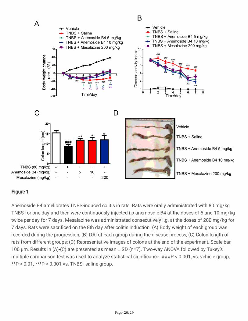
In the model group, we observed IBD-like colitis, diarrhoea and bloody mucopurulent stools. The averagebody weight of the rats in the model group declined one day after TNBS administration. Compared withthe mock-treated group, TNBS-induced rats showed a signi�cantly higher DAI score and body weight loss.In TNBS-induced severe acute colitis model, both anemoside B4 and mesalazine inhibited body weightloss during disease progression (Fig. 1A). TNBS induced a signi�cant increase in DAI score, an importantmarker of IBD. In contrast, anemoside B4 ameliorated TNBS-induced DAI score in a dose-dependentmanner (Fig. 1B). TNBS treatment induced a signi�cant decrease in colon length. Furthermore, treatmentwith anemoside B4 ameliorated TNBS-induced colon length shortening in a dose-dependent manner and
Page 9/29
mesalazine was slightly less effective (Fig. 1C and D). These results demonstrate that inhibited effects ofanemoside B4 on colitis is slightly more effective than mesalazine.
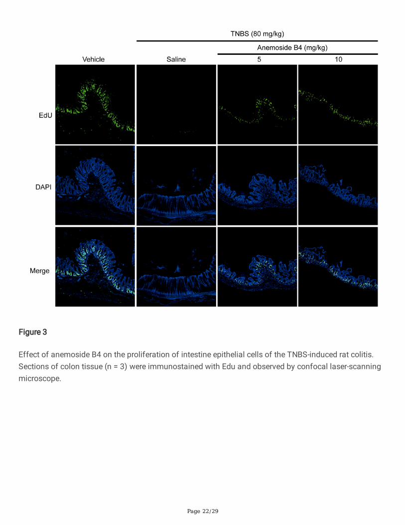
Anemoside B4 suppressed TNBS-induced in�ammationHE staining showed that the colon tissues of the rats in TNBS-induced groups had pathologicalstructures, including obvious in�ltration of in�ammatory cells, decreased number of intestinal villus,epithelial cell damage and apoptosis. There were mark difference of pathological score between normalgroup and TNBS group. However, anemoside B4 relieved all pathological changes and score in a dose-dependent manner (Fig. 2B).
TNBS-induced model of acute colitis maintains many pathological features of human colitis, especiallyincreasing type I in�ammatory response and helper T cell 1 (Th1)-related cytokines, such as TNF-α orinterferon-γ (IFN-γ). During in�ammation, monocytes are recruited to in�amed sites and lymphoid tissues,where macrophage differentiation plays an important role during the beginning and resolution ofin�ammatory processes [32]. Therefore, we measured the effect of anemoside B4 on immune cellin�ltration and in�ammatory cytokine expression in rats with TNBS-induced acute colitis. Compared withthe rats in vehicle group, those exposed to TNBS had signi�cantly (P < 0.05) increased levels of colonicproin�ammatory cytokines and chemokines, including IL-6, TNF-α, and IL-1β. However, anemoside B4signi�cantly reduced the levels of these colonic pro-in�ammatory cytokines (Fig. 2C-E).
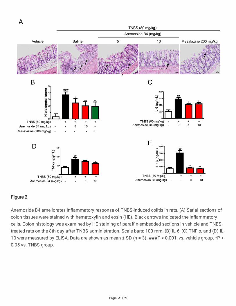
Anemoside B4 suppressed TNBS-induced proliferation andapoptosisNext, we sought to the effects of anemoside B4 on the proliferation and apoptosis induced by TNBS. TheEdU in vivo imaging kit was used to assess cell proliferation. 5-EdU was injected intraperitoneally 5 hbefore the rats were sacri�ced, the colons were then harvested and operated according to themanufacturer’s instruction. Normal colonic epithelial cells have regenerative proliferation and stainedwith green. More EdU stained spots were observed in vehicle rats. These results demonstrated TNBSdecreased the ability of regenerative proliferation. However, anemoside B4 partially restored the ability ofregenerative proliferation (Fig. 3).
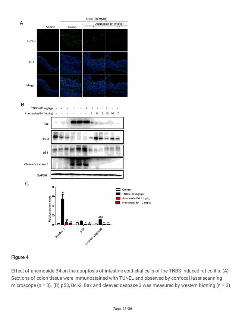
It has been previously reported that TNBS-induced colitis are associated with apoptosis of the colonicepithelial cells and its toxicology is thought to be related to the induction of apoptosis and the destructionof the intestinal mucosal barrier [33, 34]. In IBD, high levels of apoptosis have been observed in theintestinal epithelium of patients [35]. In the intracellular machinery of apoptosis, the Bcl-2 family proteinsinvolving anti-apoptotic (Bcl-2) and pro-apoptotic (Bax) members mainly control the intrinsic pathway.The up-regulation of Bax, down-regulation of Bcl-2 and cleavage of caspase 3 are all mediated by p53activation, which plays critical roles in the induction of apoptosis [36]. The increased number of apoptoticepithelial cells and enhanced Bax but attenuated Bcl-2 signaling during active colitis may lead to adefective epithelial barrier and result in pathogenic microorganism in�ltration.
Page 10/29
In the present study, TUNEL assay was carried out to detect apoptotic cells. More TUNEL stained spotswere observed in the colon tissue obtained from TNBS-induced colitis rats. Anemoside B4 treatmentsigni�cantly decreased the TUNEL stained spots. Further, Western blotting was used to detect apoptosisand related protein expression levels, respectively. Consistent with previous reports, we found that theTNBS group had markedly up-regulated Bax, p53 and cleaved caspase 3 but down-regulated Bcl-2 in thecolon. In contrast, anemoside B4 signi�cantly increased Bcl-2/Bax ratio and inhibited cleaved caspase3,p53 protein levels in a dose dependent manner, suggesting that anemoside B4 could reduce colitisapoptosis in colon sample (Fig. 4). These observations indicated that anemoside B4 was obviouslyeffective in mitigating TNBS-induced acute colitis in rats.
Anemoside B4 inhibited NF-κB activation induced by LPS incolonic epithelial cellsThe above experiment data showed that anemoside B4 had anti-in�ammatory activity and inhibitsin�ammatory cytokine secretion induced by colitis. It is well known that NF-κB plays a critical role inin�ammation involved various human diseases including IBD and in animal disease models. ActivatedNF-κB pathway promotes the expression of various pro-in�ammatory genes and in�uences the course ofmucosal in�ammation [37].
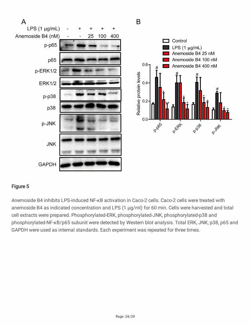
In order to explore the mechanism of anti-in�ammatory activity of anemoside B4, we investigated theeffect of anemoside B4 on the activation of the NF-κB signaling pathway, as it has been documented tobe related to in�ammation. As expected, the level of p-NF-κB/p65 protein is increased upon LPStreatment, while it was signi�cantly reduced by anemoside B4 (Fig. 5). In addition, LPS activated NF-κBupstream signaling pathway, including p-p38, p-JNK, and p-ERK in colonic epithelial cells, while theselevels were similarly reduced by anemoside B4.
Proteomic identi�cation of the differentially regulated proteins by anemoside B4
The above studies showed that 10 mg/kg anemoside B4 remarkably diminished the TNBS-induced colitisand the therapeutic effect was comparable to that of mesalazine. Therefore, in the following experiments,anemoside B4 was used at the dose of 10 mg/kg.
To further investigate the mechanisms of the therapeutic effect of anemoside B4 on TNBS-inducedcolitis, we performed a label-free quantitative proteomic analysis to identify the differentially expressedproteins in the colon samples from TNBS-induced group and anemoside B4-treated group.
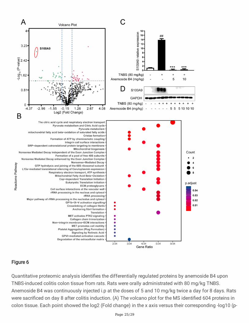
In total, we identi�ed 604 proteins and quanti�ed 591 proteins, whose ratios were shown in the volcanoplot. Among the quanti�ed proteins, 56 protein groups with fold change > 1.50 or < 0.67 and P < 0.05 weremarked with red (upregulation) or green (downregulation) dots in the anemoside B4-treated groupcompared with the TNBS-induced group (Fig. 6A).
Page 11/29
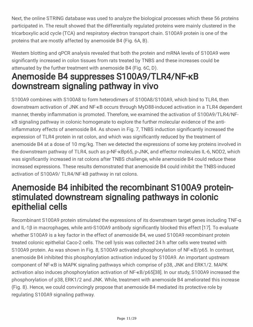
Next, the online STRING database was used to analyze the biological processes which these 56 proteinsparticipated in. The result showed that the differentially regulated proteins were mainly clustered in thetricarboxylic acid cycle (TCA) and respiratory electron transport chain. S100A9 protein is one of theproteins that are mostly affected by anemoside B4 (Fig. 6A, B).
Western blotting and qPCR analysis revealed that both the protein and mRNA levels of S100A9 weresigni�cantly increased in colon tissues from rats treated by TNBS and these increases could beattenuated by the further treatment with anemoside B4 (Fig. 6C, D).
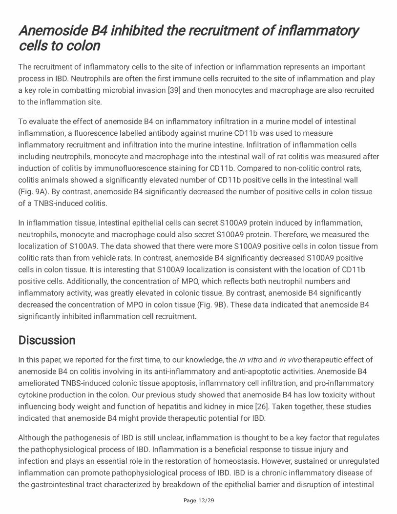
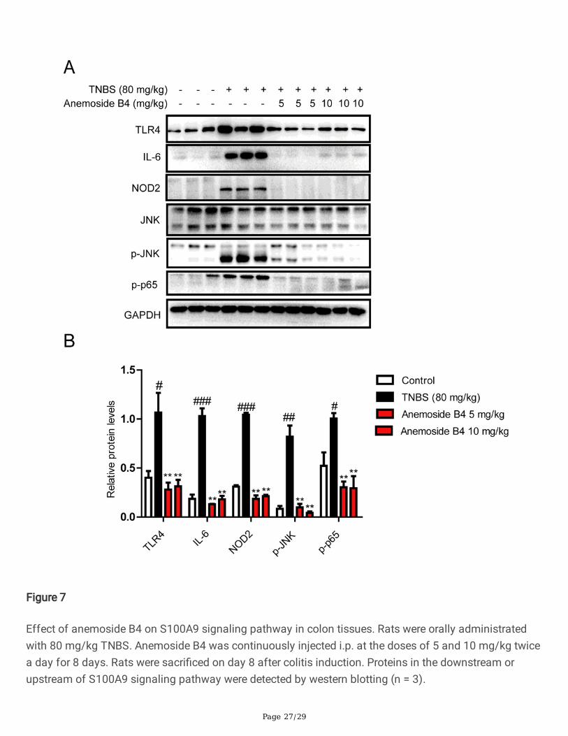
Anemoside B4 suppresses S100A9/TLR4/NF-κBdownstream signaling pathway in vivoS100A9 combines with S100A8 to form heterodimers of S100A8/S100A9, which bind to TLR4, thendownstream activation of JNK and NF-κB occurs through MyD88-induced activation in a TLR4 dependentmanner, thereby in�ammation is promoted. Therefore, we examined the activation of S100A9/TLR4/NF-κB signaling pathway in colonic homogenate to explore the further molecular evidence of the anti-in�ammatory effects of anemoside B4. As shown in Fig. 7, TNBS induction signi�cantly increased theexpression of TLR4 protein in rat colon, and which was signi�cantly reduced by the treatment ofanemoside B4 at a dose of 10 mg/kg. Then we detected the expressions of some key proteins involved inthe downstream pathway of TLR4, such as p-NF-κBp65, p-JNK, and effector molecules IL-6, NOD2, whichwas signi�cantly increased in rat colons after TNBS challenge, while anemoside B4 could reduce theseincreased expressions. These results demonstrated that anemoside B4 could inhibit the TNBS-inducedactivation of S100A9/ TLR4/NF-kB pathway in rat colons.
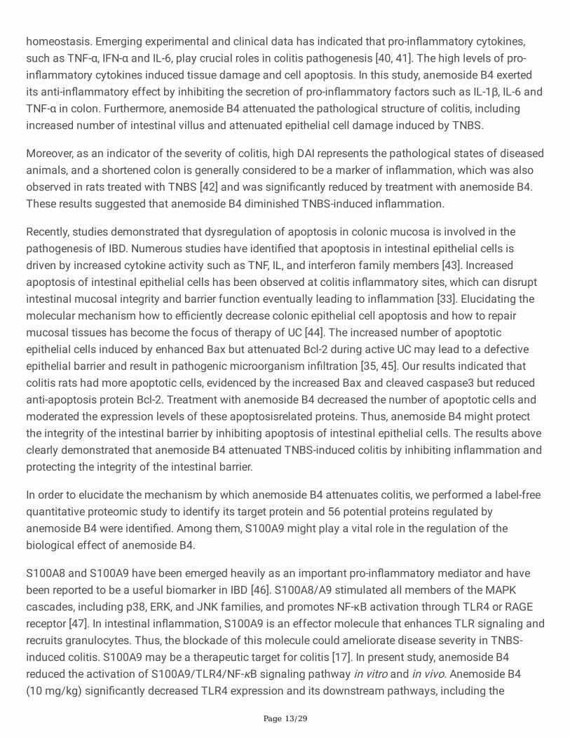
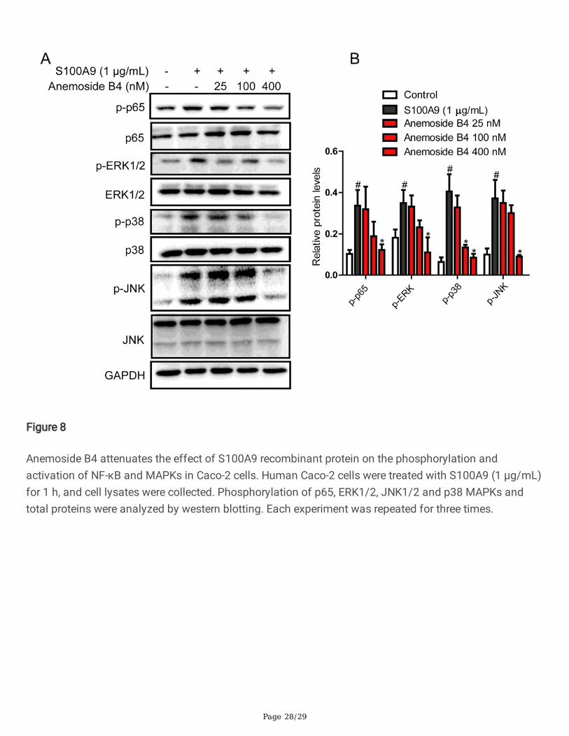
Anemoside B4 inhibited the recombinant S100A9 protein-stimulated downstream signaling pathways in colonicepithelial cellsRecombinant S100A9 protein stimulated the expressions of its downstream target genes including TNF-αand IL-1β in macrophages, while anti-S100A9 antibody signi�cantly blocked this effect [17]. To evaluatewhether S100A9 is a key factor in the effect of anemoside B4, we used S100A9 recombinant proteintreated colonic epithelial Caco-2 cells. The cell lysis was collected 24 h after cells were treated withS100A9 protein. As was shown in Fig. 8, S100A9 activated phosphorylation of NF-κB/p65. In contrast,anemoside B4 inhibited this phosphorylation activation induced by S100A9. An important upstreamcomponent of NF-κB is MAPK signaling pathways which comprise of p38, JNK and ERK1/2. MAPKactivation also induces phosphorylation activation of NF-κB/p65[38]. In our study, S100A9 increased thephosphorylation of p38, ERK1/2 and JNK. While, treatment with anemoside B4 ameliorated this increase(Fig. 8). Hence, we could convincingly propose that anemoside B4 mediated its protective role byregulating S100A9 signaling pathway.
Page 12/29
Anemoside B4 inhibited the recruitment of in�ammatorycells to colonThe recruitment of in�ammatory cells to the site of infection or in�ammation represents an importantprocess in IBD. Neutrophils are often the �rst immune cells recruited to the site of in�ammation and playa key role in combatting microbial invasion [39] and then monocytes and macrophage are also recruitedto the in�ammation site.
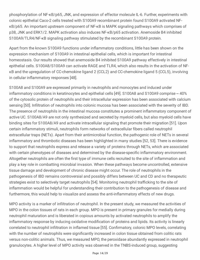
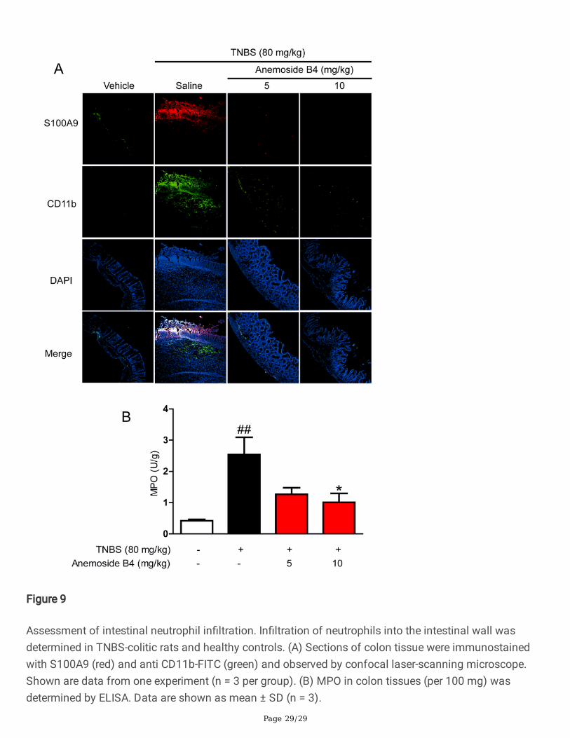
To evaluate the effect of anemoside B4 on in�ammatory in�ltration in a murine model of intestinalin�ammation, a �uorescence labelled antibody against murine CD11b was used to measurein�ammatory recruitment and in�ltration into the murine intestine. In�ltration of in�ammation cellsincluding neutrophils, monocyte and macrophage into the intestinal wall of rat colitis was measured afterinduction of colitis by immuno�uorescence staining for CD11b. Compared to non-colitic control rats,colitis animals showed a signi�cantly elevated number of CD11b positive cells in the intestinal wall(Fig. 9A). By contrast, anemoside B4 signi�cantly decreased the number of positive cells in colon tissueof a TNBS-induced colitis.
In in�ammation tissue, intestinal epithelial cells can secret S100A9 protein induced by in�ammation,neutrophils, monocyte and macrophage could also secret S100A9 protein. Therefore, we measured thelocalization of S100A9. The data showed that there were more S100A9 positive cells in colon tissue fromcolitic rats than from vehicle rats. In contrast, anemoside B4 signi�cantly decreased S100A9 positivecells in colon tissue. It is interesting that S100A9 localization is consistent with the location of CD11bpositive cells. Additionally, the concentration of MPO, which re�ects both neutrophil numbers andin�ammatory activity, was greatly elevated in colonic tissue. By contrast, anemoside B4 signi�cantlydecreased the concentration of MPO in colon tissue (Fig. 9B). These data indicated that anemoside B4signi�cantly inhibited in�ammation cell recruitment.
DiscussionIn this paper, we reported for the �rst time, to our knowledge, the in vitro and in vivo therapeutic effect ofanemoside B4 on colitis involving in its anti-in�ammatory and anti-apoptotic activities. Anemoside B4ameliorated TNBS-induced colonic tissue apoptosis, in�ammatory cell in�ltration, and pro-in�ammatorycytokine production in the colon. Our previous study showed that anemoside B4 has low toxicity withoutin�uencing body weight and function of hepatitis and kidney in mice [26]. Taken together, these studiesindicated that anemoside B4 might provide therapeutic potential for IBD.
Although the pathogenesis of IBD is still unclear, in�ammation is thought to be a key factor that regulatesthe pathophysiological process of IBD. In�ammation is a bene�cial response to tissue injury andinfection and plays an essential role in the restoration of homeostasis. However, sustained or unregulatedin�ammation can promote pathophysiological process of IBD. IBD is a chronic in�ammatory disease ofthe gastrointestinal tract characterized by breakdown of the epithelial barrier and disruption of intestinal
Page 13/29
homeostasis. Emerging experimental and clinical data has indicated that pro-in�ammatory cytokines,such as TNF-α, IFN-α and IL-6, play crucial roles in colitis pathogenesis [40, 41]. The high levels of pro-in�ammatory cytokines induced tissue damage and cell apoptosis. In this study, anemoside B4 exertedits anti-in�ammatory effect by inhibiting the secretion of pro-in�ammatory factors such as IL-1β, IL-6 andTNF-α in colon. Furthermore, anemoside B4 attenuated the pathological structure of colitis, includingincreased number of intestinal villus and attenuated epithelial cell damage induced by TNBS.
Moreover, as an indicator of the severity of colitis, high DAI represents the pathological states of diseasedanimals, and a shortened colon is generally considered to be a marker of in�ammation, which was alsoobserved in rats treated with TNBS [42] and was signi�cantly reduced by treatment with anemoside B4.These results suggested that anemoside B4 diminished TNBS-induced in�ammation.
Recently, studies demonstrated that dysregulation of apoptosis in colonic mucosa is involved in thepathogenesis of IBD. Numerous studies have identi�ed that apoptosis in intestinal epithelial cells isdriven by increased cytokine activity such as TNF, IL, and interferon family members [43]. Increasedapoptosis of intestinal epithelial cells has been observed at colitis in�ammatory sites, which can disruptintestinal mucosal integrity and barrier function eventually leading to in�ammation [33]. Elucidating themolecular mechanism how to e�ciently decrease colonic epithelial cell apoptosis and how to repairmucosal tissues has become the focus of therapy of UC [44]. The increased number of apoptoticepithelial cells induced by enhanced Bax but attenuated Bcl-2 during active UC may lead to a defectiveepithelial barrier and result in pathogenic microorganism in�ltration [35, 45]. Our results indicated thatcolitis rats had more apoptotic cells, evidenced by the increased Bax and cleaved caspase3 but reducedanti-apoptosis protein Bcl-2. Treatment with anemoside B4 decreased the number of apoptotic cells andmoderated the expression levels of these apoptosisrelated proteins. Thus, anemoside B4 might protectthe integrity of the intestinal barrier by inhibiting apoptosis of intestinal epithelial cells. The results aboveclearly demonstrated that anemoside B4 attenuated TNBS-induced colitis by inhibiting in�ammation andprotecting the integrity of the intestinal barrier.
In order to elucidate the mechanism by which anemoside B4 attenuates colitis, we performed a label-freequantitative proteomic study to identify its target protein and 56 potential proteins regulated byanemoside B4 were identi�ed. Among them, S100A9 might play a vital role in the regulation of thebiological effect of anemoside B4.
S100A8 and S100A9 have been emerged heavily as an important pro-in�ammatory mediator and havebeen reported to be a useful biomarker in IBD [46]. S100A8/A9 stimulated all members of the MAPKcascades, including p38, ERK, and JNK families, and promotes NF-κB activation through TLR4 or RAGEreceptor [47]. In intestinal in�ammation, S100A9 is an effector molecule that enhances TLR signaling andrecruits granulocytes. Thus, the blockade of this molecule could ameliorate disease severity in TNBS-induced colitis. S100A9 may be a therapeutic target for colitis [17]. In present study, anemoside B4reduced the activation of S100A9/TLR4/NF-κB signaling pathway in vitro and in vivo. Anemoside B4(10 mg/kg) signi�cantly decreased TLR4 expression and its downstream pathways, including the
Page 14/29
phosphorylation of NF-κB/p65, JNK, and expression of effector molecule IL-6. Further, experiments withcolonic epithelial Caco-2 cells treated with S100A9 recombinant protein found S100A9 activated NF-κB/p65. An important upstream component of NF-κB is MAPK signaling pathways which comprises ofp38, JNK and ERK1/2. MAPK activation also induces NF-κB/p65 activation. Anemoside B4 inhibitedS100A9/TLR4/NF-κB signaling pathway stimulated by the recombinant S100A9 protein.
Apart from the known S100A9 functions under in�ammatory conditions, little has been shown on theexpression mechanism of S100A9 in intestinal epithelial cells, which is important for intestinalhomeostasis. Our results showed that anemoside B4 inhibited S100A9 pathway effectively in intestinalepithelial cells. S100A8/S100A9 can activate RAGE and TLR4, which also results in the activation of NF-κB and the upregulation of CC-chemokine ligand 2 (CCL2) and CC-chemokine ligand 5 (CCL5), involvingin cellular in�ammatory responses [48].
S100A8 and S100A9 are expressed primarily in neutrophils and monocytes and induced underin�ammatory conditions in keratinocytes and epithelial cells [49]. S100A8 and S100A9 comprise ~ 40%of the cytosolic protein of neutrophils and their intracellular expression has been associated with calciumsensing [50]. In�ltration of neutrophils into colonic mucosa has been associated with the severity of IBD.The presence of neutrophils in the intestinal mucosa constitutes a prominent in�ammatory component ofactive UC. S100A8/A9 are not only synthesized and secreted by myeloid cells, but also myeloid cells havebinding sites for S100A8/A9 and activate intracellular signaling that promote their migration [51]. Uponcertain in�ammatory stimuli, neutrophils form networks of extracellular �bers called neutrophilextracellular traps (NETs). Apart from their antimicrobial function, the pathogenic role of NETs in severalin�ammatory and thrombotic diseases has been highlighted in many studies [52, 53]. There is evidenceto support that neutrophils express and release a variety of proteins through NETs, which are associatedwith certain phenotypes of diseases and determined by the disease-speci�c in�ammatory environment.Altogether neutrophils are often the �rst type of immune cells recruited to the site of in�ammation andplay a key role in combatting microbial invasion. When these pathways become uncontrolled, extensivetissue damage and development of chronic disease might occur. The role of neutrophils in thepathogenesis of IBD remains controversial and possibly differs between UC and CD and no therapeuticstrategies exist to selectively target neutrophils [54]. Monitoring neutrophil tra�cking to the site ofin�ammation would be helpful for understanding their contribution to the pathogenesis of disease andfurthermore, this would help to visualize and assess the anti-in�ammatory effects of new drugs.
MPO activity is a marker of in�ltration of neutrophil. In the present study, we measured the activities ofMPO in the colon tissues of rats in each group. MPO is present in primary granules for medially duringneutrophil maturation and is liberated in copious amounts by activated neutrophils to amplify thein�ammatory response by inducing oxidative modi�cation of proteins and lipids. Its activity is linearlycorrelated to neutrophil in�ltration in in�amed tissue [55]. Con�rmatory, colonic MPO levels, correlatingwith the number of neutrophils were signi�cantly increased in colon tissue obtained from colitic ratsversus non-colitic animals. Thus, we measured MPO, the peroxidase abundantly expressed in neutrophilgranulocytes. A higher level of MPO activity was observed in the TNBS-induced group, suggesting
Page 15/29
neutrophil accumulation [56, 57]. When compared with that of the TNBS-induced group, treatment withanemoside B4 signi�cantly suppressed the elevated MPO activity. Further, our immuno�uorescenceexperiment found that S100A9 was distributed on the surface of colon. TNBS induced myeloid cells tomigrate into damaged colon mucosa. S100A9 shared the same location in tissue. These datademonstrated that anemoside B4 inhibited the myeloid cell migration and S100A9 expression. Based onMPO experiment, our data indicated that anemoside B4 inhibited neutrophil accumulation.
ConclusionIn summary, our study demonstrated that anemoside B4 exhibited signi�cant protective effects on TNBSinduced colitis via suppressing the S100A9/NF-κB signaling pathway (Fig. 10).
AbbreviationsAOM, azoxymethane; CD, Crohn’s disease; CCL2, CC-chemokine ligand 2; CCL5, CC-chemokine ligand 5;DAI, Disease activity index; DSS, dextran sulfate sodium; Edu, ethynyl-2’-deoxyuridine; HMGB1, interacthigh-mobility group box 1; IBD, in�ammatory bowel disease; IFN-γ, Interferon-γ; IL-1β, interleukin-1β; IL-6,interleukin-6; MAPK, mitogen-activated protein kinases; MPO, myeloperoxidase; MyD88, myeloiddifferentiation factor 88; NETs, neutrophil extracellular traps; NF-κB, nuclear factor kappa-light-chain-enhancer of activated B cells; RAGE, receptor for advanced glycation end products; TCA, tricarboxylic acidcycle; Th1, helper T cell 1; TIR, Toll/interleukin-1 receptor; TLR4, toll-like receptor 4; TNBS, 2, 4, 6-trinitrobenzene sulfonic acid; TNF-α, tumor necrosis factor; TRAM, TRIF-related adaptor molecule; TRIF,Toll/IL-1 receptor domain-containing adaptor-inducing IFN-β; TUNEL, terminal deoxynucleotidyltransferase dUTP nick-end labeling; UC, ulcerative colitis.
Declarations
Availability of data and materialsNot applicable.
Ethics approval and consent to participate
The study was established according to the ethical guidelines and approved by the Ethics Committee onLaboratory Animal Management of Soochow University.
Consent for publication
We declare that the Publisher has the Author’s permission to publish the relevant contribution.
Competing interests
Page 16/29
The authors declare no competing con�ict of interests.
FundingThis work was jointly supported by the National Key R&D Program of China (2018YFC1705505), SuzhouScience and Technology Plan Project (SYS2019032), National Natural Science Foundation of China (No.81903896, 81273402, 81573548), Jiangsu Key Laboratory of Neuropsychiatric Diseases (BM2013003),and a project funded by Priority Academic Program Development (PAPD) of Jiangsu Higher EducationInstitutions.
Authors’ contributionsXQ, LY and XG designed the research. ZY, ZZX, SWH and LD conducted the experiments. ZY, LY and XQwrote the manuscript. ZY, LY, KN, CZ, XQ, XG revised the manuscript. All authors read and approved the�nal manuscript.
AcknowledgementsNot applicable.
References1. Gasparetto M, Guariso G. Highlights in IBD Epidemiology and Its Natural History in the Paediatric
Age. Gastroenterol Res Pract. 2013; 2013:1–12.
2. de Souza HS, Fiocchi C. Immunopathogenesis of IBD: current state of the art. Nat Rev GastroenterolHepatol. 2016;13(1):13–27.
3. Gebhardt C, Nemeth J, Angel P, et al. S100A8 and S100A9 in in�ammation and cancer. BiochemPharmacol. 2006;72(11):1622–31.
4. Henke MO, Renner A, Rubin BK, et al. Up-regulation of S100A8 and S100A9 protein in bronchialepithelial cells by lipopolysaccharide. Exp Lung Res. 2006;32(8):331–47.
5. Mork G, Schjerven H, Mangschau L, et al. Proin�ammatory cytokines upregulate expression ofcalprotectin (L1 protein, MRP-8/MRP-14) in cultured human keratinocytes. Br J Dermatol.2003;149(3):484–91.
�. Edgeworth J, Gorman M, Bennett R, et al. Identi�cation of p8,14 as a highly abundant heterodimericcalcium binding protein complex of myeloid cells. J Biol Chem. 1991;266(12):7706–13.
7. Turovskaya O, Foell D, Sinha P, et al. RAGE, carboxylated glycans and S100A8/A9 play essential rolesin colitis-associated carcinogenesis. Carcinogenesis. 2008;29(10):2035–43.
Page 17/29
�. Ichikawa M, Williams R, Wang L, et al. S100A8/A9 activate key genes and pathways in colon tumorprogression. Mol Cancer Res. 2011;9(2):133–48.
9. Kang JH, Hwang SM, Chung IY. S100A8, S100A9 and S100A12 activate airway epithelial cells toproduce MUC5AC via extracellular signal-regulated kinase and nuclear factor-kappaB pathways.Immunology. 2015;144(1):79–90.
10. Li J, Schmidt AM. Characterization and functional analysis of the promoter of RAGE, the receptor foradvanced glycation end products. J Biol Chem. 1997;272(26):16498–506.
11. Foell D, Wittkowski H, Vogl T, et al. S100 proteins expressed in phagocytes: a novel group of damage-associated molecular pattern molecules. J Leukoc Biol. 2007;81(1):28–37.
12. Hsu K, Champaiboon C, Guenther BD, et al. Anti-Infective Protective Properties of S100 Calgranulins.Antiin�amm Antiallergy Agents Med Chem. 2009;8(4):290–305.
13. Donato R, Cannon BR, Sorci G, et al. Functions of S100 proteins. Curr Mol Med. 2013;13(1):24–57.
14. Viemann D, Barczyk K, Vogl T, et al. MRP8/MRP14 impairs endothelial integrity and induces acaspase-dependent and -independent cell death program. Blood. 2007;109(6):2453–60.
15. Gomes LH, Raftery MJ, Yan WX, et al. S100A8 and S100A9-oxidant scavengers in in�ammation. FreeRadic Biol Med. 2013;58:170–86.
1�. Sands BE. Biomarkers of In�ammation in In�ammatory Bowel Disease. Gastroenterology.2015;149(5):1275–85 e1272.
17. Zhang X, Wei L, Wang J, et al. Suppression Colitis and Colitis-Associated Colon Cancer by Anti-S100a9 Antibody in Mice. Front Immunol. 2017;8:1774.
1�. Hindryckx P, Vande Casteele N, Novak G, et al. The Expanding Therapeutic Armamentarium forIn�ammatory Bowel Disease: How to Choose the Right Drug[s] for Our Patients? J Crohns Colitis.2018;12(1):105–19.
19. Bonovas S, Fiorino G, Allocca M, et al. Biologic Therapies and Risk of Infection and Malignancy inPatients With In�ammatory Bowel Disease: A Systematic Review and Network Meta-analysis. ClinGastroenterol Hepatol. 2016;14(10):1385–97 e1310.
20. Danese S. New therapies for in�ammatory bowel disease: from the bench to the bedside. Gut.2012;61(6):918–32.
21. Jackson LN, Zhou Y, Qiu S, et al. Alternative medicine products as a novel treatment strategy forin�ammatory bowel disease. Am J Chin Med. 2008;36(5):953–65.
22. Lu SW, Liu HJ, Zhao W, et al. Molecular mechanisms involved in the treatment of in�ammatorybowel disease by Pulsatilla decoction. Zhongguo Ying Yong Sheng Li Xue Za Zhi. 2011;27(1):106–9.
23. Hu Y, Chen X, Duan H, et al. Pulsatilla decoction and its active ingredients inhibit secretion of NO, ET-1, TNF-alpha, and IL-1 alpha in LPS-induced rat intestinal microvascular endothelial cells. CellBiochem Funct. 2009;27(5):284–8.
24. Wang X, Fan F, Cao Q. Modi�ed Pulsatilla decoction attenuates oxazolone-induced colitis in micethrough suppression of in�ammation and epithelial barrier disruption. Mol Med Rep.
Page 18/29
2016;14(2):1173–9.
25. Tang JL, Liu BY, Ma KW. Traditional Chinese Medicine Lancet. 2008;372(9654):1938–40.
2�. He L, Zhang Y, Kang N, et al. Anemoside B4 attenuates nephrotoxicity of cisplatin without reducinganti-tumor activity of cisplatin. Phytomedicine. 2019;56:136–46.
27. Kang N, Shen W, Zhang Y, et al. Anti-in�ammatory and immune-modulatory properties of anemosideB4 isolated from Pulsatilla chinensis in vivo. Phytomedicine. 2019;64:152934.
2�. Kiyohara H, Sujino T, Teratani T, et al. Toll-Like Receptor 7 Agonist-Induced Dermatitis Causes SevereDextran Sulfate Sodium Colitis by Altering the Gut Microbiome and Immune Cells. Cell MolGastroenterol Hepatol. 2019;7(1):135–56.
29. Oh SR, Ok S, Jung TS, et al. Protective effect of decursin and decursinol angelate-rich Angelica gigasNakai extract on dextran sulfate sodium-induced murine ulcerative colitis. Asian Pac J Trop Med.2017;10(9):864–70.
30. Elson CO, Cong Y, Brandwein S, et al. Experimental models to study molecular mechanismsunderlying intestinal in�ammation. Ann N Y Acad Sci. 1998;859:85–95.
31. Ham M, Moss AC. Mesalamine in the treatment and maintenance of remission of ulcerative colitis.Expert Rev Clin Pharmacol. 2012;5(2):113–23.
32. Davies LC, Jenkins SJ, Allen JE, et al. Tissue-resident macrophages. Nat Immunol. 2013;14(10):986–95.
33. Araki Y, Mukaisyo K, Sugihara H, et al. Increased apoptosis and decreased proliferation of colonicepithelium in dextran sulfate sodium-induced colitis in mice. Oncol Rep. 2010;24(4):869–74.
34. M I. Apoptosis of crypt epithelial cells in ulcerative colitis. The Journal of pathology. 1996;2(180).
35. Nunes T, Bernardazzi C, de Souza HS. Cell death and in�ammatory bowel diseases: apoptosis,necrosis, and autophagy in the intestinal epithelium. Biomed Res Int. 2014; 2014:218493.
3�. Liao W, Liu J, Liu B, et al. JIB04 induces cell apoptosis via activation of the p53/Bcl2/caspasepathway in MHCC97H and HepG2 cells. Oncol Rep. 2018P;40(6):3812–20.
37. Ren T, Tian T, Feng X, et al. An adenosine A3 receptor agonist inhibits DSS-induced colitis in micethrough modulation of the NF-kappaB signaling pathway. Sci Rep. 2015;5:9047.
3�. Ma X, Dang C, Kang H, et al. Saikosaponin-D reduces cisplatin-induced nephrotoxicity by repressingROS-mediated activation of MAPK and NF-kappaB signalling pathways. Int Immunopharmacol.2015;28(1):399–408.
39. Nemeth T, Sperandio M, Mocsai A. Neutrophils as emerging therapeutic targets. Nat Rev DrugDiscov.2020.
40. Hibi T, Inoue N, Ogata H, et al. Introduction and overview: recent advances in the immunotherapy ofin�ammatory bowel disease. J Gastroenterol. 2003;38(Suppl):15:36–42.
41. Neurath MF. Cytokines in in�ammatory bowel disease. Nat Rev Immunol. 2014;14(5):329–42.
42. Han F, Zhang H, Xia X, et al. Porcine beta-defensin 2 attenuates in�ammation and mucosal lesions indextran sodium sulfate-induced colitis. J Immunol. 2015;194(4):1882–93.
Page 19/29
43. Edelblum KL, Yan F, Yamaoka T, et al. Regulation of apoptosis during homeostasis and disease in theintestinal epithelium. In�amm Bowel Dis. 2006;12(5):413–24.
44. Di Sabatino A, Ciccocioppo R, Luinetti O, et al. Increased enterocyte apoptosis in in�amed areas ofCrohn's disease. Dis Colon Rectum. 2003;46(11):1498–507.
45. Qu C, Yuan ZW, Yu XT, et al. Patchouli alcohol ameliorates dextran sodium sulfate-inducedexperimental colitis and suppresses tryptophan catabolism. Pharmacol Res. 2017;121:70–82.
4�. Tibble J, Teahon K, Thjodleifsson B, et al. A simple method for assessing intestinal in�ammation inCrohn's disease. Gut. 2000;47(4):506–13.
47. Donato R. RAGE: a single receptor for several ligands and different cellular responses: the case ofcertain S100 proteins. Curr Mol Med. 2007;7(8):711–24.
4�. Zackular JP, Chazin WJ, Skaar EP. Nutritional Immunity. S100 Proteins at the Host-PathogenInterface. J Biol Chem. 2015;290(31):18991–8.
49. Wang S, Song R, Wang Z, et al. S100A8/A9 in In�ammation. Front Immunol. 2018;9:1298.
50. Hobbs JA, May R, Tanousis K, et al. Myeloid cell function in MRP-14 (S100A9) null mice. Mol CellBiol. 2003;23(7):2564–76.
51. Sinha P, Okoro C, Foell D, et al. Proin�ammatory S100 proteins regulate the accumulation of myeloid-derived suppressor cells. J Immunol. 2008;181(7):4666–75.
52. Mitsios A, Arampatzioglou A, Arelaki S, et al. NETopathies Unraveling the Dark Side of Old Diseasesthrough Neutrophils. Front Immunol. 2016;7:678.
53. Jorch SK, Kubes P. An emerging role for neutrophil extracellular traps in noninfectious disease. NatMed. 2017;23(3):279–87.
54. Wera O, Lancellotti P, Oury C. The Dual Role of Neutrophils in In�ammatory Bowel Diseases. J ClinMed. 2016; 5(12).
55. Dou W, Zhang J, Ren G, et al. Mangiferin attenuates the symptoms of dextran sulfate sodium-induced colitis in mice via NF-kappaB and MAPK signaling inactivation. Int Immunopharmacol.2014;23(1):170–8.
5�. Okayasu I, Hatakeyama S, Yamada M, et al. A novel method in the induction of reliable experimentalacute and chronic ulcerative colitis in mice. Gastroenterology. 1990;98(3):694–702.
57. Okayasu I. Development of ulcerative colitis and its associated colorectal neoplasia as a model ofthe organ-speci�c chronic in�ammation-carcinoma sequence. Pathol Int. 2012;62(6):368–80.
Figures
Page 20/29
Figure 1
Anemoside B4 ameliorates TNBS-induced colitis in rats. Rats were orally administrated with 80 mg/kgTNBS for one day and then were continuously injected i.p anemosdie B4 at the doses of 5 and 10 mg/kgtwice per day for 7 days. Mesalazine was administrated consecutively i.g. at the doses of 200 mg/kg for7 days. Rats were sacri�ced on the 8th day after colitis induction. (A) Body weight of each group wasrecorded during the progression; (B) DAI of each group during the disease process; (C) Colon length ofrats from different groups; (D) Representative images of colons at the end of the experiment. Scale bar,100 μm. Results in (A)-(C) are presented as mean ± SD (n=7). Two-way ANOVA followed by Tukey’smultiple comparison test was used to analyze statistical signi�cance. ###P < 0.001, vs. vehicle group,**P < 0.01, ***P < 0.001 vs. TNBS+saline group.
Page 21/29
Figure 2
Anemoside B4 ameliorates in�ammatory response of TNBS-induced colitis in rats. (A) Serial sections ofcolon tissues were stained with hematoxylin and eosin (HE). Black arrows indicated the in�ammatorycells. Colon histology was examined by HE staining of para�n-embedded sections in vehicle and TNBS-treated rats on the 8th day after TNBS administration. Scale bars: 100 mm. (B) IL-6, (C) TNF-α, and (D) IL-1β were measured by ELISA. Data are shown as mean ± SD (n = 3). ###P < 0.001, vs. vehicle group. *P <0.05 vs. TNBS group.
Page 22/29
Figure 3
Effect of anemoside B4 on the proliferation of intestine epithelial cells of the TNBS-induced rat colitis.Sections of colon tissue (n = 3) were immunostained with Edu and observed by confocal laser-scanningmicroscope.
Page 23/29
Figure 4
Effect of anemoside B4 on the apoptosis of intestine epithelial cells of the TNBS-induced rat colitis. (A)Sections of colon tissue were immunostained with TUNEL and observed by confocal laser-scanningmicroscope (n = 3). (B) p53, Bcl-2, Bax and cleaved caspase 3 was measured by western blotting (n = 3).
Page 24/29
Figure 5
Anemoside B4 inhibits LPS-induced NF-κB activation in Caco-2 cells. Caco-2 cells were treated withanemoside B4 as indicated concentration and LPS (1 μg/ml) for 60 min. Cells were harvested and totalcell extracts were prepared. Phosphorylated-ERK, phosphorylated-JNK, phosphorylated-p38 andphosphorylated-NF-κB/p65 subunit were detected by Western blot analysis. Total ERK, JNK, p38, p65 andGAPDH were used as internal standards. Each experiment was repeated for three times.
Page 25/29
Figure 6
Quantitative proteomic analysis identi�es the differentially regulated proteins by anemoside B4 uponTNBS-induced colitis colon tissue from rats. Rats were orally administrated with 80 mg/kg TNBS.Anemoside B4 was continuously injected i.p at the doses of 5 and 10 mg/kg twice a day for 8 days. Ratswere sacri�ced on day 8 after colitis induction. (A) The volcano plot for the MS identi�ed 604 proteins incolon tissue. Each point showed the log2 (Fold change) in the x axis versus their corresponding -log10 (p-
Page 26/29
value) in the y axis. The �lter boundaries were set so that a fold change above 1.5, or a fold change below0.67, were considered signi�cant, and a p-value of 0.05 was used as the cutoff value. The horizontaldotted line indicates the cutoff p-value of 0.05 and all points above this line have p < 0.05. The blackpoints indicate 591 quanti�ed unaltered proteins. The green points and red points (26) indicated thedown-regulated and (30) up-regulated proteins, respectively. The arrow indicated the proteins used for thebiochemical validation. (B) Reactive pathway analysis discovers that S100A9 is associated with proteinprocessing in neutrophil degranulation. (C) Quanti�cation of S100A9 mRNA expression. (D) Westernblotting analysis of S100A9 protein level. Data are shown as mean ± SD (n = 3). ##P < 0.001, vs. vehiclegroup; ***P < 0.001 vs. TNBS group.
Page 27/29
Figure 7
Effect of anemoside B4 on S100A9 signaling pathway in colon tissues. Rats were orally administratedwith 80 mg/kg TNBS. Anemoside B4 was continuously injected i.p. at the doses of 5 and 10 mg/kg twicea day for 8 days. Rats were sacri�ced on day 8 after colitis induction. Proteins in the downstream orupstream of S100A9 signaling pathway were detected by western blotting (n = 3).
Page 28/29
Figure 8
Anemoside B4 attenuates the effect of S100A9 recombinant protein on the phosphorylation andactivation of NF-κB and MAPKs in Caco-2 cells. Human Caco-2 cells were treated with S100A9 (1 μg/mL)for 1 h, and cell lysates were collected. Phosphorylation of p65, ERK1/2, JNK1/2 and p38 MAPKs andtotal proteins were analyzed by western blotting. Each experiment was repeated for three times.
Page 29/29
Figure 9
Assessment of intestinal neutrophil in�ltration. In�ltration of neutrophils into the intestinal wall wasdetermined in TNBS-colitic rats and healthy controls. (A) Sections of colon tissue were immunostainedwith S100A9 (red) and anti CD11b-FITC (green) and observed by confocal laser-scanning microscope.Shown are data from one experiment (n = 3 per group). (B) MPO in colon tissues (per 100 mg) wasdetermined by ELISA. Data are shown as mean ± SD (n = 3).