A novel unsaturated β-glucuronyl hydrolase involved in ulvan … · 2014-01-09 · A novel...
26
A novel unsaturated β-glucuronyl hydrolase 1 A novel unsaturated β-glucuronyl hydrolase involved in ulvan degradation unveils the versatility of stereochemistry requirements in family GH105 Pi Nyvall Collén 1† , Alexandra Jeudy 1 , Jean-François Sassi 2 , Agnès Groisillier 1 Mirjam Czjzek 1 , Pedro M. Coutinho 3 and William Helbert 1,4 1 Centre National de la Recherche Scientifique; Université Pierre et Marie Curie-Paris 6, Unité Mixte de Recherche 7139 "Marine Plants and Biomolecules", Station Biologique, F-29682 Roscoff Cedex, France. 2 Centre d’Etudes et de Valorisation des Algues, Presqu’île de Pen Lan, BP3, 22610 Pleubian, France. 3 Architecture et Fonction des Macromolécules Biologiques, Unité Mixte de Recherche 7257, Centre National de la Recherche Scientifique, Aix-Marseille Université, 163 Avenue de Luminy, Marseille 13288, France 4 Centre de Recherches sur les Macromolécules Végétales (CERMAV, UPR-CNRS 5301), affiliated with the Université Joseph Fourier (UJF) and member of the Institut de Chimie Moléculaire de Grenoble (ICMG, FR-CNRS 2607), Grenoble Cedex 9, France. *Running title: A novel unsaturated β-glycuronyl hydrolase To whom correspondence should be addressed: William Helbert, CERMAV-CNRS, Rue de la chimie, BP53x, F-38041 Grenoble cedex 9, France, Tel: +33 (0)4 76 03 76 61; Fax: +33 (0)4 76 54 72 03; E-mail: [email protected] † Present address: Amadeite SAS, Pôle Biotechnologique du haut du bois, F-56580 Brehan, France Keywords: Ulvan; glycoside hydrolases; family GH105; enzyme catalysis; crystal structure Background: Biodegradation of green algal cell wall requires specialized enzymatic machinery, which is not yet well characterized. Results: Structural and biochemical characterization of a new β-glucuronyl hydrolase belonging to family GH105 active on oligo-ulvans. Conclusion: The GH105 family encompasses enzymes cleaving both α- and β-linked glycosides. Significance: Investigations of enzymatic degradation of marine polysaccharides reveals enzymes with unique characteristics. ABSTRACT Ulvans are cell wall matrix polysaccharides in green algae belonging to the genus Ulva. Enzymatic degradation of the polysaccharide by ulvan lyases leads to the production of oligosaccharides with an unsaturated β-glucuronyl residue located at the non-reducing end. Exploration of the genomic environment around the Nonlabens ulvanivorans (previously Percicivirga ulvanivorans) ulvan lyase revealed a gene highly similar to known unsaturated uronyl hydrolases http://www.jbc.org/cgi/doi/10.1074/jbc.M113.537480 The latest version is at JBC Papers in Press. Published on January 9, 2014 as Manuscript M113.537480 Copyright 2014 by The American Society for Biochemistry and Molecular Biology, Inc.
Transcript of A novel unsaturated β-glucuronyl hydrolase involved in ulvan … · 2014-01-09 · A novel...
A novel unsaturated β-glucuronyl hydrolase
1
A novel unsaturated β-glucuronyl hydrolase involved in ulvan degradation unveils the versatility of stereochemistry requirements in family GH105
Pi Nyvall Collén1†, Alexandra Jeudy1, Jean-François Sassi2, Agnès Groisillier1 Mirjam Czjzek1,
Pedro M. Coutinho3 and William Helbert1,4
1Centre National de la Recherche Scientifique; Université Pierre et Marie Curie-Paris 6, Unité Mixte de Recherche 7139 "Marine Plants and Biomolecules", Station Biologique, F-29682 Roscoff Cedex, France.
2Centre d’Etudes et de Valorisation des Algues, Presqu’île de Pen Lan, BP3, 22610 Pleubian, France.
3Architecture et Fonction des Macromolécules Biologiques, Unité Mixte de Recherche 7257, Centre National de la Recherche Scientifique, Aix-Marseille Université, 163 Avenue de Luminy, Marseille
13288, France
4Centre de Recherches sur les Macromolécules Végétales (CERMAV, UPR-CNRS 5301), affiliated with the Université Joseph Fourier (UJF) and member of the Institut de Chimie Moléculaire de Grenoble
(ICMG, FR-CNRS 2607), Grenoble Cedex 9, France.
*Running title: A novel unsaturated β-glycuronyl hydrolase
To whom correspondence should be addressed: William Helbert, CERMAV-CNRS, Rue de la chimie, BP53x, F-38041 Grenoble cedex 9, France, Tel: +33 (0)4 76 03 76 61; Fax: +33 (0)4 76 54 72 03; E-mail: [email protected] †Present address: Amadeite SAS, Pôle Biotechnologique du haut du bois, F-56580 Brehan, France Keywords: Ulvan; glycoside hydrolases; family GH105; enzyme catalysis; crystal structure Background: Biodegradation of green algal cell wall requires specialized enzymatic machinery, which is not yet well characterized. Results: Structural and biochemical characterization of a new β-glucuronyl hydrolase belonging to family GH105 active on oligo-ulvans. Conclusion: The GH105 family encompasses enzymes cleaving both α- and β-linked glycosides. Significance: Investigations of enzymatic degradation of marine polysaccharides reveals enzymes with unique characteristics.
ABSTRACT Ulvans are cell wall matrix
polysaccharides in green algae belonging to the genus Ulva. Enzymatic degradation of the polysaccharide by ulvan lyases leads to the production of oligosaccharides with an unsaturated β-glucuronyl residue located at the non-reducing end. Exploration of the genomic environment around the Nonlabens ulvanivorans (previously Percicivirga ulvanivorans) ulvan lyase revealed a gene highly similar to known unsaturated uronyl hydrolases
http://www.jbc.org/cgi/doi/10.1074/jbc.M113.537480The latest version is at JBC Papers in Press. Published on January 9, 2014 as Manuscript M113.537480
Copyright 2014 by The American Society for Biochemistry and Molecular Biology, Inc.
A novel unsaturated β-glucuronyl hydrolase
2
classified in the CAZy glycoside hydrolase family 105. The gene was cloned, the protein was overexpressed in E. coli, and enzymology experiments demonstrated its unsaturated β-glucuronyl activity. Kinetic analysis of purified oligo-ulvans incubated with the new enzyme showed that the full substrate specificity is attained by three subsites that preferentially bind anionic residues (sulfated rhamnose, glucuronic/iduronic acid). The 3D crystal structure of the native enzyme reveals that a trimeric organization is required for substrate binding and recognition at the +2 binding subsite. This novel unsaturated β-glucuronyl hydrolase is part of a previously uncharacterized subgroup of GH105 members and exhibits only a very limited sequence similarity to known unsaturated β-glucuronyl sequences previously found only in family GH88. Clan-O formed by families GH88 and GH105 was singular in the fact that it covered families acting on both axial and equatorial glycosidic linkages, respectively. The overall comparison of active site structures between enzymes from these two families highlights how that within family GH105, and unlike for classical glycoside hydrolysis, the hydrolysis of vinyl ether groups from unsaturated saccharides occurs independently of the α- or β-configuration of the cleaved linkage.
Green algae are well-known for their
proliferation in eutrophicated coastal waters, resulting in so-called “green tides.” One of the most spectacular green tides occurred along the shores of Qingdao just before the 2008 Olympic Games in Beijing, China (1). Due to the strong environmental impact caused by the massive proliferation of such algae, there is a need for strategies for managing and utilizing this biomass, which has high industrial potential. Current applications based on bioconversion of green algae biomass are very limited; the main present use is as a food source, consumed under the name of “sea lettuce” (Ulva lactuca) in Asian and Mediterranean countries. However, the cell wall matrix polysaccharides found in the genus Ulva, —
described as ulvans — have promising potential with regard to their original chemical structure and physico-chemical properties (see for review, (2)). In contrast to the cell wall polysaccharides found in brown algae (e.g. alginates) or red algae (e.g. agars, carrageenans), both of which are widely used in industrial applications for their gelling and thickening properties, green algal polysaccharides have been less investigated to date.
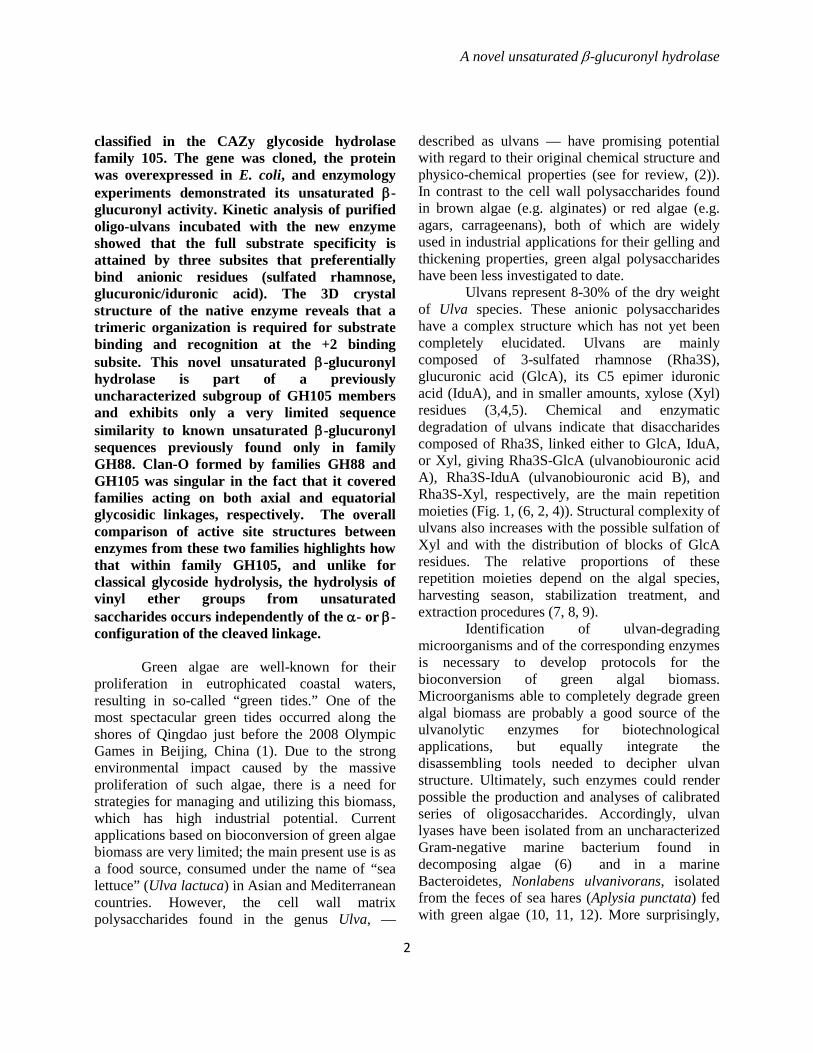
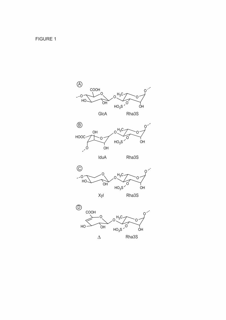
Ulvans represent 8-30% of the dry weight of Ulva species. These anionic polysaccharides have a complex structure which has not yet been completely elucidated. Ulvans are mainly composed of 3-sulfated rhamnose (Rha3S), glucuronic acid (GlcA), its C5 epimer iduronic acid (IduA), and in smaller amounts, xylose (Xyl) residues (3,4,5). Chemical and enzymatic degradation of ulvans indicate that disaccharides composed of Rha3S, linked either to GlcA, IduA, or Xyl, giving Rha3S-GlcA (ulvanobiouronic acid A), Rha3S-IduA (ulvanobiouronic acid B), and Rha3S-Xyl, respectively, are the main repetition moieties (Fig. 1, (6, 2, 4)). Structural complexity of ulvans also increases with the possible sulfation of Xyl and with the distribution of blocks of GlcA residues. The relative proportions of these repetition moieties depend on the algal species, harvesting season, stabilization treatment, and extraction procedures (7, 8, 9).
Identification of ulvan-degrading microorganisms and of the corresponding enzymes is necessary to develop protocols for the bioconversion of green algal biomass. Microorganisms able to completely degrade green algal biomass are probably a good source of the ulvanolytic enzymes for biotechnological applications, but equally integrate the disassembling tools needed to decipher ulvan structure. Ultimately, such enzymes could render possible the production and analyses of calibrated series of oligosaccharides. Accordingly, ulvan lyases have been isolated from an uncharacterized Gram-negative marine bacterium found in decomposing algae (6) and in a marine Bacteroidetes, Nonlabens ulvanivorans, isolated from the feces of sea hares (Aplysia punctata) fed with green algae (10, 11, 12). More surprisingly,
A novel unsaturated β-glucuronyl hydrolase
3
ulvan lyase activity has also been isolated from a Proteobacteria species, Ochrobactrum tritici, found in soil (13). In all cases, the main ulvan-degrading enzymes observed thus far are ulvan lyases that catalyze the cleavage of the glycosidic bond between a sulfated rhamnose and uronic residues through a β-elimination mechanism.
The products released by polysaccharide lyases are oligosaccharides with a degree of polymerization higher than two, terminated by an unsaturated uronyl residue at the non-reducing end (14, 15). To complete the degradation of these products, microbial genomes equally encode enzymes that can further cleave of the unsaturated sugar. For instance, the end-products of glycosaminoglycan lyase action are terminated by an unsaturated β-glucuronyl residue, which can be specifically digested by an unsaturated β-glucuronyl hydrolase (16, 17) found in glycoside hydrolase family 88 (GH88, CAZy database, http://www.cazy.org/, (18)). Similarly, rhamnogalacturonan lyase action yields oligosaccharides with unsaturated α-galacturonyl non-reducing ends, digested by unsaturated α-galacturonyl hydrolase (from family GH105) (19, 20). The new hydration mechanism of these enzymes was recently elegantly demonstrated by the use of synthesized substrate analogs probed by NMR, showing that the reaction directly catalyzed is the syn hydration of a vinyl ether to give an unstable hemiketal, and it is the collapse of this species that leads to glycosidic bond cleavage (21). Despite the fact that hydrolysis of vinyl ether groups in unsaturated saccharides occurs independently of the α- or β-configuration, to date all characterized members of either family proceed with conserved specificity with respect to the anomeric configuration of the glycosidic bond.
Our recent study describing the sequencing of the genomic environment around the ulvan lyase gene found in N. ulvanivorans (11) revealed a gene whose sequence is similar to uronyl hydrolases belonging to the GH105 family. The biochemical and structural characterization of this novel unsaturated uronyl hydrolase reported here, reveals a first GH105 member acting specifically on the
unsaturated end-products of ulvan lyase action cleaving an equatorial β-glycosidic bond. EXPERIMENTAL PROCEDURES Sequencing of the unsaturated β-glucuronyl hydrolase gene — The sequence of the unsaturated β- glycuronyl hydrolase gene was determined using the TAIL-PCR method (22) starting from the gene of ulvan lyase. The nested specific primers used were 5’CTATCCTTAAAAGCTGGCTCTGGAAAAGCACC3’ (TailGH105 1R); 5’GTACCCAACCTACACGA CCATCATCATGC3’ (TailGH105 2R); and 5’GTATGGAAAGCACTACCACTAACCTCACCG3’ (TailGH105 3R). Five different arbitrary degenerate primers (AD primers) were chosen from sequences found in the literature (22, 23): TGWGNAGWANCASAGA (AD1); AGWGNAGWA NCAWAGG (AD2); WGTGNAGWANCANAGA (AD3); NTCGASTWTSGWGTT (AD4); NGTCGAS WGANAWGAA (AD5). Primary TAIL reactions were performed in 20 µL containing 15 ng of genomic DNA, 1 µL GoTaq PCR buffer, 1.5 mM MgCl2, 0.2 mM each of deoxyribonucleotide triphosphate (dNTP), 0.2 µM of the first specific primer, one AD primer (5 µM AD1 and 2, 4 µM AD3, 2 µM AD4, or 3 µM AD5), and 1.25 units of GoTaq (Promega).
The conditions for the secondary TAIL reactions were identical to the first, except that 1 µL of a 1:50 dilution of the primary TAIL reaction was used as a template, and the second specific primer was used in combination with the same AD primer as used in the primary TAIL reaction. For the tertiary TAIL reaction, 1 µL of a 1:50 dilution of the secondary TAIL reaction was used as template, and the third specific primer was used. The PCR programs were different for the three TAIL reactions and were based on published programs (22), but adapted to the thermocyclers available in the laboratory (11). Heterologous expression and purification of the GH105 protein — Primers were designed to amplify the GH105 gene from N. ulvanivorans genomic DNA and to incorporate BamHI and EcoRI restriction sites into the 5’ and 3’ ends, respectively. The forward primer sequence was
A novel unsaturated β-glucuronyl hydrolase
4
GGGGGGGGATCCTGTACTGATACTGAAAAAACACCATTA and the reverse CCCCCCGAATTCTTAT CTCATTTTTAGAATTTCACTTCCAGC. Standard PCR conditions were used with an annealing temperature of 50 °C and 30 cycles with 10 ng N. ulvanivorans genomic DNA as template. The resulting PCR products were purified, digested with appropriate restriction enzymes, and subcloned into the pFO4 expression vector, modified from pET15 (Novagen) to be compatible with the BamHI/EcoRI ligation strategy and the production of his-tagged protein, possible to purify on a Ni2+ chelating column. Recombinant plasmids were used to transform Escherichia coli strain BL21 (DE3). Transformed colonies were grown for 3 h at 37°C in LB medium containing ampicillin and 0.5% glucose, where after an equivalent volume of cold LB medium containing 0.6% lactose, 20 mM Hepes pH 7.0, and 1 mM IPTG was added, and the culture was incubated 18 h at 20°C (24) for expression. After centrifugation, the bacterial pellet was resuspended in a buffer containing 20 mM Tris-HCl, 500 mM NaCl and 5 mM imidazole at a pH of 7.4. The cells were lysed using a French press followed by centrifugation to remove bacterial debris. The resulting supernatant was applied to a Nickel-Sepharose column charged with 100 mM NiSO4 (GE Healthcare). After washing, the bound proteins were eluted with a linear gradient of imidazole ranging from 5 mM to 500 mM. The active fractions were pooled and injected on a Superdex 75 HiLoad prepacked column (1.6 x 60 cm; GE Healthcare) equilibrated in 20 mM Tris-HCl pH 8.0 with 200 mM NaCl run at 1 mL per min. During purification, the active fractions were analyzed by SDS-PAGE. Protein quantification was performed on 2 µL aliquots using a NanoDrop spectrophotometer (Thermo Scientific) using the extinction coefficient 109 780 M-1 cm-1 calculated from the amino acid sequence. Analytical gel filtration was performed by injecting 200 µL on a Superdex 200 HiLoad prepacked column (1.0 x 30 cm; GE Healthcare) run at 0.5 mL per min with the same buffer as used for the preparative gel filtration.
The seleno-L-methionine (Se-Met) labeling procedure was performed by growing
recombinant E. coli BL21 (DE3) in 200 mL of PASM 5052 medium (25). The purification procedure was the same as the native enzyme, except that the final buffer contained 5 mM of tris(2-carboxyethyl)phosphin. The Se-Met labeled Nu_GH105 enzyme was concentrated to 10 mg/ml. Crystallization and crystal structure determination of the GH105 enzyme—Suitable crystallization conditions were first identified by screening 192 conditions (Quiagen JCSG+ and PACT commercial kits) using a nano-drop dispensing robot (Honeybee, Proteomics Solution) and corning 96 well crystallization plates. The best condition identified contained 25% Peg 3350 and 100 mM KNO3. After optimization of these conditions performing a grid-screen in 24-well Linbro plates, single crystals of native and Se-Met labeled Nu_GH105, were grown by mixing 2 µL of protein with 1 µL of reservoir solution, containing 100 mM Tris buffer at pH 8.5, 24 % (w/v) PEG MME 2000, 0.2 M potassium nitrate and 100 mM Tris buffer at pH 8.5, 22 % (w/v) PEG MME 3350, 0.2 M potassium nitrate, respectively. The hanging drops were equilibrated against 0.5 mL of reservoir solution. Structure determination and refinement—Prior to data collection, the crystals were rapidly soaked in a cryo-buffer that was identical to the reservoir solution supplemented with 10 % glycerol, and subsequently frozen in a nitrogen gas stream at 100 K. Native data were collected at the European Synchrotron Radiation Facility (ESRF, Grenoble, France) on beamline ID23-1 equipped with an ADSC Qunatum315 detector. The single anomalous diffraction data were collected on the beamline PROXIMA1 at the synchrotron SOLEIL (Saint-Aubin, France) at the K absorption edge of selenium peak. The beamline is equipped with a Pilatus detector. The software program package XDS (26)was used for all data reduction and scaling. Native and Se-Met labeled Nu_GH105 crystals belonged to the same orthorhombic space group P212121 with three molecules per asymmetric unit. All further data collection statistics are summarized in Table 1.
Phases were determined using the program PHASER (27) to find 34 selenium sites via the
A novel unsaturated β-glucuronyl hydrolase
5
graphical user interface CCP4i. Initial phases, with an overall figure of merit of 0.34 were improved by non crystallographic symmetry averaging and solvent flattening using the program PARROT (28), which provided an interpretable electron density map with an overall figure of merit of 0.872 after 15 cycles. Automatic model building with ARP/WARP (29) correctly built ~ 86 % of the three molecules in the asymmetric unit. This model was used for molecular replacement in the native dataset at 1.9 Å resolution. The missing parts of the model were traced manually using COOT (30). The construction cycles were alternated with positional refinement using REFMAC5 (31) including all data at 1.9 Å. Solvent molecules were added with COOT and alternative positions were added manually and refined with REFMAC5. Spectrophotometric enzyme assay—A mix of unsaturated oligosaccharides obtained by degradation of ulvan by ulvan lyase was used for the protein activity assay. Activity was characterized by a decrease in the absorbance at 235 nm. Incubations were performed directly in the spectrophotometer (U-2401, Shimadzu) equipped with a temperature-controlled cuvette holder (TCC-controller 240A, Shimadzu) set at 30°C. The standard reaction buffer was composed of 100 mM Tris-HCl pH 7.7, 100 mM NaCl and 100 µM ulvan oligosaccharides, the concentration calculated using the extinction coefficient of the double bond (4800 M-1 cm-1). A total of 4.4 nM of protein (5 µL) was added to a reaction volume of 500 µL in a quartz cuvette with a 1 cm light path, unless otherwise specified. The pH optimum was determined in the pH range 6.5 to 9 and the standard pH buffer was replaced by 100 mM MOPS (pH 6.5, 6.9, 7.2 and 7.7) or Tris-HCl (pH 7.2, 7.7, 8.15, 8.6 and 9.0). The temperature optimum was determined in the standard reaction buffer for temperatures between 20°C and 55°C in 5°C increments. The effect of salt was evaluated in the range of 0 to 500 mM NaCl. Purification of ulvan oligosaccharides to use as a substrate for the GH105 protein—Oligo-ulvans were produced using the ulvan lyase present in the culture supernatant of N. ulvanivorans. A
vial containing 100 mL ZoBell medium (5g trypton, 1g yeast extract in 1L 80% seawater) and 0.4% ulvan was inoculated with N. ulvanivorans and incubated at 20°C at 200 rpm during 48h before harvest by centrifugation (11). The supernatant was concentrated 8 times and buffered with 20 mM Tris HCl pH 8.0, 100 mM NaCl by diafiltration on a 10 kDa cutoff membrane using an 50 mL filtration cell (Amicon). This enzyme preparation (2 mL) was used to degrade 1 g of ulvan (CEVA, Pleubian, France) in 30 mL of 200 mM NaCl, 20 mM Tris HCl pH 9.3 at 30°C for 24 h. The resistant fraction was removed by filtration on a 5000 Da cutoff membrane using filtration cell (Amicon). The oligosaccharide mixture was filtered (0.2 µm) and injected on three Pharmacia Superdex 30 columns (2.6 × 60 cm; GE Healthcare) in series. The elution was conducted in 50 mM (NH4)2CO3 at 20°C using an isocratic Gilson 306 pump working at a flow rate of 1.5 mL min-1. Oligosaccharides were detected by differential refractometry (Spectra System RI-50, Thermo Separation products) and fractions were collected using a Gilson 215 liquid handler system between 450 and 900 mL. High-performance anion-exchange chromatography (HPAEC)—The purity of the oligosaccharide fractions and the degradation kinetics of pure oligosaccharides was analyzed by HPAEC on a Dionex chromatograph ICS 3000 equipped with a 20 µL injection loop, an AS100XR automated injection system (Thermo Separation Products) and an AS11 anion exchange column (4 mm × 250 mm, Dionex IonPac) with an AG11 precolumn (4 mm × 50 mm, Dionex IonPac). The system was operated in conductivity mode using an ED40 detector (Dionex) and a Dionex ASRS ultra-4mm surpressor with a current of 300 mA. Mobile phases were ultra-purified water and 290 mM NaOH. Elution was conducted at a flow rate of 0.5 mL min-1 with a GP40 gradient pump. The gradient used was 0 min, 3% B; 1.5 min 1% B; 4.1 min 5% B; 6.5min 10% B; 10.0 min 18% B; 26 min 22% B; 28 min 40% B; 30 min 100% B; 30.1 min 3% B; 37 min 3% B. Separation and elution of the oligosaccharides occur during the first 30 min of the gradient and is followed by a
A novel unsaturated β-glucuronyl hydrolase
6
wash step and re-equilibration of the column for the next injection. The Chromeleon-peak Net software (Dionex) was used for data acquisition and transfer. Mass spectrometry—Protein molecular mass was determined with a Voyager DE-STR MALDI-TOF mass spectrometer (Applied Biosystems). Protein solutions (1 µL) were diluted (1/1; 1/10 and 1/100) with sinapinic acid matrix (Sigma; 10 mg mL-1 in 30% Acetonitrile, 0.1% Trifluoroacetic acid) and spotted onto the MALDI target. Spectra were acquired in positive ion linear mode under 25 kV accelerating voltage and a mass range of 5–100 kDa. External calibration was performed using bovine serum albumin single- and double-charged ions at m/z 66,434 and 33,216 Da. 1H NMR Spectroscopy—1H NMR spectra were recorded at 298 K on a Bruker Avance 500 spectrometer equipped with an inverse 5 mm 1H/13C/15N TCI cryoprobe. Prior to analysis, samples were exchanged twice in D2O and redissolved in 99.97 atom% D2O. Chemical shifts are expressed in ppm in reference to an external standard (trimethylsilylpropionic acid). No suppression of the HOD signal was performed. Hierarchical classification of sequences—A total of 482 sequences with defined full-length catalytic modules of family GH105 were extracted from CAZy (http://www.cazy.org/, (17)) on November 2013. Related sequences were grouped using a threshold of 80% sequence identity using cd-hit (http://weizhong-lab.ucsd.edu/cd-hit/). To insure that all sequences corresponding to structural or biochemical characterizations were present a few sequences were added manually to form a set of 251 representative sequences. These sequences were aligned with Muscle 3.7 (http://www.drive5.com/muscle/) and a distance matrix was built from the multiple sequence alignment using the maximum likelihood (ML) model and the JTT substitution model (32) using an in-house modified version of Jalview (http://www.jalview.org/). Potential subfamilies were estimated following application of the Ward hierarchical clustering method (33) to the original distance matrix . The resulting tree was analyzed
under Dendroscope 3.2.9 (http://ab.inf.uni-tuebingen.de/software/dendroscope/). RESULTS The gene for a divergent GH105 glycoside hydrolase—An ulvan lyase gene from N. ulvanivorans has previously been sequenced and the heterologous-expression of the corresponding protein demonstrated that it was the first representative of a novel polysaccharide lyase family (11). Here, we determined the genome sequence flanking the ulvan lyase gene using TAIL-PCR and revealed the presence of two other genes in the same reading frame. Downstream on the 3’ side, the sequence corresponds to a protein of about 70 kDa with unpredicted function. This protein was successfully overexpressed; however, we did not observe any ulvan or oligo-ulvan degradation activity (not shown). Upstream of the ulvan lyase gene, there is a gene sequence of 1130 bp (accession number JQ403607), which translated into a protein of 377 amino acids with a theoretical molecular mass of 43.75 kDa. The LipoP program predicted a clear signal peptide of 16 amino acids with cleavage between a serine in position 16 and a cysteine in position 17 suggesting that it is a lipoprotein potentially anchored on the cell-wall.
Similarity searches were performed using BLASTP (34), against all non-redundant protein databases and the 337 amino-acid protein showed similarity to sequences classified in the glycoside hydrolase family GH105 found in CAZy. The highest similarities were observed with the 347 amino acid sequence of a Formosae agariphila KMM 3901 protein (identity 76%, accession number CDF79934) and with the 344 amino acid sequence of an Echinicola pacifica protein (identity 68%, accession number WP_018473370), respectively, but these proteins have not yet been biochemically characterized. On the contrary, the two characterized proteins from the GH105 family, the unsaturated rhamnogalacturonyl hydrolases YteR and YesR (Bacillus subtilis subsp. subtilis str. 168, accession numbers, CAB14990 and CAB12519), share only 28-29% identity (over 312-335 aa) with the N. ulvanivorans sequence. These enzymes have an α-galacturonyl hydrolase
A novel unsaturated β-glucuronyl hydrolase
7
activity, catalyzing the removal of the unsaturated uronic residue located at the non-reducing end of rhamno-galacturonan oligosaccharides produced by galacturonan lyases.
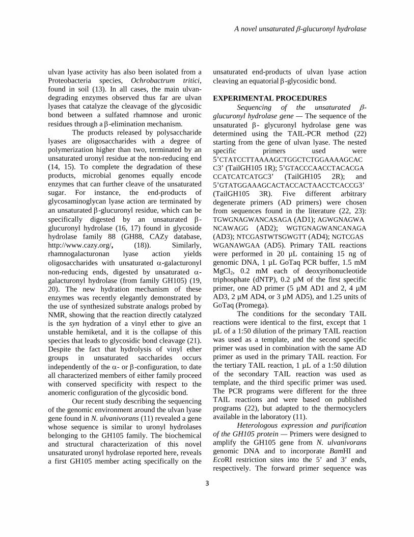
An alignment of the N. ulvanivorans sequence with the characterized GH105 sequences and those for which a 3D structure is available is shown in Fig. 2. The catalytic acid/base of family GH105 enzymes that directly protonates the ring-double bond has previously been identified to be D143 (numbering of YteR; D148 in Nu_GH105 (also described as Nu_AFQ98272-4CE7) by Itoh and co-workers (20), using site directed mutagenesis. Biochemical characterization of the novel β-glucuronyl hydrolase—The N. ulvanivorans GH105 gene fused with an N-terminal histidine tag was successfully overexpressed in E. coli BL21without its signal peptide. Expression levels were of about 50 mg of protein per liter of medium using standard culture and induction conditions. The protein was purified on a nickel Sepharose chelating resin and eluted at 375 mM immidazole. The active fractions were pooled and analyzed using SDS-PAGE and MALDI-TOF MS. The most intense band migrated as expected at 40 kDa which was measured at 43.7 kDa by mass spectrometry. However, this intense band was always associated with a second band of about 80 kDa which increased in intensity if the protein mixed with SDS-sample buffer was not boiled (data not shown), suggesting incomplete denaturation of the protein. The active fractions eluted as a single peak in permeation gel chromatography on a Superdex 75 HiLoad prepacked column (1.6 x 60 cm; GE Healthcare). The active fractions were pooled and used for crystallography and biochemical analysis. Analytical gel filtration of the active fractions on a superdex 200 (1.0 x30 cm; GE Healthcare) separated two peaks eluting at 11.2 and 13.1 mL. Calibration of the column with known protein standards allowed determination of the peak at 13.1 mL to 128.8 kDa, which corresponds well with the expected size of a trimer. The peak at 11.2 mL is outside the range of the calibration i.e. larger than 200 kDa. From the genomic environment and the putative unsaturated uronyl hydrolase activity
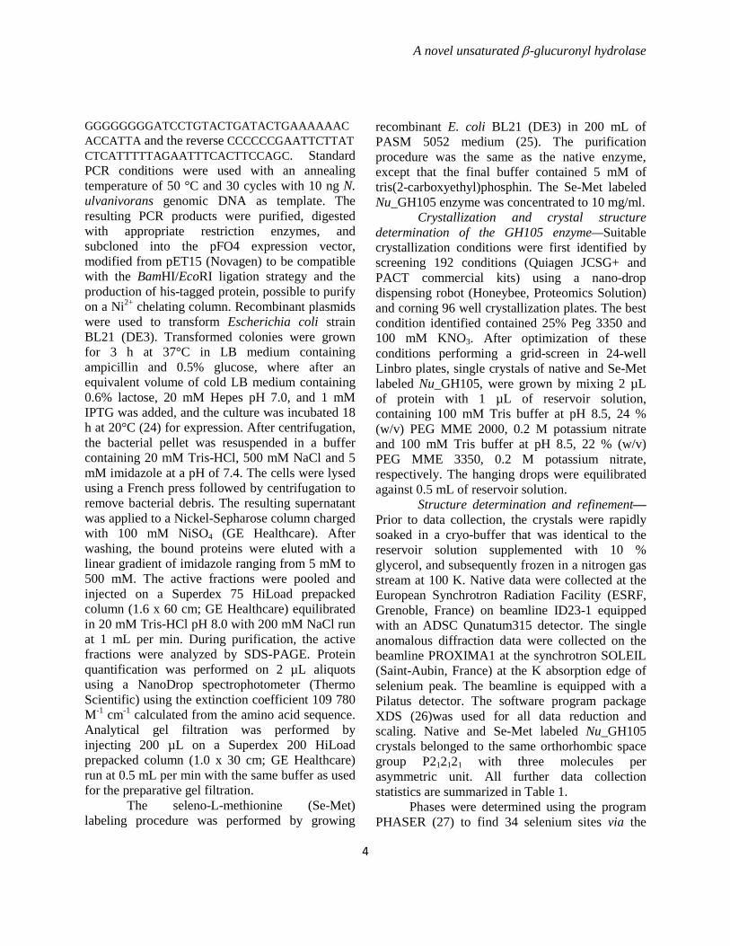
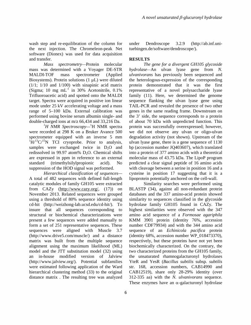
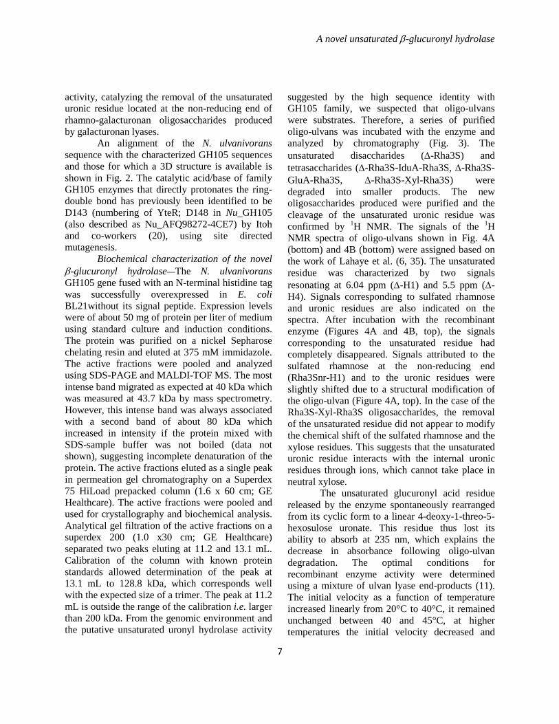
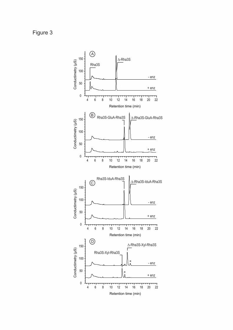
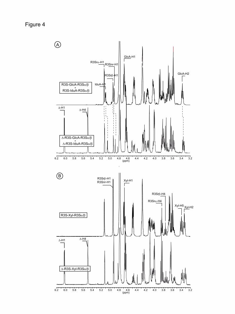
suggested by the high sequence identity with GH105 family, we suspected that oligo-ulvans were substrates. Therefore, a series of purified oligo-ulvans was incubated with the enzyme and analyzed by chromatography (Fig. 3). The unsaturated disaccharides (∆-Rha3S) and tetrasaccharides (∆-Rha3S-IduA-Rha3S, ∆-Rha3S-GluA-Rha3S, ∆-Rha3S-Xyl-Rha3S) were degraded into smaller products. The new oligosaccharides produced were purified and the cleavage of the unsaturated uronic residue was confirmed by 1H NMR. The signals of the 1H NMR spectra of oligo-ulvans shown in Fig. 4A (bottom) and 4B (bottom) were assigned based on the work of Lahaye et al. (6, 35). The unsaturated residue was characterized by two signals resonating at 6.04 ppm (∆-H1) and 5.5 ppm (∆-H4). Signals corresponding to sulfated rhamnose and uronic residues are also indicated on the spectra. After incubation with the recombinant enzyme (Figures 4A and 4B, top), the signals corresponding to the unsaturated residue had completely disappeared. Signals attributed to the sulfated rhamnose at the non-reducing end (Rha3Snr-H1) and to the uronic residues were slightly shifted due to a structural modification of the oligo-ulvan (Figure 4A, top). In the case of the Rha3S-Xyl-Rha3S oligosaccharides, the removal of the unsaturated residue did not appear to modify the chemical shift of the sulfated rhamnose and the xylose residues. This suggests that the unsaturated uronic residue interacts with the internal uronic residues through ions, which cannot take place in neutral xylose.
The unsaturated glucuronyl acid residue released by the enzyme spontaneously rearranged from its cyclic form to a linear 4-deoxy-1-threo-5-hexosulose uronate. This residue thus lost its ability to absorb at 235 nm, which explains the decrease in absorbance following oligo-ulvan degradation. The optimal conditions for recombinant enzyme activity were determined using a mixture of ulvan lyase end-products (11). The initial velocity as a function of temperature increased linearly from 20°C to 40°C, it remained unchanged between 40 and 45°C, at higher temperatures the initial velocity decreased and
A novel unsaturated β-glucuronyl hydrolase
8
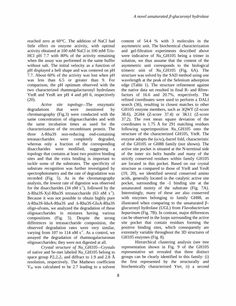
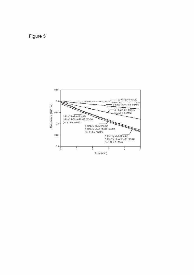
reached zero at 60°C. The addition of NaCl had little effect on enzyme activity, with optimal activity obtained at 100 mM NaCl in 100 mM Tris-HCl pH 7.7 with 80% of the activity remaining when the assay was performed in the same buffer without salt. The initial velocity as a function of pH displayed a bell shape and was centered on pH 7.7. About 60% of the activity was lost when pH was less than 6.5 or greater than 9. For comparison, the pH optimum observed with the two characterized rhamnogalacturonyl hydrolases YteR and YesR are pH 4 and pH 6, respectively (20). Active site topology—The enzymatic degradations that were monitored by chromatography (Fig.3) were conducted with the same concentration of oligosaccharides and with the same incubation times as used for the characterization of the recombinant protein. The three ∆-Rha3S non-reducing end-containing tetrasaccharides were completely degraded, whereas only a fraction of the corresponding disaccharides were modified, suggesting a topology that contains at least two positive binding sites and that the extra binding is important to tackle some of the substrates. The specificity of substrate recognition was further investigated by spectrophotometry and the rate of degradation was recorded (Fig. 5). As in the chromatography analysis, the lowest rate of digestion was observed for the disaccharides (34 nM s-1), followed by the ∆-Rha3S-Xyl-Rha3S tetrasaccharide (63 nM s-1). Because it was not possible to obtain highly pure ∆-Rha3S-IduA-Rha3S and ∆-Rha3S-GluA-Rha3S oligo-ulvans, we analyzed the degradation of these oligosaccharides in mixtures having various compositions (Fig. 5). Despite the strong differences in tetrasaccharide composition, the observed degradation rates were very similar, varying from 107 to 114 nM s-1. As a control, we assayed the degradation of rhamnogalacturonan oligosaccharides; they were not digested at all. Crystal structure of Nu_GH105—Crystals of native and Se-met labeled Nu_GH105 belong to space group P212121 and diffract to 1.9 and 2.8 Å resolution, respectively. The Mathews coefficient VM was calculated to be 2.7 leading to a solvent
content of 54.4 % with 3 molecules in the asymmetric unit. The biochemical characterization and gel-filtration experiments described above were indicative of Nu_GH105 being a trimer in solution, we thus assume that the content of the asymmetric unit corresponds to the biological trimeric unit of Nu_GH105 (Fig. 6A). The structure was solved by the SAD method using one wavelength at the peak of the Selenium adsorption edge (Table 1). The structure refinement against the native data set resulted in final R- and Rfree-factors of 16.6 and 20.7%, respectively. The refined coordinates were used to perform a DALI search (36), resulting in closest matches to other GH105 enzyme members, such as 3QWT (Z-score 38.6), 2GH4 (Z-score 37.4) or 3K11 (Z-score 37.2). The root mean square deviation of the coordinates is 1.75 Å for 291 matching residues following superimposition Nu_GH105 onto the structure of the characterized GH105, YteR. The enzyme adopts the (α/α)6-barrel fold, characteristic of the GH105 or GH88 family (not shown). The active site pocket is situated at the N-terminal side of the inner six helix bundle and many of the strictly conserved residues within family GH105 are located in this pocket. Based on our crystal structure as compared to those of YteR and YesR (19, 20), we identified several conserved amino acids, generally located in the catalytic active site pocket, surrounding the -1 binding site at the unsaturated moiety of the substrate (Fig. 7A). Interestingly, many of these are also conserved with enzymes belonging to family GH88, as illustrated when comparing to the unsaturated β-glucuronyl hydrolase (UGL) from Flavobacterium heparinum (Fig. 7B). In contrast, major differences can be observed in the loops surrounding the active site pocket that contain residues forming the positive binding sites, which consequently are extremely variable throughout the 3D structures of GH105 enzymes (Fig. 8).
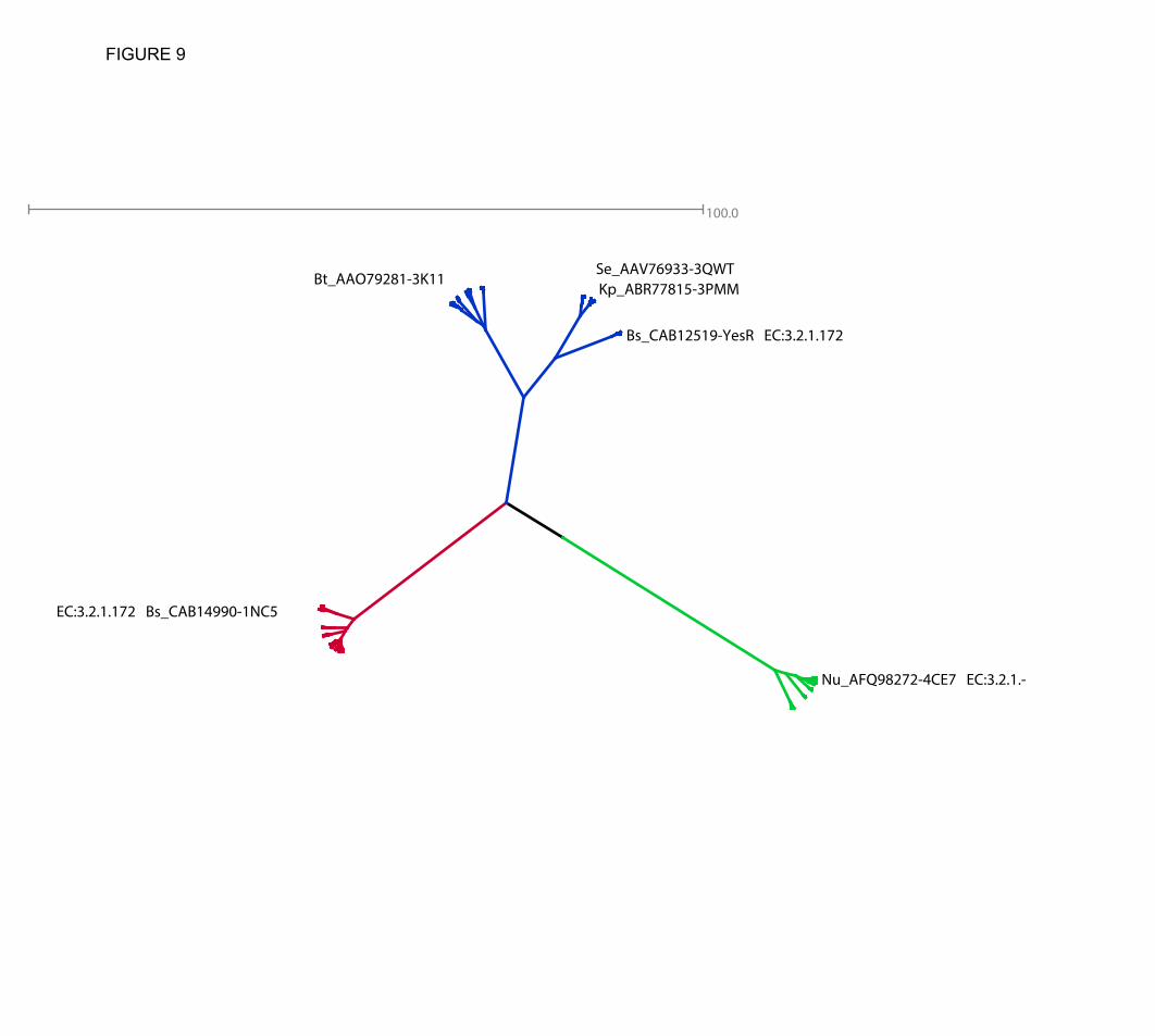
Hierarchical clustering analysis (see tree representation shown in Fig. 9 of the GH105 representative set revealed that three distinct groups can be clearly identified in this family: (i) the first represented by the structurally and biochemically characterized Yter, ii) a second
A novel unsaturated β-glucuronyl hydrolase
9
represented by the equally characterized YesR, but also with three other structural representatives without known activities, and, significantly, iii) a third set represented by the newly characterized protein Nu_GH105 (see below).
DISCUSSION We have identified and overexpressed a gene with similarity to the GH 105 family from the marine bacterium N. ulvanivorans. In agreement with its genetic location next to the recently described ulvan lyase this novel glycosyl hydrolase is capable of degrading the unsaturated oligosaccharides produced by the ulvan lyase. The degradation kinetics suggests that the active site preferentially accommodates tetrasaccharides rather than disaccharides, which were more slowly digested. Furthermore, the presence of an uronic residue at subsite +2 was more readily accommodated than a xylose residue. Subsites -1 and +1 accommodate the unsaturated glucuronyl residue and the sulfated rhamnose, respectively, while the third subsite (subsite +2) can accommodate the xylose residue, but iduronic or glucuronic residues are preferred, suggesting that a specific interaction with a negatively charged carboxyl group plays a central role in the interaction. Interestingly, the enzyme was not very sensitive to the stereochemistry of the uronic units within the chain; iduronic and glucuronic units located between the two sulfated rhamnose residues were recognized equally well. Based on these kinetic experiments, we assumed that the active site must contain at least three subsites. Regarding the crystal structure of a monomeric molecule of Nu_GH105, intriguingly only two binding sites can be defined within the active site pocket, namely sub-binding sites -1 and +1. Any additional sugar unit would stick out into the solvent region, outside the range of possible contacts to enzyme residues. However, when considering the trimeric arrangement of Nu_GH105, contacts with a neighboring symmetric molecule can stabilize additional sugar units, as illustrated in Fig. 6B, showing the position of a modeled substrate molecule
containing a putative +2 unit in proximity of R106 from a neighboring molecule. This non-conserved arginine residue of the symmetric Nu_GH105 molecule is ideally positioned to form a tight salt bridge with charged iduronic or glucuronic units, in agreement with the preference of charged units at the +2 binding site in the biochemical experiments. This feature appears to be unique to Nu_GH105 (Fig. 2), pointing towards possible substrate diversity of GH105 enzymes.
Sequence similarity of the novel β-glucuronyl hydrolase clearly places this sequence to family GH105. However, upon alignment the sequence identities with the previously characterized members of GH105 range between 24% and 25 %, reaching at most 76% with the most similar sequences of to date biochemically uncharacterized enzymes. Significantly, the sequence identity with the characterized members of GH88 ranges from 17% to 21% (over 327-362 aligned amino-acid residues). Moreover, both chromatography and NMR demonstrated that the N. ulvanivorans enzyme is an unsaturated β-glucuronyl hydrolase able to remove the non-reducing unsaturated β-linked end ∆-Rha3S of ∆-Rha3S-IduA-Rha3S, ∆-Rha3S-GluA-Rha3S, and ∆-Rha3S-Xyl-Rha3S into a new series of fully saturated oligo-ulvans. Previously known unsaturated β-glucuronyl hydrolases were found only in glycoside hydrolase GH88 family, known to hydrolyse the unsaturated disaccharides produced by lyases degrading glycosaminoglycans, such as hyaluronan and chondroitin (16) Glycosaminoglycans are linear molecules with a repeating disaccharide unit composed of an uronic acid residue linked to an amino sugar residue (glucosamine or galactosamine), which can be non-sulfated as in hyaluronan or sulfated at positions 4 and/or 6. The most striking feature of the inclusion of Nu_GH105 in the GH105 family, is that the presently characterized enzymes of this family are known to cleave unsaturated α-anomeric galacturonyl (syn: glucuronyl) residues instead of β-anomeric uronyl residues. This constitutes the first instance of a family cleaving both equatorially and axially linked glycosidic bonds, that needs to be explained based on the
A novel unsaturated β-glucuronyl hydrolase
10
singularity of the mechanism and structural information.
Both families GH105 and GH88 form clan GH-O as both structure and catalytic residues are clearly shared between these families (Fig. 8). In fact, structures from both families present an (α/α)6 barrel fold, sharing a significant number of the amino acids next to subsite -1, including the putative catalytic aspartate amino acid, a condition that suggests that the catalytic mechanism is also conserved (16, 17, 19, 20). In both families, the cleavage of the glycosidic linkage does not proceed according to classical hydrolysis mechanisms usually encountered in other glycoside hydrolases. It has been shown that cleavage starts with the addition of a proton on the carbon 4 of the unsaturated uronyl residue, which undergoes molecular rearrangements that ultimately lead to the cleavage of the glycosidic bond (37, 21). Our hypothesis is that with this mechanism, the α- or β-anomeric configurations of the unsaturated uronyl residue impose fewer constraints to the catalytic machinery and vice-versa, than in the case of classical retaining or inverting hydrolysis mechanisms. This is in agreement with the mechanistic study of a GH88 unsaturated β-glucuronyl hydrolase by Jongkees and Withers (21), who showed that the alternate anomer of a phenyl glycoside, having stereochemistry opposite to that of the natural substrate at carbon 1, was also accepted by UGL as a substrate. It is therefore possible that the cleavage of the α- and β-anomers can both be observed within the same glycoside hydrolase family, as far as it does not play a critical role for the enzymatic mechanism. This was firstly demonstrated by Jongkees and Withers (21) using a synthetic substrate, while our characterized enzyme represents the first naturally occurring example of glycosidic bonds with opposite stereochemistry being cleaved by enzymes from the same family.
The chemical structure of the rhamnogalacturonan and sulfated-rhamnoglucuronan (oligo-ulvans) oligosaccharides are sufficiently similar to explain the high protein sequence identity of the corresponding uronyl
degrading enzymes. The divergence observed between GH88 and GH105 protein sequences may be associated with subtle mechanistic variations in addition to different evolutionary pathways, but is more likely caused by the structural and conformational differences of the substrates at the positive binding sites that are possible due to fewer constraints on the stereochemistry of the cleaved bond. This is illustrated by the sequence alignments (Fig. 2) in the light of the structural superimposition of all known GH105 enzymes (Fig. 8) showing that extreme variability is observed in the loops and residues forming the positive binding sites, shaping for a large diversity of substrates even within GH105 subfamilies. We therefore predict that a much larger diversity of unsaturated glycuronyl-substrates than expected will be cleaved by members of this clan. Initial analysis of the tree resulting from hierarchical clustering suggests that the subgroups or subfamilies represented by the structurally and biochemically characterized Yter and by Nu_GH105 are more homogeneous when compared to the subgroup represented by the biochemically characterized YesR and the other remaining uncharacterized structures. The latter subgroup could therefore have an enlarged set of substrates or of different actions on these. Following this though, the subfamily represented by Nu_GH105 may only recognize very short segments of the substrate before cleavage, as suggested from the structure. In addition, it must single out that closely related sequences identified in the tree are from Akkermansia muciniphila and Pedobacter heparinus, known to be able to use mucin and heparin as carbon source, respectively (38,39). This supports the hypothesis that very diverse substrates may be found in the corresponding subfamily.
At the moment only two other protein-coding sequences, out of the over 500 sequences classified, in this family have been biochemically characterized and the situation is very similar for the GH88 family with six out of 306 sequences characterized. Knowledge about the substrates of additional members of the GH88 and GH105 families will be helpful to confirm the large

A novel unsaturated β-glucuronyl hydrolase
11
substrate diversity, as well as the co-existence of α- and β-cleaving enzymes within the same family or even subfamily.
Furthermore, the description of this novel glycosyl hydrolase from N. ulvanivorans together with the recently characterized ulvan lyase leads us one step further in elucidating the degradation pathway of ulvan. These enzymatic tools can therefore be used to produce novel oligosaccharides or to facilitate the methanolisation of green algal biomass.
A novel unsaturated β-glucuronyl hydrolase
12
REFERENCES
1. Gao, S., Chen, X., Yi, Q., Wang, G., Pan, G., Lin, A., and Peng, G. (2010) A Strategy for the proliferation of Ulva prolifera, main causative species of green tides, with formation of sporangia by fragmentation. Plos One 5, 1-7.
2. Lahaye, M., and Robic, A. (2007) Structure and functional properties of ulvan, a polysaccharide from green seaweeds. Biomacromolecules 8, 1765-1774.
3. Percival, E., and McDowell, R. H. (1967) Chemistry and enzymology of marine algal polysaccharides. Academic Press Inc. (London) Ltd.
4. Quemener, B., Lahaye, M., and Bobin-Dubigeon, C. (1997) Sugar determination in ulvans by a chemical-enzymatic method coupled to high performance anion exchange chromatography. J. Appl. Phycol. 9, 179-188.
5. Ray, B., and Lahaye, M. (1995) Cell wall polysaccharides from the marine green alga Ulva “rigida” (Ulvales, Chlorophyta). Extraction and chemical composition). Carbohydr. Res. 274, 313-318.
6. Lahaye, M., Brunel, M., and Bonin, E. (1997) Fine chemical structure analysis of oligosaccharides produced by an ulvan-lyase degradation of the water-soluble cell-wall polysaccharides from Ulva sp. (Ulvales, Chlorophyta). Carbohydr. Res. 304, 325-333.
7. Robic, A., Rondeau-Mouro, C., Sassi, J. F., Lerat, Y., and Lahaye, M. (2009) Structure and interactions of ulvan in the cell wall of the marine green algae Ulva rotundata (Ulvales, Chlorophyta). Carbohydr. Polym. 77, 206–216.
8. Robic, A., Sassi J. F., Dion, P., Lerat, Y., and Lahaye, M. (2009) Seasonal variability of physicochemical and rheological properties of ulvan in two Ulva species from the Brittany coast. J. Phycol. 45, 962–973.
9. Robic, A., Sassi, J-F., and Lahaye, M. (2008) Impact of stabilization treatments of the green seaweed Ulva rotundata (Chlorophyta) on the extraction yield, the physic-chemical and rheological properties of ulvan. Carbohydr. Polym. 74, 344–352.
10. Barbeyron, T., Lerat, Y., Sassi, J-F., Le Panse, S., Helbert, W., and Nyvall Collén, P. (2011) Persicivirga ulvanivorans sp. nov., a marine member of the family Flavobacteriaceae that degrades ulvan from green algae. Int. J. Syst. Evol. Microbiol. 61, 1899–1905.
11. Nyvall Collén, P., Sassi, J-F., Rogniaux, H., Marfaing, H., and Helbert, W. (2011) Ulvan lyases isolated from the Flavobacteria Persicivirga ulvanivorans are the first members of a new polysaccharide lyase family. J. Biol. Chem. 286, 42063-42071.
12. Yi, H., and Chun, J. (2012) Unification of the genera Nonlabens, Persicivirga, Sandarakinotalea and Stenothermobacter into a single emended genus, Nonlabens, and description of Nonlabens agnitus sp. nov. Syst. Appl. Microbiol. 35, 150– 155.
13. Boutachfaiti, R., Pheulpin, P., Courtois, B., and Courtois-Sambourg, J. (2009) Method for enzyme cleavage of polysaccharides derived from algae. WO 2009/016275 A2.
14. Garron, M.-L., and Cygler, M. (2010) Structural and mechanistic classification of uronic acid-containing polysaccharide lyases. Glycobiology 20, 1547-1573.
15. Yip, V. L.Y., and Withers, S. G. (2006) Breakdown of oligosaccharides by the process of elimination. Curr. Op.Chem. Biol. 10, 147–155.
16. Itoh, T., Akao, S., Hashimoto, W., Mikami, B., and Murata, K. (2004) Crystal structure of unsaturated glucuronyl hydrolase, responsible for the degradation of glycosaminoglycan, from Bacillus sp. GL1 at 1.8 angstrom resolution. J. Biol. Chem. 279, 31804–31812.
A novel unsaturated β-glucuronyl hydrolase
13
17. Maruyama, Y., Nakamichi, Y., Itoh, T., Mikami, B., Hashimoto, W., and Murata, K. (2009) Substrate specificity of streptococcal unsaturated glucuronyl hydrolases for sulfated glycosaminoglycan. J. Biol. Chem. 284, 18059-18069.
18. Cantarel B.L., Coutinho, P.M., Rancurel, C., Bernard, T., Lombard, V., and Henrissat, B. (2009) The Carbohydrate-Active EnZymes database (CAZy): an expert resource for Glycogenomics. Nucleic Acids Res. 37, D233-D238.
19. Itoh, T., Ochiai, A., Mikami, B., Hashimoto, W., and Murata, K. (2006) Structure of unsaturated rhamnogalacturonyl hydrolase complexed with substrate. Biochem. Biophys. Res. Com. 347, 1021-1029.
20. Itoh, T., Ochiai, A., Mikami, B., Hashimoto, W., and Murata, K. (2006) A novel glycoside hydrolase family 105: the structure of family 105 unsaturated rhamnogalacturonyl hydrolase complexed with a disaccharide in comparison with family 88 enzyme complexed with the disaccharide. J. Mol. Biol. 360, 573-585.
21. Jongkees, S. A. K., and Withers, S. G. (2011) Glycoside cleavage by a new mechanism in unsaturated glucuronyl hydrolases. J. Am. Chem. Soc. 133, 19334-19337.
22. Liu, Y. G., and Whittier, R. F. (1995) Thermal Asymmetric Interlaced PCR: Automatable amplification and sequencing of insert end fragments from PI and YAC Clones for chromosome walking. Genomics 25, 674-681.
23. Liu, Y. G., Mitsukawa, N., Oosumi, T., and Whittier, R. F. (1995) Efficient isolation and mapping of Arabidopsis thaliana T-DNA insert junctions by thermal asymmetric interlaced PCR. Plant J. 8, 457-463.
24. Korf, U., Kohl, T., vand der Zandt, H., Zahn, R., Schleeger, S., Ueberle, B., Wandschneider, S., Bechtel, S., Schöler, M., Ottleben, H., Wiemann, S., and Poutska, A. (2005) Large scale protein expression for proteome research. Proteomics 5, 3571-3580.
25. Studier, F.W. (2005) Protein production by auto-induction in high-density shaking cultures Protein Expr. Purif. 41, 207–34.
26. Kabsch, W. (2010) Xds. Acta Crystallogr D 66: 125-132. 27. McCoy, A. J., Grosse-Kunstleve, R. W., Adams, P. D., Winn, M. D., Storoni, L.C., and Read, R.J
(2007) Phaser crystallographic software. J. Appl. Cryst. 40, 658-674. 28. Zhang K. Y. J., Cowtan, K., Main, P. (1997) Combining constraints for electron-density
modification. Methods in Enzymology 277, 53-64. 29. Perrakis, A., Sixma, T.K., Wilson, K.S., and Lamzin, V.S. (1997) wARP: improvement and
extension of crystallographic phases by weighted averaging of multiple-refined dummy atomic models. Acta Cryst. D53, 448-455.
30. Emsley, P., and Cowtan, K. (2004) Coot: model-building tools for molecular graphics. Acta Cryst. D60, 2126-2132.
31. Murshudov, G.N., Vagin, A.A., and Dodson, E.J. (1997) Refinement of macromolecular structures by the maximum-likelihood method. Acta Cryst. D53, 240-255.
32. Jones, D.T., Taylor ,W.R., and Thornton, J.M. (1992) The rapid generation of mutation data matrices from protein sequences. Comput. Appl. Biosci. 8, 275-282.
33. Ward, J. H., Jr. (1963) Hierarchical Grouping to Optimize an Objective Function. J. Amer. Statist. Assoc. 58, 236–244.
34. Johnson, M., Zaretskaya, I., Raytselis, Y., Merezhuk, Y., McGinnis, S., and Madden, T.L. (2008) NCBI BLAST: a better web interface. Nucleic Acids Res. 36, W5-W9.
35. Lahaye, M., Inizan F., and Vigouroux, J. (1998) NMR analysis of the chemical structure of ulvan and of ulvan-boron complex formation. Carbohydr. Polym. 36, 239-249.
A novel unsaturated β-glucuronyl hydrolase
14
36. Holm, L., and Rosenström, P. (2010) Dali server: conservation mapping in 3D. Nucl. Acids Res. 38, W545-549.
37. Hashimoto, W., Itoh, T., Maruyama, Y., Mikami, B., and Murata, K. (2007) Hydration of vinyl ether groups by unsaturated glycoside hydrolases and their role in bacterial pathogenesis. Int. Microbiol. 10, 233-243.
38. Derrien, M., Vaughan E.E., Plugge, C.M., and de Vos, W.M. (2004) Akkermansia muciniphila gen. nov., sp. nov., a human intestinal mucin-degrading bacterium. Int. J. Syst. Evol. Microbiol. 54:1469-76.
39. Han, C., Spring, S., Lapidus, A., Del Rio, T.G., Tice, H., Copeland, A., Cheng, J.F., Lucas, S., Chen, F., Nolan, M., Bruce, D., Goodwin, L., Pitluck, S., Ivanova, N., Mavromatis, K., Mikhailova, N., Pati, A., Chen, A., Palaniappan, K., Land, M., Hauser, L., Chang, Y.J., Jeffries, C.C., Saunders, E., Chertkov, O., Brettin, T., Göker, M., Rohde, M., Bristow, J., Eisen, J.A., Markowitz, V., Hugenholtz, P., Kyrpides, N.C., Klenk, H.P., Detter, J.C. (2009) Complete genome sequence of Pedobacter heparinus type strain (HIM 762-3). Stand. Genomic Sci. 1:54-62.
40. Gouet, P., Robert, X., and Courcelle, E. (2003) ESPript/ENDscript: Extracting and rendering sequence and 3D information from atomic structures of proteins. Nucleic Acids Res. 31, 3320-3323.
Acknowledgements—We are grateful to the Biogenouest core facility for sequencing and for technical support, Stephane Cerantola at the Service Commun de Résonance Magnétique Nucléaire, Université de Bretagne Occidentale, Brest for NMR analyses, and Fanny Gaillard for mass spectrometry analyses at the Centre de Ressources Biologiques Marines, Station Biologique de Roscoff. We thank Soleil (St Auban, France) and the European Synchrotron Radiation Facilities (ESRF, Grenoble, France) for according regular beamtime and we are indebted to the staff on beamline ID23-I, and Pierre Legrand on Proxima 1 for technical support during data collection and treatment. This work was supported in part by the French National Research Agency (ANR; Recherche et Innovation en Biotechnologie, grant no. ANR-07-RIB-019 for the Ulvoligo Project), the Pôle Mer Bretagne competitiveness cluster, the Centre National de la Recherche Scientifique (CNRS) and the Brittany Regional Council. Footnote— The atomic coordinates and structure factors (code 4CE7) have been deposited in the Protein Data Bank, Research Collaboratory for Structural Bioinformatics, Rutgers University, New Brunswick, NJ (http://www.rcsb.org/). ABBREVIATIONS Rha3S, 3-sulfated rhamnose; GlcA , glucuronic acid; IduA, iduronic acid; Xyl, xylose; GH, glycolsyl hydrolase; TAIL-PCR, thermal asymmetric interlaced PCR; AD primers, arbitrary degenerate primers; dNTP, deoxyribonucleotide triphosphate; HPAEC, High-performance anion-exchange chromato-graphy; PAGE, polyacrylamide gel electrophoresis.
A novel unsaturated β-glucuronyl hydrolase
15
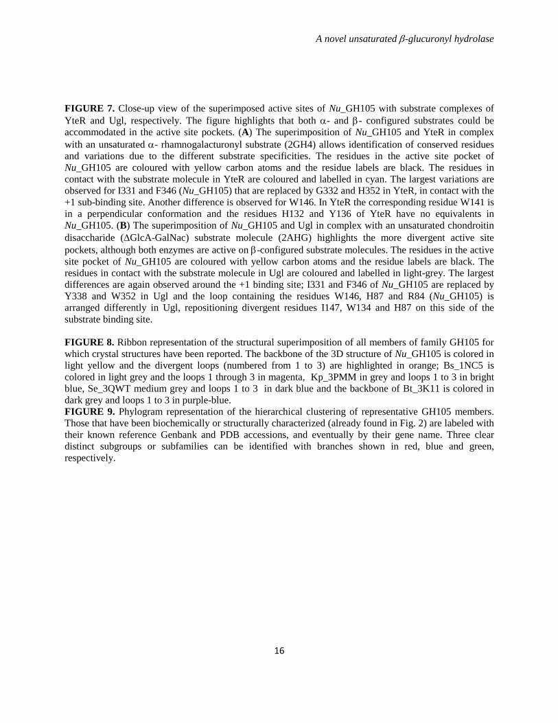
FIGURE LEGENDS TABLE 1. Data collection, phasing and refinement statistics of Nu_GH105.
FIGURE 1. Chemical structure of the three main disaccharide repetition moieties encountered in ulvans and the unsaturated oligosaccharide produced by lyase degradation. (A) and (B) are ulvanobiouronic acid A and B, respectively, (C) ulvanobiose, (D) 4-deoxy-L-threo-hex-4-enopyranosiduronic acid (∆) linked to the sulfated rhamnose occuring at the non-reducing end after cleavage of the glycosidic linkage by ulvan lyase. Rha3S: rhamnose-3-sulfate; GlcA: glucuronic acid; IduA: iduronic acid; Xyl: xylose. FIGURE 2. Amino acid sequence alignment of the N. ulvanivorans unsaturated β-glucuronyl hydrolase sequence (Nu_GH105: Nu_AFQ98272-4CE7) with the two characterized proteins belonging to the GH105 family (YesR: Bs_CAB12519-YesR and YteR: Bs_CAB14990-1NC5 from B. subtilis), as well as the three other members of GH105 with known 3D structure Kp_ABR77815-3PMM, Se_AAV76933-3QWT and Bt_AAO79281-3K11. Conserved amino acids involved in the recognition of the unsaturated uronyl sugar surrounding the -1 binding site are marked by a gray oval; the catalytic acid/base is marked by a star and two important residues in Nu_GH105 not conserved in other GH105 are marked by a gray open square. The secondary structure elements of Nu_GH105 are symbolized above the sequences and the numbering corresponds to the mature Nu_GH105 enzyme. The figure was prepared using Espript (40). FIGURE 3. High-performance anion-exchange chromatography of purified oligosaccharides incubated with unsaturated β-glucuronyl hydrolase. (A) Δ-Rha3S was partially degraded by the enzyme. (B) and (C) The tetrasaccharides Δ-Rha3S-Glc-Rha3S and Δ-Rha3S-Idu-Rha3S were completely degraded. (D) Partial degradation of the tetrasaccharide Δ-Rha3S-Xyl-Rha3S.*: uronic tetrasaccharides eluting like in B and C. - enz: without unsaturated β-glucuronyl hydrolase. +enz: with unsaturated β-glucuronyl hydrolase. FIGURE 4. 1H NMR of the purified end-products of the unsaturated β-glucuronyl hydrolase. (A) Spectrum of a mixture of 50:50 Δ-Rha3S-Glc-Rha3S:Δ-Rha3S-Idu-Rha3S (bottom) and trisaccharides obtained after incubation with unsaturated β-glucuronyl hydrolase (top). (B) Same experiment as in (A) but with the Δ-R3S-Xyl-R3Sα/β tetrasaccharide as the starting substrate. FIGURE 5. Kinetics of degradation of purified oligosaccharides. Structure of the unsaturated oligo-ulvans are indicated with the normalized degradation rates (s-1) in parentheses. The activities were determined as the absorbance decrease at 235nm determined in a quartz cuvette with a 1 cm light path at 30°C starting with 100 µM oligosaccharide and 4.4 nM enzyme in a final volume of 500 µL. FIGURE 6. The trimeric organisation of Nu_GH105 provides the residues responsible for the observed substrate specificity in the +2 sub-bindingsite. (A) The crystallographic trimer organisation of Nu_GH105. The cartoon representations of the three independent molecules, arranged around the non crystallographic three fold axis, are coloured in purple, blue and grey, respectively. The loops from residues 104 to 111 of each monomer are coloured in green and the arginine residue R106 that could play a role in binding a iduronic or glucuronic acid at +2 of the Nu_GH105 substrate is coloured in orange. (B) The superimposition of Nu_GH105 with YteR and Ugl in complex with substrate molecules allowed modelling a putative substrate molecule that binds to subsites -1 to +2, highlighting the possibility that R106 from a neighbouring molecule could bind to a carboxylic acid function of the 6th position of a sugar unit bound to +2.
A novel unsaturated β-glucuronyl hydrolase
16
FIGURE 7. Close-up view of the superimposed active sites of Nu_GH105 with substrate complexes of YteR and Ugl, respectively. The figure highlights that both α- and β- configured substrates could be accommodated in the active site pockets. (A) The superimposition of Nu_GH105 and YteR in complex with an unsaturated α- rhamnogalacturonyl substrate (2GH4) allows identification of conserved residues and variations due to the different substrate specificities. The residues in the active site pocket of Nu_GH105 are coloured with yellow carbon atoms and the residue labels are black. The residues in contact with the substrate molecule in YteR are coloured and labelled in cyan. The largest variations are observed for I331 and F346 (Nu_GH105) that are replaced by G332 and H352 in YteR, in contact with the +1 sub-binding site. Another difference is observed for W146. In YteR the corresponding residue W141 is in a perpendicular conformation and the residues H132 and Y136 of YteR have no equivalents in Nu_GH105. (B) The superimposition of Nu_GH105 and Ugl in complex with an unsaturated chondroitin disaccharide (∆GlcA-GalNac) substrate molecule (2AHG) highlights the more divergent active site pockets, although both enzymes are active on β-configured substrate molecules. The residues in the active site pocket of Nu_GH105 are coloured with yellow carbon atoms and the residue labels are black. The residues in contact with the substrate molecule in Ugl are coloured and labelled in light-grey. The largest differences are again observed around the +1 binding site; I331 and F346 of Nu_GH105 are replaced by Y338 and W352 in Ugl and the loop containing the residues W146, H87 and R84 (Nu_GH105) is arranged differently in Ugl, repositioning divergent residues I147, W134 and H87 on this side of the substrate binding site. FIGURE 8. Ribbon representation of the structural superimposition of all members of family GH105 for which crystal structures have been reported. The backbone of the 3D structure of Nu_GH105 is colored in light yellow and the divergent loops (numbered from 1 to 3) are highlighted in orange; Bs_1NC5 is colored in light grey and the loops 1 through 3 in magenta, Kp_3PMM in grey and loops 1 to 3 in bright blue, Se_3QWT medium grey and loops 1 to 3 in dark blue and the backbone of Bt_3K11 is colored in dark grey and loops 1 to 3 in purple-blue. FIGURE 9. Phylogram representation of the hierarchical clustering of representative GH105 members. Those that have been biochemically or structurally characterized (already found in Fig. 2) are labeled with their known reference Genbank and PDB accessions, and eventually by their gene name. Three clear distinct subgroups or subfamilies can be identified with branches shown in red, blue and green, respectively.
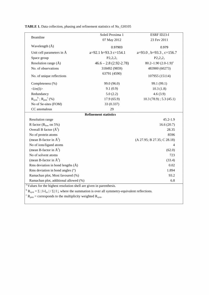
TABLE 1. Data collection, phasing and refinement statistics of Nu_GH105
Beamline Soleil Proxima 1
07 May 2012 ESRF ID23-I 23 Fev 2011
Wavelength (Å) 0.97903 0.979 Unit cell parameters in Å a=92.1 b=93.3 c=154.1 a=93.0 , b=93.3 , c=156.7 Space group P212121 P212121
Resolution range (Å) 46.6 – 2.8 (2.92-2.78) 80.2–1.90 (2.0-1.9)a
No. of observations 318492 (9859) 483900 (60273)
No. of unique reflections 63791 (4590)
107955 (15114)
Completeness (%) 99.0 (96.0) 99.1 (99.1) <I/σ(I)> 9.1 (0.9) 10.3 (1.8) Redundancy 5.0 (2.2) 4.6 (3.9) Rsym
b ; Rpimc (%) 17.9 (65.9) 10.3 (78.9) ; 5.3 (45.1)
No of Se-sites (FOM) 33 (0.337) CC anomalous 29
Refinement statistics Resolution range 45.2-1.9 R factor (Rfree on 5%) 16.6 (20.7) Overall B factor (Å2) 28.35 No of protein atoms (mean B-factor in Å2)
8596 (A 27.95; B 27.35; C 28.18)
No of ions/ligand atoms (mean B-factor in Å2)
4 (62.0)
No of solvent atoms (mean B-factor in Å2)
723 (33.4)
Rms deviation in bond lengths (Å) 0.02 Rms deviation in bond angles (°) 1.894 Ramachan plot, Most favoured (%) 93.2 Ramachan plot, additional allowed (%) 6.8
a Values for the highest resolution shell are given in parenthesis. b Rsym = Σ | I-Iav | / Σ| I |, where the summation is over all symmetry-equivalent reflections. c Rpim = corresponds to the multiplicity weighted Rsym.
H3C
H3C
H3C
OOO
HO OHO
OOH
OCOOH
HO3S
OOO
O
O
O
FIGURE 1
OO
O OO
O
HOOC
OHOH
OHOH
OH
HO3S
HOHO3S
Rha3SGlcA
Rha3SIduA
Rha3SXyl
A
B
C
OO
HO OH
OH3C
OOH
OCOOH
HO3S
Rha3S∆
D
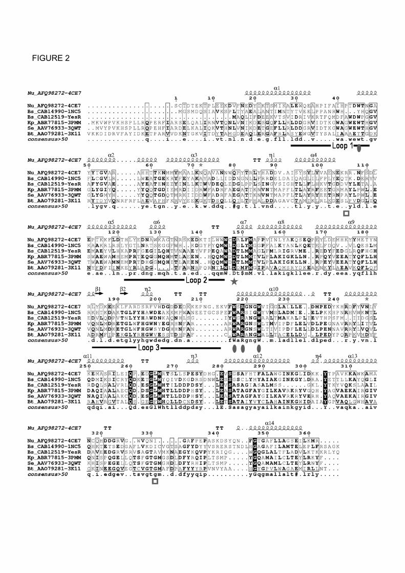
FIGURE 2
Con
duct
imet
ry (µ
S)
∆-Rha3SRha3S
∆-Rha3S-GluA-Rha3SRha3S-GluA-Rha3S
∆-Rha3S-IduA-Rha3SRha3S-IduA-Rha3S
∆-Rha3S-Xyl-Rha3S
Rha3S-Xyl-Rha3S
A
D
C
B
4 6 8 10 12 14 16 18 20 220
50
100
150
Retention time (min)
4 6 8 10 12 14 16 18 20 220
50
100
150
Retention time (min)
4 6 8 10 12 14 16 18 20 220
50
100
150
Retention time (min)
4 6 8 10 12 14 16 18 20 220
50
100
150
Retention time (min)
Con
duct
imet
ry (µ
S)
Con
duct
imet
ry (µ
S)
Con
duct
imet
ry (µ
S)
*
*
Figure 3
- enz
+ enz
- enz
+ enz
- enz
+ enz
- enz
+ enz
A
∆-H1
6.2 3.84.04.24.44.64.85.05.25.45.65.86.0 3.23.43.6(ppm)
R3Srα-H1
R3Srβ-H1
IduA-H1
R3Snr-H1
GlcA-H2
GlcA-H1
∆-H4
∆-R3S-GlcA-R3Sα/β
∆-R3S-IduA-R3Sα/β+
R3S-GlcA-R3Sα/β
R3S-IduA-R3Sα/β+
6.2 3.84.04.24.44.64.85.05.25.45.65.86.0 3.23.43.6(ppm)
R3S-Xyl-R3Sα/β
Xyl-H2Xyl-H5
R3Srβ-H1R3Snr-H1 Xyl-H1
R3Srβ-H4
R3Srα-H4
∆-H1 ∆-H4
∆-R3S-Xyl-R3Sα/β
B
Figure 4
0.3
0.35
0.4
0.45
0.5
0.55
1 2 3 4 5
∆-Rha3S (v= 34 ± 4 nM/s)
∆-Rha3S-Xyl-Rha3S(v= 63 ± 3 nM/s)
∆-Rha3S-IduA-Rha3S/∆-Rha3S-GluA-Rha3S (70/30)(v= 114 ± 2 nM/s)
0
∆-Rha3S-IduA-Rha3S/∆-Rha3S-GluA-Rha3S (50/50)(v= 112 ± 7 nM/s)
∆-Rha3S-IduA-Rha3S/∆-Rha3S-GluA-Rha3S (30/70)(v=107 ± 3 nM/s)
Abs
orba
nce
(265
nm
)
Time (min)
∆-Rha (v= 0 nM/s)
Figure 5
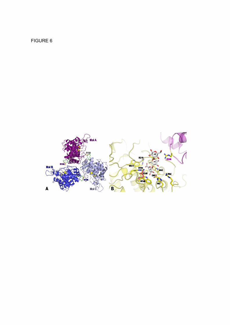
FIGURE 6
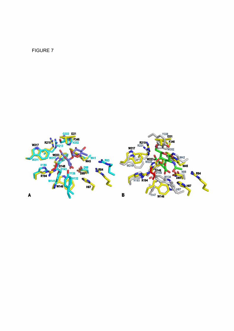
FIGURE 7
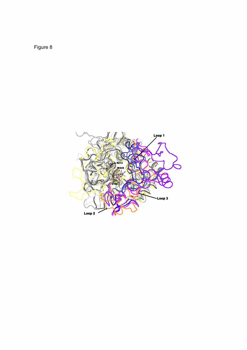
Figure 8
EC:3.2.1.172 Bs_CAB14990-1NC5
Bt_AAO79281-3K11Se_AAV76933-3QWTKp_ABR77815-3PMM
Bs_CAB12519-YesR EC:3.2.1.172
Nu_AFQ98272-4CE7 EC:3.2.1.-
100.0
FIGURE 9
![[Terzaghi] Unsaturated Soil Mechanics (2007)](https://static.fdocument.org/doc/165x107/545096f2b1af9f4c648b4d35/terzaghi-unsaturated-soil-mechanics-2007.jpg)