Original Article Suppressing the malignant phenotypes … · Original Article Suppressing the...
10
Int J Clin Exp Pathol 2013;6(11):2323-2332 www.ijcep.com /ISSN:1936-2625/IJCEP1309002 Original Article Suppressing the malignant phenotypes of glioma cells by lentiviral delivery of small hairpin RNA targeting hypoxia-inducible factor-1α Chang Gui Dong 1* , William K K Wu 2* , Su Yin Feng 3* , Jun Yu 2 , Jun Fei Shao 4 , Gui Mei He 1 1 Institute of Molecular Ecology and Evolution, IAIR, East China Normal University, Shanghai 200062, PR China; 2 Institute of Digestive Disease, Department of Medicine & Therapeutics and LKS Institute of Health, The Chi- nese University of Hong Kong, Shatin, NT, Hong Kong, China; 3 Department of Neurosurgery, Wuxi NO.4 People’s Hospital, Affiliated to Jiangnan University, Wuxi 214062, PR China; 4 Department of Neurosurgery, Wuxi People’s Hospital of Nanjing Medical University, Wuxi 214023, China. * Equal contributors. Received September 1, 2013; Accepted October 3, 2013; Epub October 15, 2013; Published November 1, 2013 Abstract: Hypoxic microenvironment of solid tumors is known to shape malignant phenotypes of cancer cells through the dimeric transcription factor hypoxia-inducible factor (HIF)-1. In the present study, the therapeutic effect of target- ing α subunit of HIF-1 in glioma cells via lentiviral delivery of small hairpin RNA (shRNA) was evaluated. Data from quantitative real-time PCR and immunohistochemistry demonstrated that HIF-1α was progressively upregulated during the development of gliomas. Lentiviral shRNA targeting HIF-1α led to substantial loss of cell viability, G 0 /G 1 - phase cell cycle arrest, apoptosis, and impairment of cell motility and invasiveness in human glioma U87MG cells. Xenograft experiments in nude mice further showed that HIF-1α-shRNA inhibited tumor growth and caused persis- tent repression of HIF-1α and its target genes, including VEGF, GLUT1 and MMP2, up to 25 days post-inoculation. Taken together, lentiviral delivery of shRNA is a promising therapeutic approach for targeting HIF-1α in glioma. Keywords: Hypoxia-inducible factor, glioma, short hairpin RNA, lentivirus Introduction Many solid tumors contain regions of hypoxia where the supply of oxygen fails to meet the metabolic need of actively proliferating tumor cells, leading to the formation of central necrot- ic zone surrounded by a rim of viable cells. Such hypoxic intratumoral microenvironment has been shown to modulate the intracellular sig- naling of tumor cells and promote the acquisi- tion of aggressive phenotypes, such as unchecked cell proliferation, evasion of apopto- sis, increased cell motility and invasiveness, stemness, and enhanced ability to recruit new blood vessels [1]. Emerging evidence suggests that tumor hypoxia is assoicated with poor responses to chemotherapy and radiotherapy as well as lower overall patient survival. The mechanism by which hypoxic conditions con- tribute to tumor aggressiveness is manifold and may include induction of endoplasmic reticulum stress [2], increased DNA damage and impaired DNA mismatch repair [3], activa- tion of mitogen-activated protein kinase (MAPK) signaling [4], and induction of transcription fac- tors, such as activator protein (AP)-1 [5], nucle- ar factor (NF)-κB [6] and hypoxia-inducible fac- tor (HIF)-1 [7]. HIF-1 is a heterodimeric transcription factor composed of an oxygen-regulated α subunit and a constitutively expressed β subunit. Under normoxic conditions, proline residues 402 and 564 of HIF-1α are hydroxylated, allowing inter- action with the E3 ubiquitin ligase von Hippel- Lindau protein that tags HIF-1α with polyubiqui- tin for proteasomal degradation. In hypoxic conditions, proline hydroxylation of HIF-1α is impaired and thereby stabilizing the protein [8]. In addition to hypoxia, HIF-1α is known to be upregulated by transforming viruses (e.g. Epstein-Barr virus, hepatitis B virus, and human papilloma virus) and bioactive lipid mediators (e.g. prostaglandin E 2 and leukotrienes) [7].
Transcript of Original Article Suppressing the malignant phenotypes … · Original Article Suppressing the...
Int J Clin Exp Pathol 2013;6(11):2323-2332www.ijcep.com /ISSN:1936-2625/IJCEP1309002
Original Article Suppressing the malignant phenotypes of glioma cells by lentiviral delivery of small hairpin RNA targeting hypoxia-inducible factor-1α
Chang Gui Dong1*, William K K Wu2*, Su Yin Feng3*, Jun Yu2, Jun Fei Shao4, Gui Mei He1
1Institute of Molecular Ecology and Evolution, IAIR, East China Normal University, Shanghai 200062, PR China; 2Institute of Digestive Disease, Department of Medicine & Therapeutics and LKS Institute of Health, The Chi-nese University of Hong Kong, Shatin, NT, Hong Kong, China; 3Department of Neurosurgery, Wuxi NO.4 People’s Hospital, Affiliated to Jiangnan University, Wuxi 214062, PR China; 4Department of Neurosurgery, Wuxi People’s Hospital of Nanjing Medical University, Wuxi 214023, China. *Equal contributors.
Received September 1, 2013; Accepted October 3, 2013; Epub October 15, 2013; Published November 1, 2013
Abstract: Hypoxic microenvironment of solid tumors is known to shape malignant phenotypes of cancer cells through the dimeric transcription factor hypoxia-inducible factor (HIF)-1. In the present study, the therapeutic effect of target-ing α subunit of HIF-1 in glioma cells via lentiviral delivery of small hairpin RNA (shRNA) was evaluated. Data from quantitative real-time PCR and immunohistochemistry demonstrated that HIF-1α was progressively upregulated during the development of gliomas. Lentiviral shRNA targeting HIF-1α led to substantial loss of cell viability, G0/G1-phase cell cycle arrest, apoptosis, and impairment of cell motility and invasiveness in human glioma U87MG cells. Xenograft experiments in nude mice further showed that HIF-1α-shRNA inhibited tumor growth and caused persis-tent repression of HIF-1α and its target genes, including VEGF, GLUT1 and MMP2, up to 25 days post-inoculation. Taken together, lentiviral delivery of shRNA is a promising therapeutic approach for targeting HIF-1α in glioma.
Keywords: Hypoxia-inducible factor, glioma, short hairpin RNA, lentivirus
Introduction
Many solid tumors contain regions of hypoxia where the supply of oxygen fails to meet the metabolic need of actively proliferating tumor cells, leading to the formation of central necrot-ic zone surrounded by a rim of viable cells. Such hypoxic intratumoral microenvironment has been shown to modulate the intracellular sig-naling of tumor cells and promote the acquisi-tion of aggressive phenotypes, such as unchecked cell proliferation, evasion of apopto-sis, increased cell motility and invasiveness, stemness, and enhanced ability to recruit new blood vessels [1]. Emerging evidence suggests that tumor hypoxia is assoicated with poor responses to chemotherapy and radiotherapy as well as lower overall patient survival. The mechanism by which hypoxic conditions con-tribute to tumor aggressiveness is manifold and may include induction of endoplasmic reticulum stress [2], increased DNA damage
and impaired DNA mismatch repair [3], activa-tion of mitogen-activated protein kinase (MAPK) signaling [4], and induction of transcription fac-tors, such as activator protein (AP)-1 [5], nucle-ar factor (NF)-κB [6] and hypoxia-inducible fac-tor (HIF)-1 [7].
HIF-1 is a heterodimeric transcription factor composed of an oxygen-regulated α subunit and a constitutively expressed β subunit. Under normoxic conditions, proline residues 402 and 564 of HIF-1α are hydroxylated, allowing inter-action with the E3 ubiquitin ligase von Hippel-Lindau protein that tags HIF-1α with polyubiqui-tin for proteasomal degradation. In hypoxic conditions, proline hydroxylation of HIF-1α is impaired and thereby stabilizing the protein [8]. In addition to hypoxia, HIF-1α is known to be upregulated by transforming viruses (e.g. Epstein-Barr virus, hepatitis B virus, and human papilloma virus) and bioactive lipid mediators (e.g. prostaglandin E2 and leukotrienes) [7].
Lentiviral shRNA silencing of HIF-1α in glioma
2324 Int J Clin Exp Pathol 2013;6(11):2323-2332
Upon activation, HIF-1 translocates into the nucleus where it induces expression of genes (e.g. VEGF, GLUT1, MMP2) related to angiogen-esis, de-differentiation, glycolysis, and metas-tasis, through binding to the consensus sequence 5’-RCGTG-3’ [9-12].
Malignant gliomas are the most common pri-mary brain tumors, which are associated with high mortality and morbidity. Despite recent advancements in chemotherapy, radiotherapy and neurosurgery, the prognosis of patients with malignant gliomas remains dismal, high-lighting an urgent need to develop novel and effective therapeutics [13]. Hypoxia is a pre-dominant feature of gliomas and has been found to maintain glioma stem cells and confer resistance to therapy [14]. Thus, targeting dys-regulated signals associated with tumor hypox-ia has been promulgated as a therapeutic approach for glioma. To this end, accumulating evidence suggest that HIF-1α is an attractive molecular target. A small hairpin RNA (shRNA) is a sequence of RNA that self-anneals to form a hairpin structure that can be employed to silence target gene expression via RNA interfer-ence [15]. The development of lentiviral vectors for ectopic expression of shRNA has been a recent advance in the field of gene therapy. The therapeutic application of lentiviral delivery of shRNA to target HIF-1α in gliomas, however, has not yet been explored. In the present study, we first show that HIF-1α was increasingly upregulated during the progression of gliomas. We also demonstrate that targeting HIF-1α with
shRNA delivered by a lentiviral vector could cause sustained reversal of malignant pheno-types and prolonged repression of HIF-1α and its target genes in glioma cells.
Materials and methods
Cell line and reagents
The human glioblastoma cell line U87MG and U251 were obtained from the Institute of Biochemistry and Cell Biology, Chinese Academy of Science (Shanghai, China). Primary antibodies were purchased from Santa Cruz Biotechnology (Santa Cruz, CA, USA). All other chemicals were purchased from Sigma unless otherwise specified.
Patient samples and immunohistochemistry
Paraffin-embedded tissue samples consisting of a total of 5 decompression brain tissues as control and 55 glioma tissues (20 cases of glio-blastoma multiforme, 13 cases of astrocytoma, 8 cases of oligodendroglioma and 4 cases of ependymoma) were used. The average age of the glioma patients was 47 years and the male to female ratio was 1.2:1. Immunohistochemical staining of HIF-1α was performed using poly-clonal anti-HIF-1α antibody (sc-13515, Santa Cruz) with standard avidin-biotin method. A pro-portion score based on staining intensity and percentage of positively stained cells was used to assess the expression of HIF-1α. Age-matched normal and cancer tissues were obtained from anonymised excess tissues that
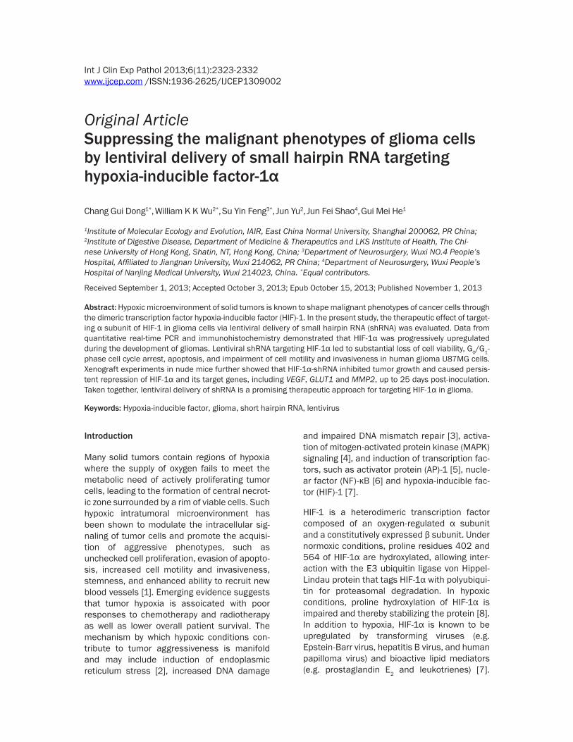
Figure 1. Successful cloning of HIF-1α-shRNA-encoding sequences into lentiviral vector was confirmed by Sanger sequencing.
Lentiviral shRNA silencing of HIF-1α in glioma
2325 Int J Clin Exp Pathol 2013;6(11):2323-2332
were no longer needed for diagnostic or clinical purposes. Use of these tissues was approved by the Ethics Committee of Nanjing Medical University (reference number: 2013-2).
Construction of HIF-1α-shRNA-expressing lentiviral vector
The shRNAs targeting HIF-1α mRNA were designed online (www.invitrogen.com/rnai): homo-LV-shRNA-HIF-1α-1168, 5’-GTGATGAA- AGAATTACCGAAT-3’; homo-LV-shRNA-HIF-1α- 1577, 5’-GAAGGAACCTGATGCTTTA-3’; homo-LV-shRNA-HIF-1α-2264, 5’-CCAGTTATGATTG- TGAAGTTA-3’; A scrambled sequence that shared no homology with the mammalian genome was used as control. Successful clon-
well in 96-well plates. After incubation for 2 h to allow cell attachment to the bottom of the well, 10 μl CCK-8 solution were added to each well at the indicated time points, and the plates were incubated for another 2 h. The optical density was then determined at 450 nm using a microplate reader. For cell cycle analysis, U87MG cells were fixed with ice-cold 70% etha-nol in phosphate-buffered saline, followed by incubation with 50 μg/ml propidium iodide, 3.8 mmol/l sodium citrate and 0.5 μg/ml RNase A at 4°C for 3 h and analyzed by flow cytometry. To quantify apoptosis, annexin V/7-amino-actinomycin (7-AAD) staining was performed, and apoptosis was evaluated by flow cytometry analysis. Briefly, after transduction, both float-ing and attached cells were collected and sub-ject to annexin V/7-AAD staining using an annexin V-FITC Apoptosis Detection kit (BioVision, Palo Alto, CA), according to the man-ufacturer’s protocol. The resulting fluorescence was measured by flow cytometry using a FACS flow cytometer (BD Biosciences, San Jose, CA).
Cell migration and invasion assays
A monolayer cell migration assay and BD BioCoat Matrigel invasion chambers (8-μm
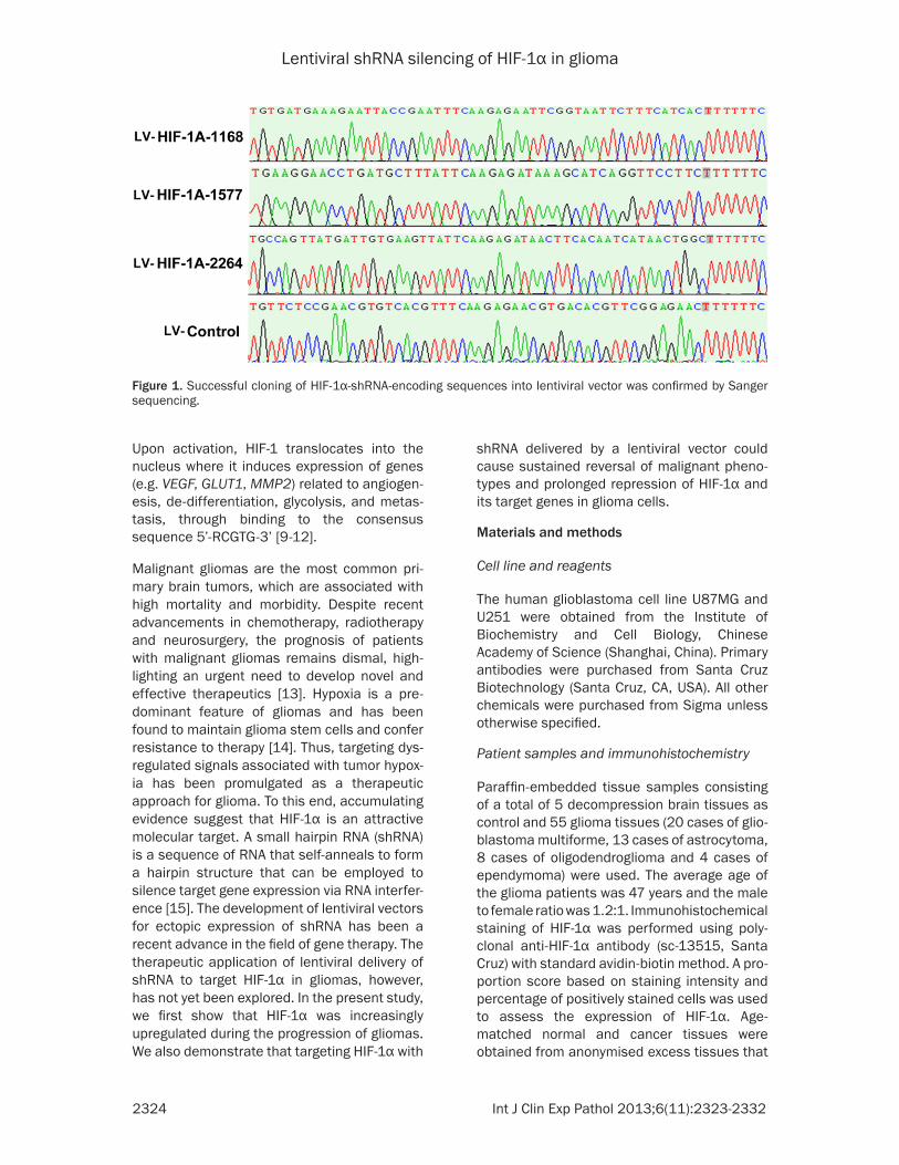
Figure 2. Upregulation of HIF-1α in glioma tissues. A: promi-nent diffuse expression of HIF-1α protein was detected in high-grade glioma tissues as revealed by immunohistochemis-try. HIF-1α protein expression was negligible in non-cancerous intradecompression brain (IDB) tissues. B: HIF-1α mRNA was upregulated in glioma tissues in a tumor grade-dependent manner as measured by real-time PCR. **P < 0.01, signifi-cantly different from IDB group.
ing of these sequences into the pLV-U6-Puro-GFP lentivector was confirmed by Sanger sequencing (Figure 1). The expression of shRNA was driven by U6 promoter. This vector also encoded a green fluorescence protein (GFP). The pPACK-H1 Lentivector Packaging System (SBI) and the 293TN cell line (SBI) were used for the production of pseudoviral particles according to the manufacturer’s instructions. U87MG cells were then transduced at a multiplicity of infection (MOI) of 2.
Cell culture and assays for cell prolifera-tion, cell cycle and apoptosis
The U87MG human glioma cells were maintained in DMEM (Invitrogen, Carl- sbad, CA, USA), supplemented with 10% fetal bovine serum (Thermo Scientific), 100 U/ml penicillin and 100 μg/ml strep-tomycin (Invitrogen) at 37°C in a humidi-fied atmosphere of 5% CO2 and 95% air. Cell proliferation was measured by the colorimetric Cell Counting Kit-8 (CCK-8) assay (Dojindo). The transfected cells were plated at a density of 5,000 cells/
Table 1. Scoring of HIF-1α immunoreactivity in intra decompression brain and glioma tissues
Sample no.
HIF-1α expression Positivity (%)- + ++ +++
IDB 5 5 0 0 0 0Grade I~II 20 17 3 0 0 15Grade III 16 1 3 10 2 93.78Grade IV 19 0 2 6 11 100
Lentiviral shRNA silencing of HIF-1α in glioma
2326 Int J Clin Exp Pathol 2013;6(11):2323-2332
pores) (BD Biosciences) were used to measure the effects of HIF-1α knockdown on the lateral motility and invasiveness of U87MG cells, respectively. For cell migration assay, U87MG cells were seeded onto 24-well plates and cul-tured in complete medium until confluent. The confluent monolayers were washed twice with PBS and incubated in a serum-free medium. Small linear wounds were created by gently striking a pipette tip across the monolayers. The healing of the wounds through cell migra-tion was assessed by measuring the wound dis-tance. For cell invasion assay, the transduced cells were seeded on the top chamber of each insert with complete medium added to the bot-tom chamber. After 48 h, cells on the mem-brane were wiped off with a cotton swab. Fixed and stained with H&E, cells on the underside of the membrane were counted from 4 micro-scope fields (magnification, x 200). The mean number of invading cells was expressed as a percentage relative to the control.
RNA extraction and real-time reverse transcrip-tion-PCR
Total RNA was isolated after transduction with Trizol reagent (Invitrogen, USA). A nanodrop spectrophotometer (Gene, USA) was used to measure the concentration of total RNA. Relative levels of mRNA were examined using SYBR green real-time quantitative reverse tran-scription-PCR (qRT-PCR) (Applied Biosystems) and normalized with GAPDH. The amplification reaction was performed using MJ real-time PCR (Bio-Rad, Hercules, CA, USA) for 40 cycles. Relative expression was calculated using the 2-∆∆Ct method.
Western blots
Total proteins were extracted after solubiliza-tion in lysis buffer containing 62.5 mM Tris-HCl (pH 6.8), 2% SDS, 10% glycerol, 1 mM sodium vanadate, and 1 mM sodium fluoride. Protein
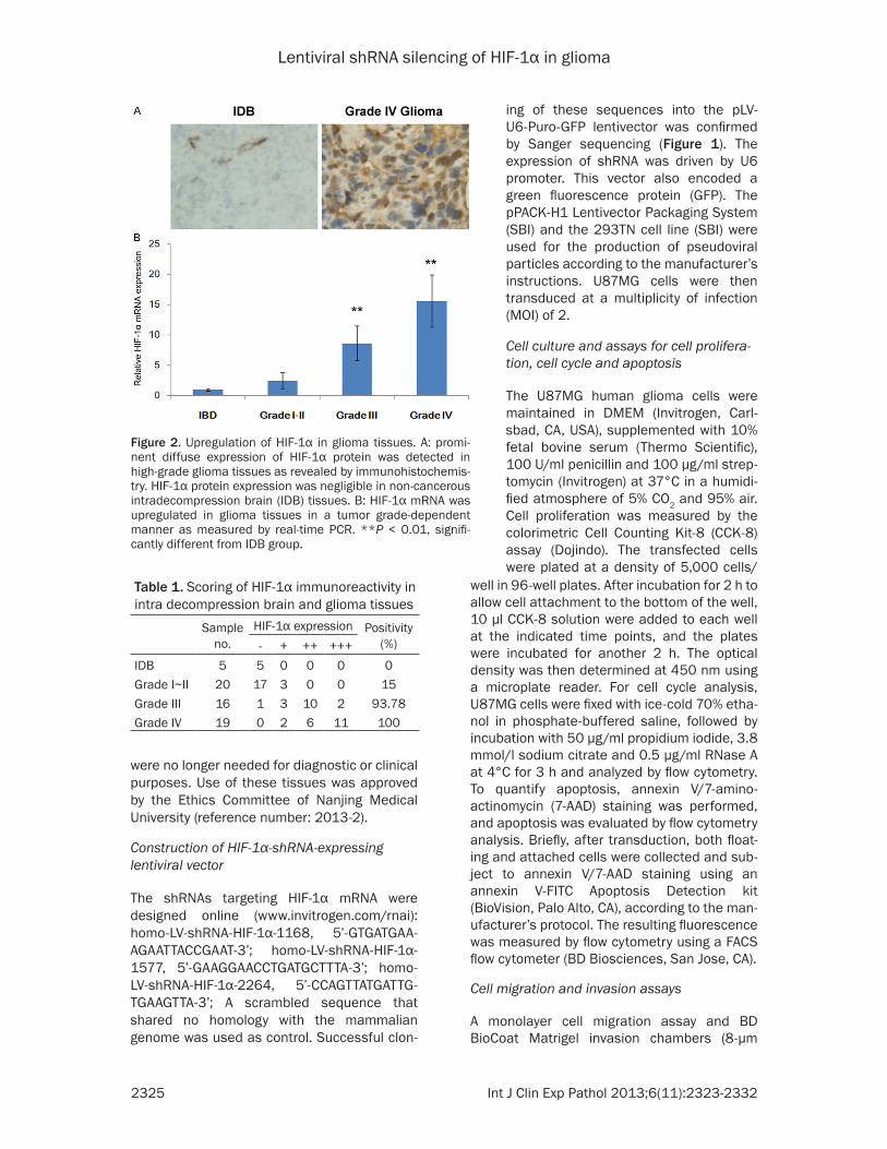
Figure 3. Validation of transduction efficiencies and knockdown efficacies of HIF-1α-shRNA-encoding lentiviral vector in U87MG cells. A: transduction efficiencies of lentivirus were confirmed to be over 99% by fluorescence microscopic detection of lentiviral expression of GFP. B: All three HIF-1α-shRNA-encoding constructs reduced HIF-1α mRNA expression as determined by real-time PCR. C: The knockdown efficacies were further confirmed by Western blots for HIF-1α. The results are the representative of three independent experiments. **P < 0.01, significantly dif-ferent from LV-control group.
Lentiviral shRNA silencing of HIF-1α in glioma
2327 Int J Clin Exp Pathol 2013;6(11):2323-2332
extracts were resolved on Tris-HCl gels and Western blotting was performed using stan-dard methodologies. Blots were developed using the enhanced chemiluminescence (ECL) reagents (Amersham Pharmacia, Bucking- hamshire, UK) and visualized using the Gene-Genius Imaging System (Syngene, Frederick, MD, USA).
Nude mice xenograft model
Transduced U87MG cells stably expressing fire-fly luciferase were trypsinized, collected and re-suspended in PBS. Then 1 × 10⁷ U87MG cells in 0.2 ml PBS were injected subcutaneously into the flank region of 6-week-old female BALB/c nu/nu mice. After inoculation, the mice were maintained under sterile condition and the size of tumor formed was measured by bio-luminescence detection using the Xenogen IVIS system. Tumor volume (V) was also estimated
according to the following formula: V = L × W2/2, where L is the mid-axis length and W is the mid-axis width. At the end of the experi-ment, the mice were sacrificed and the tumors were excised. All experimental procedures were conducted in conformity with institutional guidelines for the care and use of laboratory animals, and protocols were approved by the Institutional Animal Care and Use Committee of Nanjing Medical University. Undertaken or supervised by trained animal technicians and performed so as to minimize any discomfort to the mice.
Results
Progressive upregulation of HIF-1α during the progression of gliomas
Intermediate-to-high levels of cytoplasmic and nuclear expression of HIF-1α were observed in
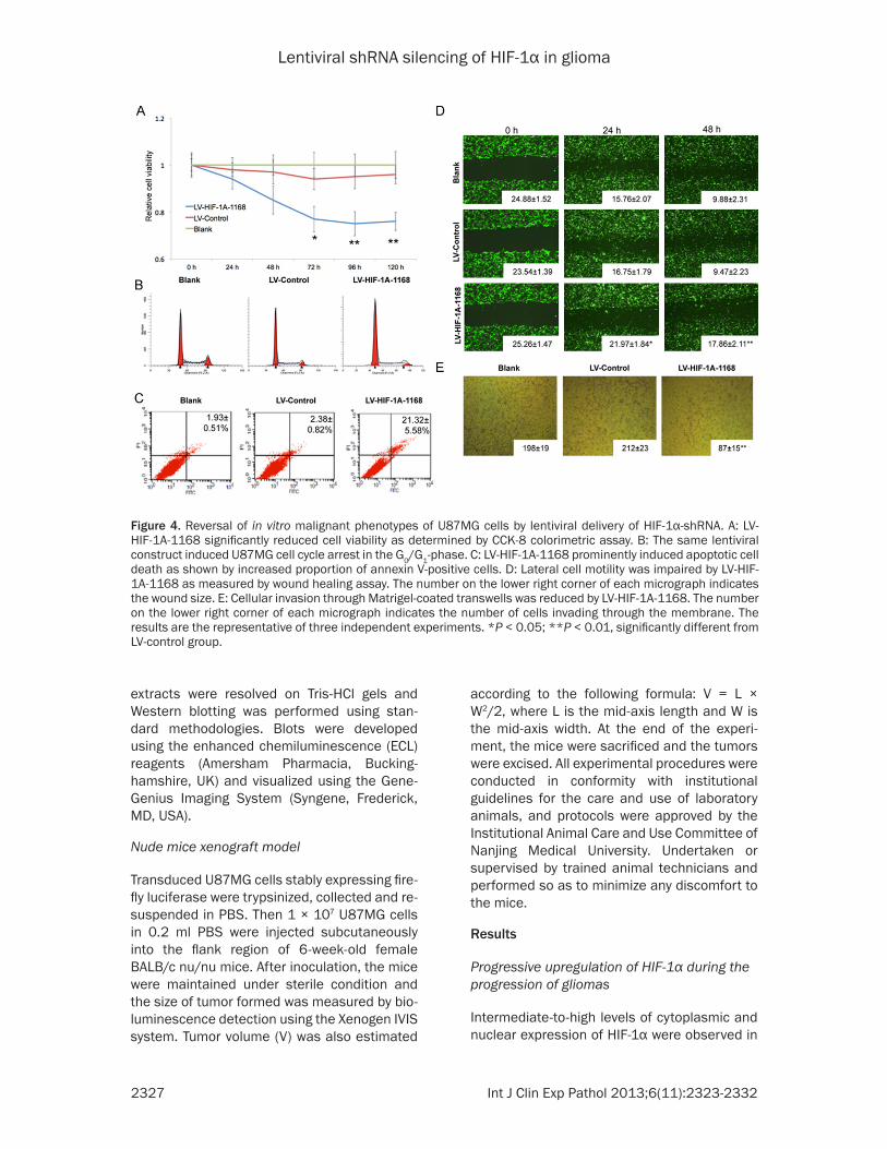
Figure 4. Reversal of in vitro malignant phenotypes of U87MG cells by lentiviral delivery of HIF-1α-shRNA. A: LV-HIF-1A-1168 significantly reduced cell viability as determined by CCK-8 colorimetric assay. B: The same lentiviral construct induced U87MG cell cycle arrest in the G0/G1-phase. C: LV-HIF-1A-1168 prominently induced apoptotic cell death as shown by increased proportion of annexin V-positive cells. D: Lateral cell motility was impaired by LV-HIF-1A-1168 as measured by wound healing assay. The number on the lower right corner of each micrograph indicates the wound size. E: Cellular invasion through Matrigel-coated transwells was reduced by LV-HIF-1A-1168. The number on the lower right corner of each micrograph indicates the number of cells invading through the membrane. The results are the representative of three independent experiments. *P < 0.05; **P < 0.01, significantly different from LV-control group.
Lentiviral shRNA silencing of HIF-1α in glioma
2328 Int J Clin Exp Pathol 2013;6(11):2323-2332
glioma tissues as revealed by immunohisto-chemical staining. By contrast, there was only negligible expression of HIF-1α in non-cancer-ous intradecompression brain tissues (Figure 2A). The upregulation of HIF-1α was assoicated with tumor grade (Table 1) but not histological subtypes. Concordant with immunohistochemi-cal staining, real-time PCR showed the stage-dependent upregulation of HIF-1α mRNA in glio-ma tissues, with more than 15-fold increase of HIF-1α mRNA in grade IV glioma samples as compared with intradecompression brain tis-sues (Figure 2B).
Effective knockdown of HIF-1α and repression of HIF-1α target genes by shRNA in U87MG glioma cells
Three shRNA-encoding constructs, namely HIF-1Α-1168, HIF-1A-1577 and HIF-1A-2264, tar-geting different sites of HIF-1α transcript were packaged into pseudotyped lentiviral particles. The transduction efficiencies of all three lentivi-ral vectors as well as the control vector were confirmed to be more than 99% in U87MG glio-ma cells by fluorescence microscopy (Figure 3A). Transduction of U87MG cells with HIF-1α-shRNA-encoding lentiviral vectors also signifi-cantly reduced the mRNA levels of HIF-1α (Figure 3B). Among these three shRNA-encod-ing vectors, HIF-1Α-1168 showed the strongest inhibition, with more than 80% repression of HIF-1α mRNA expression. The reduction of HIF-1α mRNA levels was paralleled by decreased
HIF-1α protein levels as measured by Western blots (Figure 3C), in which HIF-1Α-1168 also exhibited the strongest inhibitory effect. The HIF-1Α-1168-encoding lentiviral vector was therefore used in subsequent experiments.
Induction of cell cycle arrest and apoptosis and impairment of cell motility and invasive-ness by HIF-1α-shRNA in U87MG
Functional assays were performed to charac-terize phenotypic changes after knockdown of HIF-1α. The viability of U87MG cells transduced with HIF-1Α-1168-encoding or control vector was measured by CCK-8 assay. Knockdown of HIF-1α significantly reduced the viability of U87MG cells from day 3 to day 5 post-transduc-tion (Figure 4A). In line with this finding, cell cycle analysis revealed that knockdown of HIF-1α induced G0/G1-phase cell cycle arrest with concomitant reduction of proportion of cells in S and G2/M phases (Figure 4B). In addition to the effect on cell cycle, HIF-1α knockdown induced apoptosis in U87MG where phosphoti-dylserine externalization was increased by ~9- fold as measured by Annexin V staining (Figure 4C). As enhanced cell motility and invasiveness have been implicated in the oncogenic action of HIF-1α, these malignant phenotypes were determined by monolayer wound healing and transwell invasion assays, respectively. Results showed that HIF-1α knockdown substantially reduced the lateral motility and invasiveness of U87MG cells (Figure 4D and 4E).
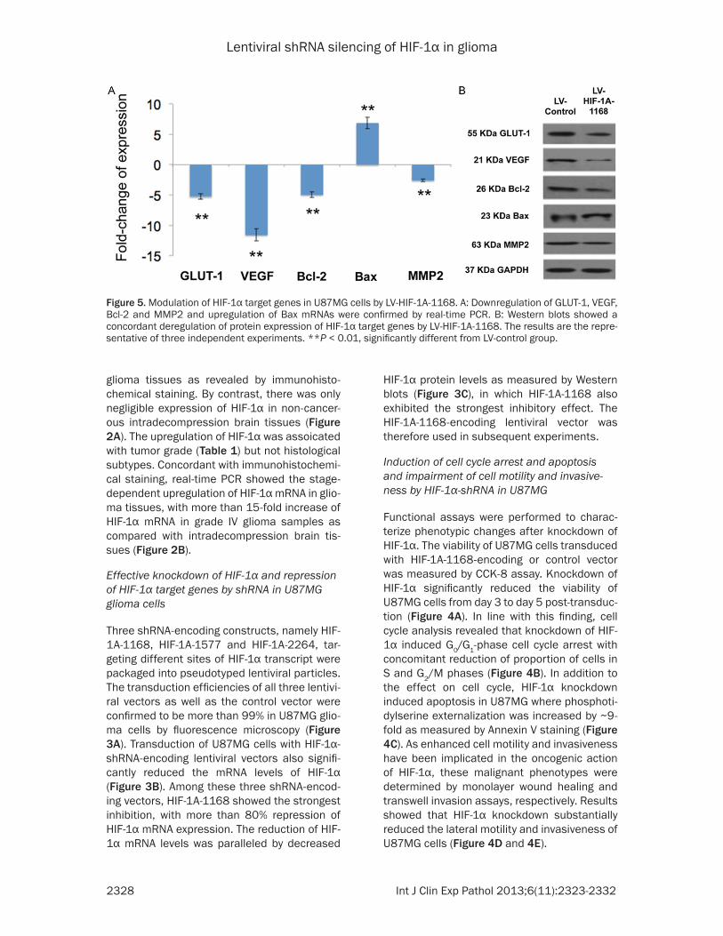
Figure 5. Modulation of HIF-1α target genes in U87MG cells by LV-HIF-1A-1168. A: Downregulation of GLUT-1, VEGF, Bcl-2 and MMP2 and upregulation of Bax mRNAs were confirmed by real-time PCR. B: Western blots showed a concordant deregulation of protein expression of HIF-1α target genes by LV-HIF-1A-1168. The results are the repre-sentative of three independent experiments. **P < 0.01, significantly different from LV-control group.
Lentiviral shRNA silencing of HIF-1α in glioma
2329 Int J Clin Exp Pathol 2013;6(11):2323-2332
Modulation of HIF-1α target genes by HIF-1α-shRNA
HIF-1α has been shown to induce GLUT-1, VEGF, Bcl-2 and MMP2 but repress the expression of Bax. To confirm if HIF-1α-shRNA could inhibit the transcriptional activity of HIF-1α, the mRNA and protein levels of these important cancer-related genes were determined. Results showed that HIF-1Α-1168-encoding vector sub-stantially reduced both the mRNA and protein expression of GLUT-1, VEGF, Bcl-2 and MMP2 but induced Bax in U87MG cells (Figure 5).
Sustained repression of glioma growth and tar-get gene HIF-1α expression in vivo by shRNA
To determine whether lentiviral delivery of shRNA could elicit sustained repression of HIF-1α and persistent inhibition of malignant phe-notypes of glioma cells in vivo, firefly luciferase-
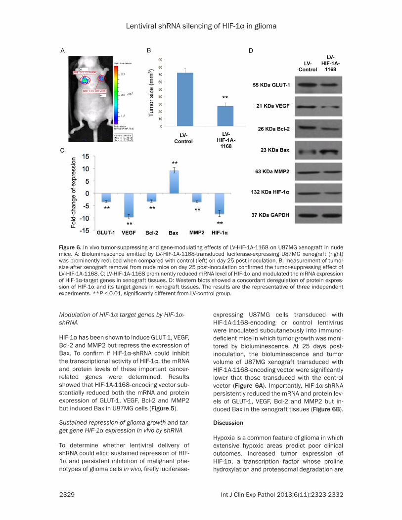
expressing U87MG cells transduced with HIF-1Α-1168-encoding or control lentivirus were inoculated subcutaneously into immuno-deficient mice in which tumor growth was moni-tored by bioluminescence. At 25 days post-inoculation, the bioluminescence and tumor volume of U87MG xenograft transduced with HIF-1Α-1168-encoding vector were significantly lower that those transduced with the control vector (Figure 6A). Importantly, HIF-1α-shRNA persistently reduced the mRNA and protein lev-els of GLUT-1, VEGF, Bcl-2 and MMP2 but in- duced Bax in the xenograft tissues (Figure 6B).
Discussion
Hypoxia is a common feature of glioma in which extensive hypoxic areas predict poor clinical outcomes. Increased tumor expression of HIF-1α, a transcription factor whose proline hydroxylation and proteasomal degradation are
Figure 6. In vivo tumor-suppressing and gene-modulating effects of LV-HIF-1A-1168 on U87MG xenograft in nude mice. A: Bioluminescence emitted by LV-HIF-1A-1168-transduced luciferase-expressing U87MG xenograft (right) was prominently reduced when compared with control (left) on day 25 post-inoculation. B: measurement of tumor size after xenograft removal from nude mice on day 25 post-inoculation confirmed the tumor-suppressing effect of LV-HIF-1A-1168. C: LV-HIF-1A-1168 prominently reduced mRNA level of HIF-1α and modulated the mRNA expression of HIF-1α-target genes in xenograft tissues. D: Western blots showed a concordant deregulation of protein expres-sion of HIF-1α and its target genes in xenograft tissues. The results are the representative of three independent experiments. **P < 0.01, significantly different from LV-control group.
Lentiviral shRNA silencing of HIF-1α in glioma
2330 Int J Clin Exp Pathol 2013;6(11):2323-2332
reduced in hypoxic conditions, has been report-ed in glioma patients and is positively correlat-ed with tumor progression and poor prognosis. In line with these findings, immunohistochemi-cal staining revealed that HIF-1α protein expres-sion was upregulated in glioma tissues in a tumor grade-dependent manner. In addition to post-translational regulation, sustained hypox-ia has been shown to induce the transcription of HIF-1α [16]. To address if transcriptional acti-vation is involved in HIF-1α upregulation in glio-ma tissues, HIF-1α mRNA level was determined by real-time PCR and found to be elevated. This finding indicates that transcriptional mecha-nism at least in part accounts for HIF-1α upreg-ulation in glioma. In this respect, upregulation of HIF-1α mRNA has been sporadically reported in other types of solid tumors, such as gastric and prostate cancer [17, 18]. However, it is the first time to observe such a phenomenon in gli-oma. The mechanism by which hypoxia induces the mRNA expression of HIF-1α remains unclear but reactive oxygen species-mediated overex-pression and activation of Rho GTPase has been reported to induce HIF-1α mRNA in hypox-ic renal cell carcinoma [19]. Our finding also corroborated the idea that targeting HIF-1α mRNA by shRNA-mediated RNA interference is a sound approach of therapeutic intervention.
In the present study, lentiviral delivery of HIF-1α-shRNA triggered substantial phenotypic changes in glioma cells, including lowered cell proliferation, G0/G1-phase cell cycle arrest, apoptosis and reduced cell motility and inva-siveness. These observations are concordant with previous findings that HIF-1α plays a cen-tral and pleotropic role in shaping oncogenic phenotypes of cancer cells through its exten-sive signaling network. In this regard, HIF-1α-mediated expression of GLUT-1, a glucose transporter, has been implicated in the meta-bolic adaptation of cancer cells to hypoglyce-mic microenvironment [20]. Aberrant activation of HIF-1α also represses apoptosis through deregulating the balance between pro- (e.g. Bax) and anti-apoptotic (e.g. Bcl-2) members of the Bcl-2 family [21]. In addition, HIF-1α may promote angiogenesis and cellular invasive-ness through induction of VEGF and MMP-2, respectively [9, 22-24]. Importantly, all these HIF-1α-associated molecular abnormalities could be abrogated by lentiviral delivery of HIF-1α-shRNA in glioma cells. Our findings
substantiated the therapeutic value of target-ing HIF-1α in the treatment of glioma.
HIF-1α is a promising therapeutic target for cancer treatment. Research effort has been put forth to identify novel inhibitors for inhibit-ing this oncogenic transcription factor. Existing small-molecule inhibitors mainly mediate their actions through decreasing HIF-1α protein level, DNA binding or transactivation. Never- theless, their off-target actions may undermine the clinical utility [7]. Novel methods are there-fore urgently needed for specific targeting of HIF-1α. In this regard, gene-specific silencing of shRNA together with the high efficiency of gene transfer and long-term expression of trans-genes by lentivirus open a window of cancer gene therapy. Lentivirus also has relatively low immunogenicity when compared with other viral vectors [25]. Nevertheless, insertional mutagenesis caused by transgene integration may still pose some clinical concerns. The use of integrase-defective lentivirus may circum-vent this issue but the stability of transgene expression remains questionable [26].
In summary, our results demonstrate that lenti-viral delivery of shRNA to target HIF-1α signifi-cantly reduces the malignant phenotypes of glioma cells and restores the deregulation of multiple target genes of HIF-1α. Such antican-cer effects persist after xenograft implantation into nude mice 25-day post-inoculation. Our findings support that the delivery of HIF-1α-shRNA through lentivirus may be a clinically useful treatment approach for glioma patients.
Acknowledgements
This work was supported by grants from the National Natural Science Foundation of China (NSFC, 81272791) and the Natural Science Foundation of Jiangsu Province (BK, 2009078).
Disclosure of conflict of interest
None of the authors has a financial relationship with a commercial entity that has an interest in the subject of this manuscript.
Address correspondence to: Jun F Shao, Department of Neurosurgery, Wuxi People’s Hospital of Nanjing Medical University, Wuxi, 214023, PR China. Tel: 86-0510-82700778; E-mail: [email protected]; Gui M He, Institute of Molecular Ecology and
Lentiviral shRNA silencing of HIF-1α in glioma
2331 Int J Clin Exp Pathol 2013;6(11):2323-2332
Evolution, IAIR, East China Normal University, Shanghai 200062, China. Tel: 86-021-62235786; E-mail: [email protected]
References
[1] Wilson WR and Hay MP. Targeting hypoxia in cancer therapy. Nat Rev Cancer 2011; 11: 393-410.
[2] Koumenis C. ER stress, hypoxia tolerance and tumor progression. Curr Mol Med 2006; 6: 55-69.
[3] Keysar SB, Trncic N, Larue SM and Fox MH. Hy-poxia/reoxygenation-induced mutations in mammalian cells detected by the flow cytome-try mutation assay and characterized by mu-tant spectrum. Radiat Res 2010; 173: 21-6.
[4] Zeng M, Kikuchi H, Pino MS and Chung DC. Hy-poxia activates the K-ras proto-oncogene to stimulate angiogenesis and inhibit apoptosis in colon cancer cells. PLoS One 2010; 5: e10966.
[5] Yao KS, Xanthoudakis S, Curran T and O’Dwyer PJ. Activation of AP-1 and of a nuclear redox factor, Ref-1, in the response of HT29 colon cancer cells to hypoxia. Mol Cell Biol 1994; 14: 5997-6003.
[6] Koong AC, Chen EY and Giaccia AJ. Hypoxia causes the activation of nuclear factor kappa B through the phosphorylation of I kappa B al-pha on tyrosine residues. Cancer Res 1994; 54: 1425-30.
[7] Tang CM and Yu J. Hypoxia-inducible factor-1 as a therapeutic target in cancer. J Gastroen-terol Hepatol 2012; 28: 401-405.
[8] Maxwell PH, Wiesener MS, Chang GW, Clifford SC, Vaux EC, Cockman ME, Wykoff CC, Pugh CW, Maher ER and Ratcliffe PJ. The tumour suppressor protein VHL targets hypoxia-induc-ible factors for oxygen-dependent proteolysis. Nature 1999; 399: 271-275.
[9] Shyu KG, Hsu FL, Wang MJ, Wang BW and Lin S. Hypoxia-inducible factor 1alpha regulates lung adenocarcinoma cell invasion. Exp Cell Res 2007; 313: 1181-1191.
[10] Semenza GL, Jiang BH, Leung SW, Passantino R, Concordet JP, Maire P and Giallongo A. Hy-poxia response elements in the aldolase A, enolase 1, and lactate dehydrogenase A gene promoters contain essential binding sites for hypoxia-inducible factor 1. J Biol Chem 1996; 271: 32529-32537.
[11] Forsythe JA, Jiang BH, Iyer NV, Agani F, Leung SW, Koos RD and Semenza GL. Activation of vascular endothelial growth factor gene tran-scription by hypoxia-inducible factor 1. Mol Cell Biol 1996; 16: 4604-4613.
[12] Ebert BL, Firth JD and Ratcliffe PJ. Hypoxia and mitochondrial inhibitors regulate expression of
glucose transporter-1 via distinct Cis-acting se-quences. J Biol Chem 1995; 270: 29083-29089.
[13] Dahlrot RH, Kristensen BW, Hjelmborg J, Herrstedt J and Hansen S. A population-based study of high-grade gliomas and mutated isoci-trate dehydrogenase 1. Int J Clin Exp Pathol 2013; 6: 31-40.
[14] Yang L, Lin C, Wang L, Guo H and Wang X. Hy-poxia and hypoxia-inducible factors in glioblas-toma multiforme progression and therapeutic implications. Exp Cell Res 2012; 318: 2417-2426.
[15] Castanotto D and Rossi JJ. The promises and pitfalls of RNA-interference-based therapeu-tics. Nature 2009; 457: 426-433.
[16] Pialoux V, Mounier R, Brown AD, Steinback CD, Rawling JM and Poulin MJ. Relationship be-tween oxidative stress and HIF-1 alpha mRNA during sustained hypoxia in humans. Free Rad-ic Biol Med 2009; 46: 321-326.
[17] Pipinikas CP, Carter ND, Corbishley CM and Fenske CD. HIF-1alpha mRNA gene expression levels in improved diagnosis of early stages of prostate cancer. Biomarkers 2008; 13: 680-691.
[18] Yoshikawa T, Tsuburaya A, Miyagi Y, Sekiguchi H, Kimura M, Cho H and Kobayashi O. Up-regu-lation of hypoxia-inducible factor-1 alpha and VEGF mRNAs in peritoneal dissemination of patients with gastric cancer. Anticancer Res 2006; 26: 3849-3853.
[19] Turcotte S, Desrosiers RR and Beliveau R. HIF-1alpha mRNA and protein upregulation in-volves Rho GTPase expression during hypoxia in renal cell carcinoma. J Cell Sci 2003; 116: 2247-2260.
[20] Williams KJ, Telfer BA, Airley RE, Peters HP, Sheridan MR, van der Kogel AJ, Harris AL and Stratford IJ. A protective role for HIF-1 in re-sponse to redox manipulation and glucose de-privation: implications for tumorigenesis. On-cogene 2002; 21: 282-290.
[21] Sasabe E, Tatemoto Y, Li D, Yamamoto T and Osaki T. Mechanism of HIF-1alpha-dependent suppression of hypoxia-induced apoptosis in squamous cell carcinoma cells. Cancer Sci 2005; 96: 394-402.
[22] Zhou J, Li K, Gu Y, Feng B, Ren G, Zhang L, Wang Y, Nie Y and Fan D. Transcriptional up-regulation of RhoE by hypoxia-inducible factor (HIF)-1 promotes epithelial to mesenchymal transition of gastric cancer cells during hypox-ia. Biochem Biophys Res Commun 2011; 415: 348-354.
[23] Luo Y, He DL, Ning L, Shen SL, Li L, Li X, Zhau HE and Chung LW. Over-expression of hypoxia-inducible factor-1alpha increases the invasive potency of LNCaP cells in vitro. BJU Int 2006; 98: 1315-1319.
Lentiviral shRNA silencing of HIF-1α in glioma
2332 Int J Clin Exp Pathol 2013;6(11):2323-2332
[24] Damert A, Ikeda E and Risau W. Activator-pro-tein-1 binding potentiates the hypoxia-induc-iblefactor-1-mediated hypoxia-induced tran-scriptional activation of vascular-endothelial growth factor expression in C6 glioma cells. Biochem J 1997; 327: 419-423.
[25] Nayak S and Herzog RW. Progress and pros-pects: immune responses to viral vectors. Gene Ther 2010; 17: 295-304.
[26] Banasik MB and McCray PB Jr. Integrase-de-fective lentiviral vectors: progress and applica-tions. Gene Ther 2010; 17: 150-157.