Impact of fluorination on proteolytic stability of peptides: a case study with α-chymotrypsin and...
12
1 3 DOI 10.1007/s00726-014-1819-7 Amino Acids (2014) 46:2733–2744 ORIGINAL ARTICLE Impact of fluorination on proteolytic stability of peptides: a case study with α‑chymotrypsin and pepsin Vivian Asante · Jérémie Mortier · Gerhard Wolber · Beate Koksch Received: 22 June 2014 / Accepted: 28 July 2014 / Published online: 6 September 2014 © Springer-Verlag Wien 2014 in proteolytic stability was observed only in a few cases. Thus, this systematic study shows that the proteolytic sta- bility of fluorinated peptides is not predictable, but rather is a very complex phenomenon that depends on the particu- lar enzyme, the position of the substitution relative to the cleavage site and the fluorine content of the side chain. Keywords Chymotrypsin · Pepsin · Aminobutyric acid · Difluoroethylglycine · Trifluoroethylglycine Abbreviations Abz o-Aminobenzoic acid HPLC High performance liquid chromatography Fmoc Fluorenylmethoxy carbonyl MD Molecular dynamics DIC Diisopropylcarbodiimide HOBT 1-Hydroxybenzotriazole HOAT 1-Hydroxy-7-azabenzotriazole TFA Trifluoroacetic acid TIS Triisopropylsilane SPPS Solid phase peptide synthesis Introduction Peptides have great potential as drug candidates, provided that a few small limitations can be overcome. They have many advantages over small molecules on the one hand and antibodies on the other hand in terms of affinity, and specificity with respect to their targets. Their use as drugs has been hampered by different factors such as low bioavailability, relatively low solubility, membrane per- meability and most profoundly low proteolytic stability (Saffran et al. 1986; Giannis and Kolter 1993; McGregor 2008). Proteolysis, the process by which proteases break Abstract Protease stability is a key consideration in the development of peptide-based drugs. A major approach to increase the bioavailability of pharmacologically active peptides is the incorporation of non-natural amino acids. Due to the unique properties of fluorine, fluorinated organic molecules have proven useful in the development of ther- apeutically active small molecules as well as in materials and crop science. This study presents data on the ability of fluorinated amino acids to influence proteolytic stability when present in peptide sequences that are based on ideal protease substrates. Different model peptides containing fluorinated amino acids or ethylglycine in the P2, P1′or P2′ positions were designed according to the specificities of the serine protease, α-chymotrypsin (EC 3.4.21.1) or the aspar- tic protease, pepsin (EC 3.4.23.1). The proteolytic stability of the peptides toward these enzymes was determined by an analytical RP-HPLC assay with fluorescence detection and compared to a control sequence. Molecular modeling was used to support the interpretation of the structure– activity relationship based on the analysis of potential ligand-enzyme interactions. Surprisingly, an increase V. Asante and J. Mortier have contributed equally to this work. Electronic supplementary material The online version of this article (doi:10.1007/s00726-014-1819-7) contains supplementary material, which is available to authorized users. V. Asante · B. Koksch (*) Institute of Chemistry and Biochemistry, Freie Universität Berlin, Takustraße 3, 14195 Berlin, Germany e-mail: [email protected] J. Mortier · G. Wolber Institute of Pharmacy, Department Pharmaceutical and Medicinal Chemistry, Freie Universität Berlin, Königin-Luise-Str. 2+4, 14195 Berlin, Germany
Transcript of Impact of fluorination on proteolytic stability of peptides: a case study with α-chymotrypsin and...
1 3
DOI 10.1007/s00726-014-1819-7Amino Acids (2014) 46:2733–2744
ORIGINAL ARTICLE
Impact of fluorination on proteolytic stability of peptides: a case study with α‑chymotrypsin and pepsin
Vivian Asante · Jérémie Mortier · Gerhard Wolber · Beate Koksch
Received: 22 June 2014 / Accepted: 28 July 2014 / Published online: 6 September 2014 © Springer-Verlag Wien 2014
in proteolytic stability was observed only in a few cases. Thus, this systematic study shows that the proteolytic sta-bility of fluorinated peptides is not predictable, but rather is a very complex phenomenon that depends on the particu-lar enzyme, the position of the substitution relative to the cleavage site and the fluorine content of the side chain.
Keywords Chymotrypsin · Pepsin · Aminobutyric acid · Difluoroethylglycine · Trifluoroethylglycine
AbbreviationsAbz o-Aminobenzoic acidHPLC High performance liquid chromatographyFmoc Fluorenylmethoxy carbonylMD Molecular dynamicsDIC DiisopropylcarbodiimideHOBT 1-HydroxybenzotriazoleHOAT 1-Hydroxy-7-azabenzotriazoleTFA Trifluoroacetic acidTIS TriisopropylsilaneSPPS Solid phase peptide synthesis
Introduction
Peptides have great potential as drug candidates, provided that a few small limitations can be overcome. They have many advantages over small molecules on the one hand and antibodies on the other hand in terms of affinity, and specificity with respect to their targets. Their use as drugs has been hampered by different factors such as low bioavailability, relatively low solubility, membrane per-meability and most profoundly low proteolytic stability (Saffran et al. 1986; Giannis and Kolter 1993; McGregor 2008). Proteolysis, the process by which proteases break
Abstract Protease stability is a key consideration in the development of peptide-based drugs. A major approach to increase the bioavailability of pharmacologically active peptides is the incorporation of non-natural amino acids. Due to the unique properties of fluorine, fluorinated organic molecules have proven useful in the development of ther-apeutically active small molecules as well as in materials and crop science. This study presents data on the ability of fluorinated amino acids to influence proteolytic stability when present in peptide sequences that are based on ideal protease substrates. Different model peptides containing fluorinated amino acids or ethylglycine in the P2, P1′or P2′ positions were designed according to the specificities of the serine protease, α-chymotrypsin (EC 3.4.21.1) or the aspar-tic protease, pepsin (EC 3.4.23.1). The proteolytic stability of the peptides toward these enzymes was determined by an analytical RP-HPLC assay with fluorescence detection and compared to a control sequence. Molecular modeling was used to support the interpretation of the structure–activity relationship based on the analysis of potential ligand-enzyme interactions. Surprisingly, an increase
V. Asante and J. Mortier have contributed equally to this work.
Electronic supplementary material The online version of this article (doi:10.1007/s00726-014-1819-7) contains supplementary material, which is available to authorized users.
V. Asante · B. Koksch (*) Institute of Chemistry and Biochemistry, Freie Universität Berlin, Takustraße 3, 14195 Berlin, Germanye-mail: [email protected]
J. Mortier · G. Wolber Institute of Pharmacy, Department Pharmaceutical and Medicinal Chemistry, Freie Universität Berlin, Königin-Luise-Str. 2+4, 14195 Berlin, Germany
2734 V. Asante et al.
1 3
down peptides and proteins, underlies the basic principle of peptide based drugs. Understanding this principle and the involved mechanisms provides a key strategy to pro-duce peptide or protein-based drugs. Over the past few decades, numerous reports have been published in which a number of strategies employed to combat these limita-tions are outlined: cyclization of peptides (Hummel et al. 2006; Hruby 2002; Ferrie et al. 2013), obstruction of N- or C-terminal ends (N-acylation, N-pyroglutamate, C-amida-tion, N-terminal esterification (phosphoester)) or addition of carbohydrate chains (glycosylation: glucose, xylose, hexose), PEGylation (Dasgupta et al. 2002; Lee et al. 2003; Werle and Bernkop-Schnürch 2006; Vlieghe et al. 2010), the introduction of noncanonical amino acids such as d-amino acids, β-amino acids, N-methylated amino acids and to lesser extent fluorinated analogues (Łegowska et al. 2009; Sani et al. 2006; March et al. 2012). In general, the introduction of noncanonical amino acids with newly designed side-chain functionalities serves as a powerful tool to improve kinetic and thermodynamic properties, proteolytic and structural stabilization of peptides and pro-teins that are not accessible using exclusively the 20 pro-teinogenic amino acids (Dougherty 2000; Salwiczek et al. 2009a, b; Gottler et al. 2008; Frackenpohl et al. 2001; Koksch et al. 1996a, b).
Due to its unique properties, fluorine has become an important element in various industrial fields like agri-culture, materials science, and the pharmaceutical and mining industry (Isanbor and O’Hagan 2006; Filler and Saha 2009). The introduction of fluorine into molecules has led to the improvement of therapeutic agents (Mul-ler et al. 2007; Meng et al. 2008) typically by increasing hydrophobicity and metabolic stability, which in turn leads to improved bioactivity and bioavailability (Böhm et al. 2004). The use of fluorine in the design of artificial amino acids is now being explored in peptide and protein thera-peutics, a class of promising agents with high specificity but low metabolic stability (Sato et al. 2006).
Studies on the protease-catalyzed incorporation of dif-ferent fluoroalkyl-substituted amino acids and their impact on structure, stability and activity of biologically active peptides have shown the importance of fluorine content and the position of fluorination within an amino acid sequence of peptides and proteins (Koksch et al. 1996a, b, 1997; Smits and Koksch 2006; Salwiczek et al. 2009a, 2012).
However, recent investigations of the inclusion of fluorinated amino acids into therapeutic peptides (Meng and Kumar 2007), host defense antimicrobial peptides (Meng et al. 2008), globular proteins (Baker and Montclare 2011), histone acetyltransferase (HAT) tGN5 (Voloshchuk et al. 2009), tripeptide epoxyketone proteasome inhibitor (Geurink et al. 2010) and model peptides (Asante et al. 2013) have indicated that, this
incorporation can have controversial effects on activ-ity and stability. Introduction of hexafluoroleucine (a sterically demanding extensively fluorinated amino acid) into the therapeutic peptide, “glucagon-like peptide-1(GLP-1[7-36])” showed protection against regulatory protease (Dipeptidylpeptidase 4, DPP IV) (Meng et al. 2008). On the contrary to these observations, the global incorporation of monofluorinated phenylalanines into histone acetyltransferase (HAT) tGN5 led to reduced pro-teolytic stability against chymotrypsin (Voloshchuk et al. 2009). Even though elastase has specificity for Ala, the replacement of Ala with TfeGly and DfeGly at different positions within a 10-amino acid model peptide led to proteolytic resistance in two cases while a decreased pro-teolytic stability was observed only in one case (Asante et al. 2013).
Proteases are found in all organisms and play important roles in (1) physiological processes including digestion, haemostasis, apoptosis, signal transduction, reproduction and the immune response and (2) disease states, such as cancer, viral infection, Alzheimer’s disease, inflammatory and cardiovascular disorders (Coughlin 2000). Commer-cially, proteases are valuable tools in the pharmaceutical and biotechnological industries (Saeki et al. 2007). Pro-teases are grouped according to their mechanism of cataly-sis, structural features and common evolutionary origin. Serine and aspartic proteases are two major groups that have been well studied.
α-Chymotrypsin EC 3.4.21.1 is a serine endopeptidase for which the three-dimensional structure, mode of cataly-sis and substrate specificity have been well characterized. Cleavage of the scissile peptide bond is carried out by a cat-alytic triad in which the three amino acid residues Ser195, Asp102 and His57 function together as a ‘charge-relay’ system (Blow et al. 1969). An important part of the cata-lytic scheme is the formation of an unusually short ‘cata-lytic’ hydrogen bond between histidine and aspartate that makes the histidine more basic and aids in the deprotona-tion of the serine (Derewenda et al. 1994). α-Chymotrypsin cleaves preferably peptide bonds on the C-terminal side of large hydrophobic residues, such as phenylalanine, tyros-ine, tryptophan and leucine (Keil 1992; Czapinska and Otlewski 1999; Hedstrom 2002; Polgár 2005).
Pepsin EC 3.4.23.1, an aspartic endopeptidase, is the major digestive protease in the gastric juice of verte-brates and has also been widely investigated since its first crystallization was reported in 1929 by Northrop (North-rop 1930). Aspartic proteases have various mechanisms, but the most commonly used is a general acid–base mechanism which involves the coordination of a water molecule between two highly conserved aspartic acid residues, Asp32 and Asp215 (Antonov et al. 1978, 1981; Suguna et al. 1987; Davies 1990). In such a regime, one
2735Impact of fluorination on proteolytic stability of peptides
1 3
of the aspartic acids acts as a nucleophile and induces hydrolysis between the P1 and P1′ residues of the sub-strate. Pepsin is known to hydrolyze peptide bonds that connect bulky hydrophobic/aromatic residues, such as Phe-Trp, Phe-Tyr and Phe–Phe (Keil 1992; Fruton 1970; Powers et al. 1977).
In fact, properties of fluorine in protein environments still remain difficult to predict and more research is required. The current study presents a systematic investi-gation of the impact of side-chain fluorination on the sta-bility of peptides towards degradation by α-chymotrypsin and pepsin. The main focus of this study is to contribute to the development of “rules of thumb” for the application of fluorine in the design of proteolytically stable variants of clinically relevant peptides.
Materials and methods
Peptide synthesis and purification
All commercially obtained reagents were of synthetic grade and were used as supplied. All peptides were synthe-sized from the C-terminal to the N-terminal end on a solid support by means of an Fmoc/tert-butyl protecting group strategy on preloaded Fmoc-Lysine (Boc) Wang resin using either a Multi-Syntech Syro XP peptide synthesizer (Mul-tiSynTech GmbH, Witten, Germany) or by manual cou-pling in 10-mL polypropylene reactors. Amino acids and
coupling reagents were introduced in four-fold excess and double coupled without capping. The fluorinated amino acids were used in two-fold excess and activated by DIC, HOBt/HOAt to form active esters and the coupling times were extended from 4 to 18 h to ensure completion of the reaction. All peptides (sequences in Fig. 1) were finally N-terminally labeled with o-aminobenzoic acid (Abz) to enable photometric detection, as the use of fluorophores within synthetic substrates is a well established method employed to gain fast, sensitive and reproducible quantita-tive monitoring of proteolytic activity.
The peptides were cleaved from the resin by treatment with 3 mL TFA/TIS/H2O (95%:5 %:10 µL) for 3 h and pre-cipitated with cold diethyl ether. The crude peptides were purified by reversed-phase high performance liquid chro-matography (RP-HPLC) on a smartline system (Knauer GmbH, Berlin), equipped with a smartline manager 5000, two smartline pumps 1000 and a UV detector 2500, with a Phenomenex® Luna C8 (2) column [10-µm particle size, 300 Å pore size, 250 × 21.20 mm (inner diameter)]. A maximum of 20-mg crude peptide per run was purified with a linear gradient of 95 % A to 50 % B over 30 min, where solvent A is 100 % water with 0.1 % TFA (v/v) and solvent B is 100 % acetonitrile with 0.1 % TFA (v/v) at a flow rate of 20 mL/min. Absorbance was recorded at λabs = 320 nm. Purity of the peptides was controlled by analytical HPLC and verification of peptide molecular weight was carried out by means of mass spectrometry. All peptides were >96 % pure as monitored by analytical HPLC.
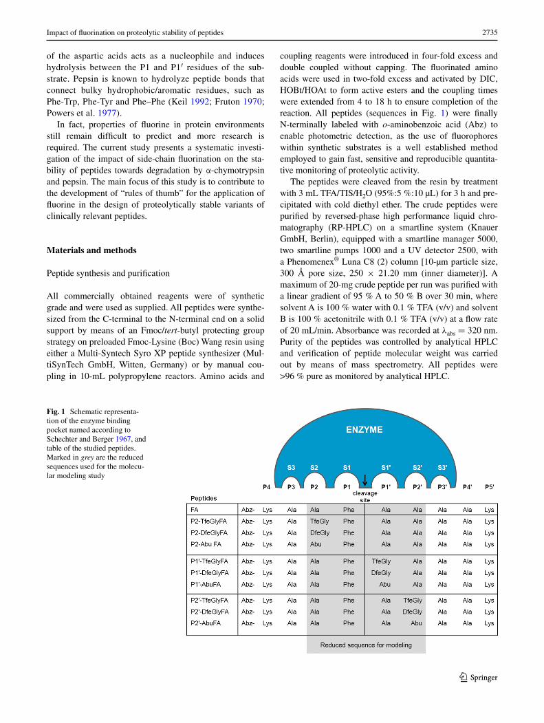
Fig. 1 Schematic representa-tion of the enzyme binding pocket named according to Schechter and Berger 1967, and table of the studied peptides. Marked in grey are the reduced sequences used for the molecu-lar modeling study
2736 V. Asante et al.
1 3
Protease degradation assay
The proteolytic stability of peptides toward α-chymotrypsin (from bovine pancreas, EC 3.4.21.1, 80.0 units/mg) and pepsin (from porcine stomach mucosa EC 3.4.23.1, ≥250 units/mg) (Sigma Aldrich, USA) was analyzed by an ana-lytical RP-HPLC equipped with a fluorescence detector. All peptides employed in the degradation studies were used as the trifluoroacetic acid (TFA) salts obtained after lyophilization. Stock solutions of α-chymotrypsin and pepsin were prepared at concentrations of 0.1 mg/mL in 10-mM phosphate buffer, pH 7.4, and 0.5 mg/mL in 10 mM acetate buffer, pH 4.0, respectively. Peptides were prepared as 20-µmol/mL stocks in DMSO and incu-bated with the respective enzyme at 30 °C with shaking at 300 rpm in a thermomixer over a period of 2 h. Aliquots of 5 µL were removed at fixed time points (0, 15, 30, 60, 90 and 120 min) and either quenched with 95-µL acetonitrile containing 0.1 % TFA, in the case of α-chymotrypsin, or 2 % aqueous ammonia, in the case of pepsin. A monolithic reversed-phase C8 analytical column (Merck’s Chromo-lith® Column, C8 endcapped, 100–4.6 mm) was used to resolve and quantify the products of digestion.
All samples were either immediately subjected to ana-lytical HPLC or frozen down (−20 °C). Detection based on the Abz label was carried out using a fluorescence detector with λex = 320 nm and λem = 420 nm.
In all cases, the peaks corresponding to the educts (full-length peptides) or the N-terminal fragment (product) were integrated and used to determine the velocity of the reac-tion. The FA peptide carrying alanine in positions P2, P1′, and P2′ was used as a reference. The enzyme concentration was adjusted to provide a system in which the reference would be degraded by ~40 % after 120 min since, peptide/protein-based drugs have been shown to exhibit less than 1 % oral bioavailability; the ability to improve this to at least 30–50 % will be of much significance (Vincent et al. 1991; Shaji and Patole 2008).
Each fragment cleaved from the full-length peptide was identified by ESI–MS on an Agilent 6210 ESI-TOF (Agi-lent Technologies, Santa Clara, CA, USA). All experiments were performed in triplicate.
Molecular modeling
The peptide ligands were built in an extended confor-mation using MOE (Molecular Operating Environment 2011) provided by the Chemical Computing Group (http://www.chemcomp.com). The Protein Data Bank (PDB) (Berman et al. 2000) entry code 4CHA (Tsu-kada and Blow 1985) was selected as the starting struc-ture for α-chymotrypsin. The protein was prepared for docking with the LigX module (default parameters) as
implemented in MOE with a particular emphasis on the enzymatic cavity and the protonation state of the catalytic triad. Docking studies were carried out using the soft-ware GOLD 5.1 (Jones et al. 1995, 1997) using a 100 % search efficiency parameter. The active site was deter-mined by selecting all residues within 10 Å around the hydroxyl group of Ser214. A stepwise increasing number of constrains was set until a conformation in agreement with the literature was obtained. Three constraints were required to orient the peptides in a conformation in agree-ment with the literature (Fujinaga et al. 1995, 2000; Hed-strom 2002): a distance range from 2 to 5 Å between (1) the backbone CO of Ser214 (protein) and backbone NH of Phe at position P1 (ligand), (2) the backbone NH of Gly193 (protein) and the backbone CO of Phe at position P1 (ligand), (3) the backbone CO of Phe41 (protein) and the backbone NH of the P2′ position (ligand). The PDB entry 1PSA (Chen et al. 1992) was selected as the start-ing structure for pepsin. The enzyme was prepared for docking with MOE (Molecular Operating Environment 2011) and the water molecule involved in the hydroly-sis mechanism but absent from the crystal structure was manually added between the two carboxylic acids Asp32 and Asp215. Docking studies were also carried out using GOLD 5.1 with a 100 % search efficiency parameter and activated toggle + spin options for the water molecule. The active site was determined by selecting all residues within 6 Å around the ligand, pepstatin A, co-crystallized within the pepsin binding cleft. With pepsin, only one constraint was sufficient to generate a peptide orientation in agreement with the literature: a distance range from 1 to 3 Å between the backbone CO of Gly34 (protein) and the backbone NH in position P2 (ligand). After docking, all valid poses were minimized using the MMFF94 force field as implemented in LigandScout version 3.1 (Wolber and Langer 2005; Seidel et al. 2010; Wolber et al. 2006). LigandScout was also used for visualization and analysis of all docking results.
Results
Structure and design
The substrate specificity of both enzymes relies on mainly large hydrophobic residues, such as Phe, Trp and Tyr at the P1 position (Keil 1992). Therefore, a positive control peptide (FA peptide: name based on amino acid residues at the P1 and P1’ positions, Phe and Ala, respectively) was designed with a central phenylalanine residue in the P1 position. This design also ensures both that the incorpo-ration of the nonproteinogenic amino acids will not gen-erate a new cleavage site, and that the protease-mediated
2737Impact of fluorination on proteolytic stability of peptides
1 3
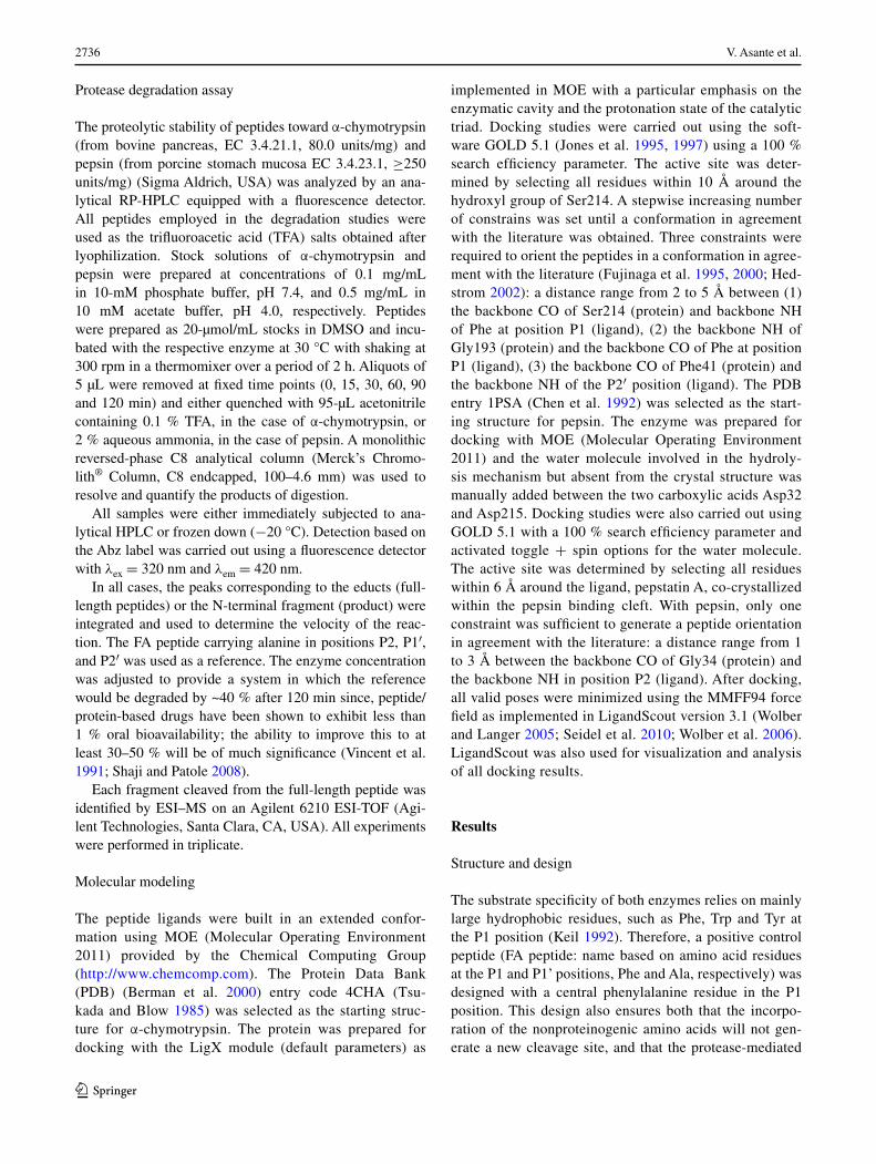
hydrolysis of the peptides used in this study will be site specific. The sequence of the FA peptide is Abz-Lys-Ala-Ala-Phe-Ala-Ala-Ala-Ala-Lys, as illustrated in Fig. 1. Because the substrate binds to the enzyme’s active site in an extended conformation, the alanines in positions P3, P3′, and P4′ act as spacers. Lysines were introduced at the ends of the peptides to enhance solubility, and o-amin-obenzoic acid (Abz) was used as a fluorescence label. Each alanine residue at the P2, P1′ or P2′ position was singly replaced by the amino acid in question, as shown in Fig. 2. The fluorinated amino acids trifluoroethylglycine (TfeGly) and difluoroethylglycine (DfeGly) differ in their degree of fluorination and hence are expected to place dif-ferent steric demands on the enzyme’s binding pocket. Additionally, such fluoroalkyl side chains have been shown to polarize neighboring C–H bonds with some-times appreciable effects on intermolecular noncovalent interactions (Salwiczek et al. 2009a, b). Aminobutyric acid (Abu) was also included in this study to distinguish between effects relating to fluorine and those relating to the hydrocarbon chain itself. This strategy led to the syn-thesis of nine FA variants carrying nonproteinogenic resi-dues in positions that are keys for the recognition of the substrate by the protease.
Protease stability of peptides
The stability of the peptides towards hydrolysis by the pro-teases was determined by an analytical RP-HPLC assay with fluorescence detection. The use of HPLC for analyz-ing cleavage products is a well-established method which offers a fast, sensitive and reproducible way to monitor the cleavage reaction (Ferrie et al. 2013; Aguilar 2004; Hua and Huang 2010; Shrimpton et al. 2000).
Each peptide was incubated with each enzyme and the products were quantified and characterized by means of the chromatography software (EZChrom Elite Version 3.1.7),
based on peak areas and mass spectrometry. Figure S1 (supporting information) shows the time course of one of the peptides in the presence of α-chymotrypsin and Fig. 3 summarizes the results of all the digestion experiments.
Molecular modeling analysis
To gain insight into the interaction between the peptide ligand and the enzyme binding site, the three FA pep-tide series were further investigated by molecular mod-eling. Each peptide/protease combination was subjected to molecular docking using GOLD (Jones et al. 1995, 1997). This software implements a genetic algorithm to represent the rotational, translational and torsion angles of a mole-cule to explore the conformational space of a ligand within an enzyme binding site. Such an approach remains chal-lenging with peptide structures that have multiple rotatable bonds. To decrease the number of atoms and bonds for the calculation, each sequence was built from the P2 to the P2′ unit, with the carboxy-terminus and the amino-terminus methylated (Me). The substrates were then constructed according to the following short sequence: MeHN-P2-P1-P1′-P2′-Me. For the 10 peptide variants (Fig. 1), docking conformations were generated within the α-chymotrypsin and pepsin enzymatic cavities. The poses were consid-ered valid when the phenylalanine P1-residue was oriented towards the hydrophobic S1 pocket of the enzyme and the backbone of the peptide was stabilized in agreement with previously published studies (Fujinaga et al. 1995, 2000; Hedstrom 2002), as summarized in Table 1. Our hypothesis was that a peptide well stabilized in the active site in silico would be an indication of good enzyme recognition and faster degradation.
Discussion
α-Chymotrypsin
In the molecular modeling study, a majority of the docking poses generated for each peptide displayed a conformation highly stabilized by the interactions reported in Table 1.
These results show excellent complementarity between the residues from P2 to P2′ of the ligand and their corre-sponding protein subsites S2 to S2′. Moreover, the side chain of Ser195 is always ideally oriented between the P1 and P1′ positions which are in agreement with the key role of this residue in the enzymatic mechanism of α-chymotrypsin. These conformations were therefore con-sidered to be a solid basis to support structural arguments regarding the ligand-enzyme interactions studied here, and their influence on proteolytic stability.
Increasing side chain volume and hydrophobicity
H2N COOH
H
CH3
Abu
H2N COOH
H
H2N COOH
H
DfeGly TfeGly
H FF
F FF
Fig. 2 Structures of the nonproteinogenic amino acids (S)-ethyl-glycine, or aminobutyric acid (Abu), (S)-4,4-difluoroethylglycine (DfeGly) and (S)-4,4,4-trifluoroethylglycine (TfeGly) used in this study
2738 V. Asante et al.
1 3
0
20
40
60
80
100
% educt
0 15 30 60 90 120Time (min)
A -Chymotrypsin B Pepsin
P2
0
20
40
60
80
100
0 15 30 60 90 120
% educt
Time (min)
0
20
40
60
80
100
0 15 30 60 90 120
% educt
Time (min)
FAP2-TfeGlyFAP2-DfeGlyFAP2-AbuFA
P1'
0
20
40
60
80
100
0 15 30 60 90 120
% educt
Time (min)
FAP1’-TfeGlyFAP1’-DfeGlyFAP1’-AbuFA
P2'
0
20
40
60
80
100
0 15 30 60 90 120
% educt
Time (min)
0
20
40
60
80
100
0 15 30 60 90 120
% educt
Time (min)
FAP2’-TfeGlyFAP2’-DfeGlyFAP2’-AbuFA
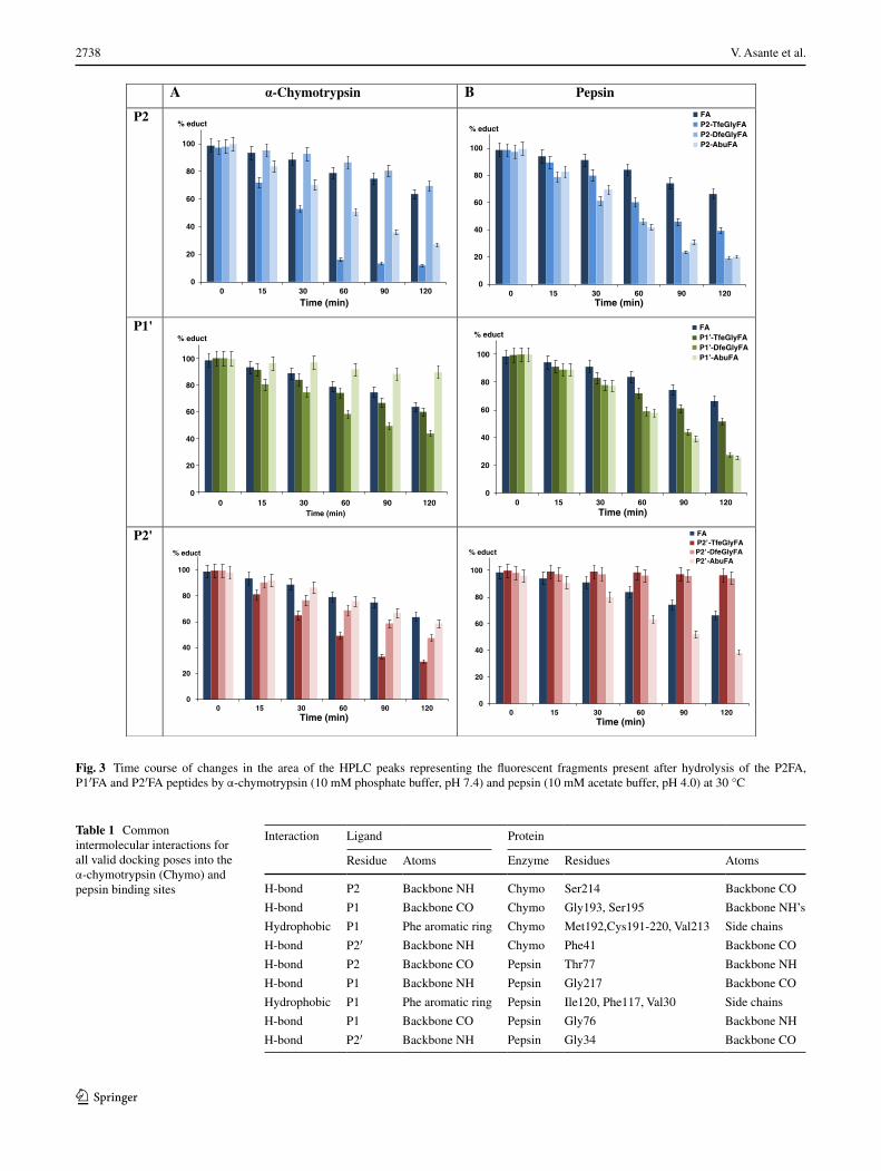
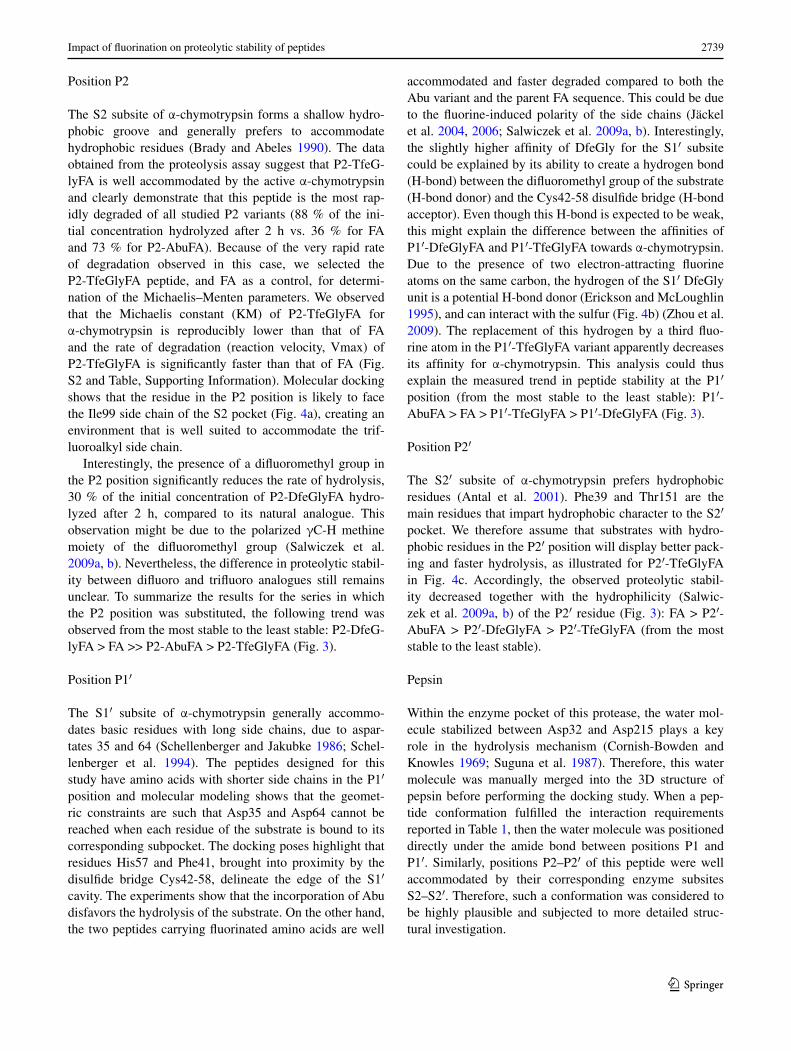
Fig. 3 Time course of changes in the area of the HPLC peaks representing the fluorescent fragments present after hydrolysis of the P2FA, P1′FA and P2′FA peptides by α-chymotrypsin (10 mM phosphate buffer, pH 7.4) and pepsin (10 mM acetate buffer, pH 4.0) at 30 °C
Table 1 Common intermolecular interactions for all valid docking poses into the α-chymotrypsin (Chymo) and pepsin binding sites
Interaction Ligand Protein
Residue Atoms Enzyme Residues Atoms
H-bond P2 Backbone NH Chymo Ser214 Backbone CO
H-bond P1 Backbone CO Chymo Gly193, Ser195 Backbone NH’s
Hydrophobic P1 Phe aromatic ring Chymo Met192,Cys191-220, Val213 Side chains
H-bond P2′ Backbone NH Chymo Phe41 Backbone CO
H-bond P2 Backbone CO Pepsin Thr77 Backbone NH
H-bond P1 Backbone NH Pepsin Gly217 Backbone CO
Hydrophobic P1 Phe aromatic ring Pepsin Ile120, Phe117, Val30 Side chains
H-bond P1 Backbone CO Pepsin Gly76 Backbone NH
H-bond P2′ Backbone NH Pepsin Gly34 Backbone CO
2739Impact of fluorination on proteolytic stability of peptides
1 3
Position P2
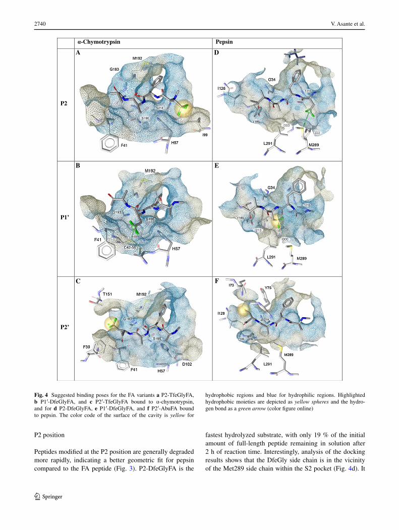
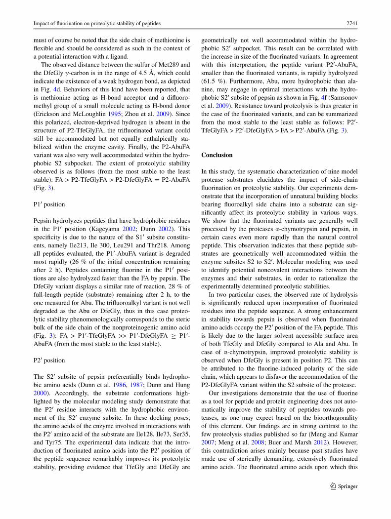
The S2 subsite of α-chymotrypsin forms a shallow hydro-phobic groove and generally prefers to accommodate hydrophobic residues (Brady and Abeles 1990). The data obtained from the proteolysis assay suggest that P2-TfeG-lyFA is well accommodated by the active α-chymotrypsin and clearly demonstrate that this peptide is the most rap-idly degraded of all studied P2 variants (88 % of the ini-tial concentration hydrolyzed after 2 h vs. 36 % for FA and 73 % for P2-AbuFA). Because of the very rapid rate of degradation observed in this case, we selected the P2-TfeGlyFA peptide, and FA as a control, for determi-nation of the Michaelis–Menten parameters. We observed that the Michaelis constant (KM) of P2-TfeGlyFA for α-chymotrypsin is reproducibly lower than that of FA and the rate of degradation (reaction velocity, Vmax) of P2-TfeGlyFA is significantly faster than that of FA (Fig. S2 and Table, Supporting Information). Molecular docking shows that the residue in the P2 position is likely to face the Ile99 side chain of the S2 pocket (Fig. 4a), creating an environment that is well suited to accommodate the trif-luoroalkyl side chain.
Interestingly, the presence of a difluoromethyl group in the P2 position significantly reduces the rate of hydrolysis, 30 % of the initial concentration of P2-DfeGlyFA hydro-lyzed after 2 h, compared to its natural analogue. This observation might be due to the polarized γC-H methine moiety of the difluoromethyl group (Salwiczek et al. 2009a, b). Nevertheless, the difference in proteolytic stabil-ity between difluoro and trifluoro analogues still remains unclear. To summarize the results for the series in which the P2 position was substituted, the following trend was observed from the most stable to the least stable: P2-DfeG-lyFA > FA >> P2-AbuFA > P2-TfeGlyFA (Fig. 3).
Position P1′
The S1′ subsite of α-chymotrypsin generally accommo-dates basic residues with long side chains, due to aspar-tates 35 and 64 (Schellenberger and Jakubke 1986; Schel-lenberger et al. 1994). The peptides designed for this study have amino acids with shorter side chains in the P1′ position and molecular modeling shows that the geomet-ric constraints are such that Asp35 and Asp64 cannot be reached when each residue of the substrate is bound to its corresponding subpocket. The docking poses highlight that residues His57 and Phe41, brought into proximity by the disulfide bridge Cys42-58, delineate the edge of the S1′ cavity. The experiments show that the incorporation of Abu disfavors the hydrolysis of the substrate. On the other hand, the two peptides carrying fluorinated amino acids are well
accommodated and faster degraded compared to both the Abu variant and the parent FA sequence. This could be due to the fluorine-induced polarity of the side chains (Jäckel et al. 2004, 2006; Salwiczek et al. 2009a, b). Interestingly, the slightly higher affinity of DfeGly for the S1′ subsite could be explained by its ability to create a hydrogen bond (H-bond) between the difluoromethyl group of the substrate (H-bond donor) and the Cys42-58 disulfide bridge (H-bond acceptor). Even though this H-bond is expected to be weak, this might explain the difference between the affinities of P1′-DfeGlyFA and P1′-TfeGlyFA towards α-chymotrypsin. Due to the presence of two electron-attracting fluorine atoms on the same carbon, the hydrogen of the S1′ DfeGly unit is a potential H-bond donor (Erickson and McLoughlin 1995), and can interact with the sulfur (Fig. 4b) (Zhou et al. 2009). The replacement of this hydrogen by a third fluo-rine atom in the P1′-TfeGlyFA variant apparently decreases its affinity for α-chymotrypsin. This analysis could thus explain the measured trend in peptide stability at the P1′ position (from the most stable to the least stable): P1′-AbuFA > FA > P1′-TfeGlyFA > P1′-DfeGlyFA (Fig. 3).
Position P2′
The S2′ subsite of α-chymotrypsin prefers hydrophobic residues (Antal et al. 2001). Phe39 and Thr151 are the main residues that impart hydrophobic character to the S2′ pocket. We therefore assume that substrates with hydro-phobic residues in the P2′ position will display better pack-ing and faster hydrolysis, as illustrated for P2′-TfeGlyFA in Fig. 4c. Accordingly, the observed proteolytic stabil-ity decreased together with the hydrophilicity (Salwic-zek et al. 2009a, b) of the P2′ residue (Fig. 3): FA > P2′-AbuFA > P2′-DfeGlyFA > P2′-TfeGlyFA (from the most stable to the least stable).
Pepsin
Within the enzyme pocket of this protease, the water mol-ecule stabilized between Asp32 and Asp215 plays a key role in the hydrolysis mechanism (Cornish-Bowden and Knowles 1969; Suguna et al. 1987). Therefore, this water molecule was manually merged into the 3D structure of pepsin before performing the docking study. When a pep-tide conformation fulfilled the interaction requirements reported in Table 1, then the water molecule was positioned directly under the amide bond between positions P1 and P1′. Similarly, positions P2–P2′ of this peptide were well accommodated by their corresponding enzyme subsites S2–S2′. Therefore, such a conformation was considered to be highly plausible and subjected to more detailed struc-tural investigation.
2740 V. Asante et al.
1 3
P2 position
Peptides modified at the P2 position are generally degraded more rapidly, indicating a better geometric fit for pepsin compared to the FA peptide (Fig. 3). P2-DfeGlyFA is the
fastest hydrolyzed substrate, with only 19 % of the initial amount of full-length peptide remaining in solution after 2 h of reaction time. Interestingly, analysis of the docking results shows that the DfeGly side chain is in the vicinity of the Met289 side chain within the S2 pocket (Fig. 4d). It
-Chymotrypsin Pepsin
P2
P1’
P2’
A D
B E
C F
Fig. 4 Suggested binding poses for the FA variants a P2-TfeGlyFA, b P1′-DfeGlyFA, and c P2′-TfeGlyFA bound to α-chymotrypsin, and for d P2-DfeGlyFA, e P1′-DfeGlyFA, and f P2′-AbuFA bound to pepsin. The color code of the surface of the cavity is yellow for
hydrophobic regions and blue for hydrophilic regions. Highlighted hydrophobic moieties are depicted as yellow spheres and the hydro-gen bond as a green arrow (color figure online)
2741Impact of fluorination on proteolytic stability of peptides
1 3
must of course be noted that the side chain of methionine is flexible and should be considered as such in the context of a potential interaction with a ligand.
The observed distance between the sulfur of Met289 and the DfeGly γ-carbon is in the range of 4.5 Å, which could indicate the existence of a weak hydrogen bond, as depicted in Fig. 4d. Behaviors of this kind have been reported, that is methionine acting as H-bond acceptor and a difluoro-methyl group of a small molecule acting as H-bond donor (Erickson and McLoughlin 1995; Zhou et al. 2009). Since this polarized, electron-deprived hydrogen is absent in the structure of P2-TfeGlyFA, the trifluorinated variant could still be accommodated but not equally enthalpically sta-bilized within the enzyme cavity. Finally, the P2-AbuFA variant was also very well accommodated within the hydro-phobic S2 subpocket. The extent of proteolytic stability observed is as follows (from the most stable to the least stable): FA > P2-TfeGlyFA > P2-DfeGlyFA = P2-AbuFA (Fig. 3).
P1′ position
Pepsin hydrolyzes peptides that have hydrophobic residues in the P1′ position (Kageyama 2002; Dunn 2002). This specificity is due to the nature of the S1′ subsite constitu-ents, namely Ile213, Ile 300, Leu291 and Thr218. Among all peptides evaluated, the P1′-AbuFA variant is degraded most rapidly (26 % of the initial concentration remaining after 2 h). Peptides containing fluorine in the P1′ posi-tions are also hydrolyzed faster than the FA by pepsin. The DfeGly variant displays a similar rate of reaction, 28 % of full-length peptide (substrate) remaining after 2 h, to the one measured for Abu. The trifluoroalkyl variant is not well degraded as the Abu or DfeGly, thus in this case proteo-lytic stability phenomenologically corresponds to the steric bulk of the side chain of the nonproteinogenic amino acid (Fig. 3): FA > P1′-TfeGlyFA >> P1′-DfeGlyFA ≥ P1′-AbuFA (from the most stable to the least stable).
P2′ position
The S2′ subsite of pepsin preferentially binds hydropho-bic amino acids (Dunn et al. 1986, 1987; Dunn and Hung 2000). Accordingly, the substrate conformations high-lighted by the molecular modeling study demonstrate that the P2′ residue interacts with the hydrophobic environ-ment of the S2′ enzyme subsite. In these docking poses, the amino acids of the enzyme involved in interactions with the P2′ amino acid of the substrate are Ile128, Ile73, Ser35, and Tyr75. The experimental data indicate that the intro-duction of fluorinated amino acids into the P2′ position of the peptide sequence remarkably improves its proteolytic stability, providing evidence that TfeGly and DfeGly are
geometrically not well accommodated within the hydro-phobic S2′ subpocket. This result can be correlated with the increase in size of the fluorinated variants. In agreement with this interpretation, the peptide variant P2′-AbuFA, smaller than the fluorinated variants, is rapidly hydrolyzed (61.5 %). Furthermore, Abu, more hydrophobic than ala-nine, may engage in optimal interactions with the hydro-phobic S2′ subsite of pepsin as shown in Fig. 4f (Samsonov et al. 2009). Resistance toward proteolysis is thus greater in the case of the fluorinated variants, and can be summarized from the most stable to the least stable as follows: P2′-TfeGlyFA > P2′-DfeGlyFA > FA > P2′-AbuFA (Fig. 3).
Conclusion
In this study, the systematic characterization of nine model protease substrates elucidates the impact of side-chain fluorination on proteolytic stability. Our experiments dem-onstrate that the incorporation of unnatural building blocks bearing fluoroalkyl side chains into a substrate can sig-nificantly affect its proteolytic stability in various ways. We show that the fluorinated variants are generally well processed by the proteases α-chymotrypsin and pepsin, in certain cases even more rapidly than the natural control peptide. This observation indicates that these peptide sub-strates are geometrically well accommodated within the enzyme subsites S2 to S2′. Molecular modeling was used to identify potential noncovalent interactions between the enzymes and their substrates, in order to rationalize the experimentally determined proteolytic stabilities.
In two particular cases, the observed rate of hydrolysis is significantly reduced upon incorporation of fluorinated residues into the peptide sequence. A strong enhancement in stability towards pepsin is observed when fluorinated amino acids occupy the P2′ position of the FA peptide. This is likely due to the larger solvent accessible surface area of both TfeGly and DfeGly compared to Ala and Abu. In case of α-chymotrypsin, improved proteolytic stability is observed when DfeGly is present in position P2. This can be attributed to the fluorine-induced polarity of the side chain, which appears to disfavor the accommodation of the P2-DfeGlyFA variant within the S2 subsite of the protease.
Our investigations demonstrate that the use of fluorine as a tool for peptide and protein engineering does not auto-matically improve the stability of peptides towards pro-teases, as one may expect based on the bioorthogonality of this element. Our findings are in strong contrast to the few proteolysis studies published so far (Meng and Kumar 2007; Meng et al. 2008; Buer and Marsh 2012). However, this contradiction arises mainly because past studies have made use of sterically demanding, extensively fluorinated amino acids. The fluorinated amino acids upon which this
2742 V. Asante et al.
1 3
study is based, however, provide a low number of fluo-rine substituents within shorter side chains. In such cases, fluorine seems often to become the key factor that triggers acceleration of peptide degradation by the here studied enzymes. Thus, our results highlight the fact that fluorine mediates interactions with proteins in ways that are not available to the hydrocarbon analogues. The here reported results are even more important in the field of medici-nal chemistry, as this tendency cannot be reconciled with the generally improved metabolic stabilities observed for small drug molecules containing fluorine atoms. Hence, the impact of side-chain fluorination on proteolytic stability of peptides is a complex phenomenon that depends upon the position of the substitution relative to the cleavage site, the type of enzyme, the chemical nature and fluorination state of the side chain, as all of these parameters dictate the pre-cise nature of the noncovalent interactions that the substrate and enzyme will engage in. For this reason, the improved resistances against hydrolysis highlighted with some vari-ants (fluorine at P2′ position with pepsin and DfeGly at P2 with α-chymotrypsin) are valuable observations that have the potential to make an impact on the design of more bio-available peptides or peptidomimetics. We are currently also investigating other aspartic acid and serine proteases to expand the studies reported here.
Acknowledgments We are grateful to the Rosa-Luxemburg-Stif-tung (RLS) and the Deutsche Forschungsgemeinschaft (Research Training Group “Fluorine as Key Element”) for financial support of Vivian Asante. Jérémie Mortier is grateful to WBI—Wallonie-Brux-elles-International for the award of a postdoctoral grant. We also express our sincere thanks to Dr. Allison Berger for her suggestions and careful editing of the text.
Conflict of interest The authors declare that they have no conflict of interest.
References
Aguilar MI (2004) HPLC of peptides and proteins: methods and Pro-tocols. Humana Press, Totowa
Antal J, Pál G, Asbóth B, Buzás Z, Patthy A, Gráf L (2001) Speci-ficity assay of serine proteinases by reverse-phase high-perfor-mance liquid chromatography analysis of competing oligopep-tide substrate library. Anal Biochem 288:156–167. doi:10.1006/abio.2000.4886
Antonov VK, Ginodman LM, Kapitannikov YV, Barshevskaya TN, Gurova AG, Rumsh LD (1978) Mechanism of pepsin catalysis: general base catalysis by the active site carboxylate ion. FEBS Lett 88:87–90
Antonov VK, Ginodman LM, Rumsh LD, Kapitannikov YV, Bar-shevskaja TN, Yavashev LP, Gurova AG, Volkova LI (1981) Stud-ies on mechanism of action of proteolytic enzymes using heavy oxygen exchange. Eur J Biochem 117:195–200
Asante V, Mortier J, Schlüter H, Koksch B (2013) Impact of fluorina-tion on proteolytic stability of peptides in human blood plasma. Bioorg Med Chem 21:3542–3546. doi:10.1016/j.bmc.2013. 03.051
Baker PJ, Montclare JK (2011) Enhanced refoldability and thermoac-tivity of fluorinated phosphotriesterase. ChemBioChem 12:1845–18481. doi:10.1002/cbic.201100221
Berman HL, Westbrook J, Feng Z, Gilliland G, Bhat TN, Weissig H, Shindyalov IN, Bourne PE (2000) The protein data bank. Nucl Acids Res 28:235–242
Blow DM, Birktoft JJ, Hartley BS (1969) Role of a buried acid group in the mechanism of action of chymotrypsin. Nature 221:337–340
Böhm HJ, Banner D, Bendels S, Kansy M (2004) Fluorine in medici-nal chemistry. ChemBioChem 5:637–643
Brady K, Abeles RH (1990) Inhibition of chymotrypsin by peptidyl-trifluoromethyl: determinants of slow-binding kinetics. Biochem-istry 29:7608–7617
Buer BC, Marsh ENG (2012) Fluorine: a new element in protein design. Prot Sci 21:453–462
Chen L, Erickson JW, Rydel TJ, Park CH, Neidhart D, Luly J, Abad-Zapatero C (1992) Structure of a pepsin/renin inhibitor complex reveals a novel crystal packing induced by minor chemical altera-tions in the inhibitor. Acta Crystallogr B 48(4):476–488
Cornish-Bowden AJ, Knowles JR (1969) The pH-dependence of pep-sin-catalysed reactions. Biochem J 113:353–362
Coughlin SR (2000) Thrombin signalling and protease-activated receptors. Nature 407:258–264
Czapinska H, Otlewski J (1999) Structural and energetic determi-nants of the S1-site specificity in serine proteases. Eur J Biochem 260:571–595
Dasgupta P, Singh A, Mukherjee R (2002) N-terminal acylation of somatostatin analog with long chain fatty acids enhances its sta-bility and anti-proliferative activity in human breast adenocarci-noma cells. Biol Pharm Bull 25:29–36
Davies DR (1990) The structure and function of the aspartic protein-ases. Annu Rev Biophys Chem 19:189–215
Derewenda ZS, Derewenda U, Kobos PM (1994) (His) Cε-H…O = C < hydrogen bond in the active sites of serine. J Mol Biol 241:83–93
Dougherty DA (2000) Unnatural amino acids as probes of protein structure and function. Curr Opin Chem Biol 4:645–652
Dunn BM (2002) Structure and mechanism of the pepsin-like family of aspartic peptidases. Chem Rev 102:4431–4458
Dunn BM, Hung S (2000) The two sides of enzyme-substrate speci-ficity: lessons from the aspartic proteinases. Biochim Biophys Acta 1477:231–240
Dunn BM, Jimenez M, Parten BF, Valler MJ, Rolph CE, Kay J (1986) A systematic series of synthetic chromophoric substrates for aspartic proteinases. Biochem J 237:899–906
Dunn BM, Valler MJ, Rolph CE, Foundling SI, Jimenez M, Kay J (1987) The pH dependence of the hydrolysis of chromogenic sub-strates of the type, Lys-Pro-Xaa-Yaa-Phe-(NO2) Phe-Arg-Leu, by selected aspartic proteinases: evidence for specific interactions in subsites S3 and S2. Biochim Biophys Acta 913:122–130
Erickson JA, McLoughlin JI (1995) Hydrogen bond donor properties of the difluoromethyl group. J Org Chem 60:1626–1631. doi:10.1021/jo00111a021
Ferrie JJ, Gruskos JJ, Goldwaser AL, Decker ME, Guarracino DA (2013) A comparative protease stability study of synthetic macro-cyclic peptides that mimic two endocrine hormones. Bioorg Med Chem Lett 23:989–995
Filler R, Saha R (2009) Fluorine in medicinal chemistry: a century of progress and a 60-years retrospective of selected highlights. Future Med Chem 1:777–791
Frackenpohl J, Arvidsson PI, Schreiber JV, Seebach D (2001) The outstanding biological stability of β- and γ-peptides toward pro-teolytic enzymes: an in vitro investigation with fifteen peptidases. ChemBioChem 2:445–455
Fruton JS (1970) The specificity and mechanism of pepsin action. Adv Enzymol Relat Areas Mol Biol 33:401–443
2743Impact of fluorination on proteolytic stability of peptides
1 3
Fujinaga M, Chernaia MM, Tarasova NI, Mosimann SC, James MNG (1995) Crystal-structure of human pepsin and its complex with pepstatin. Protein Sci 4:960–972
Fujinaga M, Cherney MM, Tarasova NI, Bartlett PA, Hanson JE, James MNG (2000) Structural study of the complex between human pepsin and a phosphorus-containing peptidic—transition-state analog. Acta Crystallogr Sect D56:272–279. doi:10.1107/S0907444999016376Wiley
Geurink PP, Liu N, Spaans MP, Downey SL, van den Nieuwendijk AMCH, van der Marel GA, Kisselev AF, Florea BI, Overkleeft HS (2010) Incorporation of fluorinated phenylalanine generates highly specific inhibitor of proteasome’s chymotrypsin-like sites. J Med Chem 53:2319–2323
Giannis A, Kolter T (1993) Peptidomimetics for receptor ligands-dis-covery, development and medicinal perspectives. Angew Chem Int Ed Engl 32:1244–1267
Gottler LM, Lee HY, Shelburne CE, Ramamoorthy A, Marsh ENG (2008) Modulating the biological activity of an antimicrobial peptide using fluorous amino acids. ChemBioChem 9:370–373
Hedstrom L (2002) Serine protease mechanism and specificity. Chem Rev 102:4501–4523
Hruby VJ (2002) Designing peptide receptor agonists and antagonists. Nat Rev Drug Discov 1:847–858
Hua J, Huang K-L (2010) A reversed phase HPLC method for the analysis of nucleotides to determine 5′-PDE enzyme activity. Bull Chem Soc Ethiop 24:167–174
Hummel G, Reineke U, Reimer U (2006) Translating peptides into small molecules. Mol BioSyst 2:499–508
Isanbor C, O’Hagan D (2006) Fluorine in medicinal chemistry: a review of anticancer agent. J Fluorine Chem 127:303–319
Jäckel C, Koksch B (2005) Fluorine in peptide design and protein engineering. Eur J Org Chem 21:4482–4503
Jäckel C, Seufert W, Thust S, Koksch B (2004) Evaluation of the molecular interactions of fluorinated amino acids with native pol-ypeptides. ChemBioChem 5:717–720
Jäckel C, Salwiczek M, Koksch B (2006) Fluorine in a native protein environment—how space filling and polarity of fluorinated alkyl groups affect protein folding. Angew Chem Int Ed 45:4198–4203
Jones G, Willett P, Glen RC (1995) Molecular recognition of receptor sites using a genetic algorithm with a description of desolvation. J Mol Biol 245:43–53. doi:10.1016/S0022-2836(95)80037-9
Jones G, Willett P, Glen RC, Leach AR, Taylor R (1997) Develop-ment and validation of a genetic algorithm for flexible docking. J Mol Biol 267:727–748. doi:10.1006/jmbi.1996.0897
Kageyama T (2002) Pepsinogens, progastricsins, and prochymosins: structure, function, evolution, and development. Cell Mol Life Sci 59:288–306
Keil B (1992) Specificity of proteolysis. Springer, Berlin 335Koksch B, Sewald N, Burger K, Jakubke H-D (1996a) Peptide modi-
fication by incorporation of α-trifluoromethyl substituted amino acids. Amino Acids 11:425–434
Koksch B, Sewald N, Jakubke H-D, Burger K (1996b) Synthesis and incorporation of α-trifluoromethyl-substituted amino acids into peptides. In: Ojima I, McCarthy JR, Welch JT (eds) Biomedical frontiers of fluorine chemistry, ACS Symposium Series, vol 639. American Chemical Society, Washington DC, pp 42–58
Koksch B, Sewald N, Hofmann H-J, Burger K, Jakubke H-D (1997) Proteolytically stable peptides by incorporation of alpha-Tfm amino acids. J Peptide Sci 3:157–167
Lee H, Jang IH, Ryu SH, Park TG (2003) N-terminal site-specific mono-PEGylation of epidermal growth factor. Pharm Res 20:818–825
Łegowska A, Debowski D, Lesner A, Wysocka M, Rolka K (2009) Intro-duction of non-natural amino acid residues into the substrate-spe-cific P1 position of trypsin inhibitor SFTI-1 yields potent chymot-rypsin and cathepsin G inhibitors. Bioorg Med Chem 17:3302–3307
March TL, Johnston MR, Duggan PJ, Gardiner J (2012) Synthesis, structure, and biological applications of α-fluorinated β-amino acids and derivatives. Chem Biodiversity 9:2410–2441. doi:10.1002/cbdv.201200307
McGregor DP (2008) Discovering and improving novel peptide thera-peutics. Curr Opin Pharmacol 8:616–619
Meng H, Kumar K (2007) Antimicrobial activity and protease stabil-ity of peptides containing fluorinated amino acids. J Am Chem Soc 129:15615–15622
Meng H, Krishnaji ST, Beinborn M, Kumar K (2008) Influence of selective fluorination on the biological activity and proteolytic stability of glucagon-like peptide-1. J Med Chem 51:7303–7307. doi:10.1021/jm8008579
Muller K, Faeh C, Diederich F (2007) Fluorine in pharmaceuticals: looking beyond intuition. Science 317:1881–1886
Northrop JH (1930) Crystalline pepsin. I. Isolation and tests of purity. J Gen Physiol 13:739–766
Polgár L (2005) The catalytic triad of serine peptidases. Cell Mol Life Sci 62:2161–2172. doi:10.1007/s00018-005-5160-x
Powers JC, Harley AD, Myers DV (1977) Subsite specificity of por-cine pepsin. Adv Exp Med Biol 95:141–157
Saeki K, Ozaki K, Kobayashi T, Ito S (2007) Enzymatic properties, genes, and crystal structures. J Biosci Bioeng 103:501–508
Saffran M, Kumar G, Savariar C, Burnham J, Williams F, Neckers D (1986) A new approach to the oral administration of insulin and other peptide drugs. Science 233:1081–1084
Salwiczek M, Samsonov S, Vagt T, Baldauf C, Nyakatura E, Fleige E, Numata J, Cölfen H, Pisabarro MT, Koksch B (2009a) Posi-tion dependent effects of fluorinated amino acids on hydropho-bic core formation of a coiled coil heterodimer. Chem Eur J 15:7628–7636
Salwiczek M, Vagt T, Koksch B (2009b) Artificial model systems designed to investigate fluorinated amino acids in native protein environments. In: Ojima I (ed) Fluorine in bioorganic and medic-inal chemistry. Blackwell Publishers, pp 391–409
Salwiczek M, Nyakatura EK, Gerling UIM, Ye S, Koksch B (2012) Fluorinated amino acids: compatibility with native protein struc-tures and effects on protein–protein interactions. Chem Soc Rev 41:2135–2171. doi:10.1039/C1CS15241F
Samsonov SA, Salwiczek M, Anders G, Koksch B, Pisabarro MT (2009) Fluorine in protein environments: a QM and MD study. J Phys Chem B 113:16400–16408
Sani M, Sinisi R, Viani F (2006) Peptidyl fluoro-ketones as proteo-lytic enzyme inhibitors. Curr Top in Med Chem 6:1545–1566
Sato AK, Viswanathan M, Kent RB, Wood CR (2006) Therapeutic peptides: technological advances driving peptides into develop-ment. Curr Opin Biotechnol 17:638–642
Schechter I, Berger A (1967) On the size of the active site in pro-teases. Biochem Biophys Res Com 27:157–162
Schellenberger V, Jakubke HD (1986) A spectrophotometric assay for the characterization of the S’ subsite specificity of alpha-chymot-rypsin. Biochim Biophys Acta 869:54–60
Schellenberger V, Turck CW, Rutter WJ (1994) Role of the S’ sub-sites in serine protease catalysis-active-site mapping of rat chy-motrypsin, rat trypsin, alpha-lytic protease, and cercarial protease from Schistosoma-Mansoni. Biochem 33:4251–4257
Seidel T, Ibis G, Bendix F, Wolber G (2010) Strategies for 3D phar-macophore-based virtual screening. Drug Discov Today Tecnol 7:221–228. doi:10.1016/j.ddtec.2010.11.004
Shaji J, Patole V (2008) Protein and peptide drug delivery: oral approaches. Indian J Pharm Sci 70:269–277
Shrimpton CN, Abbenante G, Lew RA, Smith AI (2000) Develop-ment and characterization of novel potent and stable inhibitors of endopeptidase EC 3.4.24.15. Biochem J 345:351–356
Smits R, Koksch B (2006) How C-alpha-fluoroalkyl amino acids and peptides interact with enzymes: studies concerning the influence
2744 V. Asante et al.
1 3
on proteolytic stability, enzymatic resolution and peptide cou-pling. Curr Top Med Chem 16:1483–1498
Suguna K, Padlan EA, Smith CW, Carlson WD, Davies DR (1987) Binding of a reduced peptide inhibitor to the aspartic proteinase from Rhizopus chinensis: implications for a mechanism of action. Proc Natl Acad Sci USA 84:7009–7013
Tsukada H, Blow DM (1985) Structure of α-chymotrypsin refined at 1.6 Å resolution. J Mol Biol 184:703–711
Vincent HL, Satish DK, George MG, Werner R (1991) Oral route of protein and peptide drug delivery. In: Vincent HL (ed) Peptide and protein drug delivery. Marcel Dekker, New York, pp 691–738
Vlieghe P, Lisowski V, Martinez J, Khrestchatisky M (2010) Synthetic therapeutic peptides: science and market. Drug Discov Today 15:40–56
Voloshchuk N, Zhu AY, Snydacker D, Montclare JK (2009) Posi-tional effects of monofluorinated phenylalanines on histone acetyltransferase stability and activity. Bioorg Med Chem Lett 19:5449–5451
Werle M, Bernkop-Schnürch A (2006) Strategies to improve plasma half life time of peptide and protein drugs. Amino Acids 30:351–367. doi:10.1007/s00726-005-0289-3
Wolber G, Langer T (2005) LigandScout: 3-D pharmacophores derived from protein-bound ligands and their use as virtual screening filters. J Chem Inf Model 45:160–169. doi:10.1021/ci049885e
Wolber G, Dornhofer AA, Langer T (2006) Efficient overlay of small molecules using 3-D pharmacophores. J Comput-Aid Mol Des 20:773–788. doi:10.1007/s10822-006-9078-7
Jones G, Willett P, Glen RC http://www.ccdc.cam.ac.uk/products/life_sciences/gold/
Zhou P, Tian F, Lv F, Shang Z (2009) Geometric characteristics of hydrogen bonds involving sulfur atoms in proteins. Proteins 76:151–163. doi:10.1002/prot.22327
![Index []Beer foam carbonation, 314 fatty acids, 71 hops, 46 proteolytic enzymes, 264 total soluble nitrogen, 29 Beer industry advertising, 15 beer sales, 7 beer segments, 12 classification](https://static.fdocument.org/doc/165x107/5f1733991d40922a93492bd1/index-beer-foam-carbonation-314-fatty-acids-71-hops-46-proteolytic-enzymes.jpg)


![Index [] · The Power of Functional Resins in Organic Synthesis. ... α-chymotrypsin 603 Aβ (β-amyloid (1-42)) synthesis 504, 507, 508 Accurel MP 1000 373 acetal-protected carbonyls](https://static.fdocument.org/doc/165x107/5f6421717515ab779846508d/index-the-power-of-functional-resins-in-organic-synthesis-chymotrypsin.jpg)