Hydrogen Bonds and Heat Diffusion in α-Helices: A Computational Study
10
Hydrogen Bonds and Heat Diffusion in α‑Helices: A Computational Study German Miñ o,* ,†,‡,§ Raul Barriga, † and Gonzalo Gutierrez † † Group of NanoMaterials, Departamento de Física, Facultad de Ciencias, Universidad de Chile, Casilla 653, Santiago, Chile ‡ Centro Interdisciplinario de Neurociencias de Valparaíso (CINV), Universidad de Valparaíso, Valparaíso, Chile § Facultad de Ciencias Biologicas, Centro de Bioinformatica y Biologia Integrativa, Universidad Andres Bello, Av.Republica 239, Santiago, Chile ABSTRACT: Recent evidence has shown a correlation between the heat diffusion pathways and the known allosteric communication pathways in proteins. Allosteric communication in proteins is a central, yet unsolved, problem in biochemistry, and the study and characterization of the structural determinants that mediate energy transfer among different parts of proteins is of major importance. In this work, we characterized the role of hydrogen bonds in diffusivity of thermal energy for two sets of α-helices with different abilities to form hydrogen bonds. These hydrogen bonds can be a constitutive part of the α-helices or can arise from the lateral chains. In our in vacuo simulations, it was observed that α-helices with a higher possibility of forming hydrogen bonds also had higher rates of thermalization. Our simulations also revealed that heat readily flowed through atoms involved in hydrogen bonds. As a general conclusion, according to our simulations, hydrogen bonds fulfilled an important role in heat diffusion in structural patters of proteins. ■ INTRODUCTION Proteins play the most important role in living organisms. In this respect, energy transport in proteins is an open problem in biochemistry that involves important topics conformational changes, enzyme catalysis, allosteric cooperativity, and inter- molecular affinities, among other process. 1 The necessary biological energy can originate from several sources such as thermal gradients, chemical or photochemical changes in substrates at active sites of enzymes, or protein−ligand binding. 1 In that respect, nonlinear excitations such as breathers, 2 solitons, 3−5 low- and high-energy vibrational modes 1,6 have been drawing increasing attention as mediators of energy transport in biomolecules. Due their polymeric nature, when proteins fold to reach their biological active state, they generate a complex network of contacts. Previous reports have shown that thermal energy flows in proteins according to the physical connectivity of this network, 7,8 with velocities of propagation in the orders of 10 Å ps −1 . 6 This flow of thermal energy mimics the heat transport of percolation clusters, where the energy flows anisotropically, that is, fast along physically connected channels and rather slow along numerous pathways that reach dead-ends. 6 Recent evidence has shown a correlation between the heat diffusion pathways and the known allosteric communication pathways in proteins. 9−11 Allosteric communication determines the processes of signal transmission through proteins, on both short (3 Å) and long (100 Å) range scales. This phenomenon is involved in crucial cellular and physiological functions and is a determinant for serious human diseases. 12−14,41 Allosteric communication can be determined using both experimental 15−21,26,27 and theoreti- cal 1,6−11,19,22−25 techniques, and their occurrence has been clearly established. Large efforts have been made to study the energy flow in proteins and its relation in protein dynamics. 28−33,36 Research on heat flow in different structures, such as peptide helices, 31 heme cofactors, 32 beta sheets 33 and functionalized materials 34,35 have been reported in literature. These reports have shown that the excess of energy deposited in particular sites can propagate along the structures through the covalent backbone of the molecules 31,35 and also through weaker interactions, such as hydrogen bonds. 33,34 Additionally, the energy deposited in particular sites can flow toward the solvent. 31 The solvent can play an important role in energy diffusion through the proteins, as in the Ca 2+ ATPase; 28,29 at the surface of proteins, as in PDZ-2 domain 27 and between protein subunits, as in hemoglobin. 8 Hydrogen bonds have been an inexhaustible source of research for decades. 37 Their role as stabilizing agents of protein structure have been clearly established. 38 Also, they act in the modulation chemical reactivity in proteins 43−45 and, as has been earlier proposed from the field of physics, can act as energy carriers in protein structures. 3 In this regard, the role of hydrogen bonds in thermal conduction, has been recently reported, revealing their importance in heat diffusion across of β-sheet structure of the spider silk protein. 33 In a similar way Martinez et al. 40 have shown Received: April 7, 2014 Revised: July 13, 2014 Article pubs.acs.org/JPCB © XXXX American Chemical Society A dx.doi.org/10.1021/jp503420e | J. Phys. Chem. B XXXX, XXX, XXX−XXX
Transcript of Hydrogen Bonds and Heat Diffusion in α-Helices: A Computational Study
Hydrogen Bonds and Heat Diffusion in α‑Helices: A ComputationalStudyGerman Mino,*,†,‡,§ Raul Barriga,† and Gonzalo Gutierrez†
†Group of NanoMaterials, Departamento de Física, Facultad de Ciencias, Universidad de Chile, Casilla 653, Santiago, Chile‡Centro Interdisciplinario de Neurociencias de Valparaíso (CINV), Universidad de Valparaíso, Valparaíso, Chile§Facultad de Ciencias Biologicas, Centro de Bioinformatica y Biologia Integrativa, Universidad Andres Bello, Av.Republica 239,Santiago, Chile
ABSTRACT: Recent evidence has shown a correlation between the heatdiffusion pathways and the known allosteric communication pathways inproteins. Allosteric communication in proteins is a central, yet unsolved,problem in biochemistry, and the study and characterization of the structuraldeterminants that mediate energy transfer among different parts of proteinsis of major importance. In this work, we characterized the role of hydrogenbonds in diffusivity of thermal energy for two sets of α-helices with differentabilities to form hydrogen bonds. These hydrogen bonds can be aconstitutive part of the α-helices or can arise from the lateral chains. In ourin vacuo simulations, it was observed that α-helices with a higher possibilityof forming hydrogen bonds also had higher rates of thermalization. Oursimulations also revealed that heat readily flowed through atoms involved inhydrogen bonds. As a general conclusion, according to our simulations,hydrogen bonds fulfilled an important role in heat diffusion in structural patters of proteins.
■ INTRODUCTION
Proteins play the most important role in living organisms. In thisrespect, energy transport in proteins is an open problem inbiochemistry that involves important topics conformationalchanges, enzyme catalysis, allosteric cooperativity, and inter-molecular affinities, among other process.1 The necessarybiological energy can originate from several sources such asthermal gradients, chemical or photochemical changes insubstrates at active sites of enzymes, or protein−ligand binding.1In that respect, nonlinear excitations such as breathers,2
solitons,3−5 low- and high-energy vibrational modes1,6 havebeen drawing increasing attention as mediators of energytransport in biomolecules.Due their polymeric nature, when proteins fold to reach their
biological active state, they generate a complex network ofcontacts. Previous reports have shown that thermal energy flowsin proteins according to the physical connectivity of thisnetwork,7,8 with velocities of propagation in the orders of 10 Åps−1.6 This flow of thermal energy mimics the heat transport ofpercolation clusters, where the energy flows anisotropically, thatis, fast along physically connected channels and rather slow alongnumerous pathways that reach dead-ends.6 Recent evidence hasshown a correlation between the heat diffusion pathways and theknown allosteric communication pathways in proteins.9−11
Allosteric communication determines the processes of signaltransmission through proteins, on both short (3 Å) and long(100 Å) range scales. This phenomenon is involved in crucialcellular and physiological functions and is a determinant forserious human diseases.12−14,41 Allosteric communication can be
determined using both experimental15−21,26,27and theoreti-cal1,6−11,19,22−25 techniques, and their occurrence has beenclearly established.Large efforts have been made to study the energy flow in
proteins and its relation in protein dynamics.28−33,36 Research onheat flow in different structures, such as peptide helices,31 hemecofactors,32 beta sheets33 and functionalized materials34,35 havebeen reported in literature. These reports have shown that theexcess of energy deposited in particular sites can propagate alongthe structures through the covalent backbone of themolecules31,35 and also through weaker interactions, such ashydrogen bonds.33,34 Additionally, the energy deposited inparticular sites can flow toward the solvent.31 The solvent canplay an important role in energy diffusion through the proteins,as in the Ca2+ ATPase;28,29 at the surface of proteins, as in PDZ-2domain27 and between protein subunits, as in hemoglobin.8
Hydrogen bonds have been an inexhaustible source of researchfor decades.37 Their role as stabilizing agents of protein structurehave been clearly established.38 Also, they act in the modulationchemical reactivity in proteins43−45 and, as has been earlierproposed from the field of physics, can act as energy carriers inprotein structures.3 In this regard, the role of hydrogen bonds inthermal conduction, has been recently reported, revealing theirimportance in heat diffusion across of β-sheet structure of thespider silk protein.33 In a similar wayMartinez et al.40 have shown
Received: April 7, 2014Revised: July 13, 2014
Article
pubs.acs.org/JPCB
© XXXX American Chemical Society A dx.doi.org/10.1021/jp503420e | J. Phys. Chem. B XXXX, XXX, XXX−XXX
that residues, such as arginine, lysine, aspartate, and glutamate,are responsible for the vibrational energy flow among segmentsof the binding domains in thyroid hormone receptors. Moreover,the mutation of these key residues severely impaired thefunctionality of these receptors. Due to their charged nature,these residues can form charge-assisted hydrogen bonds,41 inaddition to also having the potential to form short hydrogenbonds.42 The role that short hydrogen bonds have in theenhanced catalytic properties of proteins has been experimen-tally studied,43 while theoretical studies have examined theirelectronic properties and the effect that they have on thechemical reactivity of surrounding structural elements.44,45
Keeping in mind the significance of allosteric communicationin proteins, the study and characterization of structuraldeterminants that mediate energy transfer among differentparts of proteins is of major importance.Interestingly, there exists a model regarding the role of
hydrogen bonds in heat diffusion in peptide helices, the so-calledDavydov model.3 This model describes how the released energyfrom, e.g., ATP hydrolysis, is trapped and how it then propagatesalong peptide helices. The released energy is captured by theamide I vibrational mode of the nearby peptide groups (CONH)of the helix. Peptide groups give rise to the hydrogen bonds thatstabilize the peptide helix. The amide I vibrational mode includeda major component from the stretching of carbonyl group (CO),and relatively small components from both the C−N in-planestretching and N−H in-plane bending vibration. The excitationof amide I modes provokes a local deformation of the helix. Theinterplay between the deformation and excitation creates a wavethat propagates along the structure, a soliton.3−5 Some authorshave made research based on this hypothesis, e.g., refs 3 and 4,nevertheless its validity at relevant biological temperatures hasbeen put in doubt.39
In this work, we characterized the role of hydrogen bonds inthe heat diffusion of α-helices. Using a molecular dynamicssimulation for a set of α-helices, we estimated the energy flux atselected points of the structure. The results were analyzed interms of the chemical nature of the α-helix building blocks; thatis, in terms of their ability to form hydrogen bonds and on thelength of the lateral chain. Increased thermal diffusivity wasfound in structures that presented a shorter lateral chain and thatformed extra hydrogen bonds. Our results are in directconcordance with recent reports about the role of hydrogenbonds in the enhanced heat diffusion in structural patterns ofproteins, such as β-sheets33 and in other functionalizedmaterials.34
■ METHODS AND COMPUTATIONAL DETAILS
The evaluation of thermal conductivity was performed for 10 α-helical structures, five of which presented nonpolar lateral chainsand five of which presented polar lateral chains. Each α-helix wascomposed by only one type of amino acid residue, and each had alength of 33 residues. The nonpolar group was constituted byphenilalanine (PHE), isoleucine (ILE), leucine (LEU),methionine (MET), and valine (VAL). The polar group wasconstituted by threonine (THR), glutamine (GLN), asparagine(ASN), serine (SER), and cysteine (CYS). The α-helices wereacetylated and amidated at their N and C-terminal, respectively,thus rendering them electrically neutral.Figure 1 shows the structure of polyserine and two types of
hydrogen bonds. One type of bond, termed the central hydrogenbonds, was a constitutive part of the α-helix. The other type ofhydrogen bonds was termed the lateral hydrogen bonds, a bondthat can form between lateral chains or between lateral chainsand the backbone of the α-helix. A difference between the polarand the nonpolar groups was that the former gave rise to centralhydrogen bonds and lateral hydrogen bonds while the latter onlygave rise to central hydrogen bonds.All simulations were performed with the NAMD Molecular
Dynamics Software v.2.946 using the CHARMM2247 potentialenergy function with CMAP correction. CMAP is an energycorrection based on quantum calculations that improves proteinbackbone behavior and thus yields more accurate prediction ofprotein structure and dynamics.48
The structures were minimized over 105 steps and furtherequilibrated in vacuo for 1 ns at a constant temperature of 300 Kusing Langevin dynamics with a damping coefficient of γ = 5 ps−1.An integration time-step of 1 fs was used with a uniform dielectricconstant of 1 and a cutoff of nonbonded forces with a switchingfunction that started at a distance of 10 Å and reached zero at 13.5Å. All of the selected structures kept an α-helical structurewithout the need for any specific constraints. After this, all thestructures were cooled and re-equilibrated to 10 K for 1 ns. Thenthe heating procedure was applied with this initial temperature of10 K. Heat was then injected by increasing the kinetic energy ofthe carbonyl group (CO) (see Figure 1) at the N-acetylmoiety to a target temperature of 300 K. For this, the sameCOgroup was subjected to a confining potential of 5 kcal/mol·A2,and two specific atoms were fixed. These fixed atoms werelocated in the N-acetylated moiety (carbon atom of the methylgroup) and in the C-amidated moiety (nitrogen atom of theamide group). These two atoms, in combination with the
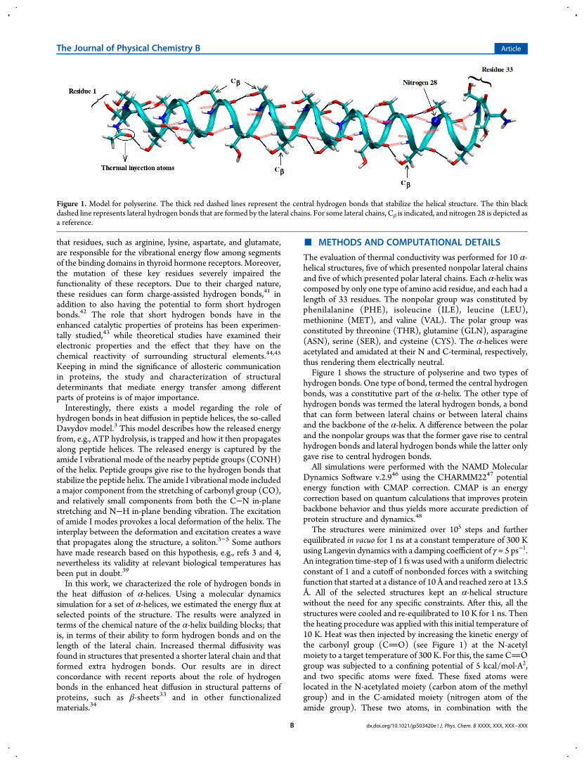
Figure 1. Model for polyserine. The thick red dashed lines represent the central hydrogen bonds that stabilize the helical structure. The thin blackdashed line represents lateral hydrogen bonds that are formed by the lateral chains. For some lateral chains, Cβ is indicated, and nitrogen 28 is depicted asa reference.
The Journal of Physical Chemistry B Article
dx.doi.org/10.1021/jp503420e | J. Phys. Chem. B XXXX, XXX, XXX−XXXB
confining potential of the CO group, prevented the loss of thehelical structure during the heating process. These conditionsalso avoided the dissipation of energy by internal rotation alongthe axis connecting the fixed atoms in the N- and C-terminals.The heat flow was determined in selected atoms through arunning average of 1000 time-steps. With this data, the average
power was computed when needed. Structurally, the formationof total hydrogen bonds, central hydrogen bonds, and lateralhydrogen bonds was measured alongside simulations for bothgroups. All the simulations were done in vacuo. There isexperimental evidence that 70% of the injected energy in peptidehelices is dissipated to the solvent.8 Thus, as our interest in this
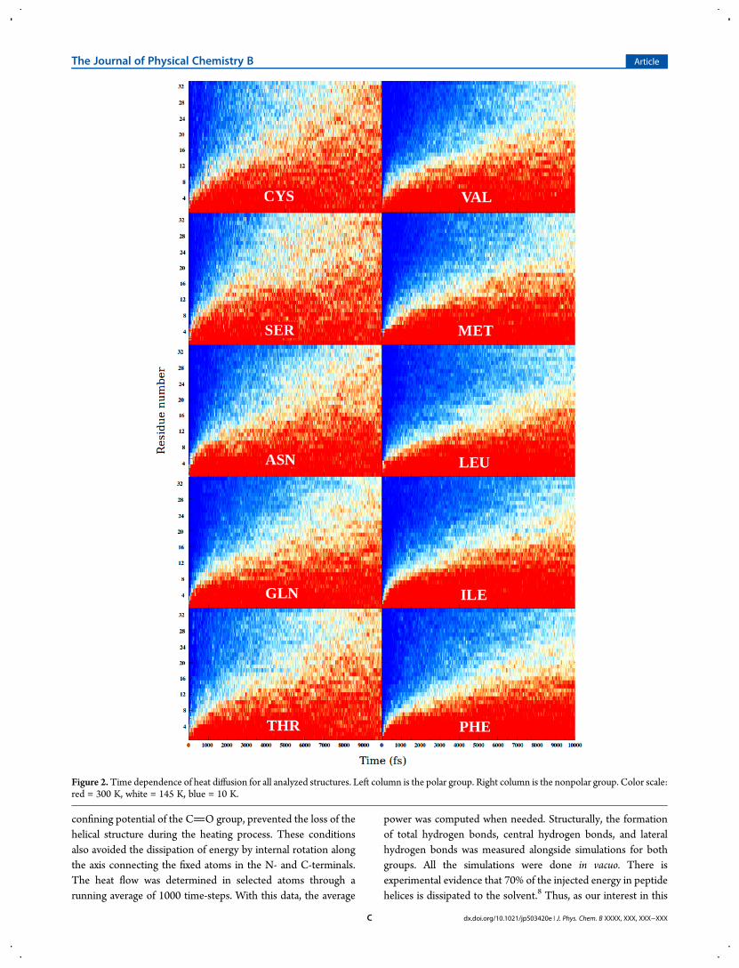
Figure 2.Time dependence of heat diffusion for all analyzed structures. Left column is the polar group. Right column is the nonpolar group. Color scale:red = 300 K, white = 145 K, blue = 10 K.
The Journal of Physical Chemistry B Article
dx.doi.org/10.1021/jp503420e | J. Phys. Chem. B XXXX, XXX, XXX−XXXC
work is to study the heat flow through the structure, we chose to
do the simulations in vacuo to maximize this process. Our model
setup seeks to emulate a nonfluxtional α-helix and its response to
a local energy excitation.
For use as a comparison, a cooling procedure was alsoperformed. Using the equilibrated structures at 300 K, the kineticenergy was removed. At the same time, the atoms of residues 1and 33 were coupled to a thermal bath at 0 K. The coolingprocedure ran toward a target temperature of 0 K. No constraints
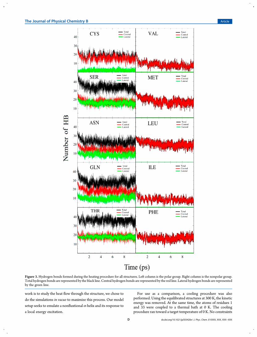
Figure 3.Hydrogen bonds formed during the heating procedure for all structures. Left column is the polar group. Right column is the nonpolar group.Total hydrogen bonds are represented by the black line. Central hydrogen bonds are represented by the red line. Lateral hydrogen bonds are representedby the green line.
The Journal of Physical Chemistry B Article
dx.doi.org/10.1021/jp503420e | J. Phys. Chem. B XXXX, XXX, XXX−XXXD
in position or confining potentials were applied to this set ofsimulations.The heating and cooling procedures used the T-couple
algorithm of NAMD2. Visualization and data analysis werecarried out using the VMD 1.9.1 software.49
■ RESULTS AND DISCUSSIONThe time dependence of heat diffusion for the heating procedureof polar and nonpolar groups is shown in Figure 2. These plotsgive a qualitative picture of the thermal diffusivity along eachstructure. Moreover, inspection of Figure 2 clearly showsdifferences between the two groups. In the polar group (leftcolumn panel), the high temperature regime (around 300 K)reached the C-terminal site of the protein at 10 000 fs of thesimulation time; in the nonpolar group (right column panel), thissame temperature regime only reached half of the α-helix. Fromthe beginning of the heating procedure, the polar group showedenhanced diffusivity along the α-helix, and this feature was morepronounced between 5000 and 10 000 fs. Given this, the polargroup took less time to transfer heat and thermalize the otherside of their respective α-helices.As the main difference between the analyzed groups was the
ability to form hydrogen bonds, these structures were analyzedand quantified during the simulations. Effectively, significantdifferences were shown through the quantification of hydrogenbonds. Figure 3 shows the number of central hydrogen bonds,lateral hydrogen bonds, and total hydrogen bonds formed byboth groups. As can be observed, the polar group (left columnpanel) formed both central hydrogen bonds and lateral hydrogenbonds, while the nonpolar group (right column) only formedcentral hydrogen bonds. In general, this gave rise to an increasednumber of total hydrogen bonds for the polar group. Analysissuggested that the extra hydrogen bonds formed by the polargroup was the cause for the observed increase in diffusivity.In order to gain numerical insight into the heat diffusion
process, the average power was determined for the backboneatoms N, O, C, and Cα at residue 28 of each structure in theinterval between 0 to 10 000 fs. These results are shown in Table1. From this determination, the groups were classified into tworanges of average power. The power of the polar group rankedfrom 364.2 to 285.7 pW, while the power of the nonpolar groupranked from 259.6 to 215.3 pW. No apparent correlation couldbe made with the molecular weight (MW) of the monomer thatcomposed each α-helix for both groups, but an inverse relationwas established between power and the size of the lateral chain.For the nonpolar group, this relation was readily observed, wherean increased size of the lateral chain led to a lower average powerat position 28. This observation is reminiscent of the dead-endconcept, in which a longer or more branched lateral chain leads toa lower power. When taking into account the sequences VAL,LEU, ILE, and PHE, it was possible to follow the bifurcation inatomic connectivity. In the case of MET, an intermediary effectbetween VAL and LEU was observed.This effect was also observed in the polar group, in which a
more bifurcated side chain was translated in lower power at site28. Considering the sequence CYS, SER, ASN, and GLN, it waspossible to follow the effect of the length of the lateral chain in themeasured power at site 28. As the lateral chain increased inlength, power decreased. In the case of THR, the bifurcation of amethyl group (−CH3) led to a dead-end that could impose anextra delay in heat transfer to site 28, with a value of 285.7 pW.The replacement of the hydroxyl group (−OH) in THR by−CH3 led to VAL of the nonpolar group with a value of 259.6
pW, and this was the next lateral chain after THR in the sequencepower measurements at position 28.Another comparison was established among members of the
two groups that shared a similar MW and degree of ramification.The pairs ASN/LEU and ASN/ILE, which shared a similar MWand ramification, showed increased power for the polar member.The same comparison was applied to the pairs THR/VAL andGLN/MET. These two polar/nonpolar pairs shared a similarMW and bifurcation with the respective polar member, showinga higher average power at the reference position number 28.These comparisons suggest that lateral hydrogen bonds is animportant factor in the process of thermalization for these α-helices.Measuring the kinetic energy of the Cβ atom is also useful for
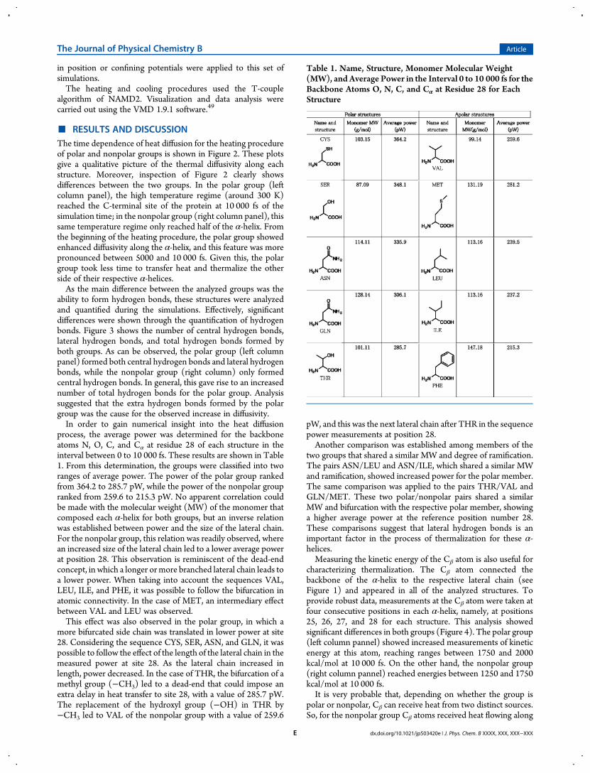
characterizing thermalization. The Cβ atom connected thebackbone of the α-helix to the respective lateral chain (seeFigure 1) and appeared in all of the analyzed structures. Toprovide robust data, measurements at the Cβ atom were taken atfour consecutive positions in each α-helix, namely, at positions25, 26, 27, and 28 for each structure. This analysis showedsignificant differences in both groups (Figure 4). The polar group(left column pannel) showed increased measurements of kineticenergy at this atom, reaching ranges between 1750 and 2000kcal/mol at 10 000 fs. On the other hand, the nonpolar group(right column pannel) reached energies between 1250 and 1750kcal/mol at 10 000 fs.It is very probable that, depending on whether the group is
polar or nonpolar, Cβ can receive heat from two distinct sources.So, for the nonpolar group Cβ atoms received heat flowing along
Table 1. Name, Structure, Monomer Molecular Weight(MW), and Average Power in the Interval 0 to 10 000 fs for theBackbone Atoms O, N, C, and Cα at Residue 28 for EachStructure
The Journal of Physical Chemistry B Article
dx.doi.org/10.1021/jp503420e | J. Phys. Chem. B XXXX, XXX, XXX−XXXE
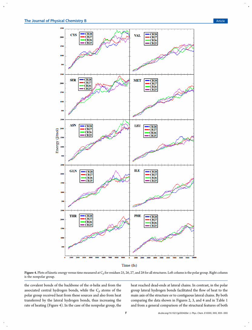
the covalent bonds of the backbone of the α-helix and from theassociated central hydrogen bonds, while the Cβ atoms of thepolar group received heat from these sources and also from heattransferred by the lateral hydrogen bonds, thus increasing therate of heating (Figure 4). In the case of the nonpolar group, the
heat reached dead-ends at lateral chains. In contrast, in the polargroup lateral hydrogen bonds facilitated the flow of heat to themain axis of the structure or to contiguous lateral chains. By bothcomparing the data shown in Figures 2, 3, and 4 and in Table 1and from a general comparison of the structural features of both
Figure 4. Plots of kinetic energy versus timemeasured at Cβ for residues 25, 26, 27, and 28 for all structures. Left column is the polar group. Right columnis the nonpolar group.
The Journal of Physical Chemistry B Article
dx.doi.org/10.1021/jp503420e | J. Phys. Chem. B XXXX, XXX, XXX−XXXF
groups, it is possible to envision the existence of two pathways forheat diffusion. The first pathway could occur through thecovalent bonds of the α-helices and through the central hydrogenbonds that are necessary for the occurrence of the α-helicalstructure. This pathway was common to both groups. Thesecond pathway could occur through lateral hydrogen bonds thatwere only formed by the polar group. It seems that these lateralhydrogen bonds gave rise to the enhanced heat diffusivityobserved in this group. All analyses approximately preserved thetrend shown in Table 1, and our results suggest an important roleof hydrogen bonds in heat transfer for these α-helical structuresand proteins.Other features of heat conduction along the α-helices were
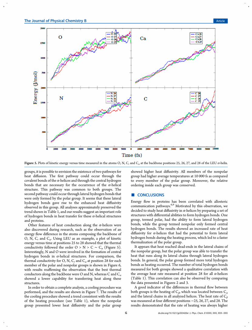
also discovered during research, such as the observation of anenergy flow difference in the atoms composing the backbone ofO, N, C, and Cα. Using LEU as an example, a plot of kineticenergy versus time at positions 25 to 28 showed that the thermalconductivity followed the order O > N > C ∼ Cα (Figure 5).Interestingly, N and O were involved in the formation of centralhydrogen bonds in α-helical structures. For comparison, thethermal conductivity for O, N, C, and Cα at position 28 for eachmember of the polar and nonpolar groups is shown in Figure 6,with results reaffirming the observation that the best thermalconductors along the backbone were O and N, whereas C and Cα
showed a lower capability for transferring heat along thesestructures.In order to obtain a complete analysis, a cooling procedure was
performed, and the results are shown in Figure 7. The results ofthe cooling procedure showed a trend consistent with the resultsof the heating procedure (see Table 1), where the nonpolargroup presented lower heat diffusivity and the polar group
showed higher heat diffusivity. All members of the nonpolargroup had higher average temperatures at 10 000 fs as comparedto every member of the polar group. Moreover, the relativeordering inside each group was conserved.
■ CONCLUSIONS
Energy flow in proteins has been correlated with allostericcommunication pathways.6,9 Motivated by this observation, wedecided to study heat diffusivity in α-helices by preparing a set ofstructures with differential abilities to form hydrogen bonds. Onegroup, termed polar, had the ability to form lateral hydrogenbonds, while the group termed nonpolar only formed centralhydrogen bonds. The results showed an increased rate of heatdiffusivity for α-helices that had the potential to form lateralhydrogen bonds during the heating process, which led to a fasterthermalization of the polar group.It appears that heat reached dead-ends in the lateral chains of
the nonpolar group, but the polar group was able to transfer theheat that runs along its lateral chains through lateral hydrogenbonds. In general, the polar group formed more total hydrogenbonds as heating occurred. The number of total hydrogen bondsmeasured for both groups showed a qualitative correlation withthe average heat rate measured at position 28 for all α-helices(Table 1). This correlation can also be observed by comparingthe data presented in Figures 2 and 3.A good indicator of the differences in thermal flow between
both groups is the heating of Cβ, which was located between Cα
and the lateral chains in all analyzed helices. The heat rate of Cβ
was measured at four different positions−25, 26, 27, and 28. Theresults demonstrated that the rate of heating was always higher
Figure 5. Plots of kinetic energy versus time measured in the atoms O, N, C, and Cα at the backbone positions 25, 26, 27, and 28 of the LEU α-helix.
The Journal of Physical Chemistry B Article
dx.doi.org/10.1021/jp503420e | J. Phys. Chem. B XXXX, XXX, XXX−XXXG
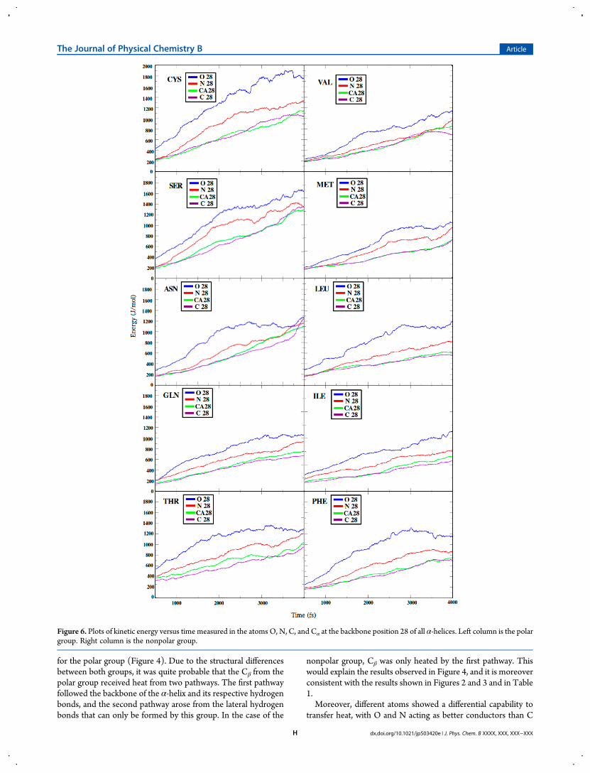
for the polar group (Figure 4). Due to the structural differencesbetween both groups, it was quite probable that the Cβ from thepolar group received heat from two pathways. The first pathwayfollowed the backbone of the α-helix and its respective hydrogenbonds, and the second pathway arose from the lateral hydrogenbonds that can only be formed by this group. In the case of the
nonpolar group, Cβ was only heated by the first pathway. Thiswould explain the results observed in Figure 4, and it is moreoverconsistent with the results shown in Figures 2 and 3 and in Table1.Moreover, different atoms showed a differential capability to
transfer heat, with O and N acting as better conductors than C
Figure 6. Plots of kinetic energy versus time measured in the atoms O, N, C, and Cα at the backbone position 28 of all α-helices. Left column is the polargroup. Right column is the nonpolar group.
The Journal of Physical Chemistry B Article
dx.doi.org/10.1021/jp503420e | J. Phys. Chem. B XXXX, XXX, XXX−XXXH
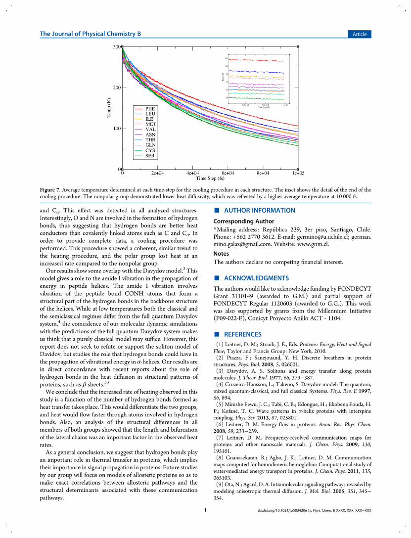
and Cα. This effect was detected in all analyzed structures.Interestingly, O and N are involved in the formation of hydrogenbonds, thus suggesting that hydrogen bonds are better heatconductors than covalently linked atoms such as C and Cα. Inorder to provide complete data, a cooling procedure wasperformed. This procedure showed a coherent, similar trend tothe heating procedure, and the polar group lost heat at anincreased rate compared to the nonpolar group.Our results show some overlap with the Davydov model.3 This
model gives a role to the amide I vibration in the propagation ofenergy in peptide helices. The amide I vibration involvesvibration of the peptide bond CONH atoms that form astructural part of the hydrogen bonds in the backbone structureof the helices. While at low temperatures both the classical andthe semiclassical regimes differ from the full quantum Davydovsystem,4 the coincidence of our molecular dynamic simulationswith the predictions of the full quantum Davydov system makesus think that a purely classical model may suffice. However, thisreport does not seek to refute or support the soliton model ofDavidov, but studies the role that hydrogen bonds could have inthe propagation of vibrational energy in α-helices. Our results arein direct concordance with recent reports about the role ofhydrogen bonds in the heat diffusion in structural patterns ofproteins, such as β-sheets.33
We conclude that the increased rate of heating observed in thisstudy is a function of the number of hydrogen bonds formed asheat transfer takes place. This would differentiate the two groups,and heat would flow faster through atoms involved in hydrogenbonds. Also, an analysis of the structural differences in allmembers of both groups showed that the length and bifurcationof the lateral chains was an important factor in the observed heatrates.As a general conclusion, we suggest that hydrogen bonds play
an important role in thermal transfer in proteins, which impliestheir importance in signal propagation in proteins. Future studiesby our group will focus on models of allosteric proteins so as tomake exact correlations between allosteric pathways and thestructural determinants associated with these communicationpathways.
■ AUTHOR INFORMATION
Corresponding Author*Mailing address: Republica 239, 3er piso, Santiago, Chile.Phone: +562 2770 3612. E-mail: [email protected]; [email protected]. Website: www.gnm.cl.
NotesThe authors declare no competing financial interest.
■ ACKNOWLEDGMENTS
The authors would like to acknowledge funding by FONDECYTGrant 3110149 (awarded to G.M.) and partial support ofFONDECYT Regular 1120603 (awarded to G.G.). This workwas also supported by grants from the Millennium Initiative(P09-022-F), Conicyt Proyecto Anillo ACT - 1104.
■ REFERENCES(1) Leitner, D. M.; Straub, J. E., Eds. Proteins: Energy, Heat and SignalFlow; Taylor and Francis Group: New York, 2010.(2) Piazza, F.; Sanejouand, Y. H. Discrete breathers in proteinstructures. Phys. Biol. 2008, 5, 026001.(3) Davydov, A. S. Solitons and energy transfer along proteinmolecules. J. Theor. Biol. 1977, 66, 379−387.(4) Cruzeiro-Hansson, L.; Takeno, S. Davydov model: The quantum,mixed quantum-classical, and full classical Systems. Phys. Rev. E 1997,56, 894.(5) Mimshe Fewu, J. C.; Tabi, C. B.; Edongue, H.; Ekobena Fouda, H.P.; Kofane, T. C. Wave patterns in α-helix proteins with interspinecoupling. Phys. Scr. 2013, 87, 025801.(6) Leitner, D. M. Energy flow in proteins. Annu. Rev. Phys. Chem.2008, 59, 233−259.(7) Leitner, D. M. Frequency-resolved communication maps forproteins and other nanoscale materials. J. Chem. Phys. 2009, 130,195101.(8) Gnanasekaran, R.; Agbo, J. K.; Leitner, D. M. Communicationmaps computed for homodimeric hemoglobin: Computational study ofwater-mediated energy transport in proteins. J. Chem. Phys. 2011, 135,065103.(9) Ota, N.; Agard, D. A. Intramolecular signaling pathways revealed bymodeling anisotropic thermal diffusion. J. Mol. Biol. 2005, 351, 345−354.
Figure 7. Average temperature determined at each time-step for the cooling procedure in each structure. The inset shows the detail of the end of thecooling procedure. The nonpolar group demonstrated lower heat diffusivity, which was reflected by a higher average temperature at 10 000 fs.
The Journal of Physical Chemistry B Article
dx.doi.org/10.1021/jp503420e | J. Phys. Chem. B XXXX, XXX, XXX−XXXI
(10) Liu, J.; Tawa, G. J.; Wallqvist, A. Identifying cytochrome p450functional networks and their allosteric regulatory elements. PLoS One2013, 8, e81980.(11) Burendahl, S.; Nilsson, L. Computational studies of LXRmolecular interactions reveal an allosteric communication pathway.Proteins 2012, 80, 294−306.(12) Laine, E.; Auclair, C.; Tchertanov, L. Allosteric communicationacross the native and mutated KIT receptor tyrosine kinase. PLoSComput. Biol. 2012, 8, e1002661.(13) Noinaj, N.; Bhasin, S. K.; Song, E. S.; Scoggin, K. E.; Juliano, M.A.; Juliano, L.; Hersh, L. B.; Rodgers, D. W. Identification of theallosteric regulatory site of insulysin. PLoS One 2011, 6, e20864.(14) Seldeen, K. L.; Deegan, B. J.; Bhat, V.; Mikles, D. C.; McDonald,C. B.; Farooq, A. Energetic coupling along an allosteric communicationchannel drives the binding of Jun-Fos heterodimeric transcription factorto DNA. FEBS J. 2011, 278, 2090−104.(15) Ackers, G. K.; Hazzard, J. H. Transduction of binding energy intohemoglobin cooperativity. Trends Biochem. Sci. 1993, 18, 385−90.(16) Koshland, D. E., Jr.; Nemethy, G.; Filmer, D. Comparison ofexperimental binding data and theoretical models in proteins containingsubunits. Biochemistry 1966, 5, 365−85.(17) Monod, J.; Wyman, J.; Changeux, J. P. On the nature of allosterictransitions: A plausible model. J. Mol. Biol. 1965, 12, 88−118.(18) Fuentes, E. J.; Gilmore, S. A.; Mauldin, R. V.; Lee, A. L. Evaluationof energetic and dynamic coupling networks in a PDZ domain protein. J.Mol. Biol. 2006, 364, 337−51.(19) Jiao, W.; Parker, E. J. Using a combination of computational andexperimental techniques to understand the molecular basis for proteinallostery. Adv. Protein. Chem. Struct. Biol. 2012, 87, 391−413.(20) Gianni, S.; Walma, T.; Arcovito, A.; Calosci, N.; Bellelli, A.;Engstrom, A.; Travaglini-Allocatelli, C.; Brunori, M.; Jemth, P.; Vuister,G. W. Demonstration of long-range interactions in a PDZ domain byNMR, kinetics, and protein engineering. Structure. 2006, 14, 1801−1809.(21) Popovych, N.; Sun, S.; Ebright, R. H.; Kalodimos, C. G.Dynamically driven protein allostery. Nat. Struct. Mol. Biol. 2006, 13,831−838.(22) Lockless, S. W.; Ranganathan, R. Evolutionarily conservedpathways of energetic connectivity in protein families. Science 1999, 286,295−299.(23) Rivalta, I.; Sultan, M. M.; Lee, N. S.; Manley, G. A.; Loria, J. P.;Batista, V. S. Allosteric pathways in imidazole glycerol phosphatesynthase. Proc. Natl. Acad. Sci. U. S. A. 2012, 109, E1428−36.(24) Sharp, K.; Skinner, J. J. Pump-probe molecular dynamics as a toolfor studying protein motion and long range coupling. Proteins 2006, 65,347−361.(25) Kong, Y.; Karplus, M. Signaling pathways of PDZ2 domain: Amolecular dynamics interaction correlation analysis. Proteins 2009, 74,145−154.(26) Li, G.; Magana, D.; Dyer, R. B. Anisotropic energy flow andallosteric ligand binding in albumin. Nat. Commun. 2014, 5, 3100.(27) Buchli, B1; Waldauer, S. A.; Walser, R.; Donten, M. L.; Pfister, R.;Blochliger, N.; Steiner, S.; Caflisch, A.; Zerbe, O.; Hamm, P. Kineticresponse of a photoperturbed allosteric protein. Proc. Natl. Acad. Sci. U.S. A. 2013, 110, 11725−11730.(28) Lervik, A.; Bresme, F.; Kjelstrup, S.; Bedeaux, D.; Miguel Rubi, J.Heat transfer in protein-water interfaces. Phys. Chem. Chem. Phys. 2010,12 (7), 1610−7.(29) Kjelstrup, S.; Rubi, J. M.; Bedeaux, D. Energy dissipation inslipping biological pumps. Phys. Chem. Chem. Phys. 2005, 7, 4009−4018.(30) Helbing, J.; Devereux, M.; Nienhaus, K.; Nienhaus, G. U.; Hamm,P.; Meuwly, M. Temperature dependence of the heat diffusivity ofproteins. J. Phys. Chem. A 2012, 116, 2620−8.(31) Botan, V.; Backus, E. H.; Pfister, R.; Moretto, A.; Crisma, M.;Toniolo, C.; Nguyen, P. H.; Stock, G.; Hamm, P. Energy transport inpeptide helices. Proc. Natl. Acad. Sci. U. S. A. 2007, 104, 12749−12754.(32) Sagnella, D. E.; Straub, J. E. Directed Energy “Funneling”Mechanism for Heme Cooling Following Ligand Photolysis or Direct
Excitation in Solvated Carbonmonoxy Myoglobin. J. Phys. Chem. B2001, 105, 7057−7063.(33) Zhang, L.; Chen, T.; Bana, H.; Liu, L. Hydrogen bonding-assistedthermal conduction in β-sheet crystals of spider silk protein. Nanoscale2014, 6, 7786−7791.(34) Schoena, P. A. E.; Michelb, B.; Curionib, A.; Poulikakosa, D.Hydrogen-bond enhanced thermal energy transport at functionalized,hydrophobic and hydrophilic silica−water interfaces. Chem. Phys. Lett.2009, 476, 271−276.(35) Lin, Z.; Rubtsov, I. V. Constant-speed vibrational signaling alongpolyethyleneglycol chain up to 60-Å distance. Proc. Natl. Acad. Sci. U. S.A. 2012, 109, 1413−1418.(36) Kjelstrup, S.; Rubi, J. M.; Bedeaux, D. Energy dissipation inslipping biological pumps. Phys. Chem. Chem. Phys. 2005, 7, 4009−4018.(37) Pimentel, G. C.; McClellan, L. The Hydrogen Bond; W. H.Freeman and Company: San Francisco and London, 1960.(38) Nick Pace, C.; Scholtz, J. M.; Grimsley, G. R. Forces stabilizingproteins. FEBS Lett. 2014, 588, 2177−2184.(39) Lomdahl, P. S.; Kerr, W. C. Do Davydov Solitons Exist at 300 K?Phys. Rev. Lett. 1985, 55, 1235.(40) Martínez, L.; Figueira, A.; Webb, P.; Polikarpov, I.; Skaf, M. S.Mapping the Intramolecular Vibrational Energy Flow in ProteinsReveals Functionally Important Residues. J. Phys. Chem. Lett. 2011, 2,2073−2078.(41) Weinhold, F.; Landis, C. Valency and Bonding: A Natural BondOrbital Donor−Acceptor Perspective; Cambridge University Press:Cambridge, 2005.(42) Lopes Jesus, A. J.; Redinha, J. S. Charge-assisted intramolecularhydrogen bonds in disubstituted cyclohexane derivatives. J. Phys. Chem.A 2011, 115, 14069−14077.(43) Frey, P. A.; Whitt, S. A.; Tobin, J. B. A low-barrier hydrogen bondin the catalytic triad of serine proteases. Science 1994, 264, 1927−30.(44) Mino, G.; Contreras, R. On the role of short and strong hydrogenbonds on the mechanism of action of a model chymotrypsine active site.J. Phys. Chem. A 2009, 113, 5769−5772.(45) Mino, G.; Contreras, R. Non-electrostatic components of shortand strong hydrogen bonds induced by compression inside fullerenes.Chem. Phys. Lett. 2010, 486, 119−122.(46) Kale, L.; Skeel, R.; Bhandarkar, M.; Brunner, R.; Gursoy, A.;Krawetz, N.; Phillips, J.; Shinozaki, A.; Varadarajan, K.; Schulten, K.NAMD2: Greater scalability for parallel molecular dynamics. J. Comput.Phys. 1999, 151, 283−312.(47)MacKerell, A. D., Jr; Bashford, D.; Bellott, M.; Dunbrack, R. L., Jr.;Evanseck, J.; Field, M. J.; Fischer, S.; Gao, J.; Guo, H.; Ha, S.; et al. All-atom empirical potential for molecular modeling and dynamics studiesof proteins. J. Phys. Chem. B 1998, 102, 3586−3616.(48) Mackerell, A. D., Jr.; Feig, M.; Brooks, C. L., 3rd. Extending thetreatment of backbone energetics in protein force fields: limitations ofgas-phase quantum mechanics in reproducing protein conformationaldistributions in molecular dynamics simulations. J. Comput. Chem. 2004,25, 1400−1415.(49) Humphrey, W.; Dalke, A.; Schulten, K. VMDVisual moleculardynamics. J. Mol. Graph. 1996, 14, 33−38.
The Journal of Physical Chemistry B Article
dx.doi.org/10.1021/jp503420e | J. Phys. Chem. B XXXX, XXX, XXX−XXXJ