Human CCL4/MIP-1β Quantikine - RnDSystems.com CCL4/MIP-1β Immunoassay Quantikine ... H400, G-26,...
16
Human CCL4/MIP-1β Immunoassay Quantikine ® ELISA This package insert must be read in its entirety before using this product. For research use only. Not for use in diagnostic procedures. Catalog Number DMB00 Catalog Number SMB00 Catalog Number PDMB00 For the quantitative determination of human Macrophage Inflammatory Protein 1 beta (MIP-1β) concentrations in cell culture supernates, serum, and plasma. Note: The standard reconstitution method has changed. Please read this package insert in its entirety before using this product.
Transcript of Human CCL4/MIP-1β Quantikine - RnDSystems.com CCL4/MIP-1β Immunoassay Quantikine ... H400, G-26,...
Human CCL4/MIP-1β Immunoassay
Quantikine® ELISA
This package insert must be read in its entirety before using this product. For research use only. Not for use in diagnostic procedures.
Catalog Number DMB00 Catalog Number SMB00 Catalog Number PDMB00
For the quantitative determination of human Macrophage Inflammatory Protein 1 beta (MIP-1β) concentrations in cell culture supernates, serum, and plasma.
Note: The standard reconstitution method has changed. Please read this package insert in its entirety before using this product.
MANUFACTURED AND DISTRIBUTED BY:
USA & Canada | R&D Systems, Inc. 614 McKinley Place NE, Minneapolis, MN 55413, USATEL: (800) 343-7475 (612) 379-2956 FAX: (612) 656-4400E-MAIL: [email protected]
DISTRIBUTED BY:
UK & Europe | R&D Systems Europe, Ltd.19 Barton Lane, Abingdon Science Park, Abingdon OX14 3NB, UKTEL: +44 (0)1235 529449 FAX: +44 (0)1235 533420E-MAIL: [email protected]
China | R&D Systems China Co., Ltd.24A1 Hua Min Empire Plaza, 726 West Yan An Road, Shanghai PRC 200050TEL: +86 (21) 52380373 FAX: +86 (21) 52371001E-MAIL: [email protected]
TABLE OF CONTENTS
SECTION PAGE
INTRODUCTION ....................................................................................................................................................................1
PRINCIPLE OF THE ASSAY ..................................................................................................................................................2
LIMITATIONS OF THE PROCEDURE ................................................................................................................................2
TECHNICAL HINTS ................................................................................................................................................................2
MATERIALS PROVIDED & STORAGE CONDITIONS ..................................................................................................3
OTHER SUPPLIES REQUIRED ............................................................................................................................................4
PRECAUTIONS ........................................................................................................................................................................4
SAMPLE COLLECTION & STORAGE ................................................................................................................................4
SAMPLE PREPARATION.......................................................................................................................................................4
REAGENT PREPARATION ....................................................................................................................................................5
ASSAY PROCEDURE ............................................................................................................................................................6
CALCULATION OF RESULTS ..............................................................................................................................................7
TYPICAL DATA ........................................................................................................................................................................7
PRECISION ...............................................................................................................................................................................8
RECOVERY................................................................................................................................................................................8
SENSITIVITY ............................................................................................................................................................................8
LINEARITY ................................................................................................................................................................................9
CALIBRATION .........................................................................................................................................................................9
SAMPLE VALUES ....................................................................................................................................................................9
SPECIFICITY .......................................................................................................................................................................... 10
REFERENCES ........................................................................................................................................................................ 11
PLATE LAYOUT .................................................................................................................................................................... 12
www.RnDSystems.com 1
INTRODUCTIONThe macrophage inflammatory proteins (MIP-1α and MIP-1β) are members of the β or CC subfamily of chemokines. These two closely related proteins were initially co-purified from the conditioned media of a lipopolysaccharide-stimulated mouse macrophage cell line (1). At least seven human cDNAs that are homologous to the mouse MIP-1β, including Act-2, pAT744, H400, G-26, HIMAP, HC21, and MAD-5, have been cloned. The predicted protein products encoded by these human MIP-1β cDNAs are between 94-98% identical (2-5). There is evidence to suggest that multiple non-allelic genes for human MIP-1β exist in the human genome.
The cDNAs for human MIP-1β encode precursor proteins with a 23 amino acid (aa) residue signal peptide that is cleaved to generate the 69 aa residue, non-glycosylated mature protein. The mRNAs for MIP-1α and MIP-1β are not expressed in unstimulated cells, but the two are coordinately expressed after activation of T cells, B cells, monocytes and mast cells. In addition, both mRNAs have also been detected in macrophages associated with human atherosclerotic plaques. Mature human MIP-1α and MIP-1β are about 75% and 77% homologous, respectively, to their mouse counterparts. The mature human MIP-1α and human MIP-1β are also 70% homologous in their protein sequences. The mouse and human proteins show cross-species bioactivities (2-5).
Chemokines are small, secreted cytokines that are involved in a variety of immune and inflammatory responses, acting primarily as chemoattractants and activators of specific leukocytes. Members of each chemokine subfamily have unique as well as overlapping activities. Like other CC chemokines, both MIP-1β and MIP-1α can chemoattract monocytes (5). In addition, MIP-1α and MIP-1β can chemoattract and induce the adhesion of T lymphocytes (5). MIP-1α, but not MIP-1β, has been shown to chemoattract B cells (6), to chemoattract and degranulate eosinophils (7), to induce histamine release from basophils and mast cells, and to chemoattract basophils (8). MIP-1α has been identified as a stem cell inhibitor (SCI) that can inhibit the proliferation of hematopoietic stem cells both in vitro and in vivo (9). MIP-1β was reported to be twenty-fold less active than MIP-1α on the same cell populations (10).
Chemokine activities are mediated by G-protein-coupled seven transmembrane domain receptors (5, 11). The β chemokine receptor designated CC chemokine receptor-1 (CC CKR-1), alternately named MIP-1α/RANTES receptor, has been shown to bind MIP-1α, RANTES, MIP-1β and MCP-1 with varying affinities (12, 13). When transfected into a kidney cell line however, CC CKR-1 transduces a signal only in response to MIP-1α and RANTES. Based on functional analysis of MIP-1β, one or more additional β chemokine receptors that transduce MIP-1β signals must also exist. MIP-1α and MIP-1β have been shown to lack affinity for the erythrocyte surface chemokine receptor/Duffy antigen that binds many α as well as β chemokines (11, 14).
The Quantikine® Human CCL4/MIP-1β Immunoassay is a 3.0-4.5 hour solid phase ELISA designed to measure human MIP-1β in cell culture supernates, serum, and plasma. It contains E. coli-expressed recombinant human MIP-1β (Act-2 variant) and antibodies raised against the recombinant factor. This immunoassay has been shown to accurately quantitate the recombinant human MIP-1β. Results obtained using natural human MIP-1β showed linear curves that were parallel to the standard curves obtained using the Quantikine® kit standards. These results indicate that this kit can be used to determine relative mass values for natural human MIP-1β.
For research use only. Not for use in diagnostic procedures.2
PRINCIPLE OF THE ASSAYThis assay employs the quantitative sandwich enzyme immunoassay technique. A monoclonal antibody specific for human MIP-1β has been pre-coated onto a microplate. Standards and samples are pipetted into the wells and any MIP-1β present is bound by the immobilized antibody. After washing away any unbound substances, an enzyme-linked polyclonal antibody specific for human MIP-1β is added to the wells. Following a wash to remove any unbound antibody-enzyme reagent, a substrate solution is added to the wells and color develops in proportion to the amount of MIP-1β bound in the initial step. The color development is stopped and the intensity of the color is measured.
LIMITATIONS OF THE PROCEDURE• FOR RESEARCH USE ONLY. NOT FOR USE IN DIAGNOSTIC PROCEDURES.
• The kit should not be used beyond the expiration date on the kit label.
• Do not mix or substitute reagents with those from other lots or sources.
• It is important that the calibrator diluent selected for the standard curve be consistent with the samples being assayed.
• If samples generate values higher than the highest standard, further dilute the samples with the appropriate calibrator diluent and repeat the assay.
• Any variation in standard diluent, operator, pipetting technique, washing technique, incubation time or temperature, and kit age can cause variation in binding.
• Variations in sample collection, processing, and storage may cause sample value differences.
• This assay is designed to eliminate interference by other factors present in biological samples. Until all factors have been tested in the Quantikine® Immunoassay, the possibility of interference cannot be excluded.
TECHNICAL HINTS• When mixing or reconstituting protein solutions, always avoid foaming.
• To avoid cross-contamination, change pipette tips between additions of each standard level, between sample additions, and between reagent additions. Also, use separate reservoirs for each reagent.
• To ensure accurate results, proper adhesion of plate sealers during incubation steps is necessary.
• When using an automated plate washer, adding a 30 second soak period following the addition of Wash Buffer, and/or rotating the plate 180 degrees between wash steps may improve assay precision.
• Substrate Solution should remain colorless until added to the plate. Keep Substrate Solution protected from light. Substrate Solution should change from colorless to gradations of blue.
• Stop Solution should be added to the plate in the same order as the Substrate Solution. The color developed in the wells will turn from blue to yellow upon addition of the Stop Solution. Wells that are green in color indicate that the Stop Solution has not mixed thoroughly with the Substrate Solution.
www.RnDSystems.com 3
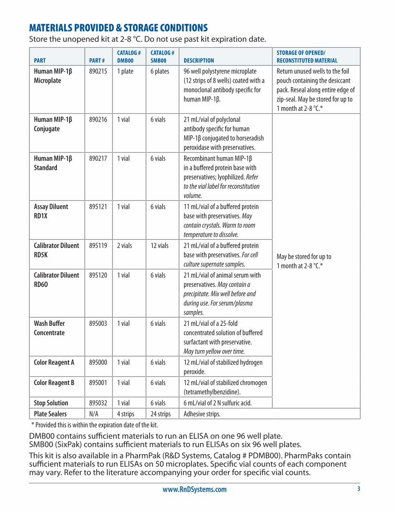
MATERIALS PROVIDED & STORAGE CONDITIONSStore the unopened kit at 2-8 °C. Do not use past kit expiration date.
PART PART #CATALOG # DMB00
CATALOG # SMB00 DESCRIPTION
STORAGE OF OPENED/ RECONSTITUTED MATERIAL
Human MIP-1β Microplate
890215 1 plate 6 plates 96 well polystyrene microplate (12 strips of 8 wells) coated with a monoclonal antibody specific for human MIP-1β.
Return unused wells to the foil pouch containing the desiccant pack. Reseal along entire edge of zip-seal. May be stored for up to 1 month at 2-8 °C.*
Human MIP-1β Conjugate
890216 1 vial 6 vials 21 mL/vial of polyclonal antibody specific for human MIP-1β conjugated to horseradish peroxidase with preservatives.
May be stored for up to 1 month at 2-8 °C.*
Human MIP-1β Standard
890217 1 vial 6 vials Recombinant human MIP-1β in a buffered protein base with preservatives; lyophilized. Refer to the vial label for reconstitution volume.
Assay Diluent RD1X
895121 1 vial 6 vials 11 mL/vial of a buffered protein base with preservatives. May contain crystals. Warm to room temperature to dissolve.
Calibrator Diluent RD5K
895119 2 vials 12 vials 21 mL/vial of a buffered protein base with preservatives. For cell culture supernate samples.
Calibrator Diluent RD6O
895120 1 vial 6 vials 21 mL/vial of animal serum with preservatives. May contain a precipitate. Mix well before and during use. For serum/plasma samples.
Wash Buffer Concentrate
895003 1 vial 6 vials 21 mL/vial of a 25-fold concentrated solution of buffered surfactant with preservative. May turn yellow over time.
Color Reagent A 895000 1 vial 6 vials 12 mL/vial of stabilized hydrogen peroxide.
Color Reagent B 895001 1 vial 6 vials 12 mL/vial of stabilized chromogen (tetramethylbenzidine).
Stop Solution 895032 1 vial 6 vials 6 mL/vial of 2 N sulfuric acid.
Plate Sealers N/A 4 strips 24 strips Adhesive strips.
* Provided this is within the expiration date of the kit.
DMB00 contains sufficient materials to run an ELISA on one 96 well plate. SMB00 (SixPak) contains sufficient materials to run ELISAs on six 96 well plates.This kit is also available in a PharmPak (R&D Systems, Catalog # PDMB00). PharmPaks contain sufficient materials to run ELISAs on 50 microplates. Specific vial counts of each component may vary. Refer to the literature accompanying your order for specific vial counts.
For research use only. Not for use in diagnostic procedures.4
OTHER SUPPLIES REQUIRED• Microplate reader capable of measuring absorbance at 450 nm, with the correction
wavelength set at 540 nm or 570 nm.
• Pipettes and pipette tips.
• Deionized or distilled water.
• Squirt bottle, manifold dispenser, or automated microplate washer.
• 500 mL graduated cylinder.
• Test tubes for dilution of standards and samples.
• Human MIP-1β Controls (optional; R&D Systems, Catalog # QC20).
PRECAUTIONSCalibrator Diluent RD6O contains sodium azide which may react with lead and copper plumbing to form explosive metallic azides. Flush with large volumes of water during disposal.
The Stop Solution provided with this kit is an acid solution.
Some components in this kit contain a preservative which may cause an allergic skin reaction. Avoid breathing mist.
Color Reagent B may cause skin, eye, and respiratory irritation. Avoid breathing fumes.
Wear protective gloves, clothing, eye, and face protection. Wash hands thoroughly after handling. Refer to the MSDS on our website prior to use.
SAMPLE COLLECTION & STORAGEThe sample collection and storage conditions listed below are intended as general guidelines. Sample stability has not been evaluated.
Cell Culture Supernates - Remove particulates by centrifugation and assay immediately or aliquot and store samples at ≤ -20 °C. Avoid repeated freeze-thaw cycles.
Serum - Use a serum separator tube (SST) and allow samples to clot for 30 minutes at room temperature before centrifugation for 15 minutes at 1000 x g. Remove serum and assay immediately or aliquot and store samples at ≤ -20 °C. Avoid repeated freeze-thaw cycles.
Plasma - Collect plasma using EDTA, heparin, or citrate as an anticoagulant. Centrifuge for 15 minutes at 1000 x g within 30 minutes of collection. Assay immediately or aliquot and store samples at ≤ -20 °C. Avoid repeated freeze-thaw cycles.
SAMPLE PREPARATIONCell culture supernate samples may require up to a 20-fold dilution into Calibrator Diluent RD5K. A suggested 20-fold dilution is 25 μL of sample + 475 μL of Calibrator Diluent RD5K.
www.RnDSystems.com 5
REAGENT PREPARATIONBring all reagents to room temperature before use.
Wash Buffer - If crystals have formed in the concentrate, warm to room temperature and mix gently until the crystals have completely dissolved. Add 20 mL of Wash Buffer Concentrate to deionized or distilled water to prepare 500 mL of Wash Buffer.
Substrate Solution - Color Reagents A and B should be mixed together in equal volumes within 15 minutes of use. Protect from light. 200 μL of the resultant mixture is required per well.
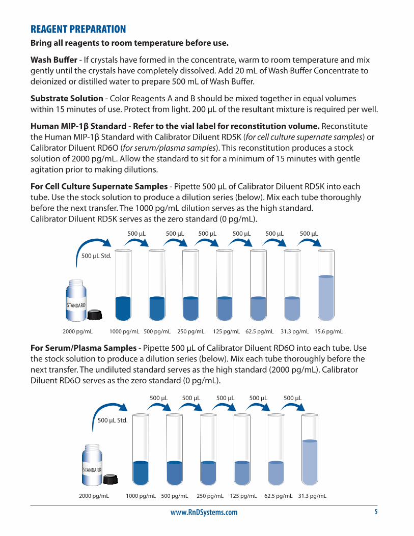
Human MIP-1β Standard - Refer to the vial label for reconstitution volume. Reconstitute the Human MIP-1β Standard with Calibrator Diluent RD5K (for cell culture supernate samples) or Calibrator Diluent RD6O (for serum/plasma samples). This reconstitution produces a stock solution of 2000 pg/mL. Allow the standard to sit for a minimum of 15 minutes with gentle agitation prior to making dilutions.
For Cell Culture Supernate Samples - Pipette 500 μL of Calibrator Diluent RD5K into each tube. Use the stock solution to produce a dilution series (below). Mix each tube thoroughly before the next transfer. The 1000 pg/mL dilution serves as the high standard. Calibrator Diluent RD5K serves as the zero standard (0 pg/mL).
500 µL Std.
2000 pg/mL 1000 pg/mL 500 pg/mL 250 pg/mL 125 pg/mL 62.5 pg/mL 31.3 pg/mL 15.6 pg/mL
500 µL 500 µL 500 µL 500 µL 500 µL 500 µL
500 µL Std.
2000 pg/mL 1000 pg/mL 500 pg/mL 250 pg/mL 125 pg/mL 62.5 pg/mL 31.3 pg/mL
500 µL 500 µL 500 µL 500 µL 500 µL
For Serum/Plasma Samples - Pipette 500 μL of Calibrator Diluent RD6O into each tube. Use the stock solution to produce a dilution series (below). Mix each tube thoroughly before the next transfer. The undiluted standard serves as the high standard (2000 pg/mL). Calibrator Diluent RD6O serves as the zero standard (0 pg/mL).
For research use only. Not for use in diagnostic procedures.6
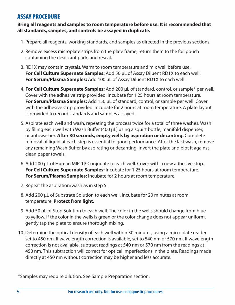
ASSAY PROCEDURE Bring all reagents and samples to room temperature before use. It is recommended that all standards, samples, and controls be assayed in duplicate.
1. Prepare all reagents, working standards, and samples as directed in the previous sections.
2. Remove excess microplate strips from the plate frame, return them to the foil pouch containing the desiccant pack, and reseal.
3. RD1X may contain crystals. Warm to room temperature and mix well before use. For Cell Culture Supernate Samples: Add 50 μL of Assay Diluent RD1X to each well. For Serum/Plasma Samples: Add 100 μL of Assay Diluent RD1X to each well.
4. For Cell Culture Supernate Samples: Add 200 μL of standard, control, or sample* per well. Cover with the adhesive strip provided. Incubate for 1.25 hours at room temperature. For Serum/Plasma Samples: Add 150 μL of standard, control, or sample per well. Cover with the adhesive strip provided. Incubate for 2 hours at room temperature. A plate layout is provided to record standards and samples assayed.
5. Aspirate each well and wash, repeating the process twice for a total of three washes. Wash by filling each well with Wash Buffer (400 μL) using a squirt bottle, manifold dispenser, or autowasher. After 30 seconds, empty wells by aspiration or decanting. Complete removal of liquid at each step is essential to good performance. After the last wash, remove any remaining Wash Buffer by aspirating or decanting. Invert the plate and blot it against clean paper towels.
6. Add 200 μL of Human MIP-1β Conjugate to each well. Cover with a new adhesive strip. For Cell Culture Supernate Samples: Incubate for 1.25 hours at room temperature. For Serum/Plasma Samples: Incubate for 2 hours at room temperature.
7. Repeat the aspiration/wash as in step 5.
8. Add 200 μL of Substrate Solution to each well. Incubate for 20 minutes at room temperature. Protect from light.
9. Add 50 μL of Stop Solution to each well. The color in the wells should change from blue to yellow. If the color in the wells is green or the color change does not appear uniform, gently tap the plate to ensure thorough mixing.
10. Determine the optical density of each well within 30 minutes, using a microplate reader set to 450 nm. If wavelength correction is available, set to 540 nm or 570 nm. If wavelength correction is not available, subtract readings at 540 nm or 570 nm from the readings at 450 nm. This subtraction will correct for optical imperfections in the plate. Readings made directly at 450 nm without correction may be higher and less accurate.
*Samples may require dilution. See Sample Preparation section.
www.RnDSystems.com 7
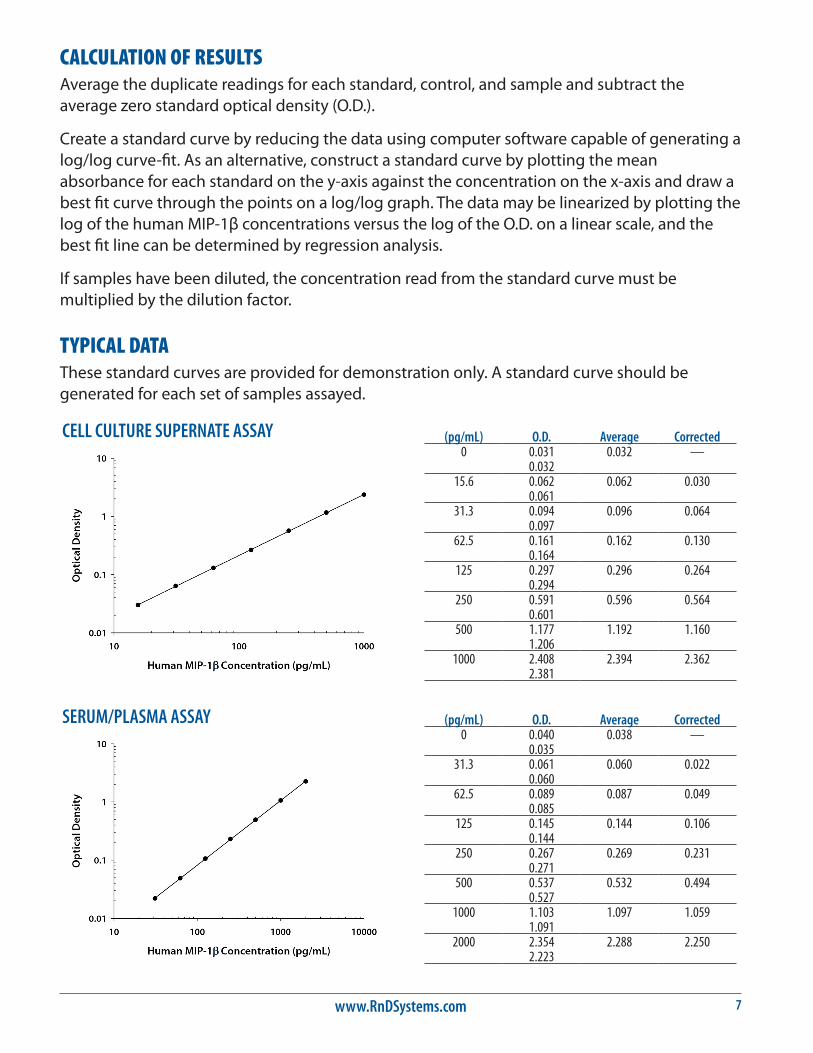
CALCULATION OF RESULTSAverage the duplicate readings for each standard, control, and sample and subtract the average zero standard optical density (O.D.).
Create a standard curve by reducing the data using computer software capable of generating a log/log curve-fit. As an alternative, construct a standard curve by plotting the mean absorbance for each standard on the y-axis against the concentration on the x-axis and draw a best fit curve through the points on a log/log graph. The data may be linearized by plotting the log of the human MIP-1β concentrations versus the log of the O.D. on a linear scale, and the best fit line can be determined by regression analysis.
If samples have been diluted, the concentration read from the standard curve must be multiplied by the dilution factor.
TYPICAL DATAThese standard curves are provided for demonstration only. A standard curve should be generated for each set of samples assayed.
(pg/mL) O.D. Average Corrected0 0.031 0.032 —
0.03215.6 0.062 0.062 0.030
0.06131.3 0.094 0.096 0.064
0.09762.5 0.161 0.162 0.130
0.164125 0.297 0.296 0.264
0.294250 0.591 0.596 0.564
0.601500 1.177 1.192 1.160
1.2061000 2.408 2.394 2.362
2.381
(pg/mL) O.D. Average Corrected0 0.040 0.038 —
0.03531.3 0.061 0.060 0.022
0.06062.5 0.089 0.087 0.049
0.085125 0.145 0.144 0.106
0.144250 0.267 0.269 0.231
0.271500 0.537 0.532 0.494
0.5271000 1.103 1.097 1.059
1.0912000 2.354 2.288 2.250
2.223
CELL CULTURE SUPERNATE ASSAY
SERUM/PLASMA ASSAY
For research use only. Not for use in diagnostic procedures.8
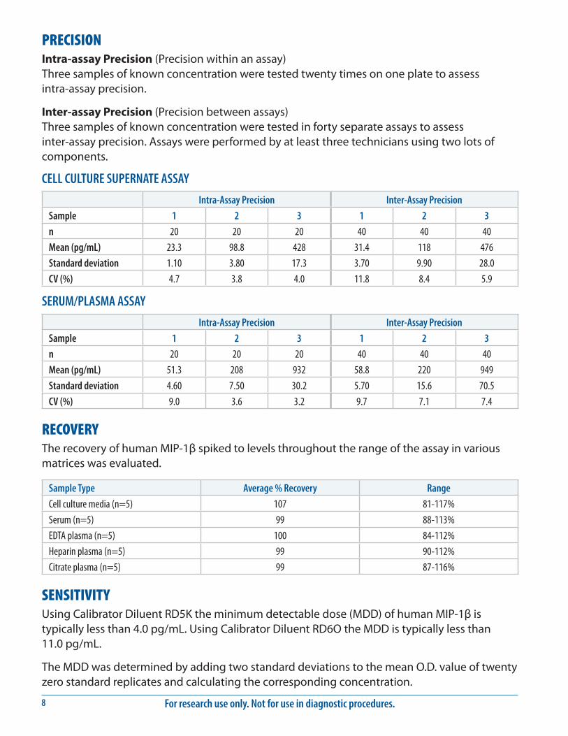
PRECISIONIntra-assay Precision (Precision within an assay) Three samples of known concentration were tested twenty times on one plate to assess intra-assay precision.
Inter-assay Precision (Precision between assays) Three samples of known concentration were tested in forty separate assays to assess inter-assay precision. Assays were performed by at least three technicians using two lots of components.
CELL CULTURE SUPERNATE ASSAY
Intra-Assay Precision Inter-Assay Precision
Sample 1 2 3 1 2 3
n 20 20 20 40 40 40
Mean (pg/mL) 23.3 98.8 428 31.4 118 476
Standard deviation 1.10 3.80 17.3 3.70 9.90 28.0
CV (%) 4.7 3.8 4.0 11.8 8.4 5.9
SERUM/PLASMA ASSAY
Intra-Assay Precision Inter-Assay Precision
Sample 1 2 3 1 2 3
n 20 20 20 40 40 40
Mean (pg/mL) 51.3 208 932 58.8 220 949
Standard deviation 4.60 7.50 30.2 5.70 15.6 70.5
CV (%) 9.0 3.6 3.2 9.7 7.1 7.4
RECOVERYThe recovery of human MIP-1β spiked to levels throughout the range of the assay in various matrices was evaluated.
Sample Type Average % Recovery Range
Cell culture media (n=5) 107 81-117%
Serum (n=5) 99 88-113%
EDTA plasma (n=5) 100 84-112%
Heparin plasma (n=5) 99 90-112%
Citrate plasma (n=5) 99 87-116%
SENSITIVITYUsing Calibrator Diluent RD5K the minimum detectable dose (MDD) of human MIP-1β is typically less than 4.0 pg/mL. Using Calibrator Diluent RD6O the MDD is typically less than 11.0 pg/mL.
The MDD was determined by adding two standard deviations to the mean O.D. value of twenty zero standard replicates and calculating the corresponding concentration.
www.RnDSystems.com 9
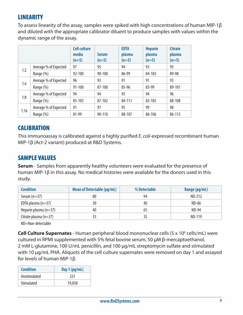
LINEARITYTo assess linearity of the assay, samples were spiked with high concentrations of human MIP-1β and diluted with the appropriate calibrator diluent to produce samples with values within the dynamic range of the assay.
Cell culture media (n=5)
Serum (n=5)
EDTA plasma (n=5)
Heparin plasma (n=5)
Citrate plasma (n=5)
1:2Average % of Expected 97 95 94 93 95
Range (%) 92-100 90-100 86-99 84-103 89-98
1:4Average % of Expected 96 93 91 91 93
Range (%) 91-100 87-100 85-96 83-99 89-101
1:8Average % of Expected 94 94 93 94 96
Range (%) 85-103 87-102 84-111 83-105 88-108
1:16Average % of Expected 91 97 95 99 98
Range (%) 81-99 90-110 88-107 86-106 86-115
CALIBRATIONThis immunoassay is calibrated against a highly purified E. coli-expressed recombinant human MIP-1β (Act-2 variant) produced at R&D Systems.
SAMPLE VALUESSerum - Samples from apparently healthy volunteers were evaluated for the presence of human MIP-1β in this assay. No medical histories were available for the donors used in this study.
Condition Mean of Detectable (pg/mL) % Detectable Range (pg/mL)
Serum (n=37) 80 94 ND-212
EDTA plasma (n=37) 30 40 ND-86
Heparin plasma (n=37) 40 65 ND-94
Citrate plasma (n=37) 33 35 ND-119
ND=Non-detectable
Cell Culture Supernates - Human peripheral blood mononuclear cells (5 x 10⁶ cells/mL) were cultured in RPMI supplemented with 5% fetal bovine serum, 50 μM β-mercaptoethanol, 2 mM L-glutamine, 100 U/mL penicillin, and 100 μg/mL streptomycin sulfate and stimulated with 10 μg/mL PHA. Aliquots of the cell culture supernates were removed on day 1 and assayed for levels of human MIP-1β.
Condition Day 1 (pg/mL)
Unstimulated 231
Stimulated 19,058
For research use only. Not for use in diagnostic procedures.10
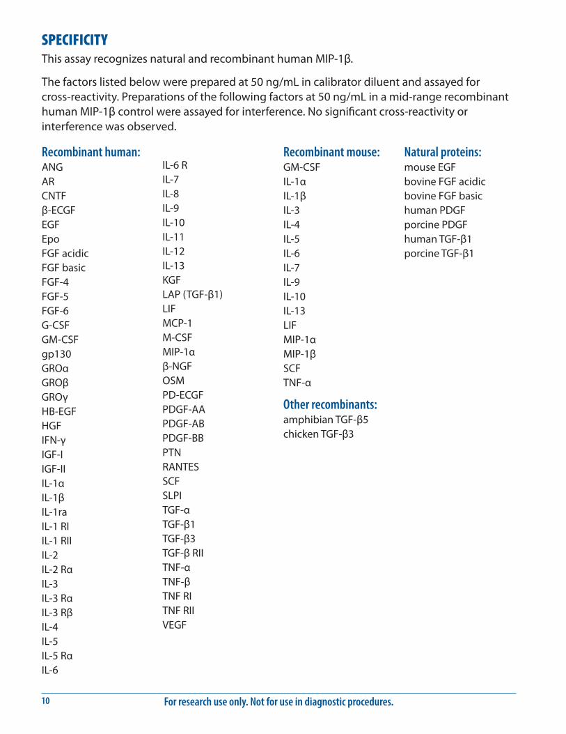
SPECIFICITYThis assay recognizes natural and recombinant human MIP-1β.
The factors listed below were prepared at 50 ng/mL in calibrator diluent and assayed for cross-reactivity. Preparations of the following factors at 50 ng/mL in a mid-range recombinant human MIP-1β control were assayed for interference. No significant cross-reactivity or interference was observed.
Recombinant human:ANGARCNTFβ-ECGFEGFEpoFGF acidicFGF basicFGF-4FGF-5FGF-6G-CSFGM-CSFgp130GROαGROβGROγHB-EGFHGFIFN-γIGF-IIGF-IIIL-1αIL-1βIL-1raIL-1 RIIL-1 RIIIL-2IL-2 RαIL-3IL-3 RαIL-3 RβIL-4IL-5IL-5 RαIL-6
IL-6 RIL-7IL-8IL-9IL-10IL-11IL-12IL-13KGFLAP (TGF-β1)LIFMCP-1M-CSFMIP-1αβ-NGFOSMPD-ECGFPDGF-AAPDGF-ABPDGF-BBPTNRANTESSCFSLPITGF-αTGF-β1TGF-β3TGF-β RIITNF-αTNF-βTNF RITNF RIIVEGF
Recombinant mouse:GM-CSFIL-1αIL-1βIL-3IL-4IL-5IL-6IL-7IL-9IL-10IL-13LIFMIP-1αMIP-1βSCFTNF-α
Other recombinants:amphibian TGF-β5chicken TGF-β3
Natural proteins:mouse EGFbovine FGF acidicbovine FGF basichuman PDGFporcine PDGFhuman TGF-β1porcine TGF-β1
www.RnDSystems.com 11
REFERENCES1. Wolpe, S.D. and A. Cerami (1989) FASEB J. 3:2565.
2. Oppenheim, J.J. et al. (1991) Annu. Rev. Immunol. 9:617.
3. Schall, T.J. (1991) Cytokine 3:165.
4. Miller, M.D. and M.S. Krangel (1992) Critical Rev. Immunol. 12:17.
5. Schall, T.J. (1994) in The Cytokine Handbook, 2nd edition, A. Thomson ed., Academic Press, New York, p. 419.
6. Schall, T.J. et al. (1993) J. Exp. Med. 177:1821.
7. Rot, A. et al. (1992) J. Exp. Med. 176:1489.
8. Alam, R. et al. (1992) J. Exp. Med. 176:781.
9. Graham, G.J. et al. (1990) Nature 344:442.
10. Graham, G.J. et al. (1993) Cell Growth Diff. 4:137.
11. Murphy, P.M. (1994) Annu. Rev. Immunol. 12:593.
12. Neote, K. et al. (1993) Cell 72:415.
13. Gao, J.L. et al. (1993) J. Exp. Med. 177:1421.
14. Neote, K. et al. (1993) J. Biol. Chem. 268:12247.
For research use only. Not for use in diagnostic procedures.12
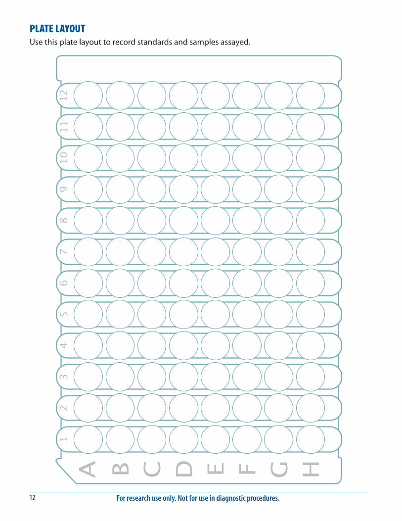
PLATE LAYOUTUse this plate layout to record standards and samples assayed.

www.RnDSystems.com 13
NOTES
For research use only. Not for use in diagnostic procedures.14
NOTES
07.96 750126.11 6/16
©2016 R&D Systems, Inc.
All trademarks and registered trademarks are the property of their respective owners.