Mouse IFN-β Quantikine · 2016-12-30 · Mouse IFN-β Immunoassay Quantikine® ELISA This package...
16
Mouse IFN-β Immunoassay Quantikine ® ELISA This package insert must be read in its entirety before using this product. For research use only. Not for use in diagnostic procedures. Catalog Number MIFNB0 For the quantitative determination of mouse Interferon beta (IFN-β) concentrations in cell culture supernates, serum, and plasma.
Transcript of Mouse IFN-β Quantikine · 2016-12-30 · Mouse IFN-β Immunoassay Quantikine® ELISA This package...
Mouse IFN-β Immunoassay
Quantikine® ELISA
This package insert must be read in its entirety before using this product. For research use only. Not for use in diagnostic procedures.
Catalog Number MIFNB0
For the quantitative determination of mouse Interferon beta (IFN-β) concentrations in cell culture supernates, serum, and plasma.
MANUFACTURED AND DISTRIBUTED BY:
USA & Canada | R&D Systems, Inc. 614 McKinley Place NE, Minneapolis, MN 55413, USATEL: (800) 343-7475 (612) 379-2956 FAX: (612) 656-4400E-MAIL: [email protected]
DISTRIBUTED BY:
UK & Europe | R&D Systems Europe, Ltd.19 Barton Lane, Abingdon Science Park, Abingdon OX14 3NB, UKTEL: +44 (0)1235 529449 FAX: +44 (0)1235 533420E-MAIL: [email protected]
China | R&D Systems China Co., Ltd.24A1 Hua Min Empire Plaza, 726 West Yan An Road, Shanghai PRC 200050TEL: +86 (21) 52380373 FAX: +86 (21) 52371001E-MAIL: [email protected]
TABLE OF CONTENTS
SECTION PAGE
INTRODUCTION .....................................................................................................................................................................1
PRINCIPLE OF THE ASSAY ...................................................................................................................................................2
LIMITATIONS OF THE PROCEDURE .................................................................................................................................2
TECHNICAL HINTS .................................................................................................................................................................2
MATERIALS PROVIDED & STORAGE CONDITIONS ...................................................................................................3
OTHER SUPPLIES REQUIRED .............................................................................................................................................4
PRECAUTIONS .........................................................................................................................................................................4
SAMPLE COLLECTION & STORAGE .................................................................................................................................4
REAGENT PREPARATION .....................................................................................................................................................5
ASSAY PROCEDURE .............................................................................................................................................................6
CALCULATION OF RESULTS ...............................................................................................................................................7
TYPICAL DATA .........................................................................................................................................................................7
PRECISION ................................................................................................................................................................................8
RECOVERY.................................................................................................................................................................................8
LINEARITY .................................................................................................................................................................................8
SENSITIVITY .............................................................................................................................................................................9
CALIBRATION ..........................................................................................................................................................................9
SAMPLE VALUES .....................................................................................................................................................................9
SPECIFICITY ........................................................................................................................................................................... 10
REFERENCES ......................................................................................................................................................................... 11
PLATE LAYOUT ..................................................................................................................................................................... 12
www.RnDSystems.com 1
INTRODUCTIONInterferon beta (IFN-β), also known as fibroblast IFN, is a secreted, approximately 22 kDa member of the type I interferon family of molecules (1). Mature mouse IFN-β shares 75% and 47% amino acid sequence identity with the rat and human proteins, respectively. Fibroblasts are the major producers of IFN-β, but it can also be produced by dendritic cells, macrophages, and endothelial cells in response to pathogens (2). It is transcriptionally regulated by TRAF3, IRF3, IRF7, and NF-κB (3, 4). IFN-β signals through the heterodimeric IFN-α/β receptor, which is constitutively associated with JAK1 and TYK2, and leads to the phosphorylation and activation of several Signal Transducer and Activator of Transcription (STAT) family members (1, 5-7). IFN-β can modulate immune responses by inducing the expression of the anti-inflammatory cytokine IL-10, promoting the generation of regulatory T cells, and inhibiting inflammasome activation (8-10).
The immune modulatory role of IFN-β appears to be important for protection against a variety of autoimmune diseases (1). IFN-β-deficient mice show increased susceptibility to experimental autoimmune encephalomyelitis (EAE), a disease model of human multiple sclerosis (MS) (11). Furthermore, IFN-β has been shown to suppress the Th17 cell response in both MS and EAE and has commonly been used as a treatment for MS (12-16). IFN-β also has protective effects when given to mice with dextran-sulphate sodium (DSS)-induced colitis (17). These immunosuppressive effects may be due in part to IFN-β-dependent induction of regulatory T cells (9, 18-20).
The Quantikine® Mouse IFN-β Immunoassay is a 4.5 hour solid phase ELISA designed to measure mouse IFN-β in cell culture supernates, serum, and plasma. It contains HEK293-expressed recombinant mouse IFN-β and antibodies raised against the recombinant factor. This immunoassay has been shown to quantitate the recombinant factor accurately. Results obtained using natural mouse IFN-β showed dose-response curves that were parallel to the standard curves obtained using the recombinant kit standards. These results indicate that this kit can be used to determine relative mass values for natural mouse IFN-β.
For research use only. Not for use in diagnostic procedures.2
PRINCIPLE OF THE ASSAYThis assay employs the quantitative sandwich enzyme immunoassay technique. A monoclonal antibody specific for mouse IFN-β has been pre-coated onto a microplate. Standards, control, and samples are pipetted into the wells and any IFN-β present is bound by the immobilized antibody. After washing away any unbound substances, an enzyme-linked monoclonal antibody specific for mouse IFN-β is added to the wells. Following a wash to remove any unbound antibody-enzyme reagent, a substrate solution is added to the wells. The enzyme reaction yields a blue product that turns yellow when the Stop Solution is added. The intensity of the color measured is in proportion to the amount of IFN-β bound in the initial step. The sample values are then read off the standard curve.
LIMITATIONS OF THE PROCEDURE• FOR RESEARCH USE ONLY. NOT FOR USE IN DIAGNOSTIC PROCEDURES.
• The kit should not be used beyond the expiration date on the kit label.
• Do not mix or substitute reagents with those from other lots or sources.
• If samples generate values higher than the highest standard, dilute the samples with calibrator diluent and repeat the assay.
• Any variation in standard diluent, operator, pipetting technique, washing technique, incubation time or temperature, and kit age can cause variation in binding.
• Variations in sample collection, processing, and storage may cause sample value differences.
• This assay is designed to eliminate interference by other factors present in biological samples. Until all factors have been tested in the Quantikine® Immunoassay, the possibility of interference cannot be excluded.
TECHNICAL HINTS• When mixing or reconstituting protein solutions, always avoid foaming.
• To avoid cross-contamination, change pipette tips between additions of each standard level, between sample additions, and between reagent additions. Also, use separate reservoirs for each reagent.
• To ensure accurate results, proper adhesion of plate sealers during incubation steps is necessary.
• Substrate Solution should remain colorless until added to the plate. Keep Substrate Solution protected from light. Substrate Solution should change from colorless to gradations of blue.
• Stop Solution should be added to the plate in the same order as the Substrate Solution. The color developed in the wells will turn from blue to yellow upon addition of the Stop Solution.
www.RnDSystems.com 3
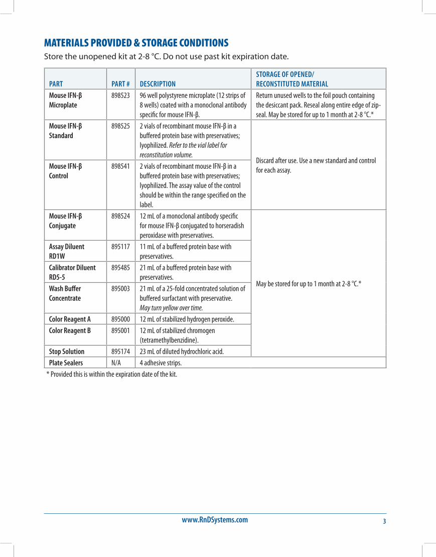
MATERIALS PROVIDED & STORAGE CONDITIONSStore the unopened kit at 2-8 °C. Do not use past kit expiration date.
PART PART # DESCRIPTIONSTORAGE OF OPENED/ RECONSTITUTED MATERIAL
Mouse IFN-β Microplate
898523 96 well polystyrene microplate (12 strips of 8 wells) coated with a monoclonal antibody specific for mouse IFN-β.
Return unused wells to the foil pouch containing the desiccant pack. Reseal along entire edge of zip-seal. May be stored for up to 1 month at 2-8 °C.*
Mouse IFN-β Standard
898525 2 vials of recombinant mouse IFN-β in a buffered protein base with preservatives; lyophilized. Refer to the vial label for reconstitution volume.
Discard after use. Use a new standard and control for each assay.Mouse IFN-β
Control898541 2 vials of recombinant mouse IFN-β in a
buffered protein base with preservatives; lyophilized. The assay value of the control should be within the range specified on the label.
Mouse IFN-β Conjugate
898524 12 mL of a monoclonal antibody specific for mouse IFN-β conjugated to horseradish peroxidase with preservatives.
May be stored for up to 1 month at 2-8 °C.*
Assay Diluent RD1W
895117 11 mL of a buffered protein base with preservatives.
Calibrator Diluent RD5-5
895485 21 mL of a buffered protein base with preservatives.
Wash Buffer Concentrate
895003 21 mL of a 25-fold concentrated solution of buffered surfactant with preservative. May turn yellow over time.
Color Reagent A 895000 12 mL of stabilized hydrogen peroxide.
Color Reagent B 895001 12 mL of stabilized chromogen (tetramethylbenzidine).
Stop Solution 895174 23 mL of diluted hydrochloric acid.
Plate Sealers N/A 4 adhesive strips.
* Provided this is within the expiration date of the kit.
For research use only. Not for use in diagnostic procedures.4
OTHER SUPPLIES REQUIRED• Microplate reader capable of measuring absorbance at 450 nm, with the correction
wavelength set at 540 nm or 570 nm.
• Pipettes and pipette tips.
• Deionized or distilled water.
• Squirt bottle, manifold dispenser, or automated microplate washer.
• 500 mL graduated cylinder.
• Horizontal orbital microplate shaker (0.12" orbit) capable of maintaining a speed of 500 ± 50 rpm.
• Test tubes for dilution of standards.
PRECAUTIONSThe Stop Solution provided with this kit is an acid solution.
Some components in this kit contain a preservative which may cause an allergic skin reaction. Avoid breathing mist.
Color Reagent B may cause skin, eye, and respiratory irritation. Avoid breathing fumes.
Wear protective gloves, clothing, eye, and face protection. Wash hands thoroughly after handling. Refer to the MSDS on our website prior to use.
SAMPLE COLLECTION & STORAGEThe sample collection and storage conditions listed below are intended as general guidelines. Sample stability has not been evaluated.
Cell Culture Supernates - Remove particulates by centrifugation. Assay immediately or aliquot and store samples at ≤ -20 °C. Avoid repeated freeze-thaw cycles.
Serum - Allow blood samples to clot for 2 hours at room temperature before centrifuging for 20 minutes at 2000 x g. Remove serum and assay immediately or aliquot and store samples at ≤ -20 °C. Avoid repeated freeze-thaw cycles.
Plasma - Collect plasma using EDTA or heparin as an anticoagulant. Centrifuge for 20 minutes at 2000 x g within 30 minutes of collection. Assay immediately or aliquot and store samples at ≤ -20 °C. Avoid repeated freeze-thaw cycles.
Note: Citrate plasma has not been validated for use in this assay. Grossly hemolyzed and icteric samples are not suitable for use in this assay. Samples with abnormally high levels of albumin are not suitable for use in this assay.
www.RnDSystems.com 5
REAGENT PREPARATIONBring all reagents to room temperature before use.
Mouse IFN-β Control - Reconstitute the control with 1.0 mL of deionized or distilled water. Mix thoroughly. Assay the control undiluted.
Wash Buffer - If crystals have formed in the concentrate, warm to room temperature and mix gently until the crystals have completely dissolved. Add 20 mL of Wash Buffer Concentrate to deionized or distilled water to prepare 500 mL of Wash Buffer.
Substrate Solution - Color Reagents A and B should be mixed together in equal volumes within 15 minutes of use. Protect from light. 100 μL of the resultant mixture is required per well.
Mouse IFN-β Standard - Refer to the vial label for reconstitution volume. Reconstitute the Mouse IFN-β Standard with deionized or distilled water. This reconstitution produces a stock solution of 10,000 pg/mL. Allow the standard to sit for a minimum of 5 minutes with gentle mixing prior to making dilutions.
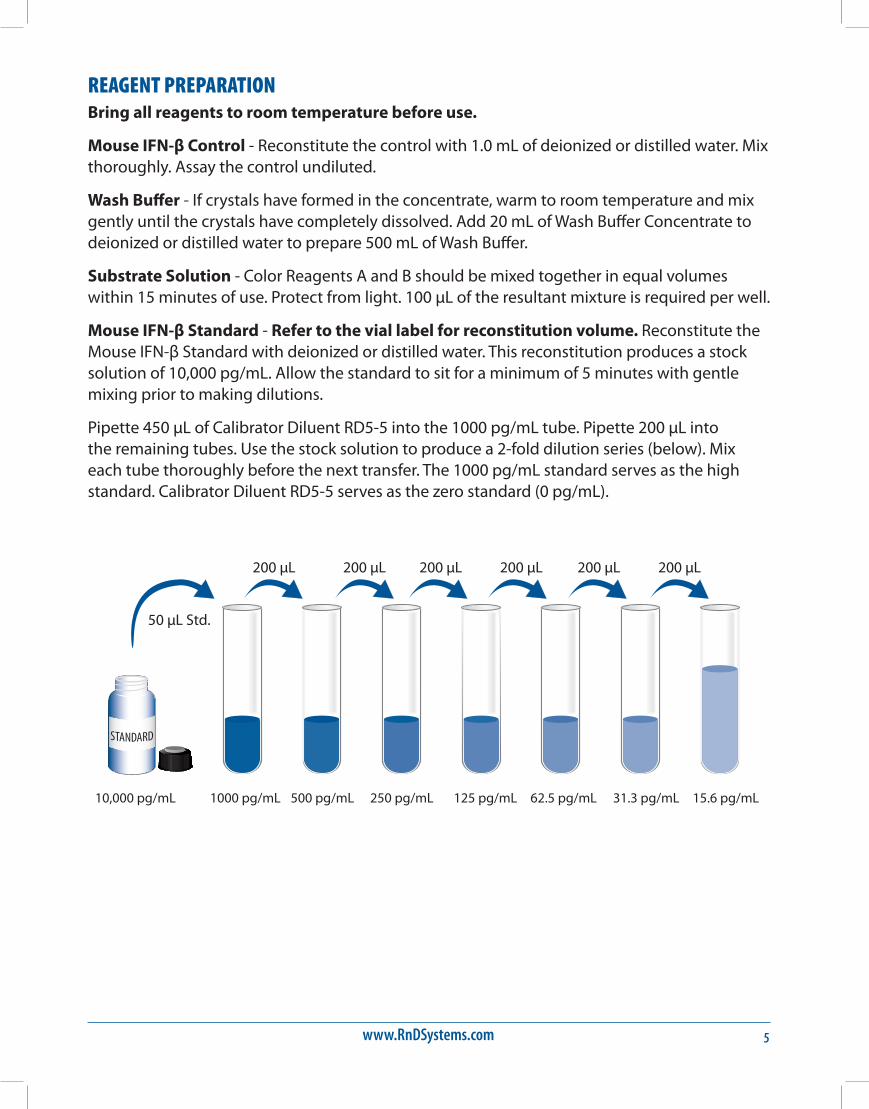
Pipette 450 μL of Calibrator Diluent RD5-5 into the 1000 pg/mL tube. Pipette 200 μL into the remaining tubes. Use the stock solution to produce a 2-fold dilution series (below). Mix each tube thoroughly before the next transfer. The 1000 pg/mL standard serves as the high standard. Calibrator Diluent RD5-5 serves as the zero standard (0 pg/mL).
50 µL Std.
10,000 pg/mL 1000 pg/mL 500 pg/mL 250 pg/mL 125 pg/mL 62.5 pg/mL 31.3 pg/mL 15.6 pg/mL
200 µL 200 µL 200 µL 200 µL 200 µL 200 µL
For research use only. Not for use in diagnostic procedures.6
ASSAY PROCEDURE Bring all reagents and samples to room temperature before use. It is recommended that all standards, control, and samples be assayed in duplicate.
1. Prepare all reagents, working standards, control, and samples as directed in the previous sections.
2. Remove excess microplate strips from the plate frame, return them to the foil pouch containing the desiccant pack, and reseal.
3. Add 50 μL of Assay Diluent RD1W to each well.
4. Add 50 μL of standard, control, or sample per well. Cover with the adhesive strip provided. Incubate for 2 hours at room temperature on a horizontal orbital microplate shaker (0.12" orbit) set at 500 ± 50 rpm. A plate layout is provided to record the standards and samples assayed.
5. Aspirate each well and wash, repeating the process three times for a total of four washes. Wash by filling each well with Wash Buffer (400 μL) using a squirt bottle, manifold dispenser, or autowasher. Complete removal of liquid at each step is essential to good performance. After the last wash, remove any remaining Wash Buffer by aspirating or decanting. Invert the plate and blot it against clean paper towels.
6. Add 100 μL of Mouse IFN-β Conjugate to each well. Cover with a new adhesive strip. Incubate for 2 hours at room temperature on the shaker.
7. Repeat the aspiration/wash as in step 5.
8. Add 100 μL of Substrate Solution to each well. Incubate for 30 minutes at room temperature on the benchtop. Protect from light.
9. Add 100 μL of Stop Solution to each well. Gently tap the plate to ensure thorough mixing.
10. Determine the optical density of each well within 30 minutes, using a microplate reader set to 450 nm. If wavelength correction is available, set to 540 nm or 570 nm. If wavelength correction is not available, subtract readings at 540 nm or 570 nm from the readings at 450 nm. This subtraction will correct for optical imperfections in the plate. Readings made directly at 450 nm without correction may be higher and less accurate.

www.RnDSystems.com 7
CALCULATION OF RESULTSAverage the duplicate readings for each standard, control, and sample and subtract the average zero standard optical density (O.D.).
Create a standard curve by reducing the data using computer software capable of generating a four parameter logistic (4-PL) curve-fit. As an alternative, construct a standard curve by plotting the mean absorbance for each standard on the y-axis against the concentration on the x-axis and draw a best fit curve through the points on the graph. The data may be linearized by plotting the log of the mouse IFN-β concentrations versus the log of the O.D. and the best fit line can be determined by regression analysis. This procedure will produce an adequate but less precise fit of the data.
If samples have been diluted, the concentration read from the standard curve must be multiplied by the dilution factor.
TYPICAL DATAThis standard curve is provided for demonstration only. A standard curve should be generated for each set of samples assayed.
(pg/mL) O.D. Average Corrected0 0.028 0.028 —
0.02815.6 0.075 0.076 0.048
0.07731.3 0.127 0.127 0.099
0.12762.5 0.230 0.232 0.204
0.233125 0.445 0.449 0.421
0.453250 0.861 0.862 0.834
0.863500 1.530 1.540 1.512
1.5491000 2.576 2.603 2.575
2.630
For research use only. Not for use in diagnostic procedures.8
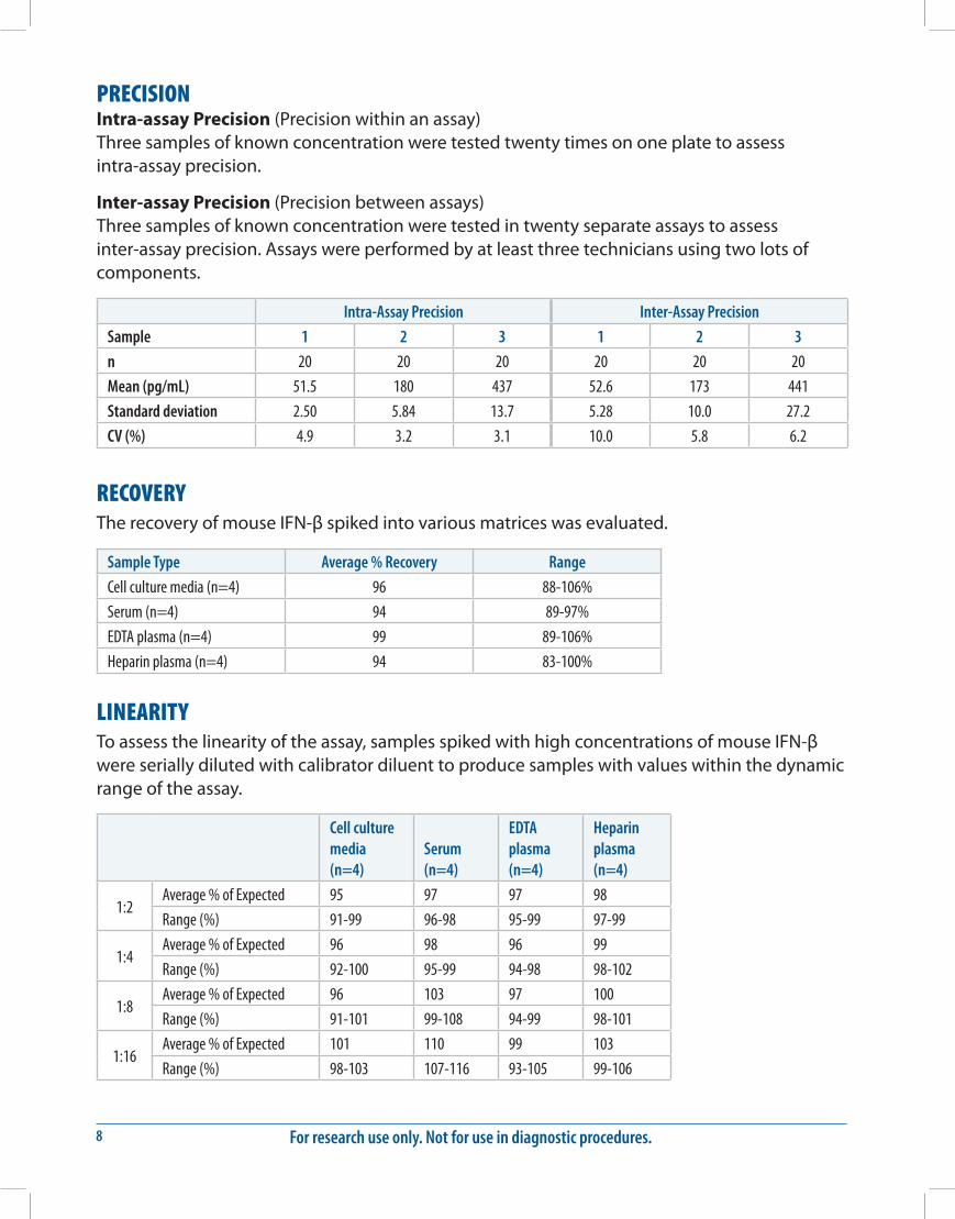
PRECISIONIntra-assay Precision (Precision within an assay) Three samples of known concentration were tested twenty times on one plate to assess intra-assay precision.
Inter-assay Precision (Precision between assays) Three samples of known concentration were tested in twenty separate assays to assess inter-assay precision. Assays were performed by at least three technicians using two lots of components.
Intra-Assay Precision Inter-Assay Precision
Sample 1 2 3 1 2 3
n 20 20 20 20 20 20
Mean (pg/mL) 51.5 180 437 52.6 173 441
Standard deviation 2.50 5.84 13.7 5.28 10.0 27.2
CV (%) 4.9 3.2 3.1 10.0 5.8 6.2
RECOVERYThe recovery of mouse IFN-β spiked into various matrices was evaluated.
Sample Type Average % Recovery Range
Cell culture media (n=4) 96 88-106%
Serum (n=4) 94 89-97%
EDTA plasma (n=4) 99 89-106%
Heparin plasma (n=4) 94 83-100%
LINEARITYTo assess the linearity of the assay, samples spiked with high concentrations of mouse IFN-β were serially diluted with calibrator diluent to produce samples with values within the dynamic range of the assay.
Cell culture media (n=4)
Serum (n=4)
EDTA plasma (n=4)
Heparin plasma (n=4)
1:2Average % of Expected 95 97 97 98
Range (%) 91-99 96-98 95-99 97-99
1:4Average % of Expected 96 98 96 99
Range (%) 92-100 95-99 94-98 98-102
1:8Average % of Expected 96 103 97 100
Range (%) 91-101 99-108 94-99 98-101
1:16Average % of Expected 101 110 99 103
Range (%) 98-103 107-116 93-105 99-106
www.RnDSystems.com 9
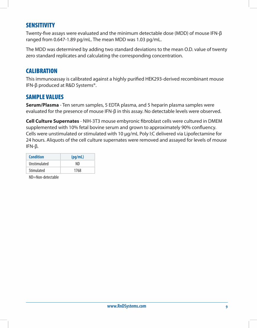
SENSITIVITYTwenty-five assays were evaluated and the minimum detectable dose (MDD) of mouse IFN-β ranged from 0.647-1.89 pg/mL. The mean MDD was 1.03 pg/mL.
The MDD was determined by adding two standard deviations to the mean O.D. value of twenty zero standard replicates and calculating the corresponding concentration.
CALIBRATIONThis immunoassay is calibrated against a highly purified HEK293-derived recombinant mouse IFN-β produced at R&D Systems®.
SAMPLE VALUESSerum/Plasma - Ten serum samples, 5 EDTA plasma, and 5 heparin plasma samples were evaluated for the presence of mouse IFN-β in this assay. No detectable levels were observed.
Cell Culture Supernates - NIH-3T3 mouse embyronic fibroblast cells were cultured in DMEM supplemented with 10% fetal bovine serum and grown to approximately 90% confluency. Cells were unstimulated or stimulated with 10 μg/mL Poly I:C delivered via Lipofectamine for 24 hours. Aliquots of the cell culture supernates were removed and assayed for levels of mouse IFN-β.
Condition (pg/mL)
Unstimulated ND
Stimulated 1768
ND=Non-detectable
For research use only. Not for use in diagnostic procedures.10
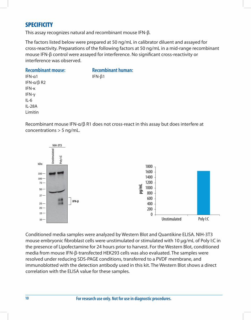
SPECIFICITYThis assay recognizes natural and recombinant mouse IFN-β.
The factors listed below were prepared at 50 ng/mL in calibrator diluent and assayed for cross-reactivity. Preparations of the following factors at 50 ng/mL in a mid-range recombinant mouse IFN-β control were assayed for interference. No significant cross-reactivity or interference was observed.
Recombinant mouse:IFN-α1IFN-α/β R2IFN-κIFN-γIL-6IL-28ALimitin
Recombinant human:IFN-β1
Recombinant mouse IFN-α/β R1 does not cross-react in this assay but does interfere at concentrations > 5 ng/mL.
IFN-β
Unst
imul
ated
Poly
I:C
150
10075
50
37
25
20
15
10
kDa
NIH-3T3pg
/mL
0200400600800
10001200140016001800
Unstimulated Poly I:C
Conditioned media samples were analyzed by Western Blot and Quantikine ELISA. NIH-3T3 mouse embryonic fibroblast cells were unstimulated or stimulated with 10 μg/mL of Poly I:C in the presence of Lipofectamine for 24 hours prior to harvest. For the Western Blot, conditioned media from mouse IFN-β-transfected HEK293 cells was also evaluated. The samples were resolved under reducing SDS-PAGE conditions, transferred to a PVDF membrane, and immunoblotted with the detection antibody used in this kit. The Western Blot shows a direct correlation with the ELISA value for these samples.
www.RnDSystems.com 11
REFERENCES1. González-Navajas, J.M. et al. (2012) Nat. Rev. Immunol. 12:125.
2. Reder, A.T. and X. Feng (2013) Front. Immunol. 4:281.
3. Schafer, S.L. et al. (1998) J. Biol. Chem. 273:2714.
4. Häcker, H. et al. (2006) Nature 439:204.
5. Platanias, L.C. (2005) Nat. Rev. Immunol. 5:375.
6. Fasler-Kan, E. et al. (1998) Eur. J. Biochem. 254:514.
7. Matikainen, S. et al. (1999) Blood 93:1980.
8. Wang, H. et al. (2011) J. Immunol 186:675.
9. Qiu, H. et al. (2008) J. Immunol. 181:2092.
10. Guarda, G. et al. (2011) Immunity 34:213.
11. Teige, I. et al. (2003) J. Immunol. 170:4776.
12. Shinohara, M.L. et al. (2008) Immunity 29:68.
13. Guo, B. et al. (2008) J. Clin. Invest. 118:1680.
14. Ramgolam, V.S. and S. Markovic-Plese (2010) Endocr. Metab. Immune Disord. Drug Targets 10:161.
15. Martín-Saavedra, F.M. et al. (2008) Mol. Immunol. 45:4008.
16. Inoue, M. and M.L. Shinohara (2013) Immunology 139:11.
17. Katakura, K. et al. (2005) J. Clin. Invest. 115:695.
18. Karaghiosoff, M. et al. (2003) Nat. Immunol. 4:471.
19. Vandenbark, A.A. et al. (2009) J. Neuroimmunol. 215:125.
20. Wang, D. et al. (2016) J. Immunol. 197:2992.
For research use only. Not for use in diagnostic procedures.12
PLATE LAYOUTUse this plate layout to record standards and samples assayed.

www.RnDSystems.com 13
NOTES
For research use only. Not for use in diagnostic procedures.14
12.16 753114.0 12/16
©2016 R&D Systems®, Inc.
NOTES
All trademarks and registered trademarks are the property of their respective owners.