Mouse IFN-γ Immunoassay Quantikine · Interferon-gamma (IFN-γ, also known as type II interferon)...
16
Mouse IFN-γ Immunoassay Quantikine ® ELISA This package insert must be read in its entirety before using this product. For research use only. Not for use in diagnostic procedures. Catalog Number MIF00 Catalog Number SMIF00 Catalog Number PMIF00 For the quantitative determination of mouse Interferon gamma (IFN-γ) concentrations in cell culture supernates and serum.
Transcript of Mouse IFN-γ Immunoassay Quantikine · Interferon-gamma (IFN-γ, also known as type II interferon)...
Mouse IFN-γ Immunoassay
Quantikine® ELISA
This package insert must be read in its entirety before using this product. For research use only. Not for use in diagnostic procedures.
Catalog Number MIF00 Catalog Number SMIF00 Catalog Number PMIF00
For the quantitative determination of mouse Interferon gamma (IFN-γ) concentrations in cell culture supernates and serum.
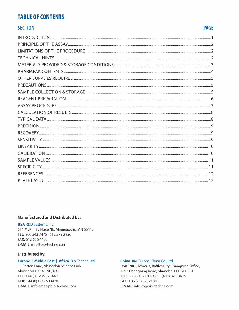
TABLE OF CONTENTS
SECTION PAGE
INTRODUCTION .....................................................................................................................................................................1PRINCIPLE OF THE ASSAY ...................................................................................................................................................2LIMITATIONS OF THE PROCEDURE .................................................................................................................................2TECHNICAL HINTS .................................................................................................................................................................2MATERIALS PROVIDED & STORAGE CONDITIONS ...................................................................................................3PHARMPAK CONTENTS .......................................................................................................................................................4OTHER SUPPLIES REQUIRED .............................................................................................................................................5PRECAUTIONS .........................................................................................................................................................................5SAMPLE COLLECTION & STORAGE .................................................................................................................................5REAGENT PREPARATION .....................................................................................................................................................6ASSAY PROCEDURE .............................................................................................................................................................7CALCULATION OF RESULTS ...............................................................................................................................................8TYPICAL DATA .........................................................................................................................................................................8PRECISION ................................................................................................................................................................................9RECOVERY.................................................................................................................................................................................9SENSITIVITY .............................................................................................................................................................................9LINEARITY .............................................................................................................................................................................. 10CALIBRATION ....................................................................................................................................................................... 10SAMPLE VALUES .................................................................................................................................................................. 11SPECIFICITY ........................................................................................................................................................................... 11REFERENCES ......................................................................................................................................................................... 12PLATE LAYOUT ..................................................................................................................................................................... 13
Manufactured and Distributed by:
USA R&D Systems, Inc. 614 McKinley Place NE, Minneapolis, MN 55413TEL: 800 343 7475 612 379 2956FAX: 612 656 4400E-MAIL: [email protected]
Distributed by:
Europe | Middle East | Africa Bio-Techne Ltd.19 Barton Lane, Abingdon Science ParkAbingdon OX14 3NB, UKTEL: +44 (0)1235 529449FAX: +44 (0)1235 533420E-MAIL: [email protected]
China Bio-Techne China Co., Ltd.Unit 1901, Tower 3, Raffles City Changning Office,1193 Changning Road, Shanghai PRC 200051TEL: +86 (21) 52380373 (400) 821-3475FAX: +86 (21) 52371001E-MAIL: [email protected]
www.RnDSystems.com 1
INTRODUCTIONInterferon-gamma (IFN-γ, also known as type II interferon) is an important immunoregulatory cytokine that was originally identified through its anti-viral activity (1, 2). It plays key roles in host defense by exerting anti-viral, anti-proliferative, and immunoregulatory activities (3, 4). On many cell types, IFN-γ induces the production of cytokines and upregulates the expression of various membrane proteins including class I and II MHC antigens, Fc receptors, leukocyte adhesion molecules, and B7 family antigens. IFN-γ is a potent activator of macrophage effector functions. It directs the synthesis, class switching, and secretion of immunoglobulins by B cells. IFN-γ also influences T-helper cell phenotype development by inhibiting Th2 differentiation and stimulating Th1 development (3, 4). IFN-γ plays a central role in the progression of inflammatory diseases such as autoimmunity and atherosclerosis (5, 6).
Biologically active IFN-γ consists of a noncovalently linked homodimer of 20-25 kDa variably glycosylated subunits (7). Mature mouse IFN-γ shares 86% amino acid (aa) sequence identity with rat IFN-γ, and 38-44% aa identity with bovine, canine, cotton rat, equine, feline, human, porcine, and rhesus IFN-γ. IFN-γ dimers bind to transmembrane IFN-γ RI (alpha subunits) which then interact with transmembrane IFN-γ RII (beta subunits) to form the functional receptor complex of two α and two β subunits (8, 9). Inclusion of IFN-γ RII in the receptor complex increases the ligand binding affinity as well as the efficiency of signal transduction (9, 10). Whereas the α-chain is expressed constitutively on many cell types, the cellular regulation of the β-chain correlates with an IFN-γ responsive state and is tightly regulated (8).
IFN-γ is produced by a number of cell types under inflammatory conditions, including dendritic epidermal/γδ T cells (11), keratinocytes (12), peripheral blood γδ T cells (13), mast cells (14), neurons (15), CD8+ T cells (16), macrophages (17), B cells (18), neutrophils (19), NK cells (20), CD4+ T cells (21), and testicular spermatids (22).
The Quantikine® Mouse IFN-γ Immunoassay is a 4.5 hour solid phase ELISA designed to measure mouse IFN-γ in cell culture supernates and mouse serum. It contains E. coli-expressed recombinant mouse IFN-γ and antibodies raised against the recombinant factor. This immunoassay has been shown to quantitate recombinant mouse IFN-γ accurately. Results obtained using natural mouse IFN-γ showed dose-response curves that were parallel to the standard curves obtained using the Quantikine® kit standards. These results indicate that this kit can be used to determine relative mass values for natural mouse IFN-γ.
For research use only. Not for use in diagnostic procedures.2
PRINCIPLE OF THE ASSAYThis assay employs the quantitative sandwich enzyme immunoassay technique. A monoclonal antibody specific for mouse IFN-γ has been pre-coated onto a microplate. Standards, control, and samples are pipetted into the wells and any IFN-γ present is bound by the immobilized antibody. After washing away any unbound substances, an enzyme-linked polyclonal antibody specific for mouse IFN-γ is added to the wells. Following a wash to remove any unbound antibody-enzyme reagent, a substrate solution is added to the wells. The enzyme reaction yields a blue product that turns yellow when the Stop Solution is added. The intensity of the color measured is in proportion to the amount of IFN-γ bound in the initial step. The sample values are then read off the standard curve.
LIMITATIONS OF THE PROCEDURE• FOR RESEARCH USE ONLY. NOT FOR USE IN DIAGNOSTIC PROCEDURES.• The kit should not be used beyond the expiration date on the kit label.• Do not mix or substitute reagents with those from other lots or sources. • If samples generate values higher than the highest standard, dilute the samples with the
appropriate calibrator diluent and repeat the assay.• Any variation in operator, pipetting technique, washing technique, incubation time or
temperature, and kit age can cause variation in binding.• Variations in sample collection, processing, and storage may cause sample value differences.• This assay is designed to eliminate interference by other factors present in biological
samples. Until all factors have been tested in the Quantikine® Immunoassay, the possibility of interference cannot be excluded.
TECHNICAL HINTS• When mixing or reconstituting protein solutions, always avoid foaming.• To avoid cross-contamination, change pipette tips between additions of each standard level,
between sample additions, and between reagent additions. Also, use separate reservoirs for each reagent.
• For best results, pipette reagents and samples into the center of each well.• It is recommended that the samples be pipetted within 15 minutes.• To ensure accurate results, proper adhesion of plate sealers during incubation steps is
necessary.• Substrate Solution should remain colorless until added to the plate. Keep Substrate Solution
protected from light. Substrate Solution should change from colorless to gradations of blue.• Stop Solution should be added to the plate in the same order as the Substrate Solution. The
color developed in the wells will turn from blue to yellow upon addition of the Stop Solution.
www.RnDSystems.com 3
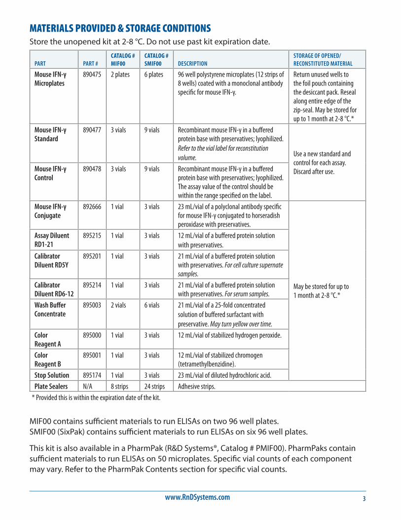
MATERIALS PROVIDED & STORAGE CONDITIONSStore the unopened kit at 2-8 °C. Do not use past kit expiration date.
PART PART #CATALOG # MIF00
CATALOG # SMIF00 DESCRIPTION
STORAGE OF OPENED/ RECONSTITUTED MATERIAL
Mouse IFN-γ Microplates
890475 2 plates 6 plates 96 well polystyrene microplates (12 strips of 8 wells) coated with a monoclonal antibody specific for mouse IFN-γ.
Return unused wells to the foil pouch containing the desiccant pack. Reseal along entire edge of the zip-seal. May be stored for up to 1 month at 2-8 °C.*
Mouse IFN-γ Standard
890477 3 vials 9 vials Recombinant mouse IFN-γ in a buffered protein base with preservatives; lyophilized. Refer to the vial label for reconstitution volume. Use a new standard and
control for each assay. Discard after use. Mouse IFN-γ
Control890478 3 vials 9 vials Recombinant mouse IFN-γ in a buffered
protein base with preservatives; lyophilized. The assay value of the control should be within the range specified on the label.
Mouse IFN-γ Conjugate
892666 1 vial 3 vials 23 mL/vial of a polyclonal antibody specific for mouse IFN-γ conjugated to horseradish peroxidase with preservatives.
May be stored for up to 1 month at 2-8 °C.*
Assay Diluent RD1-21
895215 1 vial 3 vials 12 mL/vial of a buffered protein solution with preservatives.
Calibrator Diluent RD5Y
895201 1 vial 3 vials 21 mL/vial of a buffered protein solution with preservatives. For cell culture supernate samples.
Calibrator Diluent RD6-12
895214 1 vial 3 vials 21 mL/vial of a buffered protein solution with preservatives. For serum samples.
Wash Buffer Concentrate
895003 2 vials 6 vials 21 mL/vial of a 25-fold concentrated solution of buffered surfactant with preservative. May turn yellow over time.
Color Reagent A
895000 1 vial 3 vials 12 mL/vial of stabilized hydrogen peroxide.
Color Reagent B
895001 1 vial 3 vials 12 mL/vial of stabilized chromogen (tetramethylbenzidine).
Stop Solution 895174 1 vial 3 vials 23 mL/vial of diluted hydrochloric acid.
Plate Sealers N/A 8 strips 24 strips Adhesive strips.
* Provided this is within the expiration date of the kit.
MIF00 contains sufficient materials to run ELISAs on two 96 well plates. SMIF00 (SixPak) contains sufficient materials to run ELISAs on six 96 well plates.
This kit is also available in a PharmPak (R&D Systems®, Catalog # PMIF00). PharmPaks contain sufficient materials to run ELISAs on 50 microplates. Specific vial counts of each component may vary. Refer to the PharmPak Contents section for specific vial counts.
For research use only. Not for use in diagnostic procedures.4
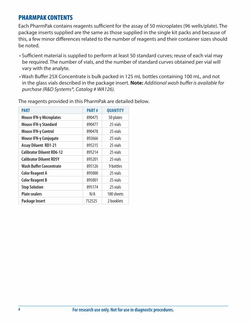
PHARMPAK CONTENTSEach PharmPak contains reagents sufficient for the assay of 50 microplates (96 wells/plate). The package inserts supplied are the same as those supplied in the single kit packs and because of this, a few minor differences related to the number of reagents and their container sizes should be noted.
• Sufficient material is supplied to perform at least 50 standard curves; reuse of each vial may be required. The number of vials, and the number of standard curves obtained per vial will vary with the analyte.
• Wash Buffer 25X Concentrate is bulk packed in 125 mL bottles containing 100 mL, and not in the glass vials described in the package insert. Note: Additional wash buffer is available for purchase (R&D Systems®, Catalog # WA126).
The reagents provided in this PharmPak are detailed below.
PART PART # QUANTITY
Mouse IFN-γ Microplates 890475 50 plates
Mouse IFN-γ Standard 890477 25 vials
Mouse IFN-γ Control 890478 25 vials
Mouse IFN-γ Conjugate 892666 25 vials
Assay Diluent RD1-21 895215 25 vials
Calibrator Diluent RD6-12 895214 25 vials
Calibrator Diluent RD5Y 895201 25 vials
Wash Buffer Concentrate 895126 9 bottles
Color Reagent A 895000 25 vials
Color Reagent B 895001 25 vials
Stop Solution 895174 25 vials
Plate sealers N/A 100 sheets
Package Insert 752525 2 booklets
www.RnDSystems.com 5
OTHER SUPPLIES REQUIRED• Microplate reader capable of measuring absorbance at 450 nm, with the correction
wavelength set at 540 nm or 570 nm.
• Pipettes and pipette tips.
• Deionized or distilled water.
• Squirt bottle, manifold dispenser, or automated microplate washer.
• 500 mL graduated cylinder.
• Polypropylene test tubes for dilution of standards.
PRECAUTIONSThe Stop Solution provided with this kit is an acid solution.
Some components in this kit contain a preservative which may cause an allergic skin reaction. Avoid breathing mist.
Color Reagent B may cause skin, eye, and respiratory irritation. Avoid breathing fumes.
Wear protective gloves, clothing, eye, and face protection. Wash hands thoroughly after handling. Refer to the SDS on our website prior to use.
SAMPLE COLLECTION & STORAGEThe sample collection and storage conditions listed below are intended as general guidelines. Sample stability has not been evaluated.
Cell Culture Supernates - Remove particulates by centrifugation and assay immediately or aliquot and store samples at ≤ -20 °C. Avoid repeated freeze-thaw cycles.
Serum - Allow blood samples to clot for 2 hours at room temperature before centrifuging for 20 minutes at 2000 x g. Remove serum and assay immediately or aliquot and store samples at ≤ -20 °C. Avoid repeated freeze-thaw cycles.
Note: Grossly hemolyzed or lipemic samples may not be suitable for use in this assay.
SAMPLE PREPARATIONUse polypropylene tubes.
Cell culture supernate samples may require a dilution.
For research use only. Not for use in diagnostic procedures.6
REAGENT PREPARATIONBring all reagents to room temperature before use.
Mouse IFN-γ Control - Reconstitute the control with 1.0 mL deionized or distilled water. Mix thoroughly. Assay the control undiluted.
Wash Buff er - If crystals have formed in the concentrate, warm to room temperature and mix gently until the crystals have completely dissolved. To prepare enough Wash Buff er for one plate, add 20 mL of Wash Buff er Concentrate to 480 mL of deionized or distilled water to prepare 500 mL of Wash Buff er.
Substrate Solution - Color Reagents A and B should be mixed together in equal volumes within 15 minutes of use. Protect from light. 100 μL of the resultant mixture is required per well.
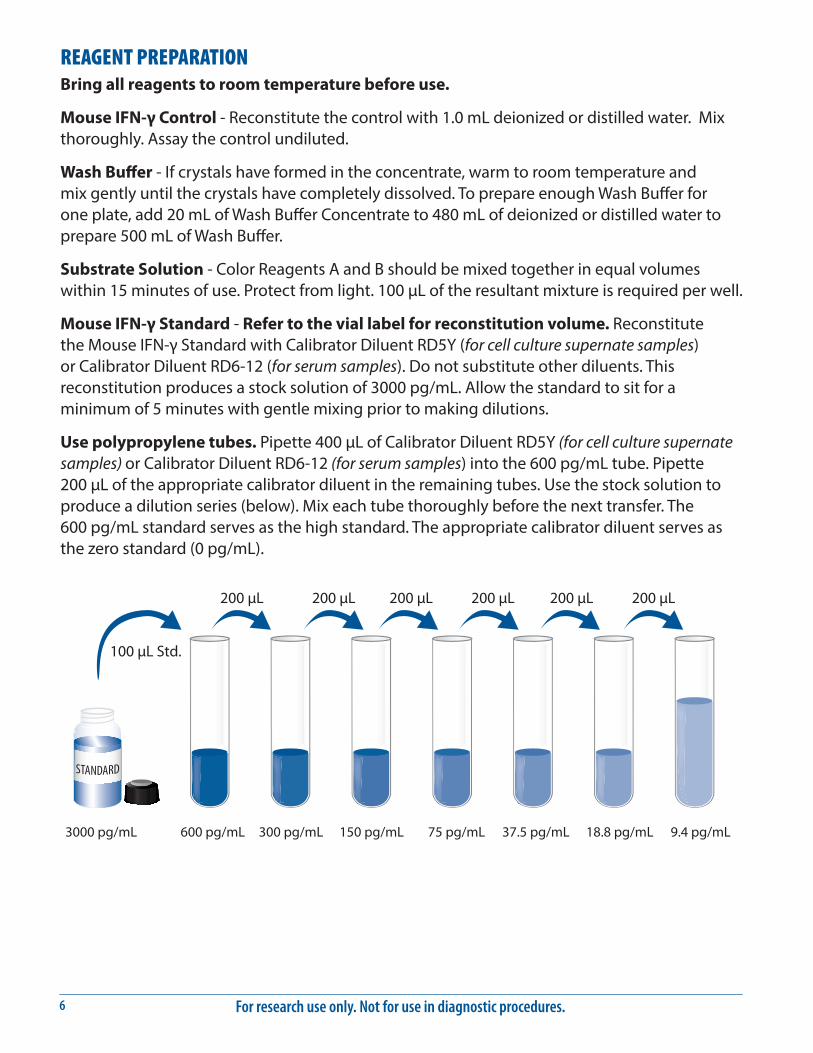
Mouse IFN-γ Standard - Refer to the vial label for reconstitution volume. Reconstitute the Mouse IFN-γ Standard with Calibrator Diluent RD5Y (for cell culture supernate samples) or Calibrator Diluent RD6-12 (for serum samples). Do not substitute other diluents. This reconstitution produces a stock solution of 3000 pg/mL. Allow the standard to sit for a minimum of 5 minutes with gentle mixing prior to making dilutions.
Use polypropylene tubes. Pipette 400 μL of Calibrator Diluent RD5Y (for cell culture supernate samples) or Calibrator Diluent RD6-12 (for serum samples) into the 600 pg/mL tube. Pipette 200 μL of the appropriate calibrator diluent in the remaining tubes. Use the stock solution to produce a dilution series (below). Mix each tube thoroughly before the next transfer. The 600 pg/mL standard serves as the high standard. The appropriate calibrator diluent serves as the zero standard (0 pg/mL).
100 µL Std.
3000 pg/mL 600 pg/mL 300 pg/mL 150 pg/mL 75 pg/mL 37.5 pg/mL 18.8 pg/mL 9.4 pg/mL
200 µL 200 µL 200 µL 200 µL 200 µL 200 µL
www.RnDSystems.com 7
ASSAY PROCEDURE Bring all reagents and samples to room temperature before use. It is recommended that all standards, control, and samples be assayed in duplicate.
1. Prepare reagents, standards, control, and samples as directed by the previous sections.
2. Remove excess microplate strips from the plate frame, return them to the foil pouch containing the desiccant pack, and reseal.
3. Add 50 μL of Assay Diluent RD1-21 to each well.
4. Add 50 μL of standard, control, or sample to each well. Mix by gently tapping the plate frame for 1 minute. Cover with the adhesive strip provided. Incubate for 2 hours at room temperature. A plate layout is provided to record standards and samples assayed.
5. Aspirate each well and wash, repeating the process four times for a total of five washes. Wash by filling each well with Wash Buffer (400 μL) using a squirt bottle, manifold dispenser, or autowasher. Complete removal of liquid at each step is essential to good performance. After the last wash, remove any remaining Wash Buffer by aspirating or decanting. Invert the plate and blot it against clean paper towels.
6. Add 100 μL of Mouse IFN-γ Conjugate to each well. Cover with a new adhesive strip. Incubate for 2 hours at room temperature.
7. Repeat the aspiration/wash as in step 5.
8. Add 100 μL of Substrate Solution to each well. Incubate for 30 minutes at room temperature. Protect from light.
9. Add 100 μL of Stop Solution to each well. Gently tap the plate to ensure thorough mixing.
10. Determine the optical density of each well within 30 minutes, using a microplate reader set to 450 nm. If wavelength correction is available, set to 540 nm or 570 nm. If wavelength correction is not available, subtract readings at 540 nm or 570 nm from the readings at 450 nm. This subtraction will correct for optical imperfections in the plate. Readings made directly at 450 nm without correction may be higher and less accurate.
For research use only. Not for use in diagnostic procedures.8
CALCULATION OF RESULTSAverage the duplicate readings for each standard, control, and sample and subtract the average zero standard optical density (O.D.).
Create a standard curve by reducing the data using computer software capable of generating a log/log curve-fit. As an alternative, construct a standard curve by plotting the mean absorbance for each standard on the y-axis against the concentration on the x-axis and draw a best fit curve through the points on a log/log graph. The data may be linearized by plotting the log of the mouse IFN-γ concentrations versus the log of the O.D. on a linear scale, and the best fit line can be determined by regression analysis.
If samples have been diluted, the concentration read from the standard curve must be multiplied by the dilution factor.
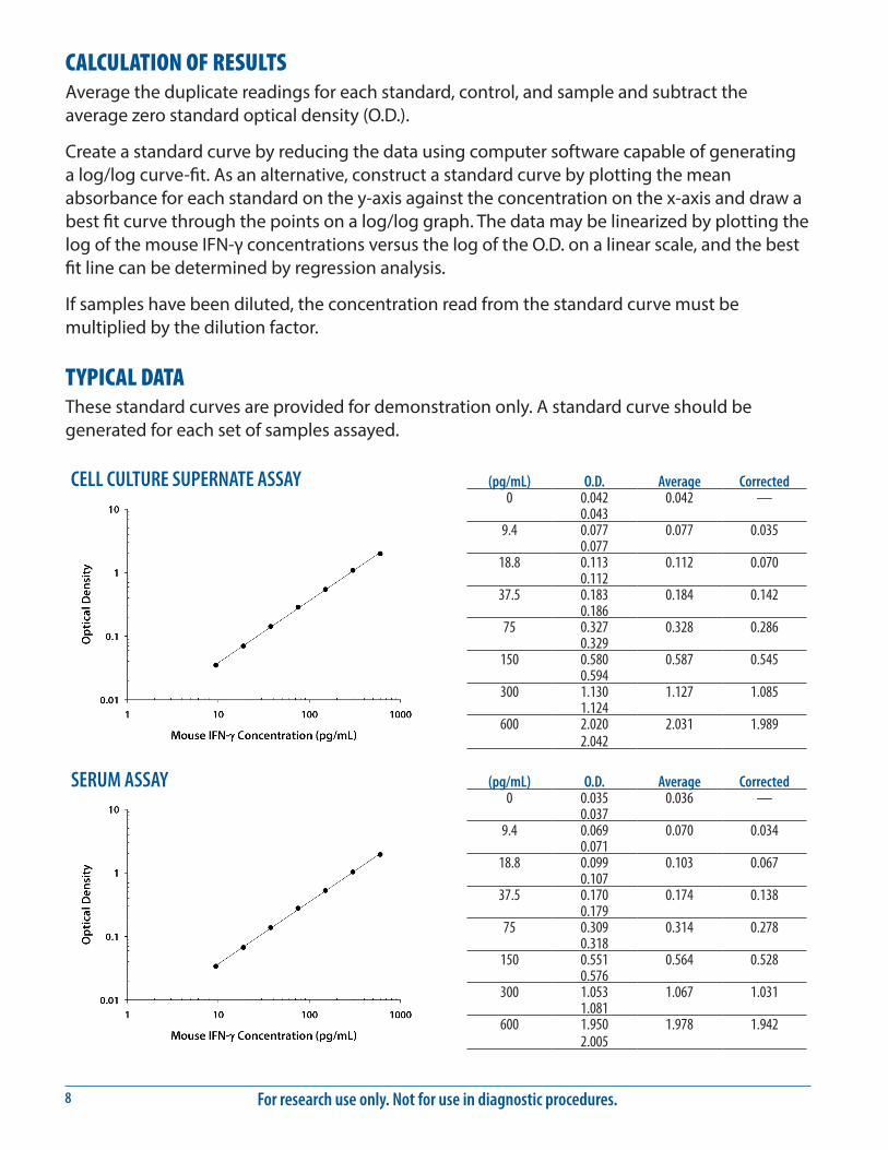
TYPICAL DATAThese standard curves are provided for demonstration only. A standard curve should be generated for each set of samples assayed.
(pg/mL) O.D. Average Corrected0 0.042 0.042 —
0.0439.4 0.077 0.077 0.035
0.07718.8 0.113 0.112 0.070
0.11237.5 0.183 0.184 0.142
0.18675 0.327 0.328 0.286
0.329150 0.580 0.587 0.545
0.594300 1.130 1.127 1.085
1.124600 2.020 2.031 1.989
2.042
(pg/mL) O.D. Average Corrected0 0.035 0.036 —
0.0379.4 0.069 0.070 0.034
0.07118.8 0.099 0.103 0.067
0.10737.5 0.170 0.174 0.138
0.17975 0.309 0.314 0.278
0.318150 0.551 0.564 0.528
0.576300 1.053 1.067 1.031
1.081600 1.950 1.978 1.942
2.005
CELL CULTURE SUPERNATE ASSAY
SERUM ASSAY
www.RnDSystems.com 9
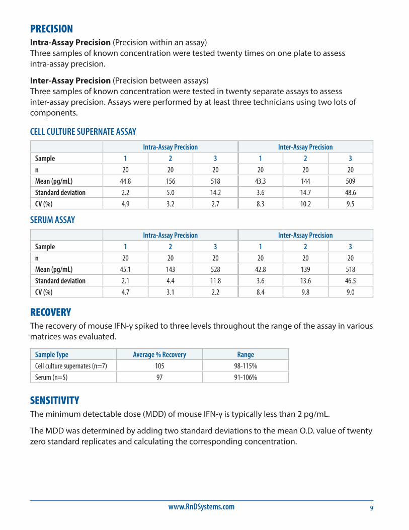
PRECISIONIntra-Assay Precision (Precision within an assay) Three samples of known concentration were tested twenty times on one plate to assess intra-assay precision.
Inter-Assay Precision (Precision between assays) Three samples of known concentration were tested in twenty separate assays to assess inter-assay precision. Assays were performed by at least three technicians using two lots of components.
CELL CULTURE SUPERNATE ASSAY
Intra-Assay Precision Inter-Assay Precision
Sample 1 2 3 1 2 3
n 20 20 20 20 20 20
Mean (pg/mL) 44.8 156 518 43.3 144 509
Standard deviation 2.2 5.0 14.2 3.6 14.7 48.6
CV (%) 4.9 3.2 2.7 8.3 10.2 9.5
SERUM ASSAY
Intra-Assay Precision Inter-Assay Precision
Sample 1 2 3 1 2 3
n 20 20 20 20 20 20
Mean (pg/mL) 45.1 143 528 42.8 139 518
Standard deviation 2.1 4.4 11.8 3.6 13.6 46.5
CV (%) 4.7 3.1 2.2 8.4 9.8 9.0
RECOVERYThe recovery of mouse IFN-γ spiked to three levels throughout the range of the assay in various matrices was evaluated.
Sample Type Average % Recovery Range
Cell culture supernates (n=7) 105 98-115%
Serum (n=5) 97 91-106%
SENSITIVITYThe minimum detectable dose (MDD) of mouse IFN-γ is typically less than 2 pg/mL.
The MDD was determined by adding two standard deviations to the mean O.D. value of twenty zero standard replicates and calculating the corresponding concentration.
For research use only. Not for use in diagnostic procedures.10
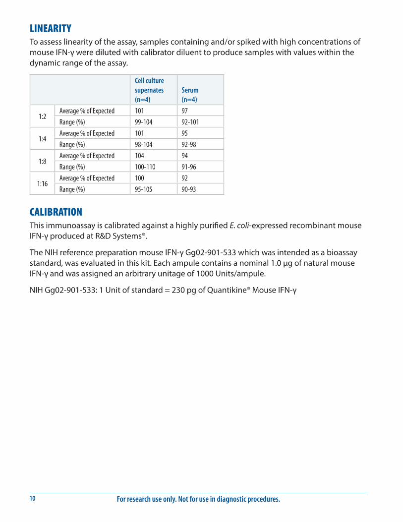
LINEARITYTo assess linearity of the assay, samples containing and/or spiked with high concentrations of mouse IFN-γ were diluted with calibrator diluent to produce samples with values within the dynamic range of the assay.
Cell culture supernates (n=4)
Serum (n=4)
1:2Average % of Expected 101 97
Range (%) 99-104 92-101
1:4Average % of Expected 101 95
Range (%) 98-104 92-98
1:8Average % of Expected 104 94
Range (%) 100-110 91-96
1:16Average % of Expected 100 92
Range (%) 95-105 90-93
CALIBRATIONThis immunoassay is calibrated against a highly purified E. coli-expressed recombinant mouse IFN-γ produced at R&D Systems®.
The NIH reference preparation mouse IFN-γ Gg02-901-533 which was intended as a bioassay standard, was evaluated in this kit. Each ampule contains a nominal 1.0 μg of natural mouse IFN-γ and was assigned an arbitrary unitage of 1000 Units/ampule.
NIH Gg02-901-533: 1 Unit of standard = 230 pg of Quantikine® Mouse IFN-γ
www.RnDSystems.com 11
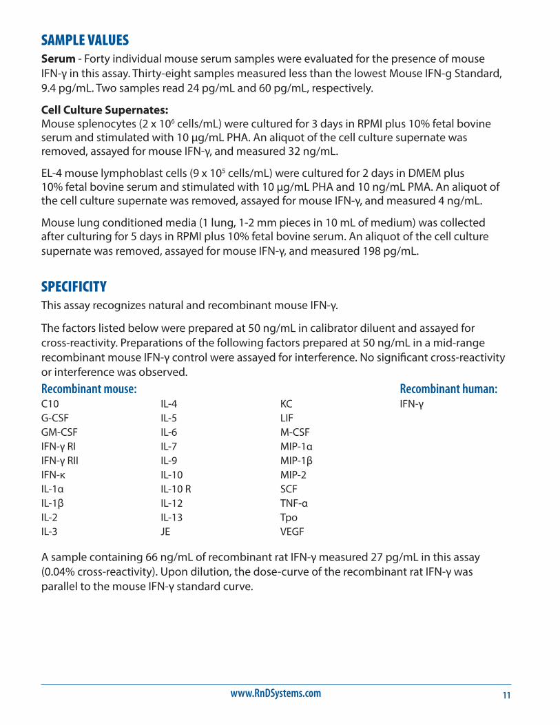
SAMPLE VALUESSerum - Forty individual mouse serum samples were evaluated for the presence of mouse IFN-γ in this assay. Thirty-eight samples measured less than the lowest Mouse IFN-g Standard, 9.4 pg/mL. Two samples read 24 pg/mL and 60 pg/mL, respectively.
Cell Culture Supernates: Mouse splenocytes (2 x 10⁶ cells/mL) were cultured for 3 days in RPMI plus 10% fetal bovine serum and stimulated with 10 μg/mL PHA. An aliquot of the cell culture supernate was removed, assayed for mouse IFN-γ, and measured 32 ng/mL.
EL-4 mouse lymphoblast cells (9 x 10⁵ cells/mL) were cultured for 2 days in DMEM plus 10% fetal bovine serum and stimulated with 10 μg/mL PHA and 10 ng/mL PMA. An aliquot of the cell culture supernate was removed, assayed for mouse IFN-γ, and measured 4 ng/mL.
Mouse lung conditioned media (1 lung, 1-2 mm pieces in 10 mL of medium) was collected after culturing for 5 days in RPMI plus 10% fetal bovine serum. An aliquot of the cell culture supernate was removed, assayed for mouse IFN-γ, and measured 198 pg/mL.
SPECIFICITYThis assay recognizes natural and recombinant mouse IFN-γ.
The factors listed below were prepared at 50 ng/mL in calibrator diluent and assayed for cross-reactivity. Preparations of the following factors prepared at 50 ng/mL in a mid-range recombinant mouse IFN-γ control were assayed for interference. No significant cross-reactivity or interference was observed.Recombinant mouse:C10G-CSFGM-CSFIFN-γ RIIFN-γ RIIIFN-κIL-1αIL-1βIL-2IL-3
IL-4IL-5IL-6IL-7IL-9IL-10IL-10 RIL-12IL-13JE
KCLIFM-CSFMIP-1αMIP-1βMIP-2SCFTNF-αTpoVEGF
Recombinant human:IFN-γ
A sample containing 66 ng/mL of recombinant rat IFN-γ measured 27 pg/mL in this assay (0.04% cross-reactivity). Upon dilution, the dose-curve of the recombinant rat IFN-γ was parallel to the mouse IFN-γ standard curve.
For research use only. Not for use in diagnostic procedures.12
REFERENCES1. Billiau, A. and P. Matthys (2009) Cytokine Growth Factor Rev. 20:97.2. Wheelock, E.F. (1965) Science 146:310.3. Schoenborn, J.R and C.B. Wilson (2007) Adv. Immunol. 96:41.4. Pestka, S. et al. (2004) Immunol. Rev. 202:8.5. Kelchtermans, H. et al. (2008) Trends Immunol. 29:479.6. McLaren, J.E. and D.P. Ramji (2009) Cytokine Growth Factor Rev. 20:125.7. Gray, P.W. and D.V. Goeddel (1983) Proc. Natl. Acad. Sci. USA 80:5842.8. Bach, E.A. et al. (1997) Annu. Rev. Immunol. 15:563.9. Marsters, S.A. et al. (1995) Proc. Natl. Acad. Sci. USA 92:5401.
10. Krause, C.D. et al. (2000) J. Biol. Chem. 275:22995.11. Sugaya, M. et al. (1999) J. Invest. Dermatol. 113:350.12. Howie, S.E.M. et al. (1996) J. Invest. Dermatol. 106:1218.13. Battistini, L. et al. (1997) J. Immunol. 159:3723.14. Gupta, A.A. et al. (1996) J. Immunol. 157:2123.15. Neumann, H. et al. (1997) J. Exp. Med. 186:2023.16. Hoiden, I. and G. Moller (1996) Scand. J. Immunol. 44:501.17. Puddu, P. et al. (1997) J. Immunol. 159:3490.18. Yoshimoto, T. et al. (1997) Proc. Natl. Acad. Sci. USA 94:3948.19. Yeaman, G.R. et al. (1998) J. Immunol. 160:5145.20. Asea, A. et al. (1996) Clin. Exp. Immunol. 105:376.21. Briscoe, D.M. et al. (1997) J. Immunol. 159:3247.22. Dejuco, N. et al. (1995) Endocrinology 136:4925.
www.RnDSystems.com 13
PLATE LAYOUTUse this plate layout to record standards and samples assayed.
For research use only. Not for use in diagnostic procedures.14
NOTES
10.96 752525.2 7/18
©2018 R&D Systems®, Inc.
All trademarks and registered trademarks are the property of their respective owners.