VeriKine-HS Mouse IFN-α All Subtype ELISA Kit the initial and progressive responses to pathogens...
16
VeriKine-HS Mouse IFN-α All Subtype ELISA Kit Product #42115 Standard Curve Range: 1.19 – 76 pg/ml Sample Detection Range: 2.38 – 152 pg/ml Store all components at 2 - 8 o C Sold under license from Pestka Biomedical Laboratories, Inc. d/b/a PBL Assay Science. For research use only. Not for diagnostic or clinical use in, or administration to, humans. Not for resale in original or any modified form, including inclusion in a kit, for any purpose. Not for use in the preparation of any commercial product. © Copyright 2017 Pestka Biomedical Laboratories, Inc. All rights reserved.
Transcript of VeriKine-HS Mouse IFN-α All Subtype ELISA Kit the initial and progressive responses to pathogens...
VeriKine-HS Mouse IFN-α All Subtype ELISA Kit
Product #42115
Standard Curve Range: 1.19 – 76 pg/mlSample Detection Range: 2.38 – 152 pg/ml
Store all components at 2 - 8oC
Sold under license from Pestka Biomedical Laboratories, Inc. d/b/a PBL Assay Science. For research use only.
Not for diagnostic or clinical use in, or administration to, humans. Not for resale in original or any modified form, including inclusion in a kit, for any purpose.
Not for use in the preparation of any commercial product.© Copyright 2017 Pestka Biomedical Laboratories, Inc. All rights reserved.
INTRODUCTION
Interferons (IFNs) are a group of cytokines which exhibit pleitropic activities that play major roles in both innate and adaptive immunity. Type I IFNs consist of multiple IFN-α genes and at least one IFN-β gene in most vertebrates, and a few other family members such as limitin in the mouse.1 In humans, IFN-α is used to treat viral diseases and cancer2-4 and is studied in mouse models of these diseases.5,6 Recent evidence has suggested that IFN-α may play a role in development of certain autoimmune diseases7,8 and that treatment with IFN-α is useful for other autoimmune diseases.9
The expression of interferons is regulated by a group of interferon regulatory factor proteins (IRF). The expression of the individual IFN-α subtypes is regulated by the cellular levels of IRF3 and IRF7. Most of the IFN-α subtypes require IRF7 for expression. IRF7 is constitutively expressed in some cell types and is inducible in others. IRF7 expression can be induced by IFN-β and IFN-α4 in mice and leads to expression of the other IFN-α subtypes. Murine IFN-β may be required for the production of IFN-α by fibroblasts but may not be required for IFN-α production by other cells.10 The IRF3/IRF7 signaling cascade is important for the initial and progressive responses to pathogens wherein hundreds of genes are regulated in a coordinated, temporal manner.11
Plasmacytoid dendritic cells are the major producers of IFN-α, but a large variety of cells can also produce lower levels of these proteins.12,13 The particular subtypes that are expressed appear to be somewhat cell and stimulus specific.14-16
42115 Rev. 00
2
The Verikine-HS Mouse IFN-α All Subtype ELISA kit has been developed to quantitate levels of IFN-α in serum, plasma, and tissue culture media.
MATERIALS PROVIDED
• Pre-coated microtiter plate(s)• Plate sealers• Wash Solution Concentrate• Mouse Interferon Alpha 4 Standard (10,000 pg/ml)• Sample Diluent• Antibody Concentrate• Antibody Diluent• HRP Conjugate Concentrate• HRP Diluent• TMB Substrate Solution• Stop Solution
ADDITIONAL MATERIALS REQUIRED (NOT PROVIDED)
• Microtiter plate reader capable of reading an OD at a wavelength of 450 nm
• Variable volume microtiter pipettes• Adjustable multi-channel pipette (50 - 200 μl)• Reagent reservoirs• Wash bottle or plate washing system• Distilled or deionized water• Serological pipettes (1, 5, 10 or 25 ml)• Disposable pipette tips (polypropylene) • Graduated cylinder• Timer
3
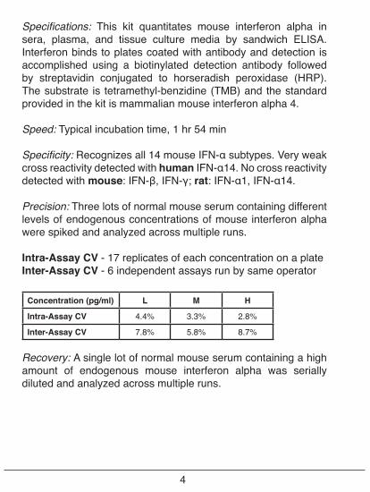
Specifications: This kit quantitates mouse interferon alpha in sera, plasma, and tissue culture media by sandwich ELISA. Interferon binds to plates coated with antibody and detection is accomplished using a biotinylated detection antibody followed by streptavidin conjugated to horseradish peroxidase (HRP). The substrate is tetramethyl-benzidine (TMB) and the standard provided in the kit is mammalian mouse interferon alpha 4.
Speed: Typical incubation time, 1 hr 54 min
Specificity: Recognizes all 14 mouse IFN-α subtypes. Very weak cross reactivity detected with human IFN-α14. No cross reactivity detected with mouse: IFN-β, IFN-γ; rat: IFN-α1, IFN-α14.
Precision: Three lots of normal mouse serum containing different levels of endogenous concentrations of mouse interferon alpha were spiked and analyzed across multiple runs.
Intra-Assay CV - 17 replicates of each concentration on a plateInter-Assay CV - 6 independent assays run by same operator
Concentration (pg/ml) L M H
Intra-Assay CV 4.4% 3.3% 2.8%
Inter-Assay CV 7.8% 5.8% 8.7%
Recovery: A single lot of normal mouse serum containing a high amount of endogenous mouse interferon alpha was serially diluted and analyzed across multiple runs.
4
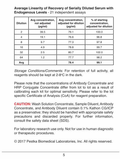
Average Linearity of Recovery of Serially Diluted Serum with Endogenous Levels - 21 independent assays
DilutionAvg concentration,
not adjusted(pg/ml)
Avg concentration, adjusted for dilution
(pg/ml)
% of starting concentration,
adjusted for dilution
2 39.5 79.1 100.0
4 19.1 76.6 96.8
8 9.7 77.3 97.8
16 4.9 78.8 99.7
32 2.5 80.7 102.0
64 1.2 77.7 98.2
Avg 78.4 99.1
Storage Conditions/Comments: For retention of full activity, all reagents should be kept at 2-8oC in the dark.
Please note that the concentrations of Antibody Concentrate and HRP Conjugate Concentrate differ from lot to lot as a result of calibrating each kit for optimal sensitivity. Please refer to the lot specific Certificate of Analysis (CoA) for reagent preparation.
CAUTION: Wash Solution Concentrate, Sample Diluent, Antibody Concentrate, and Antibody Diluent contain 0.1% Kathon CG/ICP as a preservative; they should be handled with appropriate safety precautions and discarded properly. For further information, consult the safety data sheet (SDS).
For laboratory research use only. Not for use in human diagnostic or therapeutic procedures.
© 2017 Pestka Biomedical Laboratories, Inc. All rights reserved.
5
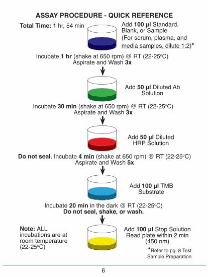
ASSAY PROCEDURE - QUICK REFERENCE
6
Total Time: 1 hr, 54 min
Incubate 1 hr (shake at 650 rpm) @ RT (22-25oC)Aspirate and Wash 3x
Incubate 30 min (shake at 650 rpm) @ RT (22-25oC)Aspirate and Wash 3x
Incubate 20 min in the dark @ RT (22-25oC)Do not seal, shake, or wash.
Add 100 μl Standard, Blank, or Sample(For serum, plasma, and media samples, dilute 1:2)*
Add 50 μl Diluted Ab Solution
Add 50 μl Diluted HRP Solution
Add 100 μl TMB Substrate
Add 100 μl Stop SolutionRead plate within 2 min
(450 nm)
Do not seal. Incubate 4 min (shake at 650 rpm) @ RT (22-25oC)Aspirate and Wash 5x
Note: ALL incubations are at room temperature (22-25oC) *Refer to pg. 8 Test
Sample Preparation
PREPARATION OF REAGENTS
Before starting the assay, Pre-coated plate, Wash Solution Con-centrate, TMB Substrate Solution, and Stop Solution should be equilibrated to room temperature (RT), 22-25oC. All other supplied components should be kept on ice (4oC) throughout the assay.
Wash Solution: The Wash Solution Concentrate may contain crystals; place the bottle in a warm water bath and gently mix until completely dissolved. Prepare a 1:20 working wash solution (e.g. add 50 ml of the Wash Solution Concentrate to 950 ml of distilled or deionized water). Mix thoroughly before use.
(Note: Prepare fresh Wash Solution for each assay run.)
Antibody Solution: Dilute Antibody Concentrate in the volume of Antibody Diluent recommended in the lot specific CoA. Prepare prior to beginning assay procedure and keep on ice (4oC).
HRP Solution: Dilute HRP Conjugate Concentrate in the volume of HRP Diluent recommended in the lot specific CoA. Prepare prior to beginning assay procedure and keep on ice (4oC).
Mouse Interferon Alpha Solution: Using the Mouse IFN Al-pha Standard, construct a standard curve as shown in Figure 1, in Sample Diluent or sample matrix. Pre-screening of serum is recommended because we have determined that a signficant portion of samples contain quantifiable levels of endogenous interferon alpha as shown in Figure 4.
(Note: Sample Diluent is viscous. Pipette slowly and remove ex-cess diluent on tip before dispensing into dilution reservoir to avoid carry over.)
7
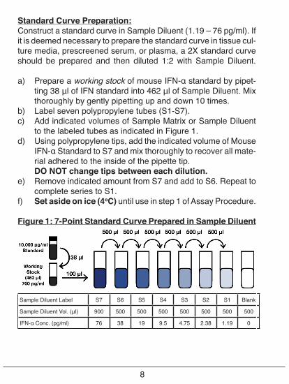
Standard Curve Preparation:Construct a standard curve in Sample Diluent (1.19 – 76 pg/ml). If it is deemed necessary to prepare the standard curve in tissue cul-ture media, prescreened serum, or plasma, a 2X standard curve should be prepared and then diluted 1:2 with Sample Diluent.
a) Prepare a working stock of mouse IFN-α standard by pipet-ting 38 μl of IFN standard into 462 μl of Sample Diluent. Mix thoroughly by gently pipetting up and down 10 times.
b) Label seven polypropylene tubes (S1-S7).c) Add indicated volumes of Sample Matrix or Sample Diluent
to the labeled tubes as indicated in Figure 1.d) Using polypropylene tips, add the indicated volume of Mouse
IFN-α Standard to S7 and mix thoroughly to recover all mate-rial adhered to the inside of the pipette tip.
DO NOT change tips between each dilution.e) Remove indicated amount from S7 and add to S6. Repeat to
complete series to S1.f) Set aside on ice (4oC) until use in step 1 of Assay Procedure.
Figure 1: 7-Point Standard Curve Prepared in Sample Diluent
Sample Diluent Label S7 S6 S5 S4 S3 S2 S1 Blank
Sample Diluent Vol. (μl) 900 500 500 500 500 500 500 500
IFN-α Conc. (pg/ml) 76 38 19 9.5 4.75 2.38 1.19 0
8
Test Sample Preparation: Prepare test samples of unknown IFN concentration using Sample Diluent as required. (For serum, plasma, and media samples, a minimum 1:2 dilution in Sample Diluent is recommended, (e.g. add 50 μl of Test Sample and 50 μl of Sample Diluent). Measurements in duplicate are recommended. Keep on ice (4oC) until use in step 1 of the Assay Procedure.
ASSAY PROCEDURE
All incubations should be performed at RT (22-25oC), keeping the plate away from drafts and other temperature fluctuations. Use plate sealers to cover the plate as directed. During all wash steps, remove contents of plate by inverting and shaking over a sink and blotting the plate on lint-free absorbent paper; tap the plate. Wash each well with a minimum of 300 μl of diluted Wash Solution for each wash step, emptying plate immediately after each wash. Extended soaking of the plate in Wash Solution may lower signal. Refer to Preparation of Reagents for details on dilution of concentrated solutions. Any alteration of the described procedures can directly affect assay performance.
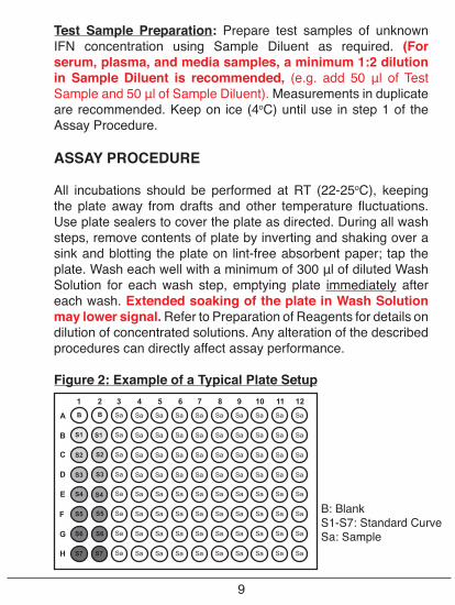
Figure 2: Example of a Typical Plate Setup
Sa
Sa
Sa
Sa
Sa
Sa
Sa
Sa
Sa
Sa
Sa
Sa
Sa
Sa
Sa
Sa
Sa
Sa
Sa
Sa
Sa
Sa
Sa
Sa
Sa
Sa
Sa
Sa
Sa
Sa
Sa
Sa
Sa
Sa
Sa
Sa
Sa
Sa
Sa
Sa
Sa
Sa
Sa
Sa
Sa
Sa
Sa
Sa
Sa
Sa
Sa
Sa
Sa
Sa
Sa
Sa
Sa
Sa
Sa
Sa
Sa
Sa
Sa
Sa
Sa
Sa
Sa
Sa
Sa
Sa
Sa
Sa
Sa
Sa
Sa
Sa
Sa
Sa
Sa
Sa
S4
S5
S6
S7
B
S1
S2
S3
S4
S5
S6
S7
B
S2
S3
E
F
G
H
C
D
A
B S1
3 4 5 6 7 8 9 10 11 121 2
9
B: BlankS1-S7: Standard CurveSa: Sample
1. Standards and Test Samples: Determine the number of microplate strips required to test the desired number of samples plus the appropriate number of wells needed to run blanks and standards. We recommend running the standard, blanks, and samples in duplicate or triplicate (see Figure 2 for an example setup). A standard curve is required for each assay. Remove extra microplate strips from the frame, seal in the foil bag provided, and store at 2-8oC. Unused strips can be used in later assays.
Add 100 μl of Standard, Blank, or Test Sample. (For Blank, add Sample Diluent or appropriate dilution matrix to each well.) For serum, plasma, or media samples, a minimum 1:2 dilution in Sample Diluent is required before adding to plate.
Cover with plate sealer and shake plate at 650 rpm at RT (22-25oC) for 1 hour.
After 1 hour, empty the contents of the plate and wash wells three times with 300 μl of diluted Wash Solution (refer to Preparation of Reagents), emptying plate immediately after each wash.
2. Antibody Solution: Add 50 μl of diluted Antibody Solution (refer to Preparation of Reagents) to each well. Cover with plate sealer and shake plate at 650 rpm at RT (22-25oC) for 30 minutes.
After 30 minutes, empty the contents of the plate and wash wells three times with 300 μl of diluted Wash Solution, emptying plate immediately after each wash.
3. HRP: Add 50 μl of diluted HRP Solution (refer to Preparation of Reagents) to each well. Do NOT cover with plate sealer and shake plate at 650 rpm at RT (22-25oC) for 4 minutes.
(Note: Do NOT allow HRP Solution to remain on plate longer than 4 minutes. It is recommended to remove the plate from shaker a few moments early to allow time for transport to wash station.)
10
After 4 minutes, empty the contents of the plate and wash wells five times with 300 μl of diluted Wash Solution, emptying plate immediately after each wash.
4. TMB Substrate: Add 100 μl of the TMB Substrate Solution to each well. Incubate, in the dark, at RT (22-25°C), for 20 minutes. Do not use a plate sealer and do not shake during the incubation.
5. Stop Solution: After the 20 minute incubation of TMB, DO NOT EMPTY THE WELLS AND DO NOT WASH. Add 100 μl of Stop Solution to each well.
6. Read: Using a microplate reader, determine the absorbance at 450 nm within 2 minutes after the addition of the Stop Solution.
CALCULATION OF RESULTS
By plotting the optical densities (OD) using a 4-parameter logistic fit for the standard curve, the interferon titer in the samples can be determined. Based on user preference, blank ODs may be subtracted from the standard and sample ODs to eliminate background.
For samples that have been diluted according to the instructions given in this manual (1:2), the concentration read from the standard curve must be multiplied by the dilution factor (x2).
A shift in optical densities is typical between users and kit lots. The back fit concentration interpolated from the standard curve is a more accurate determination of the sample titer and performance of the kit. Variations from the typical curve provided can be a result of operator technique, altered incubation time, fluctuations in temperature, and kit age.
11
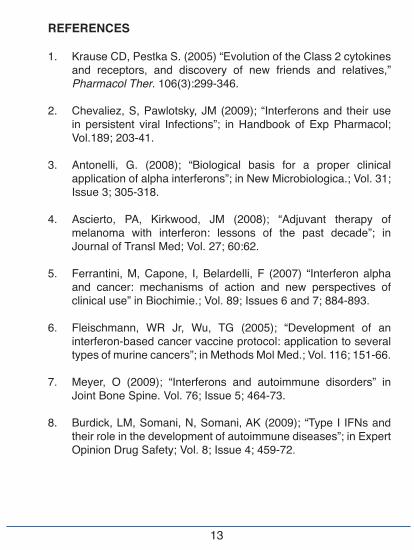
Results of a typical standard curve using a 4-parameter logistic fit are provided for illustration only and should not be used to obtain test results. A standard curve must be run in parallel with each set of samples assayed.
Figure 3: Typical Standard Curve in Sample Diluent
Figure 4: Endogenous plasma and serum levels
Endogenous levels of mouse IFN-α quantified in pooled plasma (A) and individual sera (B).
12
Abso
rban
ce (4
50 n
m)
Mouse IFN-α (pg/ml)
Mou
se IF
N-α
(pg/
ml)
REFERENCES
1. Krause CD, Pestka S. (2005) “Evolution of the Class 2 cytokines and receptors, and discovery of new friends and relatives,” Pharmacol Ther. 106(3):299-346.
2. Chevaliez, S, Pawlotsky, JM (2009); “Interferons and their use in persistent viral Infections”; in Handbook of Exp Pharmacol; Vol.189; 203-41.
3. Antonelli, G. (2008); “Biological basis for a proper clinical application of alpha interferons”; in New Microbiologica.; Vol. 31; Issue 3; 305-318.
4. Ascierto, PA, Kirkwood, JM (2008); “Adjuvant therapy of melanoma with interferon: lessons of the past decade”; in Journal of Transl Med; Vol. 27; 60:62.
5. Ferrantini, M, Capone, I, Belardelli, F (2007) “Interferon alpha and cancer: mechanisms of action and new perspectives of clinical use” in Biochimie.; Vol. 89; Issues 6 and 7; 884-893.
6. Fleischmann, WR Jr, Wu, TG (2005); “Development of an interferon-based cancer vaccine protocol: application to several types of murine cancers”; in Methods Mol Med.; Vol. 116; 151-66.
7. Meyer, O (2009); “Interferons and autoimmune disorders” in Joint Bone Spine. Vol. 76; Issue 5; 464-73.
8. Burdick, LM, Somani, N, Somani, AK (2009); “Type I IFNs and their role in the development of autoimmune diseases”; in Expert Opinion Drug Safety; Vol. 8; Issue 4; 459-72.
13
14
9. Kötter, I, Hamuryudan, V, Oztürk ,ZE, Yazici, H (Jan 7, 2010); “Interferon therapy in rheumatic diseases: state-of-theart”; in Current Opinion Rheumatology.
10. Erlandsson, L, Blumenthal, R, Eloranta, ML, Engel, H, Alm, G, Weiss, S, Leanderson, T (1998); “Interferon-beta is required for interferon-alpha production in mouse fibroblasts”; in Curr Biol.; Vol. 8; Issue 4;223-226.
11. Taniguchi T, Takaoka A (2002); “The interferon-alpha/beta system in antiviral responses: a multimodal machinery of gene regulation by the IRF family of transcription factors”; in Current Opinions in Immunology; Vol. 14; Issue 1; 111-116.
12. Jegalian AG, Facchetti F, Jaffe ES (2009); “Plasmacytoid dendritic cells: physiologic roles and pathologic states”; in Advances in Anat Pathol; Vol.16; Issue 6; 392-404.
13. Fitzgerald-Bocarsly P, Dai J, Singh S (2008); “Plasmacytoid dendritic cells and type I IFN: 50 years of convergent history”; in Cytokine Growth Factor Rev; Vol. 19; Issue 1; 3-19.
14. Li L, Sherry B (2010); “IFN-alpha expression and antiviral effects are subtype and cell type specific in the cardiac response to viral infection”; in Virology; Vol. 396; Issue 1; 59-68.
15. Baig E, Fish EN (2008); “Distinct signature type I interferon responses are determined by the infecting virus and the target cell”; in Antivir Ther.; Vol. 13; Issue 3; 409-22.
16. Delhaye S, Paul S, Blakqori G, Minet M, Weber F, Staeheli P, Michiels T (2006); “Neurons produce type I interferon during viral encephalitis”; in Proc Natl Acad Sci USA.; Vol. 103; Issue 20; 7835-7840.
15
PLATE LAYOUTUse this plate layout as a record of standards and samples assayed.
PBL Assay Science131 Ethel Road West, Suite 6, Piscataway, NJ 08854 USA
Tel: +1 732-777-9123, Fax: +1 732-777-9141Email: [email protected], Website: www.pblassaysci.com
© 2017 Pestka Biomedical Laboratories, Inc. All rights reserved.
![[PPT]PowerPoint Presentation - ITU: Committed to … · Web viewLightning flashes provide a high current (hundreds kA) in a very short time (μs), releasing a very high power in the](https://static.fdocument.org/doc/165x107/5ad34fd37f8b9a482c8d7dce/pptpowerpoint-presentation-itu-committed-to-viewlightning-flashes-provide.jpg)