Generation and characterization of β1,2-gluco ...
33
1 © The Author 2016. Published by Oxford University Press. This is an Open Access article distributed under the terms of the Creative Commons Attribution Non-Commercial License (http://creativecommons.org/licenses/by-nc/4.0/), which permits non-commercial re-use, distribution, and reproduction in any medium, provided the original work is properly cited. For commercial re-use, please contact [email protected] Generation and characterization of β1,2-gluco-oligosaccharide probes from Brucella abortus cyclic β-glucan and their recognition by C-type lectins of the immune system Hongtao Zhang 1,2 , Angelina S. Palma 1,3,* , Yibing Zhang 1 , Robert A. Childs 1 , Yan Liu 1 , Daniel A. Mitchell 4 , Leticia S. Guidolin 5 , Wilfried Weigel 6 , Barbara Mulloy 1 , Andrés E. Ciocchini 5 , Ten Feizi 1 and Wengang Chai 1,* 1 Glycosciences Laboratory, Department of Medicine, Imperial College London, London W12 0NN, UK 2 Key Laboratory of Carbohydrate Chemistry and Biotechnology, Ministry of Education, School of Biotechnology, Wuxi 214122, Jiangnan University, China 3 UCIBIO-REQUIMTE, Department of Chemistry, Faculty of Science and Technology, NOVA Universidade de Lisboa, 2829-516 Caparica, Portugal 4 CSRI-UHCW, Walsgrave Campus, University of Warwick, Coventry, CV2 2DX, UK 5 Instituto de Investigaciones Biotecnológicas “Dr. Rodolfo A. Ugalde”, Instituto Tecnológico de Chascomús (IIB-INTECH), Universidad Nacional de San Martín, San Martín 1650, Buenos Aires, Argentina; 6 SCIENION AG, Volmerstrasse 7b, 12489 Berlin, Germany * Correspondence should be addressed to A.S.P. ([email protected]; Tel: +351-21- 294-8300; Fax: +351 21-294-8550) and W.C. ([email protected]; Phone: +44-(0)20- 7594-2596; Fax: +44-(0)20-7594-7373). Glycobiology Advance Access published April 6, 2016 at Imperial College London on April 21, 2016 http://glycob.oxfordjournals.org/ Downloaded from
Transcript of Generation and characterization of β1,2-gluco ...
1
© The Author 2016. Published by Oxford University Press.
This is an Open Access article distributed under the terms of the Creative Commons Attribution Non-Commercial License
(http://creativecommons.org/licenses/by-nc/4.0/), which permits non-commercial re-use, distribution, and reproduction in any
medium, provided the original work is properly cited. For commercial re-use, please contact [email protected]
Generation and characterization of β1,2-gluco-oligosaccharide probes from Brucella
abortus cyclic β-glucan and their recognition by C-type lectins of the immune system
Hongtao Zhang1,2, Angelina S. Palma1,3,*, Yibing Zhang1, Robert A. Childs1, Yan Liu1,
Daniel A. Mitchell4, Leticia S. Guidolin5, Wilfried Weigel6, Barbara Mulloy1, Andrés E.
Ciocchini5, Ten Feizi1 and Wengang Chai1,*
1Glycosciences Laboratory, Department of Medicine, Imperial College London, London W12
0NN, UK
2Key Laboratory of Carbohydrate Chemistry and Biotechnology, Ministry of Education,
School of Biotechnology, Wuxi 214122, Jiangnan University, China
3UCIBIO-REQUIMTE, Department of Chemistry, Faculty of Science and Technology, NOVA
Universidade de Lisboa, 2829-516 Caparica, Portugal
4CSRI-UHCW, Walsgrave Campus, University of Warwick, Coventry, CV2 2DX, UK
5Instituto de Investigaciones Biotecnológicas “Dr. Rodolfo A. Ugalde”, Instituto Tecnológico
de Chascomús (IIB-INTECH), Universidad Nacional de San Martín, San Martín 1650,
Buenos Aires, Argentina; 6SCIENION AG, Volmerstrasse 7b, 12489 Berlin, Germany
*Correspondence should be addressed to A.S.P. ([email protected]; Tel: +351-21-
294-8300; Fax: +351 21-294-8550) and W.C. ([email protected]; Phone: +44-(0)20-
7594-2596; Fax: +44-(0)20-7594-7373).
Glycobiology Advance Access published April 6, 2016 at Im
perial College L
ondon on April 21, 2016
http://glycob.oxfordjournals.org/D
ownloaded from
2
Abstract
The β1,2-glucans produced by bacteria are important in invasion, survival and
immunomodulation in infected hosts be they mammals or plants. However, there has been a
lack of information on proteins which recognize these molecules. This is partly due to the
extremely limited availability of the sequence-defined oligosaccharides and derived probes
for use in the study of their interactions. Here we have used the cyclic β1,2-glucan (CβG) of
the bacterial pathogen Brucella abortus, after removal of succinyl side chains, to prepare
linearized oligosaccharides which were used to generate microarrays. We describe optimized
conditions for partial depolymerization of the cyclic glucan by acid hydrolysis and conversion
of the β1,2-gluco-oligosaccharides, with degrees of polymerization 2-13, to neoglycolipids for
the purpose of generating microarrays. By microarray analyses we show that the C-type lectin
receptor DC-SIGNR, like the closely related DC-SIGN we investigated earlier, binds to the
β1,2-gluco-oligosaccharides, as does the soluble immune effector serum mannose-binding
protein. Exploratory studies with DC-SIGN are suggestive of the recognition also of the intact
CβG by this receptor. These findings open the way to unravelling mechanisms of
immunomodulation mediated by β1,2-glucans in mammalian systems.
Key words: β1,2-glucan/carbohydrate microarray/C-type lectins/glucan recognition/
neoglycolipids
at Imperial C
ollege London on A
pril 21, 2016http://glycob.oxfordjournals.org/
Dow
nloaded from
3
Introduction
Glucan polysaccharides are of biomedical interest because of their involvement in
mechanisms of pathogen recognition and modulation of the immune system (Brown and
Gordon, 2003; Chen and Seviour, 2007). Molecular dissection of their interactions with
proteins of the immune system although desirable is not straight forward at the level of
polysaccharides on account of the inherent heterogeneities of these macromolecules. With the
advent of oligosaccharide microarray technologies (Fukui et al. 2002; Feizi and Chai 2004;
Blixt et al. 2004; Rillahan and Paulson 2011; Palma et al. 2014), it is possible now to explore
interactions with proteins using oligosaccharide probes generated from a range of
oligosaccharide sequences that can be prepared after partial depolymerization of the
polysaccharides (Pedersen et al. 2012; Palma et al. 2015).
The microarray system based on the neoglycolipid (NGL) technology (Chai et al.
2003) for preparing lipid-linked oligosaccharide probes for immobilization and binding
studies, lends itself well to analyses of glucan sequences as recognition structures within
polysaccharides. This is the basis of the ‘designer’ microarray approach (Palma et al. 2006;
Gao et al. 2014; Palma et al. 2014) whereby microarrays are generated from oligosaccharides
released from the targeted macromolecules; oligosaccharides bound by recognition proteins
may be isolated for characterization. This approach was used successfully in studies of the
ligands on glucan polysaccharides for Dectin-1, a key receptor of the innate immune system
directed against fungal pathogens (Herre et al. 2004). Dectin-1 belongs to the family of C
(calcium-dependent)-type lectin-like proteins; it lacks the canonical amino acid residues for
ligating calcium, required for carbohydrate-binding in classical C-type lectins (Drickamer
and Taylor, 2015). Nevertheless, designer microarrays (Palma et al. 2006) generated from
oligosaccharide fractions derived from fungal-type glucans (Brown and Gordon, 2001;
at Imperial C
ollege London on A
pril 21, 2016http://glycob.oxfordjournals.org/
Dow
nloaded from
4
Brown et al. 2003), established that: 1) Dectin-1 is a calcium-independent carbohydrate
binding protein; and 2) linear β1,3-linked glucose sequences with degrees of polymerization
(DP) 10 or longer are required for detection of binding.
Using the designer approach, in conjunction with a novel high-sensitivity mass
spectrometric (MS) sequencing method, we recently generated a ‘glucome’ microarray of
sequence-defined oligosaccharide probes derived from glucan polysaccharides of fungal,
bacterial and plant origins in order to use as a high-throughput screening tool for
characterizing glucan recognition systems of mammals and bacteria (Palma et al. 2015). The
probes in the microarray encompassed linear sequences with a single linkage type: 1,2-, 1,3-,
1,4- or 1,6- with α or β configurations; and mixed multiple linkage types: 1,3-, 1,4 or 1,6-;
also branched oligosaccharide sequences with 1,3 and 1,6-linkages with different DPs.
Binding of the dendritic cell-specific C-type lectin receptor DC-SIGN was noted to NGL
probes from β1,2-linked gluco-oligosaccharides DP 2 to 13, derived from the cyclic β1,2-
glucan (CβG) of the bacterial pathogen Brucella abortus, which is a major pathogenic factor
involved in B. abortus invasion and survival (Arellano-Reynoso et al. 2005) and a potent
activator of mouse and human dendritic cells (Martirosyan et al. 2012). This raised the
possibility that DC-SIGN interacts with B. abortus CβG and that this interaction participates
in modulation of the activities of DCs (Palma et al. 2015). C-type lectin receptors (CLRs)
comprise a large family of signaling receptors, which are variously involved in inflammatory
and innate immune responses to a diverse range of microbial pathogens (Hoving et al. 2014;
Drickamer and Taylor 2015). These activities occur following the binding of their
carbohydrate recognition domains (CRDs) to specific endogenous carbohydrates and those of
pathogens. The finding that DC-SIGN can bind pathogen-associated β1,2-linked gluco-
oligosaccharides raises the question whether related CLRs bind to these types of sequences,
in addition to their other well-known carbohydrate ligands.
at Imperial C
ollege London on A
pril 21, 2016http://glycob.oxfordjournals.org/
Dow
nloaded from
5
Here we describe details of the preparation of sequence-defined β1,2-linked gluco-
oligosaccharide probes for microarray analysis, including procedures for CβG hydrolysis,
oligosaccharide fractionation, with improved yields of NGLs from the longer
oligosaccharides that are difficult to derivatize. We apply the NGL microarrays to investigate
the recognition of these oligosaccharide sequences by C-type lectin immune-receptors,
including DC-SIGN and its closely related human receptor DC-SIGNR (or L-SIGN), and the
soluble serum effector mannose-binding protein (MBP). We also explore the recognition of
the intact cyclic forms of CβGs by DC-SIGN.
Results
Preparation of β1,2-gluco-oligosaccharides from cyclic β1,2-glucan
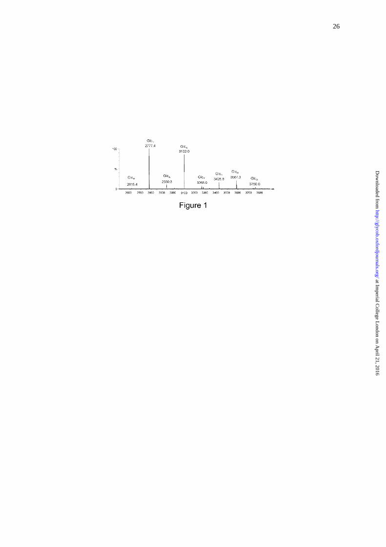
The alkali treated B. abortus CβG was analysed by MALDI-MS, and the spectrum
indicated complete removal of the succinyl side chains and preservation of the cyclic glucan
chains which consisted of DP 16-23, with DP 17 (MNa+ at m/z 2777) being the most
abundant component (Figure 1).
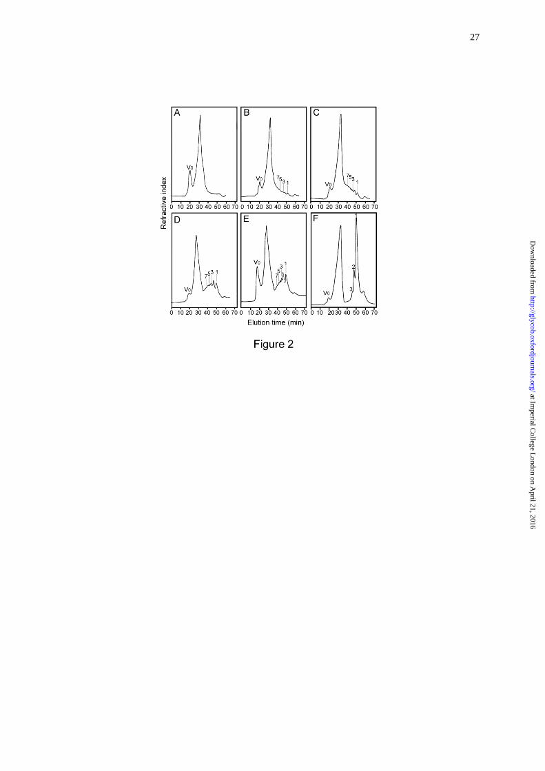
In the exploratory small scale experiments, hydrolysis of the CβG with 0.01M HCl at
100 oC was assessed by monitoring the products at different reaction times by gel filtration
(Figure 2). For monitoring of the reaction, the reagent HCl was not removed prior to analysis,
and therefore an artefactual peak related to HCl occurred at around 30 min. This has not
interfered with the evaluation of the progress of the hydrolysis. The reaction time of 120 min
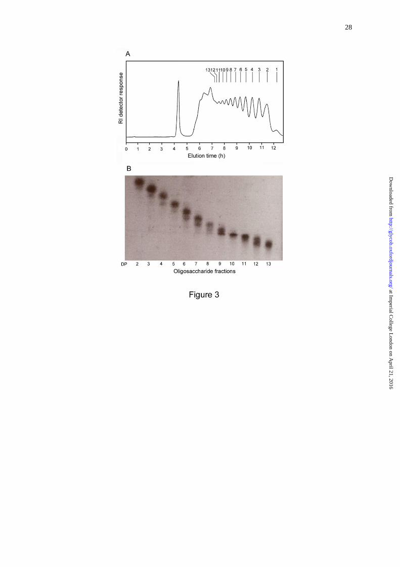
(Figure 2D) was selected for large scale experiments to obtain oligosaccharides with DPs
ranging from 2 to 13 (Figure 3A). The fractions obtained by gel filtration were analyzed by
HPTLC (Figure 3B). The identities of the major components in the higher oligosaccharide
fractions with DPs ≥ 5 were determined by MALDI-MS and of the lower oligosaccharide
at Imperial C
ollege London on A
pril 21, 2016http://glycob.oxfordjournals.org/
Dow
nloaded from
6
fractions with DPs ≤ 4 by negative-ion ESI-MS. As shown in the MALDI spectra of
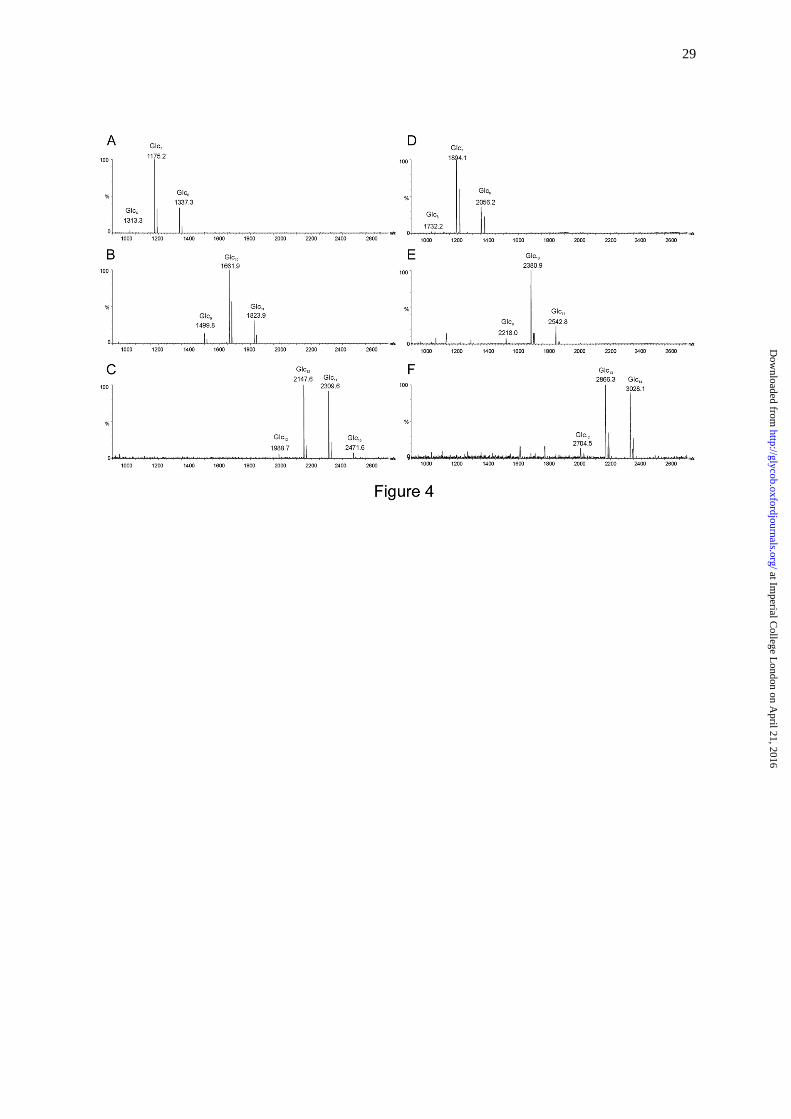
fractions DP7, DP10 and DP13 (Figure 4A-C, respectively) as representative, each fraction
contains adjacent overlapping components in addition to the main component. For example,
in fraction DP7 (Figure 4A) oligosaccharides with DP 6 and 8 were present as minor
components in addition to the main component DP 7 at m/z 1175.2 (MNa+), due to
incomplete separation by gel filtration chromatography.
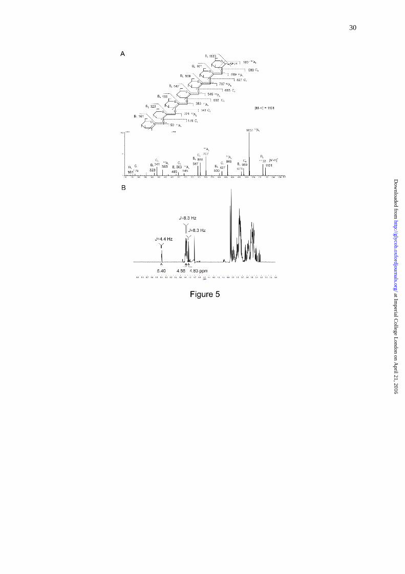
Linkage and anomeric configuration for the DP 7 fraction were investigated by
negative-ion ESI-CID-MS/MS and 1H-NMR. In the product-ion spectrum (Figure 5A), the
neutral losses of 18 Da (e.g. m/z 1133 and m/z 971) and 120 Da (e.g. m/z 1031 and m/z 869)
derived from dehydration and 0,2A-cleavage (Domon and Costello, 1988), respectively, of the
[M H] and glycosidic C-type ions (Domon and Costello, 1988) are characteristic of 1,2-
linkage of gluco-oligosaccharides (Palma et al. 2015). The β-anomeric configuration could be
readily assigned by 1H-NMR from the major anomeric doublet at 4.88 ppm with a coupling
constant of ~8.3 Hz; both - and -anomeric signals from the reducing end monosaccharide
could also be identified (Figure 5B).
Preparation of β1,2-gluco-oligosaccharide NGLs
Preparation of the NGLs of glucan oligosaccharides with DP > 7 using the
conventional method of reductive-amination (Chai et al. 2003) has been difficult and the
yield extremely low (not shown). For the higher oligomers of gluco-oligosaccharides even
with the relatively more efficient reaction in oxime-ligation (Liu et al. 2007) the yield was
again low. Improvement of conjugation conditions was attempted by modifications of several
parameters of the oxime-ligation reaction. Using the readily available 1,6-linked dextran
oligosaccharides as standards we explored the effects of different reaction temperature (22,
50 and 80 oC) and time (24, 48 and 96 h), different acidity of the reaction medium (acidic,
at Imperial C
ollege London on A
pril 21, 2016http://glycob.oxfordjournals.org/
Dow
nloaded from
7
neutral and alkaline) and different amounts of lipid reagent, but no major improvement in
reaction yield was found (not shown).
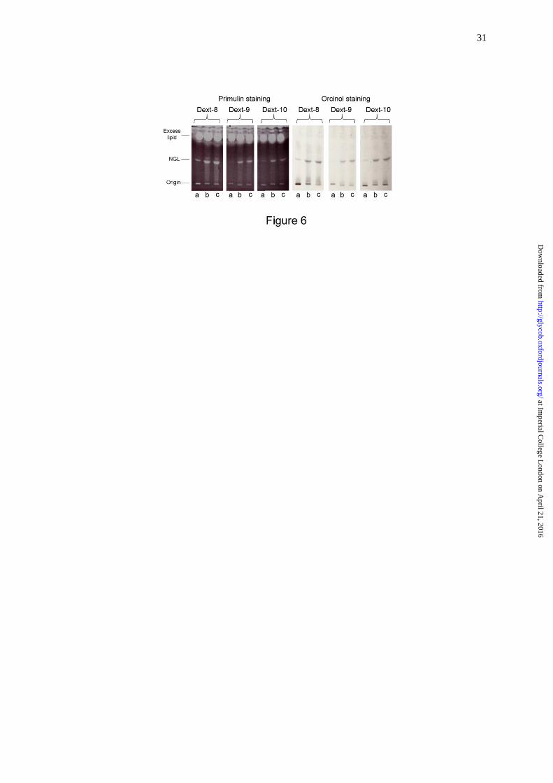
The low solubility of gluco-oligosaccharides being a well-recognized problem, we
next investigated the effect of solvent on conjugation yield. To improve the solubility of
gluco-oligosaccharides, DMSO was included in the solvent mixture for NGL conjugation.
Using dextran oligosaccharides with DP 8, 9 and 10 as examples, the solvent effect was
clearly apparent. In the presence of DMSO the yields were improved, particularly for the
higher oligomers. As shown in Figure 6, the NGL product bands in lanes b and c, in which
DMSO was included in the reaction solvent, were clearly more intense than those in lanes a.
This was apparent with both primulin (for detection of lipid) and orcinol (for detection of
glucose) staining.
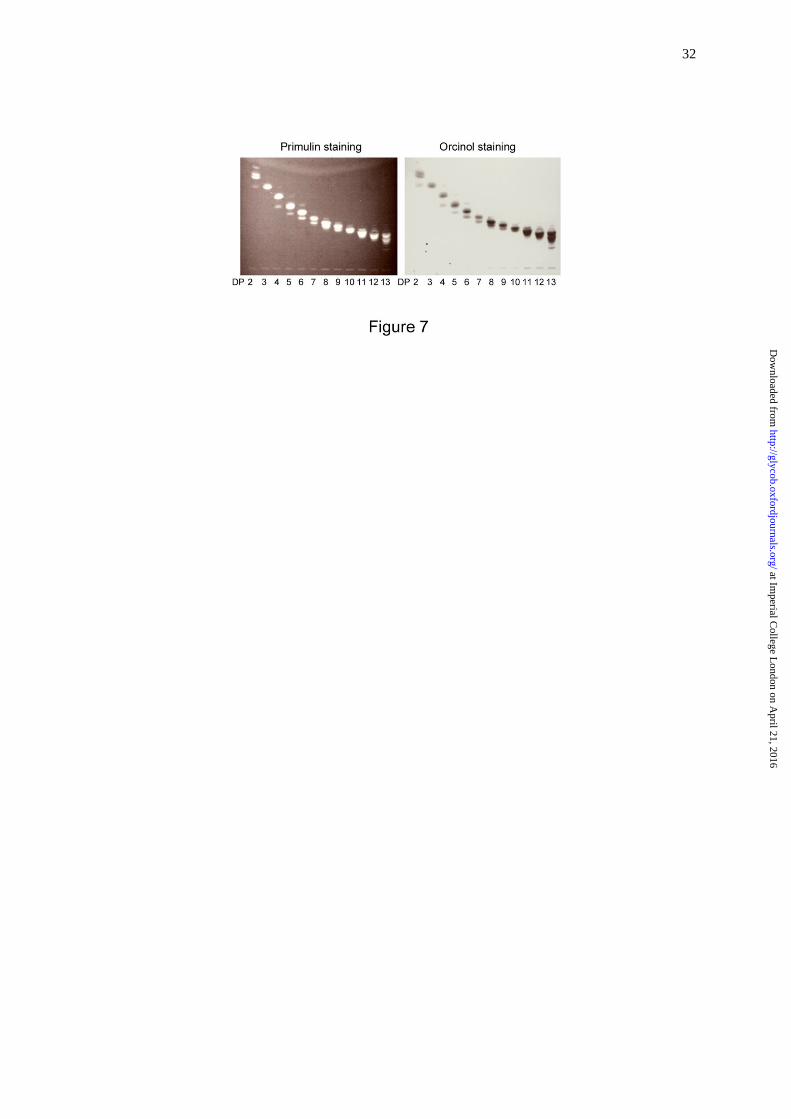
With the modified conditions (condition c in Materials and Methods), a series of
NGLs of the CβG oligosaccharides, DP 2-13 was prepared. The purified NGL probes were
analyzed by HPTLC (Figure 7) and MALDI-MS (Figure 4D-F) before printing on
nitrocellulose-coated glass slides for protein-binding experiments.
Analysis of the recognition of β1,2-gluco-oligosaccharide NGLs by CLRs of the immune
system
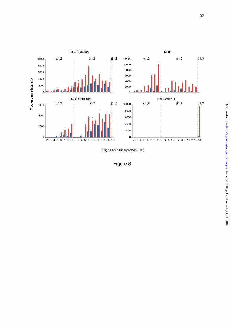
In order to investigate the recognition of the β1,2-gluco-oligosaccharides by CLRs of
the immune system, we arrayed the gluco-oligosaccharides as NGLs and included α1,2-
linked DP 2 to DP 9 and β1,3-linked DP 13 NGLs as controls (Table I). We performed
microarray analyses with proteins: DC-SIGN-bio, DC-SIGNR-bio, MBP purified from
human serum and His-Dectin-1 (Figure 8). DC-SIGN-bio showed binding to all the β1,2-
gluco-oligosaccharide probes tested; the binding pattern was similar to that previously
observed using DC-SIGN-Fc (Palma et al. 2015), namely with DP 6 showing the highest
at Imperial C
ollege London on A
pril 21, 2016http://glycob.oxfordjournals.org/
Dow
nloaded from
8
binding signal at 5 fmol glucan probe per spot (Figure S1). DC-SIGNR-bio gave binding
signals with β1,2-gluco-oligosaccharide probes with DP > 4. MBP also showed binding to
β1,2-gluco-oligosaccharide probes with DP > 2 with relatively high intensity to those with
longer chain lengths, DP 7-9. Contrasting with binding profiles of DC-SIGN-bio and DC-
SIGNR-bio, MBP showed relatively higher binding signals with the α1,2-gluco-
oligosaccharide probes, in particular the longer oligomers. His-Dectin-1 included as a control
in these microarray analyses showed the predicted binding to the β1,3-linked DP 13 from
curdlan (Palma et al. 2006), but no detectable binding to any of the CβG derived β1,2-linked
gluco-oligosaccharide probes (Figure 8), in agreement with our previous assignment (Palma
et al. 2015). Under the assay conditions, DC-SIGN-bio, DC-SIGNR-bio and MBP gave no
binding signals with the β1,3-linked DP13 probe.
In sum, the results presented here show the CLRs DC-SIGN, DC-SIGNR and MBP
can bind to linear β1,2-gluco-oligosaccharides derived from B. abortus CβG with differing
influence of chain length on the observable binding.
Exploratory studies of the recognition of cyclic β1,2-glucan by DC-SIGN CLR
In additional experiments we explored the recognition of intact forms of CβG by DC-
SIGN-Fc (Supplementary Methods and Figure S2). His-Dectin-1 was included as a control
protein. We analyzed the native form of B. abortus CβG with succinyl side chains and the
NaOH-treated CβG with these side chains removed. As these CβGs are of relatively low
molecular weight (~3 kDa, Figure 1) and cannot be readily retained on a nitrocellulose matrix,
we explored the arraying of these together with other polysaccharides as positive and
negative controls in the presence of a water soluble and photoactive terpolymer, sciPOLY3D,
(Figure S2 and Table S1). The terpolymer consists of three components:
poly(dimethylacrylamide) as the hydrophilic matrix, sodium salt of styrene sulfonate as the
at Imperial C
ollege London on A
pril 21, 2016http://glycob.oxfordjournals.org/
Dow
nloaded from
9
water solubility enhancer, and 4-methacryloxyl-oxy-benzophenone as the photo reactive
group. This polymer allows immobilization of the molecules in a 3D matrix by UV
irradiation forming covalent links between the capture molecules and the polymer and
attachment of the polymer to the slide surface. As shown in Figure S2, DC-SIGN-Fc gave
robust binding signals with α-mannan of Saccharomyces cerevisiae, which is well known to
be recognized by this receptor (Cambi et al. 2008). Binding could be detected to the two CβG
forms; also to the β1,3-glucans NSG (neutral soluble β-glucan) and PGG (poly-(1,6)-D-
glucopyranosyl-(1,3)-D-glucopyranose), as we have shown previously and in agreement with
our observation that DC-SIGN bound to β1,3-gluco-oligosaccharides with DP-2 and DP-3
(Palma et al. 2006). Both NSG and PGG, as predicted, were bound by His-Dectin-1.
Discussion
Glycan microarray technology has become established as a powerful means of glycan
ligand discovery in studies of recognition systems in functional glycomics. There is an ever-
demanding need to broaden the repertoire of sequence-defined oligosaccharide probes in
order to facilitate the studies of glycan recognition in diverse biological systems. In the
present study, we address the methodological challenge in obtaining β1,2-linked glucan
oligosaccharides with differing chain lengths and their conversion into NGL probes for
microarray construction to enable studies of their recognition by proteins. To achieve this, our
strategy has been to partially depolymerize cyclic β1,2-glucan (CβG) of B. abortus, after
removal of the succinyl side chains. Following detailed characterization of the
oligosaccharides by mass spectrometry and NMR, the gluco-oligosaccharides were
conjugated to the lipid reagent by oxime-ligation reaction using conditions we optimized for
these hard-to-derivatize oligomers. NGL probes with differing chain lengths ranging from DP
2-13 were thus obtained.
at Imperial C
ollege London on A
pril 21, 2016http://glycob.oxfordjournals.org/
Dow
nloaded from
10
The β1,2-linked CβGs are produced by different bacteria of the Proteobacteria phylum
and occur mostly in the periplasm, but they can also be secreted as extracellular
polysaccharides, to which important biological activities have been attributed (Bontemps-
Gallo and Lacroix, 2015). For example, the periplasmic CβG of the pathogen B. abortus is
essential for bacterial virulence, whereas the secreted CβG mediates interactions with
mammalian hosts (Briones et al. 2001; Arellano-Reynoso et al. 2005) and modulation of the
activities of immune cells (Martirosyan et al. 2012; Degos et al. 2015). Secreted CβGs have
been shown also to be important for invasion of the bacterial phytopathogen Xanthomonas
campestris pv campestris and suppression of systemic immune responses in plants (Rigano et
al. 2007). Linear β1,2 glucans have also been identified in some bacteria of the Proteobacteria
phylum, for example in the opportunistic pathogen Pseudomonas aeruginosa, in which they
have been shown to be involved in biofilm formation (Lequette et al. 2007). Despite the wide
occurrence and striking bioactivities attributed to these biomolecules limited information
exists about proteins that mediate their recognition.
In the present work we analyze two additional CLRs of the mammalian immune
system for their recognition of β1,2-gluco-oligosaccharides, following on from our earlier
finding that DC-SIGN binds to this type of oligosaccharides (Palma et al. 2015). We analyze
a different DC-SIGN construct, its closely related endothelial cell receptor DC-SIGNR, and
serum MBP, and show that these CLRs share the common feature of binding to β1,2-linked
glucose oligosaccharides printed as NGL probes. It has been previously shown by microarray
screening and structural analysis of the CRDs in complex with mammalian-type
oligosaccharides that DC-SIGN and DC-SIGNR have distinct ligand-binding properties
(Feinberg et al. 2001; Guo et al. 2004): both receptors bind high-mannose oligosaccharides;
but DC-SIGN can additionally interact strongly with fucosylated Lewisa and Lewisx-related
oligosaccharides. Serum MBP binds to oligosaccharides bearing terminal fucose, mannose or
at Imperial C
ollege London on A
pril 21, 2016http://glycob.oxfordjournals.org/
Dow
nloaded from
11
GlcNAc with broad specificity (Drickamer and Taylor, 2015). The binding that we observe to
the gluco-oligomers may reflect the mode of binding of these receptors to the shared high-
mannose oligosaccharide ligands through the equatorial 3-hydroxyl and 4-hydroxyl groups
(Drickamer and Taylor, 2015). This interpretation will need to be corroborated by solving the
structures of the CRD-oligosaccharide complexes.
Our findings that DC-SIGN, DC-SIGNR and MBP can interact with β1,2-gluco-
oligosaccharides derived from B. abortus CβG, raised the important question of whether the
natural intact CβG forms are recognized and thereby involved in the triggering of these
receptors of the immune system. Conformational differences between linear and cyclic β1,2-
linked oligosaccharides have been described (Mimura et al. 1996). Our exploratory analyses
suggest that DC-SIGN can interact with intact CβG forms. Further investigations of these
interactions are required and of their involvement on the immuno biological effects observed
with B. abortus CβG and β1,2-linked glucans.
The availability of β1,2-linked glucose oligosaccharide probes derived from CβG and
their effective presentation in microarrays will enable identification of oligosaccharide
epitopes recognized on CβGs by other proteins. The β1,2-linked CβGs produced by bacteria
exhibit structural diversity as they can occur in the unsubstituted form, or substituted at
glucose C6 with anionic groups, such as succinyl (Roset et al. 2006) as in the case of the
present work, phosphoglycerol (Miller et al. 1987) or methylmalonate (de Iannino and
Ugalde, 1989). These substitutions as well as branching of the backbone chain with β1,6-
linked glucose also occur in linear β1,2-glucans (Lequette et al. 2007). The strategies
presented here could well be adapted to these various types of β1,2-linked glucans and may
also be applied to the study of the influence of substitutions and branching on their
recognition by proteins. The perfection of the sciPOLY3D polymer printing and development
of new strategies to generate arrays of the intact CβGs will be important in the unraveling of
at Imperial C
ollege London on A
pril 21, 2016http://glycob.oxfordjournals.org/
Dow
nloaded from
12
these recognition systems.
Materials and Methods
Oligosaccharides and recombinant proteins
A gluco-oligosaccharide fraction with DP 13 from curdlan (with β1,3-linkage),
dextran derived fractions (with α1,6-linkage) with DP 8, 9 and 10 and Cyanobacterium
gluco-oligosaccharides (α1,2-linkage) with DP 2 to DP 9 as major components (Cyano-2 to
Cyano-9) were prepared as described (Palma et al. 2015). Recombinant, tetrameric DC-SIGN
and DC-SIGNR (complete extracellular domains, lacking the transmembrane domain) were
made and purified as described previously (Mitchell et al. 2001). These were analyzed in the
microarrays in a biotinylated form (DC-SIGN-bio and DC-SIGNR-bio, respectively),
prepared as described previously (Carroll et al. 2010); MBP purified from human serum
(Haurum et al. 1993; Jensenius 1995) was provided by Jens Christian Jensenius (Aarhus
University, Denmark); murine Dectin-1 CRD with an N-terminal His6-tag (His-Dectin-1)
was purchased from Sino Biologicals (Beijing, China). Solvents used are all of analytical
grade and the compositions of the solvents are by volume throughout the study unless
specified otherwise.
Preparation of gluco-oligosaccharides from CβG
CβG, consisting of 13-23 glucose residues, was isolated from B. abortus essentially as
described (Ciocchini et al. 2007) with some minor modifications. Cells from 200 ml of
stationary phase cultures of B. abortus strain were grown for 48 h at 37°C (200 rpm) and
harvested by centrifugation at 8,000 xg for 10 min at 4°C. Cell pellets were extracted with
ethanol (70% ethanol, 1 h at 37°C). The ethanolic extracts were centrifuged, and the
at Imperial C
ollege London on A
pril 21, 2016http://glycob.oxfordjournals.org/
Dow
nloaded from
13
supernatants were concentrated and subjected to gel filtration on a Bio-Gel P6 column (1.8 x
78 cm). Columns were eluted at room temperature with 0.5% formic acid at a flow rate of 9
ml/h, and 1.5 ml fractions were collected. Fractions corresponding to CβG were pooled,
concentrated and lyophilized. CβG was initially treated with 0.1 M NaOH at 40oC for 60 min
to remove the succinyl side chains. Following neutralization by addition of 3M HCl to pH 7.0,
the reaction mixture was desalted on a G10 column (1.6 × 30 cm). The side chain-removed
CβG eluting at the void volume was collected and freeze-dried. The successful removal of the
succinyl side chains was confirmed by matrix-assisted laser desorption/ionization mass
spectrometry (MALDI-MS) analysis.
Small scale experiments were performed initially to optimize the conditions for mild
acid hydrolysis of the NaOH-treated CβG to obtain oligosaccharide fractions with DP 2-13.
For this 5 mg of the lyophilized CβG was dissolved in 500 μl of 0.01 M HCl in a V-shaped
glass vial. The mixture was heated, with stirring, to 100oC in a heating block. For monitoring
of the reaction progress, aliquots (50 μl) of the reaction solution were taken out at various
reaction times (0, 30, 60, 120, 150, 180, and 210 min), cooled on ice and neutralized by
addition of NaOH solution (0.1 M) before injection to a FPLC system equipped with a
Superdex Peptide column (PC 3.2/30, GE Healthcare, Uppsala, Sweden). The column was
eluted with deionized water at a flow rate of 18 ml/h and the eluent was monitored with a
refractive index detector.
For large scale preparation, the reaction time of 120 min was selected. Thus, 25 mg
NaOH-treated CβG was dissolved in 2.5 ml HCl (0.01 M) and the mixture was incubated at
100 °C for 120 min. The reaction was stopped by neutralization with NaOH (0.1 M) and the
mixture was desalted on the Sephadex G10 column. The desalted hydrolysis products were
fractionated on a Bio-Gel P4 column (1.5 × 100 cm) by elution with deionized water at a
flow rate of 15 ml/h. The elution was monitored on-line by refractive index and fractions
at Imperial C
ollege London on A
pril 21, 2016http://glycob.oxfordjournals.org/
Dow
nloaded from
14
were pooled according to their glucose units.
The pooled fractions were freeze-dried, and quantified by orcinol assay for glucose
content (Chai et al. 2003). For high performance (HP) silica gel TLC analysis, an aliquot (~2
μg) of each fraction was applied to the aluminium-backed plate and a solvent system of n-
propanol/water (8:3) was used for development. The gluco-oligosaccharide bands were
detected by orcinol staining (Chai et al. 2003).
Preparation of β1,2-gluco-oligosaccharide NGLs
The β1,2-linked gluco-oligosaccharides were converted into NGLs by oxime-ligation
with the lipid reagent amino oxy-functionalized 1,2-dihexadecyl-sn-glycero-3-
phosphoethanolamine (AOPE) (Liu et al. 2007). For β1,2-linked gluco-oligosaccharides with
DP < 7, and β1,3-linked oligosaccharide with DP 13 (included as a standard control probe),
the conjugation conditions were as described (Liu et al. 2007). In brief, 50 nmol of gluco-
oligosaccharide in a glass vial were dried by lyophilization before addition of 100 nmol
AOPE (in 20 μl of CHCl3/MeOH/H2O, 10:10:1). The solvent of the mixture was evaporated
to dryness under a N2 stream. The content was re-dissolved in 50 μl of the same solvent and
the mixture was incubated at ambient temperature (22oC) for 16 h before solvent evaporation
in a heating block at 60 oC for approximately 1h.
For β1,2-linked gluco-oligosaccharides with DP > 7, the reaction conditions were
optimized to obtain higher conjugation yields. In exploratory studies, using dextran
oligosaccharides DP 8, 9 and 10 as standards, the effects of reaction time and temperature
were assessed. The reaction time was extended to 48 h and 96 h and the reaction temperature
was raised from ambient temperature to 50oC or 80oC without any apparent improvement in
reaction yield. The solvent in the reaction mixture was changed to acidic by addition of 2 μl
of acetic acid or alkaline by addition of 2 μl of triethylamine. For further improvement of
at Imperial C
ollege London on A
pril 21, 2016http://glycob.oxfordjournals.org/
Dow
nloaded from
15
solubility of the higher oligomers of the gluco-oligosaccharides, DMSO was included in the
reaction solvent. For comparison, two solvent systems CHCl3/MeOH/H2O (25:25:8) and
CHCl3/MeOH/DMSO (25:25:8) were used in the following three procedures using 50 nmol
of oligosaccharide and 1,250 nmol of AOPE: (a) oligosaccharide and AOPE in 100 μl
CHCl3/MeOH/H2O; (b) The oligosaccharide was dissolved in 15 μl DMSO before addition of
100 μl CHCl3/MeOH/H2O containing the required 1,250 nmol of AOPE; and (c) the
procedure was identical to (b) described above apart from a solvent of CHCl3/MeOH/DMSO
was used instead of CHCl3/MeOH/H2O. All the reactions were carried out at 80 oC for 96 h.
After reaction, the volatile solvent was evaporated under a stream of N2 and DMSO was
removed by repeated co-evaporation with a small amount of water by lyophilisation.
Procedure (c) was selected for preparation of the higher oligomers (DP > 7) of β1,2-linked
gluco-oligosaccharides.
NGLs of DP 2 to 5 were purified by semi-preparative HPTLC and those with DP 6 to
13 were purified using silica cartridge (Chai et al. 2003). Purified NGLs were analyzed by
HPTLC using CHCl3/MeOH/H2O (60:35:8) as the development solvent and detected by
primulin and orcinol staining (Chai et al. 2003).
Analysis of the oligosaccharides and their NGLs
MALDI-MS in the positive-ion mode was carried out on a Tof Spec-2E instrument
(Micromass, Manchester, UK) for analyses of the CG polysaccharide, oligosaccharide
fractions with DP 5-13 and all the NGLs. Sample solutions (1 μl, containing 1-10 pmol/l in
H2O for the poly- and oligosaccharides, and CHCl3/MeOH/DMSO, 25:25:8, for NGLs) were
deposited on the sample target together with the matrix of 2-(4-hydroxyphenylazo) benzoic
acid. Laser energy was 20% (coarse) and 60% (fine), and resolution was at 3,000.
Negative-ion electrospray mass spectrometry (ESI-MS) was used for shorter
at Imperial C
ollege London on A
pril 21, 2016http://glycob.oxfordjournals.org/
Dow
nloaded from
16
oligosaccharides (DP 2-4). Collision-induced dissociation tandem mass spectrometry (ESI-
CID-MS/MS) was used for sequence and linkage analysis for the heptasaccharide. ESI-MS
and CID-MS/MS were carried out on a Q-TOF mass spectrometer (Micromass, Manchester,
UK). Nitrogen was used as desolvation and nebulizer gas at a flow rate of 250 l/h and 150 l/h,
respectively. Source temperature was 80C, and the desolvation temperature 150C. A cone
voltage of 50 V was used and the capillary voltage was maintained at 3 kV. MS/MS product-
ion spectrum was obtained from CID using Argon was used as the collision gas at a pressure
of 0.17 MPa for the CID-MS/MS experiment. The collision energy was at 17 V. For analysis
oligosaccharides were dissolved in acetonitrile/water (1:1), typically at a concentration of 15
pmol/μl, of which 5 μl was loop-injected. Solvent (acetonitrile/2 mM ammonium bicarbonate,
1:1) was delivered by a Harvard syringe pump (Harvard Apparatus, Holliston, MA) at a flow
rate of 10 μl/min.
For NMR analysis, the CG derived fraction with DP 7 (150 µg) was co-evaporated
with 2H2O (99.9 atom% 2H2) twice by lyophilisation and dissolved in 550 l of high quality
2H2O (100.0 atom% 2H2), containing 0.1 l of acetone. 1H-NMR spectrum was acquired on
Varian (Palo Alto, California, USA) Unity-600 (599.89 MHz 1H) spectrometer at 25 °C and
processed with standard Varian software. The observed 1H chemical shifts were relative to
internal acetone (2.225 ppm).
Carbohydrate microarray analyses
For preparation of the microarray, the gluco-oligosaccharide NGL probes (Table I)
were printed onto 16-pad nitrocellulose-coated glass slides in duplicate at two levels, 2 and 5
fmol/spot, as described (Palma et al. 2015).
Microarray binding analyses, performed using AlexaFluor-647-labeled Streptavidin as
final readout of protein binding, imaging and data analysis were carried out essentially as
at Imperial C
ollege London on A
pril 21, 2016http://glycob.oxfordjournals.org/
Dow
nloaded from
17
described (Liu et al. 2012). The biotinylated DC-SIGN and DC-SIGNR extracellular domains
were analysed at 50 µg/ml, diluted in 0.02% casein (Pierce blocking solution) in HBS (5 mM
HEPES buffer pH7.4, 150 mM NaCl) with addition of 1% BSA and 5 mM CaCl2 (Ca-
Casein/BSA); MBP was analysed at 4 µg/ml in the blocking solution Ca-Casein/BSA,
followed by a biotinylated rabbit anti-MBP (Haurum et al. 1993) diluted at 3 µg/ml in the
same blocker; His-Dectin-1 was analysed pre-complexed with mouse monoclonal anti-poly-
histidine and biotinylated anti-mouse IgG antibodies, both from Sigma, at a ratio of 1:3:3 (by
weight) as described (Palma et al. 2015), and diluted to the final concentration of 20 µg/ml in
the blocking solution 3% (w/v) BSA from Sigma (A8577) in HBS.
Acknowledgments
We are grateful to Jens Christian Jensenius (Aarhus University, Denmark) for providing MBP.
We thank colleagues in the Glycosciences Laboratory, Colin Herbert for assistance in the
isolation of oligosaccharides and preparation of NGLs and Mark Stoll for the microarray
analysis software. We are grateful to the MRC’s Biomedical NMR Centre for NMR facilities.
This work was supported by the Wellcome Trust grants WT093378MA and WT099197MA to
TF and WC, and the UK Research Councils’ Basic Technology Initiative ‘Glycoarrays’
(GRS/79268) and EPSRC Translational Grant (EP/G037604/1) to TF; Natural Science
Foundation of China (31201384) to HZ; and the Fundação para a Ciência e Tecnologia (FCT):
FCT Investigator and PTDC/QUI-QUI/112537/2009 to ASP, RECI/BBB-BEP/0124/2012 and
UID/Multi/04378/2013 grants. HZ was supported by China Scholarship Council (CSC:
2008679005).
at Imperial C
ollege London on A
pril 21, 2016http://glycob.oxfordjournals.org/
Dow
nloaded from
18
Abbreviations
DC-SIGN, dendritic cell-specific ICAM-3-grabbing nonintegrin; DP, degree of
polymerization; NGL, neoglycolipid
References
Arellano-Reynoso B, Lapaque N, Salcedo S, Briones G, Ciocchini AE, Ugalde R, Moreno E, Moriyon
I, Gorvel JP. 2005. Cyclic β-1,2-glucan is a brucella virulence factor required for intracellular survival.
Nat Immunol 6: 618-625.
Blixt O, Head S, Mondala T, Scanlan C, Huflejt M.E, Alvarez R, Bryan MC, Fazio F, Calarese D,
Stevens J, Razi N, Stevens DJ, Skehel JJ, Van DI, Burton DR, Wilson IA, Cummings R, Bovin N,
Wong CH, Paulson JC. 2004. Printed covalent glycan array for ligand profiling of diverse glycan
binding proteins. Proc. Natl. Acad. Sci. U. S. A 101: 17033-17038.
Bontemps-Gallo S, Lacroix JM. 2015. New insights into the biological role of the osmoregulated
periplasmic glucans in pathogenic and symbiotic bacteria. Environ. Microbiol. Rep. 7: 690-697.
Briones G, Inon dI, Roset M, Vigliocco A, Paulo PS, Ugalde RA. 2001. Brucella abortus cyclic β-1,2-
glucan mutants have reduced virulence in mice and are defective in intracellular replication in HeLa
cells. Infect. Immun. 69: 4528-4535.
Brown GD, Gordon S. 2001. Immune recognition. A new receptor for beta-glucans. Nature 413: 36-
37.
Brown GD, Gordon S. 2003. Fungal beta-glucans and mammalian immunity. Immunity. 19: 311-315.
Brown GD, Herre J, Williams DL, Willment JA, Marshall AS, Gordon S. 2003. Dectin-1 mediates the
biological effects of beta-glucans. J. Exp. Med. 197: 1119-1124.
Cambi A, Netea MG, Mora-Montes HM, Gow NA, Hato SV, Lowman DW, Kullberg BJ, Torensma R,
Williams DL, Figdor CG. 2008. Dendritic cell interaction with Candida albicans critically depends on
at Imperial C
ollege London on A
pril 21, 2016http://glycob.oxfordjournals.org/
Dow
nloaded from
19
N-linked mannan. J. Biol. Chem. 283: 20590-20599.
Carroll MV, Sim RB, Bigi F, Jakel A, Antrobus R, Mitchell DA. 2010. Identification of four novel
DC-SIGN ligands on Mycobacterium bovis BCG. Protein Cell 1: 859-870.
Chai W, Stoll MS, Galustian C, Lawson AM, Feizi T. 2003. Neoglycolipid technology - deciphering
information content of glycome. Methods Enzymol. 362: 160-195.
Chen J, Seviour R. 2007. Medicinal importance of fungal -(1→3), (1→6)-glucans. Mycol. Res. 111:
635-652.
Ciocchini AE, Guidolin LS, Casabuono AC, Couto AS, de Iannino NI, Ugalde RA. 2007. A
glycosyltransferase with a length-controlling activity as a mechanism to regulate the size of
polysaccharides. Proc. Natl. Acad. Sci. U. S. A. 104: 16492-16497.
de Iannino NI, Ugalde RA. 1989. Biochemical characterization of avirulent Agrobacterium
tumefaciens chvA mutants: synthesis and excretion of -(1-2)-glucan. J. Bacteriol. 171: 2842-2849.
Degos C, Gagnaire A, Banchereau R, Moriyon I, Gorvel JP. 2015. Brucella CBG induces a dual pro-
and anti-inflammatory response leading to a transient neutrophil recruitment. Virulence. 6: 19-28.
Domon B, Costello CE. 1988. A systematic nomenclature for carbohydrate fragmentations in FAB-
MS/MS spectra of glycoconjugates. Glycoconjugate J. 5: 397-409.
Drickamer K, Taylor ME. 2015. Recent insights into structures and functions of C-type lectins in the
immune system. Curr. Opin. Struct. Biol. 34: 26-34.
Feinberg H, Mitchell DA, Drickamer K, Weis WI. 2001. Structural basis for selective recognition of
oligosaccharides by DC-SIGN and DC-SIGNR. Science 294: 2163-2166.
Feizi T, Chai W. 2004. Oligosaccharide microarrays to decipher the glyco code. Nat. Rev. Mol. Cell
Biol. 5: 582-588.
at Imperial C
ollege London on A
pril 21, 2016http://glycob.oxfordjournals.org/
Dow
nloaded from
20
Fukui S, Feizi T, Galustian C, Lawson AM, Chai W. 2002. Oligosaccharide microarrays for high-
throughput detection and specificity assignments of carbohydrate-protein interactions. Nat. Biotechnol.
20: 1011-1017.
Gao C, Liu Y, Zhang H, Zhang Y, Fukuda MN, Palma AS, Kozak RP, Childs RA, Nonaka M, Li Z,
Siegel DL, Hanfland P, Peehl DM, Chai W, Greene MI, Feizi T. 2014. Carbohydrate sequence of the
prostate cancer-associated antigen F77 assigned by a mucin O-glycome designer array. J. Biol. Chem.
289:16462-16477.
Guo Y, Feinberg H, Conroy E, Mitchell DA, Alvarez R, Blixt O, Taylor ME, Weis WI, Drickamer K.
2004. Structural basis for distinct ligand-binding and targeting properties of the receptors DC-SIGN
and DC-SIGNR. Nat. Struct. Mol. Biol. 11:591-598.
Haurum JS, Thiel S, Haagsman HP, Laursen SB, Larsen B, Jensenius JC. 1993. Studies on the
carbohydrate-binding characteristics of human pulmonary surfactant-associated protein A and
comparison with two other collectins: mannan-binding protein and conglutinin. Biochem. J. 293: 873-
878.
Herre J, Willment JA, Gordon S, Brown GD. 2004. The role of Dectin-1 in antifungal immunity. Crit
Rev. Immunol. 24: 193-203.
Hoving JC, Wilson GJ, Brown GD. 2014. Signalling C-type lectin receptors, microbial recognition
and immunity. Cell Microbiol. 16: 185-194.
Jensenius JC. 1995. MBP and innate immunity. Science 270: 1104.
Lequette Y, Rollet E, Delangle A, Greenberg EP, Bohin,J.P. 2007. Linear osmoregulated periplasmic
glucans are encoded by the opgGH locus of Pseudomonas aeruginosa. Microbiology 153: 3255-3263.
Liu Y, Childs RA, Palma AS, Campanero-Rhodes MA, Stoll MS, Chai W, Feizi T. 2012.
Neoglycolipid-Based Oligosaccharide Microarray System: Preparation of NGLs and their noncovalent
immobilization on nitrocellulose-coated glass slides for microarray analyses. Methods Mol. Biol. 808:
at Imperial C
ollege London on A
pril 21, 2016http://glycob.oxfordjournals.org/
Dow
nloaded from
21
117-136.
Liu Y, Feizi T, Campanero-Rhodes MA, Childs RA, Zhang Y, Mulloy B, Evans PG, Osborn HM, Otto
D, Crocker PR, Chai W. 2007. Neoglycolipid probes prepared via oxime ligation for microarray
analysis of oligosaccharide-protein interactions. Chem. Biol. 14: 847-859.
Martirosyan A, Perez-Gutierrez C, Banchereau R, Dutartre H, Lecine P, Dullaers M, Mello M,
Salcedo SP, Muller A, Leserman L, Levy Y, Zurawski G, Zurawski S, Moreno E, Moriyon I,
Klechevsky E, Banchereau J, Oh S, Gorvel JP. 2012. Brucella -1,2-cyclic glucan is an activator of
human and mouse dendritic cells. PLoS. Pathog. 8: e1002983.
Miller KJ, Reinhold VN, Weissborn AC, Kennedy EP. 1987. Cyclic glucans produced by
Agrobacterium tumefaciens are substituted with sn-1-phosphoglycerol residues. Biochim. Biophys.
Acta 901: 112-118.
Mimura M, Kitamura S, Gotoh S, Takeo K, Urakawa H, Kajiwara K 1996. Conformation of cyclic
and linear (1 > 2)--D-glucans in aqueous solution. Carbohydr Res. 289: 25-37.
Mitchell DA, Fadden AJ, Drickamer K. 2001. A novel mechanism of carbohydrate recognition by the
C-type lectins DC-SIGN and DC-SIGNR: Subunit organization and binding to multivalent ligands. J.
Biol. Chem. 276: 28939-28945.
Palma AS, Feizi T, Childs RA, Chai W, Liu Y. 2014. The neoglycolipid (NGL)-based oligosaccharide
microarray system poised to decipher the meta-glycome. Curr. Opin. Chem.18: 87-94.
Palma AS, Feizi T, Zhang Y, Stoll MS, Lawson AM, Diaz-Rodríguez E, Campanero-Rhodes AS,
Costa J, Brown GD, Chai W. 2006. Ligands for the beta-glucan receptor, Dectin-1, assigned using
'designer' microarrays of oligosaccharide probes (neoglycolipids) generated from glucan
polysaccharides. J Biol. Chem. 281: 5771-5779.
Palma AS, Liu Y, Zhang H, Zhang Y, McCleary BV, Yu G, Huang Q, Guidolin LS, Ciocchini AE,
Torosantucci A, Wang D, Carvalho AL, Fontes CM, Mulloy B, Childs RA, Feizi T, Chai W. 2015.
at Imperial C
ollege London on A
pril 21, 2016http://glycob.oxfordjournals.org/
Dow
nloaded from
22
Unravelling glucan recognition systems by glycome microarrays using the designer approach and
mass spectrometry. Mol. Cell Proteomics. 14: 974-988.
Pedersen HL, Fangel JU, McCleary B, Ruzanski C, Rydahl MG, Ralet MC, Farkas V, Von SL, Marcus
SE, Andersen MC, Field R, Ohlin M, Knox JP, Clausen MH, and Willats WG. 2012. Versatile high
resolution oligosaccharide microarrays for plant glycobiology and cell wall research. J. Biol. Chem.
287: 39429-39438.
Rigano LA, Payette C, Brouillard G, Marano MR, Abramowicz L, Torres PS, Yun M, Castagnaro AP,
Oirdi ME, Dufour V, Malamud F, Dow JM, Bouarab K, and Vojnov AA. 2007. Bacterial cyclic -1,2-
glucan acts in systemic suppression of plant immune responses. Plant Cell 19: 2077-2089.
Rillahan CD and Paulson JC. 2011. Glycan microarrays for decoding the glycome. Annu. Rev.
Biochem. 80:797-823.
Roset MS, Ciocchini AE, Ugalde RA, and Iannion NI. 2006. The Brucella abortus cyclic -1,2-glucan
virulence factor is substituted with O-ester-linked succinyl residues. J. Bacteriol. 188: 5003-5013.
at Imperial C
ollege London on A
pril 21, 2016http://glycob.oxfordjournals.org/
Dow
nloaded from
23
Legends to Figures
Fig. 1. MALDI mass spectrum of CβG extracted from Brucella abortus after removal of the
succinyl side chains by mild alkaline treatment.
Fig. 2. Analysis of hydrolysis products of CβG at different reaction time by gel filtration
chromatography. A, 0 min; B, 30 min; C, 60 min; D, 120 min; E, 180 min; F, 210 min. Acid
hydrolysis was carried out with 0.01 M HCl at 100oC in a v-shaped glass vial with stirring.
For gel filtration a Superdex Peptide column was used; the column was eluted with deionized
water and the eluent was monitored by refractive index. The major peak at around 30 min
was an artifact, resulting from HCl present in the reaction mixture.
Fig. 3. Preparation of CβG oligosaccharide fragments. A, Bio-Gel P4 profile of CβG
hydrolysate; B, HPTLC analysis of aliquots from each collected fractions.
Fig. 4. MALDI mass spectra of selected CβG oligosaccharides and their NGLs. A,
heptasaccharide; B, decasaccharide; C, tridecasaccharide; D, NGL of heptasaccharide; E,
NGL of decasaccharide; F, NGL of tridecasaccharide.
Fig. 5. Sequence analysis of CβG heptasaccharide by negative-ion ESI-CID-MS/MS (A) and
1H-NMR (B). The heptasaccharide structure is shown to indicate fragmentation (A). The
major doublet at 4.88 ppm with a coupling constant of 8.3 Hz was used to assign the β-
anomeric configuration; anomeric signals arising from the reducing end monosaccharide
were also identified as follows; : 5.40 ppm, 4.4 Hz; : 4.83 ppm, 8.3 Hz (B).
at Imperial C
ollege London on A
pril 21, 2016http://glycob.oxfordjournals.org/
Dow
nloaded from
24
Fig. 6. Optimization of conjugation reaction conditions. a, the reaction condition (a) was used;
b, the reactions condition (b) was used; c, the reaction condition (c) was used. Details of
conditions (a), (b) and (c) are described in Materials and Methods. Lipid was revealed by
fluorescence of primulin staining and hexose by orcinol staining
Fig. 7. HPTLC analysis of CβG oligosaccharide NGLs. 2-13 represent the degree of
polymerization (DP) of β1,2-gluco-oligosaccharides isolated from CβG.
Fig. 8. Carbohydrate microarray analysis of the interaction of C-type lectin receptors with
CβG oligosaccharides. DC-SIGN-bio and DC-SIGNR-bio were tested at 50 µg/ml, serum
MBP at 4 µg/ml and His-Dectin-1 at 20 µg/ml. The microarray consisted of lipid-linked
gluco-oligosaccharide probes (AO-NGLs) printed in duplicate on nitrocellulose-coated glass
slides. The linkage type and degree of polymerization (DP) of the major component are
indicated; their sequences are shown in Table I. The results are the means of fluorescence
intensities of duplicate spots, printed at 2 and 5 fmol/spot (blue and red bars, respectively),
and with the range indicated by error bars.
at Imperial C
ollege London on A
pril 21, 2016http://glycob.oxfordjournals.org/
Dow
nloaded from
25
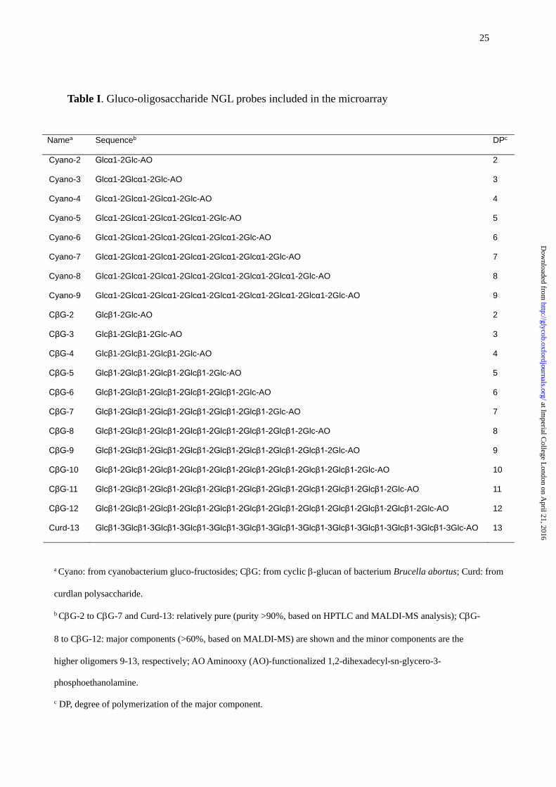
Table I. Gluco-oligosaccharide NGL probes included in the microarray
Namea Sequenceb DPc
Cyano-2 Glcα1-2Glc-AO 2
Cyano-3 Glcα1-2Glcα1-2Glc-AO 3
Cyano-4 Glcα1-2Glcα1-2Glcα1-2Glc-AO 4
Cyano-5 Glcα1-2Glcα1-2Glcα1-2Glcα1-2Glc-AO 5
Cyano-6 Glcα1-2Glcα1-2Glcα1-2Glcα1-2Glcα1-2Glc-AO 6
Cyano-7 Glcα1-2Glcα1-2Glcα1-2Glcα1-2Glcα1-2Glcα1-2Glc-AO 7
Cyano-8 Glcα1-2Glcα1-2Glcα1-2Glcα1-2Glcα1-2Glcα1-2Glcα1-2Glc-AO 8
Cyano-9 Glcα1-2Glcα1-2Glcα1-2Glcα1-2Glcα1-2Glcα1-2Glcα1-2Glcα1-2Glc-AO 9
CβG-2 Glcβ1-2Glc-AO 2
CβG-3 Glcβ1-2Glcβ1-2Glc-AO 3
CβG-4 Glcβ1-2Glcβ1-2Glcβ1-2Glc-AO 4
CβG-5 Glcβ1-2Glcβ1-2Glcβ1-2Glcβ1-2Glc-AO 5
CβG-6 Glcβ1-2Glcβ1-2Glcβ1-2Glcβ1-2Glcβ1-2Glc-AO 6
CβG-7 Glcβ1-2Glcβ1-2Glcβ1-2Glcβ1-2Glcβ1-2Glcβ1-2Glc-AO 7
CβG-8 Glcβ1-2Glcβ1-2Glcβ1-2Glcβ1-2Glcβ1-2Glcβ1-2Glcβ1-2Glc-AO 8
CβG-9 Glcβ1-2Glcβ1-2Glcβ1-2Glcβ1-2Glcβ1-2Glcβ1-2Glcβ1-2Glcβ1-2Glc-AO 9
CβG-10 Glcβ1-2Glcβ1-2Glcβ1-2Glcβ1-2Glcβ1-2Glcβ1-2Glcβ1-2Glcβ1-2Glcβ1-2Glc-AO 10
CβG-11 Glcβ1-2Glcβ1-2Glcβ1-2Glcβ1-2Glcβ1-2Glcβ1-2Glcβ1-2Glcβ1-2Glcβ1-2Glcβ1-2Glc-AO 11
CβG-12 Glcβ1-2Glcβ1-2Glcβ1-2Glcβ1-2Glcβ1-2Glcβ1-2Glcβ1-2Glcβ1-2Glcβ1-2Glcβ1-2Glcβ1-2Glc-AO 12
Curd-13 Glcβ1-3Glcβ1-3Glcβ1-3Glcβ1-3Glcβ1-3Glcβ1-3Glcβ1-3Glcβ1-3Glcβ1-3Glcβ1-3Glcβ1-3Glcβ1-3Glc-AO 13
a Cyano: from cyanobacterium gluco-fructosides; CG: from cyclic -glucan of bacterium Brucella abortus; Curd: from
curdlan polysaccharide.
b CG-2 to CG-7 and Curd-13: relatively pure (purity >90%, based on HPTLC and MALDI-MS analysis); CG-
8 to CG-12: major components (>60%, based on MALDI-MS) are shown and the minor components are the
higher oligomers 9-13, respectively; AO Aminooxy (AO)-functionalized 1,2-dihexadecyl-sn-glycero-3-
phosphoethanolamine.
c DP, degree of polymerization of the major component.
at Imperial C
ollege London on A
pril 21, 2016http://glycob.oxfordjournals.org/
Dow
nloaded from
26
at Imperial C
ollege London on A
pril 21, 2016http://glycob.oxfordjournals.org/
Dow
nloaded from
27
at Imperial C
ollege London on A
pril 21, 2016http://glycob.oxfordjournals.org/
Dow
nloaded from
28
at Imperial C
ollege London on A
pril 21, 2016http://glycob.oxfordjournals.org/
Dow
nloaded from
29
at Imperial C
ollege London on A
pril 21, 2016http://glycob.oxfordjournals.org/
Dow
nloaded from
30
at Imperial C
ollege London on A
pril 21, 2016http://glycob.oxfordjournals.org/
Dow
nloaded from
31
at Imperial C
ollege London on A
pril 21, 2016http://glycob.oxfordjournals.org/
Dow
nloaded from
32
at Imperial C
ollege London on A
pril 21, 2016http://glycob.oxfordjournals.org/
Dow
nloaded from
33
at Imperial C
ollege London on A
pril 21, 2016http://glycob.oxfordjournals.org/
Dow
nloaded from