Fibrosis A Common Pathway to Organ Injury and Failure · beta (TGF-β) pathway is important in...
12
The new england journal of medicine n engl j med 372;12 nejm.org March 19, 2015 1138 Review Article D isease-related injury in any organ triggers a complex cascade of cellular and molecular responses that culminates in tissue fibrosis. Al- though this fibrogenic response may have adaptive features in the short term, when it progresses over a prolonged period of time, parenchymal scarring and ultimately cellular dysfunction and organ failure ensue (Fig. 1). We and others have proposed four major phases of the fibrogenic response (Fig. 2). First is initiation of the response, driven by primary injury to the organ. The second phase is the activation of effector cells, and the third phase is the elaboration of extracellular matrix, both of which overlap with the fourth phase, during which the dynamic deposition (and insufficient resorption) of extracellular matrix promotes progression to fibrosis and ultimately to end-organ failure. The fact that diverse diseases in different organ systems are associated with fibrotic changes suggests common pathogenic pathways (Fig. 2). This “wounding response” is orchestrated by complex activities within different cells in which spe- cific molecular pathways have emerged. Cellular constituents include inflamma- tory cells (e.g., macrophages and T cells), epithelial cells, fibrogenic effector cells, endothelial cells, and others. Many different effector cells, including fibroblasts, myofibroblasts, cells derived from bone marrow, fibrocytes, and possibly cells derived from epithelial tissues (epithelial-to-mesenchymal transition) have been identified; there is some controversy regarding the identity of specific effectors in different organs. Beyond the multiple cells essential in the wounding response, core molecular pathways are critical; for example, the transforming growth factor beta (TGF-β) pathway is important in virtually all types of fibrosis. As fibrosis progresses, myofibroblasts proliferate and sense physical and bio- chemical stimuli in the local environment by means of integrins and cell-surface molecules; contractile mediators trigger pathological tissue contraction. This chain of events, in turn, causes physical organ deformation, which impairs organ function. Thus, the biology of fibrogenesis is dynamic, although the degree of plasticity ap- pears to vary from organ to organ. Although we understand many of the cellular and molecular processes underly- ing fibrosis, there are few effective therapies and fewer that target fibrogenesis specifically. These facts highlight the need for a deeper comprehension of the patho- genesis of fibrogenesis and the translation of this knowledge to novel treatments. Cellular and Molecular Themes in Pathogenesis Acute and chronic inflammation often trigger fibrosis (Fig. 2). Inflammation leads to injury of resident epithelial cells and often endothelial cells, resulting in enhanced release of inflammatory mediators, including cytokines, chemokines, and others. This process leads to the recruitment of a wide range of inflammatory cells, in- From the Department of Internal Medi- cine, Medical University of South Carolina (D.C.R., P.D.B.), and the Ralph H. John- son Veterans Affairs Medical Center (P.D.B.) — both in Charleston; and the Departments of Internal Medicine and Molecular Biology, University of Texas Southwestern Medical Center, Dallas (J.A.H.). Address reprint requests to Dr. Rockey at the Department of Internal Medicine, Medical University of South Carolina, 96 Jonathan Lucas St., Suite 803, MSC 623, Charleston, SC 29425, or at [email protected]. N Engl J Med 2015;372:1138-49. DOI: 10.1056/NEJMra1300575 Copyright © 2015 Massachusetts Medical Society. Dan L. Longo, M.D., Editor Fibrosis — A Common Pathway to Organ Injury and Failure Don C. Rockey, M.D., P. Darwin Bell, Ph.D., and Joseph A. Hill, M.D., Ph.D. The New England Journal of Medicine Downloaded from nejm.org by KEVIN ROSTEING on April 24, 2015. For personal use only. No other uses without permission. Copyright © 2015 Massachusetts Medical Society. All rights reserved.
Transcript of Fibrosis A Common Pathway to Organ Injury and Failure · beta (TGF-β) pathway is important in...
T h e n e w e ngl a nd j o u r na l o f m e dic i n e
n engl j med 372;12 nejm.org March 19, 20151138
Review Article
Disease-related injury in any organ triggers a complex cascade of cellular and molecular responses that culminates in tissue fibrosis. Al-though this fibrogenic response may have adaptive features in the short
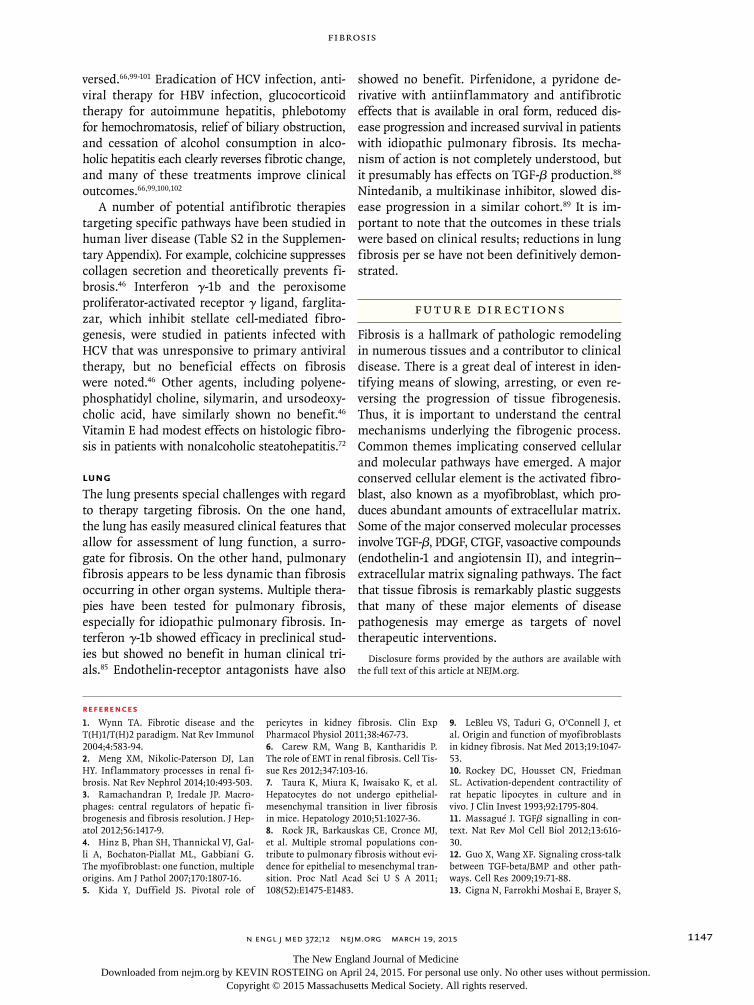
term, when it progresses over a prolonged period of time, parenchymal scarring and ultimately cellular dysfunction and organ failure ensue (Fig. 1).
We and others have proposed four major phases of the fibrogenic response (Fig. 2). First is initiation of the response, driven by primary injury to the organ. The second phase is the activation of effector cells, and the third phase is the elaboration of extracellular matrix, both of which overlap with the fourth phase, during which the dynamic deposition (and insufficient resorption) of extracellular matrix promotes progression to fibrosis and ultimately to end-organ failure.
The fact that diverse diseases in different organ systems are associated with fibrotic changes suggests common pathogenic pathways (Fig. 2). This “wounding response” is orchestrated by complex activities within different cells in which spe-cific molecular pathways have emerged. Cellular constituents include inflamma-tory cells (e.g., macrophages and T cells), epithelial cells, fibrogenic effector cells, endothelial cells, and others. Many different effector cells, including fibroblasts, myofibroblasts, cells derived from bone marrow, fibrocytes, and possibly cells derived from epithelial tissues (epithelial-to-mesenchymal transition) have been identified; there is some controversy regarding the identity of specific effectors in different organs. Beyond the multiple cells essential in the wounding response, core molecular pathways are critical; for example, the transforming growth factor beta (TGF-β) pathway is important in virtually all types of fibrosis.
As fibrosis progresses, myofibroblasts proliferate and sense physical and bio-chemical stimuli in the local environment by means of integrins and cell-surface molecules; contractile mediators trigger pathological tissue contraction. This chain of events, in turn, causes physical organ deformation, which impairs organ function. Thus, the biology of fibrogenesis is dynamic, although the degree of plasticity ap-pears to vary from organ to organ.
Although we understand many of the cellular and molecular processes underly-ing fibrosis, there are few effective therapies and fewer that target fibrogenesis specifically. These facts highlight the need for a deeper comprehension of the patho-genesis of fibrogenesis and the translation of this knowledge to novel treatments.
Cellul a r a nd Molecul a r Themes in Patho genesis
Acute and chronic inflammation often trigger fibrosis (Fig. 2). Inflammation leads to injury of resident epithelial cells and often endothelial cells, resulting in enhanced release of inflammatory mediators, including cytokines, chemokines, and others. This process leads to the recruitment of a wide range of inflammatory cells, in-
From the Department of Internal Medi-cine, Medical University of South Carolina (D.C.R., P.D.B.), and the Ralph H. John-son Veterans Affairs Medical Center (P.D.B.) — both in Charleston; and the Departments of Internal Medicine and Molecular Biology, University of Texas Southwestern Medical Center, Dallas ( J.A.H.). Address reprint requests to Dr. Rockey at the Department of Internal Medicine, Medical University of South Carolina, 96 Jonathan Lucas St., Suite 803, MSC 623, Charleston, SC 29425, or at rockey@ musc . edu.
N Engl J Med 2015;372:1138-49.DOI: 10.1056/NEJMra1300575Copyright © 2015 Massachusetts Medical Society.
Dan L. Longo, M.D., Editor
Fibrosis — A Common Pathway to Organ Injury and Failure
Don C. Rockey, M.D., P. Darwin Bell, Ph.D., and Joseph A. Hill, M.D., Ph.D.
The New England Journal of Medicine Downloaded from nejm.org by KEVIN ROSTEING on April 24, 2015. For personal use only. No other uses without permission.
Copyright © 2015 Massachusetts Medical Society. All rights reserved.
n engl j med 372;12 nejm.org March 19, 2015 1139
Fibrosis
cluding lymphocytes, polymorphonuclear leuko-cytes, eosinophils, basophils, mast cells, and mac-rophages. These inflammatory cells elicit the activation of effector cells,1 which drive the fibro-genic process. One example in which an inflam-matory lesion drives this cycle is the interstitial nephritis induced by nonsteroidal antiinflamma-tory drugs (NSAIDs), which culminates in chronic inflammation and the activation of a fibrogenic cascade. In addition, macrophages can play a prominent role in interstitial fibrosis, often driven by the TGF-β pathway.2 However, some inflam-matory cells may be protective. For example,
certain populations of macrophages phagocytose apoptotic cells that promote the fibrogenic process and activate matrix-degrading metalloproteases.3
Fibroblasts and myofibroblasts have been iden-tified as key fibrosis effectors in many organs, and as such are responsible for the synthesis of extracellular matrix proteins4 (Fig. 2). Contro-versy exists regarding the origin of these cells; for example, myofibroblasts may be derived from fibroblasts or from other mesenchymal cells, such as pericytes.5 Epithelial-to-mesenchymal tran-sition, in which epithelial cells give rise to mesen-chymal fibrogenic cells, may play a role. However,
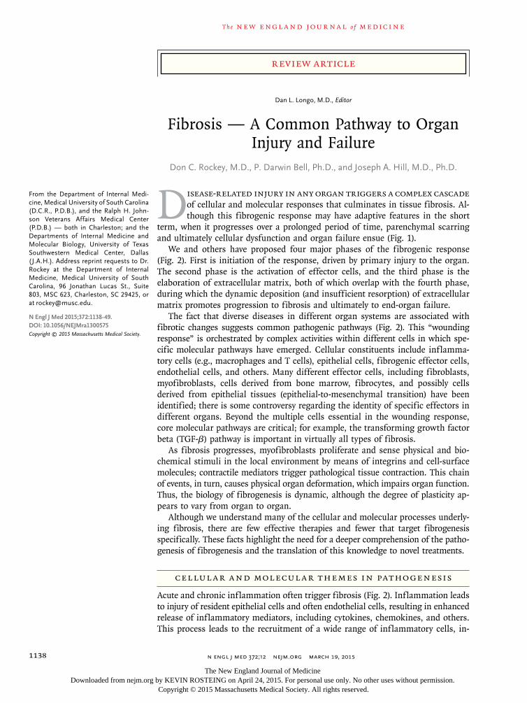
Figure 1. Fibrogenesis and Major Organ Systems.
Fibrosis is a pathologic feature of disease in virtually all organs. It has protean and often lethal consequences and accounts for substantial morbidity and mortality. Selected organs and associated diseases are highlighted.
Cirrhosis
Portal hypertensionAscites
Gastroesophageal varicesHepatorenal syndrome
Hepatopulmonary syndromePortopulmonary syndrome
Hepatic encephalopathy
Hepatocellular cancer
Cardiac fibrosis
Diastolic dysfunction
Heart failure, with reducedor preserved ejection fraction
Arrhythmia
Eye
Strabmisus
Pulmonary fibrosis
Restrictive lung disease
Pulmonary hypertension
Right-sided heart failure
Pancreatic fibrosis
Chronic pain
Diabetes mellitus
Malabsorption
Cancer
Renal fibrosis
Chronic kidney diseaseHypertension
AnemiaElectrolyte disturbances
Skin
Scleroderma
Keloid
Nephrogenic systemic fibrosis
The New England Journal of Medicine Downloaded from nejm.org by KEVIN ROSTEING on April 24, 2015. For personal use only. No other uses without permission.
Copyright © 2015 Massachusetts Medical Society. All rights reserved.
n engl j med 372;12 nejm.org March 19, 20151140
T h e n e w e ngl a nd j o u r na l o f m e dic i n e
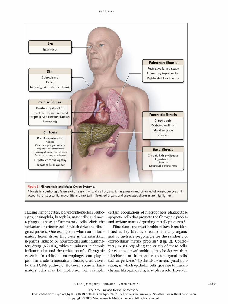
Figure 2. Cellular Injury and Fibrogenesis.
In parenchymal organs, many different types of stimuli lead to epithelial-cell injury (top), which is typically followed by an inflammatory response (shown at left). This process stimulates a fibrogenic wound-healing response that involves multiple cellular and molecular sys-tems. At the cellular level, the recruitment of inflammatory cells is central. Inflammatory cells produce a variety of mediators, cytokines, and other factors that are responsible for the stimulation and recruitment of other cells. Key among these cells are fibrogenic effector cells; these cells are of mesenchymal origin and include fibroblasts, fibrocytes, tissue-specific pericytes and myofibroblasts, and fibro-blasts derived through epithelial-to-mesenchymal transition. These effectors produce a variety of extracellular matrix proteins, which themselves may modify the wound milieu, often stimulating fibrogenic effector cells in an autocrine fashion. Indeed, in most organ sys-tems, autocrine loops in fibrogenic effector cells are prominent. Cell–cell interactions lead to further activation of effector cells. Effector cells produce a variety of extracellular matrix proteins, peptides, cytokines, and growth factors, all of which may lead to autocrine stimu-lation (see the right side of the figure), typical of most organ systems. Many forms of injury also lead to the activation and transforma-tion of other cells, such as specialized endothelial or tissue-specific cells. Injury to these cells in turn leads to a variety of downstream effects, including activation of fibrogenic effector cells. NK denotes natural killer.
Inciting injury
Basementmembrane
Recruitment ofinflammatory cells
Injuredendothelial cell
Cell–cellinteraction
Epithelialcell
Cytokines
Effector cellMacrophage
T cells
NK cells
Effector-cellactivation
Effector-cellproliferation
Injuredepithelial cell
The New England Journal of Medicine Downloaded from nejm.org by KEVIN ROSTEING on April 24, 2015. For personal use only. No other uses without permission.
Copyright © 2015 Massachusetts Medical Society. All rights reserved.
n engl j med 372;12 nejm.org March 19, 2015 1141
Fibrosis
the importance of epithelial-to-mesenchymal tran-sition has been actively debated.6-9
The matrix proteins that compose the fibrotic scar, which are highly conserved across tissues, consist predominantly of interstitial collagens (types I and III), cellular fibronectin, basement-membrane proteins such as laminin, and other, less abundant elements. In addition, myofibro-blasts, which by definition are cells that express smooth-muscle proteins, including actin (ACTA2), are contractile.10 The contraction of these cells contributes to the distortion of parenchymal ar-chitecture, which promotes disease pathogenesis and tissue failure.
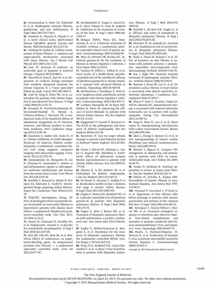
The molecular processes driving fibrosis are wide-ranging and complex (Fig. 3). The TGF-β cascade, which plays a major role in fibrosis, in-volves the binding of a ligand to a serine–threo-nine kinase type II receptor that recruits and phosphorylates a type I receptor. This type I re-ceptor subsequently phosphorylates SMADs, which function as downstream effectors, typically by modulating target gene expression. Although the TGF-β superfamily, which involves multiple sig-naling cascades, is too complex to review in detail
here (see Massagué11), its diversity highlights the complexity of the regulation of fibrosis.
TGF-β is a potent stimulator of the synthesis of extracellular matrix proteins in most fibro-genic cells. TGF-β is synthesized and secreted by inflammatory cells and by effector cells, thereby functioning in both an autocrine and paracrine fashion. The complexity of the TGF-β system is illustrated by its interactions with other cell-sig-naling pathways.12 For example, TGF-β stimulates sonic hedgehog signaling in lung fibroblasts, and sonic hedgehog signaling, in turn, regulates fibro-blast function.13 Another example of the complex-ity of the TGF-β system emerges from study of the extracellular activation of TGF-β in an inac-tive complex with a latency-associated peptide, which is subsequently activated by the αvβ6 in-tegrin.14
The molecular systems involved in fibrosis are so expansive as to preclude a detailed discussion (see Fig. 3 for an overview of several important molecular pathways). Platelet-derived growth factor (PDGF), connective-tissue growth factor (CTGF), and vasoactive peptide systems (especially angiotensin II and endothelin-1)15 play important
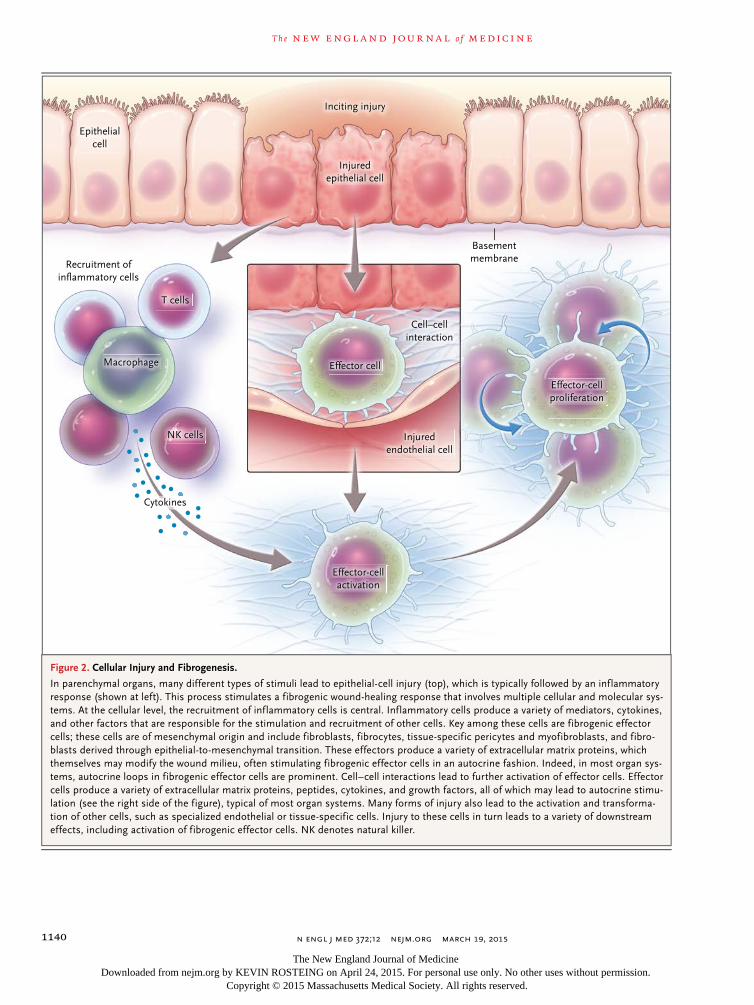
Figure 3. Molecular Pathways in Fibrosis.
Fibrogenesis in multiple tissues is effected by a number of signaling cascades. These cascades (not shown) are of-ten triggered by the exposure of effector cells to circulating or locally produced molecules that stimulate the biosyn-thesis and secretion of extracellular matrix proteins. The extracellular matrix itself may also stimulate fibrogenesis through activation of integrin signaling. Examples of major pathways are shown. MAPK denotes mitogen-activated protein kinase, ROCK Rho-associated protein kinase, and TGF-β transforming growth factor β.
TGF-βreceptor
SMADsIntegrin-linked
kinase
IntegrinsGrowthfactors
Vasoactivepeptides
Fibrogeniceffector cell
Nucleus
Cytokines
MAPKsRho/ROCK
Extracellular matrixbiosynthesis
The New England Journal of Medicine Downloaded from nejm.org by KEVIN ROSTEING on April 24, 2015. For personal use only. No other uses without permission.
Copyright © 2015 Massachusetts Medical Society. All rights reserved.
n engl j med 372;12 nejm.org March 19, 20151142
T h e n e w e ngl a nd j o u r na l o f m e dic i n e
roles. Among vasoactive systems, endothelin plays a role in fibrosis in virtually all organ systems, acting through G-protein–coupled endothelin-A or endothelin-B cell-surface receptors or both.16 Furthermore, angiogenic pathways may be im-portant in fibrosis.17 Finally, it is clear that inte-grins, which link extracellular matrix to cells, are critical in the pathogenesis of fibrosis.18,19
When injury and inflammatory responses are abrogated, resorption of extracellular matrix pro-teins occurs, promoting organ repair. When chronic injury persists, the unremitting activa-tion of effector cells results in the continuous deposition of extracellular matrix, progressive scarring, and organ damage. Thus, fibrogenesis involves the interplay between factors that pro-mote the biosynthesis, deposition, and degradation of extracellular matrix proteins. Typically, matrix synthesis is counterbalanced by matrix-degrading metalloproteases.3,20,21 In addition, fibrogenesis is governed by pathways that eliminate effectors (e.g., by means of senescence, apoptosis, or au-tophagy). For example, the apoptosis of hepatic stellate cells is associated with the reversal of fibrosis.22
An understanding of the role of genetics in the pathogenesis of fibrosis is emerging. For in-stance, in the kidney, fibrosis is a prominent fea-ture of karyomegalic interstitial nephritis, which is caused by mutations in the gene encoding Fanconi anemia-associated nuclease 1 (FAN1).23 In the liver, PNLAP3 is important in fibrosis that is mediated by ethanol and associated with fatty liver dis-ease.24,25 A number of candidate genes may be im-portant in cases of fibrosis that are mediated by infection with hepatitis C virus (HCV).26 Mutations in TERT (c:2768C→T), the gene encoding telomer-ase reverse transcriptase, and MUC5B, which en-codes mucin, are associated with pulmonary fi-brosis.27-29
The epigenetic regulation of gene expression, which includes but is not limited to DNA methyla-tion, post-translational modifications of the his-tone protein constituents of chromatin, and regu-latory noncoding RNAs (e.g., microRNAs [miRs]), is important in fibrosis. For example, fibroblasts from patients with idiopathic pulmonary fibro-sis have been found to have global changes in DNA methylation, changes that are not seen in fibroblasts from normal lungs.30 The miRs, which play an ever-expanding role in gene regulation, are also involved in the pathogenesis of fibrosis.
In diabetic nephropathy, TGF-β promotes the ex-pression of miR-192, which results in collagen de-position,31 and miR-19b regulates TGF-β signaling in hepatic stellate cells.32 In the heart, miR-21, miR-29, miR-30, and miR-133 participate in the remodeling of the myocardial matrix.33
Mech a nisms a nd A dv er se Clinic a l Effec t s
Cardiac Fibrosis
The heart undergoes extensive structural and func-tional remodeling in response to injury, central to which is the hypertrophy of cardiac myocytes, with excessive deposition of extracellular matrix.34 Myocardial fibrosis is commonly categorized as one of two types: reactive fibrosis or replacement fibrosis. Reactive fibrosis occurs in perivascular spaces and corresponds to similar fibrogenic re-sponses in other tissues; replacement fibrosis oc-curs at the site of myocyte loss.
In the heart, fibrosis is attributed to cardiac fibroblasts, the most abundant cell type in the myocardium. These cells are derived from fibro-blasts that are native to the myocardium, from circulating fibroblasts, and from fibroblasts that emerge from epithelial-to-mesenchymal transi-tion.35,36 All these cell types proliferate and dif-ferentiate into myofibroblasts in response to injury (Fig. 2), a process that is driven by classic factors such as TGF-β1, endothelin-1, and angio-tensin II.37 Cross-talk and feedback also occur between cells — in this case, between activated fibroblasts and cardiomyocytes — which further fuel fibrogenesis.38
Cardiac fibrosis contributes to both systolic and diastolic dysfunction and to perturbations of elec-trical excitation; it also disrupts repolarization (Fig. 1).39 Proarrhythmic effects are the most prominent. Collagenous septa in the failing heart contribute to arrhythmogenesis by inducing a discontinuous slowing of conduction.40 Areas of arrhythmogenic fibrosis slow conduction through junctions in the heterocellular gap that couple fibroblasts and cardiomyocytes.41 Endocardial breakthrough of microreentrant circuits occurs as a result of the heterogeneous spatial distribution of fibrosis42 and the triggering of activity caused by the depolarization of myocytes by electrically coupled myofibroblasts.43
Fibrotic scarring in the heart correlates strong-ly with an increased incidence of arrhythmias
The New England Journal of Medicine Downloaded from nejm.org by KEVIN ROSTEING on April 24, 2015. For personal use only. No other uses without permission.
Copyright © 2015 Massachusetts Medical Society. All rights reserved.
n engl j med 372;12 nejm.org March 19, 2015 1143
Fibrosis
and sudden cardiac death.44 For example, a 3% increase in the extracellular volume fraction of fibrous tissue (measured by means of magnetic resonance imaging after the administration of gadolinium) is associated with a 50% increase in the risk of adverse cardiac events.45
Hepatic Fibrosis
Hepatic fibrosis typically results from an inflam-matory process that affects hepatocytes or biliary cells. Inflammation leads to the activation of ef-fector cells, which results in the deposition of extracellular matrix. Although a variety of effec-tors synthesize extracellular matrix in the liver, hepatic stellate cells appear to be the primary source of extracellular matrix. Abundant evidence suggests that the stellate cell is pericyte-like, un-dergoing a transformation into a myofibroblast in response to injury.10
In the liver, multiple cell types, including stel-late cells, endothelial cells, Kupffer cells, bile-duct cells, and immune cells, orchestrate the cellular and molecular response to injury.46 Numerous mo-lecular pathways, similar to those found in other organs, are involved. A pathway that appears to be unique to the liver involves toll-like receptor 4 (TLR4)47; TLR4 is activated on the surface of stel-late cells by intestinal bacterial lipopolysaccha-rides derived from the gut (i.e., translocated bac-teria), triggering cell activation and fibrogenesis and thereby linking fibrosis to the microbiome.48 TLR4 expression is associated with portal inflam-mation and fibrosis in patients with fatty liver disease.49
The end result of hepatic fibrogenesis is cirrho-sis, an ominous parenchymal lesion that underlies a wide range of devastating complications that have adverse effects on survival (Fig. 1). Portal hypertension, a devastating result of injury, de-velops during the fibrogenic response after dis-ruption of the normal interaction between sinu-soidal endothelial cells and hepatic stellate cells; the resulting activation and contraction of peri-cyte-like stellate cells leads to sinusoidal constric-tion and increased intrahepatic resistance. This increase in resistance in turn activates abnormal signaling by smooth-muscle cells in mesenteric vessels. An increase in angiogenesis and collateral blood flow follows, resulting in an increase in mesenteric blood flow and a worsening of portal hypertension.50 The major clinical sequelae of por-tal hypertension, variceal hemorrhage and ascites,
emerge relatively late, after the portal pressure rises to a hepatic venous pressure gradient of more than 12 mm Hg.50
Renal Fibrosis
Events that initiate renal fibrosis are diverse, rang-ing from primary renal injury to systemic dis-eases.51,52 The kidneys are susceptible to hyper-tension and diabetes, the two leading causes of renal fibrosis. As is true in other organs, fibrosis of the kidney is mediated by cellular elements (e.g., inflammatory cells) and molecular elements (e.g., cytokines, TGF-β1, CTGF, PDGF, and endo-thelin-1) (Fig. 2 and 3).51-54 The intrarenal renin–angiotensin–aldosterone axis is particularly im-portant in hypertension-induced fibrosis.54
The kidney has a unique cellular architecture that consists of the glomeruli, tubules, intersti-tium, and capillaries. Injury at any of these sites triggers the deposition of extracellular matrix.37 The location of the initial injury is an important determinant of the clinical consequences. Inju-ries that initially target glomeruli elicit patterns of disease that are different from those that are elicited by injuries to the tubular–interstitial envi-ronment. For example, NSAIDs, urinary obstruc-tion, polycystic kidney disease, and infections can provoke tubulointerstitial fibrosis,43 whereas glomerular immune deposition (e.g., the deposi-tion of IgA) leads to glomerulonephritis.44 Glom-eruli and podocytes are sensitive to systemic and local immunologic insults45; high glomerular cap-illary pressure, exacerbated by systemic hyper-tension and diabetes, leads to proteinuria, the activation of cytokines and complement, and the infiltration of immune cells, resulting in epithe-lial cell and interstitial fibrosis.46
Glomerular fibrosis, regardless of the cause, diminishes renal blood flow, which leads to hypoxia and the activation of hypoxia-inducible factor 1, which in turn triggers nephron collapse and fibrotic replacement by means of rarefaction.47 The renal interstitium and capillaries contribute substantially to tubulointerstitial disease, as peri-tubular pericytes migrate into the interstitium, where they are transformed into myofibroblasts.48
Regardless of the initiating insult, renal fi-brosis leads to loss of function and organ failure (Fig. 1). Homeostasis can be maintained with a glomerular filtration rate as low as approximately 10% of the normal rate. As the mechanisms main-taining homeostasis are progressively disrupted,
The New England Journal of Medicine Downloaded from nejm.org by KEVIN ROSTEING on April 24, 2015. For personal use only. No other uses without permission.
Copyright © 2015 Massachusetts Medical Society. All rights reserved.
n engl j med 372;12 nejm.org March 19, 20151144
T h e n e w e ngl a nd j o u r na l o f m e dic i n e
anemia develops and the regulation of electrolyte balance and pH is disrupted.
Pulmonary Fibrosis
Pulmonary fibrosis occurs in association with a wide range of diseases, including scleroderma (sys-temic sclerosis), sarcoidosis, and infection, and as a result of environmental exposures (e.g., silica dust or asbestos), but in most patients it is idio-pathic and progressive. Idiopathic pulmonary fibrosis is characterized by progressive fibrosis without substantial inflammation.55 Its patho-genesis appears to be unlike that of other fibros-ing diseases and is poorly understood. Injury to alveolar epithelial cells activates pulmonary fibro-blasts, provoking their transformation to matrix-producing myofibroblasts.56 Activated lung fibro-blasts may cause apoptosis of alveolar cells, which leads to further fibroblast activation and a vicious cycle of injury and effector-cell activation (Fig. 2). Research has focused on TGF-β signaling (Fig. 3)57 and the interstitial pericytes present in lung fi-brosis.58
Pulmonary fibrosis is characterized by paren-chymal honeycombing, reduced lung compliance, and restrictive lung function (Fig. 1). Fibrosis of the interstitial spaces hinders gas exchange, cul-minating in abnormal oxygenation and clinical dyspnea. Progressive pulmonary fibrosis also leads to pulmonary hypertension, right-sided heart fail-ure, and ultimately respiratory failure.
Other Forms of Fibrosis
Fibrosis also occurs in the joints, bone marrow, brain, eyes, intestines, peritoneum and retroperi-toneum, pancreas, and skin, and in these cases is driven by typical cellular and molecular pro-cesses (Fig. 2 and 3). Retroperitoneal fibrosis is a rare condition characterized by inflammation and fibrosis in the retroperitoneal space; most cases are idiopathic, but secondary causes include drugs, infections, autoimmune and inflammatory stimuli, and radiation. Patients may present with pain, and the major clinical sequelae of this con-dition are related to its involvement with struc-tures in the retroperitoneum, including arteries (leading to acute and chronic renal failure) and ureters (leading to hydronephrosis). Currently, treatment of this primary fibrosing disorder is not available. In certain cancers, fibrosis is linked to TGF-β-integrin signaling.59 In scleroderma, the prototypical fibrosing skin disease, skin fibro-
blasts and myofibroblasts are activated through the TGF-β–SMAD signaling pathway.60 Nephro-genic systemic fibrosis, a debilitating condition that is marked by widespread organ fibrosis, oc-curs in patients with renal insufficiency who have been exposed to gadolinium-based contrast ma-terial. Initial systemic inflammatory-response reactions and the reaction of gadolinium (Gd3+) ions with circulating proteins and heavy metals lead to the deposition of insoluble elements in tissue.61 Since no effective therapies have been identified, prevention is key.61 A recently recog-nized IgG4-related disease appears to involve autoimmune-driven inflammation that provokes fibrosis in multiple organs, including the pancreas, retroperitoneum, lung, kidney, liver, and aorta.62
Ther a py
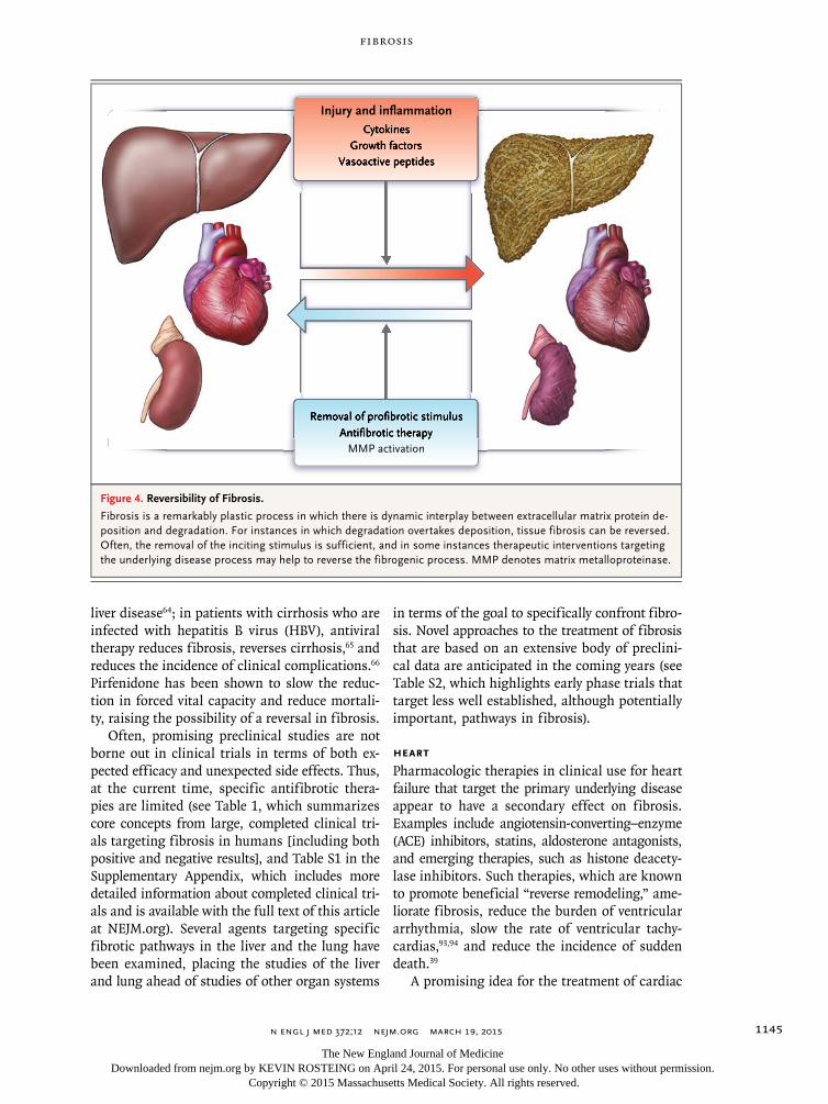
Fibrosis and resultant organ failure account for at least one third of deaths worldwide.63 Since fibrosis is common and has adverse effects in all organs, it is an attractive therapeutic target. Con-trary to the widely held perception that scar tissue is permanent, the available evidence points to the highly plastic nature of organ fibrosis; it is not irreversible “scar” but an actively remodeled tis-sue component that can, under certain circum-stances, regress. Fibrosis occurs by means of a dynamic process that involves the synthesis and deposition of extracellular matrix, and its rever-sion occurs by means of the elimination of ef-fector cells and shifts in the balance of matrix synthesis and degradation (Fig. 4). Although it is not clear what pathogenic or clinical factors pro-mote reversibility, the regression of fibrosis has been shown to lead to improved clinical outcomes. Elimination of the inciting stimulus is the first and most efficacious approach.
Fibrosis of parenchymal tissue usually pro-gresses slowly, which suggests that therapy may be required for extended periods; slowing the pro-gression of fibrosis may be a more realistic thera-peutic goal than eliminating it. One of the chal-lenges in assessing the therapeutic response is that there are few noninvasive means of measuring fi-brosis (e.g., blood or imaging tests; biopsy is typi-cally the most reliable approach) or monitoring the effects of therapeutic intervention.
The best indication that fibrosis is reversible and that this reversibility has positive effects on clinical outcomes is based on the treatment of
The New England Journal of Medicine Downloaded from nejm.org by KEVIN ROSTEING on April 24, 2015. For personal use only. No other uses without permission.
Copyright © 2015 Massachusetts Medical Society. All rights reserved.
n engl j med 372;12 nejm.org March 19, 2015 1145
Fibrosis
liver disease64; in patients with cirrhosis who are infected with hepatitis B virus (HBV), antiviral therapy reduces fibrosis, reverses cirrhosis,65 and reduces the incidence of clinical complications.66
Pirfenidone has been shown to slow the reduc-tion in forced vital capacity and reduce mortali-ty, raising the possibility of a reversal in fibrosis.
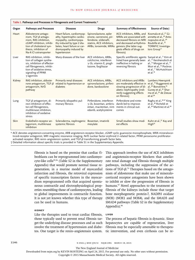
Often, promising preclinical studies are not borne out in clinical trials in terms of both ex-pected efficacy and unexpected side effects. Thus, at the current time, specific antifibrotic thera-pies are limited (see Table 1, which summarizes core concepts from large, completed clinical tri-als targeting fibrosis in humans [including both positive and negative results], and Table S1 in the Supplementary Appendix, which includes more detailed information about completed clinical tri-als and is available with the full text of this article at NEJM.org). Several agents targeting specific fibrotic pathways in the liver and the lung have been examined, placing the studies of the liver and lung ahead of studies of other organ systems
in terms of the goal to specifically confront fibro-sis. Novel approaches to the treatment of fibrosis that are based on an extensive body of preclini-cal data are anticipated in the coming years (see Table S2, which highlights early phase trials that target less well established, although potentially important, pathways in fibrosis).
Heart
Pharmacologic therapies in clinical use for heart failure that target the primary underlying disease appear to have a secondary effect on fibrosis. Examples include angiotensin-converting–enzyme (ACE) inhibitors, statins, aldosterone antagonists, and emerging therapies, such as histone deacety-lase inhibitors. Such therapies, which are known to promote beneficial “reverse remodeling,” ame-liorate fibrosis, reduce the burden of ventricular arrhythmia, slow the rate of ventricular tachy-cardias,93,94 and reduce the incidence of sudden death.39
A promising idea for the treatment of cardiac
Figure 4. Reversibility of Fibrosis.
Fibrosis is a remarkably plastic process in which there is dynamic interplay between extracellular matrix protein de-position and degradation. For instances in which degradation overtakes deposition, tissue fibrosis can be reversed. Often, the removal of the inciting stimulus is sufficient, and in some instances therapeutic interventions targeting the underlying disease process may help to reverse the fibrogenic process. MMP denotes matrix metalloproteinase.
CytokinesGrowth factors
Vasoactive peptides
Removal of profibrotic stimulusAntifibrotic therapy
MMP activation
CytokinesGrowth factors
Vasoactive peptides
Removal of profibrotic stimulusAntifibrotic therapy
Injury and inflammation
The New England Journal of Medicine Downloaded from nejm.org by KEVIN ROSTEING on April 24, 2015. For personal use only. No other uses without permission.
Copyright © 2015 Massachusetts Medical Society. All rights reserved.
n engl j med 372;12 nejm.org March 19, 20151146
T h e n e w e ngl a nd j o u r na l o f m e dic i n e
fibrosis is based on the premise that cardiac fi-broblasts can be reprogrammed into cardiomyo-cyte-like cells95,96 (Table S2 in the Supplementary Appendix) that would promote normal tissue re-generation; in a murine model of myocardial infarction and fibrosis, the retroviral expression of specific transcription factors in the myocar-dium reprogrammed cells that acquired sponta-neous contractile and electrophysiological prop-erties resembling those of cardiomyocytes, leading to global improvements in contractile function. It is not yet known whether this type of therapy can be used in humans.
Kidney
Like the therapies used to treat cardiac fibrosis, those typically used to prevent renal fibrosis tar-get the underlying disease processes and as such involve the treatment of hypertension and diabe-tes. One target is the renin–angiotensin system.
This approach involves the use of ACE inhibitors and angiotensin-receptor blockers that amelio-rate renal damage and fibrosis through multiple pathways, including the suppression of the ac-tions of TGF-β.79 Therapies based on the antago-nism of aldosterone that make use of mineralo-corticoid receptor antagonists have been shown to inhibit or slow the progression of fibrosis in humans.97 Novel approaches to the treatment of fibrosis of the kidneys include those that target bone morphogenetic protein-7, NADPH oxidase (NOX) (NOX1 and NOX4), and the SMAD3 and SMAD4 pathways (Table S2 in the Supplementary Appendix).98
Liver
The process of hepatic fibrosis is dynamic. Since hepatocytes are capable of regeneration, liver fibrosis may be especially amenable to therapeu-tic intervention, and even cirrhosis can be re-
Organ Pathways and Processes Diseases Drugs Summary of Effectiveness Source of Data†
Heart Aldosterone antago-nism, TGF-β antago-nism, RAS inhibition, cGMP inhibition, inhibi-tion of cholesterol syn-thesis, inhibition of Na-K-Cl cotransporter
Heart failure, cardiomyop-athy, hypertrophic cardio-myopathy, cardiomyopa-thy induced by type 2 dia-betes, heart failure or car-diomyopathy induced by hypertension
Spironolactone, epler enone, canrenone, pir-fenidone, sildenafil, statins, ACE inhibitors, ARBs, torsemide, MRAs
ACE inhibitors, ARBs, and MRAs are associated with decreased fibrosis on MRI and decreased arrhythmo-genesis (the latter sug-gests effects of drugs on fibrosis)
Kosmala et al.,67 Gi-annetta et al.,68 Anto-nopoulos et al.,69 Roubille et al.,70 TORAFIC Investiga-tors Group71
Liver RAS inhibition, inhibi-tion of collagen synthe-sis, inhibition of effector-cell fibrogenesis, inhibi-tion of oxidative stress, signaling of PPAR γ-agonists
Many diseases of the liver ACE inhibitors, ARBs, colchicine, interferon γ-1b, vitamin E, piogli-tazone, farglitazar
Specific antifibrotic agents listed have generally been ineffective in halting or reversing fibrosis
Sanyal et al.,72 Kim et al.,73 Kershenobich et al.,74 Morgan et al.,75 Muir et al.,76 Pockros et al.,77 McHutchison et al.78
Kidney RAS inhibition, aldoste-rone antagonism, TGF-β antagonism, Nrf2 pathway
Primarily renal diseases related to hypertension or diabetes
ACE inhibitors, ARBs, spironolactone, pirfeni-done, bardoxolone
ACE inhibitors and ARBs are moderately effective in slowing progression of di-abetic nephropathy (indi-rectly suggesting effects on fibrosis)
Lambers Heerspink et al.,79 Ruggenenti et al.,80 Bonventre,81 Guney et al.,82 Shar-ma et al.,83 de Zeeuw et al.84
Lung TGF-β antagonism, di-rect inhibition of effec-tor-cell fibrogenesis, multikinase inhibition, inhibition of oxidative stress
Primarily idiopathic pul-monary fibrosis
Pirfenidone, interferon γ-1b, bosentan, ambris-entan, macitentan, nint-edanib, acetylcysteine
Pirfenidone and ninte-danib led to improve-ments in clinical out-comes
Raghu et al.,85-87 King et al.,88 Richeldi et al.,89 Martinez et al.90
Skin Endothelin-receptor an-tagonism, multikinase inhibition
Scleroderma, nephrogenic systemic fibrosis
Bosentan, imatinib mesylate
Small studies show mod-est effects
Kuhn et al.,91 Kay and High92
* ACE denotes angiotensin-converting enzyme, ARB angiotensin-receptor blocker, cGMP cyclic guanosine monophosphate, MRA mineralocor-ticoid-receptor antagonist, MRI magnetic resonance imaging, Nrf2 nuclear factor erythroid 2–related factor, PPAR peroxisome proliferator-activated receptor, RAS renin–angiotensin system, and TGF-β transforming growth factor beta.
† Detailed information about specific trials is provided in Table S1 in the Supplementary Appendix.
Table 1. Pathways and Processes in Fibrogenesis and Current Treatments.*
The New England Journal of Medicine Downloaded from nejm.org by KEVIN ROSTEING on April 24, 2015. For personal use only. No other uses without permission.
Copyright © 2015 Massachusetts Medical Society. All rights reserved.
n engl j med 372;12 nejm.org March 19, 2015 1147
Fibrosis
versed.66,99-101 Eradication of HCV infection, anti-viral therapy for HBV infection, glucocorticoid therapy for autoimmune hepatitis, phlebotomy for hemochromatosis, relief of biliary obstruction, and cessation of alcohol consumption in alco-holic hepatitis each clearly reverses fibrotic change, and many of these treatments improve clinical outcomes.66,99,100,102
A number of potential antifibrotic therapies targeting specific pathways have been studied in human liver disease (Table S2 in the Supplemen-tary Appendix). For example, colchicine suppresses collagen secretion and theoretically prevents fi-brosis.46 Interferon γ-1b and the peroxisome proliferator-activated receptor γ ligand, farglita-zar, which inhibit stellate cell-mediated fibro-genesis, were studied in patients infected with HCV that was unresponsive to primary antiviral therapy, but no beneficial effects on fibrosis were noted.46 Other agents, including polyene-phosphatidyl choline, silymarin, and ursodeoxy-cholic acid, have similarly shown no benefit.46 Vitamin E had modest effects on histologic fibro-sis in patients with nonalcoholic steatohepatitis.72
Lung
The lung presents special challenges with regard to therapy targeting fibrosis. On the one hand, the lung has easily measured clinical features that allow for assessment of lung function, a surro-gate for fibrosis. On the other hand, pulmonary fibrosis appears to be less dynamic than fibrosis occurring in other organ systems. Multiple thera-pies have been tested for pulmonary fibrosis, especially for idiopathic pulmonary fibrosis. In-terferon γ-1b showed efficacy in preclinical stud-ies but showed no benefit in human clinical tri-als.85 Endothelin-receptor antagonists have also
showed no benefit. Pirfenidone, a pyridone de-rivative with antiinflammatory and antifibrotic effects that is available in oral form, reduced dis-ease progression and increased survival in patients with idiopathic pulmonary fibrosis. Its mecha-nism of action is not completely understood, but it presumably has effects on TGF-β production.88 Nintedanib, a multikinase inhibitor, slowed dis-ease progression in a similar cohort.89 It is im-portant to note that the outcomes in these trials were based on clinical results; reductions in lung fibrosis per se have not been definitively demon-strated.
Fu t ur e Dir ec tions
Fibrosis is a hallmark of pathologic remodeling in numerous tissues and a contributor to clinical disease. There is a great deal of interest in iden-tifying means of slowing, arresting, or even re-versing the progression of tissue fibrogenesis. Thus, it is important to understand the central mechanisms underlying the fibrogenic process. Common themes implicating conserved cellular and molecular pathways have emerged. A major conserved cellular element is the activated fibro-blast, also known as a myofibroblast, which pro-duces abundant amounts of extracellular matrix. Some of the major conserved molecular processes involve TGF-β, PDGF, CTGF, vasoactive compounds (endothelin-1 and angiotensin II), and integrin–extracellular matrix signaling pathways. The fact that tissue fibrosis is remarkably plastic suggests that many of these major elements of disease pathogenesis may emerge as targets of novel therapeutic interventions.
Disclosure forms provided by the authors are available with the full text of this article at NEJM.org.
References1. Wynn TA. Fibrotic disease and the T(H)1/T(H)2 paradigm. Nat Rev Immunol 2004; 4: 583-94.2. Meng XM, Nikolic-Paterson DJ, Lan HY. Inflammatory processes in renal fi-brosis. Nat Rev Nephrol 2014; 10: 493-503.3. Ramachandran P, Iredale JP. Macro-phages: central regulators of hepatic fi-brogenesis and fibrosis resolution. J Hep-atol 2012; 56: 1417-9.4. Hinz B, Phan SH, Thannickal VJ, Gal-li A, Bochaton-Piallat ML, Gabbiani G. The myofibroblast: one function, multiple origins. Am J Pathol 2007; 170: 1807-16.5. Kida Y, Duffield JS. Pivotal role of
pericytes in kidney fibrosis. Clin Exp Pharmacol Physiol 2011; 38: 467-73.6. Carew RM, Wang B, Kantharidis P. The role of EMT in renal fibrosis. Cell Tis-sue Res 2012; 347: 103-16.7. Taura K, Miura K, Iwaisako K, et al. Hepatocytes do not undergo epithelial-mesenchymal transition in liver fibrosis in mice. Hepatology 2010; 51: 1027-36.8. Rock JR, Barkauskas CE, Cronce MJ, et al. Multiple stromal populations con-tribute to pulmonary fibrosis without evi-dence for epithelial to mesenchymal tran-sition. Proc Natl Acad Sci U S A 2011; 108(52): E1475-E1483.
9. LeBleu VS, Taduri G, O’Connell J, et al. Origin and function of myofibroblasts in kidney fibrosis. Nat Med 2013; 19: 1047-53.10. Rockey DC, Housset CN, Friedman SL. Activation-dependent contractility of rat hepatic lipocytes in culture and in vivo. J Clin Invest 1993; 92: 1795-804.11. Massagué J. TGFβ signalling in con-text. Nat Rev Mol Cell Biol 2012; 13: 616-30.12. Guo X, Wang XF. Signaling cross-talk between TGF-beta/BMP and other path-ways. Cell Res 2009; 19: 71-88.13. Cigna N, Farrokhi Moshai E, Brayer S,
The New England Journal of Medicine Downloaded from nejm.org by KEVIN ROSTEING on April 24, 2015. For personal use only. No other uses without permission.
Copyright © 2015 Massachusetts Medical Society. All rights reserved.
n engl j med 372;12 nejm.org March 19, 20151148
T h e n e w e ngl a nd j o u r na l o f m e dic i n e
et al. The hedgehog system machinery controls transforming growth factor-β-dependent myofibroblastic differentia-tion in humans: involvement in idiopathic pulmonary fibrosis. Am J Pathol 2012; 181: 2126-37.14. Munger JS, Huang X, Kawakatsu H, et al. The integrin alpha v beta 6 binds and activates latent TGF beta 1: a mechanism for regulating pulmonary inflammation and fibrosis. Cell 1999; 96: 319-28.15. Wynn TA. Common and unique mechanisms regulate fibrosis in various fibroproliferative diseases. J Clin Invest 2007; 117: 524-9.16. Khimji AK, Rockey DC. Endothelin — biology and disease. Cell Signal 2010; 22: 1615-25.17. Johnson A, DiPietro LA. Apoptosis and angiogenesis: an evolving mechanism for fibrosis. FASEB J 2013; 27: 3893-901.18. Levine D, Rockey DC, Milner TA, Bre-uss JM, Fallon JT, Schnapp LM. Expres-sion of the integrin alpha8beta1 during pulmonary and hepatic fibrosis. Am J Pathol 2000; 156: 1927-35.19. Henderson NC, Arnold TD, Katamura Y, et al. Targeting of αv integrin identifies a core molecular pathway that regulates fibrosis in several organs. Nat Med 2013; 19: 1617-24.20. Shechter R, Raposo C, London A, Sagi I, Schwartz M. The glial scar-monocyte interplay: a pivotal resolution phase in spinal cord repair. PLoS One 2011; 6(12): e27969.21. Du X, Shimizu A, Masuda Y, et al. In-volvement of matrix metalloproteinase-2 in the development of renal interstitial fibrosis in mouse obstructive nephropa-thy. Lab Invest 2012; 92: 1149-60.22. Fallowfield JA, Mizuno M, Kendall TJ, et al. Scar-associated macrophages are a major source of hepatic matrix metallo-proteinase-13 and facilitate the resolution of murine hepatic fibrosis. J Immunol 2007; 178: 5288-95.23. Zhou W, Otto EA, Cluckey A, et al. FAN1 mutations cause karyomegalic in-terstitial nephritis, linking chronic kid-ney failure to defective DNA damage re-pair. Nat Genet 2012; 44: 910-5.24. Tian C, Stokowski RP, Kershenobich D, Ballinger DG, Hinds DA. Variant in PNPLA3 is associated with alcoholic liver disease. Nat Genet 2010; 42: 21-3.25. Romeo S, Kozlitina J, Xing C, et al. Genetic variation in PNPLA3 confers sus-ceptibility to nonalcoholic fatty liver dis-ease. Nat Genet 2008; 40: 1461-5.26. Huang H, Shiffman ML, Friedman S, et al. A 7 gene signature identifies the risk of developing cirrhosis in patients with chronic hepatitis C. Hepatology 2007; 46: 297-306.27. Gansner JM, Rosas IO, Ebert BL. Pul-monary fibrosis, bone marrow failure, and telomerase mutation. N Engl J Med 2012; 366: 1551-3.
28. Hunninghake GM, Hatabu H, Okaji-ma Y, et al. MUC5B promoter polymor-phism and interstitial lung abnormalities. N Engl J Med 2013; 368: 2192-200.29. Fingerlin TE, Murphy E, Zhang W, et al. Genome-wide association study identi-fies multiple susceptibility loci for pul-monary fibrosis. Nat Genet 2013; 45: 613-20.30. Huang SK, Scruggs AM, McEachin RC, White ES, Peters-Golden M. Lung fi-broblasts from patients with idiopathic pulmonary fibrosis exhibit genome-wide differences in DNA methylation com-pared to fibroblasts from nonfibrotic lung. PLoS One 2014; 9(9): e107055.31. Putta S, Lanting L, Sun G, Lawson G, Kato M, Natarajan R. Inhibiting microR-NA-192 ameliorates renal fibrosis in dia-betic nephropathy. J Am Soc Nephrol 2012; 23: 458-69.32. Lakner AM, Steuerwald NM, Walling TL, et al. Inhibitory effects of microRNA 19b in hepatic stellate cell-mediated fi-brogenesis. Hepatology 2012; 56: 300-10.33. van Rooij E, Olson EN. Searching for miR-acles in cardiac fibrosis. Circ Res 2009; 104: 138-40.34. Hill JA, Olson EN. Cardiac plasticity. N Engl J Med 2008; 358: 1370-80.35. Zeisberg EM, Tarnavski O, Zeisberg M, et al. Endothelial-to-mesenchymal transition contributes to cardiac fibrosis. Nat Med 2007; 13: 952-61.36. Moore-Morris T, Guimarães-Camboa N, Banerjee I, et al. Resident fibroblast lineages mediate pressure overload-in-duced cardiac fibrosis. J Clin Invest 2014; 124: 2921-34.37. Burchfield JS, Xie M, Hill JA. Patho-logical ventricular remodeling: mecha-nisms: part 1 of 2. Circulation 2013; 128: 388-400.38. Martin ML, Blaxall BC. Cardiac inter-cellular communication: are myocytes and fibroblasts fair-weather friends? J Cardiovasc Transl Res 2012; 5: 768-82.39. Spinale FG. Myocardial matrix re-modeling and the matrix metalloprotein-ases: influence on cardiac form and func-tion. Physiol Rev 2007; 87: 1285-342.40. Spach MS, Boineau JP. Microfibrosis produces electrical load variations due to loss of side-to-side cell connections: a major mechanism of structural heart dis-ease arrhythmias. Pacing Clin Electro-physiol 1997; 20: 397-413.41. Miragoli M, Gaudesius G, Rohr S. Electrotonic modulation of cardiac im-pulse conduction by myofibroblasts. Circ Res 2006; 98: 801-10.42. Tanaka K, Zlochiver S, Vikstrom KL, et al. Spatial distribution of fibrosis gov-erns fibrillation wave dynamics in the posterior left atrium during heart failure. Circ Res 2007; 101: 839-47.43. Miragoli M, Salvarani N, Rohr S. Myo-fibroblasts induce ectopic activity in car-diac tissue. Circ Res 2007; 101: 755-8.
44. Wu KC, Weiss RG, Thiemann DR, et al. Late gadolinium enhancement by car-diovascular magnetic resonance heralds an adverse prognosis in nonischemic car-diomyopathy. J Am Coll Cardiol 2008; 51: 2414-21.45. Wong TC, Piehler K, Meier CG, et al. Association between extracellular matrix expansion quantified by cardiovascular magnetic resonance and short-term mor-tality. Circulation 2012; 126: 1206-16.46. Rockey DC. Translating an under-standing of the pathogenesis of hepatic fibrosis to novel therapies. Clin Gastroen-terol Hepatol 2013; 11(3): 224-31.47. Seki E, De Minicis S, Osterreicher CH, et al. TLR4 enhances TGF-beta signaling and hepatic fibrosis. Nat Med 2007; 13: 1324-32.48. Fouts DE, Torralba M, Nelson KE, Brenner DA, Schnabl B. Bacterial translo-cation and changes in the intestinal mi-crobiome in mouse models of liver dis-ease. J Hepatol 2012; 56: 1283-92.49. Vespasiani-Gentilucci U, Carotti S, Perrone G, et al. Hepatic toll-like receptor 4 expression is associated with portal in-flammation and fibrosis in patients with NAFLD. Liver Int 2015; 35: 569-81.50. Sanyal AJ, Bosch J, Blei A, Arroyo V. Portal hypertension and its complica-tions. Gastroenterology 2008; 134: 1715-28.51. Liu Y. Cellular and molecular mecha-nisms of renal fibrosis. Nat Rev Nephrol 2011; 7: 684-96.52. Kaissling B, Lehir M, Kriz W. Renal epithelial injury and fibrosis. Biochim Biophys Acta 2013; 1832: 931-9.53. Chen J, Chen JK, Nagai K, et al. EGFR signaling promotes TGFβ-dependent re-nal fibrosis. J Am Soc Nephrol 2012; 23: 215-24.54. Mezzano SA, Ruiz-Ortega M, Egido J. Angiotensin II and renal fibrosis. Hyper-tension 2001; 38: 635-8.55. Thannickal VJ, Toews GB, White ES, Lynch JP III, Martinez FJ. Mechanisms of pulmonary fibrosis. Annu Rev Med 2004; 55: 395-417.56. Sakai N, Tager AM. Fibrosis of two: epithelial cell-fibroblast interactions in pulmonary fibrosis. Biochim Biophys Acta 2013; 1832: 911-21. 57. Wynn TA. Integrating mechanisms of pulmonary fibrosis. J Exp Med 2011; 208: 1339-50.58. Hung C, Linn G, Chow YH, et al. Role of lung pericytes and resident fibroblasts in the pathogenesis of pulmonary fibro-sis. Am J Respir Crit Care Med 2013; 188: 820-30.59. Margadant C, Sonnenberg A. Integ-rin-TGF-beta crosstalk in fibrosis, cancer and wound healing. EMBO Rep 2010; 11: 97-105.60. Jinnin M. Mechanisms of skin fibro-sis in systemic sclerosis. J Dermatol 2010; 37: 11-25.
The New England Journal of Medicine Downloaded from nejm.org by KEVIN ROSTEING on April 24, 2015. For personal use only. No other uses without permission.
Copyright © 2015 Massachusetts Medical Society. All rights reserved.
n engl j med 372;12 nejm.org March 19, 2015 1149
Fibrosis
61. Swaminathan S, Horn TD, Pellowski D, et al. Nephrogenic systemic fibrosis, gadolinium, and iron mobilization. N Engl J Med 2007; 357: 720-2.62. Umehara H, Okazaki K, Masaki Y, et al. A novel clinical entity, IgG4-related disease (IgG4RD): general concept and details. Mod Rheumatol 2012; 22: 1-14.63. Zeisberg M, Kalluri R. Cellular mech-anisms of tissue fibrosis: 1. common and organ-specific mechanisms associated with tissue fibrosis. Am J Physiol Cell Physiol 2013; 304: C216-C225.64. Liaw YF. Reversal of cirrhosis: an achievable goal of hepatitis B antiviral therapy. J Hepatol 2013; 59: 880-1.65. Marcellin P, Gane E, Buti M, et al. Re-gression of cirrhosis during treatment with tenofovir disoproxil fumarate for chronic hepatitis B: a 5-year open-label follow-up study. Lancet 2013; 381: 468-75.66. Liaw YF, Sung JJ, Chow WC, et al. La-mivudine for patients with chronic hepa-titis B and advanced liver disease. N Engl J Med 2004; 351: 1521-31.67. Kosmala W, Przewlocka-Kosmala M, Szczepanik-Osadnik H, Mysiak A, O’Moore-Sullivan T, Marwick TH. A ran-domized study of the beneficial effects of aldosterone antagonism on LV function, structure, and fibrosis markers in meta-bolic syndrome. JACC Cardiovasc Imag-ing 2011; 4: 1239-49.68. Giannetta E, Isidori AM, Galea N, et al. Chronic Inhibition of cGMP phospho-diesterase 5A improves diabetic cardio-myopathy: a randomized, controlled clin-ical trial using magnetic resonance imaging with myocardial tagging. Circu-lation 2012; 125: 2323-33.69. Antonopoulos AS, Margaritis M, Lee R, Channon K, Antoniades C. Statins as anti-inflammatory agents in atherogene-sis: molecular mechanisms and lessons from the recent clinical trials. Curr Pharm Des 2012; 18: 1519-30.70. Roubille F, Busseuil D, Merlet N, Kri-tikou EA, Rhéaume E, Tardif JC. Investi-gational drugs targeting cardiac fibrosis. Expert Rev Cardiovasc Ther 2014; 12: 111-25.71. TORAFIC Investigators Group. Ef-fects of prolonged-release torasemide ver-sus furosemide on myocardial fibrosis in hypertensive patients with chronic heart failure: a randomized, blinded-end point, active-controlled study. Clin Ther 2011; 33: 1204.e3-13.e3.72. Sanyal AJ, Chalasani N, Kowdley KV, et al. Pioglitazone, vitamin E, or placebo for nonalcoholic steatohepatitis. N Engl J Med 2010; 362: 1675-85.73. Kim MY, Cho MY, Baik SK, et al. Ben-eficial effects of candesartan, an angio-tensin-blocking agent, on compensated alcoholic liver fibrosis — a randomized open-label controlled study. Liver Int 2012; 32: 977-87.
74. Kershenobich D, Vargas F, Garcia-Ts-ao G, Perez Tamayo R, Gent M, Rojkind M. Colchicine in the treatment of cirrho-sis of the liver. N Engl J Med 1988; 318: 1709-13.75. Morgan TRWD, Weiss DG, Nem-chausky B, et al. Colchicine treatment of alcoholic cirrhosis: a randomized, place-bo-controlled clinical trial of patient sur-vival. Gastroenterology 2005; 128: 882-90.76. Muir AJ, Sylvestre PB, Rockey DC. In-terferon gamma-1b for the treatment of fibrosis in chronic hepatitis C infection. J Viral Hepat 2006; 13: 322-8.77. Pockros PJ, Jeffers L, Afdhal N, et al. Final results of a double-blind, placebo-controlled trial of the antifibrotic efficacy of interferon-gamma1b in chronic hepati-tis C patients with advanced fibrosis or cirrhosis. Hepatology 2007; 45: 569-78.78. McHutchison J, Goodman Z, Patel K, et al. Farglitazar lacks antifibrotic activity in patients with chronic hepatitis C infec-tion. Gastroenterology 2010; 138: 1365-73.79. Lambers Heerspink HJ, de Borst MH, Bakker SJ, Navis GJ. Improving the effi-cacy of RAAS blockade in patients with chronic kidney disease. Nat Rev Nephrol 2013; 9: 112-21.80. Ruggenenti P, Cravedi P, Remuzzi G. The RAAS in the pathogenesis and treat-ment of diabetic nephropathy. Nat Rev Nephrol 2010; 6: 319-30.81. Bonventre JV. Can we target tubular damage to prevent renal function decline in diabetes? Semin Nephrol 2012; 32: 452-62.82. Guney I, Selcuk NY, Altintepe L, Ata-lay H, Başarali MK, Büyükbaş S. Antifi-brotic effects of aldosterone receptor blocker (spironolactone) in patients with chronic kidney disease. Ren Fail 2009; 31: 779-84.83. Sharma K, Ix JH, Mathew AV, et al. Pirfenidone for diabetic nephropathy. J Am Soc Nephrol 2011; 22: 1144-51.84. de Zeeuw D, Akizawa T, Audhya P, et al. Bardoxolone methyl in type 2 diabetes and stage 4 chronic kidney disease. N Engl J Med 2013; 369: 2492-503.85. Raghu G, Brown KK, Bradford WZ, et al. A placebo-controlled trial of interferon gamma-1b in patients with idiopathic pulmonary fibrosis. N Engl J Med 2004; 350: 125-33.86. Raghu G, Behr J, Brown KK, et al. Treatment of idiopathic pulmonary fibro-sis with ambrisentan: a parallel, random-ized trial. Ann Intern Med 2013; 158: 641-9.87. Raghu G, Million-Rousseau R, Mor-ganti A, et al. Macitentan for the treat-ment of idiopathic pulmonary fibrosis: the randomised controlled MUSIC trial. Eur Respir J 2013; 42: 1622-32.88. King TE Jr, Bradford WZ, Castro-Ber-nardini S, et al. A phase 3 trial of pirfeni-done in patients with idiopathic pulmo-
nary fibrosis. N Engl J Med 2014; 370: 2083-92.89. Richeldi L, du Bois RM, Raghu G, et al. Efficacy and safety of nintedanib in idiopathic pulmonary fibrosis. N Engl J Med 2014; 370: 2071-82.90. Martinez FJ, de Andrade JA, Anstrom KJ, et al. Randomized trial of acetylcyste-ine in idiopathic pulmonary fibrosis. N Engl J Med 2014; 370: 2093-101.91. Kuhn A, Haust M, Ruland V, et al. Ef-fect of bosentan on skin fibrosis in pa-tients with systemic sclerosis: a prospec-tive, open-label, non-comparative trial. Rheumatology (Oxford) 2010; 49: 1336-45.92. Kay J, High WA. Imatinib mesylate treatment of nephrogenic systemic fibro-sis. Arthritis Rheum 2008; 58: 2543-8.93. Massare J, Berry JM, Luo X, et al. Di-minished cardiac fibrosis in heart failure is associated with altered ventricular ar-rhythmia phenotype. J Cardiovasc Elec-trophysiol 2010; 21: 1031-7.94. Dimas V, Ayers C, Daniels J, Joglar JA, Hill JA, Naseem RH. Spironolactone ther-apy is associated with reduced ventricular tachycardia rate in patients with cardio-myopathy. Pacing Clin Electrophysiol 2011; 34: 309-14.95. Song K, Nam YJ, Luo X, et al. Heart repair by reprogramming non-myocytes with cardiac transcription factors. Nature 2012; 485: 599-604.96. Qian L, Huang Y, Spencer CI, et al. In vivo reprogramming of murine cardiac fibroblasts into induced cardiomyocytes. Nature 2012; 485: 593-8.97. Bianchi S, Bigazzi R, Campese VM. Antagonists of aldosterone and protein-uria in patients with CKD: an uncon-trolled pilot study. Am J Kidney Dis 2005; 46: 45-51.98. Tampe D, Zeisberg M. Potential ap-proaches to reverse or repair renal fibro-sis. Nat Rev Nephrol 2014; 10: 226-37.99. Dufour JF, DeLellis R, Kaplan MM. Reversibility of hepatic fibrosis in auto-immune hepatitis. Ann Intern Med 1997; 127: 981-5.100. Hammel P, Couvelard A, O’Toole D, et al. Regression of liver fibrosis after biliary drainage in patients with chronic pancreatitis and stenosis of the common bile duct. N Engl J Med 2001; 344: 418-23.101. Berenguer J, Alvarez-Pellicer J, Mar-tín PM, et al. Sustained virological re-sponse to interferon plus ribavirin reduc-es liver-related complications and mortality in patients coinfected with hu-man immunodeficiency virus and hepati-tis C virus. Hepatology 2009; 50: 407-13.102. Maylin S, Martinot-Peignoux M, Moucari R, et al. Eradication of hepatitis C virus in patients successfully treated for chronic hepatitis C. Gastroenterology 2008; 135: 821-9.Copyright © 2015 Massachusetts Medical Society.
The New England Journal of Medicine Downloaded from nejm.org by KEVIN ROSTEING on April 24, 2015. For personal use only. No other uses without permission.
Copyright © 2015 Massachusetts Medical Society. All rights reserved.