prostatic carcinoma via the Wnt/β -catenin pathway LncRNA ...
19
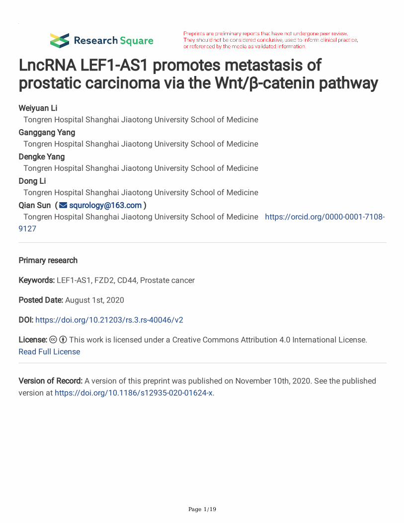
Page 1/19 LncRNA LEF1-AS1 promotes metastasis of prostatic carcinoma via the Wnt/ β -catenin pathway Weiyuan Li Tongren Hospital Shanghai Jiaotong University School of Medicine Ganggang Yang Tongren Hospital Shanghai Jiaotong University School of Medicine Dengke Yang Tongren Hospital Shanghai Jiaotong University School of Medicine Dong Li Tongren Hospital Shanghai Jiaotong University School of Medicine Qian Sun ( [email protected] ) Tongren Hospital Shanghai Jiaotong University School of Medicine https://orcid.org/0000-0001-7108- 9127 Primary research Keywords: LEF1-AS1, FZD2, CD44, Prostate cancer Posted Date: August 1st, 2020 DOI: https://doi.org/10.21203/rs.3.rs-40046/v2 License: This work is licensed under a Creative Commons Attribution 4.0 International License. Read Full License Version of Record: A version of this preprint was published on November 10th, 2020. See the published version at https://doi.org/10.1186/s12935-020-01624-x.
Transcript of prostatic carcinoma via the Wnt/β -catenin pathway LncRNA ...
Page 1/19
LncRNA LEF1-AS1 promotes metastasis ofprostatic carcinoma via the Wnt/β-catenin pathwayWeiyuan Li
Tongren Hospital Shanghai Jiaotong University School of MedicineGanggang Yang
Tongren Hospital Shanghai Jiaotong University School of MedicineDengke Yang
Tongren Hospital Shanghai Jiaotong University School of MedicineDong Li
Tongren Hospital Shanghai Jiaotong University School of MedicineQian Sun ( [email protected] )
Tongren Hospital Shanghai Jiaotong University School of Medicine https://orcid.org/0000-0001-7108-9127
Primary research
Keywords: LEF1-AS1, FZD2, CD44, Prostate cancer
Posted Date: August 1st, 2020
DOI: https://doi.org/10.21203/rs.3.rs-40046/v2
License: This work is licensed under a Creative Commons Attribution 4.0 International License. Read Full License
Version of Record: A version of this preprint was published on November 10th, 2020. See the publishedversion at https://doi.org/10.1186/s12935-020-01624-x.
Page 2/19
AbstractBackground: Long noncoding RNAs (lncRNAs), which are important functional regulators in cancer, haveemerged as critical molecular regulators in various biological processes. However, the mechanisms bywhich LEF1-AS1 modulates Androgen-Independent Prostate Cancer (AIPC) development remain largelyunknown.
Methods: The LEF1-AS1 expression level was detected in tumour tissues and adjacent normal tissues ofAIPC patients by using next-generation sequencing technology and qRT-PCR. Cell proliferation, migrationand invasion were assessed by colony formation, EDU assays and transwell assays, respectively.Xenograft assay was conducted to determine the effect of LEF1-AS1 on cell proliferation in vivo.
Results: LEF1-AS1 promoted the proliferation, migration, invasion and angiogenic ability of AIPC cells invitro and in vivo. In this mechanism, LEF1-AS1 recruited the transcription factor C-myb to the promoterregion of FZD2, which activated FZD2 transcription. Moreover, LEF1-AS1 functioned as a competingendogenous RNA (ceRNA) acting as a sponge for miR-328, which activated CD44.
Conclusion: Collectively, these data indicate that LEF1-AS1 is a tumour promoter in the development ofAIPC and that it may contribute to the improvement of AIPC diagnosis and therapy.
BackgroundProstate cancer (PCa) is the commonly diagnosed malignancy and the second-leading cause of cancer-related mortality in males in Western countries [1]. In its early stage, prostate cancer is androgendependent; however, most prostate cancers can progress to androgen resistant status as time goes on [2].It is well known that AIPC carcinogenesis is a complex biological process involving a variety of genomicvariations and cellular events [3]. Therefore, further research regarding the oncogenic signallingmechanisms in AIPC progression is urgently needed.
Long noncoding RNAs (lncRNAs) are greater than 200 bases in length and play vital roles in multiplecellular functions, including cell proliferation, apoptosis, cellular differentiation, tumourigenesis, andmetastasis [4–5]. Many lncRNAs have been reported to be involved in different types of cancers includingAIPC [6]. For example, Chakravartyet al. demonstrated that the expression of lncRNA NEAT1 wassigni�cantly higher during AIPC progression, and promoted cancer cell proliferation by regulating theepigenetic signatures of target gene promoters [7]. Human lymphoid enhancer-binding factor 1 antisenseRNA 1 (LEF1-AS1), a newly discovered lncRNA located on the plus strand of chromosome 4, waspreviously shown to be upregulated in glioblastoma (GBM) tissues and its dysregulation was postulatedto correlate with poor overall survival in patients [8–9]. In addition, several studies also demonstrated thatLEF1-AS1 promotes cell proliferation and invasion during multiple types of cancer, suggesting that LEF1-AS1 could function as an oncogene and might be a potential therapeutic target in the progress ofmalignancy [10–11]. Nevertheless, LEF1-AS1 has not been reported to be a regulatory gene associated
Page 3/19
with AIPC progression. Hence, it is necessary to explore the detailed function and the underlyingmolecular mechanism of LEF1-AS1 in the regulation of AIPC progression.
The Wnt/β-catenin pathway, one of the vital mechanisms responsible for cell proliferation, cell polarity,migration and cell fate determination during embryonic development and maintaining tissuehomeostasis, has been proven to be associated with the development of several pathologies, includingcancer [12–13]. More and more evidence elucidates that the Wnt/β-catenin pathway participates inregulating many key process during cancer development, including promoting cell proliferation,maintaining cancer stem cells (CSCs) and increasing metastasis [14–15]. In the canonical Wnt/β-cateninsignaling pathway, WNT ligands bind to corresponding frizzled receptors (FZD) and associated co-receptors in order to inhibit the downstream β-catenin complex degradation, resulting in β-cateninaccumulating and nucleus translocating where it activates transcription through T-cell factor/lymphoidenhancer factor (TCF/LEF) [16]. In recent decades, accumulating researches demonstrated Wnt/β-cateninsignaling pathway has been implicated in both normal prostate development and in PCa progression [17].Multiple molecular could activate Wnt/β-Catenin signaling during PCa progression, such as PTEN/Akt,COX-2/PGE2, PDGF, and NF-κB pathways [18–20]. However, little is known regarding the effects oflncRNA LEF1-AS1 on the wnt/β-catenin pathway in AIPC. Therefore, understanding the underlyingmolecular mechanisms of wnt/β-catenin pathway would lead to more effective diagnosis and treatmentin AIPC.
In this study, we report that LEF1-AS1 is upregulated in tumour tissues and that this lncRNA promotes theproliferation, migration, invasion and angiogenic abilities of AIPC cells as well as tumour growth in vivo.Furthermore, mechanistic investigations showed that LEF1-AS1 could initiate the activation of the Wnt/β-catenin pathway by upregulating the expression of Wnt/β-catenin pathway membrane receptor FZD2 andCD44. We speculated that LEF1-AS1 might function as an oncogene and aimed to detect the underlyingmolecular mechanisms in AIPC progression.
Methods And Materials
Patients and samplesAIPC samples were obtained from 45 patients who provided informed consent, which was in accordancewith the ethical standards of the Tongren Hospital (Shanghai, China) Review Board. All samples werereviewed by a pathologist and were con�rmed as AIPC based on a histopathological evaluation. No localor systemic treatments were administered in these patients before surgery.
Cell cultureThe AIPC cell lines PC3 and DU145 were obtained from the American Type Culture Collection (Manassas,VA, USA). The normal prostatic cell line RWPE was purchased from Jennio Biotech Co(Guangzhou,China). PC3 and DU145 cells were cultured in DMEM (Gibco, Grand Island, NY, USA) supplemented with
Page 4/19
10% heat-inactivated foetal bovine serum (FBS) (Gibco). RWPE cells were cultured in RPMI 1640 medium(Gibco) supplemented with 10% FBS. All cells were incubated at 37 °C in a 5% CO2 humidi�ed incubator.
Cell transfectionThe LEF1-AS1 pcDNA3.3 vector and empty vector (pcDNA3.3) were based on the expression vectorpcDNA3.3 (Invitrogen, USA). Small interfering RNA for LEF1-AS1, C-myb, FZD2 or CD44 (siLEF1-AS1, siC-myb) and scramble siRNA (si NC) were purchased or synthetized from RiboBio (Guangzhou, China). Thecells were seeded into 6-well plates, and transfection was performed using Lipofectamine 2000(Invitrogen, Carlsbad, CA, USA) according to the manufacturer’s instructions.
RNA isolation and RT-PCR analysesTotal RNA was extracted from AIPC samples and cells using TRIzol reagent (Invitrogen), and cDNA wassynthesised using the QuantiTect Reverse Transcription Kit (Qiagen, Valencia, CA, USA) according to themanufacturer’s protocol. qRT-PCR was then performed based on the instructions for SYBR Premix Ex Taq(Takara, Tokyo, Japan), and the expression levels were normalized to the level of GAPDH. Eachexperiment was repeated at least three times.
Chromatin immunoprecipitation (ChIP)To verify the potential transcription factors binding to the LEF1-AS1 promoter, a ChIP assay wasperformed using the EZ-Magna ChIP kit (Millipore, Shanghai, China) according to the manufacturer’sprotocol. In brief, cells were �xed with 4% paraformaldehyde and incubated with glycine for 10 min togenerate DNA–protein cross-links. Then, the cells were lysed with Cell Lysis Buffer and Nuclear LysisBuffer and sonicated to generate chromatin fragments of 400–800 bp. The chromatin fragments wereimmunoprecipitated with an anti-C-myb antibody (Cell Signaling Technology, Danvers, MA, USA) orcontrol IgG. The resulting DNA was puri�ed for PCR analyses.
RNA binding protein immunoprecipitation (RIP) assayRIP was performed according to the protocol for the EZ-Magna RIP kit (EMD Millipore, Billerica, MA, USA).Brie�y, DU145 cells grown to 80–90% con�uency were lysed in complete RIP lysis buffer, and 90% of 100µL of whole cell extract was incubated with RIP buffer containing magnetic beads conjugated with 5 µghuman anti-C-myb antibody (Abcam) or immunoglobulin G (IgG) control. Incubation with proteinase Kwith shaking was performed to digest the protein, and RNA was isolated by immunoprecipitation. Finally,the immunoprecipitated RNA was puri�ed and analysed by qRT-PCR.
Immunoblotting analysisCells (5 × 10⁶) were lysed for 20 min with lysis buffer (Beyotime Biotechnology) containing proteaseinhibitors (Roche, Indianapolis, IN, USA). Equal amounts of samples were separated by 10% sodiumdodecyl sulfate polyacrylamide gel electrophoresis and transferred to polyvinylidene �uoride membranes.The membranes were blocked with 5% (wt/vol) skimmed milk in TBS plus Tween 20 at 4 °C overnight
Page 5/19
before probing with antibodies. Membrane proteins were detected by an HRP-conjugated secondaryantibody (1:1000, Santa Cruz Biotech, Santa Cruz, CA).
Colony formation assayAIPC cells were collected, and 1000 cells were seeded in each well of a 6-well plate. After cell culture for15 days, PC3 or DU145 cells were washed with PBS and �xed in methanol for 20 min. After washing threetimes with PBS, colonies were stained with 0.1% crystal violet for 20 min. Colonies were captured using alight microscope (Olympus, Tokyo, Japan). Clusters containing ≥ 30 cells were counted as a singlecolony.
EDU assaysThe cells were cultured in 96-well plates at a density of 2 × 10³ cells/well. Then, the EDU (5′-ethynyl-2′-deoxyuridine) incorporation assay was performed to evaluate cell proliferation using a KeyFluor488 Click-iT EDU kit (KeyGENBioTECH, Nanjing, China) according to the manufacturer's instructions. Thepercentage of Edu-positive cells was calculated after �uorescence microscopy analysis.
Cell migration and invasion assaysThe cells were harvested, resuspended in serum-free media and placed into the upper chamber of aTranswell membrane �lter (Corning, NY, USA) for the migration assays or in the upper chamber of aTranswell membrane �lter coated with Matrigel (Corning) for the invasion assays. After 24 h ofincubation, cells on the upper side were removed with a cotton swab. Evaluation of invasive capacity wasperformed by counting invading cells under a microscope (40 × 10). Five random �elds of view wereanalysed for each chamber.
Immunohistochemical analysisAIPC and adjacent tissues were �xed in 4% paraformaldehyde for para�n embedding. Tissue specimenswere cut into slices, and tissue slices were depara�nized, rehydrated, and immersed in 3% hydrogenperoxide for 10 min to quench endogenous peroxidase activity. Cancer tissues were immunostained forFZD2 and CD44 (Abcam, San Francisco, CA, USA; 1:100 dilution). The signal was ampli�ed andvisualized with diaminobenzidine chromogen, followed by counterstaining with haematoxylin.
Tumourigenicity assays in nude miceWe used male nude mice (6 weeks of age) obtained from the Animal Facility of Shanghai Jiao TongUniversity School of Medicine; the mice were fed sterilized food and water. All of the animal experimentswere approved by the responsible governmental animal ethics committee and complied with the ARRIVEguidelines. Thirty-day-old male nude mice were subcutaneously injected with 1 × 10⁶ AIPC cells in 200 µLserum-free medium into the right �ank. Once palpable tumours were observed (after approximately 4weeks), the mice were sacri�ced. The tumours were isolated and weighed.
Statistical analysis
Page 6/19
The results are presented as the mean ± SD from three independent experiments performed in triplicate.The P values were calculated by using Student t test or one-way ANOVA. A P value of < 0.05 wasconsidered to indicate a statistically signi�cant result. Statistical analyses were performed usingGraphPad Prism 6.0 statistical software (GraphPad Software Inc., La Jolla, California).
Results
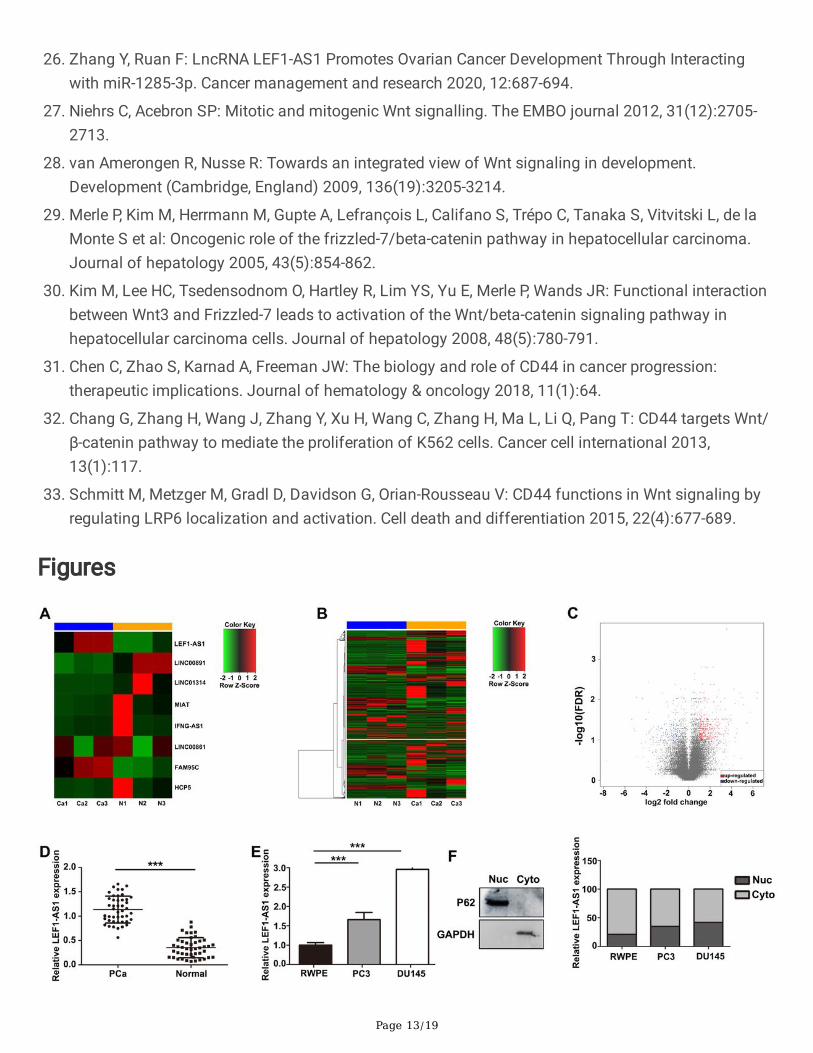
LEF1-AS1 expression is upregulated in AIPC tissues andcell linesTo identify the lncRNA pro�le and to investigate the role of lncRNAs in the development of AIPC, weperformed next-generation sequencing and compared lncRNA expression in AIPC patient samples withthat in normal samples. We identi�ed a series of abnormally expressed lncRNAs in AIPC and found thatLEF1-AS1 was signi�cantly increased in AIPC tissues (Fig. 1A). In terms of mRNAs, a total of 719 mRNAs(log2FC > 2, FDR < 0.1) were signi�cantly differentially expressed (Fig. 1B-1C). To validate the �ndingsfrom RNA sequencing, we analysed the expression of LEF1-AS1 in AIPC and adjacent tissues by qRT-PCR.The results con�rmed the accuracy of the next-generation sequencing �ndings (Fig. 1D). Furthermore, theexpression of LEF1-AS1 was increased in DU145 cells compared with PC3 and RWPE cells (Fig. 1E).These results suggested that LEF1-AS1 might function as an oncogene during AIPC progression. Theresult of subcellular fractionation showed that the LEF1-AS1 transcript was observed in the cytoplasm ofAIPC cells, whereas minimal signal was observed in the nucleus (Fig. 1F), suggesting that LEF-AS1 mightregulate gene expression at the transcriptional level.
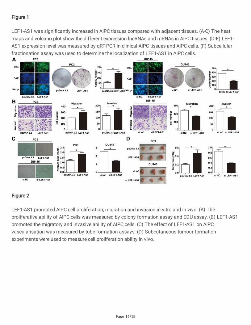
LEF1-AS1 promoted AIPC cell proliferation, migration andinvasion in vitro and in vivoWe investigated the role of LEF1-AS1 in AIPC cell proliferation using a colony formation assay and anEdu assay. The results suggested that the overexpression of LEF1-AS1 could promote the proliferation ofPC3 cells, while knockdown of LEF1-AS1 could inhibit the proliferation of DU145 cells (Fig. 3A). Next, weevaluated AIPC cell migration and invasion using Transwell-based assays. As shown in Fig. 3B, cellmigration and invasion were up-regulated in the presence of LEF1-AS1, while the opposite result wasobserved when LEF1-AS1 was inhibited. It is known that new blood vessels are essential to promotetumour development. Therefore, we utilized the endothelial tube formation assay to assess the ability ofLEF1-AS1 to affect the development of biological systems in AIPC cells. The results demonstrated thatthe neovascularization rate was especially increased after transfection with LEF1-AS1 overexpressionplasmid and decreased after transfection with siLEF1-AS1 compared with the corresponding negativecontrols (Fig. 3C). To further determine the role of LEF1-AS1 during AIPC progression in vivo, weconducted a subcutaneous tumour formation experiment in nude mice. After 4 weeks, tumour weight andvolume were dramatically increased in the LEF1-AS1 overexpression group. In contrast, tumour growth,indicated by weight and volume, was signi�cantly decreased in the siLEF1-AS1 group (Fig. 3D).
Page 7/19
Collectively, these data strongly demonstrated that LEF1-AS1 aggravated cell migration, invasion, andvasculogenic mimicry and suppressed cell proliferation in vivo during AIPC progression.
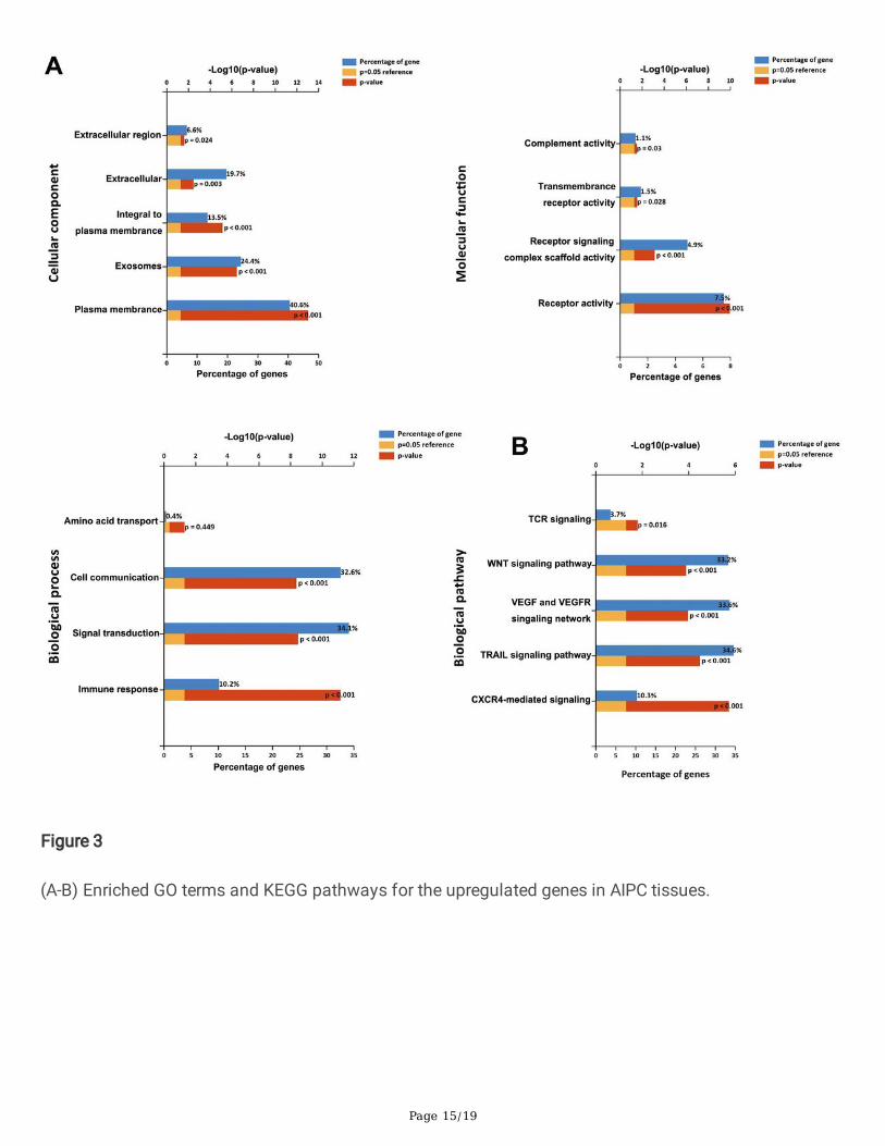
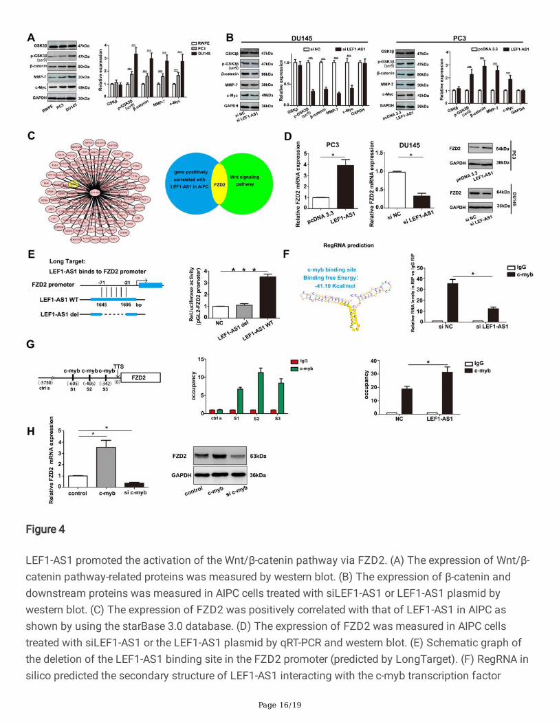
LEF1-AS1 initiated the activation of the Wnt/β-cateninpathway via frizzled class receptor 2 (FZD2)Gene Ontology (GO) analysis and KEGG pathway enrichment analysis showed that the Wnt/β-cateninsignalling pathway was one of the most enriched pathways within the set of upregulated mRNAs,suggesting that it may be involved in the pathogenesis and development of AIPC (Fig. 3A-4B). Toelucidate the functions of the molecular mechanisms induced by LEF1-AS1, the activity of the Wnt/β-catenin signalling pathway was detected in the AIPC cell lines. Compared with that of the normal cell lineRWPE, the activity of the Wnt/β-catenin signalling pathway was signi�cantly higher in PC3 and DU145cells (Fig. 4A). Next, we examined the levels of GSK3β, p-GSK3β, β-catenin, MMP-7 and c-myc protein inAIPC cells after LEF1-AS1 plasmid or siLEF1-AS1 transfection. The results showed that signi�cantlyhigher expression levels of p-GSK3β(ser9), β-catenin, MMP-7 and c-myc were observed in PC3 cellstransfected with LEF1-AS1 overexpression plasmid. Correspondingly, in the DU145 cell lines, theexpression level of Wnt/β-catenin pathway proteins was decreased in cells transfected with the siLEF1-AS1 (Fig. 4B). We next evaluated how LEF1-AS1 modulated the Wnt/β-catenin signalling pathway in AIPCprogression. Using the starBase 3.0 database, we found that multiple genes positively correlated withLEF1-AS1 in AIPC, including frizzled class receptor 2 (Fig. 4C). Moreover, as shown in Fig. 4D, asigni�cantly higher expression level of FZD2 was observed in cells transfected with the LEF1-AS1plasmid. To further dissect the molecular mechanism of FZD2 regulation by LEF1-AS1, we analysed thesequences of LEF1-AS1 and the FZD2 promoter (+ 500 to -2000 bp) using the LongTarget tool. A LEF1-AS1-FZD2 binding pattern was predicted as shown in Fig. 4E. Luciferase assays demonstrated that LEF1-AS1 with a deletion of this binding region (LEF1-AS1 del) failed to bind the FZD2 promoter as LEF1-AS1wt did. To explore how LEF1-AS1 regulates the expression of FZD2, we proposed that LEF1-AS1 mightaffect FZD2 transcription via alteration of transcription factors. We predicted the interaction of LEF1-AS1and C-myb by using an in silico analysis of regulatory RNA elements. RIP analysis further demonstratedthat LEF1-AS1 RNA could be pulled down by anti-C-myb antibody in AIPC cells, indicating the physicalbinding of LEF1-AS1 with C-myb (Fig. 4F). Finally, a ChIP assay con�rmed the occupancy of C-myb in theFZD2 promoter. AIPC cells were subjected to ChIP with anti-C-myb antibody, followed by RT-PCRmeasurements with speci�c probes targeting the FZD2 promoter or -5 kb upstream (control). The resultsindicated that the occupancy of C-myb in the FZD2 promoter region was signi�cantly increasedcompared to that of the − 5 kb upstream/control (Fig. 4G). The corresponding western blot assays withanti-FZD2 antibody showed similar FZD2 attenuation upon C-myb interference (Fig. 4H). Taken together,these data suggest that LEF1-AS1 enhances FZD2 transcription via recruitment of C-myb to the FZD2promoter region.
LEF1-AS1 increased CD44 via function through the ceRNAsponging pattern of miR-328
Page 8/19
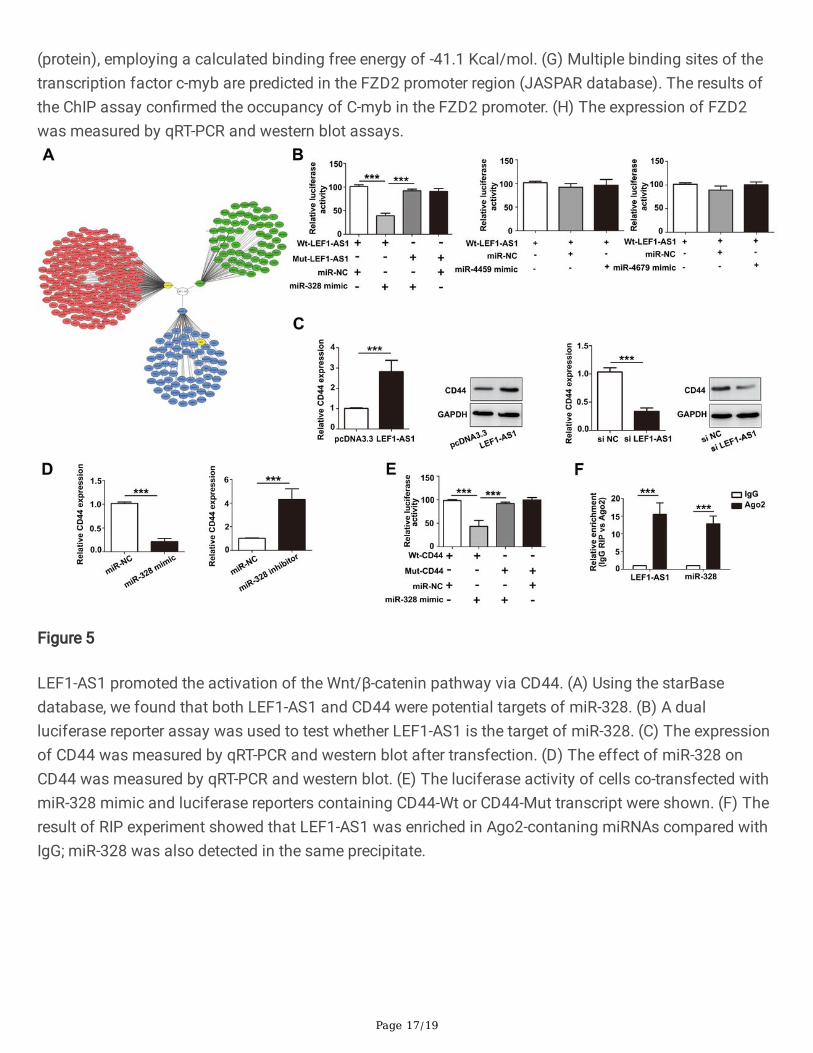
To further study the mechanism of LEF1-AS1 during Wnt/β-catenin pathway activation, we used theRegRNA database and found that LEF1-AS1 was a potential targets of 3 microRNAs (Fig. 5A). Next, wecon�rmed binding interactions between miR-328 with LEF1-AS1 by demonstrating a reduction inluciferase activity (Fig. 5B). Furthermore, multiple mRNAs were predicted by TargetScan and PITA; theregulatory networks of LEF1-AS1/miR-328 axis are shown in Fig. 5A. Among them, the CD44 gene waspredicted as a downstream target gene of the LEF1-AS1/miR-328 axis. Interestingly, an early studyreported that miR-328 promotes cell proliferation by targeting CD44 in MCF-7 cells. Therefore, wedetected whether or not LEF1-AS1 regulates CD44 expression via sponging miR-328. As shown in Fig. 5C-5D, CD44 expression was signi�cantly increased by LEF1-AS1 or miR-328 inhibitor transfection. Incontrast, CD44 expression was suppressed by LEF1-AS1 siRNAs or miR-328 mimic. In addition, a dual-luciferase reporter assay was used to test whether CD44 is a direct target of miR-328. The results showedthat luciferase activity was reduced in AIPC cells that were co-transfected with miR-328 and CD44-WT butwas not reduced in cells containing CD44-Mut (Fig. 5E). The ceRNA binding pattern between LEF1-AS1/CD44 and miR-328 was further validated by RNA immunoprecipitation. As shown in Fig. 5F, RNAwas signi�cantly more enriched in the Ago2-IP fractions of AIPC cells compared with the IgG‐IP group (P < 0.001), indicating a direct targeted relationship between LEF1-AS1 and miR‐328. Therefore, inagreement with recent �ndings, the transcriptional increase in LEF1-AS1 expression seemed to be animportant mechanism contributing to the activation of the Wnt/β-catenin pathway in AIPC cells byupregulating CD44.
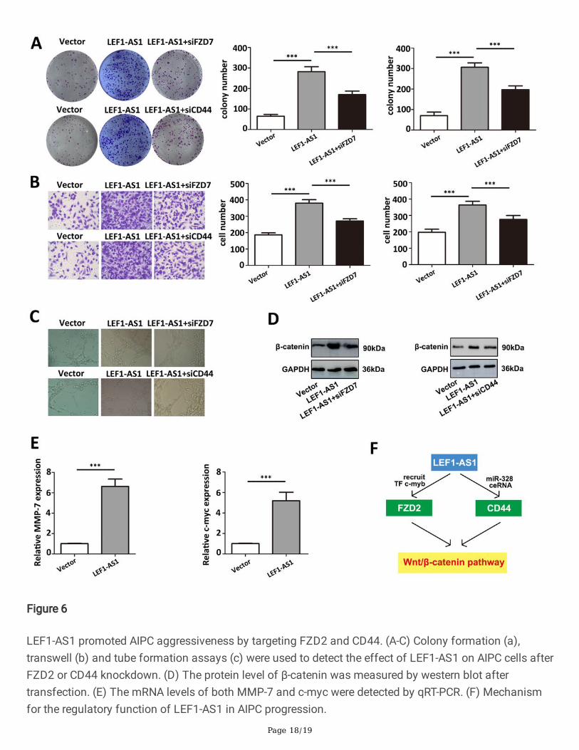
LEF1-AS1 aggravated cell proliferation and migration partlydependent on its regulation on FZD2 and CD44 in humanprostate cancer cellsAfter con�rming that LEF1-AS1 regulates FZD2 and CD44 in AIPC cells, it is necessary to furtherdetermine whether the oncogenic functions of LEF1-AS1 is dependent on its modulation of FZD2/CD44.Colony formation, transwell assays and tube formation assays were carried out to validate thatknockdown of FZD2 or CD44 that could partly inhibit the tumor promoting effect caused by LEF1-AS1overexpression. The results demonstrated that co-transfected with LEF1-AS1 and siFZD2/siCD44 partlyreversed the oncogenic roles caused by LEF1-AS1 overexpression (Fig. 6A-6C). In addition, the expressionof β-catenin and downstream targets (MMP-7 and c-myc) was also decreased after siFZD2/siCD44transfection in LEF1-AS1 overexpression cells compared with control group (Fig. 6D-E). Taken together, asshown in Fig. 6F, our �ndings demonstrated that LEF1-AS1 could enhance FZD2 transcription viarecruitment of c-myb to its promoter region and increase CD44 mRNA levels via miR-328 sponge,resulting in the activation of Wnt/β-catenin pathway.
Discussion
Page 9/19
Prostate carcinoma is a common male malignancy and a major cause of cancer-associated mortalityworldwide [21]. In recent decades, knowledge regarding the molecular changes that underlie PCa hasincreased, which has greatly increased interest in the discovery of new molecular markers for diagnosisand treatment [22]. In the present study, our results clari�ed that LEF1-AS1 may serve a role as anoncogene in AIPC by aggravating cancer cell proliferation, migration and vascularization. The underlyingmechanism of LEF1-AS1 in AIPC might be through activating Wnt/β-catenin signalling.
Increasing efforts have contributed to elucidating the molecular and cellular mechanisms underlying theprogression of cancer, among which lncRNAs have attracted increasing attention [23]. Recently, anincreasing number of studies have elucidated that long non-coding RNA LEF1-AS1 plays critical roles incellular proliferation, apoptosis, differentiation, and invasion during the progression of multiple types ofcancer [24]. Zhang et al. reported that LEF1-AS1 knockdown inhibited cell survival, proliferation andmigration, whereas enhanced cell apoptosis and induced G0/G1 cell cycle arrest in oral squamous cellcarcinoma, hinting a cancerigenic role of LEF1-AS1 in tumour progression [25]. Furthermore, LEF1-AS1also has been proven to be positively correlated with lymph node metastasis and advanced stage inovarian cancer and Enhanced expression of LEF1-AS1 may predict a poor prognosis [26]. Nevertheless,the role of LEF1-AS1 and the underlying mechanism in AIPC progression remain unclear. In the presentstudy, we performed next-generation sequencing to investigate lncRNA expression pro�les and identi�edLEF1-AS1 as a signi�cantly upregulated lncRNA in advanced AIPC. Afterwards, we detected the LEF1-AS1expression level in AIPC by using RT-qPCR. The results showed that LEF1-AS1 was signi�cantly increasedduring AIPC progression, which was consistent with the sequencing results. Moreover, our results showedthat LEF1-AS1 promoted the proliferation, migration, invasion and vasculogenic mimicry of AIPC cellsboth in vitro and in vivo. These data suggest that LEF1-AS1 acts as an oncogene during AIPCprogression.
Recent studies have demonstrated that Wnt/β-catenin and the downstream TCF/LEF complex, as keyregulators of mitogenesis and tumourigenicity, participate in regulating cell transformation, cell growthand cell cycle progression [27–28]. In view of the frequent activation of Wnt signaling pathway in tumourprogression, FZDs have an apparent role in modulating this pathway as they serve as upstreamregulators of the Wnt signaling pathway. In general, up-regulation of FZDs lead to the activation of Wnt/β-catenin pathway and, to a lesser extent, the non-canonical pathway [29–30]. In the present study, wecon�rmed that LEF1-AS1 acts in both the cytoplasm and nucleus. In the nucleus, LEF1-AS1 binds to thepromoter region of FZD2. Interestingly, LEF1-AS1 also recruits the transcription factor C-myb in thisregion. Our results con�rmed that LEF1-AS1 enhanced FZD2 transcription via recruitment of C-myb to itspromoter region.
In the cytoplasm, LEF1-AS1 may mainly serve as a sponge for regulatory miRNAs. For example, aprevious study reported that lncRNA LEF1-AS1 functions as a ceRNA by regulating the expression of miR-544a in lung cancer [11]. In this study, we demonstrated that LEF1-AS1 functions as a ceRNA bysponging miR-328, which leads to CD44 activation. CD44, as a multifunctional transmembrane adhesionglycoprotein, plays an important role in signal transduction. Recent evidence shows that the expression
Page 10/19
of CD44 is mainly regulated by speci�c signaling networks, transcriptional factors, and epigeneticmechanisms [31]. Moreover, CD44 was shown to target the Wnt/β-catenin signaling pathway in tumourprogression [32–33]. In this study, for the �rst time, we demonstrated that LEF1-AS1 acted as a powerfulregulator in AIPC and regulated the activation of Wnt/β-catenin and the downstream pathway byfunctioning as a ceRNA to increase the expression of CD44.
ConclusionIn summary, as shown in Fig. 6F, we determined that LEF1-AS1 was signi�cantly increased during AIPCprogression. Furthermore, a mechanistic study showed that LEF1-AS1 functioned as a ceRNA and servedas a regulator in the activation of the Wnt/β-catenin pathway via FZD2 and CD44. Our results providenew insight into the mechanism that links the function of LEF1-AS1 with AIPC and suggest that LEF1-AS1 may serve as a novel potential target for the improvement of AIPC therapy.
Declarations
AcknowledgementsNot applicable.
Authors’ contributionsWeiyuan Li and Ganggang Yang designed the research and wrote the manuscript; Weiyuan Li and DengkeYang performed experiments and analyzed data; Dong Li collected and analyzed data; Qian Sun revisedthe manuscript and approved the �nal submission. All authors discussed the results and reviewed themanuscript.
FundingThis work was supported by grants from Science and Technology Foundation of Shanghai(CNKW2018Y05).
Availability of data and materialsThe analyzed data sets generated during the present study are available from the corresponding authoron reasonable request.
Ethics approval and consent to participate
Page 11/19
The present study was approved by the ethical review committee of the Tongren Hospital of Shanghaijiaotong University.
Patient consent for publicationNot applicable.
Competing interestsThe authors declare that they have no competing interests.
References1. Siegel R, Naishadham D, Jemal A: Cancer statistics, 2013. CA: a cancer journal for clinicians 2013,
63(1):11-30.
2. Heidenreich A, Aus G, Bolla M, Joniau S, Matveev VB, Schmid HP, Zattoni F: EAU guidelines onprostate cancer. European urology 2008, 53(1):68-80.
3. Klein EA, Silverman R: In�ammation, infection, and prostate cancer. Current opinion in urology 2008,18(3):315-319.
4. Ponting CP, Oliver PL, Reik W: Evolution and functions of long noncoding RNAs. Cell 2009,136(4):629-641.
5. Seton-Rogers S: Non-coding RNAs: The cancer X factor. Nature reviews Cancer 2013, 13(4):224-225.
�. Schmitt AM, Chang HY: Long Noncoding RNAs in Cancer Pathways. Cancer cell 2016, 29(4):452-463.
7. Lin D: Commentary on "The oestrogen receptor alpha-regulated lncRNA NEAT1 is a critical modulatorof prostate cancer." Chakravarty D, Sboner A, Nair SS, Giannopoulou E, Li R, Hennig S, Mosquera JM,Pauwels J, Park K, Kossai M, MacDonald TY, Fontugne J, Erho N, Vergara IA, Ghadessi M, DavicioniE, Jenkins RB, Palanisamy N, Chen Z, Nakagawa S, Hirose T, Bander NH, Beltran H, Fox AH, ElementoO, Rubin MA, University of Washington-Urology, Seattle, WA. Nat Commun 2014; 5:5383. Urologiconcology 2016, 34(11):522.
�. Fagerberg L, Hallström BM, Oksvold P, Kampf C, Djureinovic D, Odeberg J, Habuka M, TahmasebpoorS, Danielsson A, Edlund K et al: Analysis of the human tissue-speci�c expression by genome-wideintegration of transcriptomics and antibody-based proteomics. Molecular & cellular proteomics :MCP 2014, 13(2):397-406.
9. Wang J, Liu X, Yan C, Liu J, Wang S, Hong Y, Gu A, Zhao P: LEF1-AS1, a long-noncoding RNA,promotes malignancy in glioblastoma. OncoTargets and therapy 2017, 10:4251-4260.
10. Sun T, Liu Z, Zhang R, Ma S, Lin T, Li Y, Yang S, Zhang W, Wang Y: Long Non-Coding RNA LEF1-AS1Promotes Migration, Invasion and Metastasis of Colon Cancer Cells Through miR-30-5p/SOX9 Axis.OncoTargets and therapy 2020, 13:2957-2972.
Page 12/19
11. Wang A, Zhao C, Gao Y: LEF1-AS1 contributes to proliferation and invasion through regulating miR-544a/ FOXP1 axis in lung cancer. 2019, 37(6):1127-1134.
12. Nusse R, Clevers H: Wnt/β-Catenin Signaling, Disease, and Emerging Therapeutic Modalities. Cell2017, 169(6):985-999.
13. Polakis P: The many ways of Wnt in cancer. Current opinion in genetics & development 2007,17(1):45-51.
14. Arend RC, Londoño-Joshi AI, Straughn JM, Jr., Buchsbaum DJ: The Wnt/β-catenin pathway inovarian cancer: a review. Gynecologic oncology 2013, 131(3):772-779.
15. Clevers H, Nusse R: Wnt/β-catenin signaling and disease. Cell 2012, 149(6):1192-1205.
1�. MacDonald BT, Tamai K, He X: Wnt/beta-catenin signaling: components, mechanisms, and diseases.Developmental cell 2009, 17(1):9-26.
17. Yu X, Wang Y, Jiang M, Bierie B, Roy-Burman P, Shen MM, Taketo MM, Wills M, Matusik RJ: Activationof beta-Catenin in mouse prostate causes HGPIN and continuous prostate growth after castration.The Prostate 2009, 69(3):249-262.
1�. Castellone MD, Teramoto H, Williams BO, Druey KM, Gutkind JS: Prostaglandin E2 promotes coloncancer cell growth through a Gs-axin-beta-catenin signaling axis. Science (New York, NY) 2005,310(5753):1504-1510.
19. Lamberti C, Lin KM, Yamamoto Y, Verma U, Verma IM, Byers S, Gaynor RB: Regulation of beta-cateninfunction by the IkappaB kinases. The Journal of biological chemistry 2001, 276(45):42276-42286.
20. Persad S, Troussard AA, McPhee TR, Mulholland DJ, Dedhar S: Tumor suppressor PTEN inhibitsnuclear accumulation of beta-catenin and T cell/lymphoid enhancer factor 1-mediatedtranscriptional activation. The Journal of cell biology 2001, 153(6):1161-1174.
21. Scher HI, Solo K, Valant J, Todd MB, Mehra M: Prevalence of Prostate Cancer Clinical States andMortality in the United States: Estimates Using a Dynamic Progression Model. PloS one 2015,10(10):e0139440.
22. Ferlay J, Soerjomataram I, Dikshit R, Eser S, Mathers C, Rebelo M, Parkin DM, Forman D, Bray F:Cancer incidence and mortality worldwide: sources, methods and major patterns in GLOBOCAN2012. International journal of cancer 2015, 136(5):E359-386.
23. de Oliveira JC, Oliveira LC, Mathias C, Pedroso GA, Lemos DS, Salviano-Silva A, Jucoski TS, Lobo-Alves SC, Zambalde EP, Cipolla GA et al: Long non-coding RNAs in cancer: Another layer ofcomplexity. 2019, 21(1):e3065.
24. Xiang C, Zhang Y, Zhang Y, Liu C, Hou Y, Zhang Y: lncRNA LEF1-AS1 Promotes Proliferation andInduces Apoptosis of Non-Small-Cell Lung Cancer Cells by Regulating miR-221/PTEN Signaling.Cancer management and research 2020, 12:3845-3850.
25. Zhang C, Bao C, Zhang X, Lin X, Pan D, Chen Y: Knockdown of lncRNA LEF1-AS1 inhibited theprogression of oral squamous cell carcinoma (OSCC) via Hippo signaling pathway. Cancer biology &therapy 2019, 20(9):1213-1222.
Page 13/19
2�. Zhang Y, Ruan F: LncRNA LEF1-AS1 Promotes Ovarian Cancer Development Through Interactingwith miR-1285-3p. Cancer management and research 2020, 12:687-694.
27. Niehrs C, Acebron SP: Mitotic and mitogenic Wnt signalling. The EMBO journal 2012, 31(12):2705-2713.
2�. van Amerongen R, Nusse R: Towards an integrated view of Wnt signaling in development.Development (Cambridge, England) 2009, 136(19):3205-3214.
29. Merle P, Kim M, Herrmann M, Gupte A, Lefrançois L, Califano S, Trépo C, Tanaka S, Vitvitski L, de laMonte S et al: Oncogenic role of the frizzled-7/beta-catenin pathway in hepatocellular carcinoma.Journal of hepatology 2005, 43(5):854-862.
30. Kim M, Lee HC, Tsedensodnom O, Hartley R, Lim YS, Yu E, Merle P, Wands JR: Functional interactionbetween Wnt3 and Frizzled-7 leads to activation of the Wnt/beta-catenin signaling pathway inhepatocellular carcinoma cells. Journal of hepatology 2008, 48(5):780-791.
31. Chen C, Zhao S, Karnad A, Freeman JW: The biology and role of CD44 in cancer progression:therapeutic implications. Journal of hematology & oncology 2018, 11(1):64.
32. Chang G, Zhang H, Wang J, Zhang Y, Xu H, Wang C, Zhang H, Ma L, Li Q, Pang T: CD44 targets Wnt/β-catenin pathway to mediate the proliferation of K562 cells. Cancer cell international 2013,13(1):117.
33. Schmitt M, Metzger M, Gradl D, Davidson G, Orian-Rousseau V: CD44 functions in Wnt signaling byregulating LRP6 localization and activation. Cell death and differentiation 2015, 22(4):677-689.
Figures
Page 14/19
Figure 1
LEF1-AS1 was signi�cantly increased in AIPC tissues compared with adjacent tissues. (A-C) The heatmaps and volcano plot show the different expression lncRNAs and mRNAs in AIPC tissues. (D-E) LEF1-AS1 expression level was measured by qRT-PCR in clinical AIPC tissues and AIPC cells. (F) Subcellularfractionation assay was used to determine the localization of LEF1-AS1 in AIPC cells.
Figure 2
LEF1-AS1 promoted AIPC cell proliferation, migration and invasion in vitro and in vivo. (A) Theproliferative ability of AIPC cells was measured by colony formation assay and EDU assay. (B) LEF1-AS1promoted the migratory and invasive ability of AIPC cells. (C) The effect of LEF1-AS1 on AIPCvascularisation was measured by tube formation assays. (D) Subcutaneous tumour formationexperiments were used to measure cell proliferation ability in vivo.
Page 15/19
Figure 3
(A-B) Enriched GO terms and KEGG pathways for the upregulated genes in AIPC tissues.
Page 16/19
Figure 4
LEF1-AS1 promoted the activation of the Wnt/β-catenin pathway via FZD2. (A) The expression of Wnt/β-catenin pathway-related proteins was measured by western blot. (B) The expression of β-catenin anddownstream proteins was measured in AIPC cells treated with siLEF1-AS1 or LEF1-AS1 plasmid bywestern blot. (C) The expression of FZD2 was positively correlated with that of LEF1-AS1 in AIPC asshown by using the starBase 3.0 database. (D) The expression of FZD2 was measured in AIPC cellstreated with siLEF1-AS1 or the LEF1-AS1 plasmid by qRT-PCR and western blot. (E) Schematic graph ofthe deletion of the LEF1-AS1 binding site in the FZD2 promoter (predicted by LongTarget). (F) RegRNA insilico predicted the secondary structure of LEF1-AS1 interacting with the c-myb transcription factor
Page 17/19
(protein), employing a calculated binding free energy of -41.1 Kcal/mol. (G) Multiple binding sites of thetranscription factor c-myb are predicted in the FZD2 promoter region (JASPAR database). The results ofthe ChIP assay con�rmed the occupancy of C-myb in the FZD2 promoter. (H) The expression of FZD2was measured by qRT-PCR and western blot assays.
Figure 5
LEF1-AS1 promoted the activation of the Wnt/β-catenin pathway via CD44. (A) Using the starBasedatabase, we found that both LEF1-AS1 and CD44 were potential targets of miR-328. (B) A dualluciferase reporter assay was used to test whether LEF1-AS1 is the target of miR-328. (C) The expressionof CD44 was measured by qRT-PCR and western blot after transfection. (D) The effect of miR-328 onCD44 was measured by qRT-PCR and western blot. (E) The luciferase activity of cells co-transfected withmiR-328 mimic and luciferase reporters containing CD44-Wt or CD44-Mut transcript were shown. (F) Theresult of RIP experiment showed that LEF1-AS1 was enriched in Ago2-contaning miRNAs compared withIgG; miR-328 was also detected in the same precipitate.
Page 18/19
Figure 6
LEF1-AS1 promoted AIPC aggressiveness by targeting FZD2 and CD44. (A-C) Colony formation (a),transwell (b) and tube formation assays (c) were used to detect the effect of LEF1-AS1 on AIPC cells afterFZD2 or CD44 knockdown. (D) The protein level of β-catenin was measured by western blot aftertransfection. (E) The mRNA levels of both MMP-7 and c-myc were detected by qRT-PCR. (F) Mechanismfor the regulatory function of LEF1-AS1 in AIPC progression.

Page 19/19
Supplementary Files
This is a list of supplementary �les associated with this preprint. Click to download.
Highlight.docx