Dr. M. A. SOFI MD; FRCP (London); FRCPEdin; FRCSEdin.
18
THALASSEMIA Dr. M. A. SOFI MD; FRCP (London); FRCPEdin; FRCSEdin
-
Upload
naomi-foster -
Category
Documents
-
view
222 -
download
5
Transcript of Dr. M. A. SOFI MD; FRCP (London); FRCPEdin; FRCSEdin.
THALASSEMIADr. M. A. SOFI
MD; FRCP (London); FRCPEdin; FRCSEdin
Thalassemia
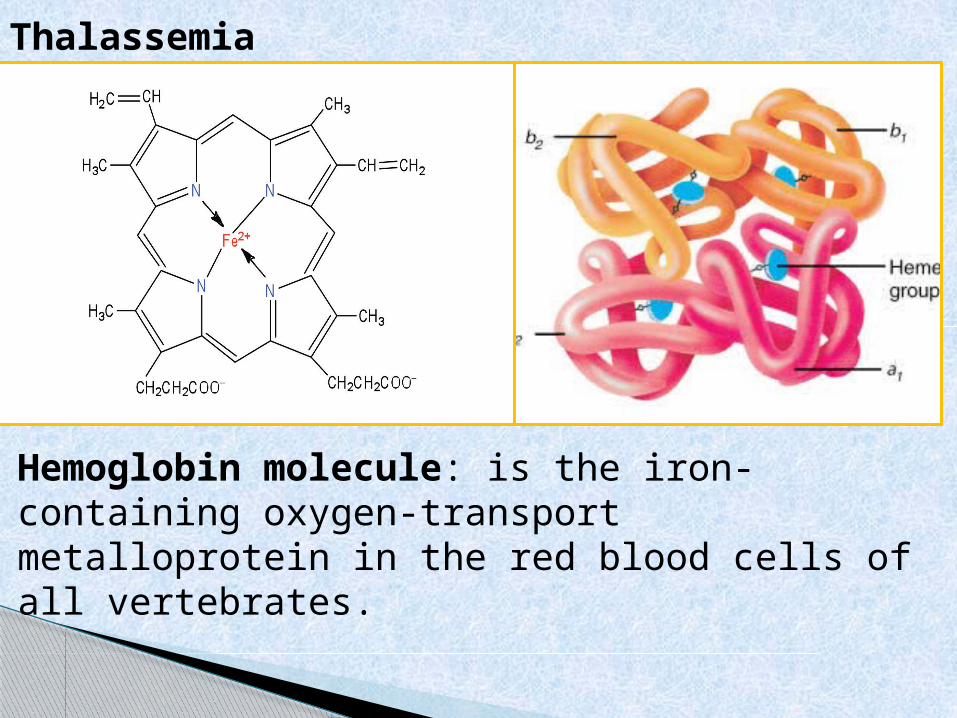
Hemoglobin molecule: is the iron-containing oxygen-transport metalloprotein in the red blood cells of all vertebrates.
The normal haemoglobin molecule has a haem base surrounded by two pairs of globin chains.
The types of globin are called alpha (α), beta (β), gamma (γ) and delta (δ).
Most types of hemoglobin have two α chains and two other identical types.
HbA, the most common form of adult haemoglobin, has two α and two β chains.
Fetal haemoglobin (HbF) has two α and two γ components (this is the predominant type of Hb before birth).
HbA2 is present in smaller amounts, with two α and two δ chains.
THALASSEMIA
The thalassaemias are a group of recessively autosomal inherited conditions characterized by decreased or absence of synthesis of one of the two polypeptide chains (α or β) that form the normal adult human haemoglobin molecule (HbA, α2/β2).
β-globin gene defects may give rise to β thalassemia, while mutations of the α globin gene may cause α thalassemia.
Over 300 mutations giving rise to thalassemia have been identified and its clinical severity varies enormously.
Thalassemia major, intermediate and minor refer largely to disease severity.
THALASSEMIA
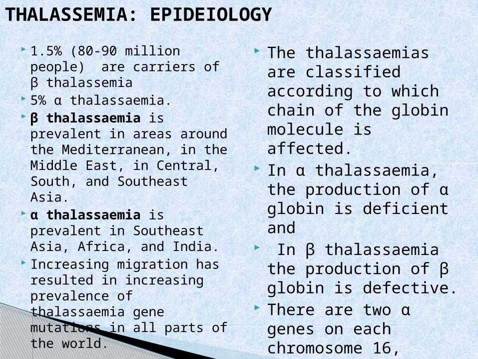
1.5% (80-90 million people) are carriers of β thalassemia
5% α thalassaemia. β thalassaemia is
prevalent in areas around the Mediterranean, in the Middle East, in Central, South, and Southeast Asia.
α thalassaemia is prevalent in Southeast Asia, Africa, and India.
Increasing migration has resulted in increasing prevalence of thalassaemia gene mutations in all parts of the world.
The thalassaemias are classified according to which chain of the globin molecule is affected.
In α thalassaemia, the production of α globin is deficient and
In β thalassaemia the production of β globin is defective.
There are two α genes on each chromosome 16, giving α thalassaemia the unique feature of gene duplication.
There is only one β-globin gene on chromosome
THALASSEMIA: EPIDEIOLOGY
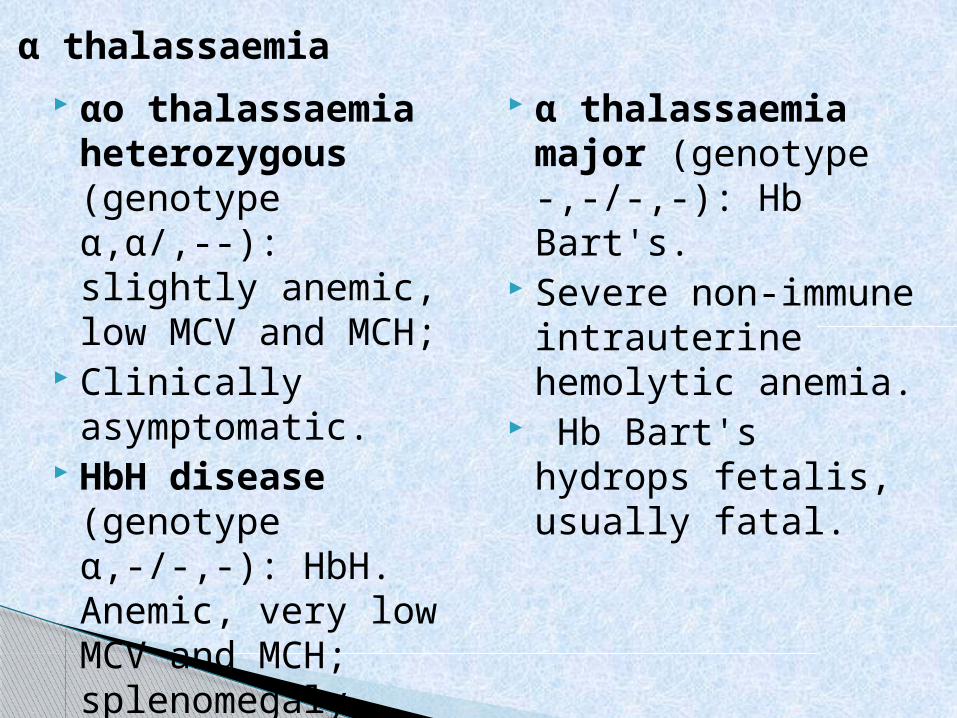
αo thalassaemia heterozygous (genotype α,α/,--): slightly anemic, low MCV and MCH;
Clinically asymptomatic.
HbH disease (genotype α,-/-,-): HbH. Anemic, very low MCV and MCH; splenomegaly, variable bone changes.
α thalassaemia major (genotype -,-/-,-): Hb Bart's.
Severe non-immune intrauterine hemolytic anemia.
Hb Bart's hydrops fetalis, usually fatal.
α thalassaemia
Normal: genotype β2/β2.
β-thalassaemia trait (genotype -/β2): HbA2 >4%. Slightly anemic, low MCV and MCH
Clinically asymptomatic.
β thalassaemia intermedia (genotype -/βo or β+/β+): high HbF, variable.
Anemic (symptoms usually develop when the hemoglobin level remains below 7.0 g/dL)
Very low MCV and MCH;
Splenomegaly Variable bone changes Variable transfusion
dependency.β thalassaemia major
(genotype -o/-o): HbF >90% (untransfused).
Severe haemolytic anaemia,
Very low MCV and MCH
Hepatosplenomegaly, Chronic transfusion
dependency.
β thalassaemia
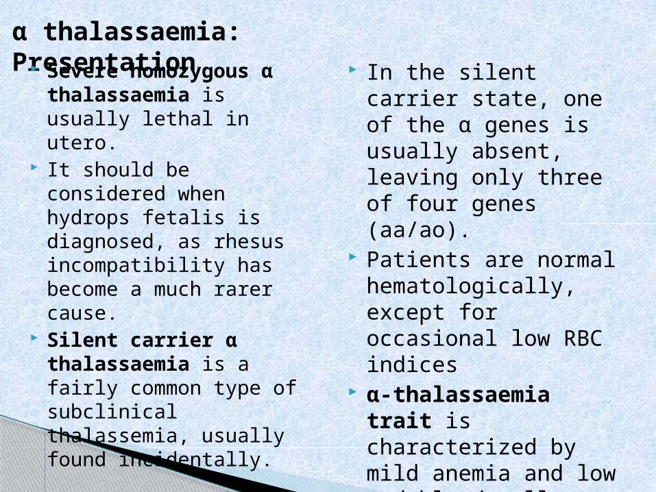
In the silent carrier state, one of the α genes is usually absent, leaving only three of four genes (aa/ao).
Patients are normal hematologically, except for occasional low RBC indices
α-thalassaemia trait is characterized by mild anemia and low red blood cell (RBC) indices.
α thalassaemia: Presentation Severe homozygous α
thalassaemia is usually lethal in utero.
It should be considered when hydrops fetalis is diagnosed, as rhesus incompatibility has become a much rarer cause.
Silent carrier α thalassaemia is a fairly common type of subclinical thalassemia, usually found incidentally.
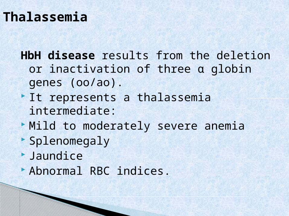
HbH disease results from the deletion or inactivation of three α globin genes (oo/ao).
It represents a thalassemia intermediate: Mild to moderately severe anemia Splenomegaly Jaundice Abnormal RBC indices.
Thalassemia
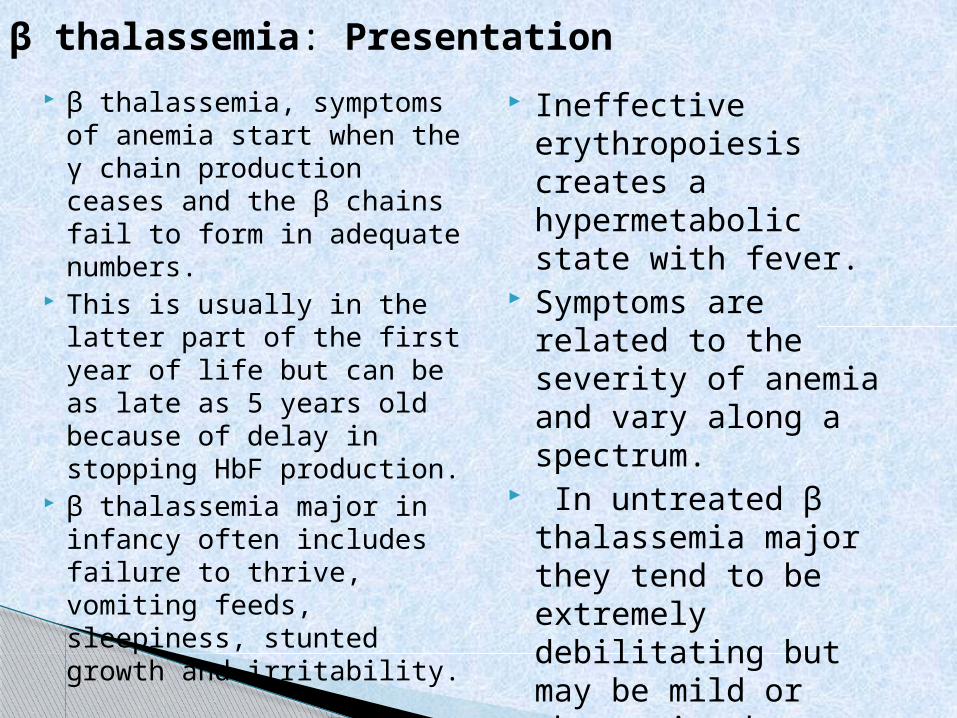
Ineffective erythropoiesis creates a hypermetabolic state with fever.
Symptoms are related to the severity of anemia and vary along a spectrum.
In untreated β thalassemia major they tend to be extremely debilitating but may be mild or absent in those with milder forms of disease.
β thalassemia, symptoms of anemia start when the γ chain production ceases and the β chains fail to form in adequate numbers.
This is usually in the latter part of the first year of life but can be as late as 5 years old because of delay in stopping HbF production.
β thalassemia major in infancy often includes failure to thrive, vomiting feeds, sleepiness, stunted growth and irritability.
β thalassemia: Presentation
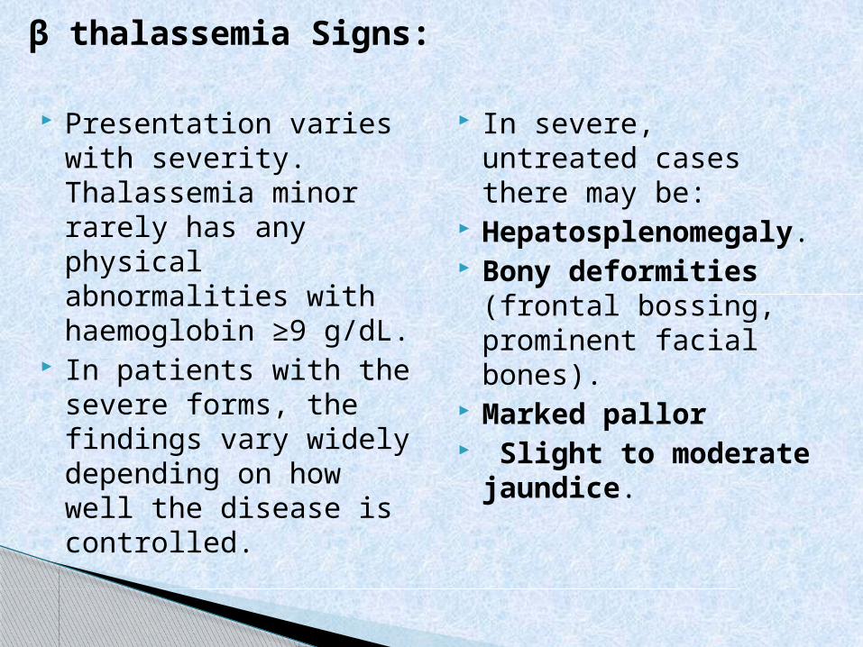
In severe, untreated cases there may be:
Hepatosplenomegaly.
Bony deformities (frontal bossing, prominent facial bones).
Marked pallor Slight to moderate
jaundice.
Presentation varies with severity. Thalassemia minor rarely has any physical abnormalities with haemoglobin ≥9 g/dL.
In patients with the severe forms, the findings vary widely depending on how well the disease is controlled.
β thalassemia Signs:
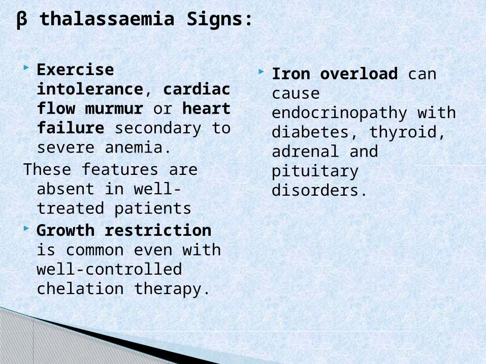
Iron overload can cause endocrinopathy with diabetes, thyroid, adrenal and pituitary disorders.
Exercise intolerance, cardiac flow murmur or heart failure secondary to severe anemia.
These features are absent in well-treated patients
Growth restriction is common even with well-controlled chelation therapy.
β thalassaemia Signs:
Hemoglobin electrophoresis usually reveals the diagnosis.
Normal HbA2 is between 1.5 and 3.0% whilst HbA2 >3.5 % is diagnostic.
DNA analysis should be offered to identify and confirm couples at risk, in prenatal testing
α-thalassemia trait requires measuring either the α-β chain synthesis ratio or using polymerase chain reaction (PCR) assay tests.
FBC shows a microcytic, hypochromic anaemia
In the severe forms hemoglobin level ranges from 2-8 g/dL.
WBC is elevated due to hemolytic process.
Platelet count may be depressed in splenomegaly.
Serum iron level is elevated with saturation as high as 80%.
Ferritin is also raised.
Investigations:
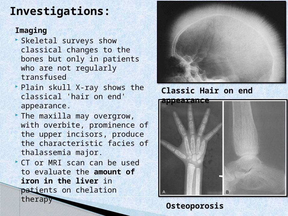
Imaging Skeletal surveys show
classical changes to the bones but only in patients who are not regularly transfused
Plain skull X-ray shows the classical 'hair on end' appearance.
The maxilla may overgrow, with overbite, prominence of the upper incisors, produce the characteristic facies of thalassemia major.
CT or MRI scan can be used to evaluate the amount of iron in the liver in patients on chelation therapy
Investigations:
Classic Hair on end appearance
Osteoporosis
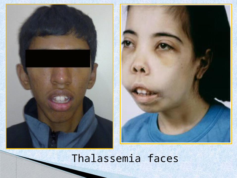
Thalassemia faces
The general principles of management include:
Asymptomatic carriers: require no specific treatment but should be protected from detrimental iron supplementation
Thalassemia intermedia or HbH disease:
Need to be closely monitored for progression of complications induced by chronic hemolytic anemia.
Occasional blood transfusion:
Infection-associated aplastic or hyper-hemolytic crises
Pregnancy Growth impairment Skeletal deformities.Hypersplenism
develops, splenectomy may be considered, although this carries severe risks of life-threatening infections, pulmonary hypertension, and thrombosis
MANAGEMENT
Thalassemia major: Regular
hypertransfusion to maintain a haemoglobin level higher than 9.5 g/dL.
Iron chelation to prevent overload syndrome.
Care by a multidisciplinary team (including hematologist, specialized nurse, social worker, psychologist, genetic counselor, cardiologist and liver specialist).
Non-drug Psychological support. Genetic counseling. Avoid food rich in iron.
Extra vitamin E, folic acid and some vitamin C may be beneficial.
Transfusion improves both quality and quantity of life in severe cases. The target is not to let Hb fall below 9.5 g/dL.
Splenectomy may be indicated if hypersplenism causes a marked increase in transfusion requirements,
Management:
Do not treat anemia with iron unless iron deficiency had been substantiated.
Desferrioxamine is given parenterally to aid iron excretion. The dose and means of delivery vary according to the needs of the patient.
Oral chelating agents have been developed and are now in use, including deferasirox and deferiprone.
Hydroxyurea may increase the expression of γ chains (HbF) and remove the excess α chains, which could potentially correct ineffective erythropoiesis.
There is hope for new combination therapies - e.g., oral deferiprone used in combination with desferrioxamine, producing a greater effect than either alone
Folic acid and vitamin E deficiency may require treatment.
Management: