CCAAT/Enhancer-binding Protein β (C/EBPβ κ High … X-tremeGENE siRNA transfection reagent....
32
CCAAT/Enhancer-binding Protein β (C/EBPβ) and Nuclear Factor Kappa B (NF-κB) Mediate High Level Expression of Chemokine Genes CCL3 and CCL4 by Human Chondrocytes in Response to Interleukin-1β (IL-1β)* Zhiqi Zhang 1, 2 , Jennifer L. Bryan 1 , Elizabeth DeLassus 1 , Li-Wei Chang 3 , Weiming Liao 2 , and Linda J. Sandell 1, 4 § Department of Orthopaedic Surgery 1 , Department of Pathology and Immunology 3 , Department of Cell Biology and Physiology 4 , Washington University School of Medicine at Barnes-Jewish Hospital, St. Louis, MO 63110, United States Department of Orthopaedic Surgery 2 , First Affiliated Hospital of Sun Yat-sen University, Guangzhou, Guangdong 510080, P. R. China Running Title: C/EBPβ and NF-κB regulate expression of chemokines §To whom correspondence should be addressed: Dr. Linda J. Sandell, Ph.D., Department of Orthopaedic Surgery, Washington University School of Medicine, 660 S. Euclid Ave, CB 8233, St. Louis, MO 63110. Tel: 314-454-7800; Fax: 314-454-5900; E-mail: [email protected] . A large set of chemokines is highly up-regulated in human chondrocytes in response to IL-1β (Sandell, L.J. et al. 2008. Osteoarthritis Cartilage 16, 1560-1571). To investigate the mechanism of transcriptional regulation, deletion constructs of selected chemokine gene promoters, the human CCL3 (MIP-1α) and CCL4 (MIP-1β), were transfected into human chondrocytes with or without IL-1β. The results show that an IL-1β-responsive element is located between -300 to -140bp of CCL3 promoter, and -222 to -100bp of CCL4 promoter. As both of these elements contain C/EBPβ motifs, the function of C/EBPβ was examined. IL-1β stimulated the expression of C/EBPβ, and the direct binding of C/EBPβ to the C/EBPβ motif was confirmed by EMSA and ChIP analyses. The -300bp CCL3 promoter and -222bp CCL4 promoter were strongly up-regulated by co-transfection with C/EBPβ expression vector. Mutation of the C/EBPβ motif and reduction of C/EBPβ expression by siRNA decreased the up-regulation. Additionally, another cytokine-related transcriptional factor, NF-κB, was also shown to be involved in the up-regulation of chemokines in response to IL-1β and the binding site was identified. The regulation of C/EBPβ and NF-κB was confirmed by the inhibition by C/EBPβ and NF-κB and by transfection with C/EBPβ and NF-κB expression vectors in the presence or absence of IL-1β. Taken together, our results suggest that C/EBPβ and NF-κB are both involved in the IL-1β-responsive up-regulation of chemokine genes in human chondrocytes. Time course experiments indicated that C/EBPβ gradually and steadily induces chemokine up-regulation, whereas NF-κB activity was highest at the early stage of chemokine up-regulation. Chemokines are associated with several diseases, including cardiovascular diseases, neuroinflammation, cancer and HIV-associated diseases (1). As mediators of cell recruitment, chemokines are known to be important in inflammatory diseases, including rheumatoid arthritis (RA), osteoarthritis (OA), inflammatory bowel disease, multiple sclerosis and transplant rejection (1,2). Chemokines are a specific class 1 http://www.jbc.org/cgi/doi/10.1074/jbc.M110.130377 The latest version is at JBC Papers in Press. Published on August 11, 2010 as Manuscript M110.130377 Copyright 2010 by The American Society for Biochemistry and Molecular Biology, Inc. by guest on July 10, 2018 http://www.jbc.org/ Downloaded from
Transcript of CCAAT/Enhancer-binding Protein β (C/EBPβ κ High … X-tremeGENE siRNA transfection reagent....
CCAAT/Enhancer-binding Protein β (C/EBPβ) and Nuclear Factor Kappa B (NF-κB) Mediate High Level Expression of Chemokine Genes CCL3 and CCL4 by Human Chondrocytes in
Response to Interleukin-1β (IL-1β)* Zhiqi Zhang 1, 2, Jennifer L. Bryan 1, Elizabeth DeLassus 1, Li-Wei Chang 3, Weiming Liao 2, and
Linda J. Sandell 1, 4§ Department of Orthopaedic Surgery1, Department of Pathology and Immunology3, Department of Cell Biology and Physiology4, Washington University School of Medicine at Barnes-Jewish Hospital, St.
Louis, MO 63110, United States Department of Orthopaedic Surgery2, First Affiliated Hospital of Sun Yat-sen University, Guangzhou,
Guangdong 510080, P. R. China Running Title: C/EBPβ and NF-κB regulate expression of chemokines
§To whom correspondence should be addressed: Dr. Linda J. Sandell, Ph.D., Department of Orthopaedic Surgery, Washington University School of Medicine, 660 S. Euclid Ave, CB 8233, St. Louis, MO 63110. Tel: 314-454-7800; Fax: 314-454-5900; E-mail: [email protected].
A large set of chemokines is highly up-regulated in human chondrocytes in response to IL-1β (Sandell, L.J. et al. 2008. Osteoarthritis Cartilage 16, 1560-1571). To investigate the mechanism of transcriptional regulation, deletion constructs of selected chemokine gene promoters, the human CCL3 (MIP-1α) and CCL4 (MIP-1β), were transfected into human chondrocytes with or without IL-1β. The results show that an IL-1β-responsive element is located between -300 to -140bp of CCL3 promoter, and -222 to -100bp of CCL4 promoter. As both of these elements contain C/EBPβ motifs, the function of C/EBPβ was examined. IL-1β stimulated the expression of C/EBPβ, and the direct binding of C/EBPβ to the C/EBPβ motif was confirmed by EMSA and ChIP analyses. The -300bp CCL3 promoter and -222bp CCL4 promoter were strongly up-regulated by co-transfection with C/EBPβ expression vector. Mutation of the C/EBPβ motif and reduction of C/EBPβ expression by siRNA decreased the up-regulation. Additionally, another cytokine-related transcriptional factor, NF-κB, was also shown to be involved in the
up-regulation of chemokines in response to IL-1β and the binding site was identified. The regulation of C/EBPβ and NF-κB was confirmed by the inhibition by C/EBPβ and NF-κB and by transfection with C/EBPβ and NF-κB expression vectors in the presence or absence of IL-1β. Taken together, our results suggest that C/EBPβ and NF-κB are both involved in the IL-1β-responsive up-regulation of chemokine genes in human chondrocytes. Time course experiments indicated that C/EBPβ gradually and steadily induces chemokine up-regulation, whereas NF-κB activity was highest at the early stage of chemokine up-regulation.
Chemokines are associated with several diseases, including cardiovascular diseases, neuroinflammation, cancer and HIV-associated diseases (1). As mediators of cell recruitment, chemokines are known to be important in inflammatory diseases, including rheumatoid arthritis (RA), osteoarthritis (OA), inflammatory bowel disease, multiple sclerosis and transplant rejection (1,2). Chemokines are a specific class
1
http://www.jbc.org/cgi/doi/10.1074/jbc.M110.130377The latest version is at JBC Papers in Press. Published on August 11, 2010 as Manuscript M110.130377
Copyright 2010 by The American Society for Biochemistry and Molecular Biology, Inc.
by guest on July 10, 2018http://w
ww
.jbc.org/D
ownloaded from
of cytokines that classically mediate chemoattraction (chemotaxis) between cells. However, the production and function of chemokines in organs and tissues have recently been recognized, and knockout of the chemokine receptors CXCR4 and CXCR2 suggest a role in development and cell senescence (3,4) and the chemokine CCL3 is a potent osteoclastogenic factor (5). There exist over 50 chemokine ligands and 20 G protein-coupled receptors (1). Chemokines have similar protein structures being 8-10 kDa, with two major subclasses having conserved cysteine residues either adjacent (CC) or separated by one amino acid (CXC) (2).
IL-1β is an important cytokine in rheumatoid and osteoarthritic joint diseases. Generally, IL-1β is viewed as a catabolic factor for cartilage, inducing enzymes that degrade the extracellular matrix (ECM) and reducing synthesis of the primary cartilage components type II collagen (COL2A1) and aggrecan (6-8), although it can also induce bone morphogenetic protein 2 (BMP-2) potentially to initiate a repair response (9). In joint diseases, IL-1β is synthesized by synovial cells (10) and cartilage chondrocytes (6,11), therefore, its effect on chondrocytes is highly relevant to the fate of cartilage.
We have shown by microarray analysis, that a large set of chemokine genes is up-regulated by the pro-inflammatory cytokine IL-1β in adult normal cartilage and from patients with OA (6). It can be expected that this increase in a wide range of chemokines will have a significant impact on the cells of cartilage and other related joint tissues, and should be considered in the pathophysiology of OA. Recently, we demonstrated that the adipokine, resistin, present in injured joints (12), also increased chemokine genes at both the transcriptional and post-transcriptional levels (13) in temporal patterns similar to the IL-1β patterns (6). The
post-transcriptional regulation of chemokines was further investigated by us in chondrocytes with resistin (13) , and by others in fibroblasts (14).
In present study, we focused on the transcriptional regulation of chemokine genes. As we reported (6), IL-1β increased expression of chemokines CCL3, CCL4, CCL20, CCL3L1, CXCL1, CXCL2, CXCL3, CXCL6, and CXCL8 (IL-8) from 25 to 75-fold in human articular chondrocytes. A computational analysis of these co-regulated genes identified NF-κB, C/EBPβ and myocyte enhancer binding factor 3 (MEF-3), as candidate transcriptional regulators. NF-κB has been shown to regulate a specific subset of chemokines (15-18), however, Amos and colleagues recently demonstrated that inhibition of NF-κB activity did not inhibit all inflammatory mediators (19), therefore, there are likely other transcriptional mechanisms involved. C/EBP has been shown to regulate a set of chemokines (4,20,21). We have previously shown that C/EBPβ is associated with IL-1β-induced and tumor necrosis factor-alpha (TNF-α)-induced down regulation of matrix genes in chondrocytes and the repression of cartilage gene expression in non-cartilaginous tissues (22-25). Here, we investigated the roles of C/EBPβ and NF-κB in the up-regulation of two chemokine genes CCL3 (macrophage inflammatory protein 1α, MIP-1α) and CCL4 (MIP-1β) in human chondrocytes in response to IL-1β.
EXPERIMENTAL PROCEDURES
Material— The materials used in this work
were purchased as follows: Dulbecco’s modified Eagle’s medium (DMEM), Ham’s F-12 medium, were from Mediatech Inc., Herndon, VA. Fetal bovine serum, pfx polymerase, SuperScript® II Reverse Transcriptase, restriction enzymes, Alexa fluor® 488 goat anti-mouse IgG, and Alexa fluor®
2
by guest on July 10, 2018http://w
ww
.jbc.org/D
ownloaded from
594 goat anti-rabbit IgG from Invitrogen, Carlsbad, CA; Penicillin/streptomycin solution, ascorbic acid, Actinomycin D, Tween 20, Triton-X-100, and CelLyticTMNu-Clear Extraction KitTM from Sigma, St.Louis, MO; 16% paraformaldehyde from Electron Microscopy Science, hatfield, PA; Recombinant human IL-1β from R&D Systems, Inc., Minneapolis, MN; RNeasy Mini Kit, QIAshredder, DNase I, from Qiagen, Inc., Valencia, CA; [γ-32P]dATP from Perkin Elmer, Downers Grove, IL; FuGENE® 6 Transfection Reagent, X-tremeGENE siRNA Transfection Reagent, Quick Spin Sephadex G-50 and G-25 column, Pronase and Collagenase P from Roche, Indianapolis, IN; pGL3-basic vector, Reporter Lysis Buffer, Luciferase Assay Reagent, β-galactosidase from Promega, Madison, WI; QuickChange® II Site-directed Mutanogenesis Kit from Stratagene, La Jolla, CA; Ready Gel Tris-HCL Precast Gels, and non-fat dry milk from Bio-Rad, Hercules, CA; anti-C/EBPβ, SREBP1, anti-c-Rel, normal rabbit IgG and Actin antibodies from Santa Cruz Biotechnology, Santa Cruz, CA; SuperSignal® West Pico chemiluminescent substrate from Thermo Fisher Scientific Inc., Waltham, MA; SYBR Green PCR Master Mix was from Applied Biosystens, Foster City, CA; Cell-permeable NEMO binding domain (NBD) synthetic peptides (IKK-NBD peptide and IKK-NBD control peptide), were from BIOMOL, Plymouth Meeting, PA; SB203580 was from Calbiochem, Gibbstown, NJ; EZ-ChIPTM, Immobilon-P and Immobilon-FL transfer membrane were from Millipore, Bedford, MA.
Plasmid Constructs— A series of CCL3 and CCL4 promoter 5’-deletion constructs were made by PCR and subcloned into pGL3-basic vector using the pGL2-CCL3 (-1972/+75) and pGL3-CCL4 (-1281/+12) as previously described (26,27). The CCL3 and CCL4 promoter constructs, C/EBPβ and IkappaB kinase 2 (IKK2) expression
vector, and pNF-κB luciferase reporter were provided by the following: the human pGL2-CCL3 (-1972/+75) was from Dr. G. David Roodman (University of Pittsburgh) (26); human pGL3-CCL4 (-1281/+12) was obtained from Dr. Sheau-Farn Yeh (National Yang-Ming University, Taipei) (27); human IKK2 in the pCDNA3 vector and pNF-κB luciferase reporter were from Dr. Yousef Abu-Amer (Washington University) (28); human C/EBP-full-length in the pCDNA3 vector was from Dr. Erika Crouch (Washington University) (29). The empty expression vectors were made by excision of cDNAs from the corresponding C/EBP expression vectors (22). To facilitate subcloning of the amplified fragments, the antisense primer contained a HindIII restriction site adaptor, and the sense primer contained an XhoI or SmaI site. The PCR fragments and the luciferase expression vector pGL3-basic vector were digested with XhoI or SmaI and HindIII before ligation. All constructs were confirmed by DNA sequence analysis using a GL2 primer and RV3 primer.
Cell Culture— Human primary chondrocytes were obtained from articular cartilage obtained at the time of total joint replacement or from above the knee amputation, with approval of the Washington University Human Studies Review Board and permission of the patient (IRB No. 05-0279). Chondrocytes were isolated following previously published procedures (6) and plated at a density of 2.5×10⁵ cells/cm2 in DMEM/F12 media plus 10% fetal bovine serum (FBS), 50 μg/ml ascorbate and antibiotics (50 U/ml penicillin and 50 μg/ml streptomycin). Cells were allowed to rest for 24 h and IL-1β was added at the concentrations and times indicated. IL-1β was reconstituted in sterile phosphate-buffered saline (PBS) containing 0.1% bovine serum albumin. T/C-28a2 human chondrocyte cell line were also used (a gift from Dr. Mary B. Goldring, Cornell
3
by guest on July 10, 2018http://w
ww
.jbc.org/D
ownloaded from
University), and cultured like human articular chondrocytes. For RNA or nuclear protein extraction, T/C-28 human chondrocyte cell line was plated in 3×10⁴ cells/cm2 densities and cultured overnight. Human IL-1β was then added to the medium at the concentrations indicated.
Transient Transfection and Luciferase Assay— DNA transfections of T/C-28a2 cells were performed using FuGENE 6TM transfection reagent or X-tremeGENE siRNA transfection reagent. 2×10⁵ of T/C-28a2 cells were cultured in a 6-well plate overnight. The transfection mixture containing 3-9 μl of transfection reagent (6:1 ratio of trasfection reagent μl to DNA μg), 500 ng of various promoter constructs, and 100 ng or 200 ng of pCMV-β-gal was then added, and the cells were cultured for 8 h or 24 h with or without IL-1β as indicated. For co-transfection assay, 500 ng of C/EBPβ expression vectors or empty vector, and 162 pM of siRNAs were added to the 100 μl transfection mixture as indicated. The sequences of siRNAs used were described previously (21). Due to low translation efficiency of C/EBPβ, we used higher amounts of plasmid (500 ng) for transfection, as shown previously (22,24). FBS was added to transfection medium 4 h later to a final concentration of 10%. After 24 h of incubation, cells were replaced with fresh complete medium and incubated for an additional 8 h or 24h with or without IL-1β. The cells were then harvested with Reporter Lysis BufferTM, and the lysate was analyzed for luciferase activity using Promega Luciferase Assay ReagentTM. The β-galactosidase activities were also measured to normalize variations in transfection efficiency. Each transfection experiment was performed in duplicate or triplicate and repeated at least three times.
Preparation of Nuclear Extracts— Nuclear extracts from T/C-28a2 cells were isolated using
Nu-Clear Extraction KitTM according to the manufacturer’s instructions. Protein concentration of nuclear extract was determined using Bio-Rad protein assay kit.
Electrophoretic Mobility Shift Assay (EMSA)— Fragment A (between -300 and -141 bp relative to the human CCL3 translation start site), and Fragment B (between -222 and -101 bp relative to the human CCL4 translation start site) were amplified by PCR. All oligonucleotides were synthesized by Invitrogen, and complementary oligonucleotide was annealed to make double-stranded oligonucleotide (30). The fragment A, B and various double-stranded oligonucleotides were end-labeled using T4 polynucleotide kinase and [γ-32P]dATP. Bandshifts were performed by incubating 5 μg of nuclear extracts in the mobility shift buffer (10mM Hepes–KOH, pH 7.9, 100mM NaCl, 1mM EDTA, 10% glycerol, and 1mM DTT) and 2 μg of poly(dI-dC) with the DNA probe on ice for 30 min. For the competition studies, the cold DNA fragments were added at a 100-fold molar excess compared with the probe and incubated for 30 min on ice before adding the DNA probe. For the antibody interference experiments, the nuclear extracts and 2 μl of antibody were preincubated in the buffer for 1 h at 4 °C. DNA protein complexes were resolved on a 5% polyacrylamide gel at 100 V for about 90 minutes. The gels were dried and autoradiographed.
Western Blotting— Fifteen μg of nuclear extracts were denatured in SDS sample buffer contains 0.1 M dithiothreitol at 100 °C for 3 min and separated on a 12% Bio-Rad Ready Gel in Tris/glycine/SDS buffer. The gels were then transferred to Immobilon-P transfer membrane in Tris/glycine buffer, pH 8.3, containing 20% methanol. The membranes were saturated in 5% non-fat dry milk in PBS at 4°C for overnight and reacted with anti-C/EBPβ antibody diluted to
4
by guest on July 10, 2018http://w
ww
.jbc.org/D
ownloaded from
1:1000 or Actin antibody diluted to 1:500 in PBS containing 0.05% Tween 20. The bound antibodies were recognized by IgG antibodies coupled to horseradish peroxidase, and the secondary antibodies were detected by autoradiograph using SuperSignal
Chemiluminescent Substrate from Thermo Scientific. For western blotting using Odyssey imaging system, 10 μg of nuclear extracts were used, and the gels were transferred and scanned as the protocol recommended by the manufacturer (LI-COR).
RNA Isolation and Real Time Quantitative PCR— Total RNA was isolated from T/C-28 cells and human primary articular chondrocytes with RNeasy Mini Kit with DNase I treatment, following the protocol recommended by the manufacturer (Qiagen). Total RNA (1 μg) was reverse-transcribed with a SuperScriptTM II Reverse Transcriptase to synthesize cDNA. The cDNA was then used for real-time quantitative PCR (qPCR). Real-time quantitative PCR was performed in a total volume of 20 μl reaction mixture containing 10 μl of SYBR Green PCR Master Mix, 2.5 μl of cDNA, and 200 nM of primers using a 7300 Real-Time PCR System (Applied Biosystems), and done in triplicate. Primers used for qPCR were optimized for each gene, and the dissociation curve was determined by the Real-Time PCR System. The parameters of primer design included a primer size of 18 to 21 bp, a product size of 80 to 150 bp, a primer annealing temperature of 59° to 61°, and a primer GC content of 45% to 55%. Results were normalized to glyceraldehyde-3- phosphate dehydrogenase (GAPDH). The primer sequences are as follows: human GAPDH, 5’-ACCCAGAAGACTGTGGATGG-3’(sense) and 5’-GAGGCAGGGATGATGTTCTG-3’ (antisense); human C/EBPβ, 5’-CTCGCAGGTC- AAGAGCAAGG-3’ (sense) and 5’-TCGTCGCT-
GTGCTTGTCC-3’ (antisense) (22); human CCL3, 5’-GCAACCAGTTCTCTGCATCA-3’(sense), 5’-TGGCTGCTCGTCTCAAAGTA-3’ (antisense); human CCL4, 5’-GCTTTTCTTACACTGCGAG- GA-3’ (sense), 5’-CCAGGATTCACTGGGATC- AG-3’ (antisense); human NF-κB1 (p50), 5’-CCTGGATGACTCTTGGGAAA-3’ (sense), 5’-TCAGCCAGCTGTTTCATGTC-3’ (antisense); human NF-κB2 (p52), 5’-GAACAGCCTTGCAT- CTAGCC-3’ (sense), 5’-TTTTCAGCATGGAT- GTCAGC-3’ (antisense); human RelA (p65), 5’-TCTGCTTCCAGGTGACAGTG-3’ (sense), 5’-GCCAGAGTTTCGGTTCACTC-3’ (antisense); human c-Rel, 5’-CGAACCCAATTTATGACAA- CCG-3’ (sense), 5’-TTTTGTTTCTTTGCTTTA- TTGCCG-3’(antisense) (31); human RelB, 5’-CTGCTTCCAGGCCTCATATC-3’ (sense), 5’-CGCAGCTCTGATGTGTTTGT-3’ (antisense); human IκBα, 5’-GATCCGCCAGGTGAAGGG-3’ (sense), 5’-GCAATTTCTGGCTGGTTGG-3’ (antisense) (32). The cycle threshold (Ct) values for GAPDH and those of genes of interest was measured for each sample, and the relative transcript levels were calculated as χ= 2-∆∆Ct, in which ∆∆Ct= ∆Treatment - ∆C and ∆Treatment = Cttreatment - CtGAPDH; ∆C = Ctcontrol - CtGAPDH.
Chromatin immunoprecipitation (ChIP) Assay— Chromatin immuneprecipitations (IPs) were carried out using the EZ ChIP™ assay kit following the protocol recommended by the manufacturer. One 15-cm dish of T/C-28 cells (4 x10⁶) in 15-cm culture dishes was used for one IP reaction. Cross-linking of DNA/proteins was induced by addition of formaldehyde (1% final concentration) directly to the culture medium for 10 min at 37 ºC. Cells were lysed and DNA in the supernatant was sheared by sonication. A 10 μl aliquot of the sonicated chromatin sample (“input”) was removed for PCR analysis and the remainder was used for IPs. To each IP was added either 5 μg of C/EBPβ antibody or normal rabbit IgG, and
5
by guest on July 10, 2018http://w
ww
.jbc.org/D
ownloaded from
reactions were incubated overnight with constant mixing at 4 ºC. Protein G beads were collected by centrifugation and protein/DNA complexes were eluted from the beads followed by a cross-link reversal step by addition of 5M NaCl (8 μl) to the eluted sample (200 μl) for overnight at 65 ºC. DNA from each IP reaction (2 μl) was used for PCR with primer pairs to amplify the 140 bp region of interest in CCL3 (-300/-140) and the 110 bp in CCL4 (-222/-100): CCL3, 5’-TCATGCACAGACCAGTTCTTATGA-3’ (sense primer), 5’-CTCTAACTCTCAGCTCTC- AACTCA-3’ (antisense primer); CCL4, 5’-CTGTACCACTTCCCTTTTCTTCTC-3’ (sense primer), 5’-CTGAAGCTAGCTGAGTG- AGGAGTT-3’ (antisense primer). Input or immunoprecipitated DNA was amplified by PCR [94 ºC, 20 s; 59 ºC, 30 s; 72 ºC, 2 min] for 32 cycles (CCL3) or 34 cycles (CCL4).
Immunofluorescence— 5×10⁴ human articular chondrocytes were cultured in each well of 8-well chamber slides (from Lab Tek). Cells were allowed to rest for 24 h and IL-1β was then added at the times indicated. Cells were incubated in 4% paraformaldehyde in PBS for 10 min, 0.2% Triton-X-100 in PBS for 5 min, and 10% normal goat serum in PBS for 2 h at room temperature. Cells were reacted with rabbit anti-C/EBPβ and mouse anti-c-Rel antibodies diluted to 1:400 in 2% normal goat serum in PBS for overnight at 4 ºC. The secondary antibodies, Alexa fluor 488 dye-labeled goat anti-mouse IgG diluted to 1: 250 and Alexa fluor 594 dye-labeled goat anti-rabbit IgG diluted to 1:400 in 2% normal goat serum in PBS were then added to the cells for 1h at room temperature. Immunoreactivity was detected by fluorescence microscopy.
RESULTS
IL-1β Stimulates the Expression of
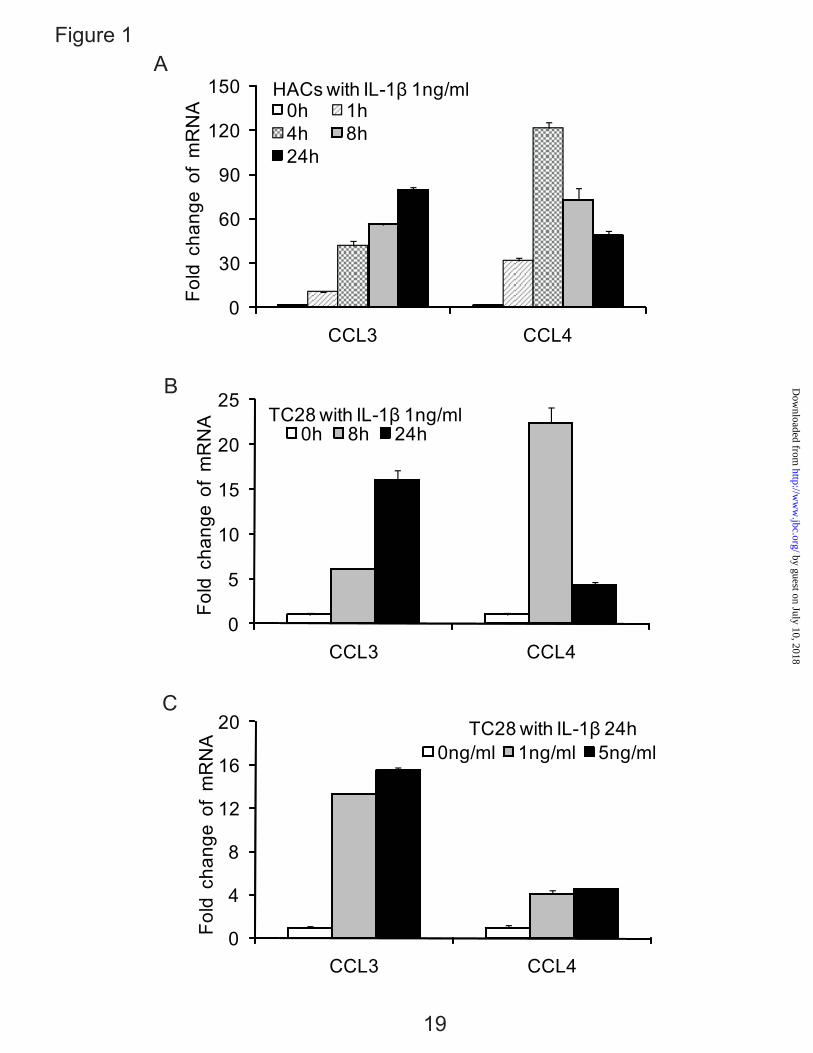
Chemokine Genes CCL3 and CCL4 in Normal Human Articular Chondrocytes and the Chondrocyte-derived Cell Line, T/C-28 — We have reported that a large set of chemokines that are up-regulated by the IL-1β in adult normal cartilage and from patients with OA (6). Here, we showed that two representative genes, CCL3 and CCL4, were increased in human articular chondrocytes and in the cell line T/C-28 with the same kinetics in the 24 h time period, although the expression level of CCL3 and CCL4 in human primary chondrocytes was higher than T/C-28 chondrocytes cell line (Fig. 1, A-C). CCL3 demonstrated continuous increase to 24h to approximately an 80 fold increase in primary cells. CCL4 was up-regulated by 4 h, but then decreased during the remaining time period to about a 45 fold increase at 24h. A dose response test of IL-1β showed that 1 ng/ml is adequate for significant up-regulation (Fig. 1C). The primary chondrocytes and T/C-28 chondrocyte cell line showed a similar pattern of induction (Fig. 1A and B).
An IL-1β-responsive Element is Located Between -300 to -140 bp of the CCL3 Promoter, and -222 to -100 bp of the CCL4 Promoter— To identify the IL-1β-responsive element within CCL3 and CCL4 promoters, five 5’-deletion constructs were transiently transfected into T/C-28 cells and incubated in the absence or presence of IL-1β (Fig. 2, A and B). Although there are many exceptions, most transcriptional regulation occurs within the first 1000 nucleotides of the promoter (33). For the CCL3 promoter constructs, the -1395, -522, -420, -300 bp CCL3 promoter constructs were up-regulated by the IL-1β treatment, however the response was lost in the -140 bp CCL3 constructs (Fig. 2A). For CCL4 promoter constructs, the expression of the -1281, -521, -372 and -222 bp CCL4 promoter constructs were up-regulated by the IL-1β
6
by guest on July 10, 2018http://w
ww
.jbc.org/D
ownloaded from
treatment, whereas the response was lost in the -100 bp CCL4 construct (Fig. 2B). These data suggest that in these constructs, the IL-1β-responsive element is located between -300 to -140 bp of the CCL3 promoter, and -222 to -100 bp of the CCL4 promoter.
IL-1β Stimulates the Expression of C/EBPβ in Human Articular Chondrocytes and T/C-28 Cells— Our computational database of regulatory motifs revealed that there were potential binding sites for C/EBPβ between -300 to -140 bp of the CCL3 promoter (2 sites), and -222 to -100 bp of the CCL4 promoter (1 site), therefore, we examined whether IL-1β stimulates the expression of C/EBPβ in primary chondrocytes and T/C-28 cells. The expression level of C/EBPβ mRNA was stimulated by IL-1β over 24 h (Fig. 3, A-C). Type II collagen mRNA decreased to about 60% within 24 h in human chondrocytes as expected (Fig. 3A) as we have shown previously for rat chondrosarcoma cells (22) and human articular chondrocytes (6). Western blots for C/EBPβ revealed that in the absence of IL-1β treatment, C/EBPβ protein was present in the nuclei. When cells were stimulated with IL-1β, C/EBPβ protein expression was increased about 3-fold (Fig. 3D). Multiple C/EBPβ isoforms with stimulatory or inhibitory activity can be translated from a single mRNA by use of alternative translation initiation sites within the same open reading frame (34,35). The IL-1β treatment stimulated the expression of two of C/EBPβ forms, 38 kDa and 36 kDa liver-enriched transcriptional activator proteins (LAP), and 16 kDa liver-enriched inhibitory protein (LIP) (23,36), however, the up-regulation of protein expression of C/EBPβ LAP was highest.
C/EBPβ Binds to the IL-1β-responsive Element of the CCL3 and CCL4 Promoters— To determine whether C/EBPβ functions within the IL-1β-responsive element, EMSA was carried out using nuclear extract from the T/C-28 cells.
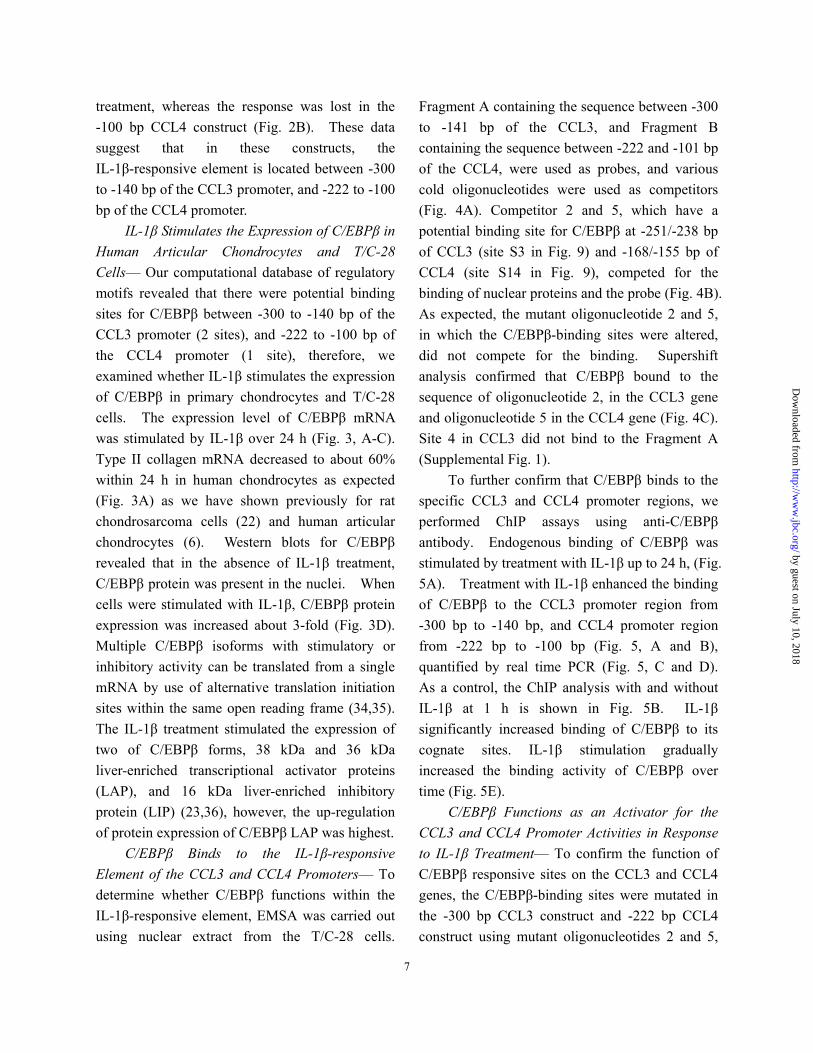
Fragment A containing the sequence between -300 to -141 bp of the CCL3, and Fragment B containing the sequence between -222 and -101 bp of the CCL4, were used as probes, and various cold oligonucleotides were used as competitors (Fig. 4A). Competitor 2 and 5, which have a potential binding site for C/EBPβ at -251/-238 bp of CCL3 (site S3 in Fig. 9) and -168/-155 bp of CCL4 (site S14 in Fig. 9), competed for the binding of nuclear proteins and the probe (Fig. 4B). As expected, the mutant oligonucleotide 2 and 5, in which the C/EBPβ-binding sites were altered, did not compete for the binding. Supershift analysis confirmed that C/EBPβ bound to the sequence of oligonucleotide 2, in the CCL3 gene and oligonucleotide 5 in the CCL4 gene (Fig. 4C). Site 4 in CCL3 did not bind to the Fragment A (Supplemental Fig. 1).
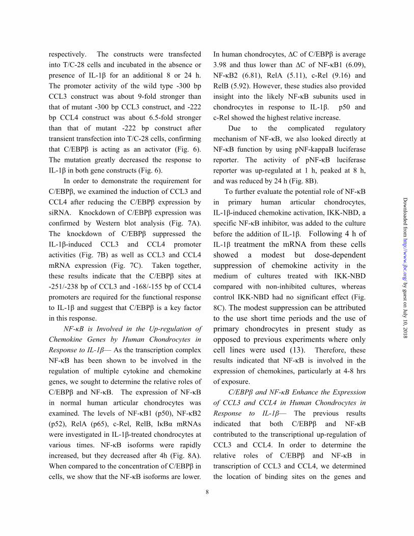
To further confirm that C/EBPβ binds to the specific CCL3 and CCL4 promoter regions, we performed ChIP assays using anti-C/EBPβ antibody. Endogenous binding of C/EBPβ was stimulated by treatment with IL-1β up to 24 h, (Fig. 5A). Treatment with IL-1β enhanced the binding of C/EBPβ to the CCL3 promoter region from -300 bp to -140 bp, and CCL4 promoter region from -222 bp to -100 bp (Fig. 5, A and B), quantified by real time PCR (Fig. 5, C and D). As a control, the ChIP analysis with and without IL-1β at 1 h is shown in Fig. 5B. IL-1β significantly increased binding of C/EBPβ to its cognate sites. IL-1β stimulation gradually increased the binding activity of C/EBPβ over time (Fig. 5E).
C/EBPβ Functions as an Activator for the CCL3 and CCL4 Promoter Activities in Response to IL-1β Treatment— To confirm the function of C/EBPβ responsive sites on the CCL3 and CCL4 genes, the C/EBPβ-binding sites were mutated in the -300 bp CCL3 construct and -222 bp CCL4 construct using mutant oligonucleotides 2 and 5,
7
by guest on July 10, 2018http://w
ww
.jbc.org/D
ownloaded from
respectively. The constructs were transfected into T/C-28 cells and incubated in the absence or presence of IL-1β for an additional 8 or 24 h. The promoter activity of the wild type -300 bp CCL3 construct was about 9-fold stronger than that of mutant -300 bp CCL3 construct, and -222 bp CCL4 construct was about 6.5-fold stronger than that of mutant -222 bp construct after transient transfection into T/C-28 cells, confirming that C/EBPβ is acting as an activator (Fig. 6). The mutation greatly decreased the response to IL-1β in both gene constructs (Fig. 6).
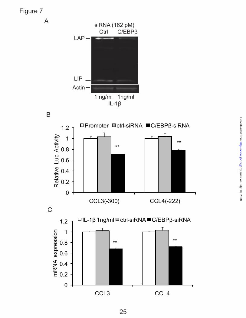
In order to demonstrate the requirement for C/EBPβ, we examined the induction of CCL3 and CCL4 after reducing the C/EBPβ expression by siRNA. Knockdown of C/EBPβ expression was confirmed by Western blot analysis (Fig. 7A). The knockdown of C/EBPβ suppressed the IL-1β-induced CCL3 and CCL4 promoter activities (Fig. 7B) as well as CCL3 and CCL4 mRNA expression (Fig. 7C). Taken together, these results indicate that the C/EBPβ sites at -251/-238 bp of CCL3 and -168/-155 bp of CCL4 promoters are required for the functional response to IL-1β and suggest that C/EBPβ is a key factor in this response.
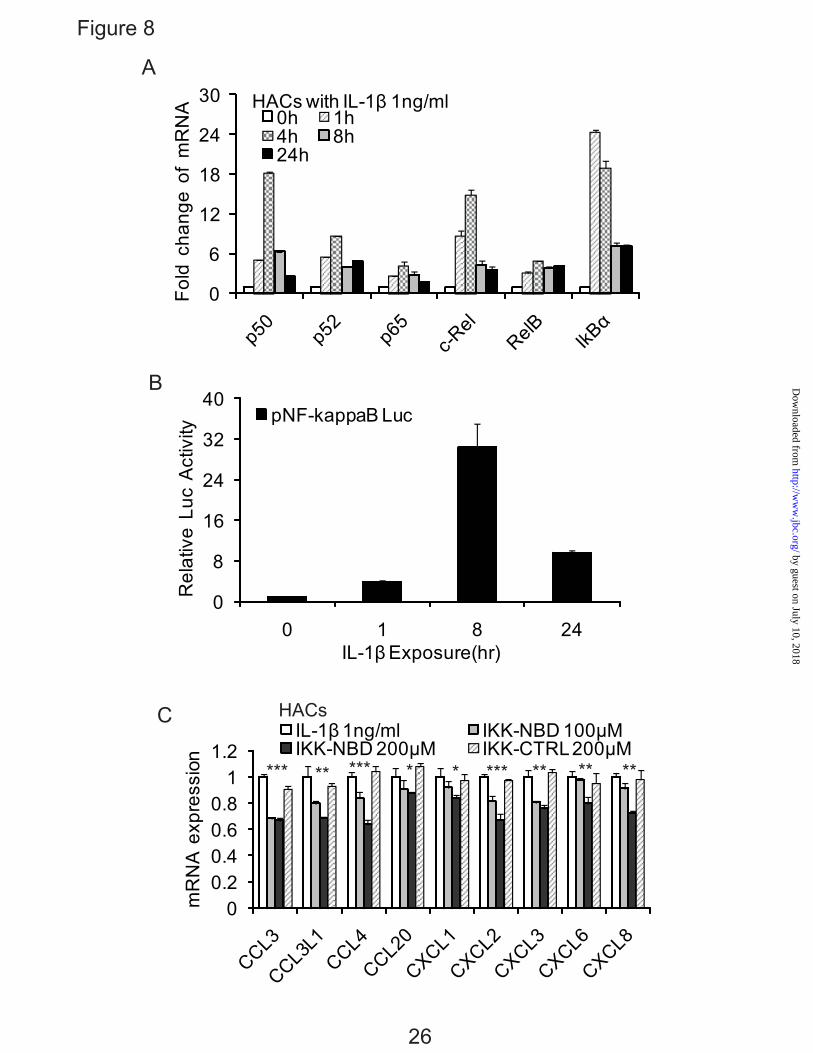
NF-κB is Involved in the Up-regulation of Chemokine Genes by Human Chondrocytes in Response to IL-1β— As the transcription complex NF-κB has been shown to be involved in the regulation of multiple cytokine and chemokine genes, we sought to determine the relative roles of C/EBPβ and NF-κB. The expression of NF-κB in normal human articular chondrocytes was examined. The levels of NF-κB1 (p50), NF-κB2 (p52), RelA (p65), c-Rel, RelB, IκBα mRNAs were investigated in IL-1β-treated chondrocytes at various times. NF-κB isoforms were rapidly increased, but they decreased after 4h (Fig. 8A). When compared to the concentration of C/EBPβ in cells, we show that the NF-κB isoforms are lower.
In human chondrocytes, ∆C of C/EBPβ is average 3.98 and thus lower than ∆C of NF-κB1 (6.09), NF-κB2 (6.81), RelA (5.11), c-Rel (9.16) and RelB (5.92). However, these studies also provided insight into the likely NF-κB subunits used in chondrocytes in response to IL-1β. p50 and c-Rel showed the highest relative increase.
Due to the complicated regulatory mechanism of NF-κB, we also looked directly at NF-κB function by using pNF-kappaB luciferase reporter. The activity of pNF-κB luciferase reporter was up-regulated at 1 h, peaked at 8 h, and was reduced by 24 h (Fig. 8B).
To further evaluate the potential role of NF-κB in primary human articular chondrocytes, IL-1β-induced chemokine activation, IKK-NBD, a specific NF-κB inhibitor, was added to the culture before the addition of IL-1β. Following 4 h of IL-1β treatment the mRNA from these cells showed a modest but dose-dependent suppression of chemokine activity in the medium of cultures treated with IKK-NBD compared with non-inhibited cultures, whereas control IKK-NBD had no significant effect (Fig. 8C). The modest suppression can be attributed to the use short time periods and the use of primary chondrocytes in present study as opposed to previous experiments where only cell lines were used (13). Therefore, these results indicated that NF-κB is involved in the expression of chemokines, particularly at 4-8 hrs of exposure.
C/EBPβ and NF-κB Enhance the Expression of CCL3 and CCL4 in Human Chondrocytes in Response to IL-1β— The previous results indicated that both C/EBPβ and NF-κB contributed to the transcriptional up-regulation of CCL3 and CCL4. In order to determine the relative roles of C/EBPβ and NF-κB in transcription of CCL3 and CCL4, we determined the location of binding sites on the genes and
8
by guest on July 10, 2018http://w
ww
.jbc.org/D
ownloaded from
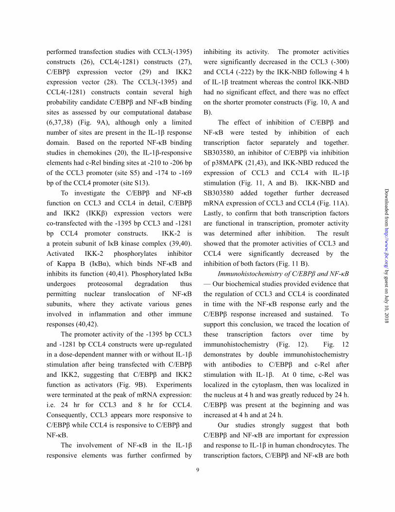
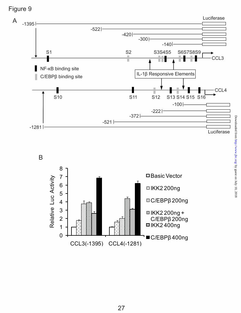
performed transfection studies with CCL3(-1395) constructs (26), CCL4(-1281) constructs (27), C/EBPβ expression vector (29) and IKK2 expression vector (28). The CCL3(-1395) and CCL4(-1281) constructs contain several high probability candidate C/EBPβ and NF-κB binding sites as assessed by our computational database (6,37,38) (Fig. 9A), although only a limited number of sites are present in the IL-1β response domain. Based on the reported NF-κB binding studies in chemokines (20), the IL-1β-responsive elements had c-Rel binding sites at -210 to -206 bp of the CCL3 promoter (site S5) and -174 to -169 bp of the CCL4 promoter (site S13).
To investigate the C/EBPβ and NF-κB function on CCL3 and CCL4 in detail, C/EBPβ and IKK2 (IKKβ) expression vectors were co-transfected with the -1395 bp CCL3 and -1281 bp CCL4 promoter constructs. IKK-2 is a protein subunit of IκB kinase complex (39,40). Activated IKK-2 phosphorylates inhibitor of Kappa B (IκBα), which binds NF-κB and inhibits its function (40,41). Phosphorylated IκBα undergoes proteosomal degradation thus permitting nuclear translocation of NF-κB subunits, where they activate various genes involved in inflammation and other immune responses (40,42).
The promoter activity of the -1395 bp CCL3 and -1281 bp CCL4 constructs were up-regulated in a dose-dependent manner with or without IL-1β stimulation after being transfected with C/EBPβ and IKK2, suggesting that C/EBPβ and IKK2 function as activators (Fig. 9B). Experiments were terminated at the peak of mRNA expression: i.e. 24 hr for CCL3 and 8 hr for CCL4. Consequently, CCL3 appears more responsive to C/EBPβ while CCL4 is responsive to C/EBPβ and NF-κB.
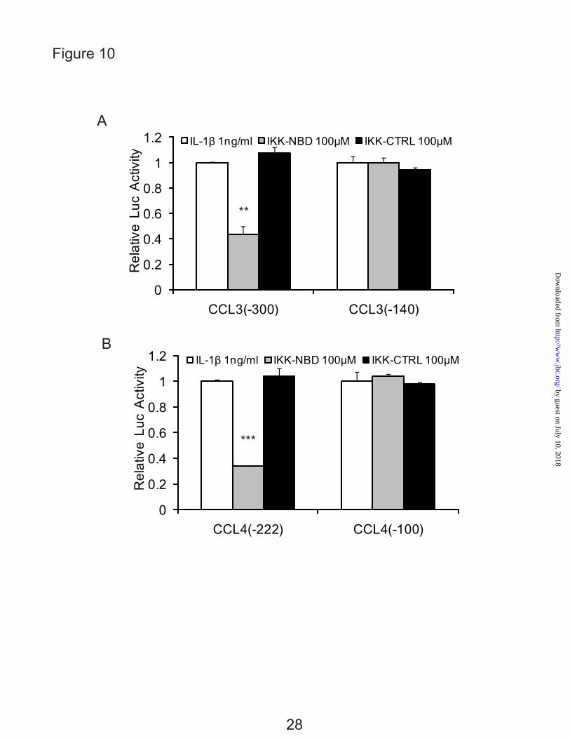
The involvement of NF-κB in the IL-1β responsive elements was further confirmed by
inhibiting its activity. The promoter activities were significantly decreased in the CCL3 (-300) and CCL4 (-222) by the IKK-NBD following 4 h of IL-1β treatment whereas the control IKK-NBD had no significant effect, and there was no effect on the shorter promoter constructs (Fig. 10, A and B).
The effect of inhibition of C/EBPβ and NF-κB were tested by inhibition of each transcription factor separately and together. SB303580, an inhibitor of C/EBPβ via inhibition of p38MAPK (21,43), and IKK-NBD reduced the expression of CCL3 and CCL4 with IL-1β stimulation (Fig. 11, A and B). IKK-NBD and SB303580 added together further decreased mRNA expression of CCL3 and CCL4 (Fig. 11A). Lastly, to confirm that both transcription factors are functional in transcription, promoter activity was determined after inhibition. The result showed that the promoter activities of CCL3 and CCL4 were significantly decreased by the inhibition of both factors (Fig. 11 B).
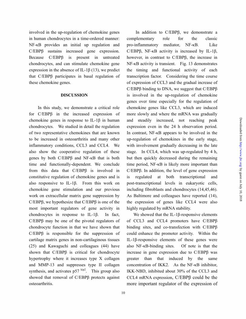
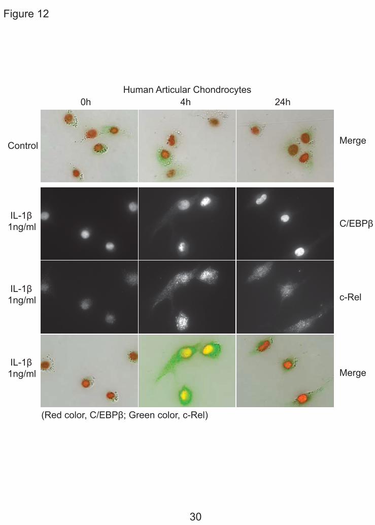
Immunohistochemistry of C/EBPβ and NF-κB — Our biochemical studies provided evidence that the regulation of CCL3 and CCL4 is coordinated in time with the NF-κB response early and the C/EBPβ response increased and sustained. To support this conclusion, we traced the location of these transcription factors over time by immunohistochemistry (Fig. 12). Fig. 12 demonstrates by double immunohistochemistry with antibodies to C/EBPβ and c-Rel after stimulation with IL-1β. At 0 time, c-Rel was localized in the cytoplasm, then was localized in the nucleus at 4 h and was greatly reduced by 24 h. C/EBPβ was present at the beginning and was increased at 4 h and at 24 h.
Our studies strongly suggest that both C/EBPβ and NF-κB are important for expression and response to IL-1β in human chondrocytes. The transcription factors, C/EBPβ and NF-κB are both
9
by guest on July 10, 2018http://w
ww
.jbc.org/D
ownloaded from
involved in the up-regulation of chemokine genes in human chondrocytes in a time-ordered manner: NF-κB provides an initial up regulation and C/EBPβ sustains increased gene expression. Because C/EBPβ is present in untreated chondrocytes, and can stimulate chemokine gene expression in the absence of IL-1β (13), we predict that C/EBPβ participates in basal regulation of these chemokine genes.
DISCUSSION
In this study, we demonstrate a critical role
for C/EBPβ in the increased expression of chemokine genes in response to IL-1β in human chondrocytes. We studied in detail the regulation of two representative chemokines that are known to be increased in osteoarthritis and many other inflammatory conditions, CCL3 and CCL4. We also show the cooperative regulation of these genes by both C/EBPβ and NF-κB that is both time and functionally-dependent. We conclude from this data that C/EBPβ is involved in constitutive regulation of chemokine genes and is also responsive to IL-1β. From this work on chemokine gene stimulation and our previous work on extracellular matrix gene suppression by C/EBPβ, we hypothesize that C/EBPβ is one of the most important regulators of gene activity in chondrocytes in response to IL-1β. In fact, C/EBPβ may be one of the pivotal regulators of chondrocyte function in that we have shown that C/EBPβ is responsible for the suppression of cartilage matrix genes in non-cartilaginous tissues (25) and Kawaguchi and colleagues (44) have shown that C/EBPβ is critical for chondrocyte hypertrophy where it increases type X collagen and MMP-13 and suppresses type II collagen synthesis, and activates p57 kip2. This group also showed that removal of C/EBPβ protects against osteoarthritis.
In addition to C/EBPβ, we demonstrate a complementary role for the classic pro-inflammatory mediator, NF-κB. Like C/EBPβ, NF-κB activity is increased by IL-1β, however, in contrast to C/EBPβ, the increase in NF-κB activity is transient. Fig. 13 demonstrates the timing and functional activity of each transcription factor. Considering the time course of expression of CCL3 and the gradual increase of C/EBPβ binding to DNA, we suggest that C/EBPβ is involved in the up-regulation of chemokine genes over time especially for the regulation of chemokine genes like CCL3, which are induced more slowly and where the mRNA was gradually and steadily increased, not reaching peak expression even in the 24 h observation period. In contrast, NF-κB appears to be involved in the up-regulation of chemokines in the early stage, with involvement gradually decreasing in the late stage. In CCL4, which was up-regulated by 4 h, but then quickly decreased during the remaining time period, NF-κB is likely more important than C/EBPβ. In addition, the level of gene expression is regulated at both transcriptional and post-transcriptional levels in eukaryotic cells, including fibroblasts and chondrocytes (14,45,46). As Baltimore and colleagues have reported (14), the expression of genes like CCL4 were also highly regulated by mRNA stability.
We showed that the IL-1β-responsive elements of CCL3 and CCL4 promoters have C/EBPβ binding sites, and co-transfection with C/EBPβ could enhance the promoter activity. Within the IL-1β-responsive elements of these genes were also NF-κB-binding sites. Of note is that the increase in gene expression due to C/EBPβ was greater than that induced by the same concentration of IKK2. As the NF-κB inhibitor, IKK-NBD, inhibited about 30% of the CCL3 and CCL4 mRNA expression, C/EBPβ could be the more important regulator of the expression of
10
by guest on July 10, 2018http://w
ww
.jbc.org/D
ownloaded from
11
chemokine genes in this cell type. Co-regulation by NF-κB and C/EBPβ was
recently shown in several genes in different cells (4,21,47,48). Teti and colleagues reported that Pseudomonas aeruginosa induced IL-8 (CXCL8) promoter expression and protein production in conjunctival epithelial cells by activating RelA and C/EBPβ and by promoting the cooperative binding of these transcription factors to the IL-8 promoter that in turn activates transcription (21). They also showed that C/EBPβ regulated the IL-8 basal transcriptional activity, but not NF-κB, which is consistent with our data on the IL-1β effect in chondrocytes. Gil and colleagues reported that IL-8, CXCL1 and CXCL5, which are CXCR2-binding chemokines, are up-regulated during replicative and oncogene-induced senescence. They demonstrated function of NF-κB and C/EBPβ in controlling the secretory program associated with cell senescence although they did not investigate the mechanism (4).
Studies have shown that the RelA (p65) subunit of NF-κB is involved in some lipopolysaccharide (LPS)-stimulated chemokines in human monocytic cells (17,18). However, we show in chondrocytes that, of the NF-κB subunits, c-Rel mRNA was increased more than p65 with IL-1β stimulation, and the computational analysis in chemokine genes CCL3 and CCL4 showed c-Rel binding sites were predicted in the IL-1β-responsive elements. Ohashi and colleagues recently reported that c-Rel was an important transcription factor in the regulation of the induction of proinflammatory cytokines (49), therefore c-Rel would be an interesting target for the further investigation.
Additional transcription factors have been shown to regulate CCL3 and CCL4. In multiple
myeloma, acute myeloid leukemia 1A (AML-1A) and AML-1B (also known as runt-related transcription factor 1, runx1) are principle regulators of CCL3 (26) although C/EBPβ was later found to be important (50). In our experiments reported here, runx1 transfection did not increase gene expression (Z. Zhang and L.J. Sandell, unpublished results). C/EBPβ it is thought to be responsible for regulating growth, proliferation and antiapoptotic responses by regulating the expression of other key transcription factors. In an early publication, Grove and Plumb (20) demonstrated that C/EBP, NF-κB and cEts family members were important in the LPS-induced stimulation of CCL3.
Levels of the proinflammatory cytokine IL-1β are increased in synovial fluid in joint diseases such as OA and RA and can induce cartilage damage and bone resorption. Chemokines function in the recruitment of neutrophils, monocytes, immature dendritic cells, B cells and activated T cells (51). However, chemokines also have functions at the individual tissue level as it has been reported that CXC family of chemokines are important in the regulation of angiogenesis, and CCL2, CCL3, and CCR2 stimulate osteoclastogenesis (52-54). In summary, the data presented here show that chondrocytes react in a cell-specific manner to IL-1β utilizing C/EBPβ and NF-κB in a combinatorial regulation of chemokine gene expression. The activity of C/EBPβ is augmented by a transient increase in activity of NF-κB and both transcription factors act independently on the chemokine genes, CCL3 and CCL4. These studies provide the foundation for the control of chemokine gene expression in chronic joint disease.
by guest on July 10, 2018http://w
ww
.jbc.org/D
ownloaded from
REFERENCES 1. Gerard, C., and Rollins, B. J. (2001) Nat Immunol 2(2), 108-115 2. Haringman, J. J., Ludikhuize, J., and Tak, P. P. (2004) Ann Rheum Dis 63(10), 1186-1194 3. Ma, Q., Jones, D., Borghesani, P. R., Segal, R. A., Nagasawa, T., Kishimoto, T., Bronson, R. T.,
and Springer, T. A. (1998) Proc Natl Acad Sci U S A 95(16), 9448-9453 4. Acosta, J. C., O'Loghlen, A., Banito, A., Guijarro, M. V., Augert, A., Raguz, S., Fumagalli, M.,
Da Costa, M., Brown, C., Popov, N., Takatsu, Y., Melamed, J., d'Adda di Fagagna, F., Bernard, D., Hernando, E., and Gil, J. (2008) Cell 133(6), 1006-1018
5. Han, J. H., Choi, S. J., Kurihara, N., Koide, M., Oba, Y., and Roodman, G. D. (2001) Blood 97(11), 3349-3353
6. Sandell, L. J., Xing, X., Franz, C., Davies, S., Chang, L. W., and Patra, D. (2008) Osteoarthritis Cartilage 16(12), 1560-1571
7. Yamasaki, K., Nakasa, T., Miyaki, S., Ishikawa, M., Deie, M., Adachi, N., Yasunaga, Y., Asahara, H., and Ochi, M. (2009) Arthritis Rheum 60(4), 1035-1041
8. Miyaki, S., Nakasa, T., Otsuki, S., Grogan, S. P., Higashiyama, R., Inoue, A., Kato, Y., Sato, T., Lotz, M. K., and Asahara, H. (2009) Arthritis Rheum 60(9), 2723-2730
9. Fukui, N., Zhu, Y., Maloney, W. J., Clohisy, J., and Sandell, L. J. (2003) J Bone Joint Surg Am 85-A Suppl 3, 59-66
10. Martel-Pelletier, J., Alaaeddine, N., and Pelletier, J. P. (1999) Front Biosci 4, D694-703 11. Tiku, K., Thakker-Varia, S., Ramachandrula, A., and Tiku, M. L. (1992) Cell Immunol 140(1),
1-20 12. Lee, J. H., Ort, T., Ma, K., Picha, K., Carton, J., Marsters, P. A., Lohmander, L. S., Baribaud, F.,
Song, X. Y., and Blake, S. (2009) Osteoarthritis Cartilage 17(5), 613-620 13. Zhang, Z., Xing, X., Hensley, G., Chang, L. W., Liao, W., Abu-Amer, Y., Sandell, L. J. (2010)
Arthritis Rheum DOI 10.1002/art.27473 14. Hao, S., and Baltimore, D. (2009) Nat Immunol 10(3), 281-288 15. Pulai, J. I., Chen, H., Im, H. J., Kumar, S., Hanning, C., Hegde, P. S., and Loeser, R. F. (2005) J
Immunol 174(9), 5781-5788 16. Rezzonico, R., Imbert, V., Chicheportiche, R., and Dayer, J. M. (2001) Blood 97(10), 2932-2940 17. Lakshmanan, U., and Porter, A. G. (2007) J Immunol 179(12), 8480-8490 18. Lim, C. A., Yao, F., Wong, J. J., George, J., Xu, H., Chiu, K. P., Sung, W. K., Lipovich, L., Vega,
V. B., Chen, J., Shahab, A., Zhao, X. D., Hibberd, M., Wei, C. L., Lim, B., Ng, H. H., Ruan, Y., and Chin, K. C. (2007) Mol Cell 27(4), 622-635
19. Amos, N., Lauder, S., Evans, A., Feldmann, M., and Bondeson, J. (2006) Rheumatology (Oxford) 45(10), 1201-1209
20. Grove, M., and Plumb, M. (1993) Mol Cell Biol 13(9), 5276-5289 21. Venza, I., Cucinotta, M., Visalli, M., De Grazia, G., Oliva, S., and Teti, D. (2009) J Biol Chem
284(7), 4191-4199 22. Okazaki, K., Li, J., Yu, H., Fukui, N., and Sandell, L. J. (2002) J Biol Chem 277(35),
31526-31533
12
by guest on July 10, 2018http://w
ww
.jbc.org/D
ownloaded from
23. Imamura, T., Imamura, C., Iwamoto, Y., and Sandell, L. J. (2005) J Biol Chem 280(17), 16625-16634
24. Imamura, T., Imamura, C., McAlinden, A., Davies, S. R., Iwamoto, Y., and Sandell, L. J. (2008) Arthritis Rheum 58(5), 1366-1376
25. Okazaki, K., Yu, H., Davies, S. R., Imamura, T., and Sandell, L. J. (2006) J Cell Biochem 97(4), 857-868
26. Choi, S. J., Oba, T., Callander, N. S., Jelinek, D. F., and Roodman, G. D. (2003) Blood 101(10), 3778-3783
27. Tseng, P. C., Hsu, H. C., Janmanchi, D., Lin, C. H., Kuo, Y. H., Chou, C. K., and Yeh, S. F. (2008) Biochem Pharmacol 76(9), 1121-1133
28. Otero, J. E., Dai, S., Alhawagri, M. A., Darwech, I., Abu-Amer, Y. (2010) J Bone Miner Res 25(6), 1282-1294
29. He, Y., and Crouch, E. (2002) J Biol Chem 277(22), 19530-19537 30. McAlinden, A., Liang, L., Mukudai, Y., Imamura, T., and Sandell, L. J. (2007) J Biol Chem
282(33), 24444-24454 31. Lu, K. T., Sinquett, F. L., Dryer, R. L., Song, C., and Covey, L. R. (2006) Blood 108(12),
3769-3776 32. Anest, V., Cogswell, P. C., and Baldwin, A. S., Jr. (2004) J Biol Chem 279(30), 31183-31189 33. Kristiansson, E., Thorsen, M., Tamas, M. J., and Nerman, O. (2009) Mol Biol Evol 26(6),
1299-1307 34. Descombes, P., and Schibler, U. (1991) Cell 67(3), 569-579 35. Poli, V. (1998) J Biol Chem 273(45), 29279-29282 36. Zhu, Y., Saunders, M. A., Yeh, H., Deng, W. G., and Wu, K. K. (2002) J Biol Chem 277(9),
6923-6928 37. Chang, L. W., Nagarajan, R., Magee, J. A., Milbrandt, J., and Stormo, G. D. (2006) Genome Res
16(3), 405-413 38. Davies, S. R., Chang, L. W., Patra, D., Xing, X., Posey, K., Hecht, J., Stormo, G. D., and Sandell,
L. J. (2007) Genome Res 17(10), 1438-1447 39. Zandi, E., Rothwarf, D. M., Delhase, M., Hayakawa, M., and Karin, M. (1997) Cell 91(2),
243-252 40. Hacker, H., and Karin, M. (2006) Sci STKE 2006(357), re13 41. DiDonato, J. A., Hayakawa, M., Rothwarf, D. M., Zandi, E., and Karin, M. (1997) Nature
388(6642), 548-554 42. Rothwarf, D. M., and Karin, M. (1999) Sci STKE 1999(5), RE1 43. Basak, C., Pathak, S. K., Bhattacharyya, A., Mandal, D., Pathak, S., and Kundu, M. (2005) J Biol
Chem 280(6), 4279-4288 44. Hirata, M., Kugimiya, F., Fukai, A., Ohba, S., Kawamura, N., Ogasawara, T., Kawasaki, Y., Saito,
T., Yano, F., Ikeda, T., Nakamura, K., Chung, U. I., and Kawaguchi, H. (2009) PLoS One 4(2), e4543
45. Fukui, N., Ikeda, Y., Ohnuki, T., Hikita, A., Tanaka, S., Yamane, S., Suzuki, R., Sandell, L. J., and Ochi, T. (2006) J Biol Chem 281(37), 27229-27241
13
by guest on July 10, 2018http://w
ww
.jbc.org/D
ownloaded from
46. Tebo, J. M., Datta, S., Kishore, R., Kolosov, M., Major, J. A., Ohmori, Y., and Hamilton, T. A. (2000) J Biol Chem 275(17), 12987-12993
47. El-Asmar, B., Giner, X. C., and Tremblay, J. J. (2009) J Mol Endocrinol 42(2), 131-138 48. Sow, F. B., Alvarez, G. R., Gross, R. P., Satoskar, A. R., Schlesinger, L. S., Zwilling, B. S., and
Lafuse, W. P. (2009) J Leukoc Biol 86(5), 1247-1258 49. Lu, Y. C., Kim, I., Lye, E., Shen, F., Suzuki, N., Suzuki, S., Gerondakis, S., Akira, S., Gaffen, S.
L., Yeh, W. C., and Ohashi, P. S. (2009) J Immunol 182(11), 7212-7221 50. Pal, R., Janz, M., Galson, D. L., Gries, M., Li, S., Johrens, K., Anagnostopoulos, I., Dorken, B.,
Mapara, M. Y., Borghesi, L., Kardava, L., Roodman, G. D., Milcarek, C., and Lentzsch, S. (2009) Blood 114(18), 3890-3898
51. Borzi, R. M., Mazzetti, I., Cattini, L., Uguccioni, M., Baggiolini, M., and Facchini, A. (2000) Arthritis Rheum 43(8), 1734-1741
52. Binder, N. B., Niederreiter, B., Hoffmann, O., Stange, R., Pap, T., Stulnig, T. M., Mack, M., Erben, R. G., Smolen, J. S., and Redlich, K. (2009) Nat Med 15(4), 417-424
53. Miyamoto, K., Ninomiya, K., Sonoda, K. H., Miyauchi, Y., Hoshi, H., Iwasaki, R., Miyamoto, H., Yoshida, S., Sato, Y., Morioka, H., Chiba, K., Egashira, K., Suda, T., Toyama, Y., and Miyamoto, T. (2009) Biochem Biophys Res Commun 383(3), 373-377
54. Rudolph, E. H., and Woods, J. M. (2005) Curr Pharm Des 11(5), 613-631
FOOTNOTES
* The authors would like to thank Drs John C. Clohisy, Robert L. Barrack, Douglas McDonald, Ryan Nunley, and Rick W. Wright, and Head Nurse Keith Foreman, for normal cartilage and cartilage from patients with osteoarthritis. The authors would like to thank Drs G. David Roodman and Sheau-Farn Yeh for the generous gifts of CCL3 and CCL4 plasmids. The authors would also like to thank Drs Xiaoyun Xing, Chikashi Kobayashi, Yousef Abu-Amer and Audrey McAlinden at the Washington University School of Medicine for valuable assistance. These studies were funded by The National Institute for Arthritis, Musculoskeletal and Skin Diseases (R01 AR 050847 and R01 AR036994). Abbreviations: C/EBPβ, CCAAT/enhancer-binding protein; NF-κB, nuclear factor kappa B; IL-1β, interleukin 1β; RA, rheumatoid arthritis; OA, osteoarthritis; ECM, extracellular matrix; COL2A1, type II procollagen gene; BMP-2, bone morphogenetic protein 2; TNF-α, tumor necrosis factor-alpha; MIP-1, macrophage Inflammatory Proteins 1; IKK-NBD, cell-permeable NEMO binding domain; IKK2, IkappaB kinase 2; EMSA, electrophoretic mobility shift assay; ChIP, chromatin immunoprecipitation; siRNA, small interfering RNA; qPCR, real time PCR; LAP, liver-enriched transcriptional activator proteins; LIP, liver-enriched inhibitory protein; MMP-13, matrix metalloproteinase 13; AML-1, acute myeloid leukemia 1; Runx1, runt-related transcription factor 1.
FIGURE LEGENDS
14
by guest on July 10, 2018http://w
ww
.jbc.org/D
ownloaded from
FIG. 1. IL-1β stimulates the expression of CCL3 and CCL4 in normal human articular chondrocytes (HACs) and T/C-28 chondrocyte cells. A, HACs were treated with 1 ng/ml IL-1β for various times as indicated. B, T/C-28 cells were treated with 1 ng/ml IL-1β for various times as indicated. C, T/C-28 cells were treated for various concentrations for 24 h. The relative expression levels were examined by quantitative real time PCR method. Each bar represents the mean ±S.D. from three experiments. FIG. 2. IL-1β-responsive element is located between -300 to -140 bp of the CCL3 promoter, and -222 to -100 bp of the CCL4 promoter. 5’-deletion constructs were transiently transfected into T/C-28a2 cells and incubated for 24h, then a further 24h (CCL3) (A) or 8 h (CCL4) (B) in the absence or presence of IL-1β (1 ng/ml) with fresh complete medium. Luciferase activities were measured and expressed relative to the activity of promoterless pGL3b (set as 1). Each bar represents the mean ±S.D. of at least three independent experiments. FIG. 3. IL-1β stimulates the expression of C/EBPβ in normal human articular chondrocytes (HACs) and T/C-28 chondrocyte cells. The relative expression levels were examined in normal chondrocytes from human articular cartilage and T/C-28 cells treated with IL-1β (1 ng/ml) using quantitative real time PCR method. A and B, Cells were treated with 1 ng/ml IL-1β for various times as indicated in HACs (A) and T/C-28 cells (B). C, T/C-28 cells were treated for various concentrations for 24 h. D, C/EBPβ proteins were examined by western blot of nuclear extracts (N.E.) from T/C-28 cells treated or without IL-1β (1 ng/ml). IL-1β increased all the isoforms of C/EBPβ: LAP (38 kDa, and 36 kDa) and LIP (16 kDa), and the both isoforms of C/EBPβ-LAP were more significant increase than LIP. Each bar represents the mean ±S.D. from three to five experiments. FIG. 4. C/EBPβ binds the sequence between -251 to -238 bp of the CCL3 promoter, and -168 and -155 bp of the CCL4 promoter. A, a diagram showing DNA fragments used for EMSA. For CCL3, Fragment A from -300 to -141 bp and relative locations of oligonucleotides 1, 2 and 3 were shown. For CCL4, Fragment B from -222 to -101 bp and relative locations of oligonucleotides 4, 5 and 6 were shown. The oligonucleotide 2 from -255 to -232 bp and oligonucleotide 5 from -175 to -151 bp contain C/EBPβ motif (underlined). Mu, mutant oligonucleotide 2 and 5 containing two base pair mutations (AA - CC) is underlined within the C/EBPβ motif. B, EMSA for T/C-28a2 nuclear extracts using fragment A or B as a probe and various cold competitors at 100-fold molar excess. The oligonucleotide 2 and 5 competed with the binding of nuclear proteins to the probe. C, EMSA and supershift analysis for T/C-28a2 nuclear extracts using oligonucleotide 2 and 5 as the probe. Mutant oligonucleotide 2 and 5 did not compete with the binding of nuclear proteins to the probe. Antibodies against C/EBPβ or SREBP1 were preincubated with the nuclear extracts. Lines showed shift band of the complex of C/EBPβ antibody (Ab), and specific retarded bands (C, control; I, IL-1β treatment). FIG. 5. C/EBPβ binding activity to the CCL3(-300/-141) and CCL4(-222/-101) promoter region is increased in the presence of IL-1β. Chromatin immunoprecipitation assay was performed using
15
by guest on July 10, 2018http://w
ww
.jbc.org/D
ownloaded from
anti-C/EBPβ antibodies in T/C-28 cells treated with IL-1β for various times as indicated. Treatment with IL-1β enhanced the binding of C/EBPβ to the promoter. A, T/C-28 cells were treated with 1 ng/ml IL-1β for 0 h, 1 h, 4 h, 8 h, 24 h. B, T/C-28 cells were treated or without IL-1β (1 ng/ml) for 1 h. C and D, T/C-28 cells were treated with 1 ng/ml IL-1β for 0 h and 24 h, and the relative expression levels of input and C/EBPβ antibody DNAs of CCL3 (C) and CCL4 (D) were examined by quantitative real time PCR method. The value was normalized to GAPDH, and the expression levels of input and C/EBPβ at 0 h were set as 1. E, T/C-28 cells were treated with 1 ng/ml IL-1β for 0 h, 4 h, 8 h and 24 h, and the relative expression level of CCL3 of input and C/EBPβ antibody DNAs were examined by quantitative real time PCR method. The value was normalized to GAPDH and input, and the expression levels of C/EBPβ at 0 h were set as 1. FIG. 6. C/EBPβ functions as an activator for the CCL3 and CCL4 promoter activities, and mutation of the C/EBP-binding site down-regulates the promoter activity of the CCL3 (-300/-1) and CCL4 (-222/-1) constructs. Site-directed mutagenesis was performed within the C/EBP-binding site of the CCL3 (-300/-1) and CCL4 (-222/-1) constructs using the mutant oligonucleotide 2 and 5 sequence. The mutant and wild type constructs were transiently transfected into T/C-28a2 cells and incubated with or without IL-1β. Luciferase activities were measured and expressed relative to the activity of promoterless pGL3b (set as 1). Further, the wild type and mutant of CCL3 (-300/-1) and CCL4 (-222/-1) were co-transfected with C/EBPβ expression plasmid into T/C-28a2 cells with IL-1β (1 ng/ml). Relative luciferase activity indicates the fold expression relative to the activity of promoterless pGL3b which co-transfected with empty vector (set as 1) in the presence of IL-1β (1 ng/ml). Each bar represents the mean ±S.D. of at least three independent experiments. FIG. 7. Silencing of C/EBPβ suppresses the IL-1β-induced CCL3 and CCL4 transcriptional activation. A, Odyssey western blot of C/EBPβ proteins in T/C-28 cells transfected with control (ctrl) or C/EBPβ siRNAs (162 pM), and stimulated with 1ng/ml IL-1β for 8h. B, T/C-28 cells were transfected with CCL3 and CCL4 promoters and where indicated cotransfected with control (ctrl) or C/EBPβ siRNAs (162 pM). Twenty-four hours after transfection, T/C-28 cells were stimulated with 1ng/ml IL-1β for 24 h (CCL3) or 8 h (CCL4). Relative luciferase activity indicates the fold expression relative to the activity of promoter with IL-1β (1 ng/ml) only (set as 1). C, T/C-28 cells transfected with control (ctrl) or C/EBPβ siRNAs were stimulated for 8 h with IL-1β (1 ng/ml). The relative expression levels were examined by quantitative real time PCR method. The p value of C/EBPβ siRNA was compared with IL-1β alone based on student’s t-test (**, means p < 0.01). Each bar represents the mean ±S.D. of at least three independent experiments. FIG. 8. Involvement of NF-κB in the up-regulation of chemokines in human chondrocytes in response to IL-1β. A, Human articular chondrocytes were treated with 1 ng/ml IL-1β for various times as indicated. B, Effect of IL-1β (1ng/ml) on the expression of pNF-κB Luc reporter in T/C-28 human chondrocytes. Relative luciferase activity indicates the fold expression relative to the activity of zero time (set as 1) in the presence of IL-1β (1 ng/ml). C, Human articular chondrocytes were pretreated with vehicle (DMSO), IKK-NBD peptide (100 μM and 200 μM), or IKK-NBD control peptide (200 μM) for 1
16
by guest on July 10, 2018http://w
ww
.jbc.org/D
ownloaded from
h and then exposed to IL-1β (1 ng/ml) for 4 h. The p value of IKK-NBD (200 μM) was compared with IL-1β alone based on student’s t-test (*, means p < 0.05; **, means p < 0.01; ***, means p < 0.001). Each bar represents the mean ±S.D. of three independent experiments. FIG. 9. Transcriptional regulation in the highly expression of CCL3 and CCL4. A, The candidate C/EBPβ and NF-κB binding sites in human CCL3 (-1395) and CCL4 (-1281) constructs. B, C/EBPβ and IKK2 stimulate the expression of CCL3 (-1395) and CCL4 (-1281) promoter in T/C-28 human chondrocytes. The CCL3 (-1395) and CCL4 (-1281) promoter constructs were co-transfected with C/EBPβ and IKK2 expression plasmid into T/C-28a2 cells with or without IL-1β (1 ng/ml) for 24 h (CCL3) or 8 h (CCL4). Relative luciferase activity indicates the fold expression relative to the activity of the construct co-transfected with empty vector (set as 1) in the present of IL-1β after each co-transfection compared with their activities in the absence of IL-1β. Each bar represents the mean ± S.D. of three independent experiments. FIG. 10. Involvement of NF-κB in IL-1β-responsive elements of chemokines in human chondrocytes. The CCL3 (-300) (A), CCL3 (-140) (A), CCL4 (-222) (B) and CCL4 (-100) (B) promoter constructs were co-transfected into T/C-28a2 cells. Twenty-four hours after transfection, T/C-28 cells were pretreated with vehicle (DMSO), IKK-NBD peptide (100 μM), or IKK-NBD control peptide (100 μM) for 1 h and then exposed to IL-1β (1ng/ml) for 4 h. The p value of IKK-NBD (100 μM) was compared with IL-1β alone based on student’s t-test (**, means p < 0.01; ***, means p < 0.001). Each bar represents the mean ±S.D. of three independent experiments. FIG. 11. IKK-NBD and SB303580 (SB) co-enhance the inhibition of chemokines by human chondrocytes with IL-1β treatment. A, T/C-28 cells were pretreated with vehicle (DMSO), IKK-NBD peptide (100 μM), SB303580 (100 μM), or IKK-NBD control peptide (100 μM) for 1 h and then exposed to IL-1β (1 ng/ml) for 4 h. After IL-1β treatment, total RNA was isolated, and real-time quantitative-PCR was performed. B, The CCL3 (-1395) and CCL4 (-1281) promoter constructs were transfected into T/C-28a2 cells and incubated for 24 h, then were pretreated with vehicle (DMSO), IKK-NBD peptide (100 μM), SB303580 (100 μM), and IKK-NBD control peptide (100 μM) for 1 h and then exposed to IL-1β (1 ng/ml) for 4 h. Relative luciferase activity indicates the fold expression relative to the activity of the construct co-transfected with empty vector (set as 1) in the presence of IL-1β (1 ng/ml). The p value of IKK-NBD (100 μM) or SB303580 (100 μM) was compared with IL-1β alone based on student’s t-test (**, means p < 0.01; ***, means p < 0.001). Each bar represents the mean ±S.D. of at least three independent experiments. FIG. 12. Subcellular localization of C/EBPβ and c-Rel in response to IL-1β. The top panel shows a merged image of immunohistochemistry for C/EBPβ and the NF-kB subunit c-Rel: C/EBPβ is present in the nucleus of cells even without IL-1β exposure: C/EBPβ is located in the nucleus while c-Rel is located diffusely throughout the cell. With addition of IL-1β, C/ EBPβ is increased in the nucleus and c-Rel is increased and translocates to the nucleus. At 24 hr, C/ EBPβ remains high in the nucleus and c-Rel is significantly reduced.
17
by guest on July 10, 2018http://w
ww
.jbc.org/D
ownloaded from
18
FIG. 13. A transcriptional model for the up-regulation of chemokine genes in human chondrocytes in response to IL-1β. Upon IL-1β stimulation, C/ EBPβ protein and DNA binding are increased up to the 24 hr tested. NF-kB shows little function without IL-1β, but increased up to 8 hr after which the function is greatly reduced. The distribution of the transcription factors (Fig 12) is consistent with the functional parameters.
by guest on July 10, 2018http://w
ww
.jbc.org/D
ownloaded from
0
4
8
12
16
20
CCL3 CCL4
Fold
cha
nge
of
mR
NA TC28 with IL-1β 24h
0ng/ml 1ng/ml 5ng/ml
0
30
60
90
120
150
CCL3 CCL4
Fold
cha
nge
of
mR
NA
HACs with IL-1β 1ng/ml0h 1h4h 8h24h
0
5
10
15
20
25
CCL3 CCL4
Fold
cha
nge
of
mR
NA TC28 with IL-1β 1ng/ml
0h 8h 24h
Figure 1A
B
C
19
by guest on July 10, 2018http://w
ww
.jbc.org/D
ownloaded from
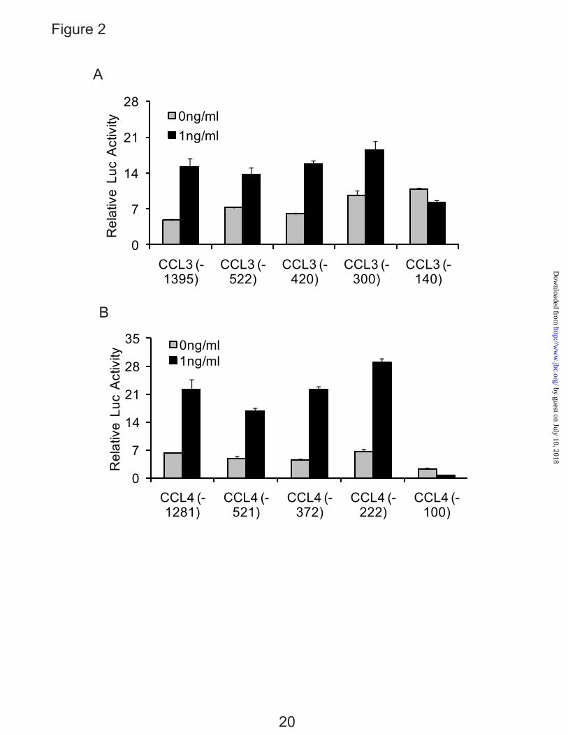
Figure 2
A
0
7
14
21
28
CCL3 (-1395)
CCL3 (-522)
CCL3 (-420)
CCL3 (-300)
CCL3 (-140)
Rel
ativ
e L
uc A
ctiv
ity 0ng/ml1ng/ml
B
0
7
14
21
28
35
CCL4 (-1281)
CCL4 (-521)
CCL4 (-372)
CCL4 (-222)
CCL4 (-100)
Rel
ativ
e L
uc A
ctiv
ity
0ng/ml1ng/ml
20
by guest on July 10, 2018http://w
ww
.jbc.org/D
ownloaded from
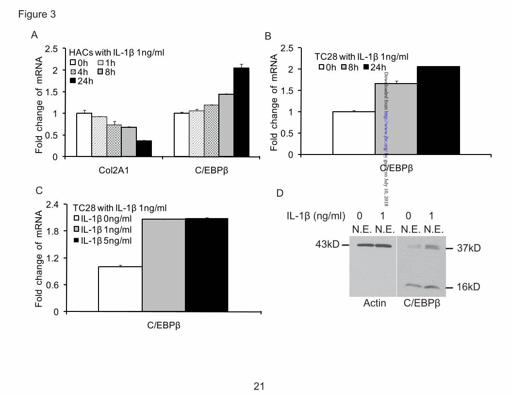
Figure 3
A B
0
0.5
1
1.5
2
2.5
C/EBPβ
Fold
cha
nge
of
mR
NA TC28 with IL-1β 1ng/ml
0h 8h 24h
0
0.5
1
1.5
2
2.5
Col2A1 C/EBPβ
Fold
cha
nge
of
mR
NA
HACs with IL-1β 1ng/ml0h 1h4h 8h24h
C D
0
0.6
1.2
1.8
2.4
C/EBPβ
Fold
cha
nge
of
mR
NA TC28 with IL-1β 1ng/ml
IL-1β 0ng/mlIL-1β 1ng/mlIL-1β 5ng/ml
IL-1β (ng/ml) 0 1 0 1 N.E. N.E. N.E. N.E.
43kD 37kD
16kDActin C/EBPβ
21
by guest on July 10, 2018http://w
ww
.jbc.org/D
ownloaded from
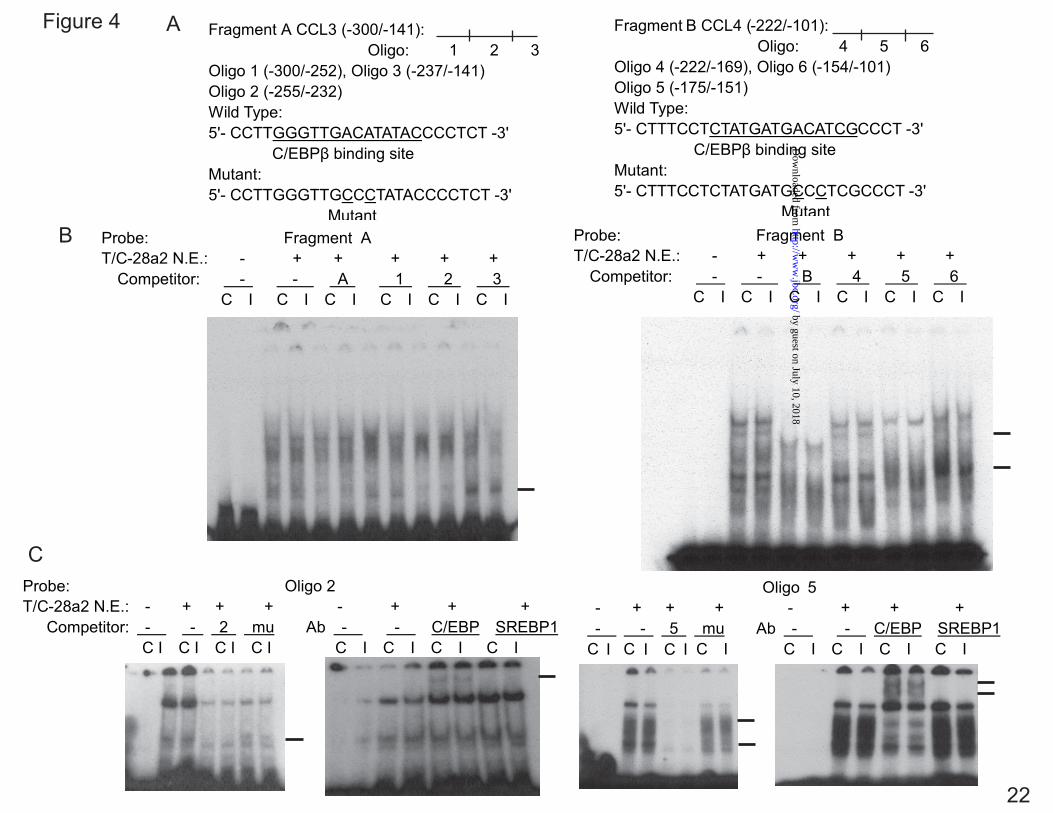
Figure 4 A
B
C
22
by guest on July 10, 2018http://w
ww
.jbc.org/D
ownloaded from
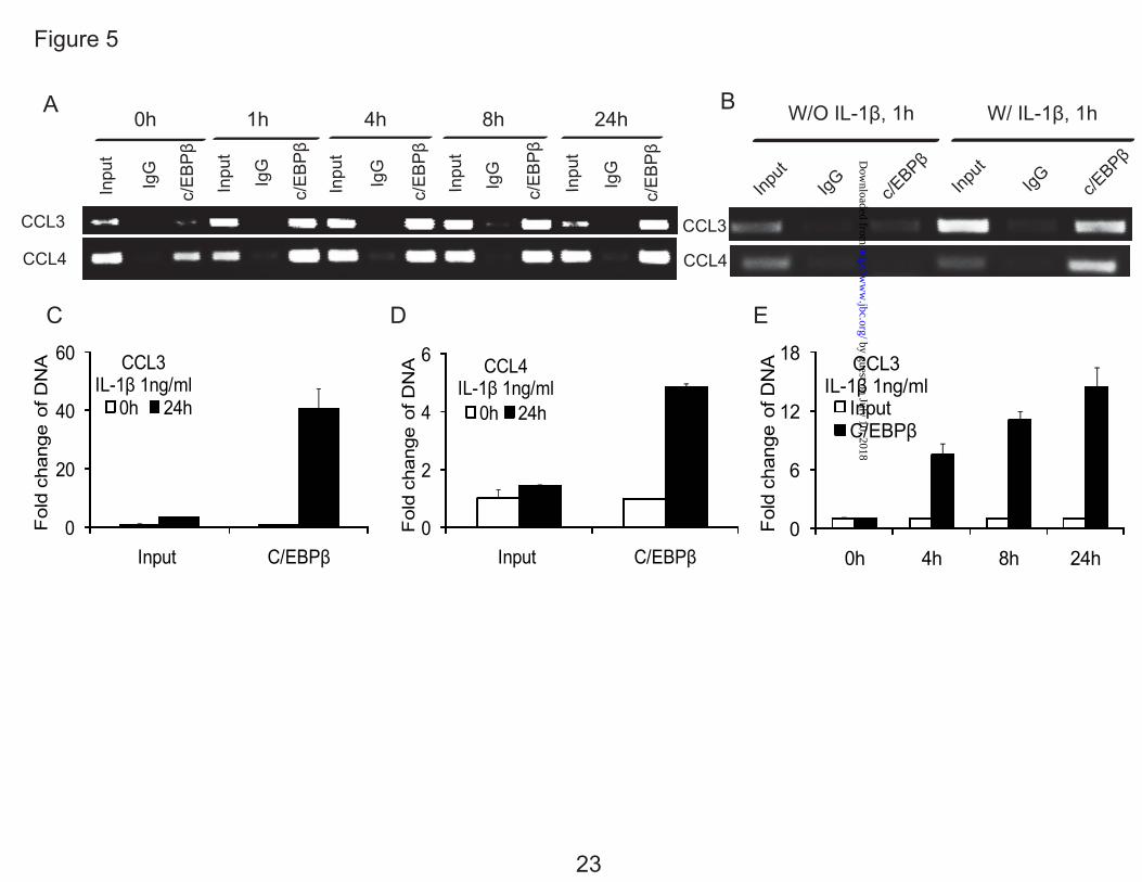
Figure 5
A B
0
20
40
60
Input C/EBPβ
Fol
d ch
ange
of D
NA
CCL3IL-1β 1ng/ml
0h 24h
0
2
4
6
Input C/EBPβ
Fol
d ch
ange
of D
NA CCL4
IL-1β 1ng/ml0h 24h
0
6
12
18
0h 4h 8h 24h
Fold
cha
nge
of D
NA CCL3
IL-1β 1ng/ml Input C/EBPβ
C D E
0h 1h 4h 8h 24h W/O IL-1β, 1h W/ IL-1β, 1h
23
by guest on July 10, 2018http://w
ww
.jbc.org/D
ownloaded from
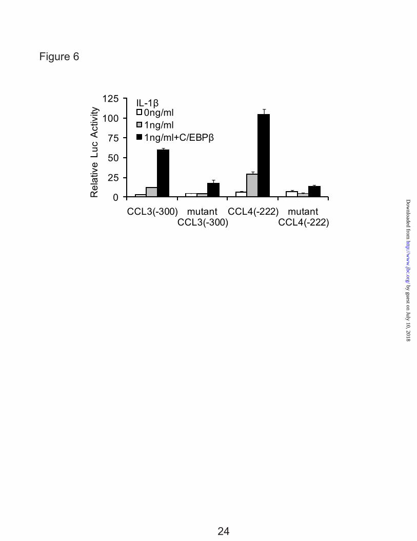
Figure 6
0
25
50
75
100
125
CCL3(-300) mutant CCL3(-300)
CCL4(-222) mutant CCL4(-222)
Rel
ativ
e L
uc A
ctiv
ityIL-1β
0ng/ml1ng/ml1ng/ml+C/EBPβ
24
by guest on July 10, 2018http://w
ww
.jbc.org/D
ownloaded from
0
0.2
0.4
0.6
0.8
1
1.2
CCL3(-300) CCL4(-222)
Rel
ativ
e L
uc A
ctiv
ity
Promoter ctrl-siRNA C/EBPβ-siRNA
siRNA (162 pM) Ctrl C/EBPβ
LAP
LIPActin
1 ng/ml 1ng/ml IL-1β
Figure 7A
0
0.2
0.4
0.6
0.8
1
1.2
CCL3 CCL4
mR
NA
exp
ress
ion
IL-1β 1ng/ml ctrl-siRNA C/EBPβ-siRNA
****
B
C
****
25
by guest on July 10, 2018http://w
ww
.jbc.org/D
ownloaded from
00.20.40.60.8
11.2
mR
NA
exp
ress
ion
IL-1β 1ng/ml IKK-NBD 100μM IKK-NBD 200μM IKK-CTRL 200μM
0
6
12
18
24
30
Fold
cha
nge
of
mR
NA HACs with IL-1β 1ng/ml
0h 1h4h 8h24h
0
8
16
24
32
40
0 1 8 24
Rel
ativ
e L
uc A
ctiv
ity
IL-1βExposure(hr)
pNF-kappaB Luc
HACs
Figure 8
A
B
C
*** ** *** * * *** ** ** **
26
by guest on July 10, 2018http://w
ww
.jbc.org/D
ownloaded from
S1 S2 S3S4S5 S6S7S8S9
S10 S11 S12 S13 S14 S15 S16
Figure 9
A
B
27
Luciferase
-522
-420
-300 -140
IL-1β Responsive Elements
-100 -222 -372 -521 -1281
Luciferase
012345678
CCL3(-1395) CCL4(-1281)
Rel
ativ
e L
uc A
ctiv
ity Basic Vector
IKK2 200ng
C/EBPβ 200ng
IKK2 200ng + C/EBPβ 200ngIKK2 400ng
C/EBPβ 400ng
by guest on July 10, 2018http://w
ww
.jbc.org/D
ownloaded from
0
0.2
0.4
0.6
0.8
1
1.2
CCL3(-300) CCL3(-140)
Rel
ativ
e L
uc A
ctiv
ityIL-1β 1ng/ml IKK-NBD 100μM IKK-CTRL 100μM
0
0.2
0.4
0.6
0.8
1
1.2
CCL4(-222) CCL4(-100)
Rel
ativ
e L
uc A
ctiv
ity
IL-1β 1ng/ml IKK-NBD 100μM IKK-CTRL 100μM
**
***
Figure 10
A
B
28
by guest on July 10, 2018http://w
ww
.jbc.org/D
ownloaded from
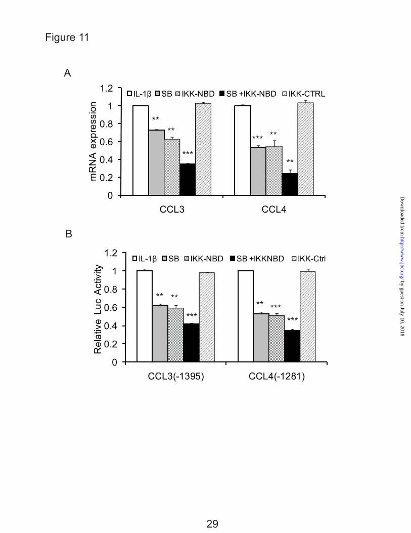
Figure 11
A
B
0
0.2
0.4
0.6
0.8
1
1.2
CCL3(-1395) CCL4(-1281)
Rel
ativ
e L
uc A
ctiv
ity
IL-1β SB IKK-NBD SB +IKKNBD IKK-Ctrl
0
0.2
0.4
0.6
0.8
1
1.2
CCL3 CCL4
mR
NA
exp
ress
ion
IL-1β SB IKK-NBD SB +IKK-NBD IKK-CTRL
**** **
**
** ****
******
******
***
29
by guest on July 10, 2018http://w
ww
.jbc.org/D
ownloaded from
0h 4h 24h
IL-1β1ng/ml
Human Articular Chondrocytes
(Red color, C/EBPβ; Green color, c-Rel)
Figure 12
30
Merge
C/EBPβ
c-Rel
Merge
Control
IL-1β1ng/ml
IL-1β1ng/ml
by guest on July 10, 2018http://w
ww
.jbc.org/D
ownloaded from
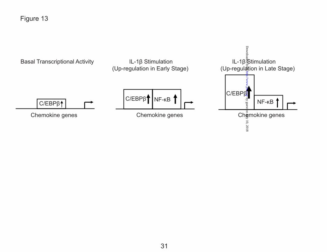
Basal Transcriptional Activity IL-1β Stimulation IL-1β Stimulation (Up-regulation in Early Stage) (Up-regulation in Late Stage)
C/EBPβ
C/EBPβ NF-κB
C/EBPβNF-κB
Chemokine genes Chemokine genes Chemokine genes
Figure 13
31
by guest on July 10, 2018http://w
ww
.jbc.org/D
ownloaded from
Linda J. SandellZhiqi Zhang, Jennifer L. Bryan, Elizabeth DeLassus, Li-Wei Chang, Weiming Liao and
)β (IL-1βchondrocytes in response to interleukin-1mediate high level expression of chemokine genes CCL3 and CCL4 by human
B)κ) and nuclear factor kappa B (NF-β (C/EBPβCCAAT/enhancer-binding protein
published online August 11, 2010J. Biol. Chem.
10.1074/jbc.M110.130377Access the most updated version of this article at doi:
Alerts:
When a correction for this article is posted•
When this article is cited•
to choose from all of JBC's e-mail alertsClick here
Supplemental material:
http://www.jbc.org/content/suppl/2010/09/02/M110.130377.DC1
by guest on July 10, 2018http://w
ww
.jbc.org/D
ownloaded from