Biochemistry Review I - University of Pittsburgh
27
Biochemistry Review I Biochemistry Review I BBSI @ Pitt 2004 BBSI @ Pitt 2004 Rajan Munshi Center for Computational Biology and Bioinformatics University of Pittsburgh School of Medicine May 25, 2004
Transcript of Biochemistry Review I - University of Pittsburgh
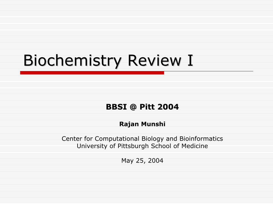
Biochemistry Review IBiochemistry Review I
BBSI @ Pitt 2004BBSI @ Pitt 2004
Rajan Munshi
Center for Computational Biology and BioinformaticsUniversity of Pittsburgh School of Medicine
May 25, 2004
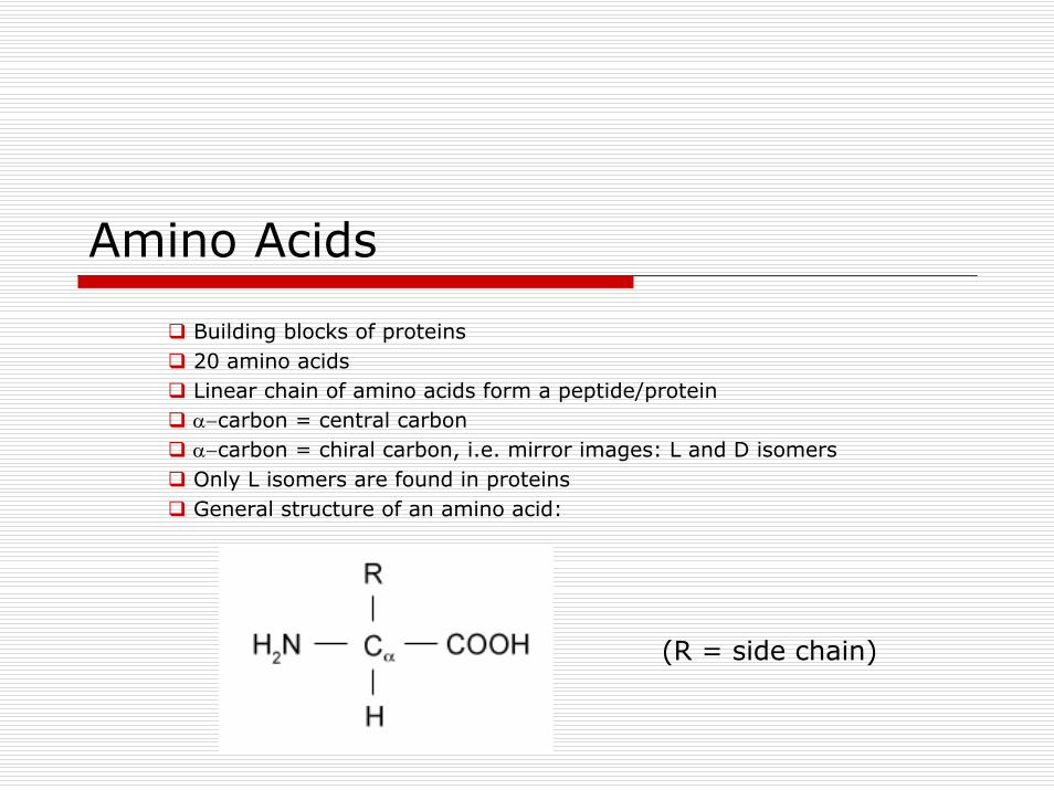
Amino Acids
Building blocks of proteins20 amino acidsLinear chain of amino acids form a peptide/proteinα−carbon = central carbonα−carbon = chiral carbon, i.e. mirror images: L and D isomersOnly L isomers are found in proteinsGeneral structure of an amino acid:
(R = side chain)
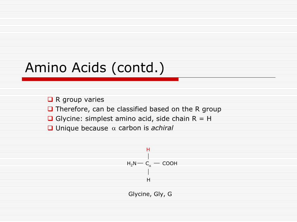
Amino Acids (contd.)
R group variesTherefore, can be classified based on the R groupGlycine: simplest amino acid, side chain R = HUnique because α carbon is achiral
H
CαH2N COOH
H
Glycine, Gly, G
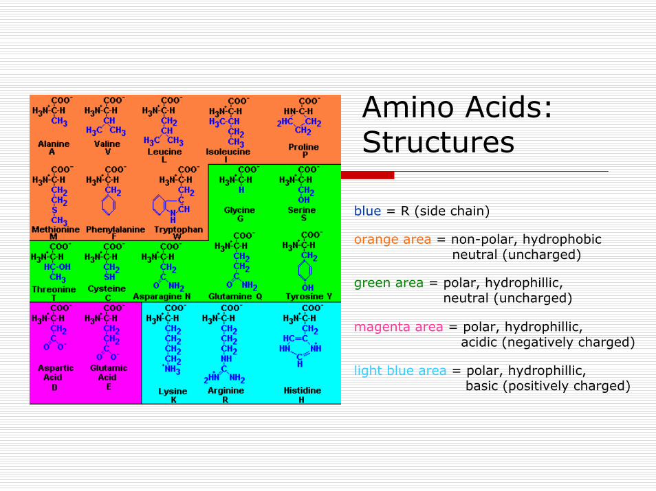
Amino Acids: Structures
blue = R (side chain)
orange area = non-polar, hydrophobicneutral (uncharged)
green area = polar, hydrophillic,neutral (uncharged)
magenta area = polar, hydrophillic,acidic (negatively charged)
light blue area = polar, hydrophillic,basic (positively charged)
Amino Acids: ClassificationNon-polar, hydrophobic,neutral (uncharged)Alanine, Ala, AValine, Val, VLeucine, Leu, LIsoleucine, Ile, IProline, Pro, PMethionine, Met, MPhenylalanine, Phe, FTryptophan, Trp, W
Polar, hydrophillic,neutral (uncharged)Glycine, Gly, GSerine, Ser, SThreonine, Thr, TCysteine, Cys, CAsparagine, Asn, NGlutamine, Gln, QTyrosine, Tyr, Y
Polar, hydrophillic,basic (positively charged)Lysine, Lys, KArginine, Arg, RHistidine, His, H
Polar, hydrophillic,Acidic (negatively charged)Aspartic acid, Asp, DGlutamic acid, Glu, E
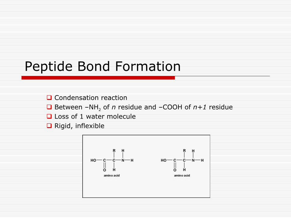
Peptide Bond Formation
Condensation reactionBetween –NH2 of n residue and –COOH of n+1 residueLoss of 1 water moleculeRigid, inflexible
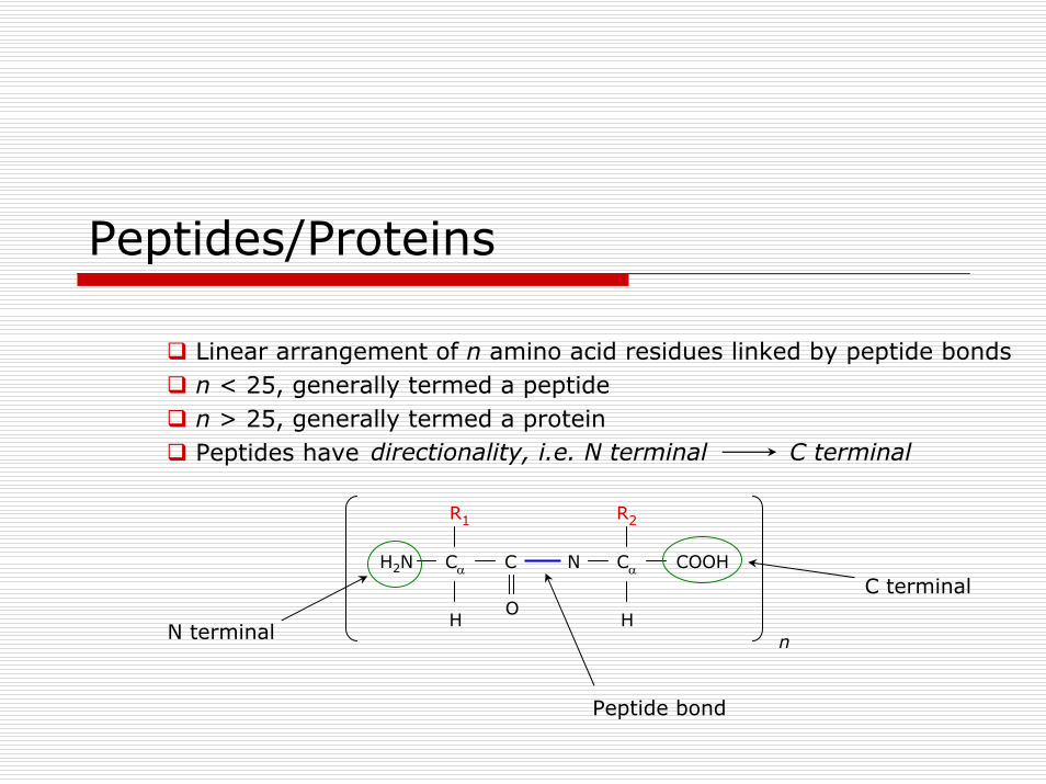
Peptides/Proteins
Linear arrangement of n amino acid residues linked by peptide bondsn < 25, generally termed a peptiden > 25, generally termed a proteinPeptides have
R1
CαH2N C
H
CαN COOH
HO
R2
n
directionality, i.e. N terminal C terminal
N terminal
C terminal
Peptide bond
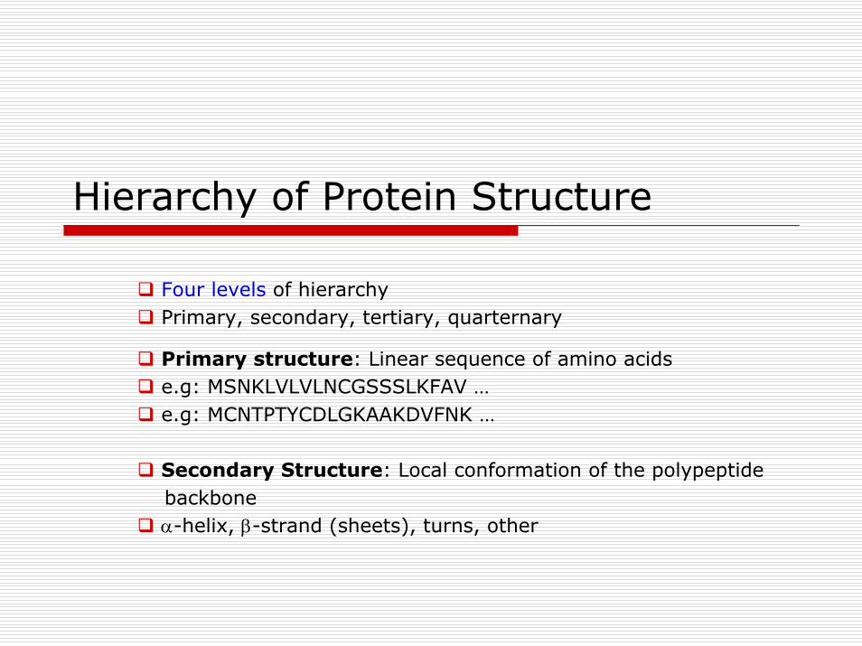
Hierarchy of Protein Structure
Four levels of hierarchyPrimary, secondary, tertiary, quarternary
Primary structure: Linear sequence of amino acidse.g: MSNKLVLVLNCGSSSLKFAV …e.g: MCNTPTYCDLGKAAKDVFNK …
Secondary Structure: Local conformation of the polypeptidebackboneα-helix, β-strand (sheets), turns, other
Secondary Structure: α-helix
Most abundant; ~35% of residues in a proteinRepetitive secondary structure3.6 residues per turn; pitch (rise per turn) = 5.4 ÅC′=O of i forms H bonds with NH of residue i+4Intra-strand H bondingC′=0 groups are parallel to the axis; side chains point away from the axisAll NH and C′O are H-bonded, except first NH and last C′OHence, polar ends; present at surfacesAmphipathic
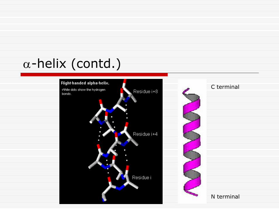
α-helix (contd.)
C terminal
N terminal
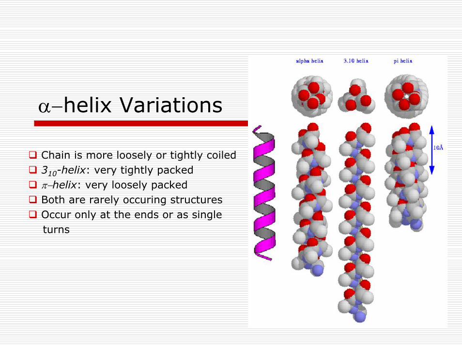
α−helix Variations
Chain is more loosely or tightly coiled310-helix: very tightly packedπ−helix: very loosely packedBoth are rarely occuring structuresOccur only at the ends or as singleturns
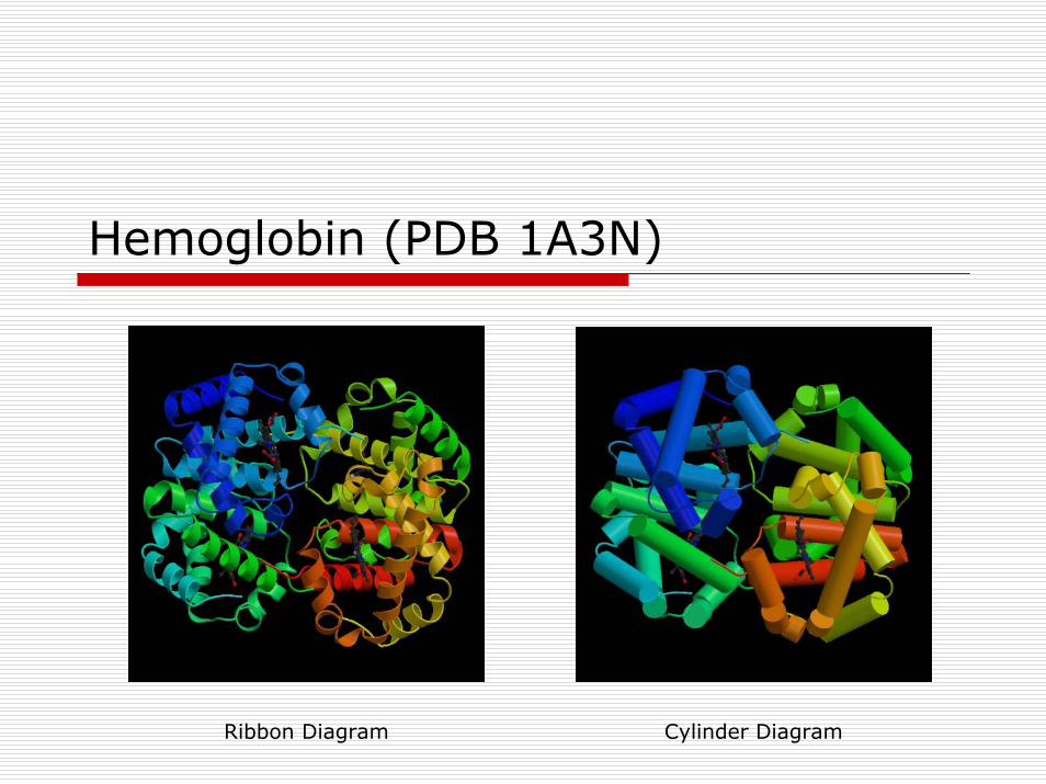
Hemoglobin (PDB 1A3N)
Ribbon Diagram Cylinder Diagram
β−sheetsOther major structural elementBasic unit is a β-strandUsually 5-10 residuesCan be parallel or anti-parallel based on the relative directions ofinteracting β-strands“Pleated” appearanceUnlike α-helices:Are formed with different parts of the sequenceH-bonding is inter-strand (opposed to intra-strand)Side chains from adjacent residues are on opposite sides of the sheetand do not interact with one anotherLike α-helices:Repeating secondary structure (2 residues per turn)Can be amphipathic
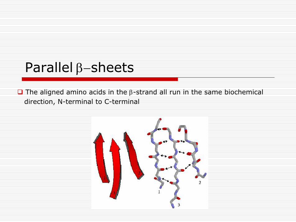
Parallel β−sheets
The aligned amino acids in the β-strand all run in the same biochemicaldirection, N-terminal to C-terminal
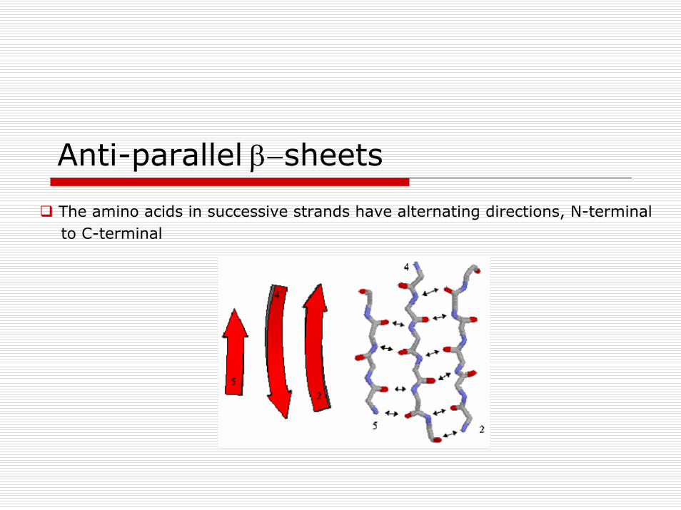
Anti-parallel β−sheets
The amino acids in successive strands have alternating directions, N-terminalto C-terminal
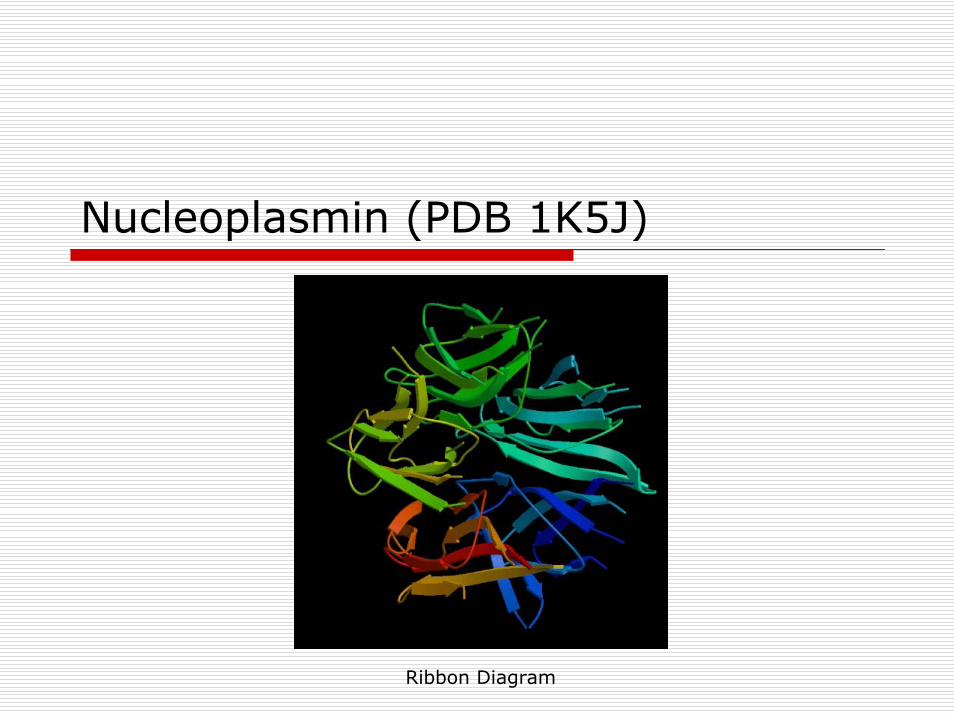
Nucleoplasmin (PDB 1K5J)
Ribbon Diagram
Amino Acid Preferences
α-helix formingThe amino acid side chain should cover and protect the backbone H-bonds inthe core of the helixAla, Leu, Met, Glu, Arg, Lys: good helix formersPro, Gly, Tyr, Ser: very poor helix formers
β-strand formingAmino acids with large bulky side chains prefer to form β-sheet structuresTyr, Trp, Ile, Val, Thr, Cys, Phe
Secondary structure disruptorsGly: side chain too smallPro: side chain linked to α-N, has no N-H to H-bond; rigid structure due to ringAsp, Asn, Ser: H-bonding side chains compete directly with backbone H-bonds
Turns/Loops
Third "classical" secondary structureReverses the direction of the polypeptide chain Located primarily on protein surface; contain polar and charged residuesThree types: I, II, III
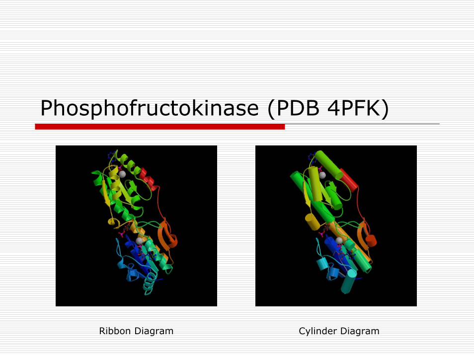
Phosphofructokinase (PDB 4PFK)
Ribbon Diagram Cylinder Diagram
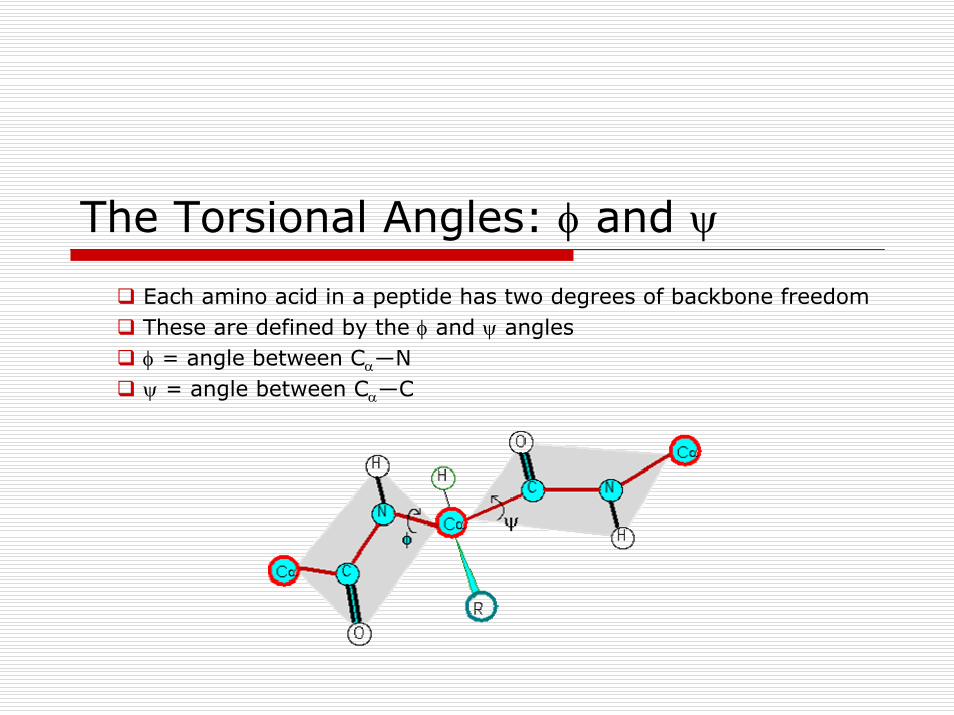
The Torsional Angles: φ and ψ
Each amino acid in a peptide has two degrees of backbone freedomThese are defined by the φ and ψ anglesφ = angle between Cα―Nψ = angle between Cα―C
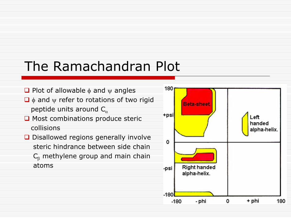
The Ramachandran Plot
Plot of allowable φ and ψ anglesφ and ψ refer to rotations of two rigidpeptide units around Cα
Most combinations produce stericcollisionsDisallowed regions generally involvesteric hindrance between side chainCβ methylene group and main chainatoms
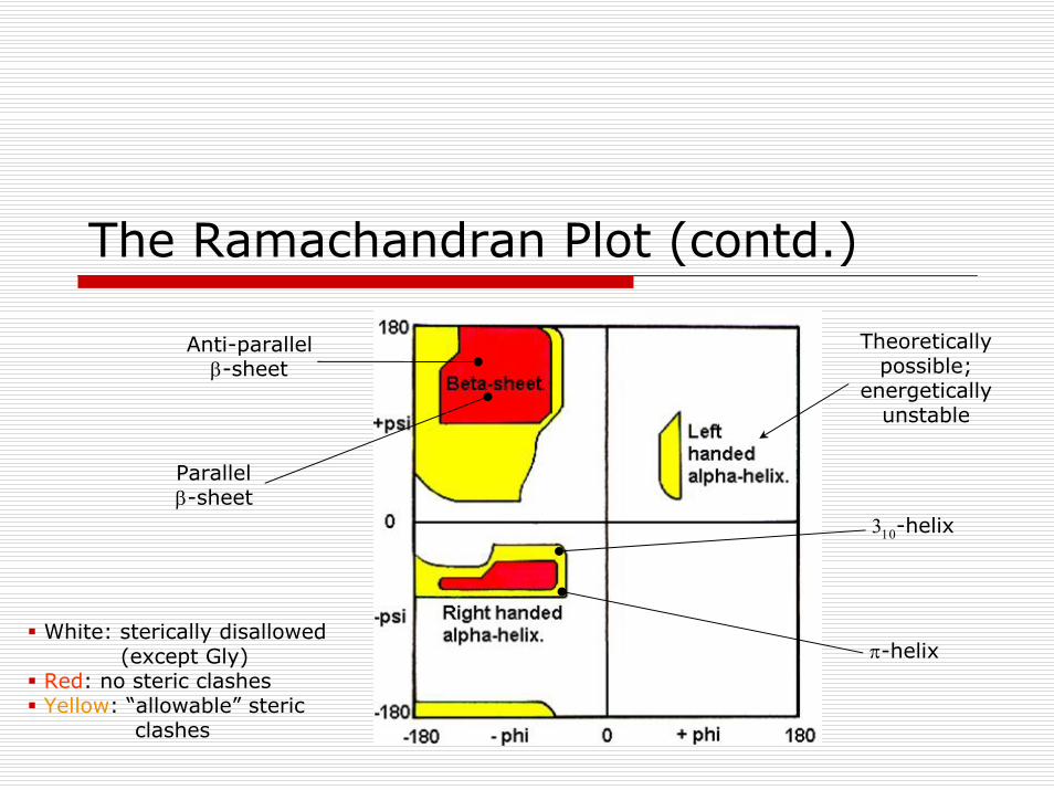
The Ramachandran Plot (contd.)
Theoreticallypossible;
energeticallyunstable
π-helix
310-helix
Anti-parallelβ-sheet
Parallelβ-sheet
White: sterically disallowed(except Gly)
Red: no steric clashesYellow: “allowable” steric
clashes
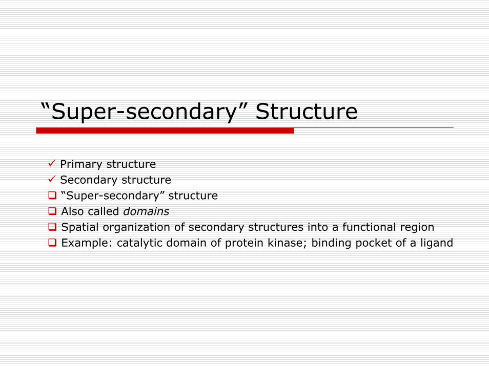
“Super-secondary” Structure
Primary structureSecondary structure“Super-secondary” structureAlso called domainsSpatial organization of secondary structures into a functional regionExample: catalytic domain of protein kinase; binding pocket of a ligand
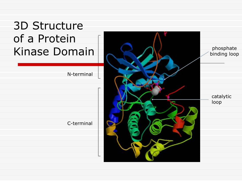
N-terminal
3D Structure of a Protein Kinase Domain phosphate
binding loop
catalyticloop
C-terminal
Quarternary Structure
Spatial organization of subunits to form a functional proteinExample: Hemoglobin2 α chains, 2 β chainsEach chain binds heme (Fe)Forms an α2-β2 tetramer
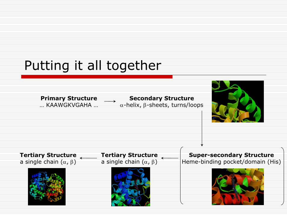
Putting it all together
Secondary Structureα-helix, β-sheets, turns/loops
Super-secondary StructureHeme-binding pocket/domain (His)
Primary Structure… KAAWGKVGAHA …
Tertiary Structurea single chain (α, β)
Tertiary Structurea single chain (α, β)
Additional Reading
General informationBiochemistry, 5th ed., Berg, Tymoczko, StryerBiochemistry, 3rd ed., Voet and VoetDetailed informationProteins, 2nd ed., CreightonIntroduction to Protein Structure, 2nd ed., Branden and ToozeInternetImages from the Protein Data Bank (PDB): www.rcsb.org/pdbNumerous wesbites (Google protein secondary structure)


![Review Article Bioactive Peptides: A Review - BASclbme.bas.bg/bioautomation/2011/vol_15.4/files/15.4_02.pdf · Review Article Bioactive Peptides: A Review ... casein [145]. Other](https://static.fdocument.org/doc/165x107/5acd360f7f8b9a93268d5e73/review-article-bioactive-peptides-a-review-article-bioactive-peptides-a-review.jpg)