Methods: Fluorescence Biochemistry 4000 Dr. Ute Kothe.
16
Methods: Fluorescence Biochemistry 4000 Dr. Ute Kothe
-
date post
18-Dec-2015 -
Category
Documents
-
view
221 -
download
1
Transcript of Methods: Fluorescence Biochemistry 4000 Dr. Ute Kothe.
Methods:Fluorescence
Biochemistry 4000
Dr. Ute Kothe

Remember: Absorbance
log (I0/I) = A = ε l c
Beer-Lambert law
Absorbance of monochromatic light reduces the intensity (I)
Measured relatively to original intensity (I0)
Depends on path length (l, often 1 cm), concentration (c) and
molar extinction coefficient (e, units: M-1 cm-1) Used to measure concentrations
Is very fast & provides information only on average ground state
of molecules; energy is set free by non-radiative decay (heat)
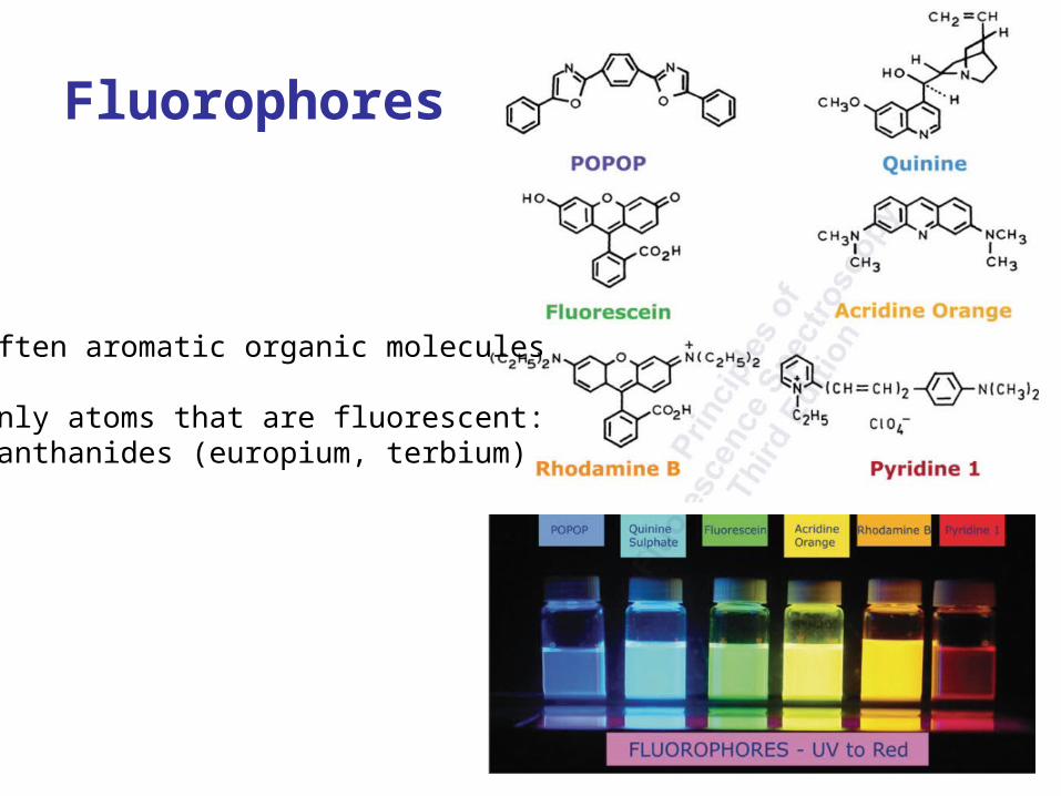
Fluorophores
Often aromatic organic molecules
Only atoms that are fluorescent:Lanthanides (europium, terbium)

What is Fluorescence?
Emission spectra – typically independent of excitation wavelength
Excitation spectra
Upon excitation of a
fluorophore, it re-emits light
at a longer wavelength.
Wavenumber = 1 / wavelength (linear to energy)
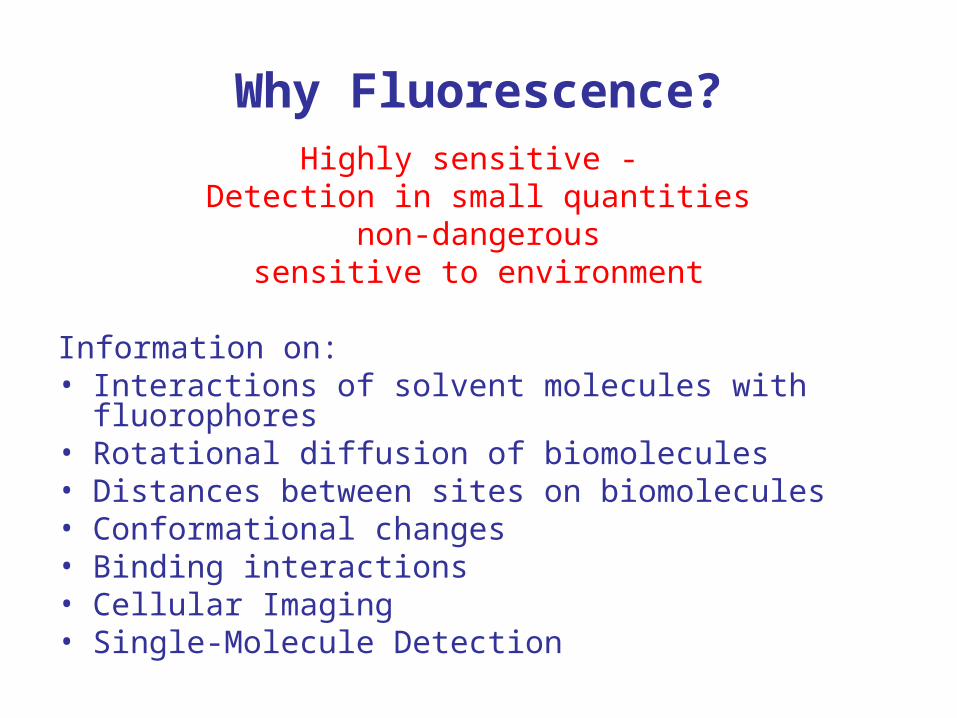
Why Fluorescence?Highly sensitive -
Detection in small quantitiesnon-dangerous
sensitive to environment
Information on:• Interactions of solvent molecules with fluorophores• Rotational diffusion of biomolecules• Distances between sites on biomolecules• Conformational changes• Binding interactions• Cellular Imaging• Single-Molecule Detection
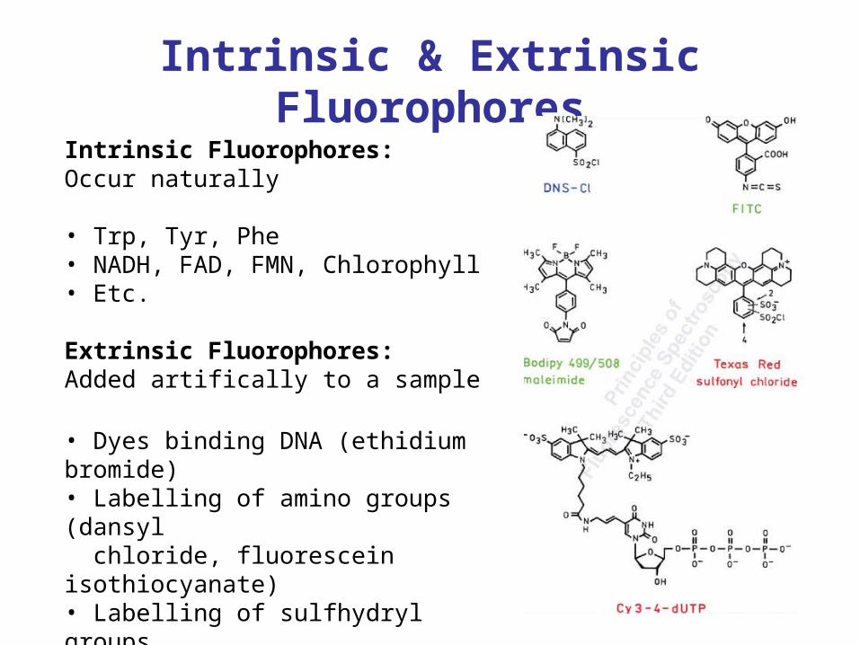
Intrinsic & Extrinsic Fluorophores
Intrinsic Fluorophores:Occur naturally
• Trp, Tyr, Phe• NADH, FAD, FMN, Chlorophyll • Etc.
Extrinsic Fluorophores:Added artifically to a sample
• Dyes binding DNA (ethidium bromide)• Labelling of amino groups (dansyl chloride, fluorescein isothiocyanate)• Labelling of sulfhydryl groups (maleimide dyes)• etc.
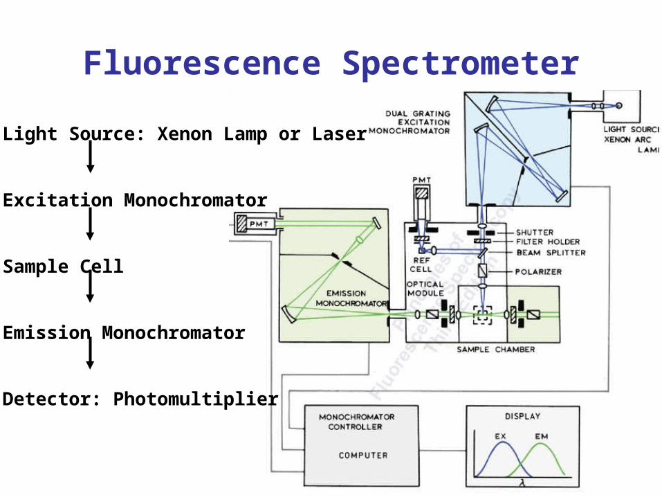
Fluorescence Spectrometer
Light Source: Xenon Lamp or Laser
Excitation Monochromator
Sample Cell
Emission Monochromator
Detector: Photomultiplier
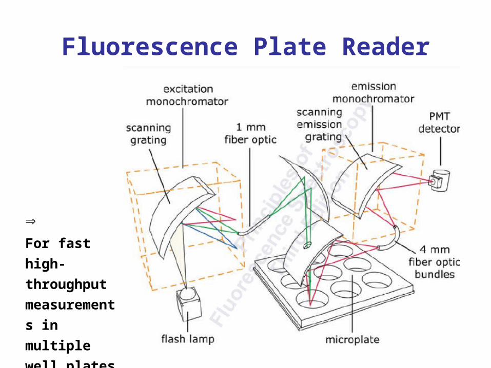
Fluorescence Plate Reader
For fast high-
throughput
measurements
in multiple
well plates
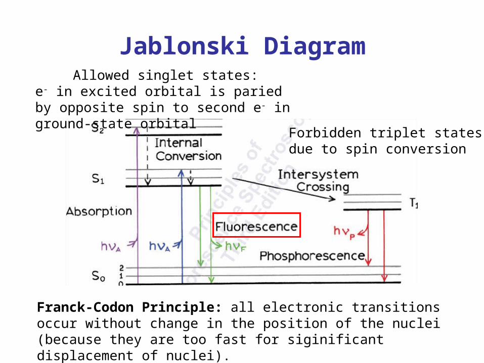
Jablonski Diagram
Franck-Codon Principle: all electronic transitions occur without change in the position of the nuclei (because they are too fast for siginificant displacement of nuclei).
Allowed singlet states:e- in excited orbital is paried by opposite spin to second e- in ground-state orbital
Forbidden triplet statesdue to spin conversion
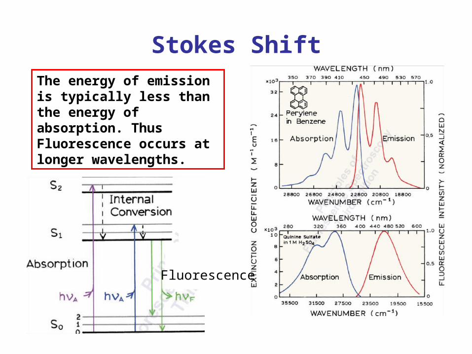
Stokes ShiftThe energy of emission is typically less than the energy of absorption. Thus Fluorescence occurs at longer wavelengths.
Fluorescence
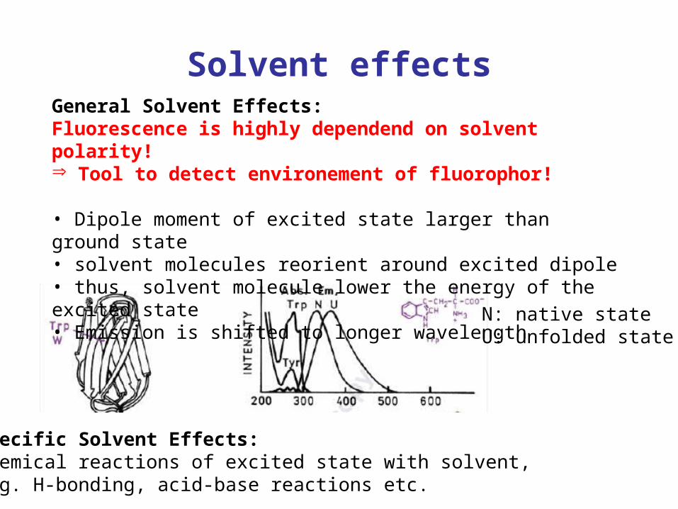
Solvent effectsGeneral Solvent Effects:Fluorescence is highly dependend on solvent polarity! Tool to detect environement of fluorophor!
• Dipole moment of excited state larger than ground state• solvent molecules reorient around excited dipole• thus, solvent molecule lower the energy of the excited state• Emission is shifted to longer wavelength
Specific Solvent Effects:Chemical reactions of excited state with solvent,e.g. H-bonding, acid-base reactions etc.
N: native stateU: unfolded state
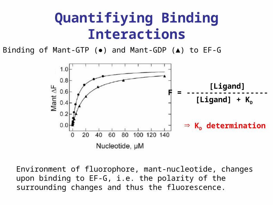
Quantifiying Binding Interactions
Binding of Mant-GTP (●) and Mant-GDP (▲) to EF-G
[Ligand]F = ------------------ [Ligand] + KD
Environment of fluorophore, mant-nucleotide, changes upon binding to EF-G, i.e. the polarity of the surrounding changes and thus the fluorescence.
KD determination
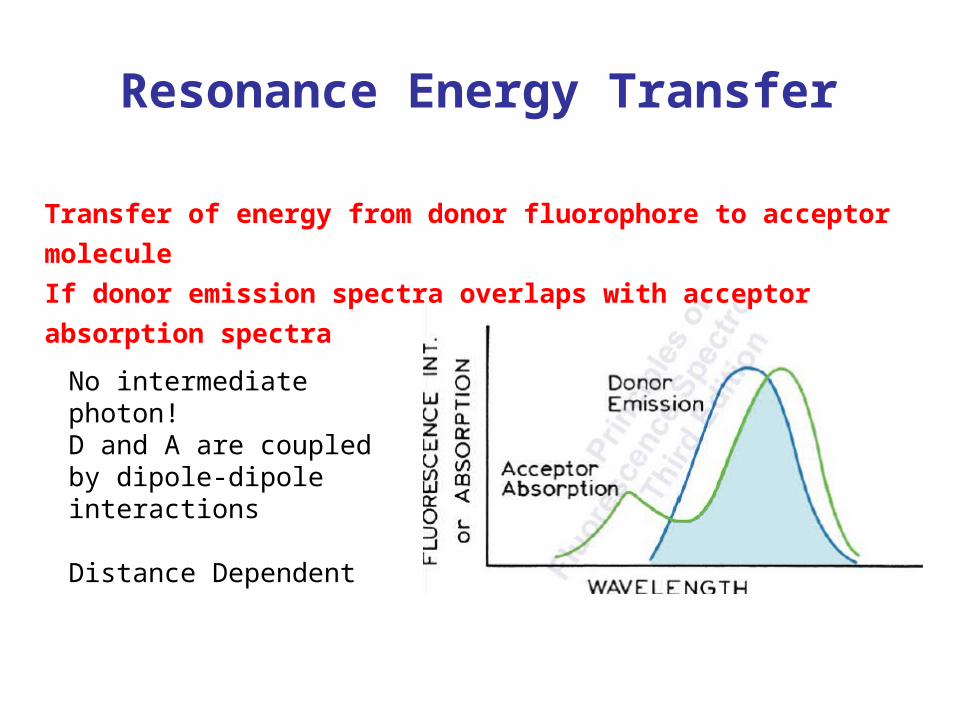
Resonance Energy Transfer
Transfer of energy from donor fluorophore to acceptor molecule
If donor emission spectra overlaps with acceptor absorption spectra
No intermediate photon!D and A are coupled by dipole-dipole interactions
Distance Dependent
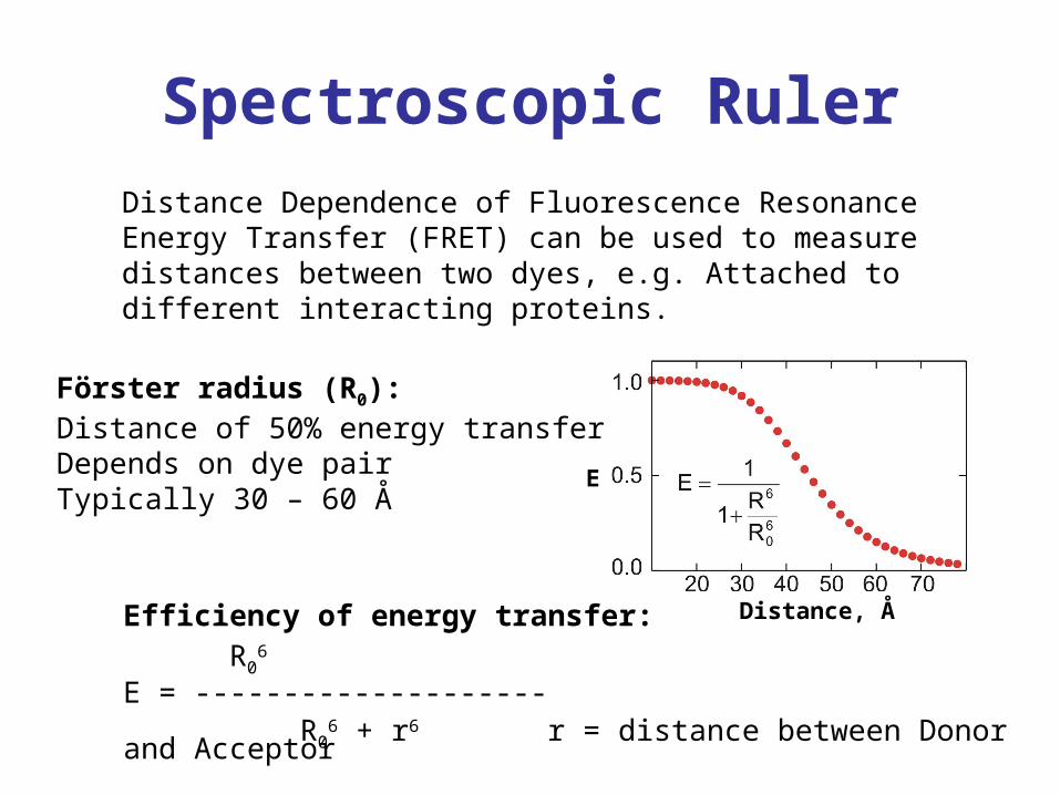
Spectroscopic Ruler
Distance, Å
E
Distance Dependence of Fluorescence Resonance Energy Transfer (FRET) can be used to measure distances between two dyes, e.g. Attached to different interacting proteins.
Förster radius (R0):Distance of 50% energy transferDepends on dye pairTypically 30 – 60 Å
Efficiency of energy transfer:R0
6
E = -------------------- R0
6 + r6 r = distance between Donor and Acceptor
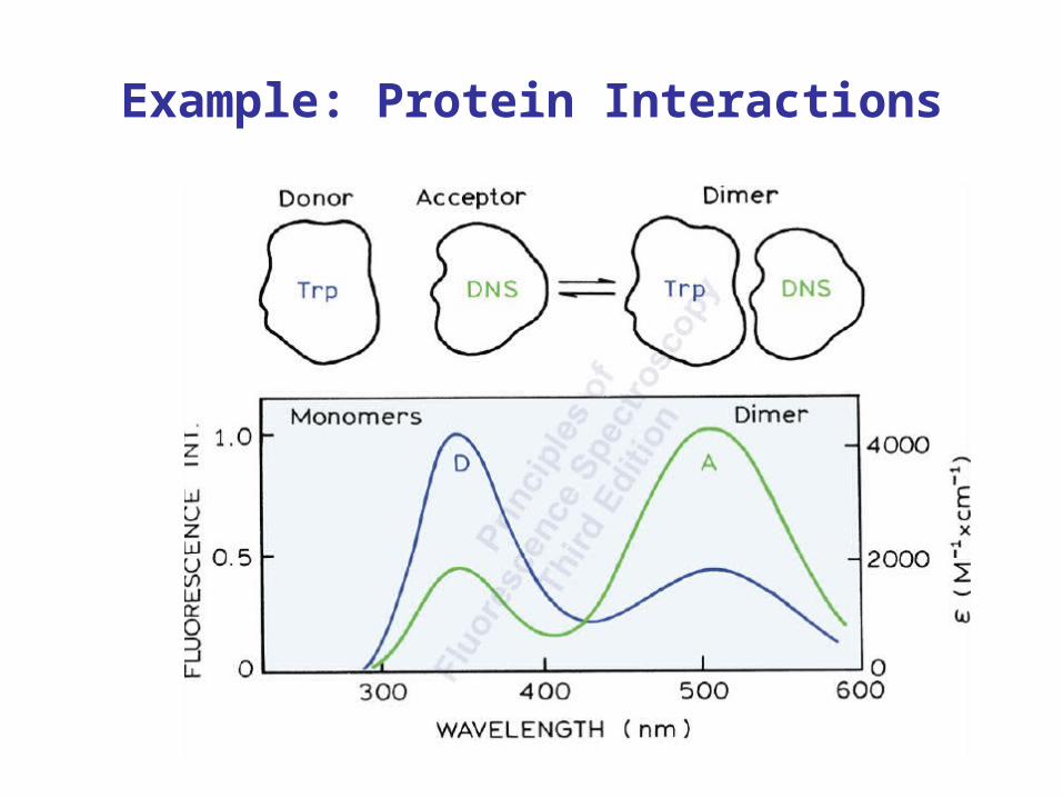
Example: Protein Interactions
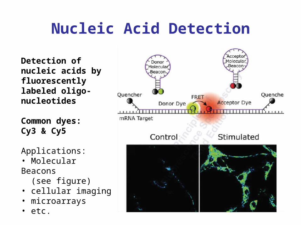
Nucleic Acid Detection
Detection of nucleic acids by fluorescently labeled oligo-nucleotides
Common dyes:Cy3 & Cy5
Applications:• Molecular Beacons (see figure)• cellular imaging• microarrays• etc.