An Epigenetic Switch Involving NF-κB, Lin28, Let-7 ... · An Epigenetic Switch Involving NF-kB,...
14
An Epigenetic Switch Involving NF- k B, Lin28, Let-7 MicroRNA, and IL6 Links Inflammation to Cell Transformation Dimitrios Iliopoulos, 1 Heather A. Hirsch, 1 and Kevin Struhl 1, * 1 Department of Biological Chemistry and Molecular Pharmacology, Harvard Medical School, Boston, MA 02115, USA *Correspondence: [email protected] DOI 10.1016/j.cell.2009.10.014 SUMMARY Inflammation is linked clinically and epidemiologi- cally to cancer, and NF-kB appears to play a causa- tive role, but the mechanisms are poorly understood. We show that transient activation of Src oncoprotein can mediate an epigenetic switch from immortalized breast cells to a stably transformed line that forms self-renewing mammospheres that contain cancer stem cells. Src activation triggers an inflammatory response mediated by NF-kB that directly activates Lin28 transcription and rapidly reduces let-7 micro- RNA levels. Let-7 directly inhibits IL6 expression, re- sulting in higher levels of IL6 than achieved by NF-kB activation. IL6-mediated activation of the STAT3 transcription factor is necessary for transformation, and IL6 activates NF-kB, thereby completing a posi- tive feedback loop. This regulatory circuit operates in other cancer cells lines, and its transcriptional signature is found in human cancer tissues. Thus, inflammation activates a positive feedback loop that maintains the epigenetic transformed state for many generations in the absence of the inducing signal. INTRODUCTION Tumorigenesis is a multistep process that requires constitutive cell division, growth, and survival. The molecular events govern- ing the onset and progression of malignant transformation involve the inactivation of tumor suppressor genes and the acquisition of oncogenic mutations (Hahn and Weinberg, 2002; Vogelstein and Kinzler, 2004). These genetic perturbations help cancer cells override the normal mechanisms controlling cellular proliferation. In addition to genetic changes, tumor suppressor genes can be inactivated by epigenetic silencing through DNA methylation (Baylin, 2005), and this can occur through an elabo- rate pathway triggered by an oncoprotein (Gazin et al., 2007). Lastly, the progression from normal cells to cancer is strongly influenced by environmental conditions and extracellular signaling pathways that affect the activity of tumor suppressors and oncoproteins. Clinical and epidemiological studies have suggested a strong association between inflammation and different types of cancer, and inflammatory molecules can provide growth signals that promote the proliferation of malignant cells (Balkwill and Manto- vani, 2001; Naugler and Karin, 2008; Pierce et al., 2009). For example, interleukin-6 (IL6) is upregulated in epithelial cancers such as breast and prostate (Sasser et al., 2007; Wegiel et al., 2008). NF-kB, a transcription factor that regulates the expression of antiapoptotic genes and activates different proinflammatory cytokines and chemokines, seems to be a key molecular link between inflammation and oncogenesis initiation and progres- sion (Naugler and Karin, 2008). Constitutively active NF-kB occurs in many types of cancer, and mouse models provide genetic and biochemical evidence for a causative role of NF-kB in malignant conversion and progression (Luedde et al., 2007; Sakurai et al., 2008). However, the mechanistic linkage between inflammation and cancer remain to be elucidated. MicroRNAs play critical roles in many biological processes including cancer by directly interacting with specific messenger RNAs (mRNAs) through base pairing and then inhibiting expres- sion of the target genes through a variety of molecular mecha- nisms (Bartel, 2009; Ventura and Jacks, 2009). MicroRNAs can undergo aberrant regulation during carcinogenesis (Lu et al., 2005), and they can act as oncogenes or tumor suppressor genes. For example, Let-7 is an important microRNA family con- sisting of 12 members located in genomic locations frequently deleted in human cancers (Calin et al., 2004). In addition, expres- sion of let-7 RNAs is reduced in non-small-cell lung cancer patients and associated with poor prognosis (Johnson et al., 2005), and let-7 overexpression substantially reduces tumor burden in a K-Ras murine lung cancer model (Kumar et al., 2008). Although microRNAs are important for cancer develop- ment and progression, there is limited understanding of their roles in the molecular pathways and regulatory circuits that are involved in the process of cellular transformation. Epigenetic inheritance is a phenomenon in which cellular phenotypes and gene expression patterns are faithfully trans- mitted through multiple generations via a mechanism that involves something beyond DNA sequence. The term was in- vented to explain how the same genome can give rise to pheno- typically different cell types. An epigenetic switch occurs when a stable cell type changes to another stable cell type without any change in DNA sequence. Epigenetic switches require an initiating event (which could be a specific molecular process or Cell 139, 693–706, November 13, 2009 ª2009 Elsevier Inc. 693
Transcript of An Epigenetic Switch Involving NF-κB, Lin28, Let-7 ... · An Epigenetic Switch Involving NF-kB,...
An Epigenetic Switch Involving NF-kB,Lin28, Let-7 MicroRNA, and IL6 LinksInflammation to Cell TransformationDimitrios Iliopoulos,1 Heather A. Hirsch,1 and Kevin Struhl1,*1Department of Biological Chemistry and Molecular Pharmacology, Harvard Medical School, Boston, MA 02115, USA
*Correspondence: [email protected]
DOI 10.1016/j.cell.2009.10.014
SUMMARY
Inflammation is linked clinically and epidemiologi-cally to cancer, and NF-kB appears to play a causa-tive role, but the mechanisms are poorly understood.We show that transient activation of Src oncoproteincan mediate an epigenetic switch from immortalizedbreast cells to a stably transformed line that formsself-renewing mammospheres that contain cancerstem cells. Src activation triggers an inflammatoryresponse mediated by NF-kB that directly activatesLin28 transcription and rapidly reduces let-7 micro-RNA levels. Let-7 directly inhibits IL6 expression, re-sulting in higher levels of IL6 than achieved by NF-kBactivation. IL6-mediated activation of the STAT3transcription factor is necessary for transformation,and IL6 activates NF-kB, thereby completing a posi-tive feedback loop. This regulatory circuit operatesin other cancer cells lines, and its transcriptionalsignature is found in human cancer tissues. Thus,inflammation activates a positive feedback loopthat maintains the epigenetic transformed state formany generations in the absence of the inducingsignal.
INTRODUCTION
Tumorigenesis is a multistep process that requires constitutive
cell division, growth, and survival. The molecular events govern-
ing the onset and progression of malignant transformation
involve the inactivation of tumor suppressor genes and the
acquisition of oncogenic mutations (Hahn and Weinberg, 2002;
Vogelstein and Kinzler, 2004). These genetic perturbations help
cancer cells override the normal mechanisms controlling cellular
proliferation. In addition to genetic changes, tumor suppressor
genes can be inactivated by epigenetic silencing through DNA
methylation (Baylin, 2005), and this can occur through an elabo-
rate pathway triggered by an oncoprotein (Gazin et al., 2007).
Lastly, the progression from normal cells to cancer is strongly
influenced by environmental conditions and extracellular
signaling pathways that affect the activity of tumor suppressors
and oncoproteins.
Clinical and epidemiological studies have suggested a strong
association between inflammation and different types of cancer,
and inflammatory molecules can provide growth signals that
promote the proliferation of malignant cells (Balkwill and Manto-
vani, 2001; Naugler and Karin, 2008; Pierce et al., 2009). For
example, interleukin-6 (IL6) is upregulated in epithelial cancers
such as breast and prostate (Sasser et al., 2007; Wegiel et al.,
2008). NF-kB, a transcription factor that regulates the expression
of antiapoptotic genes and activates different proinflammatory
cytokines and chemokines, seems to be a key molecular link
between inflammation and oncogenesis initiation and progres-
sion (Naugler and Karin, 2008). Constitutively active NF-kB
occurs in many types of cancer, and mouse models provide
genetic and biochemical evidence for a causative role of NF-kB
in malignant conversion and progression (Luedde et al., 2007;
Sakurai et al., 2008). However, the mechanistic linkage between
inflammation and cancer remain to be elucidated.
MicroRNAs play critical roles in many biological processes
including cancer by directly interacting with specific messenger
RNAs (mRNAs) through base pairing and then inhibiting expres-
sion of the target genes through a variety of molecular mecha-
nisms (Bartel, 2009; Ventura and Jacks, 2009). MicroRNAs can
undergo aberrant regulation during carcinogenesis (Lu et al.,
2005), and they can act as oncogenes or tumor suppressor
genes. For example, Let-7 is an important microRNA family con-
sisting of 12 members located in genomic locations frequently
deleted in human cancers (Calin et al., 2004). In addition, expres-
sion of let-7 RNAs is reduced in non-small-cell lung cancer
patients and associated with poor prognosis (Johnson et al.,
2005), and let-7 overexpression substantially reduces tumor
burden in a K-Ras murine lung cancer model (Kumar et al.,
2008). Although microRNAs are important for cancer develop-
ment and progression, there is limited understanding of their
roles in the molecular pathways and regulatory circuits that are
involved in the process of cellular transformation.
Epigenetic inheritance is a phenomenon in which cellular
phenotypes and gene expression patterns are faithfully trans-
mitted through multiple generations via a mechanism that
involves something beyond DNA sequence. The term was in-
vented to explain how the same genome can give rise to pheno-
typically different cell types. An epigenetic switch occurs when
a stable cell type changes to another stable cell type without
any change in DNA sequence. Epigenetic switches require an
initiating event (which could be a specific molecular process or
Cell 139, 693–706, November 13, 2009 ª2009 Elsevier Inc. 693
stochastic fluctuation), but the phenotypes of the new cell type
are inherited in the absence of the initiating signal. Epigenetic
switches occur in prokaryotes, and hence in the absence of
chromatin, although chromatin often plays an important role in
eukaryotes. As illuminated by classic studies of an epigenetic
switch in bacteriophage l (Ptashne, 2009), positive feedback
loops involving transcriptional regulatory proteins are the funda-
mental principle for epigenetic inheritance of gene expression
patterns.
Here, we show that transient activation of Src causes an
epigenetic switch in which nontransformed MCF10A cells are
converted into mammospheres that can be propagated for
many generations in the absence of the initiating signal. This
epigenetic switch is activated by an inflammatory signal, and
epigenetic inheritance is mediated by a positive feedback loop
involving the NF-kB transcription factor, the microRNA process-
ing factor Lin28, Let-7 microRNA, and IL6. This regulatory circuit
links inflammation to cellular transformation, and it appears to be
important for other forms of cancer.
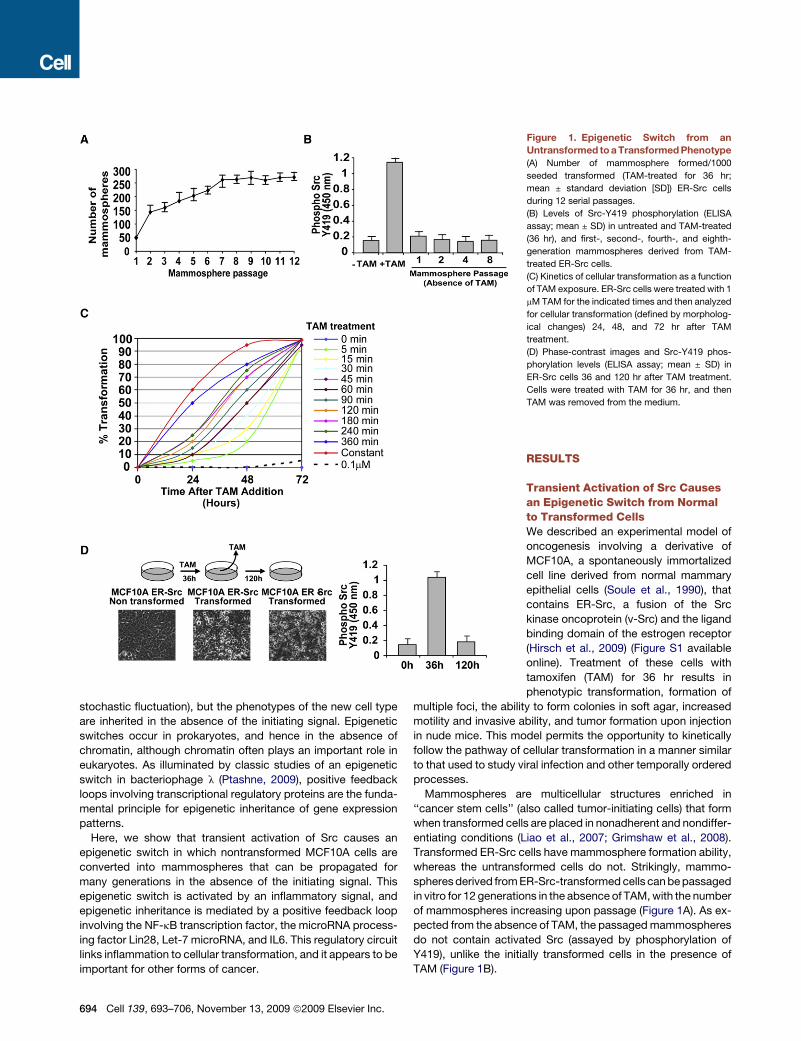
Figure 1. Epigenetic Switch from an
Untransformed to a Transformed Phenotype
(A) Number of mammosphere formed/1000
seeded transformed (TAM-treated for 36 hr;
mean ± standard deviation [SD]) ER-Src cells
during 12 serial passages.
(B) Levels of Src-Y419 phosphorylation (ELISA
assay; mean ± SD) in untreated and TAM-treated
(36 hr), and first-, second-, fourth-, and eighth-
generation mammospheres derived from TAM-
treated ER-Src cells.
(C) Kinetics of cellular transformation as a function
of TAM exposure. ER-Src cells were treated with 1
mM TAM for the indicated times and then analyzed
for cellular transformation (defined by morpholog-
ical changes) 24, 48, and 72 hr after TAM
treatment.
(D) Phase-contrast images and Src-Y419 phos-
phorylation levels (ELISA assay; mean ± SD) in
ER-Src cells 36 and 120 hr after TAM treatment.
Cells were treated with TAM for 36 hr, and then
TAM was removed from the medium.
RESULTS
Transient Activation of Src Causesan Epigenetic Switch from Normalto Transformed CellsWe described an experimental model of
oncogenesis involving a derivative of
MCF10A, a spontaneously immortalized
cell line derived from normal mammary
epithelial cells (Soule et al., 1990), that
contains ER-Src, a fusion of the Src
kinase oncoprotein (v-Src) and the ligand
binding domain of the estrogen receptor
(Hirsch et al., 2009) (Figure S1 available
online). Treatment of these cells with
tamoxifen (TAM) for 36 hr results in
phenotypic transformation, formation of
multiple foci, the ability to form colonies in soft agar, increased
motility and invasive ability, and tumor formation upon injection
in nude mice. This model permits the opportunity to kinetically
follow the pathway of cellular transformation in a manner similar
to that used to study viral infection and other temporally ordered
processes.
Mammospheres are multicellular structures enriched in
‘‘cancer stem cells’’ (also called tumor-initiating cells) that form
when transformed cells are placed in nonadherent and nondiffer-
entiating conditions (Liao et al., 2007; Grimshaw et al., 2008).
Transformed ER-Src cells have mammosphere formation ability,
whereas the untransformed cells do not. Strikingly, mammo-
spheres derived from ER-Src-transformed cells can be passaged
in vitro for 12 generations in the absence of TAM, with the number
of mammospheres increasing upon passage (Figure 1A). As ex-
pected from the absence of TAM, the passaged mammospheres
do not contain activated Src (assayed by phosphorylation of
Y419), unlike the initially transformed cells in the presence of
TAM (Figure 1B).
694 Cell 139, 693–706, November 13, 2009 ª2009 Elsevier Inc.
To further characterize the switch between nontransformed
and transformed cells, we varied the time of TAM treatment.
Remarkably, TAM treatment for only 5 min results in transforma-
tion, although the process is slower (72 hr as opposed to 36 hr;
Figures 1C and S2A). Furthermore, increasing the time of TAM
treatment progressively reduces the time necessary for the
transformed phenotype. The transformation that occurs upon
very short TAM treatment is not due to residual TAM, because
10-fold lower TAM is unable to induce transformation (Fig-
ure S2B), and transformed cells maintained in the absence of
TAM lack activated Src yet retain the transformed phenotype
(Figure 1D).
Collectively, these results demonstrate that transient activation
of Src causes an epigenetic switch from a stable nontransformed
cell line to a transformed state capable of forming self-renewing
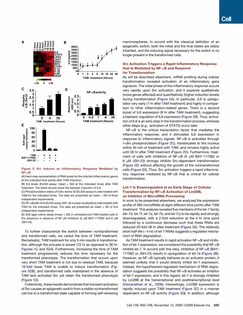
Figure 2. Src Induces an Inflammatory Response Mediated by
NF-kB
(A) Heat-map representation of RNA levels for the indicted inflammatory genes
at the indicated time points after TAM induction.
(B) IL6 levels (ELISA assay; mean ± SD) at the indicated times after TAM
treatment. The black arrows show the biphasic induction of IL6.
(C) Phosphorylation status of IkBa-serine 32 (ELISA assay) in cells treated with
TAM for the indicated times. The data are presented as mean ± SD of three
independent experiments.
(D) NF-kB/p65 ActivELISA assay (NF-kB nuclear localization) cells treated with
TAM for the indicated times. The data are presented as mean ± SD of five
independent experiments.
(E) Soft agar colony assay (mean ± SD) in untreated and TAM-treated cells in
the presence or absence of NF-kB inhibitors (5 mM BAY-117082 and 6 mM
JSH-23).
mammospheres. In accord with the classical definition of an
epigenetic switch, both the initial and the final states are stably
inherited, and the inducing signal necessary for the switch is no
longer present in the transformed cells.
Src Activation Triggers a Rapid Inflammatory Responsethat Is Mediated by NF-kB and Requiredfor TransformationAs will be described elsewhere, mRNA profiling during cellular
transformation revealed activation of an inflammatory gene
signature. The initial phase of this inflammatory response occurs
very rapidly upon Src activation, and it expands qualitatively
(more genes affected) and quantitatively (higher induction levels)
during transformation (Figure 2A). In particular, IL6 is upregu-
lated very early (1 hr after TAM treatment) and highly in compar-
ison to other inflammation-related genes. There is a second
boost of IL6 expression (8 hr after TAM treatment), suggesting
a biphasic regulation of IL6 expression (Figure 2B). Thus, activa-
tion of IL6 is an early step in the transformation process, whereas
other steps (e.g., activation of STAT3) occur later.
NF-kB is the critical transcription factor that mediates the
inflammatory response, and it stimulates IL6 expression in
response to inflammatory signals. NF-kB is activated through
I-kBa phosphorylation (Figure 2C), translocates to the nucleus
within 30 min of treatment with TAM, and remains highly active
until 36 hr after TAM treatment (Figure 2D). Furthermore, treat-
ment of cells with inhibitors of NF-kB (5 mM BAY-117082 or
6 mM JSH-23) strongly inhibits Src-dependent transformation
(Figure 2E) without affecting the growth of the nontransformed
cells (Figure S3). Thus, Src activation triggers a rapid inflamma-
tory response mediated by NF-kB that is critical for cellular
transformation.
Let-7 Is Downregulated at an Early Stage of CellularTransformation by NF-kB Activation of Lin28B,an Inhibitor of MicroRNA ProcessingIn work to be presented elsewhere, we analyzed the expression
profile of 365 microRNAs at eight different time points after TAM
treatment. This analysis revealed five members of the let-7 family
(let-7d, let-7f, let-7a, let-7b, and let-7c) to be rapidly and strongly
downregulated, with a 2-fold reduction at the 4 hr time point
followed by a continuous decrease such that these RNAs are
reduced 20-fold 36 hr after treatment (Figure 3A). The relatively
short half-life (�4 hr) of let-7 RNAs suggests a regulated mecha-
nism of RNA degradation.
As TAM treatment results in rapid activation NF-kB and inhibi-
tion of let-7 expression, we considered the possibility that NF-kB
inhibits let-7. In accord with this idea, inhibition of NF-kB (BAY-
117082 or JSH-23) results in upregulation of let-7a (Figure 3B).
However, as NF-kB typically behaves as an activator protein, it
seemed unlikely that it would directly inhibit let-7 expression.
Instead, the hypothesized regulated mechanism of RNA degra-
dation suggests the possibility that NF-kB activates an inhibitor
of let-7 expression, and in this regard, let-7 is strongly inhibited
by Lin28B at the transcriptional and posttranscriptional level
(Viswanathan et al., 2008). Interestingly, Lin28B expression is
rapidly induced upon TAM treatment (Figure 3C) in a manner
dependent on NF-kB activity (Figure 3D). In addition, although
Cell 139, 693–706, November 13, 2009 ª2009 Elsevier Inc. 695
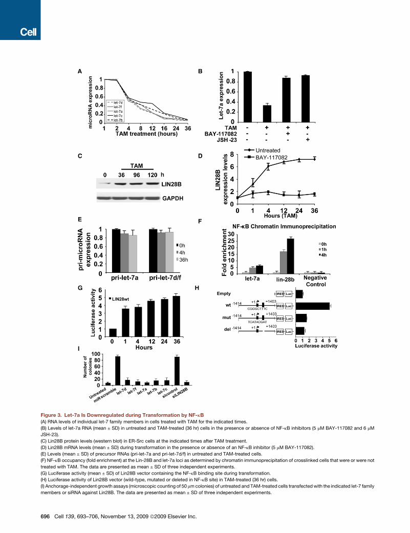
Figure 3. Let-7a Is Downregulated during Transformation by NF-kB
(A) RNA levels of individual let-7 family members in cells treated with TAM for the indicated times.
(B) Levels of let-7a RNA (mean ± SD) in untreated and TAM-treated (36 hr) cells in the presence or absence of NF-kB inhibitors (5 mM BAY-117082 and 6 mM
JSH-23).
(C) Lin28B protein levels (western blot) in ER-Src cells at the indicated times after TAM treatment.
(D) Lin28B mRNA levels (mean ± SD) during transformation in the presence or absence of an NF-kB inhibitor (5 mM BAY-117082).
(E) Levels (mean ± SD) of precursor RNAs (pri-let-7a and pri-let-7d/f) in untreated and TAM-treated cells.
(F) NF-kB occupancy (fold enrichment) at the Lin-28B and let-7a loci as determined by chromatin immunoprecipitation of crosslinked cells that were or were not
treated with TAM. The data are presented as mean ± SD of three independent experiments.
(G) Luciferase activity (mean ± SD) of Lin28B vector containing the NF-kB binding site during transformation.
(H) Luciferase activity of Lin28B vector (wild-type, mutated or deleted in NF-kB site) in TAM-treated (36 hr) cells.
(I) Anchorage-independent growth assays (microscopic counting of 50 mm colonies) of untreated and TAM-treated cells transfected with the indicated let-7 family
members or siRNA against Lin28B. The data are presented as mean ± SD of three independent experiments.
696 Cell 139, 693–706, November 13, 2009 ª2009 Elsevier Inc.
mature let-7 RNA levels decrease rapidly and strongly during
the cellular transformation process, levels of the primary let7a,
let7c, and let7e RNAs are unaffected by (Figure 3E), suggesting
that NF-kB has little if any direct effect on the rate of let-7
transcription.
Sequence analysis reveals a highly conserved NF-kB motif in
the first intron of the lin-28B gene and moderately conserved
NF-kB motifs �4 kb upstream of the let-7a gene (Figure S4).
As assayed by chromatin immunoprecipitation, NF-kB binds
both the let-7 and Lin28B regions identified above, with binding
to the highly conserved Lin28B site being stronger (Figure 3F). In
accord with the kinetics of NF-kB activation, strong binding is
observed 1 hr after TAM addition, but not in the absence of
TAM. Moreover, a Lin28B genomic fragment including the highly
conserved NF-kB site is sufficient to activate transcription of a
luciferase reporter construct during the transformation process
(Figure 3G). Similar constructs in which the NF-kB site was
mutated or deleted do not support transcriptional activity (Fig-
ure 3H). These observations suggest that NF-kB directly acti-
vates Lin28B expression through a binding site in the first intron,
and the increased levels of Lin28B inhibit let-7 expression
through a posttranscriptional mechanism.
Inhibition of Let-7 through Activation of Lin28BIs Important for Cellular TransformationTo address whether the observed inhibition of let-7 microRNAs
by Lin28B is important for cellular transformation, we examined
the phenotypic consequences of modulating the expression of
Lin28B and let-7 family members. First, inhibition of Lin28B
expression by small interfering RNA (siRNA) (Figure S5A)
strongly reduces cellular transformation (Figure 3I). Second, in
MCF-10A cells lacking the ER-Src construct, overexpression
of Lin28B results in increased cell growth and motility, the ability
to form colonies in soft agar and tumors in nude mice, reduced
levels of let-7, and increased levels of IL6 (Figures S5B–S5G).
Third, overexpression of individual let-7 microRNAs (let-7a,
let-7b, let-7c, let-7d, and let-7f) strongly inhibits anchorage-
independent growth in soft agar (Figure 3I), and it blocks the
transformed morphology and formation of foci as well as the
migratory and invasion activity of ER-Src-transformed cells (Fig-
ure S6). Thus, Lin28B and its ability to rapidly inhibit let-7 micro-
RNAs upon Src activation is a key early step that is important for
cellular transformation. Moreover, as each let-7 family member
acts as a suppressor of transformation, Lin28B is important to
coordinately inhibit all the let-7 family members. Because of
the redundancy between let-7 family members and the slightly
stronger effect of let-7a for inhibiting colony formation, migration
and invasion activity, subsequent experiments have been
performed with let-7a.
Let-7 MicroRNA Directly Inhibits Expression of IL6MicroRNAs exert their biological functions through suppression
of target genes via RNA-RNA complementarity. Using three
different selection criteria (Figure S7), we identified IL6 as a
potential gene target of let-7. Moreover, the conservation
between the microRNA and a putative target site in the 30
untranslated region (UTR) of the IL6 gene suggests the impor-
tance of this interaction during evolution (Figure 4A).
Several lines of evidence indicate that let-7a directly targets
IL6 mRNA through binding its 30 UTR. First, let-7a overexpres-
sion inhibits the activity of a luciferase reporter construct con-
taining the IL6 30 UTR (Figure 4B). Second, immunoprecipitates
of HA-Ago1, a protein that mediates microRNA functions, show
markedly higher levels of IL6 mRNA binding upon let-7a overex-
pression (Figure 4C). Third, inhibition of let-7a via introduction of
an antisense RNA or by Lin28B overexpression significantly
induces IL6 mRNA levels (Figure 4D). Fourth, phosphorylation
of STAT3, a downstream target of IL6, is inhibited by the addition
of let-7a or by inhibition of Lin28B (Figure 4E). Fifth, individual
expression of all let-7 family members tested results in reduced
levels of both IL6 protein and the angiogenic cytokine VEGF,
a direct transcriptional target of STAT3 (Figure 4F), indicating
that let-7 inhibits the IL6-dependent signaling pathway.
Let-7 Inhibits IL6 Expression Indirectly throughRas and NF-kBAs mentioned previously, HMGA2 and Ras are let-7a gene
targets (Johnson et al., 2005; Mayr et al., 2007). Although we
did not detect any difference in HMGA2 expression during trans-
formation of ER-Src cells (data not shown), transformed cells
have high levels of Ras that are inhibited by expression of
let-7a (Figure 4G). As Ras-induced secretion of IL6 is required
for tumorigenesis (Ancrile et al., 2007) and IL6 transcription is
induced directly by NF-kB, we hypothesized that let-7a might
inhibit IL6 expression indirectly through the Ras-NF-kB pathway.
In accord with this hypothesis, antisense inhibition of Ras
expression (Figure S8A) during cellular transformation reduces
IL6 protein levels, albeit less effectively than achieved by let-7a
overexpression (Figure 4H). In addition, NF-kB activity is strongly
induced upon transformation, and this induction is partially
blocked by inhibition of Ras (Figure S8B). Lastly, inhibition of
Ras expression reduces the level of transformation, although
much less effectively than inhibition of IL6 (Figure 4I). Thus,
let-7a microRNA inhibits IL6 expression both directly through
its 30 UTR and indirectly by an interaction with Ras that leads
to a reduction in NF-kB activity. However, it seems that let-7
regulates IL6 expression more effectively through direct inhibi-
tion than indirect inhibition through Ras.
IL6 Inhibition of Let-7 Expression Occurs through NF-kBand Is Important for TransformationDepletion of IL6 by a monoclonal antibody blocks the morpho-
logical changes associated with transformed cells (Figure 5A),
reduces colony formation, and inhibits cell motility (Figure S9).
At lower concentrations of the IL6 antibody that only partially
reduce IL6 levels (Figure S9), transformation is significantly
delayed (Figure 5B). Thus, IL6 is important for transformation,
and the level of IL6 affects the rate at which cells become
transformed.
Conversely, IL6 treatment for only 1 hr is sufficient to induce
transformation of MCF10A cells (soft agar assay) in the absence
of Src activation (Figure 5C), and the resulting transformed cells
were able to form tumors in xenografts (Figure 5D). The trans-
formed phenotype of these IL6-induced cells was fully acquired
96–120 hr after treatment, and it remained stable for at least
10 more days (Figure 5E). Furthermore, when these stable
Cell 139, 693–706, November 13, 2009 ª2009 Elsevier Inc. 697
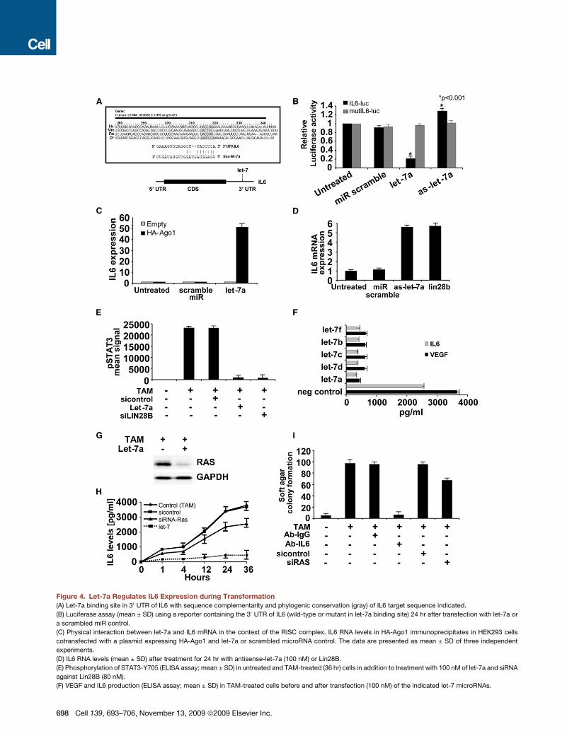
Figure 4. Let-7a Regulates IL6 Expression during Transformation
(A) Let-7a binding site in 30 UTR of IL6 with sequence complementarity and phylogenic conservation (gray) of IL6 target sequence indicated.
(B) Luciferase assay (mean ± SD) using a reporter containing the 30 UTR of IL6 (wild-type or mutant in let-7a binding site) 24 hr after transfection with let-7a or
a scrambled miR control.
(C) Physical interaction between let-7a and IL6 mRNA in the context of the RISC complex. IL6 RNA levels in HA-Ago1 immunoprecipitates in HEK293 cells
cotransfected with a plasmid expressing HA-Ago1 and let-7a or scrambled microRNA control. The data are presented as mean ± SD of three independent
experiments.
(D) IL6 RNA levels (mean ± SD) after treatment for 24 hr with antisense-let-7a (100 nM) or Lin28B.
(E) Phosphorylation of STAT3-Y705 (ELISA assay; mean ± SD) in untreated and TAM-treated (36 hr) cells in addition to treatment with 100 nM of let-7a and siRNA
against Lin28B (80 nM).
(F) VEGF and IL6 production (ELISA assay; mean ± SD) in TAM-treated cells before and after transfection (100 nM) of the indicated let-7 microRNAs.
698 Cell 139, 693–706, November 13, 2009 ª2009 Elsevier Inc.
(15 day) IL6-transformed cells were treated with the IL6 antibody,
the resulting cells were severely defective in forming colonies
in soft agar (Figure 5F), indicating that transformation per se
and maintenance of the transformed state depends on IL6
production.
Strikingly, IL6 treatment inhibits let-7a microRNA expression
in a manner that depends upon NF-kB (Figure 5G), and it leads
to increased cell motility (Figure S10A). This IL6-induced cell
transformation of MCF10A cells is inhibited by overexpression
of let-7a (Figure S10B). The finding that IL6 inhibits let-7 expres-
sion and that let-7 inhibits IL6 expression through a direct micro-
RNA targeting interaction indicates that there is a negative
feedback loop between let-7a and IL6. This negative feedback
loop is controlled by NF-kB and indeed is actually a subloop of
a positive feedback loop controlled by NF-kB (see the Discus-
sion and below).
The IL6-STAT3 Signaling Pathway Is Importantfor TransformationAs expected, depletion of IL6 results in reduced expression of
several targets of the IL6 signaling pathway, such as JAK2,
STAT3, VEGF, IL8, IL1A, and IL1B (Figure 5H). IL6 acts primarily
through its receptor to activate the JAK/STAT pathway, and
inhibition of the IL6 receptor reduces transformation and tumor-
igenicity (Figure S11). STAT3, a DNA-binding transcriptional
activator that is phosphorylated in response to IL6 and other
inflammatory cytokines, is an important mediator of cellular
transformation (Frank, 2007). Levels of STAT3 RNA (Figure 2A)
and protein (Figure S12A) are induced during ER-Src transforma-
tion, but only at late time points; i.e., after NF-kB activation, let-7
inhibition, and IL6 superactivation. IL6 inhibition strongly
reduces STAT3 expression (Figure 5H) and phosphorylation
(Figure 5I), indicating that STAT3 activation is IL6 dependent.
Inhibition of STAT3 blocks the morphological changes associ-
ated with ER-Src transformation (Figure S12B) and reduces
colony formation (Figure 5J). In addition, pharmacological inhibi-
tion of the JAK/STAT3 pathway inhibited tumor formation in nude
mice (Figure 5K). Lastly, Socs3, a negative regulator of the IL6
pathway, is downregulated during the process of cellular trans-
formation, and inhibition of Socs3 expression via siRNA causes
increased tumorigenicity (Figure S13). Overall, these results
suggest that activation of the IL6 pathway through IL6 receptor,
STAT3 activation, and downregulation of Soc3 are crucial late
steps in cellular transformation.
The Positive Feedback Loop Involving NF-kB, Lin28B,Let-7, and IL6 Is Required for Maintenance of theTransformed Phenotype and Stem Cell PopulationThe above experiments suggest that a positive feedback loop
involving NF-kB, Lin28B, let-7, and IL6 is required for transfor-
mation of MCF10A cells (see below). To test whether this positive
feedback loop is required for the maintenance and stability of the
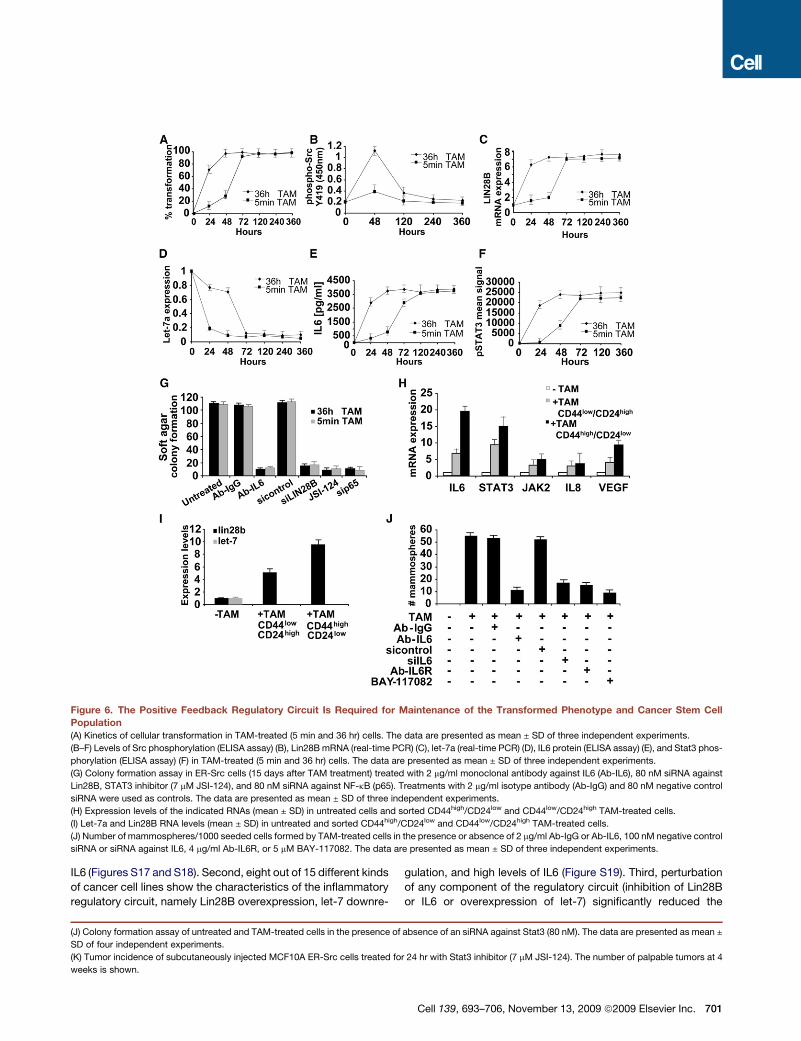
transformed phenotype, we examined transformed cells (gener-
ated by 5 min or 36 hr of TAM treatment) for up to 15 days after
removal of TAM. Under these conditions, the transformed
phenotype is maintained in the absence of Src activity, with
high levels of Lin28B, IL6, and phosphorylated STAT3 and low
levels of let-7 (Figures 6A–6F). Furthermore, breaking the regula-
tory circuit in these stably transformed cells by inhibition of IL6,
Lin28B, STAT3, or NF-kB leads to loss of tumorigenicity and
cell motility (Figures 6G and S14). Thus, the positive feedback
loop is required to maintain the stability of the transformed
phenotype.
ER-Src cells treated with TAM can form mammospheres with
self-renewal properties (Hirsch et al., 2009) (Figure 1A), suggest-
ing that they have attributes of ‘‘cancer stem cells’’ (also known
as tumor initiating cells). Cancer stem cells express high levels of
CD44 and low levels of CD24 antigen markers (Mani et al., 2008),
and �10% of the TAM-treated ER-Src cell population are stem
cells (CD44high/CD24low), whereas 90% are nonstem-trans-
formed cells (CD44low/CD24high). This ratio of stem cells and
cancer cells is typical for other cancer cell lines (Ponti et al.,
2005; Chu et al., 2009). In accord with the defining features of
cancer stem cells, the CD44high/CD24low cells can form mammo-
spheres and tumors in nude mice, while CD44low/CD24high cells
cannot.
Interestingly, the cancer stem cells display a stronger inflam-
matory gene signature (Figure 6H), more increased levels of
nuclear NF-kB (Figure S15), higher levels of Lin28B, and more
decreased let-7a expression (Figure 6I) than observed in the non-
stem cancer cells. Thus, mammosphere formation involves the
selection of a subpopulation of cancer stem cells in which the
inflammatory feedback loop is even more active than in the
majority population of cancer cells. In mammospheres derived
from ER-Src-transformed cells, inhibition of IL6, IL6 receptor, or
NF-kB activity blocks mammosphere propagation (Figure 6J),
indicating that the inflammatory feedback loop is required for
growth of cancer stem cells. Conversely, untransformed
MCF10A cells can form mammospheres after IL6 treatment.
This IL6-mediated mammosphere formation is dramatically
reduced after let-7a overexpression or NF-kB inhibition, and
the few mammospheres formed are smaller (Figure S16A).
Similarly, inhibition of let-7a or Lin28B overexpression permits
MCF10A cells to form mammospheres in a manner that depends
on IL6 (Figure S16B). These data suggest that activation of NF-kB
and the IL6 pathway through inhibition of let-7 by Lin28B is
required for the self-renewal capacity of cancer stem cells.
The Inflammatory Regulatory Circuit Is Important forCancer Cells from Diverse Developmental LineagesSeveral lines of evidence indicate that NF-kB, Lin28B, let-7a, and
IL6 are more generally involved in oncogenic transformation.
First, in MCF-10A cells, RASV12 behaves similarly to Src in acti-
vating NF-kB and mediating transformation through let-7 and
(G) Western blot analysis of RAS protein expression in 36 hr TAM-treated cells after let-7a (100 nM) overexpression; GAPDH levels were used as a loading control.
(H) IL6 production (ELISA assay) during transformation after transfection with negative control siRNA or siRNA against Ras or let-7a (100 nM). The data are
presented as mean ± SD of three independent experiments.
(I) Soft agar colony assay in TAM-treated cells after treatment with 2 mg/ml of monoclonal antibody against IL6 (Ab-IL6) and IgG isotype antibody (Ab-IgG) or
siRNA negative control and siRNA against Ras (100 nM). The data are presented as mean ± SD of three independent experiments.
Cell 139, 693–706, November 13, 2009 ª2009 Elsevier Inc. 699
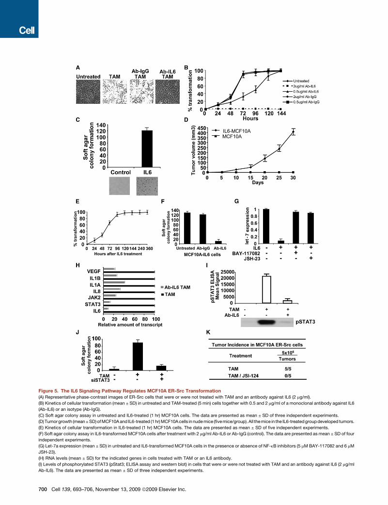
Figure 5. The IL6 Signaling Pathway Regulates MCF10A ER-Src Transformation
(A) Representative phase-contrast images of ER-Src cells that were or were not treated with TAM and an antibody against IL6 (2 mg/ml).
(B) Kinetics of cellular transformation (mean ± SD) in untreated and TAM-treated (5 min) cells together with 0.5 and 2 mg/ml of a monoclonal antibody against IL6
(Ab-IL6) or an isotype (Ab-IgG).
(C) Soft agar colony assay in untreated and IL6-treated (1 hr) MCF10A cells. The data are presented as mean ± SD of three independent experiments.
(D) Tumor growth (mean ± SD) of MCF10A and IL6-treated (1 hr) MCF10A cells in nude mice (five mice/group). All themice in the IL6-treated group developed tumors.
(E) Kinetics of cellular transformation in IL6-treated (1 hr) MCF10A cells. The data are presented as mean ± SD of five independent experiments.
(F) Soft agar colony assay in IL6-transformed MCF10A cells after treatment with 2 mg/ml Ab-IL6 or Ab-IgG (control). The data are presented as mean ± SD of four
independent experiments.
(G) Let-7a expression (mean ± SD) in untreated and IL6-transformed MCF10A cells in the presence or absence of NF-kB inhibitors (5 mM BAY-117082 and 6 mM
JSH-23).
(H) RNA levels (mean ± SD) for the indicated genes in cells treated with TAM or an IL6 antibody.
(I) Levels of phosphorylated STAT3 (pStat3; ELISA assay and western blot) in cells that were or were not treated with TAM and an antibody against IL6 (2 mg/ml
Ab-IL6). The data are presented as mean ± SD of three independent experiments.
700 Cell 139, 693–706, November 13, 2009 ª2009 Elsevier Inc.
IL6 (Figures S17 and S18). Second, eight out of 15 different kinds
of cancer cell lines show the characteristics of the inflammatory
regulatory circuit, namely Lin28B overexpression, let-7 downre-
gulation, and high levels of IL6 (Figure S19). Third, perturbation
of any component of the regulatory circuit (inhibition of Lin28B
or IL6 or overexpression of let-7) significantly reduced the
(J) Colony formation assay of untreated and TAM-treated cells in the presence of absence of an siRNA against Stat3 (80 nM). The data are presented as mean ±
SD of four independent experiments.
(K) Tumor incidence of subcutaneously injected MCF10A ER-Src cells treated for 24 hr with Stat3 inhibitor (7 mM JSI-124). The number of palpable tumors at 4
weeks is shown.
Figure 6. The Positive Feedback Regulatory Circuit Is Required for Maintenance of the Transformed Phenotype and Cancer Stem Cell
Population
(A) Kinetics of cellular transformation in TAM-treated (5 min and 36 hr) cells. The data are presented as mean ± SD of three independent experiments.
(B–F) Levels of Src phosphorylation (ELISA assay) (B), Lin28B mRNA (real-time PCR) (C), let-7a (real-time PCR) (D), IL6 protein (ELISA assay) (E), and Stat3 phos-
phorylation (ELISA assay) (F) in TAM-treated (5 min and 36 hr) cells. The data are presented as mean ± SD of three independent experiments.
(G) Colony formation assay in ER-Src cells (15 days after TAM treatment) treated with 2 mg/ml monoclonal antibody against IL6 (Ab-IL6), 80 nM siRNA against
Lin28B, STAT3 inhibitor (7 mM JSI-124), and 80 nM siRNA against NF-kB (p65). Treatments with 2 mg/ml isotype antibody (Ab-IgG) and 80 nM negative control
siRNA were used as controls. The data are presented as mean ± SD of three independent experiments.
(H) Expression levels of the indicated RNAs (mean ± SD) in untreated cells and sorted CD44high/CD24low and CD44low/CD24high TAM-treated cells.
(I) Let-7a and Lin28B RNA levels (mean ± SD) in untreated and sorted CD44high/CD24low and CD44low/CD24high TAM-treated cells.
(J) Number of mammospheres/1000 seeded cells formed by TAM-treated cells in the presence or absence of 2 mg/ml Ab-IgG or Ab-IL6, 100 nM negative control
siRNA or siRNA against IL6, 4 mg/ml Ab-IL6R, or 5 mM BAY-117082. The data are presented as mean ± SD of three independent experiments.
Cell 139, 693–706, November 13, 2009 ª2009 Elsevier Inc. 701
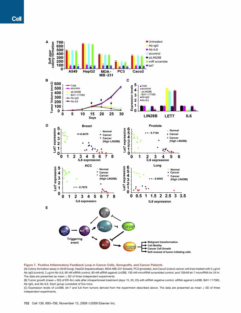
Figure 7. Positive Inflammatory Feedback Loop in Cancer Cells, Xenografts, and Cancer Patients
(A) Colony formation assay in A549 (lung), HepG2 (hepatocellular), MDA-MB-231 (breast), PC3 (prostate), and Caco2 (colon) cancer cell lines treated with 2 mg/ml
Ab-IgG (control), 2 mg/ml Ab-IL6, 80 nM siRNA control, 80 nM siRNA against Lin28B, 100 nM microRNA scrambled control, and 100nM let-7 microRNA for 24 hr.
The data are presented as mean ± SD of three independent experiments.
(B) Tumor growth (mean ± SD) of ER-Src cells after intraperitoneal treatment (days 15, 20, 25) with siRNA negative control, siRNA against Lin28B, BAY-117082,
Ab-IgG, and Ab-IL6. Each group consisted of five mice.
(C) Expression levels of Lin28B, let-7 and IL6 from tumors derived from the experiment described above. The data are presented as mean ± SD of three
independent experiments.
702 Cell 139, 693–706, November 13, 2009 ª2009 Elsevier Inc.
tumorigenicity and motility of lung (A549), hepatocellular
(HepG2), breast (MDA-MB-231), prostate (PC3), and colon
(Caco2) cancer cells (Figures 7A and S20A). In all cases, these
perturbations resulted in reduced expressionof IL6 (FigureS20B),
suggesting the importance of IL6 in maintaining the transformed
phenotype. Thus, the inflammatory feedback loop is important
for cancer cells from diverse developmental lineages.
The Inflammatory Regulatory Circuit Is Importantfor Cancer Cell Growth In VivoTo address whether the inflammatory regulatory circuit was
important for cancer growth in vivo, we injected subcutaneously
injected TAM-treated cells into 30 nu/nu mice and obtained
tumors with a size of 100 mm3 in all cases. The mice were
randomly separated into six groups and treated intraperitoneally
with siRNAs against Lin28B, the NF-kB inhibitor BAY-117082,
or a monoclonal antibody against IL6; treatments were repeated
for three cycles. After the second cycle of treatment, there was
significant suppression of tumor growth in all treated mice (Fig-
ure 7B). After 30 days, the tumors were extremely small in size,
and cells taken from these tumors had low expression of
Lin28B and IL6 and high levels of let-7 (Figure 7C). Thus, pertur-
bation of any component of the regulatory circuit strongly
suppresses tumor growth and restores gene expression patterns
typical of nontransformed cells.
Let-7a and IL6 Are Negatively Correlated in Cancerand Normal TissuesTo address whether the inflammatory feedback loop is relevant
to human cancer, we examined the expression of Lin28B,
let-7a, and IL6 in cancer and normal breast, prostate, hepatocel-
lular, and lung tissues (Figures 7D and S21). As expected, cancer
tissues had lower levels of let-7a and higher levels of IL6 relative
to normal tissues. More importantly, among both normal and
diseased individuals, there is a striking inverse relationship
between let-7a and IL6 expression levels in breast, prostate,
and hepatocellular tissues. This striking inverse relationship
strongly argues for a mechanistic relationship between let-7
and IL6 in these tissues that is amplified in cancer. In contrast,
while let-7a expression is reduced in lung cancer tissues, only
a subset of these show high IL6 levels. Furthermore, Lin28B is
overexpressed in a subset of breast (7/17), prostate (6/15), and
hepatocellular (7/9) cancer tissues, suggesting a mechanistic
relationship between Lin28B, let-7, and IL6 in different forms of
cancer. These results suggest that the regulatory pathway iden-
tified in the ER-Src model (Figure 7E) is relevant to human
disease and specifically important for certain cancer types.
DISCUSSION
A Molecular Pathway that Links Inflammation to CellularTransformationIn 1863, Rudolf Virchow proposed that chronic inflammation may
lead to development of cancer, and there are now clinical, epide-
miological, and molecular links between inflammation and onco-
genic transformation (Balkwill and Mantovani, 2001; Naugler and
Karin, 2008; Pierce et al., 2009). However, molecular pathways
linking inflammation to cellular transformation are unknown, in
large part because of the lack of experimental systems that
can follow the process by which a nontransformed cell becomes
transformed. Here, using an experimental model in which activa-
tion of an ER-Src oncoprotein by treatment with tamoxifen
converts a nontransformed epithelial cell line (MCF-10A) to the
transformed state in 24–36 hr, we describe such a molecular
pathway that involves NF-kB, Lin28B, let-7 microRNA, and IL6
(Figure 7E).
The first step in the pathway is the activation of NF-kB, which
occurs within 30 min after treatment with tamoxifen. By acti-
vating NF-kB, Src effectively provides the inflammatory signal
that is critical for cellular transformation. Src is required for
NF-kB activation in other biological contexts (Abu-Amer et al.,
1998; Lee et al., 2007), so genetic changes or environmental
conditions that activate Src might contribute to cancer via an
inflammatory pathway. However, in our model, Src does not
play a significant role in the transformation process other than
providing the initial inflammatory signal. In this regard, activation
of NF-kB by Ras or by IL6 also induces the oncogenic transition
in MCF-10A cells lacking the ER-Src protein.
Downregulation of the let-7 microRNA family is a critical early
step in the oncogenic transition of the ER-Src cells, because
expression on any individual let-7 family member blocks cellular
transformation. Although NF-kB is required for downregulation
of the level of let-7 RNAs, this inhibition occurs primarily at a post-
transcriptional level, because the amounts of the let-7 precursor
RNA are essentially unchanged during the transformation
process. Lin28B plays a critical role in processing let-7RNA
(Heo et al., 2008; Viswanathan et al., 2008), and our results indi-
cate that NF-kB inhibits let-7 RNA levels primarily by activating
transcription of lin-28B via direct binding to a highly conserved
NF-kB site in the first intron. By using lin-28B as an intermediary,
NF-kB rapidly transmits an inflammatory signal into a mechanism
for coordinate inhibition of all let-7 microRNAs, which is essential
for transformation. NF-kB also binds in vivo to a site upstream of
the let-7 RNA coding region, but it is unclear whether this binding
affects the regulation of let-7 RNA.
Expression of the cytokine IL6, a major mediator of the inflam-
matory response, is directly repressed by let-7 through a stan-
dard interaction of the microRNA with the 30 UTR of the target
mRNA. NF-kB activation and subsequent repression of let-7
therefore results in a dramatic and biphasic increase in IL6 levels,
which is necessary for cellular transformation. These high levels
of IL6 depend on both transcriptional activation by NF-kB and
inhibition of Let7 and are required for sufficient binding to the
IL6 receptor to cause phosphorylation and nuclear entry of the
STAT3 transcription factor, which then activates multiple growth
and survival genes such as VEGF (Niu et al., 2002). In the ER-Src
cells, STAT3 is a key IL6 target that is required for cellular trans-
formation. Consistent with these results, STAT3 is important for
(D) Lin28B, let-7a, and IL6 expression in breast, prostate, hepatocellular, and lung cancer and normal tissues. Each data point represents an individual sample,
and a correlation coefficient (r) between let-7a and IL6 expression is shown.
(E) Schematic overview of inflammatory positive feedback loop during cellular transformation.
Cell 139, 693–706, November 13, 2009 ª2009 Elsevier Inc. 703
colitis-associated tumorigenesis (Bollrath et al., 2009; Grivenni-
kov et al., 2009).
An Epigenetic Switch from Nontransformed toTransformed Cells Mediated by an InflammatoryPositive Feedback LoopAn epigenetic switch occurs when a stable cell type changes to
another stable cell type without any change in DNA sequence.
Epigenetic switches require an initiating event (which could
be a specific molecular process or stochastic fluctuation), but
the phenotypes of the new cell type are inherited in the absence
of the initiating signal. Here, we describe an epigenetic switch
in which a stable nontransformed cell type is converted to
a stable transformed cell type via transient activation of the Src
oncoprotein. Remarkably, this switch in cell type occurs even
when cells are exposed to tamoxifen for only 5 min. Furthermore,
mammospheres generated from the transformed cells can be
propagated for at least 12 generations over 2.5 months in the
absence of tamoxifen. As the transition between nontrans-
formed to transformed cells occurs within 24–36 hr, it is
extremely unlikely to involve changes in DNA sequence. Hence,
the phenomenon we describe here fits all the definitions of an
epigenetic switch.
As illuminated by classic studies of an epigenetic switch in
bacteriophage l, positive feedback loops involving transcrip-
tional regulatory proteins are the fundamental principle for epige-
netic inheritance of gene expression patterns (Ptashne, 2009).
NF-kB, Lin28B, let-7 microRNA, and IL6 are the key components
of the positive feedback loop underlying the epigenetic switch
from a nontransformed cell type to a transformed cell type
capable of forming self-renewing stem cells (Figure 7E). The
switch is triggered by an initial inflammatory signal (activated
Src here) that activates NF-kB. NF-kB generates high levels of
IL6 by direct activation of IL6 transcription and indirect (via
Lin28B) inhibition of let-7 microRNA. The resulting high levels
of IL6 activate NF-kB, thereby completing the positive feedback
loop that maintains the transformed phenotype, self-renewal of
mammospheres, and tumor formation in nude mice in the
absence of the triggering event (Src activation). High levels of
IL6 are crucial for activating NF-kB, and this is why Let-7 is
required for the regulatory circuit.
A related positive feedback loop involving Ras is likely to
contribute to the epigenetic switch. As is the case for IL6, Ras
is both a direct target of Let-7 and an activator of NF-kB.
However, as the oncogenic form of Ras (Ras-V12) is not present
in MCF-10A cells, we suspect that this regulatory circuit is not as
important as the feedback loop involving IL6 in our experimental
system. Our results do not exclude, and we would not be
surprised by, the existence of analogous feedback loops
involving NF-kB or other transcriptional regulatory proteins or
microRNAs.
Three classes of experiments validate the positive feedback
loop. First, overexpression of any positive factor (Lin28B, IL6)
or inhibition of a negative factor (let-7) induces cellular transfor-
mation, indicating that the loop can be started at any step.
Second, under conditions where the transformed state is stable
in the absence of the inducing factor, perturbation at any step
breaks the established positive feedback loop, thereby causing
704 Cell 139, 693–706, November 13, 2009 ª2009 Elsevier Inc.
loss of the transformed state. Third, the strength of the initial
signal (time of TAM treatment) or the amount of product gener-
ated by that signal (IL6) is inversely correlated with the rate of
observing cellular transformation.
Ultimately, the epigenetic switch is an inflammatory positive
feedback loop in which a transient inflammatory signal is con-
verted into a chronic inflammatory state that is maintained by
activated NF-kB. Once NF-kB is activated, the regulatory circuit
is sufficient to generate and maintain the chronic inflammatory
loop (i.e., the transformed state) without the original (or new)
environmental signal. Importantly, this inflammatory feedback
loop is a common feature of cellular transformation, because it
occurs and is functionally relevant in cancer cells of diverse
developmental origin.
Relevance to Human CancerOur experimental model has the advantage of studying the
process of cellular transformation and cancer stem cell forma-
tion in a dynamic and well-defined manner, analogous to studies
of viral infection. Although this model utilizes an immortalized cell
line with an artificial oncogene, the key components of the switch
between nontransformed and transformed cells, NF-kB, let-7,
and IL6, have all been linked to human disease. Activation of
the NF-kB pathway is correlated with carcinogenesis (Luo
et al., 2005), Let-7 is downregulated in several cancers (Johnson
et al., 2005; Mayr et al., 2007; Sampson et al., 2007), Lin28 is
overexpressed in primary human tumors (Viswanathan et al.,
2009) (Figure 7D), and IL6 has been implicated as a growth factor
for multiple myeloma, Hodgkin’s lymphoma, and epithelial
cancers (Kawano et al., 1988; Nagel et al., 2005; Sasser et al.,
2007). Our finding of a striking inverse relationship between
let-7 and IL6 expression in breast and prostate epithelial cancer
tissues suggests the importance of inflammatory activation and
the IL6-let-7 regulatory circuit in solid cancers. Lastly, the epige-
netic switch can be triggered by v-Src or Ras-V12, which are
oncogenes associated with many human cancers.
In our cellular transformation model, the epigenetic switch
between nontransformed and transformed cells occurs very
quickly in response to a transient inflammatory signal. Although
a transient inflammatory signal is clearly insufficient to trigger
such an epigenetic switch in normal cells, we believe that the
epigenetic switch described here is relevant to human cancer.
Specifically, we suggest that the epigenetic switch requires
cells that are genetically altered to be at an intermediate stage
in the transition between a primary cell and a cancer cell. The
epigenetic switch requires cells that can inhibit let-7 and
generate high levels of IL6 upon NF-kB stimulation, and it is
likely that this requires multiple factors in addition to NF-kB
itself. In addition, although IL6 is a key effector molecule, gener-
ation of the transformed state undoubtedly requires other
factors. These additional factors are likely to depend on devel-
opmental state, extracellular stimuli, and mutational status, thus
accounting for cell-type specificity of the epigenetic switch.
Nevertheless, the results presented here provide a paradigm
in which a key step in cancer progression involves an epigenetic
switch in response to an inflammatory (or other environmental)
signal as opposed to a mutational change in a tumor
suppressor or oncogene.
EXPERIMENTAL PROCEDURES
Cell Culture, Cellular Transformation Assays, and Isolation
of Cancer Stem Cells
MCF10A cells containing ER-Src, an integrated fusion of the v-Src oncopro-
tein, and the ligand-binding domain of estrogen receptor were grown and
induced to transform with 1 mM 4OH-TAM as described previously (Hirsch
et al., 2009). In some experiments, cells were treated with human recombinant
IL6, anti-IL6 antibody, and the NF-kB inhibitors BAY-117082 and JSH-23.
Except where otherwise indicated, morphological changes, phenotypic
transformation, and foci formation occurred 24–36 hr after TAM addition and
were monitored by phase-contrast microscopy. Cells were assayed for their
ability to grow as anchorage-independent colonies in soft agar and to form
mammospheres as described previously (Hirsch et al., 2009). For long-term
propagation, mammospheres were collected by gentle centrifugation, dissoci-
ated to single cells as described (Dontu et al., 2003), and then cultured to
obtain the next generation. Cancer stem cells (CD44high/CD24low) and non-
stem-transformed cells (CD44low/CD24high) were isolated from transformed
cell populations by flow-cytometric sorting on single-cell suspensions stained
with CD44 (FITC-conjugated) and CD24 (PE-conjugated) antibodies. Other cell
lines were grown in Dulbecco’s modified Eagle’s medium, 10% fetal bovine
serum, and penicillin/streptomycin.
Protein Analysis
Western blotting was performed by standard procedures with antibodies
against Ras, STAT3, the p65 subunit of NF-kB, Lin28B, Socs3, GAPDH, and
b-actin, and detection was performed with HRP-conjugated antisera and
chemiluminescence. ELISA assays for IL6, VEGF, NF-kB, phospho (Ser32)-
IkBa, and phospho (Tyr705)-STAT3 were performed in accord with manufac-
turers’ instructions. To examine the association of the RISC complex with IL6
mRNA, HEK293 cells were cotransfected with a plasmid that expressed
HA-Ago1 together with 100 nM microRNAs, followed by HA-Ago1 immunopre-
cipitation and analysis of IL6 RNA levels.
RNA Analysis
For analysis of mRNAs, total RNA was reverse transcribed to form comple-
mentary DNA, and the resulting material analyzed by quantitative PCR in
real-time. Levels of Let-7 microRNAs were determined with the mirVana
qRT-PCR miRNA Detection Kit and qRT-PCR Primer Sets, according to the
manufacturer’s instructions, with RNU48 used as a control. For analysis of
patient samples, we only used samples lacking the absence of infiltrating
macrophages, which was determined by expression of the macrophage
marker CD11b, which is not expressed in epithelial cells.
Chromatin Immunoprecipitation
Potential NF-kB binding sites in the vicinity of the Let-7a3 and Lin28B mRNA
initiation site were identified by DNA sequence motif, evolutionary conserva-
tion, and nucleosome occupancy. NF-kB (p65) binding in vivo to these putative
sites was analyzed by chromatin immunoprecipitation and quantitative PCR
analysis as described previously (Yang et al., 2006).
Transfection Experiments
Transcriptional activation by NF-kB and inhibition of IL6 mRNA levels by let-7
microRNA were performed standard luciferase reporter gene assays upon
transient transfection of the relevant DNAs. For other genetic experiments,
siRNAs or plasmids capable of overexpressing the desired microRNAs or
protein were transiently transfected into cells. After 24 hr, the resulting cells
were phenotypically analyzed for transformation and for levels of RNAs and
proteins of interest.
Xenograft Experiments
Injections of nontransformed and transformed MCF-10A cells into nude mice,
treatments of tumors by intraperitoneal injections, and measurements of tumor
volume were performed as described previously (Hirsch et al., 2009).
For all quantitative experiments, data are presented as mean values ± SD
from three independent experiments. Detailed experimental procedures are
provided in the Supplemental Data.
SUPPLEMENTAL DATA
Supplemental Data include Supplemental Experimental Procedures and 21
figures and can be found with this article online at http://www.cell.com/
supplemental/S0092-8674(09)01302-6.
ACKNOWLEDGMENTS
We would like to thank Richard Gregory for suggesting the idea that lin-28
might be involved in the process, Philip N. Tsichlis for providing laboratory
access and materials needed for the xenograft experiments, Fabio Petrocca
for help in designing the mammosphere and cell sorting experiments, Savina
A. Jaeger for bioinformatic analysis and identification of NF-KB binding sites
in let-7a and lin-28B, Joan Brugge for providing the MCF10A ER-Src and
control cell lines, William Farrar for providing the pGL3-IL6 luciferace vector,
George Daley for providing the pBabe.Puro-Lin28B vector, Joshua T. Mendell
for providing the Lin28B-P1 luciferase vector, and Koon Ho Wong for
construction of the mutant luciferase vectors. This work was supported by
a postdoctoral fellowship from the American Cancer Society to H.A.H. and
a research grant to K.S. from the National Institutes of Health (CA 107486).
Received: April 3, 2009
Revised: July 15, 2009
Accepted: September 25, 2009
Published online: October 29, 2009
REFERENCES
Abu-Amer, Y., Ross, F., McHugh, K., Livolsi, A., Peyron, J.F., and Teitelbaum,
S. (1998). Tumor necrosis factor-alpha activation on nuclear factor-kB in
marrow macrophages is mediated by c-Src tryrosine phosphorylation of
IKB. J. Biol. Chem. 273, 29417–29423.
Ancrile, B., Lim, K.H., and Counter, C.M. (2007). Oncogenic Ras-induced
secretion of IL6 is required for tumorigenesis. Genes Dev. 21, 1714–1719.
Balkwill, F., and Mantovani, A. (2001). Inflammation and cancer: back to
Virchow? Lancet 357, 539–545.
Bartel, D.P. (2009). MicroRNAs: target recognition and regulatory functions.
Cell 136, 215–233.
Baylin, S.B. (2005). DNA methylation and gene silencing in cancer. Nat Clin
Pract Oncol 2, S4–S11.
Bollrath, J., Phesse, T.J., von Burstin, V.A., Putoczki, T., Bennecke, M., Bate-
man, T., Nebelsiek, T., Lundgren-May, T., Canli, O., Schwitalla, S., et al. (2009).
gp130-mediated Stat3 activation in enterocytes regulates cell survival and
cell-cycle progression during colitis-associated tumorigenesis. Cancer Cell
15, 91–102.
Calin, G.A., Sevignani, C., Dumitru, C.D., Hyslop, T., Noch, E., Yendamuri, S.,
Shimizu, M., Rattan, S., Bullrich, F., Negrini, M., et al. (2004). Human microRNA
genes are frequently located at fragile sites and genomic regions involved in
cancers. Proc. Natl. Acad. Sci. USA 101, 2999–3004.
Chu, P., Clanton, D.J., Snipas, T.S., Lee, J., Mitchell, E., Nguyen, M.L., Hare,
E., and Peach, R.J. (2009). Characterization of a subpopulation of colon cancer
cells with stem cell-like properties. Int. J. Cancer 124, 1312–1321.
Dontu, G., Abdallah, W.M., Foley, J.M., Jackson, K.W., Clarke, M.F., Kawa-
mura, M.J., and Wicha, M.S. (2003). In vitro propagation and transcriptional
profiling of human mammary stem/progenitor cells. Genes Dev. 17, 1253–
1270.
Frank, D.A. (2007). STAT3 as a central mediator of neoplastic cellular transfor-
mation. Cancer Lett. 251, 199–210.
Gazin, C., Wajapeyee, N., Gobeil, S., Virbasius, C.M., and Green, M.R. (2007).
An elaborate pathway required for Ras-mediated epigenetic silencing. Nature
449, 1073–1077.
Grimshaw, M.J., Cooper, L., Papazisis, K., Coleman, J.A., Bohnenkamp, H.R.,
Chiapero-Stanke, L., Taylor-Papadimitriou, J., and Burchell, J.M. (2008).
Cell 139, 693–706, November 13, 2009 ª2009 Elsevier Inc. 705
Mammosphere culture of metastatic breast cancer cells enriches for tumori-
genic breast cancer cells. Breast Cancer Res. 10, R52.
Grivennikov, S., Karin, E., Terzic, J., Mucida, D., Yu, G.Y., Vallabhapurapu, S.,
Scheller, J., Rose-John, S., Cheroutre, H., Eckmann, L., et al. (2009). IL-6 and
Stat3 are required for survival of intestinal epithelial cells and development of
colitis-associated cancer. Cancer Cell 15, 103–113.
Hahn, W.C., and Weinberg, R.A. (2002). Rules for making human tumor cells.
N. Engl. J. Med. 347, 1593–1603.
Heo, I., Joo, C., Cho, J., Ha, M., Han, J., and Kim, V.N. (2008). Lin28 mediates
the terminal uridylation of let-7 precursor MicroRNA. Mol. Cell 32, 276–284.
Hirsch, H.A., Iliopoulos, D., Tsichlis, P.N., and Struhl, K. (2009). Metformin
selectively targets cancer stem cells and acts together with chemotherapy
to blocks tumor growth and prolong remission. Cancer Res. 69, in press.
Johnson, S.M., Grosshans, H., Shingara, J., Byrom, M., Jarvis, R., Cheng, A.,
Labourier, E., Reinert, K.L., Brown, D., and Slack, F.J. (2005). RAS is regulated
by the let-7 microRNA family. Cell 120, 635–647.
Kawano, M., Hirano, T., Matsuda, T., Taga, T., Horii, Y., Iwato, K., Asaoku, H.,
Tang, B., Tanabe, O., Tanaka, H., et al. (1988). Autocrine generation and
requirement of BSF-2/IL-6 for human multiple myelomas. Nature 332, 83–85.
Kumar, M.S., Erkeland, S.J., Pester, R.E., Chen, C.Y., Ebert, M.S., Sharp, P.A.,
and Jacks, T. (2008). Suppression of non-small cell lung tumor development
by the let-7 microRNA family. Proc. Natl. Acad. Sci. USA 105, 3903–3908.
Lee, H.S., Moon, C., Lee, H.W., Park, E.M., Cho, M.S., and Kang, J.L. (2007).
Src tyrosine kinases mediate activations of NF-kappaB and integrin signal
during lipopolysaccharide-induced acute lung injury. J. Immunol. 179,
7001–7011.
Liao, M.J., Zhang, C.C., Zhou, B., Zimonjic, D.B., Mani, S.A., Kaba, M., Gifford,
A., Reinhardt, F., Popescu, N.C., Guo, W., et al. (2007). Enrichment of a popu-
lation of mammary gland cells that form mammospheres and have in vivo
repopulating activity. Cancer Res. 67, 8131–8138.
Lu, J., Getz, G., Miska, E.A., Alvarez-Saavedra, E., Lamb, J., Peck, D., Sweet-
Cordero, A., Ebert, B.L., Mak, R.H., Ferrando, A.A., et al. (2005). MicroRNA
expression profiles classify human cancers. Nature 435, 834–838.
Luedde, T., Beraza, N., Kotsikoris, V., van Loo, G., Nenci, A., De Vos, R.,
Roskams, T., Trautwein, C., and Pasparakis, M. (2007). Deletion of NEMO/
IKKgamma in liver parenchymal cells causes steatohepatitis and hepatocel-
lular carcinoma. Cancer Cell 11, 119–132.
Luo, J.-L., Kamata, H., and Karin, M. (2005). IKK/NF-kB signaling: balancing
life and death- a new approach to cancer therapy. J. Clin. Invest. 115, 2625–
2632.
Mani, S.A., Guo, W., Liao, M.J., Eaton, E.N., Ayyanan, A., Zhou, A.Y., Brooks,
M., Reinhard, F., Zhang, C.C., Shipitsin, M., et al. (2008). The epithelial-
mesenchymal transition generates cells with properties of stem cells. Cell
133, 704–715.
Mayr, C., Hemann, M.T., and Bartel, D.P. (2007). Disrupting the pairing
between let-7 and Hmga2 enhances oncogenic transformation. Science
315, 1576–1579.
Nagel, S., Scherr, M., Quentmeier, H., Kaufmann, M., Zaborski, M., Drexler,
H.G., and MacLeod, R.A. (2005). HLXB9 activates IL6 in Hodgkin lymphoma
cell lines and is regulated by PI3K signalling involving E2F3. Leukemia 19,
841–846.
706 Cell 139, 693–706, November 13, 2009 ª2009 Elsevier Inc.
Naugler, W.E., and Karin, M. (2008). NF-kappaB and cancer-identifying targets
and mechanisms. Curr. Opin. Genet. Dev. 18, 19–26.
Niu, G., Wright, K.L., Huang, M., Song, L., Haura, E., Turkson, J., Zhang, S.,
Wang, T., Sinibaldi, D., Coppola, D., et al. (2002). Constitutive Stat3 activity
up-regulates VEGF expression and tumor angiogenesis. Oncogene 21,
2000–2008.
Pierce, B.L., Ballard-Barbash, R., Bernstein, L., Baumgartner, R.N.,
Neuhouser, M.L., Wener, M.H., Baumgartner, K.B., Gilliland, F.D., Sorensen,
B.E., McTiernan, A., et al. (2009). Elevated biomarkers of inflammation are
associated with reduced survival among breast cancer patients. J. Clin. Oncol.
27, 3437–3444.
Ponti, D., Costa, A., Zaffaroni, N., Pratesi, G., Petrangolini, G., Coradini, D.,
Pilotti, S., Pierotti, M.A., and Daidone, M.G. (2005). Isolation and in vitro prop-
agation of tumorigenic breast cancer cells with stem/progenitor cell proper-
ties. Cancer Res. 65, 5506–5511.
Ptashne, M. (2009). Binding reactions: epigenetic switches, signal transduc-
tion and cancer. Curr. Biol. 19, R234–R241.
Sakurai, T., He, G., Matsuzawa, A., Yu, G.Y., Maeda, S., Hardiman, G., and
Karin, M. (2008). Hepatocyte necrosis induced by oxidative stress and IL-1
alpha release mediate carcinogen-induced compensatory proliferation and
liver tumorigenesis. Cancer Cell 14, 156–165.
Sampson, V.B., Rong, N.H., Han, J., Yang, Q., Aris, V., Soteropoulos, P.,
Petrelli, N.J., Dunn, S.P., and Krueger, L.J. (2007). MicroRNA let-7a down-
regulates MYC and reverts MYC-induced growth in Burkitt lymphoma cells.
Cancer Res. 67, 9762–9770.
Sasser, A.K., Sullivan, N.J., Studebaker, A.W., Hendey, L.F., Axel, A.E., and
Hall, B.M. (2007). Interleukin-6 is a potent growth factor for ER-alpha-positive
human breast cancer. FASEB J. 21, 3763–3770.
Soule, H.D., Maloney, T.M., Wolman, S.R., Peterson, W.D., Brenz, R.,
McGrath, C.M., Russo, J., Pauley, R.J., Jones, R.F., and Brooks, S.C.
(1990). Isolation and characterization of a spontaneously immortallized human
breast epithelial cell line, MCF10. Cancer Res. 50, 6075–6086.
Ventura, A., and Jacks, T. (2009). MicroRNAs and cancer: short RNAs go
a long way. Cell 136, 586–591.
Viswanathan, S.R., Daley, G.Q., and Gregory, R.I. (2008). Selective blockade
of microRNA processing by Lin28. Science 320, 97–100.
Viswanathan, S.R., Powers, J.T., Einhorn, W., Hoshida, Y., Ng, T.L., Toffanin,
S., O’Sullivan, M., Lu, J., Phillips, L.A., Lockhart, V.L., et al. (2009). Lin28
promotes transformation and is associated with advanced human malignan-
cies. Nat. Genet. 41, 843–848.
Vogelstein, B., and Kinzler, K.W. (2004). Cancer genes and the pathways they
control. Nat. Med. 10, 789–799.
Wegiel, B., Bjartell, A., Culig, Z., and Persson, J.L. (2008). Interleukin-6 acti-
vates PI3K/Akt pathway and regulates cyclin A1 to promote prostate cancer
cell survival. Int. J. Cancer 122, 1521–1529.
Yang, A., Zhu, Z., Kapranov, P., McKeon, F., Church, G.M., Gingeras, T.R., and
Struhl, K. (2006). Relationships between p63 binding, DNA sequence,
transcription activity, and biological function in human cells. Mol. Cell 24,
593–602.
![GSW670P - adeltd.co.uk · LTS - Load Transfer Switch [Accessories for ACP Automatic Control Panel] The Load Transfer Switch (LTS) panel operates the power supply changeover between](https://static.fdocument.org/doc/165x107/604ba8e6b01bc235557c4ce1/gsw670p-lts-load-transfer-switch-accessories-for-acp-automatic-control-panel.jpg)