A. soli Spread of A. soli Vaccinia Virus to Cattle … test for screening...
5
LETTERS 1576 Emerging Infectious Diseases • www.cdc.gov/eid • Vol. 20, No. 9, September 2014 (9). The TMB-2–producing A. soli strain that we isolated came from a blood culture, indicating that A. soli is a potential cause of bloodstream in- fections or bacteremia. A. soli has also been detected in lice and keds of do- mestic animals (10), indicating that A. soli may inhabit natural environments and that injuries and bites by arthro- pods might present a risk for invasive infections. Isolates of Acinetobacter species, particularly those recovered from blood culture, should be identi- fied to species type to enable further evaluation of the clinical significance of carbapenem-resistant A. soli strains. This study was supported by the Min- istry of Health, Labour and Welfare, Japan (H25-Shinko-Ippan-003 and H24-Shinko- Ippan-010). Hiromitsu Kitanaka, Masa-aki Sasano, Satoru Yokoyama, Masahiro Suzuki, Wanchun Jin, Masami Inayoshi, Mitsuhiro Hori, Jun-ichi Wachino, Kouji Kimura, Keiko Yamada, and Yoshichika Arakawa Author affiliations: Nagoya University Graduate School of Medicine, Aichi, Japan (H. Kitanaka, S. Yokoyama, W. Jin, J. Wachino, K. Kimura, K. Yamada, Y. Arakawa); Okazaki City Hospital, Aichi (M. Sasano, M. Inayoshi, M. Hori); and Aichi Prefectural Institute of Public Health, Aichi (M. Suzuki) DOI: http://dx.doi.org/10.3201/eid2009.140117 References 1. Yamamoto M, Nagao M, Matsumura Y, Hotta G, Matsushima A, Ito Y, et al. Regional dissemination of Acineto- bacter species harboring metallo-β- lactamase genes in Japan. Clin Micro- biol Infect. 2013;19:729–36. http://dx.doi. org/10.1111/1469-0691.12013 2. Arakawa Y, Shibata N, Shibayama K, Kurokawa H, Yagi T, Fujiwara H, et al. Convenient test for screening metallo- β-lactamase-producing gram-negative bacteria by using thiol compounds. J Clin Microbiol. 2000;38:40–3. 3. Hattori T, Kawamura K, Arakawa Y. Comparison of test methods for detecting metallo-β-lactamase-producing Gram- negative bacteria. Jpn J Infect Dis. 2013; 66:512–8. 4. Malhotra J, Anand S, Jindal S, Rajagopal R, Lal R. Acinetobacter indicus sp. nov., isolated from a hexachlorocyclohex- ane dump site. Int J Syst Evol Micro- biol. 2012;62:2883–90. http://dx.doi.org/ 10.1099/ijs.0.037721-0 5. Kim D, Baik KS, Kim MS, Park SC, Kim SS, Rhee MS, et al. Acinetobacter soli sp. nov., isolated from forest soil. J Microbiol. 2008;46:396–401. http:// dx.doi.org/10.1007/s12275-008-0118-y 6. Pellegrino FL, Vieira VV, Baio PV, dos Santos RM, dos Santos AL, Santos NG, et al. Acinetobacter soli as a cause of bloodstream infection in a neonatal intensive care unit. J Clin Microbiol. 2011;49:2283–5. http://dx.doi. org/10.1128/JCM.00326-11 7. Endo S, Yano H, Kanamori H, Inomata S, Aoyagi T, Hatta M, et al. High frequency of Acinetobacter soli among Acineto- bacter isolates causing bacteremia at a Japanese tertiary hospital. J Clin Mi- crobiol. 2014;52:911–5. http://dx.doi. org/10.1128/JCM.03009-13 8. El Salabi A, Borra PS, Toleman MA, Samuelsen Ø, Walsh TR. Genetic and biochemical characterization of a novel metallo-β-lactamase, TMB-1, from an Achromobacter xylosoxidans strain iso- lated in Tripoli, Libya. Antimicrob Agents Chemother. 2012;56:2241–5. http://dx. doi.org/10.1128/AAC.05640-11 9. Suzuki S, Matsui M, Suzuki M, Sugita A, Kosuge Y, Kodama N, et al. Detection of Tripoli metallo-β-lactamase 2 (TMB-2), a variant of bla TMB-1 , in clinical isolates of Acinetobacter spp. in Japan. J Antimi- crob Chemother. 2013;68:1441–2. http:// dx.doi.org/10.1093/jac/dkt031 10. Kumsa B, Socolovschi C, Parola P, Rolain JM, Raoult D. Molecular detec- tion of Acinetobacter species in lice and keds of domestic animals in Oromia Regional State, Ethiopia. PLoS ONE. 2012;7:e52377. http://dx.doi.org/10.1371/ journal.pone.0052377 Address for correspondence: Yoshichika Arakawa, Department of Bacteriology, Nagoya University Graduate School of Medicine, 65 Tsurumai-cho, Showa-ku, Nagoya, Aichi 466-8550, Japan; email: [email protected] Spread of Vaccinia Virus to Cattle Herds, Argentina, 2011 To the Editor: Since 1999, sev- eral zoonotic outbreaks of vaccinia virus (VACV) infection have been re- ported in cattle and humans in rural areas of Brazil. The infections have caused exanthematous lesions on cows and persons who milk them, and thus are detrimental to the milk industry and public health services (1,2). In Brazil during the last decade, VACV outbreaks have been detected from the north to the extreme south of the country (1–4). Because Brazil shares extensive boundaries with other South American countries, humans and cattle on dairy and beef-producing farms in those countries may be at risk of expo- sure to VACV. To determine if VACV has spread from Brazil to Argentina, we investigated the presence of VACV in serum samples from cattle in Argentina. During 2011, we obtained serum samples from 100 animals (50 dairy and 50 beef cattle) on farms in Cór- doba, Corrientes, Entre Ríos, and San- ta Fe Provinces in Argentina (online Technical Appendix, panel A, http:// wwwnc.cdc.gov/EID/article/20/9/14- 0154-Techapp1.pdf). No VACV cases had been reported in humans or cattle in these provinces. However, Corrien- tes Province borders the Brazilian state of Rio Grande do Sul, where VACVs (Pelotas 1 and Pelotas 2 viruses) were isolated during an outbreak affecting horses in 2008 (2). To determine the presence of neu- tralizing antibodies in the serum sam- ples, we used an orthopoxvirus 70% plaque-reduction neutralization test as described (4). On the basis of previ- ous studies that detected viral DNA in serum samples (4–6), we used real- time PCR to amplify the highly con- served orthopoxvirus vaccinia growth factor (vgf) gene DNA (P.A. Alves, unpub. methods).
Transcript of A. soli Spread of A. soli Vaccinia Virus to Cattle … test for screening...
LETTERS
1576 EmergingInfectiousDiseases•www.cdc.gov/eid•Vol.20,No.9,September2014
(9). The TMB-2–producing A. soli strain that we isolated came from a blood culture, indicating that A. soli is a potential cause of bloodstream in-fections or bacteremia. A. soli has also been detected in lice and keds of do-mestic animals (10), indicating that A. soli may inhabit natural environments and that injuries and bites by arthro-pods might present a risk for invasive infections. Isolates of Acinetobacter species, particularly those recovered from blood culture, should be identi-fied to species type to enable further evaluation of the clinical significance of carbapenem-resistant A. soli strains.
This study was supported by the Min-istry of Health, Labour and Welfare, Japan (H25-Shinko-Ippan-003 and H24-Shinko-Ippan-010).
Hiromitsu Kitanaka, Masa-aki Sasano, Satoru Yokoyama,
Masahiro Suzuki, Wanchun Jin, Masami Inayoshi,
Mitsuhiro Hori, Jun-ichi Wachino, Kouji Kimura,
Keiko Yamada, and Yoshichika Arakawa
Author affiliations: Nagoya University Graduate School of Medicine, Aichi, Japan (H. Kitanaka, S. Yokoyama, W.Jin, J. Wachino, K. Kimura, K. Yamada, Y. Arakawa); Okazaki City Hospital, Aichi(M. Sasano, M. Inayoshi, M. Hori); andAichiPrefectural InstituteofPublicHealth,Aichi(M.Suzuki)
DOI:http://dx.doi.org/10.3201/eid2009.140117
References
1. Yamamoto M, Nagao M, Matsumura Y, Hotta G, Matsushima A, Ito Y, et al. Regional dissemination of Acineto-bacter species harboring metallo-β-lactamase genes in Japan. Clin Micro-biol Infect. 2013;19:729–36. http://dx.doi.org/10.1111/1469-0691.12013
2. Arakawa Y, Shibata N, Shibayama K, Kurokawa H, Yagi T, Fujiwara H, et al. Convenient test for screening metallo-β-lactamase-producing gram-negative bacteria by using thiol compounds. J Clin Microbiol. 2000;38:40–3.
3. Hattori T, Kawamura K, Arakawa Y. Comparison of test methods for detecting metallo-β-lactamase-producing Gram-negative bacteria. Jpn J Infect Dis. 2013; 66:512–8.
4. Malhotra J, Anand S, Jindal S, Rajagopal R, Lal R. Acinetobacter indicus sp. nov., isolated from a hexachlorocyclohex-ane dump site. Int J Syst Evol Micro-biol. 2012;62:2883–90. http://dx.doi.org/ 10.1099/ijs.0.037721-0
5. Kim D, Baik KS, Kim MS, Park SC, Kim SS, Rhee MS, et al. Acinetobacter soli sp. nov., isolated from forest soil. J Microbiol. 2008;46:396–401. http://dx.doi.org/10.1007/s12275-008-0118-y
6. Pellegrino FL, Vieira VV, Baio PV, dos Santos RM, dos Santos AL, Santos NG, et al. Acinetobacter soli as a cause of bloodstream infection in a neonatal intensive care unit. J Clin Microbiol. 2011;49:2283–5. http://dx.doi.org/10.1128/JCM.00326-11
7. Endo S, Yano H, Kanamori H, Inomata S, Aoyagi T, Hatta M, et al. High frequency of Acinetobacter soli among Acineto-bacter isolates causing bacteremia at a Japanese tertiary hospital. J Clin Mi-crobiol. 2014;52:911–5. http://dx.doi.org/10.1128/JCM.03009-13
8. El Salabi A, Borra PS, Toleman MA, Samuelsen Ø, Walsh TR. Genetic and biochemical characterization of a novel metallo-β-lactamase, TMB-1, from an Achromobacter xylosoxidans strain iso-lated in Tripoli, Libya. Antimicrob Agents Chemother. 2012;56:2241–5. http://dx. doi.org/10.1128/AAC.05640-11
9. Suzuki S, Matsui M, Suzuki M, Sugita A, Kosuge Y, Kodama N, et al. Detection of Tripoli metallo-β-lactamase 2 (TMB-2), a variant of blaTMB-1, in clinical isolates of Acinetobacter spp. in Japan. J Antimi-crob Chemother. 2013;68:1441–2. http://dx.doi.org/10.1093/jac/dkt031
10. Kumsa B, Socolovschi C, Parola P, Rolain JM, Raoult D. Molecular detec-tion of Acinetobacter species in lice and keds of domestic animals in Oromia Regional State, Ethiopia. PLoS ONE. 2012;7:e52377. http://dx.doi.org/10.1371/journal.pone.0052377
Address for correspondence: Yoshichika Arakawa, Department of Bacteriology, Nagoya University Graduate School of Medicine, 65 Tsurumai-cho, Showa-ku, Nagoya, Aichi 466-8550, Japan; email: [email protected]
Spread of Vaccinia Virus to
Cattle Herds, Argentina, 2011To the Editor: Since 1999, sev-
eral zoonotic outbreaks of vaccinia virus (VACV) infection have been re-ported in cattle and humans in rural areas of Brazil. The infections have caused exanthematous lesions on cows and persons who milk them, and thus are detrimental to the milk industry and public health services (1,2). In Brazil during the last decade, VACV outbreaks have been detected from the north to the extreme south of the country (1–4). Because Brazil shares extensive boundaries with other South American countries, humans and cattle on dairy and beef-producing farms in those countries may be at risk of expo-sure to VACV. To determine if VACV has spread from Brazil to Argentina, we investigated the presence of VACV in serum samples from cattle in Argentina.
During 2011, we obtained serum samples from 100 animals (50 dairy and 50 beef cattle) on farms in Cór-doba, Corrientes, Entre Ríos, and San-ta Fe Provinces in Argentina (online Technical Appendix, panel A, http://wwwnc.cdc.gov/EID/article/20/9/14-0154-Techapp1.pdf). No VACV cases had been reported in humans or cattle in these provinces. However, Corrien-tes Province borders the Brazilian state of Rio Grande do Sul, where VACVs (Pelotas 1 and Pelotas 2 viruses) were isolated during an outbreak affecting horses in 2008 (2).
To determine the presence of neu-tralizing antibodies in the serum sam-ples, we used an orthopoxvirus 70% plaque-reduction neutralization test as described (4). On the basis of previ-ous studies that detected viral DNA in serum samples (4–6), we used real-time PCR to amplify the highly con-served orthopoxvirus vaccinia growth factor (vgf) gene DNA (P.A. Alves, unpub. methods).
LETTERS
EmergingInfectiousDiseases•www.cdc.gov/eid•Vol.20,No.9,September2014 1577
To amplify the hemagglutinin (HA) gene DNA from the serum samples, we used real-time PCR with primers as described by de Souza Trindade et al. 2008 (7). The HA PCR products were directly sequenced in both orientations by using specific primers and capillary electrophoresis (Genetic Analyzer 3130; Applied Bio-systems, Grand Island, NY, USA). We used ClustalW (http://www.clustal.org) and MEGA4 software (http://megasoftware.net/) to align nucleotide sequences and construct a phyloge-netic tree (neighbor-joining method, 1,000 bootstraps) from the obtained HA fragment.
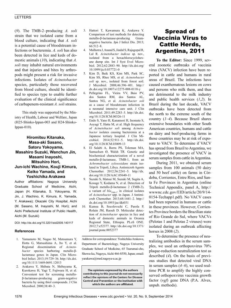
Of the 50 dairy cattle samples, 4 (8.0%) had neutralizing antibod-ies against orthopoxvirus; of these, 3 (75.0%) had titers of 100 neutral-izing units (NU)/mL, and 1 (25.0%) had a titer of 400 NU/mL. Of the 50 beef cattle, 8 (16.0%) had antibodies to orthopoxvirus, 1 (12.5%) of which had a titer of 800 NU/mL. Most of the positive samples were from cattle in Corrientes and Entre Ríos Provinc-es (Table).
Of the 100 serum samples, 5 (3 from beef and 2 from dairy cattle) were positive for vgf by real-time PCR. HA DNA was amplified from 2 of the 3 vgf PCR–positive beef cattle samples; plaque-reduction neutraliza-tion test results were also positive for the 2 samples (Table).
Alignment of the HA fragment nucleotide sequence of the isolates from Argentina showed that the se-quence was highly similar to that of
the homologous gene of VACV iso-lates from Brazil. Furthermore, the se-quences showed a signature deletion that is also present in the sequences of VACV isolates from Brazil. Com-pared with sequences for other VACV isolates, those from Argentina had 2 polymorphisms (online Technical Ap-pendix, panel C). The HA sequences from the isolates from Argentina dem-onstrated 100% identity among them-selves and exhibited higher identity with group 1 (98.2% identity) versus group 2 (93.6% identity) isolates from Brazil (online Technical Appendix, panel D). In the phylogenetic tree based on the HA nucleotide sequences (online Technical Appendix, panel B), the VACVs from Argentina clustered with several group 1 VACVs detected during outbreaks in Brazil.
Although no outbreaks of exan-thematous VACV infection have been described in cattle or humans in Ar-gentina, we detected neutralizing an-tibodies against orthopoxvirus and de-tected VACV DNA in serum samples from cattle in the country. Most of the seropositive samples were from cattle in Entre Ríos Province, which shares a border with Uruguay, and Corrientes Province, which shares a border with Rio Grande do Sul State in Brazil, where Pelotas VACVs have been iso-lated (2).
We believe that the seropositive cattle in this study may have been ex-posed to VACV, the only orthopoxvi-rus known to be circulating in South America (1–4,8–10). Despite vet-erinary surveillance efforts of border
control organizations, VACV control may be hampered by the circulation of infected rural workers and the misdi-agnosis of VACV infection; misdiag-noses occur because VACV lesions re-semble those of other exanthematous diseases. Moreover, peridomestic ro-dents have been hypothesized to act as VACV hosts, and could facilitate the spread of VACV in border areas (10). In addition, we could not rule out the circulation of autochthonous VACV in Argentina, but this is a less likely explanation. Our findings suggest that cattle herds in areas of Argentina near the border with Brazil may be exposed to VACV from Brazil and, thus, may be at risk for VACV infection. Further research is needed to determine the risk factors for VACV infection and to assess the circulation of VACV in South America
AcknowledgmentsWe thank João Rodrigues dos Santos,
Ângela Sana Lopes, Ilda Gama, and col-leagues from the Laboratório de Vírus for their excellent technical support.
Financial support was provided by the Conselho Nacional de Desenvolvi-mento Científico e Tecnológico (CNPq), Coordenação de Aperfeiçoamento de Pes-soal de Nível Superior (CAPES), Funda-ção de Amparo à Pesquisa do Estado de Minas Gerais (FAPEMIG), and Ministério da Agricultura, Pecuária e Abastecimento (MAPA). A.P.M. F.-L. was the recipient of a fellowships from CAPES, and E.G.K., C.A.B., G.S.T., and P.C.P.F. were recipi-ents of fellowships from CNPq.
Table.DiagnosisofOrthopoxvirus infectioninbeefanddairycattleduringastudyofthespreadofvacciniavirustocattleherds,Argentina,2011*
Province Cattletype No.farms sampled
No.serumsamplestested
No.positivesamples,bylevelofNU/mLagainst orthopoxvirus†
No.(%)positivebyreal-time PCR
100 200 400 800 Total(%) vgf HA Córdoba Dairy 1 25 0 0 1 0 1(4.0) 2(8.0) 0 SantaFe Dairy 1 25 3 0 0 0 3(12.0) 0 0 Corrientes Beef >2‡ 8 0 1 1 0 2(25.0) 1(12,5)§ 1(12,5)§ EntreRíos Beef 5 42 2 2 1 1 6(14.3) 2(4,8) 1(2,4)§ Total Dairyandbeef >9 100 5 3 3 1 12(12.0) 5(5.0) 2(2.0) *HA,hemagglutiningeneDNA;NU,serumdilutionatwhich70%plaquereductionpermLoccurs;vgf,orthopoxvirusvacciniagrowthfactorgeneDNA. †Determined by plaque-reductionneutralizationtest. ‡SampleswereobtainedfromseveralfarmsinCorrientesProvince. §Animalwasalsopositivebyplaque-reductionneutralizationtest.
LETTERS
1578 EmergingInfectiousDiseases•www.cdc.gov/eid•Vol.20,No.9,September2014
Ana Paula Moreira Franco-Luiz, Alexandre Fagundes-Pereira,
Galileu Barbosa Costa, Pedro Augusto Alves, Danilo Bretas Oliveira,
Cláudio Antônio Bonjardim, Paulo César Peregrino Ferreira,
Giliane de Souza Trindade, Carlos Javier Panei,
Cecilia Mónica Galosi, Jônatas Santos Abrahão, and Erna Geessien Kroon
Author affiliations: Universidade Federalde Minas Gerais, Belo Horizonte, Brazil(A.P.M.Franco-Luiz,A.Fagundes-Pereira,G.B.Costa,P.A.Alves,D.B.Oliveira,C.A.Bonjardim, P.C.P. Ferreira,G.S.Trindade,J.S.Abrahão,E.G.Kroon);UniversidadNa-cionaldelaPlata,BuenosAires,Argentina(C.J.Panei,C.M.Galosi);ConsejoNacio-nalInvestigacionesCientíficasyTécnicas,BuenosAires (C.J. Panei); and ComisióndeInvestigacionesCientíficasProvinciadeBuenosAires,,BuenosAires(C.M.Galosi)
DOI:http://dx.doi.org/10.3201/eid2009.140154
References
1. Trindade GS, Lobato ZIP, Drumond BP, Leite JA, Trigueiro RC, Guedes MIMC, et al. Isolation of two vaccinia virus strains from a single bovine vaccinia outbreak in rural area from Brazil: implications on the emergence of zoonotic orthopoxviruses. Am J Trop Med Hyg. 2006;75:486–90.
2. Campos RK, Brum MC, Nogueira CE, Drumond BP, Alves PA, Siqueira-Lima L, et al. Assessing the variability of Brazilian vaccinia virus isolates from a horse exan-thematic lesion: coinfection with distinct viruses. Arch Virol. 2011;156:275–83. http://dx.doi.org/10.1007/s00705-010-0857-z
3. Mota BEF, Trindade GS, Diniz TC, da Silva-Nunes M, Braga EM, Urbano-Ferreira M, et al. Seroprevalence of ortho-poxvirus in an Amazonian rural village, Acre, Brazil. Arch Virol. 2010;155:1139–44. http://dx.doi.org/10.1007/s00705-010- 0675-3
4. Abrahão JS, Silva-Fernandes AT, Lima LS, Campos RK, Guedes MI, Cota MM, et al. Vaccinia virus infection in mon-keys, Brazilian Amazon. Emerg Infect Dis. 2010;16:976–9. http://dx.doi.org/ 10.3201/eid1606.091187
5. Cohen JI, Hohman P, Preuss JC, Li L, Fischer SH, Fedorko DP, et al. Detection of vaccinia virus DNA, but not infectious
virus, in the blood of smallpox vaccine recipients. Vaccine. 2007;25:4571–4. http://dx.doi.org/10.1016/j.vaccine.2007. 03.044
6. Savona MR, Dela Cruz WP, Jones MS, Thornton JA, Xia D, Hadfield TL, et al. Detection of vaccinia DNA in the blood following smallpox vaccination. JAMA. 2006;295:1895–900. http://dx.doi.org/ 10. 1001/jama.295.16.1898
7. de Souza Trindade G, Li Y, Olson VA, Emerson G, Regnery RL, da Fonseca FG, et al. Real-time PCR assay to iden-tify variants of vaccinia virus: implica-tions for the diagnosis of bovine vaccinia in Brazil. J Virol Methods. 2008;152: 63–71. http://dx.doi.org/10.1016/j.jvirom-et. 2008.05.028
8. Damon IK. Poxviruses. In: Knipe DM, Howley PM, Griffin DE, Lamb RA, Martin MA, Roizman B, et al., editors. Fields virology. Vol II. 5th ed. Philadel-phia: Lippincott Williams and Wilkins; 2007. p. 2947–75.
9. Trindade GS, Emerson GL, Carroll DS, Kroon EG, Damon IK. Brazilian vac-cinia viruses and their origins. Emerg Infect Dis. 2007;13:965–72. http://dx.doi.org/10.3201/eid1307.061404
10. Abrahão JS, Guedes MI, Trindade GS, Fonseca FG, Campos RK, Mota BF, et al. One more piece in the VACV eco-logical puzzle: could peridomestic rodents be the link between wildlife and bovine vaccinia outbreaks in Brazil? PLoS ONE. 2009;4:e7428. http://dx.doi.org/10.1371/journal.pone.0007428
Address for correspondence: Erna G. Kroon, Laboratório de Vírus, Departamento de Microbiologia, Instituto de Ciências Biológicas, Universidade Federal de Minas Gerais, Av. Antônio Carlos, 6627, Caixa Postal 486, CEP 31270-901, Belo Horizonte, Minas Gerais, Brazil; email: [email protected]
Cerebellitis Associated
with Influenza A(H1N1)pdm09,
United States, 2013To the Editor: Central nervous
system (CNS) manifestations of in-fluenza are uncommon, especially in adults (1,2), and influenza-associated cerebellitis is exceedingly rare: 8 cases have been reported (3–7; online Tech-nical Appendix). We describe a case of cerebellitis caused by influenza A(H1N1)pdm09 in an adult woman.
The 37-year-old female patient who sought medical care in Florida, United States, on January 5, 2013, de-scribed a 4-day history of intermittent fever of 38.5°C, generalized fatigue, diffuse headache, mild nonproductive cough, 3 episodes of vomiting, and decreased oral intake. On January 4, she experienced acute onset of ataxia and dysarthric speech with slurred pronunciation. She reported no con-tact with sick persons, recent travel, or exposure to pets or birds. She had a medical history of asthma since child-hood, controlled by using montelukast tablets and inhaled steroids. The pa-tient denied having ever received an influenza vaccination.
The patient appeared ill; her oral temperature was elevated at 38.3°C, but other vital signs were within nor-mal limits (blood pressure 109/70 mm Hg; pulse rate 88 beats/minute; respira-tory rate 15 breaths per minute; and ox-ygen saturation 98% at room air). Mu-cosal membranes appeared normal. No neck stiffness or palpable lymph nodes were noted. Results of heart examina-tion were normal. Lungs were clear to auscultation, and the abdomen was soft, indicating no hepatosplenomegaly or palpable masses. No rash was seen. The neurologic examination revealed normal mental status but moderate ataxic dysarthria. Her cranial nerves were intact, and motor strength was 5/5 throughout. Results of a sensory
For more information please contact:Centers for Disease Control and Prevention
1600 Clifton Road NE, Atlanta, GA 30333Telephone: 1-800-CDC-INFO (232-4636)
TTY: 1-888-232-63548 Web: www.cdc.gov/Lyme
How to Correctly Remove a TickGrasp the tick firmly and as closely to the skin as possible. With a steady motion, pull the tick’s body away from the
skin. Do not be alarmed if the tick’s mouthparts remain in the skin. Cleanse the area with an antiseptic.
Page 1 of 2
Article DOI: http://dx.doi.org/10.3201/eid2009.140154
Spread of Vaccinia Virus to Cattle Herds, Argentina, 2011
Technical Appendix
Page 2 of 2
Technical Appendix Figure. A) Map with black dots indicating the 4 provinces where blood
samples were collected for the study of the spread of vaccinia virus (VACV) to cattle herds,
Argentina, 2011. B) Phylogenetic tree based on nucleotide sequences of orthopoxvirus
hemagglutinin (HA) gene. The tree was constructed with HA sequences by using the neighbor-
joining method with 1,000 bootstrap replicates and the Tamura 3-parameter model in MEGA4
(http://megasoftware.net/). Bootstrap values are represented on branches and only values
>90% are shown. Nucleotide sequences were obtained from GenBank. Scale bar represents
0.02 substitutions per nucleotide position. C) Nucleotide sequence of the VACV Argentina A56R
(HA) gene compared with homologous sequences from several other orthopoxviruses. The
samples from Argentina showed 2 polymorphisms (C–A and C–G) in comparison with other
VACV isolates. D) A56R (HA) gene nucleotide identity matrix. The analysis involved 17 nt
sequences. Evolutionary analyses were conducted in MEGA4.