A λ Cro-Like Repressor Is Essential for the Induction of Conjugative ...
12
A Cro-Like Repressor Is Essential for the Induction of Conjugative Transfer of SXT/R391 Elements in Response to DNA Damage Dominic Poulin-Laprade, Vincent Burrus Laboratory of Bacterial Molecular Genetics, Département de Biologie, Faculté des Sciences, Université de Sherbrooke, Sherbrooke, Québec, Canada ABSTRACT Integrative and conjugative elements (ICEs) of the SXT/R391 family are the main contributors to acquired multidrug resistance in the seventh pandemic lineage of Vibrio cholerae, the etiological agent of the diarrheal disease cholera. Conjugative transfer of SXT/R391 ICEs is triggered by antibiotics and agents promoting DNA damage through RecA-dependent autoproteolysis of SetR, an ICE-encoded CI-like repressor. Here, we describe the role of CroS, a distant Cro homolog, as a key component contribut- ing to the regulation of expression of the activator SetCD that orchestrates the expression of the conjugative transfer genes. We show that deletion of croS abolishes the SOS response-dependent induction of SXT despite the presence of a functional setR gene. Using quantitative reverse transcription-PCR and lacZ reporter assays, we also show that CroS represses setR and setCD expression by binding to operator sites shared with SetR. Furthermore, we provide evidence of an additional operator site bound by SetR and CroS. Finally, we show that SetCD expression generates a positive feedback loop due to SXT excision and replication in a fraction of the cell population. Together, these results refine our understanding of the genetic regulation governing the propagation of major vectors of multidrug resistance. IMPORTANCE Healthcare systems worldwide are challenged by an alarming drug resistance crisis caused by the massive and rapid propagation of antibiotic resistance genes and the associated emergence of multidrug-resistant pathogenic bacteria. SXT/R391 ICEs contrib- ute to this phenomenon not only in clinical and environmental vibrios but also in several members of the family Enterobacteria- ceae. We have identified and characterized here the regulator CroS as a key factor in the stimulation of conjugative transfer of these ICEs in response to DNA-damaging agents. We have also untangled conflicting evidence regarding autoactivation of trans- fer by the master activator of SXT/R391 ICEs, SetCD. Discovery of CroS provides a clearer and more complete understanding of the regulatory network that governs the dissemination of SXT/R391 ICEs in bacterial populations. C holera is a severe infectious disease caused by the ingestion of food or water contaminated by Vibrio cholerae. This disease remains widespread in regions with limited access to clean water and where poor sanitation allows easy dissemination of the bacte- rium in drinking water sources. Cholera is characterized by a pro- fuse watery diarrhea that rapidly induces massive fluid loss, caus- ing severe dehydration of the patient, which may lead to death within 24 h of symptom onset. Although epidemic cholera is usu- ally caused by V. cholerae O1, the unusual serogroup O139 emerged in the early 1990s as the cause of a cholera outbreak in India (1). O139 clinical isolates were found to be resistant to sul- famethoxazole and trimethoprim, two antibiotics commonly used for the treatment of severe cases of cholera (2). This resis- tance was found to be transmissible and linked to an integrative and conjugative element (ICE) named SXT (3). ICEs are self- transmissible bacterial mobile elements that play a major role in gene exchange in bacterial populations, as they are horizontally transferred via conjugation by a process similar to that used by many conjugative plasmids (4, 5). Unlike plasmids, ICEs do not remain stably in an episomal form and are rather found integrated into the chromosome. SXT integrates itself into the chromosome of V. cholerae in a site-specific manner at the 5= end of prfC, a gene coding for the peptide release factor RF3 (6). Since the discovery of SXT, SXT or related ICEs have been found to be prevalent in the seventh pandemic isolates of V. cholerae and sporadically present in other Vibrio species (7–9) and other Gammaproteobacteria of clinical origin or isolated from the aquatic environment, such as Photobacterium (10), Proteus (11), Alteromonas (12), Marinomo- nas (13), and Shewanella (9, 14) species. SXT is closely related to R391, an ICE conferring resistance to kanamycin and mercury, originally detected in a 1967 South African isolate of Providencia rettgeri (15, 16). All of the ICEs related to SXT and R391 are grouped into a single family, namely, the SXT/R391 family, be- cause they all have the same chromosomal integration site and a set of conserved genes essential for site-specific integration, con- jugative transfer, and regulation (6, 8). SXT/R391 ICEs also con- tain variable DNA insertions conferring adaptive traits, including resistance to antibiotics, heavy metals, and bacteriophage infec- tion (8, 17, 18); synthesis of the second messenger c-di-GMP (19); and homologous recombination and mutagenic repair systems (20, 21). Beside their own transfer, SXT/R391 ICEs have been shown to mobilize plasmids, phylogenetically unrelated genomic Received 31 July 2015 Accepted 26 September 2015 Accepted manuscript posted online 5 October 2015 Citation Poulin-Laprade D, Burrus V. 2015. A Cro-like repressor is essential for the induction of conjugative transfer of SXT/R391 elements in response to DNA damage. J Bacteriol 197:3822–3833. doi:10.1128/JB.00638-15. Editor: R. L. Gourse Address correspondence to Vincent Burrus, [email protected]. Supplemental material for this article may be found at http://dx.doi.org/10.1128 /JB.00638-15. Copyright © 2015, American Society for Microbiology. All Rights Reserved. 3822 jb.asm.org December 2015 Volume 197 Number 24 Journal of Bacteriology on January 29, 2018 by guest http://jb.asm.org/ Downloaded from
Transcript of A λ Cro-Like Repressor Is Essential for the Induction of Conjugative ...
A � Cro-Like Repressor Is Essential for the Induction of ConjugativeTransfer of SXT/R391 Elements in Response to DNA Damage
Dominic Poulin-Laprade, Vincent Burrus
Laboratory of Bacterial Molecular Genetics, Département de Biologie, Faculté des Sciences, Université de Sherbrooke, Sherbrooke, Québec, Canada
ABSTRACT
Integrative and conjugative elements (ICEs) of the SXT/R391 family are the main contributors to acquired multidrug resistancein the seventh pandemic lineage of Vibrio cholerae, the etiological agent of the diarrheal disease cholera. Conjugative transfer ofSXT/R391 ICEs is triggered by antibiotics and agents promoting DNA damage through RecA-dependent autoproteolysis of SetR,an ICE-encoded � CI-like repressor. Here, we describe the role of CroS, a distant � Cro homolog, as a key component contribut-ing to the regulation of expression of the activator SetCD that orchestrates the expression of the conjugative transfer genes. Weshow that deletion of croS abolishes the SOS response-dependent induction of SXT despite the presence of a functional setRgene. Using quantitative reverse transcription-PCR and lacZ reporter assays, we also show that CroS represses setR and setCDexpression by binding to operator sites shared with SetR. Furthermore, we provide evidence of an additional operator site boundby SetR and CroS. Finally, we show that SetCD expression generates a positive feedback loop due to SXT excision and replicationin a fraction of the cell population. Together, these results refine our understanding of the genetic regulation governing thepropagation of major vectors of multidrug resistance.
IMPORTANCE
Healthcare systems worldwide are challenged by an alarming drug resistance crisis caused by the massive and rapid propagationof antibiotic resistance genes and the associated emergence of multidrug-resistant pathogenic bacteria. SXT/R391 ICEs contrib-ute to this phenomenon not only in clinical and environmental vibrios but also in several members of the family Enterobacteria-ceae. We have identified and characterized here the regulator CroS as a key factor in the stimulation of conjugative transfer ofthese ICEs in response to DNA-damaging agents. We have also untangled conflicting evidence regarding autoactivation of trans-fer by the master activator of SXT/R391 ICEs, SetCD. Discovery of CroS provides a clearer and more complete understanding ofthe regulatory network that governs the dissemination of SXT/R391 ICEs in bacterial populations.
Cholera is a severe infectious disease caused by the ingestion offood or water contaminated by Vibrio cholerae. This disease
remains widespread in regions with limited access to clean waterand where poor sanitation allows easy dissemination of the bacte-rium in drinking water sources. Cholera is characterized by a pro-fuse watery diarrhea that rapidly induces massive fluid loss, caus-ing severe dehydration of the patient, which may lead to deathwithin 24 h of symptom onset. Although epidemic cholera is usu-ally caused by V. cholerae O1, the unusual serogroup O139emerged in the early 1990s as the cause of a cholera outbreak inIndia (1). O139 clinical isolates were found to be resistant to sul-famethoxazole and trimethoprim, two antibiotics commonlyused for the treatment of severe cases of cholera (2). This resis-tance was found to be transmissible and linked to an integrativeand conjugative element (ICE) named SXT (3). ICEs are self-transmissible bacterial mobile elements that play a major role ingene exchange in bacterial populations, as they are horizontallytransferred via conjugation by a process similar to that used bymany conjugative plasmids (4, 5). Unlike plasmids, ICEs do notremain stably in an episomal form and are rather found integratedinto the chromosome. SXT integrates itself into the chromosomeof V. cholerae in a site-specific manner at the 5= end of prfC, a genecoding for the peptide release factor RF3 (6). Since the discovery ofSXT, SXT or related ICEs have been found to be prevalent in theseventh pandemic isolates of V. cholerae and sporadically presentin other Vibrio species (7–9) and other Gammaproteobacteria ofclinical origin or isolated from the aquatic environment, such as
Photobacterium (10), Proteus (11), Alteromonas (12), Marinomo-nas (13), and Shewanella (9, 14) species. SXT is closely related toR391, an ICE conferring resistance to kanamycin and mercury,originally detected in a 1967 South African isolate of Providenciarettgeri (15, 16). All of the ICEs related to SXT and R391 aregrouped into a single family, namely, the SXT/R391 family, be-cause they all have the same chromosomal integration site and aset of conserved genes essential for site-specific integration, con-jugative transfer, and regulation (6, 8). SXT/R391 ICEs also con-tain variable DNA insertions conferring adaptive traits, includingresistance to antibiotics, heavy metals, and bacteriophage infec-tion (8, 17, 18); synthesis of the second messenger c-di-GMP (19);and homologous recombination and mutagenic repair systems(20, 21). Beside their own transfer, SXT/R391 ICEs have beenshown to mobilize plasmids, phylogenetically unrelated genomic
Received 31 July 2015 Accepted 26 September 2015
Accepted manuscript posted online 5 October 2015
Citation Poulin-Laprade D, Burrus V. 2015. A � Cro-like repressor is essential for theinduction of conjugative transfer of SXT/R391 elements in response to DNAdamage. J Bacteriol 197:3822–3833. doi:10.1128/JB.00638-15.
Editor: R. L. Gourse
Address correspondence to Vincent Burrus, [email protected].
Supplemental material for this article may be found at http://dx.doi.org/10.1128/JB.00638-15.
Copyright © 2015, American Society for Microbiology. All Rights Reserved.
3822 jb.asm.org December 2015 Volume 197 Number 24Journal of Bacteriology
on January 29, 2018 by guesthttp://jb.asm
.org/D
ownloaded from
islands (mobilizable genomic islands), and up to 1.5 Mb ofchromosomal DNA by high-frequency recombination transfer(22, 23).
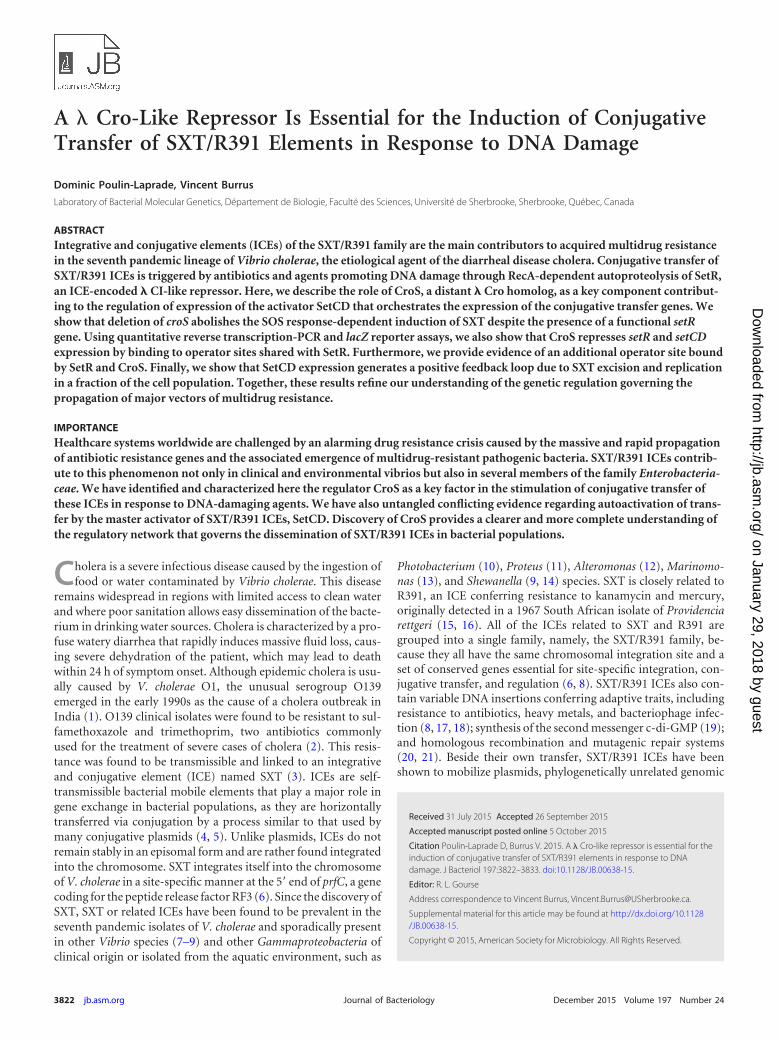
The most conserved genes (97% identity at the nucleotidelevel) shared by SXT/R391 ICEs are s086 and setR, which belong tothe regulatory module located at the rightmost end of the inte-grated ICE, near the attR attachment site (Fig. 1A) (8). The regu-latory module contains eight open reading frames (ORFs), sevenof which are in the same orientation (s086, s084, s083, s082, setD,setC, and eex), while the last gene, setR, is divergently transcribed(Fig. 1A and B) (24). eex and the convergent gene traG code for theentry exclusion system of SXT/R391 ICEs (25). The overlappinggenes setC and setD encode the SetCD activator complex. SetCDwas shown to bind upstream of the �35 sequence of 11 promotersin SXT/R391 ICEs, activating the expression of �40 genes essen-tial for site-specific and homologous recombination, ICE replica-tion and partition, and conjugative transfer (26–28). The func-tions of s082, s083, s084, and s086 remain unknown. In silicoanalysis has revealed that s086 codes for a putative small basicprotein of 9.4 kDa with a predicted helix-turn-helix (HTH) DNA-binding domain. setR codes for the main repressor of SXT/R391ICEs (24, 29). SetR contains an HTH_XRE (PF01381) motif in its
N-terminal moiety and a C-terminal LexA-like autoproteolysisdomain (PF00717). SetR is related to phage 434 CI and otherlambdoid phage repressors and has been shown to bind to fouroperator sites located between s086 and setR (Fig. 1B) (24, 30).These operators, O1, O2, O3, and OL, are part of PL and PR, twodivergent overlapping promoters located in the intergenic regionbetween s086 and setR. The relative affinities of SetR for these fouroperator sites and their positions suggest an autoregulation of setRexpression from PR (30) and a repression of the operon containings086 and setCD driven from PL (29). Repression at PL is alleviatedwhen the cellular pool of SetR drops, most likely as a result ofautoproteolysis stimulated by DNA damage-induced activation ofRecA (29).
The relative positions of setR and s086 are reminiscent of cI andcro carried by bacteriophage �. CI and Cro form a pair that gov-erns the transition between the lysogenic and lytic pathways of the� life cycle (31, 32). To date, SXT/R391 ICEs have been known tobe regulated by only two transcriptional regulators, the repressorSetR and the activator complex SetCD (24). In this study, we iden-tify S086 as a third regulator, which will be referred to as CroS forCro-like repressor of SXT. We demonstrate that CroS is a keyfactor in the alleviation of SetR repression and induction of con-
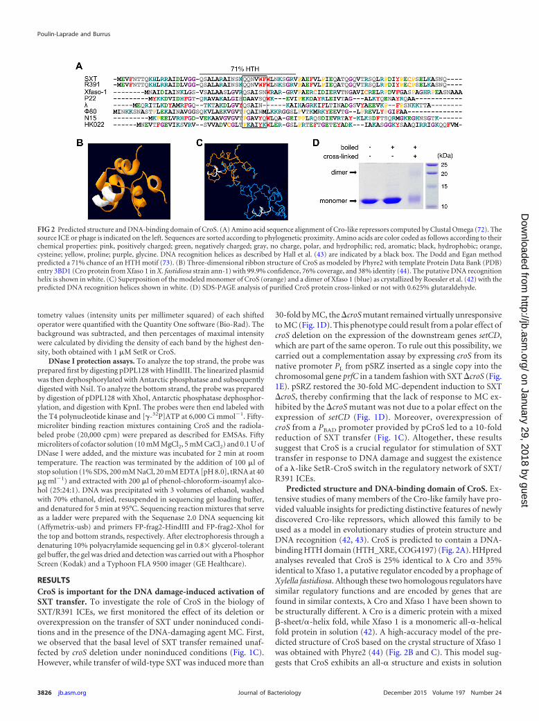
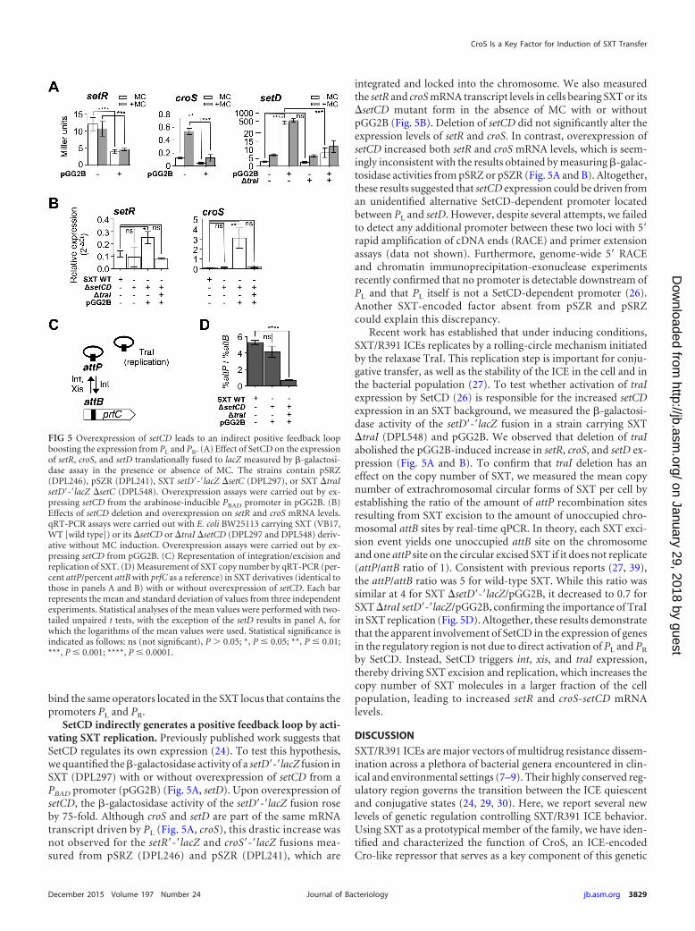
FIG 1 Effects of croS on SXT conjugative transfer. (A) Schematic representation of the regulatory module of SXT. The dotted line indicates the region enlargedin panel B. (B) The intergenic region between croS, previously referred to as s086, and setR in SXT as represented by Beaber and Waldor (30). The PL and PR
promoters, the OL, O1, O2, and O3 operators, the �10 and �35 elements, the Shine-Dalgarno (SD) sequence of croS, and the start codons of croS and setR arerepresented. (C) Effect of the deletion or overexpression of croS on SXT transfer. Conjugation assays were carried out with E. coli BW25113 SXT (VB17, WT) orits �croS mutant (DPL353, �) as the donor. Overexpression assays were carried out by expressing croS from the arabinose-inducible PBAD promoter in pCroS(DPL525). E. coli CAG18439 (Tcr) was used as the recipient. Frequencies of exconjugant formation were expressed as the number of exconjugant CFU (Sur Tmr
Tcr) per recipient CFU (Tcr). (D) Effect of croS deletion on the induction of SXT transfer by MC. The donor strains were VB17, DPL353, and DPL263 in whichcroS was expressed from its native PL promoter in pSRZ integrated in single copy in tandem with SXT �croS. The recipient strain was CAG18439. The inductionindex corresponds to the ratio of the frequencies of exconjugant formation obtained with and without MC treatment prior to transfer. The dashed line indicatesan induction index of 1, i.e., no difference between the induced and noninduced conditions. Each bar represents the mean and standard deviation of valuesobtained from at least three independent experiments. Statistical analyses of the logarithms of the mean values were performed with two-tailed unpaired t tests.Asterisks indicate that P values are �0.05 compared to the wild type (WT). (E) Schematic representation of the insert of the pSR, pSZR, and pSRZ plasmids sitespecifically integrated into the 5= end of prfC alone or in tandem with SXT. The plasmid backbone is not represented. In pSZR and pSRZ, lacZ was translationallyfused to the sixth codon of croS and setR, respectively.
CroS Is a Key Factor for Induction of SXT Transfer
December 2015 Volume 197 Number 24 jb.asm.org 3823Journal of Bacteriology
on January 29, 2018 by guesthttp://jb.asm
.org/D
ownloaded from
jugative transfer of SXT/R391 ICEs. By binding to five operatorsites shared with SetR, CroS acts as a repressor of PL and PR,thereby repressing both setCD and setR by a mechanism that isevocative of � Cro repression on CI and CII (33). We also showthat SetCD increases its own expression not by activating PL but byindirectly generating a positive feedback loop triggered by activa-tion of SXT replication, which increases the SXT copy number andultimately the level of the setCD mRNA transcript. Finally, wepropose a new model of SXT transfer regulation that includesCroS as a repressor of PL and PR, thereby promoting the SOS-dependent induction of SXT/R391 ICE transfer.
MATERIALS AND METHODSBacterial strains and media. The bacterial strains used in this study aredescribed in Table 1. The strains were routinely grown in lysogeny broth(LB-Miller; EMD) at 37°C in an orbital shaker/incubator and maintainedat �80°C in LB broth containing 15% (vol/vol) glycerol. Antibiotics wereused at the following concentrations: ampicillin, 100 �g ml�1; chloram-phenicol, 20 �g ml�1; erythromycin, 10 �g ml�1; kanamycin, 50 �g ml�1;
spectinomycin, 50 �g ml�1; sulfamethoxazole (Su), 160 �g ml�1; tetra-cycline (Tc), 12 �g ml�1; trimethoprim (Tm), 32 �g ml�1. When re-quired, bacterial cultures were supplemented with 0.1 mM isopropyl-�-D-thiogalactopyranoside (IPTG), 0.02% L-arabinose, or 100 ng ml�1
mitomycin C (MC).Bacterial conjugations. Conjugation assays were performed as de-
scribed by Burrus and Waldor (34), with the following modifications.When induction with MC was performed, overnight cultures of donorand recipient cells were diluted 1:4 and the donor cultures were supple-mented or not with MC and incubated for 1 h at 37°C with shaking. Thecells were harvested by centrifugation for 3 min at 1,200 g, washed in 1volume of LB broth, and resuspended in 1/20 volume of LB broth. Matingmixtures were then deposited on LB agar plates and incubated at 37°Cfor 2 h.
Molecular biology methods. Plasmid DNA was prepared with theEZ-10 Spin Column Plasmid DNA Minipreps kit (Bio Basic) according tothe manufacturer’s instructions. All of the enzymes used in this study werepurchased from New England BioLabs. PCR assays were performed withthe Taq, Q5, or Phusion polymerase according to the manufacturer’s in-structions. When necessary, PCR products were purified with an EZ-10
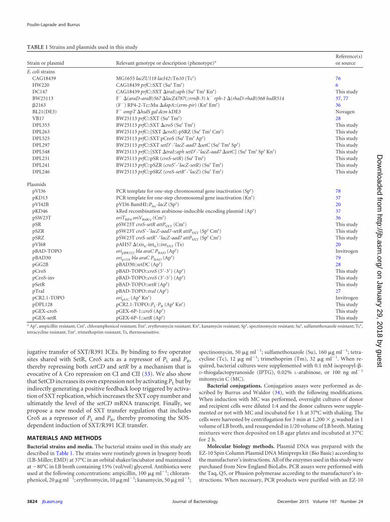
TABLE 1 Strains and plasmids used in this study
Strain or plasmid Relevant genotype or description (phenotype)a
Reference(s)or source
E. coli strainsCAG18439 MG1655 lacZU118 lacI42::Tn10 (Tcr) 76HW220 CAG18439 prfC::SXT (Sur Tmr) 6DC147 CAG18439 prfC::SXT �traI::aph (Sur Tmr Knr) This studyBW25113 F� �(araD-araB)567 �lacZ4787(::rrnB-3) �� rph-1 �(rhaD-rhaB)568 hsdR514 37, 77�2163 (F�) RP4-2-Tc::Mu �dapA::(erm-pir) (Knr Emr) 36BL21(DE3) F� ompT �hsdS gal dcm �DE3 NovagenVB17 BW25113 prfC::SXT (Sur Tmr) 28DPL353 BW25113 prfC::SXT �croS (Sur Tmr) This studyDPL263 BW25113 prfC::[SXT �croS]-pSRZ (Sur Tmr Cmr) This studyDPL525 BW25113 prfC::SXT pCroS (Sur Tmr Apr) This studyDPL297 BW25113 prfC::SXT setD=-=lacZ-aad7 �setC (Sur Tmr Spr) This studyDPL548 BW25113 prfC::[SXT �traI::aph setD=-=lacZ-aad7 �setC] (Sur Tmr Spr Knr) This studyDPL231 BW25113 prfC::pSR (croS-setR) (Sur Tmr) This studyDPL241 BW25113 prfC::pSZR (croS=-=lacZ-setR) (Sur Tmr) This studyDPL246 BW25113 prfC::pSRZ (croS-setR=-=lacZ) (Sur Tmr) This study
PlasmidspVI36 PCR template for one-step chromosomal gene inactivation (Spr) 78pKD13 PCR template for one-step chromosomal gene inactivation (Knr) 37pVI42B pVI36 BamHI::Plac-lacZ (Spr) 20pKD46 �Red recombination arabinose-inducible encoding plasmid (Apr) 37pSW23T oriTRP4 oriVR6K (Cmr) 36pSR pSW23T croS-setR attPSXT (Cmr) This studypSZR pSW23T croS=-=lacZ-aad7-setR attPSXT (Spr Cmr) This studypSRZ pSW23T croS-setR=-=lacZ-aad7 attPSXT (Spr Cmr) This studypVI68 pAH57 �(xis�-int�)::intSXT (Ts) 20pBAD-TOPO oripBR322 bla araC PBAD (Apr) InvitrogenpBAD30 orip15A bla araC PBAD (Apr) 79pGG2B pBAD30::setDC (Apr) 28pCroS pBAD-TOPO::croS (5=-3=) (Apr) This studypCroS-inv pBAD-TOPO::croS (3=-5=) (Apr) This studypSetR pBAD-TOPO::setR (Apr) This studypTraI pBAD-TOPO::traI (Apr) 27pCR2.1-TOPO oripUC (Apr Knr) InvitrogenpDPL128 pCR2.1-TOPO::PL-PR (Apr Knr) This studypGEX-croS pGEX-6P-1::croS (Apr) This studypGEX-setR pGEX-6P-1::setR (Apr) This study
a Apr, ampicillin resistant; Cmr, chloramphenicol resistant; Emr, erythromycin resistant; Knr, kanamycin resistant; Spr, spectinomycin resistant; Sur, sulfamethoxazole resistant; Tcr,tetracycline resistant; Tmr, trimethoprim resistant; Ts, thermosensitive.
Poulin-Laprade and Burrus
3824 jb.asm.org December 2015 Volume 197 Number 24Journal of Bacteriology
on January 29, 2018 by guesthttp://jb.asm
.org/D
ownloaded from
Spin Column PCR Products Purification kit (Bio Basic) according to themanufacturer’s instructions. E. coli was transformed by electroporation asdescribed by Dower et al. (35) in a Bio-Rad Gene Pulser Xcell apparatusset at 25 �F, 200 �, and 1.8 kV with 1-mm gap electroporation cuvettes.Sequencing reactions were performed by the Plateforme de Séquençage etde Génotypage du Centre de Recherche du CHUL (Québec, QC, Canada).
Plasmid and strain constructions. The plasmids and oligonucleotidesused in this study are listed in Table 1 (also see Table S1 in the supplemen-tal material). Plasmids pCroS and pSetR were constructed by amplifyingcroS and setR with primer pair s086pBADf/s086pBADr.s or setRpBADf/setRpBADr.s and genomic DNA of E. coli HW220 as the template andcloning into the TA cloning expression vector pBAD-TOPO (Invitrogen)according to the manufacturer’s instructions. pSR was constructed byPCR amplification of the croS-setR-attP region resulting from the excisionof SXT in E. coli HW220 with the primer pair PEs086/VISLR3. The2,022-bp PCR product was first cloned into pCR2.1-TOPO (Invitrogen)according to the manufacturer’s instructions. The cloned fragment wasthen recovered by NotI/BamHI digestion and then ligated to NotI/BamHI-digested pSW23T, yielding pSR. pSW23T contains the RP4 mo-bilization locus, the R6K pir-dependent conditional origin of replication,and a chloramphenicol resistance gene (36). Plasmid pDPL128 was con-structed by cloning into pCR2.1-TOPO (Invitrogen) the intergenic regionbetween setR and croS, which was amplified by PCR with primer pairRRs086-4/RRs086setRr and genomic DNA of E. coli HW220 as the tem-plate.
�croS and �traI mutant derivatives of SXT were constructed in E. coliHW220 by the one-step chromosomal gene inactivation technique (37)with primer pairs delS086F/dels086R and TraIWF2/TraIWR3, respec-tively, and the pKD13 template. SXT �traI::aph complemented withpTraI was transferred by conjugation into E. coli BW25113. To constructE. coli DPL548, the �traI::aph mutation was then transduced into E. coliDPL297 (BW25113 prfC::SXT �setCD::lacZ-aad7) by P1vir generalizedtransduction. Strains containing pSR integrated into the 5= end of prfCwere constructed by mating the RP4� pir� strain E. coli �2163/pSR withE. coli BW25113 containing pVI68 as the recipient strain. pVI68 expressesthe integrase of SXT, thereby allowing site-specific integration of pSR,which was verified by amplifying the attL and attR sites by PCR withprimer pairs EattBR/VISRF and EattBF/VISLR3, respectively. The ab-sence of multiple integrations of pSR in tandem arrays was verified withprimers VISLR3 and VISRF. The resulting strain, E. coli DPL231, containsa unique copy of the croS-setR region integrated site specifically into prfC.Translational lacZ fusions setR=-=lacZ, croS=-=lacZ, and setCD=-=lacZ wereconstructed by the one-step chromosomal gene inactivation technique(37) with primer pairs DATsetR-lacZf/DATsetR-lacZr, DATsetQ-lacZf/DATsetQ-lacZr, and DATsetCD-lacZf/DATsetCD-lacZr, respectively,and pVI42B as the template. In each construct, the sixth codon of the geneof interest was fused to the ninth codon of lacZ. The setR=-=lacZ andcroS=-=lacZ mutations targeted BW25113 derivative strains containingpSR integrated into prfC (DPL231), whereas the setCD=-=lacZ mutationtargeted SXT in E. coli VB17. SXT �croS was transferred into DPL246 byconjugation to generate DPL263.
RNA isolation and quantitative reverse transcription (qRT)-PCR.Total RNA was extracted and cDNA were synthesized as described previ-ously (19, 38). RNA integrity was verified with a 2100 Bioanalyzer (Agi-lent). Quantitative amplification of setR, croS, setD, and setC was carriedout with primer pairs RTsetR-F/RTsetR-R, RTcroS-F/RTcroS-R, RTsetD-F/RTsetD-R, and RTsetC-F/RTsetC-R, respectively. For normalization,rpoZ was amplified with primers RTrpoZcoli-F/RTrpoZcoli-R and the��CT calculation method. Experiments were repeated three times in trip-licate and combined. Melting curves were determined with the final reac-tion products (156 to 165 bp) to confirm that amplification was specific totargets. All primer pairs exhibited efficiencies between 95 and 97%.
Real-time qPCR assays for relative quantification of attB and attP.The frequency of excision and copy number of the excised circular form ofSXT were assessed by real-time quantitative PCR (qPCR) as described
elsewhere (39). Genomic DNA was obtained from cultures of E. coli VB17,DPL297/pGG2B, and DPL548/pGG2B cells grown under conditions de-scribed for the qRT-PCR assays. Bacterial cultures were grown in LB me-dium at 37°C to an optical density at 600 nm (OD600) of 0.2 and then foran additional 2 h at 37°C. prfC, attB, and attP were quantified with primerpairs prfC.qec.F1/prfC.qec.R1, attB.qec.F2/attB.qec.R2, and attP.qec.F2/attP.qec.R2, respectively (27) (see Table S1 in the supplemental material).Three biological replicates of qPCR experiments were performed on theRNomics platform of the Laboratoire de Génomique Fonctionnelle del’Université de Sherbrooke, Sherbrooke, QC, Canada (http://lgfus.ca).Normalization was performed as described previously (39).
�-Galactosidase assays. The substrate used to determine LacZ levelswas o-nitrophenyl-�-D-galactopyranoside, and the assays were carriedout as described previously (40) with LB medium and 100 �g ml�1 am-picillin for maintenance of pCroS and pGG2B. Induction of croS andsetCD expression and of the SOS response was done by supplementing,respectively, with 0.2% arabinose and/or 100 ng ml�1 MC a refreshedculture grown to an OD600 of 0.2, followed by a 2-h incubation at 37°Cwith shaking prior to cell sampling.
Expression and purification of the SetR and CroS proteins. Over-night-grown cultures of E. coli BL21(DE3) bearing pGEX-croS or pGEX-setR were diluted 1/1,000 in fresh 2 YT broth (80) supplemented with100 �g ml�1 ampicillin and incubated at 37°C with agitation. Proteinexpression was induced at mid-exponential phase (OD600 of 0.6) with 0.1mM IPTG. The cultures were grown for an additional 3 h at 37°C withagitation. Cells were collected by centrifugation, resuspended in phos-phate-buffered saline containing 1% Triton X-100, 1 mM phenylmethyl-sulfonyl fluoride, and a cocktail of protease inhibitors (Protease InhibitorCocktail; Sigma). Cells were lysed by sonication and centrifuged to pelletthe debris, and CroS and SetR were recovered by affinity chromatographywith the glutathione S-transferase purification module (GE Healthcare)and the PreScission protease (GE Healthcare) according to the manufac-turer’s instructions. Protein concentration was estimated with the Brad-ford protein assay (Bio-Rad), and purity was determined by SDS-PAGEanalysis.
Dimerization assay. Prior to dimerization assay, the purity of CroSwas estimated by SDS-PAGE analysis to be 95%. No contaminant proteinswith a size similar to that of CroS was observed. A 4.7-�g sample ofpurified CroS was incubated with 0.625% glutaraldehyde for 30 min atroom temperature. The reaction mixture was then denatured at 100°C for3 min, loaded onto a 15% SDS-PAGE gel, and run for 75 min at 150 V(41). The gel was then stained with Coomassie brilliant blue R-250. ThePrecision Plus Protein Kaleidoscope Standards ladder (Bio-Rad) was usedas a molecular weight marker.
EMSAs. The probes used in electrophoretic mobility shift assays(EMSAs) were either annealed oligonucleotides or PCR products. Puri-fied PCR products and a single oligonucleotide for each annealed pairwere labeled with [-32P]dATP with the T4 polynucleotide kinase. Anequimolar quantity of the complementary oligonucleotide was added, thevolume was brought to 100 �l in 20 mM Tris-HCl (pH 8.0)–20 mM KCl,and the mixture was denatured at 95°C for 5 min and then cooled to roomtemperature over 1 h. The -32P-labeled double-stranded DNA fragmentswere separated from unincorporated [-32P]dATP with Illustra MicroSpin G-25 columns (GE Healthcare) according to the manufacturer’s in-structions. Purified CroS and SetR proteins were preincubated in 24 �l ofbuffer I (20 mM Tris-HCl [pH 8.0], 20 mM KCl, 1 mM MgCl2, 5% glyc-erol) for 20 min at 4°C. For PCR probes 1 to 6 (Fig. 4D), nonspecificcompetitor DNA was added by supplementing buffer I with sonicatedsalmon sperm DNA at 50 ng ml�1. Radiolabeled probes (2,000 cpm) wereadded, and samples were incubated for 30 min at 37°C and then for 10 minat 4°C. Samples were immediately loaded onto a prerun 4% polyacryl-amide gel containing 0.5 Tris-borate-EDTA buffer and subjected toelectrophoresis at a constant voltage of 120 V at 4°C for 1 h 15 min. Gelswere dried and exposed to a Phosphor Screen (Kodak), and results werevisualized with a Typhoon FLA 9500 imager (GE Healthcare). The densi-
CroS Is a Key Factor for Induction of SXT Transfer
December 2015 Volume 197 Number 24 jb.asm.org 3825Journal of Bacteriology
on January 29, 2018 by guesthttp://jb.asm
.org/D
ownloaded from
tometry values (intensity units per millimeter squared) of each shiftedoperator were quantified with the Quantity One software (Bio-Rad). Thebackground was subtracted, and then percentages of maximal intensitywere calculated by dividing the density of each band by the highest den-sity, both obtained with 1 �M SetR or CroS.
DNase I protection assays. To analyze the top strand, the probe wasprepared first by digesting pDPL128 with HindIII. The linearized plasmidwas then dephosphorylated with Antarctic phosphatase and subsequentlydigested with NsiI. To analyze the bottom strand, the probe was preparedby digestion of pDPL128 with XhoI, Antarctic phosphatase dephosphor-ylation, and digestion with KpnI. The probes were then end labeled withthe T4 polynucleotide kinase and [-32P]ATP at 6,000 Ci mmol�1. Fifty-microliter binding reaction mixtures containing CroS and the radiola-beled probe (20,000 cpm) were prepared as described for EMSAs. Fiftymicroliters of cofactor solution (10 mM MgCl2, 5 mM CaCl2) and 0.1 U ofDNase I were added, and the mixture was incubated for 2 min at roomtemperature. The reaction was terminated by the addition of 100 �l ofstop solution (1% SDS, 200 mM NaCl, 20 mM EDTA [pH 8.0], tRNA at 40�g ml�1) and extracted with 200 �l of phenol-chloroform-isoamyl alco-hol (25:24:1). DNA was precipitated with 3 volumes of ethanol, washedwith 70% ethanol, dried, resuspended in sequencing gel loading buffer,and denatured for 5 min at 95°C. Sequencing reaction mixtures that serveas a ladder were prepared with the Sequenase 2.0 DNA sequencing kit(Affymetrix-usb) and primers FP-frag2-HindIII and FP-frag2-XhoI forthe top and bottom strands, respectively. After electrophoresis through adenaturing 10% polyacrylamide sequencing gel in 0.8 glycerol-tolerantgel buffer, the gel was dried and detection was carried out with a PhosphorScreen (Kodak) and a Typhoon FLA 9500 imager (GE Healthcare).
RESULTSCroS is important for the DNA damage-induced activation ofSXT transfer. To investigate the role of CroS in the biology ofSXT/R391 ICEs, we first monitored the effect of its deletion oroverexpression on the transfer of SXT under noninduced condi-tions and in the presence of the DNA-damaging agent MC. First,we observed that the basal level of SXT transfer remained unaf-fected by croS deletion under noninduced conditions (Fig. 1C).However, while transfer of wild-type SXT was induced more than
30-fold by MC, the �croS mutant remained virtually unresponsiveto MC (Fig. 1D). This phenotype could result from a polar effect ofcroS deletion on the expression of the downstream genes setCD,which are part of the same operon. To rule out this possibility, wecarried out a complementation assay by expressing croS from itsnative promoter PL from pSRZ inserted as a single copy into thechromosomal gene prfC in a tandem fashion with SXT �croS (Fig.1E). pSRZ restored the 30-fold MC-dependent induction to SXT�croS, thereby confirming that the lack of response to MC ex-hibited by the �croS mutant was not due to a polar effect on theexpression of setCD (Fig. 1D). Moreover, overexpression ofcroS from a PBAD promoter provided by pCroS led to a 10-foldreduction of SXT transfer (Fig. 1C). Altogether, these resultssuggest that CroS is a crucial regulator for stimulation of SXTtransfer in response to DNA damage and suggest the existenceof a �-like SetR-CroS switch in the regulatory network of SXT/R391 ICEs.
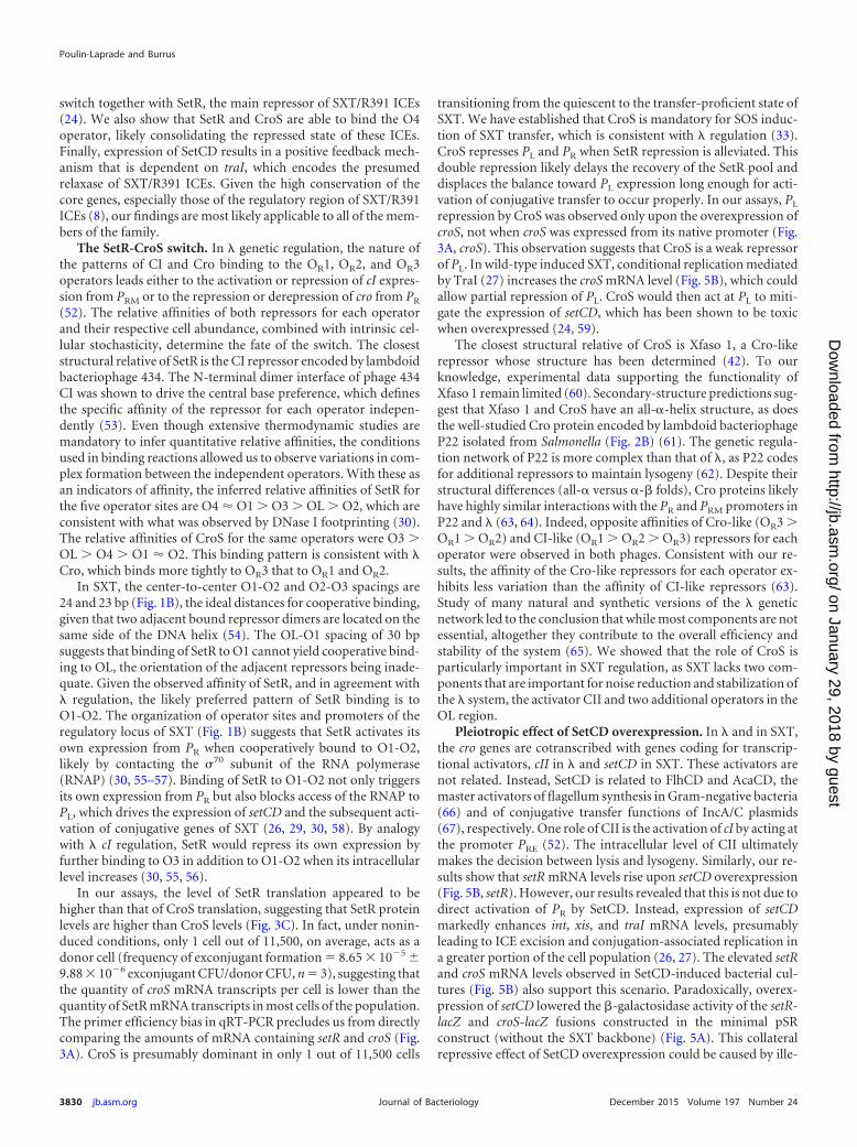
Predicted structure and DNA-binding domain of CroS. Ex-tensive studies of many members of the Cro-like family have pro-vided valuable insights for predicting distinctive features of newlydiscovered Cro-like repressors, which allowed this family to beused as a model in evolutionary studies of protein structure andDNA recognition (42, 43). CroS is predicted to contain a DNA-binding HTH domain (HTH_XRE, COG4197) (Fig. 2A). HHpredanalyses revealed that CroS is 25% identical to � Cro and 35%identical to Xfaso 1, a putative regulator encoded by a prophage ofXylella fastidiosa. Although these two homologous regulators havesimilar regulatory functions and are encoded by genes that arefound in similar contexts, � Cro and Xfaso 1 have been shown tobe structurally different. � Cro is a dimeric protein with a mixed�-sheet/ -helix fold, while Xfaso 1 is a monomeric all- -helicalfold protein in solution (42). A high-accuracy model of the pre-dicted structure of CroS based on the crystal structure of Xfaso 1was obtained with Phyre2 (44) (Fig. 2B and C). This model sug-gests that CroS exhibits an all- structure and exists in solution
FIG 2 Predicted structure and DNA-binding domain of CroS. (A) Amino acid sequence alignment of Cro-like repressors computed by Clustal Omega (72). Thesource ICE or phage is indicated on the left. Sequences are sorted according to phylogenetic proximity. Amino acids are color coded as follows according to theirchemical properties: pink, positively charged; green, negatively charged; gray, no charge, polar, and hydrophilic; red, aromatic; black, hydrophobic; orange,cysteine; yellow, proline; purple, glycine. DNA recognition helices as described by Hall et al. (43) are indicated by a black box. The Dodd and Egan methodpredicted a 71% chance of an HTH motif (73). (B) Three-dimensional ribbon structure of CroS as modeled by Phyre2 with template Protein Data Bank (PDB)entry 3BD1 (Cro protein from Xfaso 1 in X. fastidiosa strain ann-1) with 99.9% confidence, 76% coverage, and 38% identity (44). The putative DNA recognitionhelix is shown in white. (C) Superposition of the modeled monomer of CroS (orange) and a dimer of Xfaso 1 (blue) as crystallized by Roessler et al. (42) with thepredicted DNA recognition helices shown in white. (D) SDS-PAGE analysis of purified CroS protein cross-linked or not with 0.625% glutaraldehyde.
Poulin-Laprade and Burrus
3826 jb.asm.org December 2015 Volume 197 Number 24Journal of Bacteriology
on January 29, 2018 by guesthttp://jb.asm
.org/D
ownloaded from
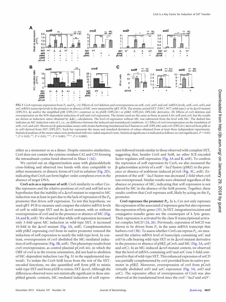
either as a monomer or as a dimer. Despite extensive similarities,CroS does not contain the cysteine residues C42 and C55 formingthe intrasubunit cystine bond observed in Xfaso 1 (42).
We carried out an oligomerization assay with glutaraldehydecross-linking and observed two bands with sizes compatible toeither monomeric or dimeric forms of CroS in solution (Fig. 2D),indicating that CroS can form higher-order complexes even in theabsence of target DNA.
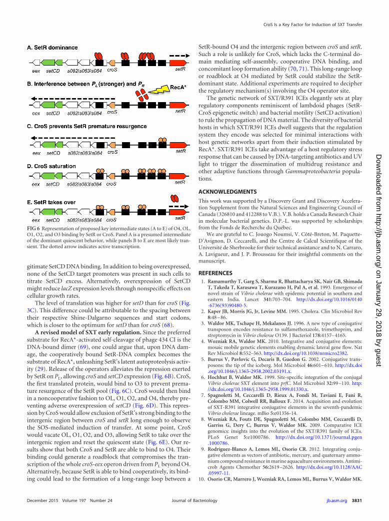
CroS acts as a repressor of setR. CroS similarity to other Cro-like repressors and the relative positions of croS and setR led us tohypothesize that the inability of a �croS mutant to respond to MCinduction was at least in part due to the lack of repression of the PR
promoter that drives setR expression. To test this hypothesis, weused qRT-PCR to measure and compare the relative mRNA levelsof setR in wild-type SXT and its �croS mutant, with or withoutoverexpression of croS and in the presence or absence of MC (Fig.3A and B, setR). We observed that while setR expression increasedonly 3-fold upon MC induction in wild-type SXT, it increased10-fold in the �croS mutant (Fig. 3A, setR). Complementationwith pSRZ expressing croS from its native promoter restored theinduction of setR expression to nearly the wild-type level. In con-trast, overexpression of croS abolished the MC-mediated induc-tion of setR expression (Fig. 3B, setR). This phenotype results fromcroS overexpression, as control plasmid pCroS-inv, in which theORF of croS is in the reverse orientation, did not lead to abolitionof MC-dependent induction (see Fig. S1 in the supplemental ma-terial). To isolate the CroS-SetR locus from the rest of the SXT-encoded functions, we also quantified setR from pSR to mimicwild-type SXT and from pSZR to mimic SXT �croS. Although thedifferences observed were not statistically significant in these sim-plified genetic contexts, MC-mediated induction of setR expres-
sion followed trends similar to those observed with complete SXT,suggesting that, besides CroS and SetR, no other ICE-encodedfactor regulates setR expression (Fig. 3A and B, setR). To confirmthe repression of setR expression by CroS, we also measured the�-galactosidase activity of a setR=-=lacZ fusion (pSRZ) in the pres-ence or absence of arabinose-induced pCroS (Fig. 3C, setR). Ex-pression of the setR=-=lacZ fusion was decreased 2-fold when croSwas overexpressed. Similar results were obtained regardless of theabsence or presence of MC, indicating that setR expression is notaltered by MC in the absence of the SetR protein. Together, theseresults confirm that CroS represses setR, likely by acting on the PR
promoter.CroS represses the promoter PL. In �, Cro not only represses
the expression of the associated cI repressor gene but also repressesthe expression of lytic genes (33). In SXT, integration/excision andconjugative-transfer genes are the counterpart of � lytic genes.Their expression is activated by the class II transcriptional activa-tor complex SetCD (24, 26). Previously, expression of SetCD wasshown to be driven from PL in the same mRNA transcript thatharbors croS (30). To assess whether CroS can repress PL, we mea-sured the relative mRNA level of transcripts containing setC andsetD in cells bearing wild-type SXT or its �croS mutant derivativein the presence or absence of pSRZ, pCroS, and MC (Fig. 3A, setDand setC). In an MC-induced �croS mutant context, we observedthat the level of mRNA containing setD and setC rose 5-fold com-pared to that of wild-type SXT. This enhanced expression of setCDwas partially complemented by croS provided from its native pro-moter in pSRZ. Moreover, overexpression of croS from pCroSvirtually abolished setD and setC expression (Fig. 3A, setD andsetC). The repressive effect of overexpression of CroS was alsoobserved at the translational level since the croS=-=lacZ and setD=-
FIG 3 CroS represses expression from PL and PR. (A) Effects of croS deletion and overexpression on setR, croS, setD, and setC mRNA levels. setR, croS, setD, andsetC mRNA transcript levels in the presence or absence of MC were measured by qRT-PCR. The strains carried SXT (VB17, WT [wild type]) or its �croS mutant(DPL353, �) and/or the simplified pSR (DPL231) construct or its pSZR (DPL241) or pSRZ (DPL263, DPL246) derivative. (B) Effects of croS deletion andoverexpression on the SOS-dependent induction of setR and croS expression. The strains used are the same as those in panel A for setR and croS, but the resultsare shown as induction ratios obtained by ��CT calculations. The level of expression without MC was subtracted from the level with MC. The dashed lineindicates an MC induction ratio of 1, i.e., no difference between the induced and noninduced conditions. (C) Effect of croS overexpression on the translation ofsetR, croS, and setD. Shown are �-galactosidase assays with strains harboring translational lacZ fusions to setR (DPL246) and croS (DPL241) derived from pSR orto setD derived from SXT (DPL297). Each bar represents the mean and standard deviation of values obtained from at least three independent experiments.Statistical analyses of the mean values were performed with two-tailed unpaired t tests. Statistical significance is indicated as follows: ns (not significant), P � 0.05;*, P � 0.05; **, P � 0.01; ***, P � 0.001; ****, P � 0.0001.
CroS Is a Key Factor for Induction of SXT Transfer
December 2015 Volume 197 Number 24 jb.asm.org 3827Journal of Bacteriology
on January 29, 2018 by guesthttp://jb.asm
.org/D
ownloaded from
=lacZ fusions yielded very low �-galactosidase activities even whenthe cells were exposed to MC (Fig. 3C, croS and setD). Expressionof croS was similar in the SXT and pSR contexts, while removal ofthe setR gene in pSRZ boosted the expression of croS, confirmingthe repressive function of SetR on PL (Fig. 3A, croS). Abolitionof the MC responsiveness in pSRZ confirms the pivotal role ofSetR for integration of the SOS signal (RecA*) to the regulatorynetwork. croS expressed from its native promoter is not sufficientto completely repress PL upon MC induction. These expressionassays confirm the importance of SetR for the MC-dependent in-duction of SXT/R391 ICE transfer and that CroS acts as a mildrepressor of the PL promoter, which drives the expression of themaster activator complex SetCD.
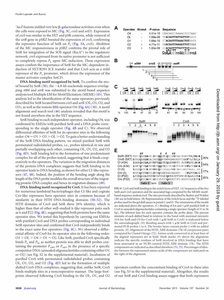
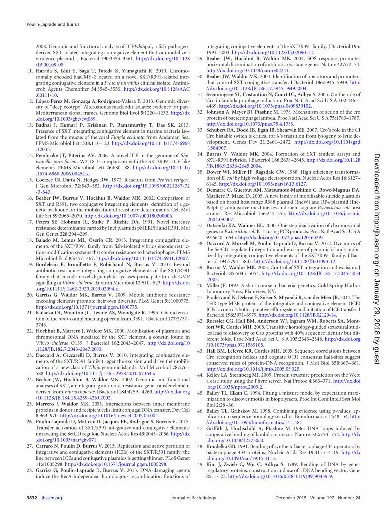
DNA-binding motif recognized by SetR. To confirm the mo-tif bound by SetR (30), the �4.8-kb nucleotide sequence overlap-ping s084 and attR was submitted to the motif-based sequenceanalysis tool Multiple EM for Motif Elicitation (MEME) (45). Thisanalysis led to the identification of the same operators previouslydescribed for SetR located between croS and setR (OL, O1, O2, andO3), as well as the remote fifth operator O4 (Fig. 4A) (30). A motifalignment and search tool (46) analysis revealed that this motif isnot found anywhere else in the SXT sequence.
SetR binding to each independent operator, including O4, wasconfirmed by EMSAs with purified SetR and a DNA probe corre-sponding to the single operator (Fig. 4B and C). We observeddifferential affinities of SetR for its operator sites in the followingorder: O4 � O1 � O3 � OL � O2. To gain a better understandingof the SetR DNA-binding pattern, we mixed purified SetR withpermutated radiolabeled probes, i.e., probes identical in size andpartially overlapping each other, containing OL, O1, O2, and O3(Fig. 4D). SetR binding led to the formation of one predominantcomplex for all of the probes tested, suggesting that it binds coop-eratively to the operators. The variation in the migration distancesof the protein-DNA complexes suggests that SetR binding to itsoperator leads to DNA bending, as shown for other CI-like repres-sors (47, 48). Indeed, the position of the bending angle along thelength of the DNA probe modifies the overall structure of the migrat-ing protein-DNA complex and hence its migration distance (49).
DNA-binding motif recognized by CroS. It has been reportedfor numerous lambdoid bacteriophages that CI-like and cognateCro-like repressors have operator sites in common because ofsimilarity in their HTH DNA-binding domains (50–52). TheHTH domains of CroS and SetR show 26% identity, which ishigher than that of other well-studied �-like repressor pairs suchas � and P22 (Fig. 4E), suggesting that both proteins have the sameoperator sites. We tested this hypothesis by carrying out EMSAswith purified CroS and DNA probes corresponding to individualSetR operator sites and confirmed that CroS independently bindsto the exact same five operators (Fig. 4C). We observed a differ-ential affinity of CroS for its operator sites in the following order:O3 � OL � O4 � O1 � O2. Like SetR, purified CroS specificallybinds PL and PR, as neither protein was able to shift probes con-taining the promoter Ps089 or Ps003 in the presence of a specificcompetitor DNA (annealed oligonucleotides containing either O1or O2) (see Fig. S2 in the supplemental material). Incubation ofpurified CroS with permutated radiolabeled probes containingOL, O1, O2, and O3 (Fig. 4D) led to the formation of multipleshifted CroS-DNA complexes, suggesting that, unlike SetR, CroSbinds multiple sites in a noncooperative manner. The large foot-prints observed following CroS binding to the OL, O1, and O2
operators confirm the concomitant binding of CroS to these sites(see Fig. S3 in the supplemental material). Altogether, the resultsof our SetR and CroS binding assays suggest that both repressors
FIG 4 CroS and SetR binding to the switch locus of SXT. (A) Sequences of the fiveSetR and CroS operators and the associated logo computed by the MEME motif-based sequence analysis tool. The sites previously described by Beaber and Waldor(30) are in bold letters. (B) Representation of the switch locus and the 32P-labeledprobes used for the gel shift assays in panels C and D. The orientations of the motifsare indicated above the operators. (C) Binding of 0.6 and 1 �M purified SetR orCroS to annealed oligonucleotides containing a single operator (length of 31 to 36bp). The leftmost lane for each operator contains the probe alone. The percentintensity of each shifted band in relation to the band with maximal intensity(O4 for SetR and O3 for CroS with 1 �M SetR or CroS) is indicated belowthe autoradiograph. (D) Bending assays with 1 �M SetR or CroS and per-muted PCR probes 1 to 6. The border lanes contain probe 1 with no addedprotein. (E) Alignment of the HTH_XRE domains (74) of corepressor pairscomputed by Clustal Omega (72). Amino acids conserved in at least four ofthe aligned repressors are in bold. Hash marks (#) above the alignmentindicate the specific residues of SetR involved in DNA binding that havebeen annotated in an NCBI-curated HTH_XRE domain (74). The HTHcomponents are indicated as described elsewhere (51, 75). Percentages of iden-tity between the represented amino acids of the corepressors are indicated onthe right of the alignment.
Poulin-Laprade and Burrus
3828 jb.asm.org December 2015 Volume 197 Number 24Journal of Bacteriology
on January 29, 2018 by guesthttp://jb.asm
.org/D
ownloaded from
bind the same operators located in the SXT locus that contains thepromoters PL and PR.
SetCD indirectly generates a positive feedback loop by acti-vating SXT replication. Previously published work suggests thatSetCD regulates its own expression (24). To test this hypothesis,we quantified the �-galactosidase activity of a setD=-=lacZ fusion inSXT (DPL297) with or without overexpression of setCD from aPBAD promoter (pGG2B) (Fig. 5A, setD). Upon overexpression ofsetCD, the �-galactosidase activity of the setD=-=lacZ fusion roseby 75-fold. Although croS and setD are part of the same mRNAtranscript driven by PL (Fig. 5A, croS), this drastic increase wasnot observed for the setR=-=lacZ and croS=-=lacZ fusions mea-sured from pSRZ (DPL246) and pSZR (DPL241), which are
integrated and locked into the chromosome. We also measuredthe setR and croS mRNA transcript levels in cells bearing SXT or its�setCD mutant form in the absence of MC with or withoutpGG2B (Fig. 5B). Deletion of setCD did not significantly alter theexpression levels of setR and croS. In contrast, overexpression ofsetCD increased both setR and croS mRNA levels, which is seem-ingly inconsistent with the results obtained by measuring �-galac-tosidase activities from pSRZ or pSZR (Fig. 5A and B). Altogether,these results suggested that setCD expression could be driven froman unidentified alternative SetCD-dependent promoter locatedbetween PL and setD. However, despite several attempts, we failedto detect any additional promoter between these two loci with 5=rapid amplification of cDNA ends (RACE) and primer extensionassays (data not shown). Furthermore, genome-wide 5= RACEand chromatin immunoprecipitation-exonuclease experimentsrecently confirmed that no promoter is detectable downstream ofPL and that PL itself is not a SetCD-dependent promoter (26).Another SXT-encoded factor absent from pSZR and pSRZcould explain this discrepancy.
Recent work has established that under inducing conditions,SXT/R391 ICEs replicates by a rolling-circle mechanism initiatedby the relaxase TraI. This replication step is important for conju-gative transfer, as well as the stability of the ICE in the cell and inthe bacterial population (27). To test whether activation of traIexpression by SetCD (26) is responsible for the increased setCDexpression in an SXT background, we measured the �-galactosi-dase activity of the setD=-=lacZ fusion in a strain carrying SXT�traI (DPL548) and pGG2B. We observed that deletion of traIabolished the pGG2B-induced increase in setR, croS, and setD ex-pression (Fig. 5A and B). To confirm that traI deletion has aneffect on the copy number of SXT, we measured the mean copynumber of extrachromosomal circular forms of SXT per cell byestablishing the ratio of the amount of attP recombination sitesresulting from SXT excision to the amount of unoccupied chro-mosomal attB sites by real-time qPCR. In theory, each SXT exci-sion event yields one unoccupied attB site on the chromosomeand one attP site on the circular excised SXT if it does not replicate(attP/attB ratio of 1). Consistent with previous reports (27, 39),the attP/attB ratio was 5 for wild-type SXT. While this ratio wassimilar at 4 for SXT �setD=-=lacZ/pGG2B, it decreased to 0.7 forSXT �traI setD=-=lacZ/pGG2B, confirming the importance of TraIin SXT replication (Fig. 5D). Altogether, these results demonstratethat the apparent involvement of SetCD in the expression of genesin the regulatory region is not due to direct activation of PL and PR
by SetCD. Instead, SetCD triggers int, xis, and traI expression,thereby driving SXT excision and replication, which increases thecopy number of SXT molecules in a larger fraction of the cellpopulation, leading to increased setR and croS-setCD mRNAlevels.
DISCUSSION
SXT/R391 ICEs are major vectors of multidrug resistance dissem-ination across a plethora of bacterial genera encountered in clin-ical and environmental settings (7–9). Their highly conserved reg-ulatory region governs the transition between the ICE quiescentand conjugative states (24, 29, 30). Here, we report several newlevels of genetic regulation controlling SXT/R391 ICE behavior.Using SXT as a prototypical member of the family, we have iden-tified and characterized the function of CroS, an ICE-encodedCro-like repressor that serves as a key component of this genetic
FIG 5 Overexpression of setCD leads to an indirect positive feedback loopboosting the expression from PL and PR. (A) Effect of SetCD on the expressionof setR, croS, and setD translationally fused to lacZ measured by �-galactosi-dase assay in the presence or absence of MC. The strains contain pSRZ(DPL246), pSZR (DPL241), SXT setD=-=lacZ �setC (DPL297), or SXT �traIsetD=-=lacZ �setC (DPL548). Overexpression assays were carried out by ex-pressing setCD from the arabinose-inducible PBAD promoter in pGG2B. (B)Effects of setCD deletion and overexpression on setR and croS mRNA levels.qRT-PCR assays were carried out with E. coli BW25113 carrying SXT (VB17,WT [wild type]) or its �setCD or �traI �setCD (DPL297 and DPL548) deriv-ative without MC induction. Overexpression assays were carried out by ex-pressing setCD from pGG2B. (C) Representation of integration/excision andreplication of SXT. (D) Measurement of SXT copy number by qRT-PCR (per-cent attP/percent attB with prfC as a reference) in SXT derivatives (identical tothose in panels A and B) with or without overexpression of setCD. Each barrepresents the mean and standard deviation of values from three independentexperiments. Statistical analyses of the mean values were performed with two-tailed unpaired t tests, with the exception of the setD results in panel A, forwhich the logarithms of the mean values were used. Statistical significance isindicated as follows: ns (not significant), P � 0.05; *, P � 0.05; **, P � 0.01;***, P � 0.001; ****, P � 0.0001.
CroS Is a Key Factor for Induction of SXT Transfer
December 2015 Volume 197 Number 24 jb.asm.org 3829Journal of Bacteriology
on January 29, 2018 by guesthttp://jb.asm
.org/D
ownloaded from
switch together with SetR, the main repressor of SXT/R391 ICEs(24). We also show that SetR and CroS are able to bind the O4operator, likely consolidating the repressed state of these ICEs.Finally, expression of SetCD results in a positive feedback mech-anism that is dependent on traI, which encodes the presumedrelaxase of SXT/R391 ICEs. Given the high conservation of thecore genes, especially those of the regulatory region of SXT/R391ICEs (8), our findings are most likely applicable to all of the mem-bers of the family.
The SetR-CroS switch. In � genetic regulation, the nature ofthe patterns of CI and Cro binding to the OR1, OR2, and OR3operators leads either to the activation or repression of cI expres-sion from PRM or to the repression or derepression of cro from PR
(52). The relative affinities of both repressors for each operatorand their respective cell abundance, combined with intrinsic cel-lular stochasticity, determine the fate of the switch. The closeststructural relative of SetR is the CI repressor encoded by lambdoidbacteriophage 434. The N-terminal dimer interface of phage 434CI was shown to drive the central base preference, which definesthe specific affinity of the repressor for each operator indepen-dently (53). Even though extensive thermodynamic studies aremandatory to infer quantitative relative affinities, the conditionsused in binding reactions allowed us to observe variations in com-plex formation between the independent operators. With these asan indicators of affinity, the inferred relative affinities of SetR forthe five operator sites are O4 � O1 � O3 � OL � O2, which areconsistent with what was observed by DNase I footprinting (30).The relative affinities of CroS for the same operators were O3 �OL � O4 � O1 � O2. This binding pattern is consistent with �Cro, which binds more tightly to OR3 that to OR1 and OR2.
In SXT, the center-to-center O1-O2 and O2-O3 spacings are24 and 23 bp (Fig. 1B), the ideal distances for cooperative binding,given that two adjacent bound repressor dimers are located on thesame side of the DNA helix (54). The OL-O1 spacing of 30 bpsuggests that binding of SetR to O1 cannot yield cooperative bind-ing to OL, the orientation of the adjacent repressors being inade-quate. Given the observed affinity of SetR, and in agreement with� regulation, the likely preferred pattern of SetR binding is toO1-O2. The organization of operator sites and promoters of theregulatory locus of SXT (Fig. 1B) suggests that SetR activates itsown expression from PR when cooperatively bound to O1-O2,likely by contacting the �70 subunit of the RNA polymerase(RNAP) (30, 55–57). Binding of SetR to O1-O2 not only triggersits own expression from PR but also blocks access of the RNAP toPL, which drives the expression of setCD and the subsequent acti-vation of conjugative genes of SXT (26, 29, 30, 58). By analogywith � cI regulation, SetR would repress its own expression byfurther binding to O3 in addition to O1-O2 when its intracellularlevel increases (30, 55, 56).
In our assays, the level of SetR translation appeared to behigher than that of CroS translation, suggesting that SetR proteinlevels are higher than CroS levels (Fig. 3C). In fact, under nonin-duced conditions, only 1 cell out of 11,500, on average, acts as adonor cell (frequency of exconjugant formation � 8.65 10�5 �9.88 10�6 exconjugant CFU/donor CFU, n � 3), suggesting thatthe quantity of croS mRNA transcripts per cell is lower than thequantity of SetR mRNA transcripts in most cells of the population.The primer efficiency bias in qRT-PCR precludes us from directlycomparing the amounts of mRNA containing setR and croS (Fig.3A). CroS is presumably dominant in only 1 out of 11,500 cells
transitioning from the quiescent to the transfer-proficient state ofSXT. We have established that CroS is mandatory for SOS induc-tion of SXT transfer, which is consistent with � regulation (33).CroS represses PL and PR when SetR repression is alleviated. Thisdouble repression likely delays the recovery of the SetR pool anddisplaces the balance toward PL expression long enough for acti-vation of conjugative transfer to occur properly. In our assays, PL
repression by CroS was observed only upon the overexpression ofcroS, not when croS was expressed from its native promoter (Fig.3A, croS). This observation suggests that CroS is a weak repressorof PL. In wild-type induced SXT, conditional replication mediatedby TraI (27) increases the croS mRNA level (Fig. 5B), which couldallow partial repression of PL. CroS would then act at PL to miti-gate the expression of setCD, which has been shown to be toxicwhen overexpressed (24, 59).
The closest structural relative of CroS is Xfaso 1, a Cro-likerepressor whose structure has been determined (42). To ourknowledge, experimental data supporting the functionality ofXfaso 1 remain limited (60). Secondary-structure predictions sug-gest that Xfaso 1 and CroS have an all- -helix structure, as doesthe well-studied Cro protein encoded by lambdoid bacteriophageP22 isolated from Salmonella (Fig. 2B) (61). The genetic regula-tion network of P22 is more complex than that of �, as P22 codesfor additional repressors to maintain lysogeny (62). Despite theirstructural differences (all- versus -� folds), Cro proteins likelyhave highly similar interactions with the PR and PRM promoters inP22 and � (63, 64). Indeed, opposite affinities of Cro-like (OR3 �OR1 � OR2) and CI-like (OR1 � OR2 � OR3) repressors for eachoperator were observed in both phages. Consistent with our re-sults, the affinity of the Cro-like repressors for each operator ex-hibits less variation than the affinity of CI-like repressors (63).Study of many natural and synthetic versions of the � geneticnetwork led to the conclusion that while most components are notessential, altogether they contribute to the overall efficiency andstability of the system (65). We showed that the role of CroS isparticularly important in SXT regulation, as SXT lacks two com-ponents that are important for noise reduction and stabilization ofthe � system, the activator CII and two additional operators in theOL region.
Pleiotropic effect of SetCD overexpression. In � and in SXT,the cro genes are cotranscribed with genes coding for transcrip-tional activators, cII in � and setCD in SXT. These activators arenot related. Instead, SetCD is related to FlhCD and AcaCD, themaster activators of flagellum synthesis in Gram-negative bacteria(66) and of conjugative transfer functions of IncA/C plasmids(67), respectively. One role of CII is the activation of cI by acting atthe promoter PRE (52). The intracellular level of CII ultimatelymakes the decision between lysis and lysogeny. Similarly, our re-sults show that setR mRNA levels rise upon setCD overexpression(Fig. 5B, setR). However, our results revealed that this is not due todirect activation of PR by SetCD. Instead, expression of setCDmarkedly enhances int, xis, and traI mRNA levels, presumablyleading to ICE excision and conjugation-associated replication ina greater portion of the cell population (26, 27). The elevated setRand croS mRNA levels observed in SetCD-induced bacterial cul-tures (Fig. 5B) also support this scenario. Paradoxically, overex-pression of setCD lowered the �-galactosidase activity of the setR-lacZ and croS-lacZ fusions constructed in the minimal pSRconstruct (without the SXT backbone) (Fig. 5A). This collateralrepressive effect of SetCD overexpression could be caused by ille-
Poulin-Laprade and Burrus
3830 jb.asm.org December 2015 Volume 197 Number 24Journal of Bacteriology
on January 29, 2018 by guesthttp://jb.asm
.org/D
ownloaded from
gitimate SetCD DNA binding. In addition to being overexpressed,none of the SetCD target promoters was present in such cells totitrate SetCD excess. Alternatively, overexpression of SetCDmight reduce lacZ expression levels through nonspecific effects oncellular growth rates.
The level of translation was higher for setD than for croS (Fig.3C). This difference could be attributable to the spacing betweentheir respective Shine-Dalgarno sequences and start codons,which is closer to the optimum for setD than for croS (68).
A revised model of SXT early regulation. Since the preferredsubstrate for RecA*-activated self-cleavage of phage 434 CI is theDNA-bound dimer (69), one could argue that, upon DNA dam-age, the cooperatively bound SetR-DNA complex becomes thesubstrate of RecA*, unleashing SetR’s latent autoproteolysis activ-ity (29). Release of the operators alleviates the repression exertedby SetR on PL, allowing croS and setCD expression (Fig. 6B). CroS,the first translated protein, would bind to O3 to prevent prema-ture resurgence of the SetR pool (Fig. 6C). CroS would then bindin a noncooperative fashion to OL, O1, O2, and O4, thereby pre-venting adverse overexpression of setCD (Fig. 6D). This repres-sion by CroS would allow exclusion of SetR’s strong binding to theintergenic region between croS and setR long enough to observethe SOS-mediated induction of transfer. At some point, CroSwould vacate OL, O1, O2, and O3, allowing SetR to take over theintergenic region and reset the quiescent state (Fig. 6E). Our re-sults show that both CroS and SetR are able to bind to O4. Theirbinding could generate a roadblock that compromises the tran-scription of the whole croS-eex operon driven from PL beyond O4.Alternatively, because SetR is able to bind cooperatively, its bind-ing could lead to the formation of a long-range loop between a
SetR-bound O4 and the intergenic region between croS and setR.Such a role is unlikely for CroS, which lacks the C-terminal do-main mediating self-assembly, cooperative DNA binding, andconcomitant loop formation ability (70, 71). This long-range loopor roadblock at O4 mediated by SetR could stabilize the SetR-dominant state. Additional experiments are required to decipherthe regulatory mechanism(s) involving the O4 operator site.
The genetic network of SXT/R391 ICEs elegantly sets at playregulatory components reminiscent of lambdoid phages (SetR-CroS epigenetic switch) and bacterial motility (SetCD activation)to rule the propagation of DNA material. The diversity of bacterialhosts in which SXT/R391 ICEs dwell suggests that the regulationsystem they encode was selected for minimal interactions withhost genetic networks apart from their induction stimulated byRecA*. SXT/R391 ICEs take advantage of a host regulatory stressresponse that can be caused by DNA-targeting antibiotics and UVlight to trigger the dissemination of multidrug resistance andother adaptive functions through Gammaproteobacteria popula-tions.
ACKNOWLEDGMENTS
This work was supported by a Discovery Grant and Discovery Accelera-tion Supplement from the Natural Sciences and Engineering Council ofCanada (326810 and 412288 to V.B.). V.B. holds a Canada Research Chairin molecular bacterial genetics. D.P.-L. was supported by scholarshipsfrom the Fonds de Recherche du Québec.
We are grateful to C. Jouogo Noumsi, V. Côté-Breton, M. Paquette-D’Avignon, D. Ceccarelli, and the Centre de Calcul Scientifique of theUniversité de Sherbrooke for their technical assistance and to N. Carraro,A. Lavigueur, and J. P. Brousseau for their insightful comments on themanuscript.
REFERENCES1. Ramamurthy T, Garg S, Sharma R, Bhattacharya SK, Nair GB, Shimada
T, Takeda T, Karasawa T, Kurazano H, Pal A, et al. 1993. Emergence ofnovel strain of Vibrio cholerae with epidemic potential in southern andeastern India. Lancet 341:703–704. http://dx.doi.org/10.1016/0140-6736(93)90480-5.
2. Kaper JB, Morris JG, Jr, Levine MM. 1995. Cholera. Clin Microbiol Rev8:48 – 86.
3. Waldor MK, Tschape H, Mekalanos JJ. 1996. A new type of conjugativetransposon encodes resistance to sulfamethoxazole, trimethoprim, andstreptomycin in Vibrio cholerae O139. J Bacteriol 178:4157– 4165.
4. Wozniak RA, Waldor MK. 2010. Integrative and conjugative elements:mosaic mobile genetic elements enabling dynamic lateral gene flow. NatRev Microbiol 8:552–563. http://dx.doi.org/10.1038/nrmicro2382.
5. Burrus V, Pavlovic G, Decaris B, Guedon G. 2002. Conjugative trans-posons: the tip of the iceberg. Mol Microbiol 46:601– 610. http://dx.doi.org/10.1046/j.1365-2958.2002.03191.x.
6. Hochhut B, Waldor MK. 1999. Site-specific integration of the conjugalVibrio cholerae SXT element into prfC. Mol Microbiol 32:99 –110. http://dx.doi.org/10.1046/j.1365-2958.1999.01330.x.
7. Spagnoletti M, Ceccarelli D, Rieux A, Fondi M, Taviani E, Fani R,Colombo MM, Colwell RR, Balloux F. 2014. Acquisition and evolutionof SXT-R391 integrative conjugative elements in the seventh-pandemicVibrio cholerae lineage. mBio 5:e01356-14.
8. Wozniak RA, Fouts DE, Spagnoletti M, Colombo MM, Ceccarelli D,Garriss G, Dery C, Burrus V, Waldor MK. 2009. Comparative ICEgenomics: insights into the evolution of the SXT/R391 family of ICEs.PLoS Genet 5:e1000786. http://dx.doi.org/10.1371/journal.pgen.1000786.
9. Rodríguez-Blanco A, Lemos ML, Osorio CR. 2012. Integrating conju-gative elements as vectors of antibiotic, mercury, and quaternary ammo-nium compound resistance in marine aquaculture environments. Antimi-crob Agents Chemother 56:2619 –2626. http://dx.doi.org/10.1128/AAC.05997-11.
10. Osorio CR, Marrero J, Wozniak RA, Lemos ML, Burrus V, Waldor MK.
FIG 6 Representation of proposed key intermediate states (A to E) of O4, OL,O1, O2, and O3 binding by SetR or CroS. Panel A is a presumed intermediateof the dominant quiescent behavior, while panels B to E are most likely tran-sient. The dotted arrow indicates active transcription.
CroS Is a Key Factor for Induction of SXT Transfer
December 2015 Volume 197 Number 24 jb.asm.org 3831Journal of Bacteriology
on January 29, 2018 by guesthttp://jb.asm
.org/D
ownloaded from
2008. Genomic and functional analysis of ICEPdaSpaI, a fish-pathogen-derived SXT-related integrating conjugative element that can mobilize avirulence plasmid. J Bacteriol 190:3353–3361. http://dx.doi.org/10.1128/JB.00109-08.
11. Harada S, Ishii Y, Saga T, Tateda K, Yamaguchi K. 2010. Chromo-somally encoded blaCMY-2 located on a novel SXT/R391-related inte-grating conjugative element in a Proteus mirabilis clinical isolate. Antimi-crob Agents Chemother 54:3545–3550. http://dx.doi.org/10.1128/AAC.00111-10.
12. López-Pérez M, Gonzaga A, Rodríguez-Valera F. 2013. Genomic diver-sity of “deep ecotype” Alteromonas macleodii isolates: evidence for pan-Mediterranean clonal frames. Genome Biol Evol 5:1220 –1232. http://dx.doi.org/10.1093/gbe/evt089.
13. Badhai J, Kumari P, Krishnan P, Ramamurthy T, Das SK. 2013.Presence of SXT integrating conjugative element in marine bacteria iso-lated from the mucus of the coral Fungia echinata from Andaman Sea.FEMS Microbiol Lett 338:118 –123. http://dx.doi.org/10.1111/1574-6968.12033.
14. Pembroke JT, Piterina AV. 2006. A novel ICE in the genome of She-wanella putrefaciens W3-18-1: comparison with the SXT/R391 ICE-likeelements. FEMS Microbiol Lett 264:80 – 88. http://dx.doi.org/10.1111/j.1574-6968.2006.00452.x.
15. Coetzee JN, Datta N, Hedges RW. 1972. R factors from Proteus rettgeri.J Gen Microbiol 72:543–552. http://dx.doi.org/10.1099/00221287-72-3-543.
16. Beaber JW, Burrus V, Hochhut B, Waldor MK. 2002. Comparison ofSXT and R391, two conjugative integrating elements: definition of a ge-netic backbone for the mobilization of resistance determinants. Cell MolLife Sci 59:2065–2070. http://dx.doi.org/10.1007/s000180200006.
17. Peters SE, Hobman JL, Strike P, Ritchie DA. 1991. Novel mercuryresistance determinants carried by IncJ plasmids pMERPH and R391. MolGen Genet 228:294 –299.
18. Balado M, Lemos ML, Osorio CR. 2013. Integrating conjugative ele-ments of the SXT/R391 family from fish-isolated vibrios encode restric-tion-modification systems that confer resistance to bacteriophages. FEMSMicrobiol Ecol 83:457– 467. http://dx.doi.org/10.1111/1574-6941.12007.
19. Bordeleau E, Brouillette E, Robichaud N, Burrus V. 2010. Beyondantibiotic resistance: integrating conjugative elements of the SXT/R391family that encode novel diguanylate cyclases participate to c-di-GMPsignalling in Vibrio cholerae. Environ Microbiol 12:510 –523. http://dx.doi.org/10.1111/j.1462-2920.2009.02094.x.
20. Garriss G, Waldor MK, Burrus V. 2009. Mobile antibiotic resistanceencoding elements promote their own diversity. PLoS Genet 5:e1000775.http://dx.doi.org/10.1371/journal.pgen.1000775.
21. Kulaeva OI, Wootton JC, Levine AS, Woodgate R. 1995. Characteriza-tion of the umu-complementing operon from R391. J Bacteriol 177:2737–2743.
22. Hochhut B, Marrero J, Waldor MK. 2000. Mobilization of plasmids andchromosomal DNA mediated by the SXT element, a constin found inVibrio cholerae O139. J Bacteriol 182:2043–2047. http://dx.doi.org/10.1128/JB.182.7.2043-2047.2000.
23. Daccord A, Ceccarelli D, Burrus V. 2010. Integrating conjugative ele-ments of the SXT/R391 family trigger the excision and drive the mobili-zation of a new class of Vibrio genomic islands. Mol Microbiol 78:576 –588. http://dx.doi.org/10.1111/j.1365-2958.2010.07364.x.
24. Beaber JW, Hochhut B, Waldor MK. 2002. Genomic and functionalanalyses of SXT, an integrating antibiotic resistance gene transfer elementderived from Vibrio cholerae. J Bacteriol 184:4259 – 4269. http://dx.doi.org/10.1128/JB.184.15.4259-4269.2002.
25. Marrero J, Waldor MK. 2005. Interactions between inner membraneproteins in donor and recipient cells limit conjugal DNA transfer. Dev Cell8:963–970. http://dx.doi.org/10.1016/j.devcel.2005.05.004.
26. Poulin-Laprade D, Matteau D, Jacques PE, Rodrigue S, Burrus V. 2015.Transfer activation of SXT/R391 integrative and conjugative elements:unraveling the SetCD regulon. Nucleic Acids Res 43:2045–2056. http://dx.doi.org/10.1093/nar/gkv071.
27. Carraro N, Poulin D, Burrus V. 2015. Replication and active partition ofintegrative and conjugative elements (ICEs) of the SXT/R391 family: theline between ICEs and conjugative plasmids is getting thinner. PLoS Genet11:e1005298. http://dx.doi.org/10.1371/journal.pgen.1005298.
28. Garriss G, Poulin-Laprade D, Burrus V. 2013. DNA-damaging agentsinduce the RecA-independent homologous recombination functions of
integrating conjugative elements of the SXT/R391 family. J Bacteriol 195:1991–2003. http://dx.doi.org/10.1128/JB.02090-12.
29. Beaber JW, Hochhut B, Waldor MK. 2004. SOS response promoteshorizontal dissemination of antibiotic resistance genes. Nature 427:72–74.http://dx.doi.org/10.1038/nature02241.
30. Beaber JW, Waldor MK. 2004. Identification of operators and promotersthat control SXT conjugative transfer. J Bacteriol 186:5945–5949. http://dx.doi.org/10.1128/JB.186.17.5945-5949.2004.
31. Svenningsen SL, Costantino N, Court DL, Adhya S. 2005. On the role ofCro in lambda prophage induction. Proc Natl Acad Sci U S A 102:4465–4469. http://dx.doi.org/10.1073/pnas.0409839102.
32. Johnson A, Meyer BJ, Ptashne M. 1978. Mechanism of action of the croprotein of bacteriophage lambda. Proc Natl Acad Sci U S A 75:1783–1787.http://dx.doi.org/10.1073/pnas.75.4.1783.
33. Schubert RA, Dodd IB, Egan JB, Shearwin KE. 2007. Cro’s role in the CICro bistable switch is critical for �’s transition from lysogeny to lytic de-velopment. Genes Dev 21:2461–2472. http://dx.doi.org/10.1101/gad.1584907.
34. Burrus V, Waldor MK. 2004. Formation of SXT tandem arrays andSXT-R391 hybrids. J Bacteriol 186:2636 –2645. http://dx.doi.org/10.1128/JB.186.9.2636-2645.2004.
35. Dower WJ, Miller JF, Ragsdale CW. 1988. High efficiency transforma-tion of E. coli by high voltage electroporation. Nucleic Acids Res 16:6127–6145. http://dx.doi.org/10.1093/nar/16.13.6127.
36. Demarre G, Guerout AM, Matsumoto-Mashimo C, Rowe-Magnus DA,Marliere P, Mazel D. 2005. A new family of mobilizable suicide plasmidsbased on broad host range R388 plasmid (IncW) and RP4 plasmid (Inc-Palpha) conjugative machineries and their cognate Escherichia coli hoststrains. Res Microbiol 156:245–255. http://dx.doi.org/10.1016/j.resmic.2004.09.007.
37. Datsenko KA, Wanner BL. 2000. One-step inactivation of chromosomalgenes in Escherichia coli K-12 using PCR products. Proc Natl Acad Sci U S A97:6640–6645. http://dx.doi.org/10.1073/pnas.120163297.
38. Daccord A, Mursell M, Poulin-Laprade D, Burrus V. 2012. Dynamics ofthe SetCD-regulated integration and excision of genomic islands mobi-lized by integrating conjugative elements of the SXT/R391 family. J Bac-teriol 194:5794 –5802. http://dx.doi.org/10.1128/JB.01093-12.
39. Burrus V, Waldor MK. 2003. Control of SXT integration and excision. JBacteriol 185:5045–5054. http://dx.doi.org/10.1128/JB.185.17.5045-5054.2003.
40. Miller JF. 1992. A short course in bacterial genetics. Cold Spring HarborLaboratory Press, Plainview, NY.
41. Pradervand N, Delavat F, Sulser S, Miyazaki R, van der Meer JR. 2014. TheTetR-type MfsR protein of the integrative and conjugative element (ICE)ICEclc controls both a putative efflux system and initiation of ICE transfer. JBacteriol 196:3971–3979. http://dx.doi.org/10.1128/JB.02129-14.
42. Roessler CG, Hall BM, Anderson WJ, Ingram WM, Roberts SA, Mont-fort WR, Cordes MH. 2008. Transitive homology-guided structural stud-ies lead to discovery of Cro proteins with 40% sequence identity but dif-ferent folds. Proc Natl Acad Sci U S A 105:2343–2348. http://dx.doi.org/10.1073/pnas.0711589105.
43. Hall BM, Lefevre KR, Cordes MH. 2005. Sequence correlations betweenCro recognition helices and cognate O(R) consensus half-sites suggestconserved rules of protein-DNA recognition. J Mol Biol 350:667– 681.http://dx.doi.org/10.1016/j.jmb.2005.05.025.
44. Kelley LA, Sternberg MJ. 2009. Protein structure prediction on the Web:a case study using the Phyre server. Nat Protoc 4:363–371. http://dx.doi.org/10.1038/nprot.2009.2.
45. Bailey TL, Elkan C. 1994. Fitting a mixture model by expectation maxi-mization to discover motifs in biopolymers. Proc Int Conf Intell Syst MolBiol 2:28 –36.
46. Bailey TL, Gribskov M. 1998. Combining evidence using p-values: ap-plication to sequence homology searches. Bioinformatics 14:48 –54. http://dx.doi.org/10.1093/bioinformatics/14.1.48.
47. Griffith J, Hochschild A, Ptashne M. 1986. DNA loops induced bycooperative binding of lambda repressor. Nature 322:750 –752. http://dx.doi.org/10.1038/322750a0.
48. Koudelka GB. 1991. Bending of synthetic bacteriophage 434 operators bybacteriophage 434 proteins. Nucleic Acids Res 19:4115– 4119. http://dx.doi.org/10.1093/nar/19.15.4115.
49. Kim J, Zwieb C, Wu C, Adhya S. 1989. Bending of DNA by gene-regulatory proteins: construction and use of a DNA bending vector. Gene85:15–23. http://dx.doi.org/10.1016/0378-1119(89)90459-9.
Poulin-Laprade and Burrus
3832 jb.asm.org December 2015 Volume 197 Number 24Journal of Bacteriology
on January 29, 2018 by guesthttp://jb.asm
.org/D
ownloaded from
50. Koudelka GB, Lam CY. 1993. Differential recognition of OR1 and OR3by bacteriophage 434 repressor and Cro. J Biol Chem 268:23812–23817.
51. Sauer RT, Yocum RR, Doolittle RF, Lewis M, Pabo CO. 1982. Homologyamong DNA-binding proteins suggests use of a conserved super-secondarystructure. Nature 298:447–451. http://dx.doi.org/10.1038/298447a0.
52. Ptashne M. 2004. A genetic switch: phage lambda revisited, 3rd ed. ColdSpring Harbor Laboratory Press, Cold Spring Harbor, NY.
53. Koudelka GB. 1998. Recognition of DNA structure by 434 repressor.Nucleic Acids Res 26:669 – 675. http://dx.doi.org/10.1093/nar/26.2.669.
54. Mao C, Carlson NG, Little JW. 1994. Cooperative DNA-protein inter-actions. Effects of changing the spacing between adjacent binding sites. JMol Biol 235:532–544.
55. Li M, Moyle H, Susskind MM. 1994. Target of the transcriptional acti-vation function of phage lambda cI protein. Science 263:75–77. http://dx.doi.org/10.1126/science.8272867.
56. Xu J, Koudelka GB. 2000. DNA sequence requirements for the activationof 434 P(RM) transcription by 434 repressor. DNA Cell Biol 19:621– 630.http://dx.doi.org/10.1089/104454900750019380.
57. Kuldell N, Hochschild A. 1994. Amino acid substitutions in the �35recognition motif of sigma 70 that result in defects in phage lambda re-pressor-stimulated transcription. J Bacteriol 176:2991–2998.
58. Meyer BJ, Maurer R, Ptashne M. 1980. Gene regulation at the rightoperator (OR) of bacteriophage lambda. II. OR1, OR2, and OR3: theirroles in mediating the effects of repressor and cro. J Mol Biol 139:163–194.
59. Armshaw P, Pembroke JT. 2013. Generation and analysis of an ICE R391deletion library identifies genes involved in the element encoded UV-inducible cell-sensitising function. FEMS Microbiol Lett 342:45–53. http://dx.doi.org/10.1111/1574-6968.12107.
60. de Mello Varani A, Souza RC, Nakaya HI, de Lima WC, Paula deAlmeida LG, Kitajima EW, Chen J, Civerolo E, Vasconcelos AT, VanSluys MA. 2008. Origins of the Xylella fastidiosa prophage-like regionsand their impact in genome differentiation. PLoS One 3:e4059. http://dx.doi.org/10.1371/journal.pone.0004059.
61. Newlove T, Konieczka JH, Cordes MH. 2004. Secondary structureswitching in Cro protein evolution. Structure 12:569 –581. http://dx.doi.org/10.1016/j.str.2004.02.024.
62. Susskind MM, Botstein D. 1978. Molecular genetics of bacteriophageP22. Microbiol Rev 42:385– 413.
63. Poteete AR, Hehir K, Sauer RT. 1986. Bacteriophage P22 Cro protein:sequence, purification, and properties. Biochemistry 25:251–256. http://dx.doi.org/10.1021/bi00349a035.
64. Poteete AR, Ptashne M. 1982. Control of transcription by the bacterio-phage P22 repressor. J Mol Biol 157:21– 48. http://dx.doi.org/10.1016/0022-2836(82)90511-3.
65. Little JW. 2010. Evolution of complex gene regulatory circuits by additionof refinements. Curr Biol 20:R724-734. http://dx.doi.org/10.1016/j.cub.2010.06.028.
66. Soutourina OA, Bertin PN. 2003. Regulation cascade of flagellar expres-sion in Gram-negative bacteria. FEMS Microbiol Rev 27:505–523. http://dx.doi.org/10.1016/S0168-6445(03)00064-0.
67. Carraro N, Matteau D, Luo P, Rodrigue S, Burrus V. 2014. The masteractivator of IncA/C conjugative plasmids stimulates genomic islands andmultidrug resistance dissemination. PLoS Genet 10:e1004714. http://dx.doi.org/10.1371/journal.pgen.1004714.
68. Chen H, Bjerknes M, Kumar R, Jay E. 1994. Determination of theoptimal aligned spacing between the Shine-Dalgarno sequence and thetranslation initiation codon of Escherichia coli mRNAs. Nucleic Acids Res22:4953– 4957. http://dx.doi.org/10.1093/nar/22.23.4953.
69. Pawlowski DR, Koudelka GB. 2004. The preferred substrate for RecA-mediated cleavage of bacteriophage 434 repressor is the DNA-bounddimer. J Bacteriol 186:1–7. http://dx.doi.org/10.1128/JB.186.1.1-7.2004.
70. Bell CE, Frescura P, Hochschild A, Lewis M. 2000. Crystal structure ofthe lambda repressor C-terminal domain provides a model for coopera-tive operator binding. Cell 101:801– 811. http://dx.doi.org/10.1016/S0092-8674(00)80891-0.
71. Bell CE, Lewis M. 2001. Crystal structure of the lambda repressor C-ter-minal domain octamer. J Mol Biol 314:1127–1136. http://dx.doi.org/10.1006/jmbi.2000.5196.
72. Sievers F, Wilm A, Dineen D, Gibson TJ, Karplus K, Li W, López R,McWilliam H, Remmert M, Soding J, Thompson JD, Higgins DG. 2011.Fast, scalable generation of high-quality protein multiple sequence align-ments using Clustal Omega. Mol Syst Biol 7:539.
73. Dodd IB, Egan JB. 1990. Improved detection of helix-turn-helix DNA-binding motifs in protein sequences. Nucleic Acids Res 18:5019 –5026.http://dx.doi.org/10.1093/nar/18.17.5019.
74. Marchler-Bauer A, Zheng C, Chitsaz F, Derbyshire MK, Geer LY, GeerRC, Gonzales NR, Gwadz M, Hurwitz DI, Lanczycki CJ, Lu F, Lu S,Marchler GH, Song JS, Thanki N, Yamashita RA, Zhang D, Bryant SH.2013. CDD: conserved domains and protein three-dimensional structure.Nucleic Acids Res 41:D348-352. http://dx.doi.org/10.1093/nar/gks1243.
75. Harrison SC, Aggarwal AK. 1990. DNA recognition by proteins with thehelix-turn-helix motif. Annu Rev Biochem 59:933–969. http://dx.doi.org/10.1146/annurev.bi.59.070190.004441.
76. Singer M, Baker TA, Schnitzler G, Deischel SM, Goel M, Dove W,Jaacks KJ, Grossman AD, Erickson JW, Gross CA. 1989. A collection ofstrains containing genetically linked alternating antibiotic resistance ele-ments for genetic mapping of Escherichia coli. Microbiol Rev 53:1–24.
77. Grenier F, Matteau D, Baby V, Rodrigue S. 2014. Complete genomesequence of Escherichia coli BW25113. Genome Announc 2:e01038-14.
78. Ceccarelli D, Daccord A, Rene M, Burrus V. 2008. Identification of theorigin of transfer (oriT) and a new gene required for mobilization of theSXT/R391 family of integrating conjugative elements. J Bacteriol 190:5328 –5338. http://dx.doi.org/10.1128/JB.00150-08.
79. Guzman LM, Belin D, Carson MJ, Beckwith J. 1995. Tight regulation,modulation, and high-level expression by vectors containing the arabi-nose PBAD promoter. J Bacteriol 177:4121– 4130.
80. Miller H. 1987. Practical aspects of preparing phage and plasmid DNA:growth, maintenance, and storage of bacteria and bacteriophage. MethodsEnzymol 152:145–170. http://dx.doi.org/10.1016/0076-6879(87)52016-X.
CroS Is a Key Factor for Induction of SXT Transfer
December 2015 Volume 197 Number 24 jb.asm.org 3833Journal of Bacteriology
on January 29, 2018 by guesthttp://jb.asm
.org/D
ownloaded from