-glutamyl cycle in plant’s adaptation to...
143
SEDE AMMINISTRATIVA: UNIVERSITÀ DEGLI STUDI DI PADOVA DIPARTIMENTO DI AGRONOMIA ANIMALI ALIMENTI RISORSE NATURALI E AMBIENTE SCUOLA DI DOTTORATO DI RICERCA IN SCIENZE ANIMALI E AGROALIMENTARI INDIRIZZO: PRODUZIONI AGROALIMENTARI CICLO XXVII γ-glutamyl cycle in plant’s adaptation to environment Direttore della Scuola: Ch.ma Prof.ssa Viviana Corich Coordinatore di Indirizzo: Ch.ma Prof.ssa Viviana Corich Supervisore: Ch.mo Prof. Antonio Masi Dottoranda: Anna Rita Trentin
Transcript of -glutamyl cycle in plant’s adaptation to...
SEDE AMMINISTRATIVA: UNIVERSITÀ DEGLI STUDI DI PADOVA
DIPARTIMENTO DI AGRONOMIA ANIMALI ALIMENTI RISORSE NATURALI E
AMBIENTE
SCUOLA DI DOTTORATO DI RICERCA IN SCIENZE ANIMALI E
AGROALIMENTARI
INDIRIZZO: PRODUZIONI AGROALIMENTARI
CICLO XXVII
γ-glutamyl cycle in plant’s adaptation to environment
Direttore della Scuola: Ch.ma Prof.ssa Viviana Corich
Coordinatore di Indirizzo: Ch.ma Prof.ssa Viviana Corich
Supervisore: Ch.mo Prof. Antonio Masi
Dottoranda: Anna Rita Trentin
CONTENTS
LIST OF ABBREVIATIONS……………….……………………………………………………… 1
ABSTRACT........................................................................................................................................5
INTRODUCTION............................................................................................................................... 8
GENERAL INTRODUCTION....................................................................................................................... 9
REACTIVE OXYGEN SPECIES.................................................................................................................. 9
ANTIOXIDANTS SYSTEM IN PLANT......................................................................................................... 10
OXIDATIVE STRESS IN PLANT................................................................................................................. 11
ROS AS SIGNALS.................................................................................................................................... 12
GLUTATHIONE FUNCTIONS IN PLANTS....................................................................................................13
GLUTATHIONE HOMEOSTASIS................................................................................................................ 14
GSH: PHYSICO-CHEMICAL PROPERTIES AND BIOCHEMICAL MECHANISMS OF REACTION.....................15
GSH BIOSYNTHESIS AND CATABOLISM.................................................................................................. 15
γ- GLUTAMYL CICLE IN PLANTS............................................................................................................. 16
GAMMA -GLUTAMYLTRASNFERASES IN ARABIDOPSIS........................................................................... 17
THE APOPLASTIC SPACE...................................................................................................................... 19
REFERENCES.................................................................................................................................. 20
OBJECTIVES.................................................................................................................................... 29
ORIGINAL PUBLICATIONS.......................................................................................................... 32
Chapter 1:“Biochemical and quantitative proteomics investigations in Arabidopsis ggt1 mutant leaves reveal a role for thegamma-glutamyl cycle in plant’s adaptationto environment”…………………...……….……………………………………..……… 33
Chapter 2:“Role of apoplastic GGT1 in plant’s response to UV-B radiation” and “Gamma-glutamyl cycle in plants: a bridge connecting the environment to the plant cell?”........75
Chapter 3:“Regulation of ROS homeostasis in apoplastic space by thiols”…………..……….. 114
CONCLUSIONS............................................................................................................................. 137
LIST OF PUBLICATIONS............................................................................................................. 139

1
LIST OF ABBREVIATIONS
2
2-DE, two-dimension polyacrylamide gel electrophoresis;
ANOVA Analysis of variance,
ASC ascorbate,
CAT catalase,
DEPMPO5-(diethoxyphosphoryl)-5-methyl-1-pyroline N-oxide,
DETAPAC or DTPA Diethylenetriaminepentaacetic acid,
DHA Dehydroascorbate,
DHAR dehydroascorbate reductase,
DNA Deoxyribonucleic acid,
DNPH, 2,4-dinitrophenylhydrazine;
DP dipeptidase,
DTT dithiothreitol,
ECWF, extracellular washing fluid;
EPR Electron Paramagnetic Resonance,
ES, enrichment score;
FDR False discovery rates,
FW, fresh weight;
GGCT γ-glutamylcyclo-transferase,
GGT, gamma-glutamyl transferase/transpeptidase;
GGT1 gamma-glutamyl transferase 1 isoform,
GHs Glycosyl hydrolases,
GLM General linear models,
GO, gene ontology;
GPX glutathione peroxidase,
GR glutathione reductase,
GRX, glutaredoxins;
GSH, reduced glutathione;
3
GSNO S-Nitrosoglutathione,
GSSG, oxidized glutathione;
GSTs Glutathione S-Transferases,
GUS β-glucuronidase fusion proteins,
HL High Light,
HPLC High pressure liquid chromatography,
HR hypersensitive response,
ICP-OES Inductively Coupled Plasma Optical Emission Spectroscopy,
IEF, isoelectric focusing;
iTRAQ Isobaric tags for relative and absolute quantification,
LC-MS-MS Liquid Chromatography Mass Spectrometry,
LMW low molecular weight;
MBDs membrane bound dipeptidases,
mRNA messenger Ribonucleic acid,
NAC, N-acetylcysteine;
NADH β-Nicotinamide adenine dinucleotide, reduced form,
NADPH Nicotinamide adenine dinucleotide phosphate reduced salt,
NO nitric oxide,
PCD programmed cell death,
PM, plasma membrane;
PNPs Plant natriuretic peptides,
POD peroxidase
PRPs Pathogenesis-related proteins,
PRX, peroxyredoxins;
PUFAs polyunsaturated fatty acid,
ROS, reactive oxygen species;
RT, room temperature;
4
SAG, senescence-associated gene;
SBD-F, ammonium 7-fluoro 2,1,3-benzooxadiazole-4-sulfonate;
SCX, strong cation exchange;
SDS Sodium dodecyl sulfate,
SOD Superoxide dismutase,
TEAB Triethyl ammonium bicarbonate,
TEM, transmission electron microscopy;
TRX, thioredoxins;
UV-B Ultraviolet-B radiation,
UVR8 UV-B photoreceptor 8,
WT, wild-type.

5
ABSTRACT
6
ABSTRACT
This thesis work focusses on the gamma-glutamyl cycle in plants, with the aim to address the
physiological significance of this cycle in plant adaptation to the environment. It is composed of
three sections, where different approaches have been developed to understand different aspects of
the cycle. In consideration that alternative and converging strategies may provide tools for
deciphering plant metabolism,two main approaches were adopted: the application of stress
conditions, and the use of mutants.
In the first work, integrated biochemical, immunocytochemical, and quantitative proteomics analyses
were performed in leaves of Arabidopsis thalianaggt1 knockout mutant (lacking apoplastic GGT1
isoform) and itscorresponding wild-type (WT). The ggt1 knockout leaves exhibited an increased
ascorbate and GSH content, increased apoplastic GSH content, and enhanced protein carbonylations
in the low-molecular-weight range compared to WT. Proteome data showed that disruption of
gamma-glutamyl cycle in ggt1 knockout-leaves was associated with the induction of genes encoding
four GSTs, a GSH peroxidase (GPX1), and glyoxylase II, suggesting that GGT1 plays a role in redox
signaling. The disruption of the gamma-glutamyl cycle in the ggt1 mutant results in pleiotropic effects
related to biotic and abiotic stress response, antioxidant metabolism, senescence, carbohydrate
metabolism and photosynthesis, with strong implications for plant’s adaptation to environment.
The objective of the second contribution wastounderstand how the ggt1 mutant line responds when
it is exposed to an external oxidative stress by UV-B radiation. The response of ggt1 knockout
Arabidopsis leaves to UV-B radiation was assessed by investigating changes in extracellular
glutathione and ascorbate content and their redox state, and in apoplastic protein
composition.Results show that, upon UV-B exposure, soluble antioxidants are altered in both
genotypes. Rearrangements occur in their apoplastic protein composition, both in the wildtype
under UV-B and in the ggt1 mutant in physiological conditions. This suggeststhe involvement of
H2O2, which may ultimately act as a signal. I argue that oxidative stress conditions imposed by UV-
B and disruption of the gamma-glutamyl cycle result in similar stress-induced responses, to some
degree at least.
Since the gamma-glutamyl transferase operates in the extracellular space, aim of the third
contribution was to better investigate the reactions involvingLMW thiols (glutathione,cysteine and
cysteinyl-glycine), metals and enzymes related to ROS metabolism in the cell wall. Resultsindicate
that LMW thiolsexhibit quenching capacity for reactive oxygen species generated in the apoplastic
7
spaceandpoint to a role for LMW thiols, which are metabolically related to each other in the
gamma-glutamyl cycle, in modulating redox reactions in plant cell walls.
RIASSUNTO
L’argomento della mia tesi di dottorato è stato il ciclo del gamma-glutammile nelle piante con lo
scopo generale di investigare il significato fisiologico di questo ciclo nell’adattamento delle piante
all’ambiente. Il lavoro è composto da tre contributi sperimentali, nei qualidifferentiapprocci sono stati
adottati per capire diversi aspetti del ciclo. Quando si vogliono approfondire le conoscenze per
decifrare il metabolismo si possono usare strategiealternative e convergenti, due sono i principali
approcci che sono stati adottati: sono stati imposti degli stress ossidativi esterni per valutare la risposta
della pianta e si è fatto uso di mutanti.
Nel primo lavoro, sono state eseguite analisi biochimiche, immunocitochimiche e proteomiche in
foglie di Arabidopsis thaliana del mutante ggt1 (mancante dell’isoforma apoplastica di GGT1) e nel
corrispondente wild-type (WT). Comparando lefoglie wild-tipe con il mutante ggt1,quest’ultimo
presentava un incremento del contenuto di ascorbato e glutatione, anche il GSH apoplastico risultava
aumentato e un cambiamento nelle carbonilazioni delle proteinea basso peso molecolare. I dati
proteomici evidenziavano che l’interruzione del ciclo del gamma-glutammile nelle foglie del mutante
ggt1 era associato con l’induzione di geni codificanti per quattro glutatione-sulfo-transferasi (GSTs),
una glutatione perossidasi (GPX1), e la gliossilasi II, suggerendo che la proteina GGT1 ha un ruolo
nel redox signaling. Quindi l’interruzione del ciclo del gamma glutammile nel mutante ggt1 porta ad
effetti pleiotropici legati alla risposta a stress biotici e abiotici, altera il metabolismo degli
antiossidanti, la senescenza, il metabolismo dei carboidrati e la fotosintesi, con forti implicazioni
nell’adattamento delle piante all’ambiente.
L’obiettivo del secondo contributo era capire come il mutante ggt1 risponde quando è esposto ad un
stress ossidativo esterno, è stato scelto di usare la radiazione UV-B. La risposta del mutante ggt1alla
radiazione UV-B è stata valutata investigando i cambiamenti nello spazio apoplastico della
composizione proteica e del contenuto di glutatione e ascorbato e il loro stato redox. I risultati
evidenziano che, l’esposizione all’UV-B, altera gli antiossidanti solubili in entrambi i genotipi. I
riarrangiamentiche avvengono nella composizione proteica dell’apoplasto, nel wild-type sottoposto a
UV-B e nel mutante ggt1 in condizioni fisiologiche, suggeriscono un coinvolgimento del perossido di
idrogeno (H2O2), il quale potrebbe agire come segnale. Questo mi porta a dedurre che le condizioni
di stress ossidativo (imposte con l’UV-B) a l’interruzione del ciclo del gamma glutammile,in una
certa misura, portano a una simile risposta indotta da stress.Poichè la gamma-glutammil transferase
8
agisce nello spazio extracellulare, scopo del terzo contributo è stato investigare le reazioni che
avvengono tra i tioli a basso peso molecolare (glutathione, cisteina e cisteinil-glicina), i metalli e gli
enzimi legati al metabolismo dei ROS nella parete cellulare. I risultati indicano che i tioli LMW
sono in grado di quenchare le specie attive dell’ossigeno generate nell’ apoplasto e evidenziano un
ruolo per i tioli, i quali sono metabolicamente correlati tra loro nel ciclo del gamma-glutammile, nel
modulare le reazioni redox nella parete cellulare.
INTRODUCTION
9
GENERAL INTRODUCTION
Climate changes are a selective force able to produce a pressure on natural populations. Effects of
global changes are multiple: e.g. shift in geographical distribution, increase of parasites and/or
competitor species, alteration in life-cycle (growth, reproduction and senescence), loss of habitat
(change of sea-level rise, increased fire frequency, altered weather patterns, glacial recession),
extinction or extirpation of species (Mawdsley et al., 2009). In recent years, climate changes are
rapid and unpredictable and they are likely to override plants’ capacity to adapt. Due to their sessile
life-style, plants must endure a range of biotic and abiotic stress conditions and they develop a
modified tolerance to survive. Subsequently, these unfavourable situations can cause a restricted
plant growth and development. Most notably, if we consider crop plants, these changes were
reflected in reduction of productivity, that cause worldwide economic costs (Nakabayashi and Saito
2015;Suzuki et al., 2014). To avoid damage from abiotic and biotic factors, plants adapt to the
changes in their environment by activating evolved self-defense mechanisms. Adverse conditions
increase the formation of reactive oxygen species (ROS), and consequently, plants develop
enzymatic and non-enzymatic antioxidant molecules (Pitzschke et al., 2006). Understanding plant
responses to environmental changes is a need which demands modern and novel strategies. This
research could represent a further step to help and improve plant’s adaptation to the environment,
with important consequences on crop productivity and crop-derived food quality and nutritional
value.
1. REACTIVE OXYGEN SPECIES
The formation of reactive oxygen species is a common consequence of an oxygen-containing
atmosphere; therefore, every organism, to survive, has developed some mechanisms to limit the
damages of ROS. This class of molecules includes hydrogen peroxide (H2O2), superoxide anion
radicals (•O2-), hydroperoxyl radical (•HO2), hydroxyl radical (•OH), singlet oxygen (•O2) and other
highly oxidizing molecules. These molecules are generated from O2 by energy transfer or electron
transfer reactions; the first step requires an energy input, but afterwards, it occurs spontaneously
(Karuppanapandian et al., 2011). ROS exert their action by reacting with organic molecules, which
in turn can be damaged or undergo redox modifications (Masi et al., 2015).
Compared to O2, •O2 is a highly reactive molecule (Mittler, 2002; Halliwell, 2006) and can interact
with target biomolecules; the preferred ones are double bond moieties, such as polyunsaturated fatty
acids (PUFAs) or guanine bases of DNA. In biological systems, •O2 is produced either by UV-B
radiation or, in chloroplasts, due to chlorophyll photosensitization (Bischof et al., 2003).
10
•O2- is a short-lived ROS (half-life of approximately 1 µs), it can not cross membranes and it is
usually quickly dismutated to H2O2. •O2- can also react with •NO and give rise to peroxynitrite
(OONO ).
•HO2 molecules are formed from O2¯ by protonation; they can cross biomembranes and react with
PUFAs thus initiating lipid auto-oxidation (Halliwell and Gutteridge, 2006). H2O2 is a relatively
long-lived molecule (half-life, 1 ms), moderately reactive and can diffuse short distances. By
travelling across membranes, H2O2 can act as signal messenger in the stress response (Halliwell,
2006; Moller et al., 2007).
•OH is the most reactive oxidant in the ROS family, and it is not considered to have signalling
function. •OH reacts with all biomolecules (lipids and DNA, pigments, proteins). Plant cells can not
scavenge this highly reactive ROS and its production in excess induce programmed cell death
(PCD), (Vranova et al., 2002; Manoharan et al., 2005; Karuppanapandian et al., 2011).
When exposed to biotic and abiotic stress, an increase in ROS concentration is often reported in
plant cells, but they are also result from normal metabolic activity. Several metabolic pathways
induce ROS generation in different cellular compartments such as chloroplasts (during
photosynthesis), mitochondria (by electron transport in aerobic respiration), peroxisomes, cytosol,
vacuoles, endoplasmic reticulum and plasma membranes (by oxidoreductase enzymes and metal
catalyzed oxidation) (Corpas et al., 2001; del Rio et al., 2002; Mittler, 2002; Asada, 2006; Navrot et
al., 2007).
1.1 ANTIOXIDANTS SYSTEM IN PLANT
Cells have evolved a complex array of different biological strategies to ameliorate the harmful
effects of ROS: one system is the prevention or avoidance of ROS formation, another way is
scavenging ROS by enzymatic and non-enzymatic processes (e.g. low molecular weight
antioxidants) to maintain and control low concentrations inside the cell (Nakabayashi and Saito
2015). Various enzymes are involved in ROS-scavenging, most notable are: superoxide dismutase
(SOD) , catalase (CAT), ascorbate oxidase, glutathione peroxidase (GPX) reductase (GR) and
sulfo-transferase (GSTs). Superoxide dismutase (SOD) catalyzes the conversion of two superoxide
anions into a molecule of hydrogen peroxide (H2O2) and oxygen (O2). In the peroxisomes, the
enzyme catalase converts H2O2 to water and oxygen, and thus completes the detoxification initiated
by SOD. Glutathione peroxidases is a group of selenium-enzymes, which also catalyze the
degradation of hydrogen peroxide. Glutathione reductases convert oxidised glutathione to reduce
form.
11
There are also a number of small molecules that are involved in ROS detoxification: ascorbic acid,
glutathione, tocopherols are the main non-enzymatic antioxidants. However, there are also many
secondary compounds (e.g. flavonoids, phenolic acids and other phenols, alkaloids, nitrogen
compounds, aminoacids and amines, carotenoids and chlorophyll derivatives). They quench free
radicals and ROS directly or by activation of scavenging defense systems controlled by cascades
system (Gill and Tuteja 2010).
Ascorbic acid is a water soluble molecule capable of reducing ROS, while vitamin E (α-tocopherol)
is a lipid soluble molecule that has been suggested to play a similar role in membranes. Glutathione
plays an important role in the intra- and extra-cellular defense against the negative effects of
reactive oxygen species. Reactions with ROS molecules oxidize glutathione, but the reduced form
is regenerated in a redox reaction by an NADPH-dependent reductase. The ratio of the oxidized
form of glutathione (GSSG) and the reduced form (GSH) is a dynamic indicator of the oxidative
stress of an organism.
1.2 OXIDATIVE STRESS IN PLANT
In plants, various environmental perturbations induce oxidative stress, such as salinity, drought,
high light intensity (HL), wind, heat, UV-B radiation, chilling, wounding, ozone (O3), herbicides,
parasites, heavy metals and pathogens. The overproduction of ROS results in an imbalance between
the accumulation and the removal of these molecules in tissues and by plants antioxidant systems.
This condition leads to non-specific damages to macromolecules such as DNA, proteins and lipids
(Apel and Hirt, 2004; Munne-Bosch and Alegre, 2004; Karuppanapandian et al., 2006a,b,c, 2008,
2009, 2011). One of the most known indicators of the presence of free radicals is lipid peroxidation.
Unsaturated fatty acids, in cellular membranes, are a common target for these molecules. Lipid
peroxides are unstable and decompose to form a complex series of compounds, which include
reactive carbonyl compounds, such as malondialdehyde. These injures can potentially result in cell
death, and in the worst case lead to the organism death.
To check free radical formation, and consequently the presence of oxidative stress, it is common to
investigate on antioxidants systems: they provide information on cellular redox state, and influence
gene expression of defense response associated with biotic and abiotic stimuli (Foyer and Noctor
2005). In the cytosol, redox homeostasis is maintained thanks to a pool of low molecular weight
antioxidants, mainly glutathione, ascorbate and tocopherol. The ability of these molecules to act as
redox buffers in plant cells is one of their important function: glutathione reacts through its thiol (–
SH) group and interacts with a number of other molecules in a series of redox reactions to scavenge
12
damaged species, and one of its main partner is ascorbate (GSH-ASC cycle). Ascorbate (or vitamin
C) is synthesized in high concentrations in plant cells, and in addition to GSH contributes to redox
buffering of hydrophilic molecules. Tocopherols (or vitamin E), due to its nature, is an important
liposoluble redox buffer (Foyer and Noctor 2005).
1.3 ROS AS SIGNALS
Redox regulation has been demonstrated to be involved in different processes, not only in stress-
regulated gene expression and disease resistance, but also in stomatal closure, organ development
and control of the plant architecture, hormone signaling, and signal transduction (Potters et al.,
2010). Recent works provide evidence that ROS have a role in cell signalling, and they are involved
in the regulation of various developmental and physiological processes and in pathogen defense
(i.e., the HR - hypersensitive response), including apoptosis, gene expression, and the activation of
cell signalling cascades (Guan and Scandalios, 2000; Pei et al., 2000; Mittler et al., 2004; Foyer and
Noctor, 2005; Vellosillo et al., 2010). In many plant metabolic processes, ROS act as diffusible
signals and secondary messengers in signal transduction pathways (Foyer and Noctor 2005). It
should be noted that ROS can serve as both intra- and intercellular messengers. This evidence
highlights in plants a delicate balance between ROS production and scavenging, that allows
coexisting with different functions (Karuppanapandian et al., 2011). Their combined action as
metabolism signals and stress factors constitutes plant redox homeostasis, in which ROS and
antioxidants acts as a metabolic interface (Foyer and Noctor 2005). Of course, not all reactive
oxygen species have the same potential to act as signaling molecules, only ROS that are able to
cross biomembranes are likely to be implicated. Most interesting from this point of view is H2O2 for
a number of reasons: it is produced by different enzyme systems; its half-life is relatively long; and
it is present and tolerated in higher concentration compared to other ROS. Moreover, H2O2 can
transmit redox signals through the vascular system (long distance) or in the apoplast (short distance)
(Foyer and Noctor 2005).
In plants, as in animals, a common mechanism to get rid of ROS as a relatively stable product is by
the oxidation of thiol-containing domains; thiol oxidation plays a key role also on protein
phosphorylation controlled by kinase pathway (Kovtun et al., 2000; Gupta and Luan, 2003; Rentel
et al., 2004; Waszczak et al., 2015; Reczek and Chandel 2015).Glutathione redox adjustments are
as important as enhanced ROS pools in signalling (Creissen et al., 1999; Mou et al., 2003; Ball et
al., 2004; Gomez et al., 2004; Evans et al., 2005). Glutathione pool is oxidized and increased in
catalase-deficient plants (Noctor et al., 2002a; Rizhsky et al., 2002). Moreover, different works
demonstrated a relationship between salicylic acid, catalase, ascorbate peroxidase, and glutathione
13
in oxidative and reductive steps during plant–pathogen interactions (Vanacker et al., 2000; Mou et
al., 2003). GSH status influences also cytosolic calcium concentration (Gomez et al., 2004; Evans
et al., 2005), which is known to increase together with ROS production in early events of pathogen
responses (Dangl and Jones, 2001; Lecourieux et al., 2002).
2. GLUTATHIONE FUNCTIONS IN PLANTS
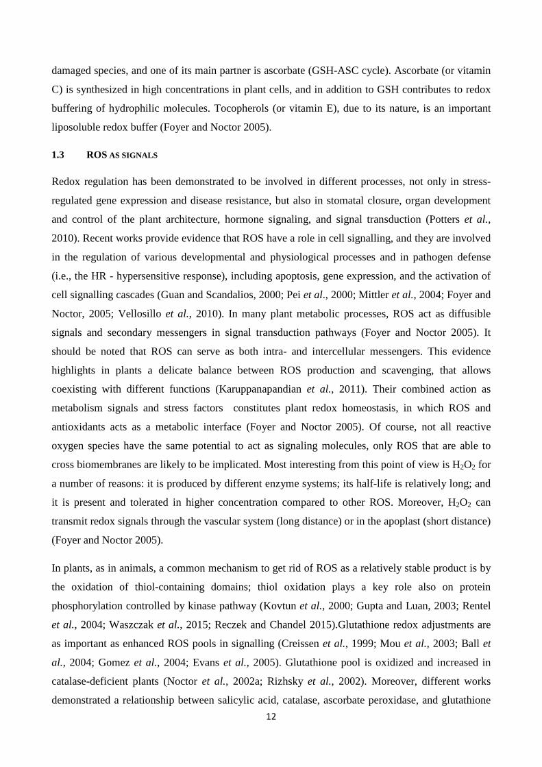
Glutathione (GSH; γ-glutamyl-cysteinyl-glycine) is a tripeptide, the main and most abundant low
molecular weight thiol and one of the major non-protein antioxidant molecules in plant cells. It is
involved in a plethora of metabolic pathways and biological functions: e.g. it regulates protein
function, flowering and lateral root growth, cell division, mRNA translation, xenobiotic
detoxification, sulphur nutrition and storage, cellular redox state and signalling; moreover it is a
precursor of phytochelatins (Potters et al., 2010) (see Fig.1). All these functions make glutathione a
node point for regulation of plant development and responses to the environment (Noctor et al.,
2011). GSH can move through plasma membrane, and because of its thiol (–SH) moiety, it is
involved both in short and long distance sulphur transport by xylem and phloem fluids. Indeed,
cysteine amine group is linked to the carboxyl group of the glutamate to form a gamma peptide that
can not be cleaved by proteases.
Fig. 1: An overview of glutathione functions.
14
2.1 GLUTATHIONE HOMEOSTASIS
The subcellular distribution of glutathione in plants is important for plant growth and development
(Kocsy et al., 2013). Its concentration, inside cells, occurs between 3 and 10 mM and varies due to
different tissue and developmental stage of plants (Leustek and Saito 1999). Glutathione is involved
in the regulation of sulphur metabolism in plants: it constitutes the main non-protein sink for
reduced sulphur, being the major sulfur reservoir (Kopriva and Rennenberg2004).
The contribution of GSH to cellular homeostasis is demonstrated by the use of A. thaliana mutants
in several functional genomic studies, that indicate a correlation between a decreased glutathione
content and a series of damaging events. Among others, they include auxin transport and
metabolism disruption, camalexin content decrease, root apical meristem developmental failure,
increased sensitivity to cadmium, loss of apical dominance and reduced secondary root production,
and enhanced sensitivity to pathogens (Bloem et al., 2007; Pivato et al., 2014). Environmental
stress conditions provoke alterations in subcellular glutathione contents, therefore GSH is used also
as stress marker. These information are helpful to understand the role of this metabolite during
stress condition in plants(Zechmann, 2014).
The ratio of GSH-GSSG is the central indicator of cellular redox state: notably GSSG is
accumulated in oxidative stress, whereas glutathione is in its reduced form (GSH) under
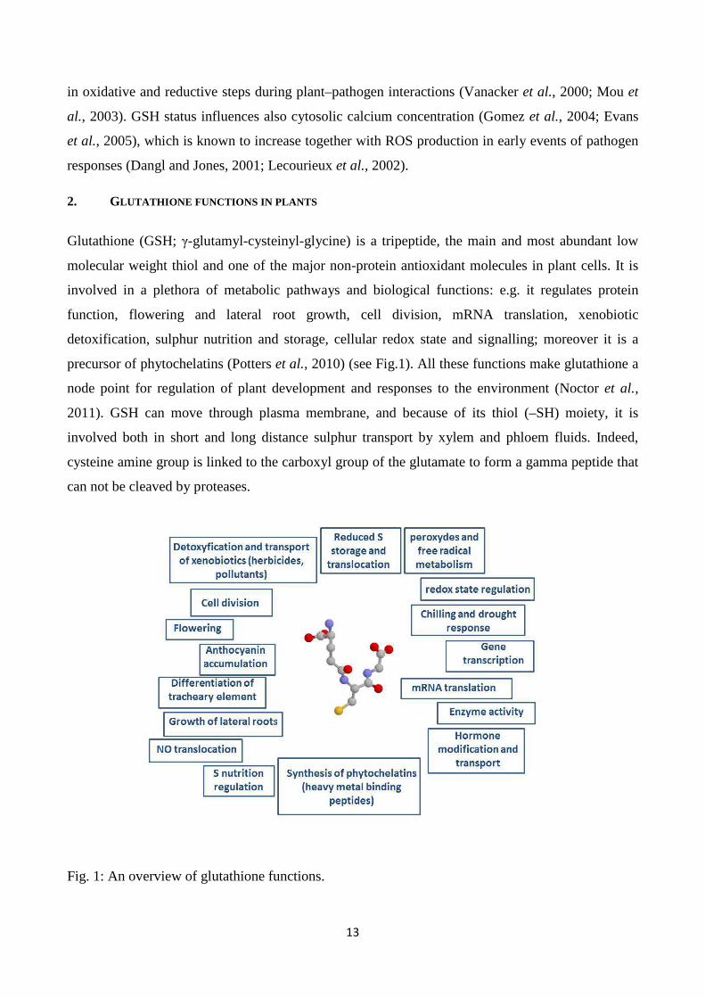
physiological condition (Pivato et al., 2014). Moreover, in plants glutathione cooperates with
ascorbate in the so called ascorbate–glutathione cycle (Fig 2)to detoxify reactive oxygen species
(ROS) by direct chemical interaction. Ascorbate is oxidized by ROS to monodehydroascorbate and
dehydroascorbate (DHA), whose reduction is coupled to glutathione oxidation (then GSSG is
enzymatically reduced by glutathione reductase GR). Since glutathione is deeply connected to
ascorbate, knowing the amount of ascorbate and glutathione and their redox states (reduced vs.
oxidized) is very important in the study of plant responses to stress (Zechmann, 2014).
15
Fig. 2:Ascorbate–glutathione cycle (from Foyer and Noctor 2011).
2.2 GSH: PHYSICO-CHEMICAL PROPERTIES AND BIOCHEMICAL MECHANISMS OF REACTION
Glutathione is linked at the γ-carboxyl group of glutamate to the amino group of cysteine: this
property confers stability to the molecule (proteases resistance) and allows programmed
degradation by amino acid transferases. Inside cells GSH is predominantly maintained in its
reduced form by glutathione reductases, that are present in cytosol, plastids, mitochondria, and
peroxisomes (Halliwell and Foyer, 1978;Smith et al., 1989; Edwards et al., 1990; Jiménez et al.,
1997; Chew et al., 2003; Kataya and Reumann, 2010).
Oxidized glutathione is produced by the formation of a disulfide bond between the cysteine thiol
moieties of two glutathione molecules. The reversible redox reactions of cysteine ensure many GSH
functions. In general, oxidized forms includes either disulfides with another glutathione molecule
(GSSG) or with a different thiol to form mixed disulfides. ROS are the molecules mainly involved
in GSH oxidation, that serves as scavenger or sacrificial nucleophile. Glutathione oxidation can also
be catalysed by enzymes, that reduce H2O2 or peroxides to water or to the corresponding alcohol
(Pivato et al., 2014), or that oxidize glutathione for ascorbate regeneration (dehydroascorbate
reductase, DHAR) (Foyer and Mullineaux, 1998).
Glutathione forms conjugates with a vast array of endogenous electrophilic species and with
xenobiotics, acting as a detoxification agent (Wang and Ballatori, 1998; Dixon and Edwards, 2010).
In particular, with nitric oxide (NO) it forms a conjugate (GSNO) which is receiving particular
attention for its physiological significance as signalling molecule (Lindermayr et al., 2005).
Therefore, in its oxidized/reduced forms, glutathione constitutes a redox buffer that on the one hand
guarantees cellular redox homeostasis, and on the other hand it participates in signalling processes.
A clear example of this double function is the interaction between glutathione and proteins: on the
one hand oxidized protein thiols can be reverted to their reduced state thanks to glutathione (redox
homeostasis), on the other hand the linkage between glutathione and proteins (glutathionylation)
can act as signalling process (e.g. it can control activity of transcription factors) (Mejer and Hell
2005).
2.3 GSH BIOSYNTHESIS AND CATABOLISM
Glutathione biosynthesis is similar between plants and other organisms (Rennenberg and Filner
1982; Meister 1988; Noctor et al., 2002b). The tripeptide is formed from Glu, Cys and Gly by two
ATP-dependent enzymes (namely GSH1 and GSH2). The first step occurs in the plastid with the
16
synthesis of γ-glutamylcysteine (γ-Glu-Cys). This intermediate molecule is exported in the cytosol
and/or cloroplast where the addition of glycine occurs (Pivato et al., 2014). Notably, the γ-Glu-Cys
synthetase (GSH1) is the rate limiting enzyme of GSH production: the increase of glutathione
contents was shown both by artificial elevation of cysteine content by exogenous supplementation
and by the overexpression of genes and enzymes involved in cysteine synthesis (Gullner et al.,
1999; Harms et al., 2000; Bloem et al., 2004 and 2007; Zechmann et al., 2007 and2008; Noji and
Saito, 2002; Wirtz and Hell, 2007).
In plant cells there are two alternative degradation pathways for GSH. In the cytosol a γ-
glutamylcyclo-transferase (GGCT) pathway operates (Ohkama-Ohtsu et al., 2008), meanwhile in
apoplast and vacuole γ-glutamyltransferase/transpeptidases are active (GGT, EC 2.3.2.2) (Ohkama-
Ohtsu et al., 2007a,b; Tolin et al., 2013;Masi et al., 2015). The two degradation pathways coexist
and operate independently of one another, and have therefore distinct physiological significance and
regulation. Indeed, the gamma glutamyl cycle is functional to the recovery of extracellular
glutathione, while the γ-glutamylcyclo-transferase/5 oxoproline pathway participates to the control
of cytosolic glutathione homeostasis.
2.4 γ- GLUTAMYL CYCLE IN PLANTS
The existence of the extracellular enzyme gamma-glutamyl-transferase (GGT; E.C. 2.3.2.2)
degrading GSH has been reported in plants as in animals (Martin et al., 2007; Meister and Anderson
1983). This is a crucial enzyme in the gamma-glutamyl cycle, consisting of intracellular glutathione
synthesis, extrusion to the extracellular space and recovery by gamma-glutamyltransferase (GGT)
and cys-glydipeptidase (DP). The degradation into its constituent amino acids has now been
demonstrated both in animals (Fig. 3) (Meister and Anderson 1983) and also in plants (Martin et
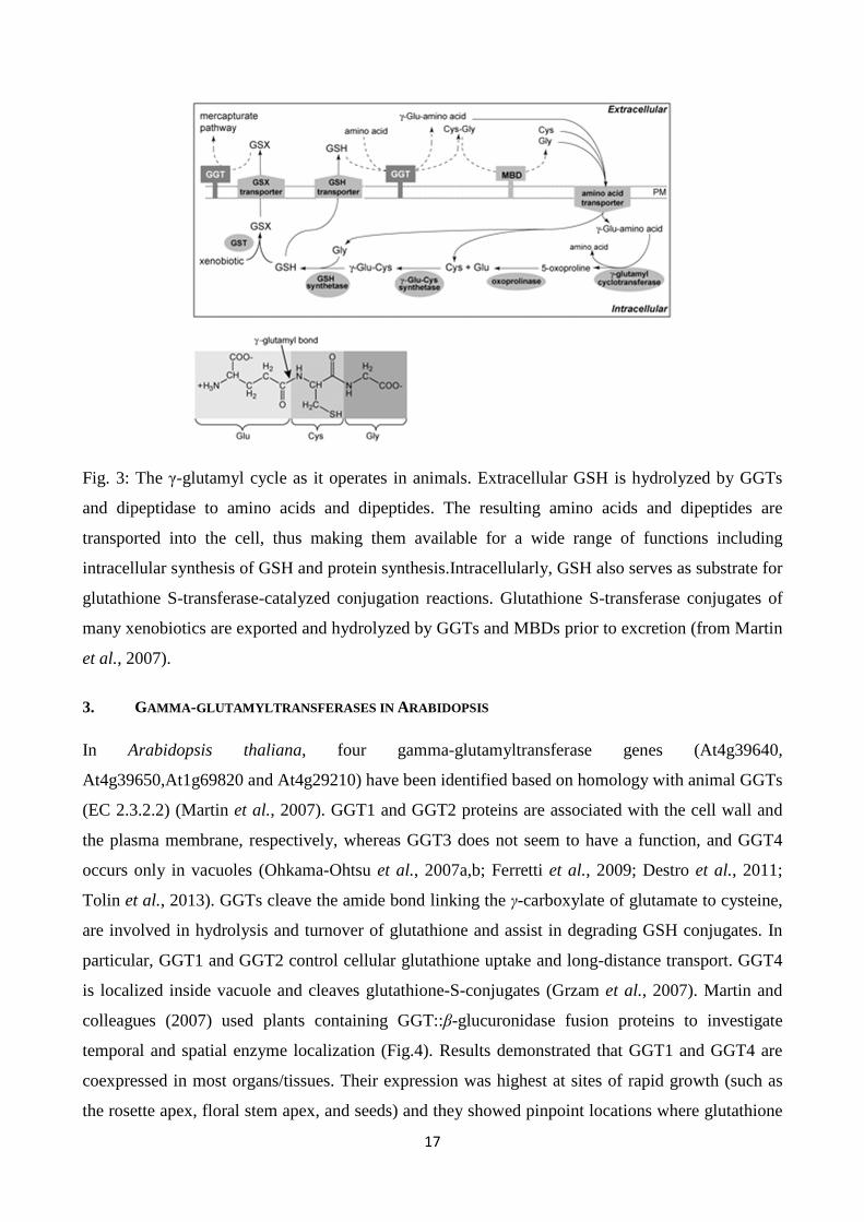
al.,2007; Ferretti et al., 2009). Amino acids are then taken up and glutathione is reassembled inside
the cell. GSH/GGT-dependent processes have been described of pivotal importance in redox
homeostasis in modulation of health and stress condition in evolutionarily distant organisms
(Pennacchio et al., 2014).
17
Fig. 3: The γ-glutamyl cycle as it operates in animals. Extracellular GSH is hydrolyzed by GGTs
and dipeptidase to amino acids and dipeptides. The resulting amino acids and dipeptides are
transported into the cell, thus making them available for a wide range of functions including
intracellular synthesis of GSH and protein synthesis.Intracellularly, GSH also serves as substrate for
glutathione S-transferase-catalyzed conjugation reactions. Glutathione S-transferase conjugates of
many xenobiotics are exported and hydrolyzed by GGTs and MBDs prior to excretion (from Martin
et al., 2007).
3. GAMMA -GLUTAMYLTRANSFERASES IN ARABIDOPSIS
In Arabidopsis thaliana, four gamma-glutamyltransferase genes (At4g39640,
At4g39650,At1g69820 and At4g29210) have been identified based on homology with animal GGTs
(EC 2.3.2.2) (Martin et al., 2007). GGT1 and GGT2 proteins are associated with the cell wall and
the plasma membrane, respectively, whereas GGT3 does not seem to have a function, and GGT4
occurs only in vacuoles (Ohkama-Ohtsu et al., 2007a,b; Ferretti et al., 2009; Destro et al., 2011;
Tolin et al., 2013). GGTs cleave the amide bond linking the γ-carboxylate of glutamate to cysteine,
are involved in hydrolysis and turnover of glutathione and assist in degrading GSH conjugates. In
particular, GGT1 and GGT2 control cellular glutathione uptake and long-distance transport. GGT4
is localized inside vacuole and cleaves glutathione-S-conjugates (Grzam et al., 2007). Martin and
colleagues (2007) used plants containing GGT::β-glucuronidase fusion proteins to investigate
temporal and spatial enzyme localization (Fig.4). Results demonstrated that GGT1 and GGT4 are
coexpressed in most organs/tissues. Their expression was highest at sites of rapid growth (such as
the rosette apex, floral stem apex, and seeds) and they showed pinpoint locations where glutathione
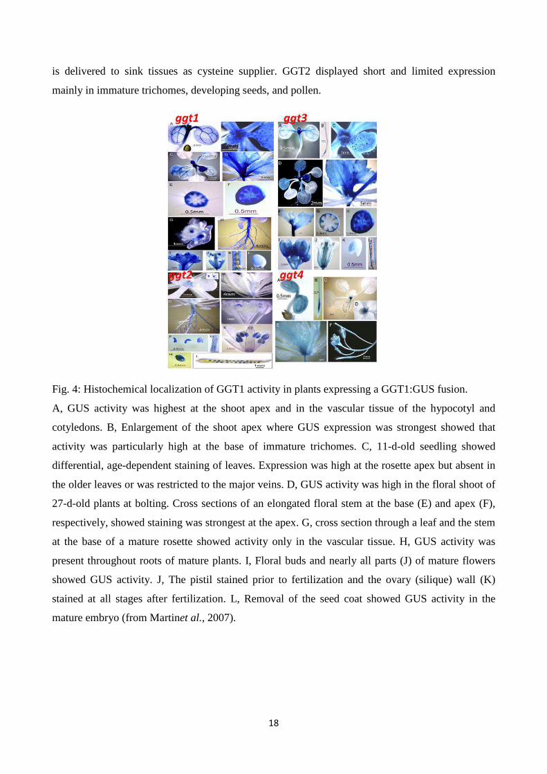
18
is delivered to sink tissues as cysteine supplier. GGT2 displayed short and limited expression
mainly in immature trichomes, developing seeds, and pollen.
Fig. 4: Histochemical localization of GGT1 activity in plants expressing a GGT1:GUS fusion.
A, GUS activity was highest at the shoot apex and in the vascular tissue of the hypocotyl and
cotyledons. B, Enlargement of the shoot apex where GUS expression was strongest showed that
activity was particularly high at the base of immature trichomes. C, 11-d-old seedling showed
differential, age-dependent staining of leaves. Expression was high at the rosette apex but absent in
the older leaves or was restricted to the major veins. D, GUS activity was high in the floral shoot of
27-d-old plants at bolting. Cross sections of an elongated floral stem at the base (E) and apex (F),
respectively, showed staining was strongest at the apex. G, cross section through a leaf and the stem
at the base of a mature rosette showed activity only in the vascular tissue. H, GUS activity was
present throughout roots of mature plants. I, Floral buds and nearly all parts (J) of mature flowers
showed GUS activity. J, The pistil stained prior to fertilization and the ovary (silique) wall (K)
stained at all stages after fertilization. L, Removal of the seed coat showed GUS activity in the
mature embryo (from Martinet al., 2007).

19
4. THE APOPLASTIC SPACE
All plant tissues can be divided into two main compartments: symplastic (located in the inner side
of the plasma membrane) and apoplastic space (located in the outer side, extracellular) (Fig. 5).
Fig. 5:Apoplast and symplast in plant cell.
Plasmodesmata are symplastic connection of cells that consist of microscopic channels used for the
transport of small molecules (Giraldo and Valent 2013). The apoplastic route facilitates the
transport of water and solutes across a tissue or organ: this process is known as apoplastic transport.
It is also a site for cell-to-cell communication.
The apoplast includes the cell wall, with its interfibrillar and intermicellar space, and the xylem,
with its gas and water-filled intercellular space (Sattelmacher 2000:). It contains proteins and other
molecules involved in plant cell’s sensing and signalling of biotic and abiotic stress (Dietz, 1997;
Agrawal et al., 2010).
It is at the interphase between the cell and the external environment, where fast fluctuations occur
due to different stresses, such as salinity (Hernandez et al., 2001), ozone (Jaspers et al., 2005)
drought (Hu et al., 2005), UV-B radiation (Pristov et al., 2013) and pathogens (Delaunois et al.,
2014). As a consequence, many molecules in the apoplastic space change their redox state and
concentrations.
Any external environmental condition is sensed by the plant thanks to the generation of an
extracellular signal, which is transmitted to the inner compartments and, following specific
transduction pathways, induces plant response to readjust cell metabolism to the new condition.
Many players are required for this system: specific genes expression, post-transcriptional and post-
translational regulation, hormones and cell regulators (Masi et al., 2015).
During local oxidative stress, hydrogen peroxide and superoxide anion can diffuse through apoplast
and transmit a warning signal to neighbouring cells. In addition, a local alkalinization of the
20
apoplast due to such stress can travel within minutes to the rest of the plant via the xylem and
trigger systemic acquired resistance.
REFERENCES
Agrawal G.K., Jwa N.S., Lebrun M.H., Job D., Rakwal R. (2010). “Plant secretome: unlocking
secrets of the secreted proteins”. Proteomics 10 pp. 799-827.
Apel K., Hirt H. (2004). “Reactive oxygen species: metabolism, oxidative stress, and signal
transduction”. Annu Rev Plant Biol 55 pp. 373–399.
Asada K. (2006).“Production and scavenging of reactive oxygen species in chloroplasts and their
functions”. Plant Physiol 141 pp. 391–396.
Ball L., Accotto G., Bechtold U., Creissen G., Funck D., Jimenez A., Kular B., Leyland N., Mejia-
Carranza J., Reynolds H., Karpinski S., and Mullineaux P.M. (2004). “ Evidence for a direct link
between glutathione biosynthesis and stress defense gene expression in Arabidopsis”. Plant Cell 16
pp. 2448–2462.
Bischof K., Janknegt P.J., Buma A.G.J., Rijstenbil J.W., Peralta G., Breeman A.M. (2003).
“Oxidative stress and enzymatic scavenging of superoxide radicals induced by solar UV-B radiation
in Ulvacanopies from southern Spain”. Sci Mar 67 (3) pp. 353-359.
Bloem E., Haneklaus S., Salac I., Wickenhäuser P., Schnug E. (2007). “Facts and fiction about
sulfur metabolism in relation to plantpathogen interactions”. Plant Biol9 pp. 596–607.
Bloem E., Riemenschneider A., Volker J., Papenbrock J., Schmidt A., Salac I., et al. (2004).
“Sulphur supply and infection with Pyrenopeziza brassicae influence l-cysteine desulphydrase
activity in Brassica napus L”. J Exp Bot 55 pp. 2305–2312.
Chew O., Whelan J., Millar A.H. (2003). “Molecular definition of the ascorbate-glutathione cycle
in Arabidopsis mitochondria reveals dual targeting of antioxidant defenses in plants”. J Biol Chem
278 pp. 46869–46877.
Corpas F.J., Barroso J.B., del Río L.A. (2001). “Peroxisomes as a source of reactive oxygen species
and nitric oxide signal molecules in plant cells”. Trends Plant Sci 6 pp. 145–150.
21
Creissen G., Firmin J., Fryer M., Kular B., Leyland M., Reynolds H., Pastori G., Wellburn F.,
Baker N.R., Wellburn A., and Mullineaux P. (1999). “Elevated glutathione biosynthetic capacity in
the chloroplasts of transgenic tobacco paradoxically causes increased oxidative stress”.Plant Cell11
pp. 1277–1291.
Dangl J.L., and Jones J.D.G. (2001). “Plant pathogens and integrated defence responses to
infection”. Nature 411 pp. 826–833.
del Rio L.A., Corpas F.J., Sandalio L.M., Palma J.M., Gomez M., Barroso J.B. (2002). “Reactive
oxygen species, antioxidant systems and nitric oxide in peroxisomes”. J Exp Bot 53 pp. 1255–1272.
Delaunois B., Jeandet P., Clément C., Baillieul F., Dorey S., Cordelier S. (2014). “Uncovering
plant-pathogen crosstalk through apoplastic proteomic studies”. Front PlantSci5, 249.
doi:10.3389/fpls.2014.00249.
DestroT., Prasad D., Martignago D., Bernet I. L., et al., (2011).“Compensatory expression and
substrate inducibility of gamma-glutamyl transferase GGT2 isoform in Arabidopsis thaliana”. J
Exp Bot62 pp. 805-814.
Dietz, K.J. (1997). Functions and responses of the leaf apoplast under stress. Progress in Botany 58
pp. 221-254.
Dixon D.P., Edwards R. (2010). “Glutathione S-transferases”.The Arabidopsis Book. doi:
10.1199:tab.0131
Edwards E.A., Rawsthorne S., Mullineaux P.M. (1990). “Subcellular distribution of multiple forms
of glutathione reductase in pea (Pisum sativum L.)”. Planta 180 pp. 278–284.
Evans N.H., McAinsh M.R., Hetherington A.M., and Knight M.R. (2005). “ROS perception in
Arabidopsis thaliana: The ozone-induced calcium response”. Plant J 41 pp. 615–626.
Ferretti M., Destro T., Tosatto S.C.E., La Rocca N., Rascio N., Masi A. (2009). “Gamma-glutamyl
transferase in the cell wall participates in extracellular glutathione salvage from the root apoplast”.
New Phytol 181 pp. 115–126.
Foyer C.H., Mullineaux P.M. (1998). “The presence of dehydroascorbate and dehydroascorbate
reductase in plant tissues”. FEBS Lett425 pp. 528–529.
Foyer C.H., Noctor G. (2005). “Redox homeostasis and antioxidant signaling: a metabolic interface
between stress perception and physiological responses”. Plant Cell 17(7) pp. 1866-75.
22
Foyer C.H., Noctor G. (2011). “Ascorbate and Glutathione: the heart of the redox hub”. Plant
Physiol155 pp. 2-18.
Gill S.S., Tuteja N. (2010). “Reactive oxygen species and antioxidant machinery in abiotic stress
tolerance in crop plants”. Plant Physiol Biochem 48(12) pp. 909-30. doi:
10.1016/j.plaphy.2010.08.016.
Giraldo M.C., Valent B. (2013). “Apoplastic and symplastic compartments in plants”. Nature Rev
Microbiol 11 pp. 800–814. doi:10.1038/nrmicro3119.
Gomez L.D., Noctor G., Knight M., and Foyer C.H. (2004). “Regulation of calcium signaling and
gene expression by glutathione”. J Exp Bot 55 pp. 1851–1859.
Grzam A., Martin M.N., Hell R., Meyer A.J. (2007). “Gamma-Glutamyl transpeptidase GGT4
initiates vacuolar degradation of glutathione S-conjugates in Arabidopsis”. FEBS Lett 581(17) pp.
3131-8.
Guan L.M., Scandalios J.G. (2000). “Hydrogen peroxide-mediated catalase gene expression in
response to wounding”. Free Rad Biol Med 28 pp.1182–1190.
Gullner G., Tóbiás I., Fodor J., Kömives T. (1999). “Elevation of glutathione level and activation of
glutathione-related enzymes affect virus infection in tobacco”. Free Radic Res 31(Suppl.), S155–
S161. doi: 10.1080/10715769900301451.
Gupta R., and Luan S. (2003). “Redox control of protein tyrosine phosphatases and mitogen-
activated protein kinases in plants”. Plant Physiol132 pp. 1149–1152.
Halliwell B. (2006). “Reactive Species and Antioxidants. Redox Biology Is a Fundamental Theme
of Aerobic Life”. Plant Physiol 141(2) pp. 312–322. doi: 10.1104/pp.106.077073.
Halliwell B., Foyer C.H. (1978). “Properties and physiological function of a glutathione reductase
purified from spinach leaves by affinity chromatography”. Planta 139 pp. 9–17.
Halliwell B., Gutteridge J.M.C.(2006). “Free radicals in biology and medicine”. Oxford: Oxford
University Press. UK, Fourth Edition.
Harms K., von Ballmoos P., Brunold C., Hofgen R., Hesse H. (2000). “Expression of a bacterial
serine acetyltransferase in transgenic potato plants leads to increased levels of cysteine and
glutathione”. Plant J 22 pp. 335–343.

23
Hernandez J.A., Ferrer M.A., Jimenez A., Barcelo A.R., Sevilla F. (2001). “Antioxidant systems
and O2-/ H2O2 production in the apoplast of Pea leaves. Its relation with salt-induced necrotic
lesions in minor veins”. Plant Physiol 127 pp. 817–831.
Hu J.F., Li G.F., Gao Z.H., Chen L., Ren H B., Jia W.S. (2005). “Regulation of water deficit-
induced abscisic acid accumulation by apoplastic ascorbic acid in maize seedlings”. J IntegrPlant
Biol 47 pp. 1335-1344.
Jaspers, P., Kollist, H., Langebartels, C., Kangasjarvi, J. (2005). “Plant responses to ozone,” in
Antioxidants and reactive oxygen species in plants, ed. N. Smirnoff (Oxford: Blackwell Publishing),
pp. 268-292.
Jiménez A., Hernández J.A., del Río L., Sevilla F. (1997). “Evidence for the presence of the
ascorbate-glutathione cycle in mitochondria and peroxisomes of pea leaves”. Plant Physiol 114 pp.
275–284.
Karuppanapandian T., Sinha P.B., Kamarul Haniya A., Manoharan K. (2006a).“Differential
antioxidative responses of ascorbate-glutathione cycle enzymes and metabolites to chromium stress
in green gram (Vigna radiata L. Wilczek) leaves”. J Plant Biol 49 pp. 440–447.
Karuppanapandian T., Sinha P.B., Kamarul Haniya A,. Premkumar G., Manoharan K.
(2006b).“Aluminium-induced changes in antioxidative enzyme activities, hydrogen peroxide
content and cell wall peroxidase activity in green gram (Vigna radiataL. cv. Wilczek) roots”. J
Plant Biol 33 pp. 241–246.
Karuppanapandian T., Sinha P.B., Premkumar G., Manoharan K. (2006c). “Chromiumtoxicity:
Correlated with increased in degradation of photosynthetic pigments and total soluble protein and
increased peroxidase activity in green gram (Vigna radiataL.) seedlings”. J Swamy Bot-Cl 23 pp.
117–122.
Karuppanapandian T., Saranyadevi A.R., Jeyalakshmi K., Manoharan K. (2008).“Mechanism,
control and regulation of leaf senescence in plants”. J Plant Biol 35 pp. 141–155.
Karuppanapandian T., Sinha P.B., Kamarul Haniya A., Manoharan K. (2009).“Chromium-induced
accumulationof peroxide content, stimulation of antioxidative enzymes and lipid peroxidation in
green gram (Vigna radiata L. cv. Wilczek) leaves”. Afr J Biotechnol 8 pp. 475–479.
24
Karuppanapandian T., Moon J.C., Kim C., Manoharan K., Kim W. (2011). “Reactive oxygen
species in plants: their generation, signal transduction, and scavenging mechanisms”.AJCS5(6) pp.
709-725.
Kataya A.M.R., Reumann S. (2010). “Arabidopsis glutathione reductase 1 is dually targeted to
peroxisomes and the cytosol”. Plant Signal Behav 5 pp. 171–175.
Kocsy G., Tari I., Vankova R., Zechmann B., Gulyas Z., Poor P., Galiba G. (2013). “Redox control
of plant growth and development”. Plant Sci211 pp. 77–91. doi: 10.1016/j.plantsci.2013.07.004.
Kopriva S., Rennenberg H. (2004). “Control of sulphate assimilation and glutathione synthesis:
interaction with N and C metabolism”. J Exp Bot55 pp. 1831–1842.
Kovtun Y., Chi, W.-L., Ten, G., and Shee, J. (2000). “Functional analysis of oxidative stress-
activated mitogen-activated protein kinase cascade in plants”. Proc Natl Acad SciUSA 97 pp. 2940–
2945.
Lecourieux D., Mazars C., Pauly N., Ranjeva R., and Pugin A. (2002). “Analysis and effects of
cytosolic free calcium increases in response to elicitors in Nicotiana plumbaginifolia cells”. Plant
Cell14 pp. 2627–2641.
Leustek T., and Saito K. (1999). “Sulfate Transport and Assimilation in Plants”. Plant Physiol
120(3) pp. 637-644.
Lindermayr C., Saalbach G., Dürner J. (2005). “Proteomic identification of S-nitrosylated proteins
in Arabidopsis”. Plant Physiol137 pp. 921–930.
Manoharan K., Karuppanapandian T., Sinha P.B., Prasad R. (2005). “Membrane degradation,
accumulation of phosphatidic acid, stimulation of catalase activity and nuclear DNA fragmentation
during 2,4-D-induced leaf senescence in mustard”. J Plant Biol 48 pp. 394–403.
Martin M.N., Saladores P.H., Lambert E., Hudson A.O., Leustek T. (2007). “Localization of
members of the γ-glutamyl transpeptidase family identifies sites of glutathione and glutathione S-
conjugate hydrolysis”. Plant Physiol 144 pp. 1715-1732.
Masi A., Trentin A.R., Agrawal, G.K., RakwalR. (2015). “Gamma-glutamyl cycle in plants: a
bridge connecting the environment to the plant cell?” Front Plant Sci 16 April 2015 |
http://dx.doi.org/10.3389/fpls.2015.00252.
25
Mawdsley J.R., O’Maley R., Ojima D.S. (2009).“A Review of Climate-Change Adaptation
Strategies for Wildlife Management and Biodiversity Conservation”. Conservation Biology 23(5)
pp. 1080–1089.
Meister A., (1988). “Glutathione Metabolism and Its Selective Modification”. J Biol Chem 263(33)
pp. 17205-17208.
Meister A., and Anderson M.E. (1983). “Glutathione”. Annu Rev Biochem 52 pp. 711-760.
Mejer A.J., Hell R. (2005). “Glutathione homeostasis and redox-regulation by sulfhydryl groups”.
Photosynth Res 86(3) pp. 435–457.
Mittler R. (2002). “Oxidative stress, antioxidants and stress tolerance”. Trends Plant Sci 7(9) pp.
405-10.
Mittler R., Vanderauwera S., Gollery M., Van Breusegem F. (2004).“Reactive oxygen gene
network of plants”. Trends Plant Sci 9 pp. 490–498.
Moller I.M., Jensen P.E., Hansson A. (2007). “Oxidative modifications to cellular components in
plants”. Annu Rev Plant Biol 58 pp. 459–481.
Mou Z., Fan W., and Dong X. (2003). “Inducers of plant systemic acquired resistance regulate
NPR1 function through redox changes”. Cell 113 pp. 935–944.
Munne-Bosch S., Alegre L. (2004). “Die and let live: leaf senescence contributes to plant survival
under drought stress”. Funct Plant Biol 31 pp. 203–216 .
Nakabayashi R. and Saito K. (2015). “Integrated metabolomics for abiotic stress responses in
plants”.Curr Opin Plant Biol 24 pp. 10–16.
Navrot N., Roubier N., Gelbaye E., Jacquot J-P. (2007).“Reactive oxygen species generation and
antioxidant systems in plant mitochondria”. Physiol Plant 129 pp. 185–195.
Noctor G., Veljovic-Jovanovic S., Driscoll S., Novitskaya L., and Foyer C.H. (2002a). “Drought
and oxidative load in wheat leaves: A predominant role for photorespiration?” Ann Bot 89 pp. 841–
850.
NoctorG., GomezL., VanackerH., FoyerC.H. (2002b). “Interactions between biosynthesis,
compartimentation and transport in the control of glutathione homeostasis and signalling”.J Exp Bot
53(372)pp. 1283–1304.
26
Noctor G., Queval G., Mhamdi A., Chaouch S., Foyer C.H. (2011). “Glutathione”. Arabidopsis
Book doi: 10.1199/tab.0142.
Noji M., Saito K. (2002). “Molecular and biochemical analysis of serine acetyltransferase and
cysteine synthase towards sulfur metabolic engineering in plants”. Amino Acids 22 pp. 231–243.
Ohkama-Ohtsu N., Radwan S., Peterson A., Zhao P., et al. (2007a). “Characterization of the
extracellular γ-glutamyl transpeptidases, GGT1 and GGT2, in Arabidopsis”. Plant J 49 pp. 865-
877.
Ohkama-Ohtsu, N., Zhao, P., Xiang, C., Oliver, D. J. (2007b). “Glutathione-conjugates in the
vacuole are degraded by γ-glutamyl transpeptidase GGT3 in Arabidopsis”. Plant J 49 pp. 878-888.
Ohkama-Ohtsu N., Oikawa A., Zhao P., Xiang C., Saito K., Oliver D. J. (2008). “A gamma-
glutamyl transpeptidase-independent pathway of glutathione catabolism to glutamate via 5-
oxoproline in Arabidopsis”. Plant Physiol 148 pp. 1603–1613.
Pei Z-M., Murata Y., Benning G., Thomine S., Klusener B., Allen G.J., Grill E., Schroeder J.I.
(2000). “Calcium channels activated by hydrogen peroxide mediate abscisic acid signalling in guard
cells”. Nature 406 pp. 731–734.
Pennacchio F., Masi A., Pompella A. (2014). “Glutathione levels modulation as a strategy in host-
parasite interactions—insights for biology of cancer”. Front Pharmacol 2014; 5: 180.
doi: 10.3389/fphar.2014.00180.
Pitzschke, A.,Fornazi, C., Hirt, H. (2006). Reactive oxygen species signalling in plants.
AntioxidRedox Sign 8 pp. 1757–1764.
Pivato M., Fabrega-Prats M., Masi A. (2014). “Low-molecular-weight thiols in plants: Functional
and analytical implications”.Arch Biochem Biophys 560 pp. 83–99.
PottersG., Horemans N., JansenM. K. (2010). “The cellular redox state in plant stress biology a
charging concept”. Plant Physiol Biochem 48 pp. 292–300. doi:10.1016/j.plaphy.2009.12.007.
Pristov J.B., Jovanović S.V., Mitrović A., Spasojević I. (2013). “UV-irradiation provokes
generation of superoxide on cell wall polygalacturonic acid”. Physiol Plant 148 pp. 574–581.
Reczek CR, Chandel NS. (2015). “ROS-dependent signal transduction”. Curr Opin Cell Biol 33 pp.
8-13. doi: 10.1016/j.ceb.2014.09.010.
27
RennenbergH., FilnerP. (1982).“Stimulation of h(2)s emission from pumpkin leaves by inhibition
of glutathione synthesis”.Plant Physiol 69(4) pp. 766–770.
Rentel M.C., Lecourieux D., Ouaked F., Usher S.L., Peterson L., Okamoto H., Knight H., Peck
S.C., Grierson C.S., Hirt H., and Knight M.R. (2004). “OXI1 kinase is necessary for oxidative
burst-mediated signalling in Arabidopsis”. Nature 427 pp. 858–861.
Rizhsky L., Hallak-Herr E., Van Breusegem F., Rachmilevitch S., Barr J.E., Rodermel S., Inzé D.,
and Mittler R. (2002). “Double antisense plants lacking ascorbate peroxidase and catalase are less
sensitive to oxidative stress than single antisense plants lacking ascorbate peroxidase or catalase”.
Plant J32 pp. 329–342.
Sattelmacher B. (2000). “The apoplast and its significance for plant mineral nutrition”. New
Phytol149(2)pp. 167–192.
Smith I.K., Vierheller T.L., Thorne C.A.(1989). “Properties and functions of glutathione reductase
in plants”. Physiol Plant 77 pp. 449–456.
Suzuki N., Rivero R.M., Shulaev V., Blumwald E., Mittler R. (2014). “Abiotic and biotic stress
combinations”. New Phytol 203(1) pp. 32–43.
Tolin S., Arrigoni G., Trentin A.R., Veljovic-Jovanovic S., Pivato M., Zechman B., Masi A. (2013).
“Biochemical and quantitative proteomics investigations in Arabidopsisggt1 mutant leaves reveal a
role for the gamma-glutamyl cycle in plant’s adaptation to environment”. Proteomics 13 pp. 2031–
2045.
Vanacker H., Carver T.L.W., and Foyer C.H. (2000). “Early H2O2 accumulation in mesophyll cells
leads to induction of glutathione during the hypersensitive response in the barley-powdery mildew
interaction”. Plant Physiol 123 pp. 1289–1300.
Vellosillo T., Vicente J., Kulasekaran S., Hamberg M., Castresana C. (2010). “Emerging
complexity in reactive oxygen species production and signaling during the response of plants to
pathogens”. Plant Physiol 154 pp. 444–448.
Vranova E., Inze D., Van Breusegem F. (2002). “Signal transduction during oxidative stress”. J Exp
Bot 53 pp. 1227–1236.
Wang W., Ballatori N. (1998). “Endogenous glutathione conjugates: occurrence and biological
functions”. PharmacolRev50 pp. 335–355.
28
Waszczak C., Akter S., Jacques S., Huang J., Messens J., and Van Breusegem F. (2015). “Oxidative
post-translational modifications of cysteine residues in plant signal transduction”. J Exp Bot doi:
10.1093/jxb/erv084.
Wirtz M., Hell R. (2007). “Dominant-negative modification reveals the regulatory function of the
multimeric cysteine synthase protein complex in transgenic tobacco”. Plant Cell 19 pp. 625–639.
Zechmann B., Zellnig G., Urbanek-Krajnc A., Müller M. (2007). “Artificial elevation of glutathione
affects symptom development in ZYMV-infected Cucurbita pepo L. plants”. Arch Virol 152 pp.
747–762.
Zechmann B., Müller M., Zellnig G. (2008). “Modified levels of cysteine affect glutathione
metabolism in plant cells”.In Sulfur Assimilation and Abiotic Stress in Plants, eds Khan N. A.,
Singh S., Umar S., editors. (Berlin: Springer; ) pp. 193–206.
ZechmannB. (2014).”Compartment-specific importance of glutathione during abiotic and biotic
stress”.Front Plant Sci 5: 566. doi: 10.3389/fpls.2014.00566.

29
OBJECTIVES
30
OBJECTIVES
Plants growth and composition result from a complex interplay of genotypic features and
environmental and nutritional factors. With the dual aim to improve crop production and plant-
derived food quality, many scientists worldwide are studying the physiological determinants of
plant adaptation to environmental stress conditions.
The appearance of reactive oxygen species (ROS) is an unavoidable consequence of life in
oxygenic atmosphere, but their production is enhanced under unfavourable environmental
conditions. In living cells, antioxidants act to contrast free radicals and permit to control and limit
damages caused by toxins, medications, stress, pollution, poor diet, trauma, infections and radiation
(Halliwell and Gutteridge, 2006).Oxidative stress conditions negatively affect plant growth and
development, and accelerate scenescence.
In recent years, a great number of nutritionists and consumers have also shown an increasing
interest towards food therapeutic value, long shelf life and consequently on natural plants
antioxidants (Sreeramulu et al., 2013). Plant foods, due to their antioxidant activity, help to stay
healthy and prevent disease (Kaur and Kapoor, 2001).
All these considerations point to the importance of studying antioxidants metabolism in plant cells.
A major soluble antioxidant in plant cells is ascorbate,and human diet relies on plant food intake for
correct supply.Another main non-proteic antioxidant is GSH, mainly localised intracellularly;
however, it is also found in the apoplast, where it is involved in the so called gamma-glutamyl
cycle.
An interesting peculiarity of the cycle is that it occurs between inside and outside the cell: GSH is
synthetized in the cytosol, carried out trough plasma membranesto the extracellular space and here
it is cleaved by gamma-glutamyltransferase (GGT) to produce cysteinyl-glycine (cys-gly) and
glutamate.
A major aim of my thesis was to better clarify if the gamma-glutamyl cycle is involved in
mechanisms that regulate redox responses and antioxidant levels in plants.
To do so, I decided to investigate: i) what are the metabolic consequences of the ggt1 mutation at
proteomic level; ii) how Arabidopsis thalianaggt1 mutants respond to oxidative conditions; iii)
what are the signals arising in the apoplast, involving LMW thiols and cell wall components, that
might mediate the redox responses.
31
REFERENCES
Halliwell B., Gutteridge J.M.C. (2006). “Free Radicals in Biology and Medicine”. Ed 4. Clarendon
Press, Oxford.
Sreeramulu D., Reddy C.V.K., Chauhan A., Balakrishna N., Raghunath M. (2013). “Natural
Antioxidant Activity of Commonly Consumed Plant Foods in India: Effect of Domestic
Processing”. Oxid Med Cell Longev 2013 12;2013:369479. http://dx.doi.org/10.1155/2013/369479.
Kaur C., Kapoor H.C. (2001). “Antioxidants in fruits and vegetables—the millennium's health”.
IntJ Food Sci Tech 36(7) pp. 703–725

32
ORIGINAL PUBLICATIONS
33
CHAPTER 1: BIOCHEMICAL AND QUANTITATIVE PROTEOMICS INVESTIGATIONS IN ARABIDOPSIS
GGT1 MUTANT LEAVES REVEAL A ROLE FOR THE GAMMA -GLUTAMYL CYCLE IN PLANT ’S
ADAPTATION TO ENVIRONMENT
Serena Tolin, Giorgio Arrigoni, Anna Rita Trentin , Sonja Veljovic-Jovanovic, Micaela Pivato, B.
Zechman and Antonio Masi
Proteomics 2013 Jun;13(12-13):2031-45. doi: 10.1002/pmic.201200479
34
Biochemical and quantitative proteomics investigations in Arabidopsis ggt1 mutant leaves
reveal a role for the gamma-glutamyl cycle in plant’s adaptation to environment
Serena Tolin1,2, Giorgio Arrigoni2,3, Anna Rita Trentin1, Sonja Veljovic-Jovanovic4, Micaela
Pivato1, Bernd Zechman5 and Antonio Masi1
1DAFNAE, University of Padova, Viale dell’Universita’ 16, 35020 Legnaro (PD).
2Proteomics Center of Padova University, VIMM, and Padova University Hospital, Via G. Orus 2b,
35129 Padova, Italy.
3Department of Biomedical Sciences, University of Padova, Viale G. Colombo 3, 35121 Padova,
Italy
4Institute of Multidisciplinary Research, Kneza Viseslava 1, University of Belgrade, Serbia
5Karl-Franzens-University of Graz, Institute of Plant Sciences, Schubertstrasse 51, 8010 Graz,
Austria
Abbreviations: 2-DE, two-dimension polyacrylamide gel electrophoresis; DNPH, 2,4-
dinitrophenylhydrazine; ECWF, extracellular washing fluid; ES, enrichment score; FDR, false
discovery rate; FW, fresh weight; GGT, gamma-glutamyl transferase/transpeptidase; GO, gene
ontology; GRX, glutaredoxins; GSH, reduced glutathione; GSSG, oxidized glutathione; IEF,
isoelectric focusing; LMW low molecular weight; NAC, N-acetylcysteine; PM, plasma membrane;
ROS, reactive oxygen species; SCX, strong cation exchange; PRX, peroxyredoxins; RT, room
temperature; SAG, senescence-associated gene; SBD-F, ammonium 7-fluoro 2,1,3-
benzooxadiazole-4-sulfonate; TRX, thioredoxins; TEM, transmission electron microscopy; WT,
wild-type.
35
Key words: antioxidants; differential proteomics; gamma-glutamyl cycle; glutathione; oxidative
stress.
ABSTRACT
The existence of a gamma-glutamyl cycle consisting of intracellular GSH synthesis, extrusion to the
apoplastic space and recovery by gamma-glutamyl transferase (GGT)-assisted degradation into its
constituent amino acids, has been demonstrated in plants. To address the significance of this cycle
in plant cells, we performed integrated biochemical, immunocytochemical, and quantitative
proteomics analyses in the Arabidopsis thaliana ggt1 knockout mutant (lacking apoplastic GGT1
isoform) and its corresponding wild-type (WT). The ggt1 knockout leaves exhibited an increased
ascorbate and GSH content, increased apoplastic GSH content, and enhanced protein carbonylations
in the low-molecular-weight range compared to WT. The combined iTRAQ and LC-MS/MS based
quantitative proteomics approach identified 70 proteins (out of 1,013 identified proteins) whose
abundance was significantly different in leaves of ggt1 mutant compared to WT, with a fold change
≥1.5. Mining of the proteome data for GSH-associated genes showed that disruption of gamma-
glutamyl cycle in ggt1 knockout-leaves was associated with the induction of genes encoding four
GSTs in the phi class (GSTF2, GSTF6, GSTF9, and GSTF10), a GSH peroxidase (GPX1), and
glyoxylase II. Proteins with a lower abundance compared to the WT are involved in chloroplast
functions, carbohydrate/maltose metabolism and vegetative storage protein synthesis. Present
findings suggest that GGT1 plays a role in redox signaling. The disruption of the gamma-glutamyl
cycle in the ggt1 mutant results in pleiotropic effects related to biotic and abiotic stress response,
antioxidant metabolism, senescence, carbohydrate metabolism and photosynthesis, with strong
implications for plant’s adaptation to environment.
36
1. INTRODUCTION
Climate changes are so rapid and unpredictable in recent years that they are likely to override
plants’ capacity to adapt, resulting in restricted plant growth and development, and consequently in
reduced productivity. Developing crop plants with a modified tolerance to abiotic and biotic stresses
is therefore a necessity, which demands modern, novel strategies to gain a thorough understanding
of how plants respond to environmental changes. Intrinsically, plants adapt to the changes in their
environment and avoid damage from abiotic and biotic factors, by activating evolved self-defense
mechanisms.
There is now a wealth of evidence to indicate that many adverse environmental factors
affecting plants at the cellular level take effect, at least in part, by promoting oxidative stress [1-3].
These effects are mediated by changes in the level of reactive oxygen species (ROS) in the apoplast
- a complex structure surrounding plant cells. The role and properties of extracellular gamma-
glutamyl transferase/transpeptidase (GGT, (5-L-glutamyl)-peptide:amino-acid 5-glutamyl
transferase; EC 2.3.2.2.) in oxidative stress have been widely studied in animal cells [4], and more
recently in plants [5-10]. The GGT is an ectoenzyme promoting cleavage of the gamma-glutamyl
moiety of GSH and gamma-glutamyl-related compounds.
GGTs exist in all organisms and in multiple isoforms, sharing the characteristic of being extra-
cytosolic. In animals, they are located on the plasma membrane (PM) with the catalytic site facing
outwards [4]. In Arabidopsis thaliana, the two isoforms GGT1 and GGT2 are reportedly apoplastic,
where GGT1 is cell-wall bound [10] and GGT2 is PM associated; GGT4 is vacuolar and assists in
degradation of the GSH conjugates [6,9]. A fourth isoform, GGT3 is believed to be non-functional
because it contains a truncated sequence [8].
While GGT is known to promote GSH degradation to cysteinylglycine (cys-gly) and glutamic
acid, the significance of this reaction in plant metabolism is largely unclear. This step is part of the
37
gamma-glutamyl cycle consisting of the extrusion of GSH to the extracellular space, degradation
into its constituent amino acids, followed by their reabsorption by means of amino acid transporters.
In mammals, this cycle has been correlated with antioxidative responses involving GSH and has
been implicated in intercellular and inter-organ cysteine delivery [11]. The GGT knockout mice
exhibit abnormal growth and die prematurely, within 2 months of birth [12].
In plants, the involvement for GGT in antioxidant response remains poorly characterized.
Under photo-oxidative stress induced by ultraviolet B exposure, cys-gly content in leaves was
reported to increase throughout the period of exposure to radiation [13]. In another study aiming to
isolate genes involved in protection against oxidative damage, a cDNA from Arabidopsis encoding
a putative GGT was isolated and its expression in yeast conferred an enhanced tolerance to the
thiol-oxidizing drug diamide [14]. While the vacuolar AtGGT4 has been demonstrated to drive the
metabolism of GSH-conjugates, the functions of the apoplatic isoforms are less clear.
A functional genomics approach using knockout mutant lines has indicated no clear
phenotype in ggt1A. thaliana lines, apart from a shorter life cycle represented by early flowering
and premature senescence [15,8]. This phenotype was interpreted as the result of a difficulty in
adapting to the environment, the shorter life cycle being an escape mechanism similar to the
strategy adopted by plants that survive in harsh conditions, e.g. drought [16]. It should be noted
that, in terms of their dependence on cysteine availability, animal cells behave very differently from
plant cells: the former rely on external GGT for intracellular cysteine availability, whereas plant
cells have an autonomous capacity for cysteine biosynthesis. The GGT mutations may therefore
have less dramatic effects on plants than on animals.
With an aim to shed light on the metabolic readjustments due to the mutation in the ggt1 knockout,
we applied biochemical, immunocytochemical and quantitative proteomics approaches on leaf
proteins from the ggt1 and wild-type (WT) plants. For the quantitative proteomics, we utilized
iTRAQ isobaric tags for relative and absolute quantitation in combination with liquid
38
chromatography tandem mass spectrometry (LC-MS/MS) on a high mass accuracy Orbitrap mass
spectrometer. Our results indicate that the gamma-glutamyl cycle is part of the cell’s coordinated
response to the environment.
2. MATERIALS AND METHODS
2.1 Plant materials
After 4 days of stratification at 4°C in the dark, seeds from A. thaliana L. ecotype Columbia (Col-0)
and a ggt1 knockout mutant line were sown on soil and grown in a greenhouse. The ggt1 knockout
mutant (ecotype Columbia) was identified in the mutant collection [17] established at the Salk
Institute, and is available from the Nottingham A. thaliana Stock Centre (http://nasc.nott.ac.uk;
polymorphism SALK_080363). Leaf samples were harvested at the fully-expanded rosette stage,
approximately one week before bolting, and stored at -80°C until use. For proteome analysis,
samples were obtained after pooling leaves from ten independent plants per genotype.
2.2 Electron microscopy/immunogold labeling of ascorbate and GSH
Sample preparation for electron microscopy and immunogold labeling of ascorbate and GSH was
performed as described previously [18,19]. Fixation of leaves was performed for 90 minutes in a
mixture of 2.5% paraformaldehyde and 0.5% glutardialdehyde dissolved in 0.06 M phosphate
buffer (pH 7.2). Samples were then washed for 60 minutes in buffer and dehydrated for 20 minutes
at each step in increasing concentrations (50%, 70%, and 90%) of acetone. Infiltration was
performed with LR-White resin (30%, 60% and 100%; London Resin Company Ltd., Berkshire,
UK) and the samples were polymerized for 48 hours at 50 °C. Sections with a thickness of 80 nm
were blocked for 20 minutes with 2% bovine serum albumine (BSA) dissolved in phosphate
buffered saline (PBS, pH 7.2). Subsequently they were treated for 120 minutes with the primary
antibodies (anti ascorbate rat polyclonal IgG, Abcam plc, Cambridge, UK and anti GSH rabbit
polyclonal IgG, Millipore Corp., Billerica, MA, USA) diluted 1:300 (ascorbate antibody) and 1:50
39
(GSH antibody) in PBS containing 1% BSA (for ascorbate labeling) and 1% goat serum (for GSH
labeling). After three short rinses in PBS sections were incubated for 90 minutes with 10 nm gold-
conjugated secondary antibodies (goat anti rat IgG for ascorbate, goat anti rabbit IgG for GSH,
British BioCell International, Cardiff, UK) diluted 1:100 (for ascorbate labeling) and 1:50 (for GSH
labeling) in PBS. Sections were finally rinsed with distilled water. At least 20 (vacuoles and
peroxisomes) to 60 (all other cell compartments) sectioned cell structures of a minimum of 15
different cells from at least four different samples per plant were analyzed for gold particle density.
The data were statistically evaluated with the Mann-Whitney U test using Statistica (Stat-Soft,
Tulsa, OK, USA, 2002) and presented as the number of gold particles per µm-2.
2.3 Soluble antioxidant extraction
Frozen leaf samples (250 mg) from at least five biological replicates were ground with a mortar and
pestle to extract soluble antioxidants with 0.1 N HCl and 1 mM EDTA. Following centrifugation at
10,000 g for 10 min, extracts were rapidly tested for ascorbate and low-molecular-weight (LMW)
thiol levels.
2.4 Extracellular washing fluid extraction
A vacuum infiltration procedure was used for apoplast protein extraction [20]. Briefly, one gram of
leaf tissue was washed in chilled H2O and then submerged in 100 ml chilled vacuum infiltration
buffer (50 mM KPi, pH 6.1) containing 5 µM N-acetylcysteine (NAC) in a vacuum desiccator. A
vacuum was applied for 10 min at a pressure of 20 kPa using a vacuum pump to remove the gas
from the apoplastic spaces. Excess buffer was removed from the WTand ggt1 mutant leaves. Each
leaf was then positioned vertically in a 20 ml syringe. The syringes were placed in centrifuge tubes
and centrifuged at 200 g for 20 min at 4°C. Apoplastic extracellular washing fluid (ECWF) extracts
were collected from the bottom of the tubes. NAC was used as a tracer and internal standard. It
elutes in a chromatographic region devoid of other peaks, several minutes after the elution of GSH,
40
which is usually the last endogenous thiol compound to remain visible in chromatograms. ECWF
extracts were then used to measure the LMW thiol and ascorbate content.
2.5 Ascorbate content
The ascorbate content was determined spectrophotometrically by measuring the absorbance at 265
nm, according to the Hewitt and Dickes method [21].
2.6 LMW thiol content
Prepared extract (50 µL) was derivatized with SBD-F fluorophore (Sigma-Aldrich, St. Louis, USA).
LMW thiols were separated by isocratic HPLC using the method described in Masi et al. [13] with
some adaptations. The mobile phase was 3% methanol in 75 mM NH4+-formiate, pH 2.9.
2.7 Carbonylated proteins analysis
Proteins were extracted from leaves of the ggt1 mutant and WT plants using extraction buffer A (20
mM Tris pH 8.0, 3 M NaCl, 1 mM EDTA, and 1% SDS) in a ratio of 1:5 (w/v). Extracted proteins
were subsequently precipitated with acetone. The pellet thus obtained was then resuspended in a
solution (5 M urea, 2 M thiourea, 2% CHAPS, and 0.4% ampholites) compatible to two-dimension
polyacrylamide gel electrophoresis (2-DE). Collected supernatant was subjected to protein
quantification using the modified Lowry total protein kit (Sigma-Aldrich, St. Louis, USA),
according to the manufacturer’s instructions. Total protein (100 µg) from each sample was loaded
onto IPG strips (7 cm, 3-10 pH range) (GE Healthcare Bio-Science AB, Uppsala, Sweden) for
isoelectric focusing (IEF). After IEF, the strips were incubated with 10 mM DNPH solution (to
derivatize the carbonylated proteins) in 10% TFA, followed by two washes with a washing solution
(8 M urea, 20% glycerol, 1% SDS, and 0.5 M Tris HCl, pH 6.8). Treated IPG strips were then
subjected to 2-DE using a 12% SDS-PAGE.
41
The 2D gels were then subjected to Western blot analysis, where the protein spots were
transferred onto a nitrocellulose membrane (using Hoefer mini-VE transblot apparatus, Amersham
Biosciences, Piscataway, USA) and saturated with the T-PBS blocking buffer (Triton X100 0.1% in
PBS pH 7.4, plus 3% skimmed milk) for 1 h at room temperature (RT). The membranes were
incubated with rabbit anti-DNP antibodies (Serologicals Corporation, Norcross, USA) (dilution
1:20,000) in T-PBS plus 3% milk for 1 h at RT, followed by washing with T-PBS. The membranes
were incubated with secondary antibody (anti-goat anti-rabbit, dilution 1:50,000) for 1 h at RT. The
peroxide-luminol reaction was used to detect cross-reacting proteins; the chemiluminescent reaction
buffer was mixed with chemiluminescent reagent in a ratio of 2:1 (v/v) (Sigma-Aldrich, St. Louis,
USA). Membranes were incubated in the prepared chemiluminescent solution for 5 min at RT and
developed in a dark room.
2.8 Statistical analysis
Data from at least five replicates were submitted to an analysis of variance (ANOVA). The Tukey
honest significant difference multiple comparisons procedure was used to discriminate between
means. A p < 0.05 was considered significant for all comparisons.
2.9 Total leaf protein extraction for iTRAQ labeling and MS analyses
Leaves (0.5 g) were homogenized in liquid nitrogen to fine powder. Leaf powder was suspended in
an extraction buffer (50 mM HEPES pH 8, 1% Triton X100, 1M NaCl, 1mM
phenylmethanesulfonylfluoride, 1 mM benzamidine) with thorough mixing, followed by
centrifugation at 10,000 rpm for 15 min at 4°C. To the collected clear supernatant, cold acetone was
added to precipitate proteins at -20° C overnight, and centrifuged at 12,000 rpm for 15 min at 4°C.
The protein pellet thus obtained was used for protein quantification using the modified Lowry
method, as described above, for iTRAQ labeling.
42
2.10 iTRAQ labeling
The principle behind the method is described in Ross et al. [22]. Labeling was done with an
iTRAQ® Reagents Multiplex Kit (AB Sciex, MA, USA). The iTRAQ experiment was performed
on protein samples derived from pooled leaves (collected from 10 independent plants of each ggt1
mutant and WT) as described above. Two technical replicates were carried out with tag swapping
for each pooled sample. Protein content from each sample (WT and ggt1 mutant leaves) was
carefully quantified using the bicinchoninic acid (BCA) method (Sigma-Aldrich kit, St. Louis,
USA). 500 µg of proteins from each sample were precipitated with cold acetone and kept overnight
at -20 °C. The protein pellets were resuspended in an iTRAQ-compatible buffer (TEAB 0.5 M, SDS
0.1%) to a final concentration of 2-5 µg/µL. To avoid any variability in the precipitation and
resuspension processes, protein content was quantified again using the bicinchoninic acid method.
100 µg of each sample was reduced, alkylated and digested with trypsin, according to the iTRAQ
manufacturer’s protocol. To check the digestion efficiency, 1 µg of each sample was analyzed by
LC-MS/MS (details of the instruments and instrumental methods are given in the following
section).Control and mutant samples were split into two identical 50 µg aliquots and labeled with
different iTRAQ tags: the WT sample was labeled with tags 114 and 116, whereas the ggt1 mutant
sample with tags 115 and 117. Before labeling, to each sample, trypsin-digested BSA (2.5 pmol)
was spiked as an internal standard for monitoring labeling efficiency. The labeling procedure was
completed as specified in the iTRAQ manufacture’s protocol. Prior to mixing the samples in a
1:1:1:1 ratio, 1 µg of each labeled sample was analyzed separately by LC-MS/MS to test labeling
efficiency. The resulting data were checked against the database as explained below, setting the
iTRAQ labeling as a variable modification. All the peptides were correctly identified as being
iTRAQ-modified at the N-terminus and at each lysine residue. The samples were then pooled and
dried under vacuum.
43
2.11 Strong cation exchange fractionation
Strong cation exchange (SCX) chromatography was performed on a SCX cartridge (AB Sciex,
MA, USA). The labeled samples were dissolved in 500 µL of buffer A (10 mM KH2PO4, 25%
acetonitrile, pH 3) and loaded onto the cartridge using a syringe pump with a 50 µL/min flow rate.
The cartridge was washed 3 times with 500 µL of buffer A. Peptides were eluted in a stepwise
manner with increasing concentrations of KCl in buffer A. The labeled peptides were eluted in 8
fractions (500 µL per fraction) with the following concentrations of KCl in the buffer A: 40, 60, 80,
100, 130, 170, 200, and 350 mM. The volume of each fraction was reduced under vacuum to
remove acetonitrile. Samples were desalted using C18 cartridges (Sep-Pack, C18, Waters, Milford,
MA, USA) according to the manufacturer’s instructions. Samples were finally dried under vacuum
and kept at -20°C until MS analysis.
2.12 LC-MS/MS analyses
Samples were resuspended in H2O/0.1% formic acid and 1 µg of each fraction underwent LC-
MS/MS analysis. The MS analyses were conducted with a LTQ-Orbitrap XL mass spectrometer
(Thermo Fisher Scientific, Pittsburgh, CA, USA) coupled online with a nano-HPLC Ultimate 3000
(Dionex-Thermo Fisher Scientific). Samples were loaded onto a homemade 10 cm chromatographic
column packed into a pico-frit (75 mm I.D., 10 mm tip, New Objectives) with C18 material
(ReproSil, 300 Å, 3 µm). Peptides were eluted with a linear gradient of acetonitrile/0.1% formic
acid from 3% to 50% in 90 min at a flow rate of 250 nL/min. According to the method described by
Köcher et al. [23], the instrument performed a full scan at high resolution (60000) on the Orbitrap,
followed by MS/MS scans on the three most intense ions with CID fragmentation on the linear trap.
MS/MS scans were performed on the same ions with HCD fragmentation on the Orbitrap (with a
resolution of 7500) to obtain low mass range data suitable for protein quantification. The peptides
reliably identified in each sample were inserted in an exclusion list that was used to perform (under
44
the same chromatographic and instrumental conditions) a second LC-MS/MS run for each sample
fraction.
2.13 Data analysis
The raw LC-MS/MS files were analyzed using Proteome Discoverer 1.2 (Thermo Fisher Scientific,
Pittsburgh, USA). The software was connected to a Mascot Search Engine server, version 2.2.4
(Matrix Science, London, UK). The spectra were searched against an A. thaliana database
(downloaded from TAIR version dated February 2012 with 33,596 entries and 13,743,691 residues)
concatenated with a database of contaminant proteins commonly found in proteomic experiments.
Enzyme specificity was set to trypsin with 2 missed cleavages. Peptide and fragment tolerance was
set to 10 ppm and 0.6 Da, respectively. Methylthiocysteine, 4-plex iTRAQ at the N-terminus and
Lys were set as fixed modifications, while methionine oxidation was selected as a variable
modification. Based on the search against the corresponding randomized database, false discovery
rates (FDR) of 5% and 1% were calculated by the Proteome Discoverer. The data were pre-filtered
to exclude MS/MS spectra containing less than 5 peaks or with a total ion count below 50. Were
considered as positive hits all proteins identified and quantified with at least two independent
peptides with a high degree of confidence (FDR 1%). The quantification was performed
normalizing the results on the median value of all measured iTRAQ reporter ratios. The list of
quantified proteins was exported to Excel for further filtering and statistical analyses, which were
conducted after removing from the final list the proteins that showed a discordant trend in the
replicates. A ratio of mutant to WT ≥ 1.5 (fold change ≥ +50%) was set as the threshold for
increased content, while a ratio of WT to mutant ≥ 1.5 was taken to indicate decreased protein
content.

45
2.14 Bioinformatics analysis
The QuickGO web-based browser, provided by the UniProt-GOA group, was used to analyze Gene
Ontology (GO) terms and annotations. The GO-term functional annotation was done using DAVID
[24], which relies on a modified Fisher’s exact p-value to cluster GO-terms in functional annotation
groups, establishing whether the term is over-represented in a given proteomic data set. DAVID
generates an enrichment score (ES) for each cluster, defined as the geometric mean of all the
enrichment p-values for each annotation term associated with the gene members in the background
group [25]. An ES higher than 1.3 is considered statistically significant (1.3 is equivalent to p-value
< 0.05).
3. RESULTS
Consistent with previous findings [15], the ggt1 mutants exhibited no obvious phenotype, rather
only a shorter life cycle, presumably due to a general difficulty in adapting to the environment. On
average, the ggt1 knockout plants flowered three to four days earlier than the WT, followed by a
premature senescence.
3.1 Soluble antioxidant content in leaves and in the leaf apoplastic space
Total ascorbate content, on a total leaf content basis, was approximately 35% higher in ggt1
mutants (2.71±0.28 µmol per g FW-1) than in WT (1.99±0.22 µmol per g FW-1). The ggt1 knockout
leaves also carried higher total GSH, but not cys-gly, content compared to WT (Table 1).
Leaf ECWF
GSH Cys-Gly GSH Cys-Gly
Col- 0 855.72 (± 44.61) 5.03 (± 0.14) 8.59 (± 1.19) 0.87 (± 0.23)
ggt1- 1183.25 (± 37.69) * 4.76 (± 0.21) 13.74 (± 3.64)* 0.11(± 0.05)**
46
Table 1.GSH and cysteinylglycine content measured by chromatographic separation and
quantitation of their SBD-derivatives. Analyses were carried out on total leaf extracts and on leaf
extracellular washing fluids (ECWF) obtained from ggt1 mutant and WT plants. Values are
calculated as the mean of at least 5 different experiments and are expressed in nmol·(g-1) FW. * and
**, indicate significance at the 0.05, 0.01 level of confidence, respectively.
The LMW thiols were measured in the ECWF. Contamination level of the extracts identified by the
infiltration-centrifugation technique was assessed by means of malic dehydrogenase activity
measurements, and was consistently below 2% (data not shown). The quality of the extracts was
also indirectly confirmed by the absence of γEC, a precursor in GSH biosynthesis and metabolically
restricted in the cytoplasm, which was not detected in the ECWF. Our results point to a higher GSH
content, and a drop in cys-gly content (approximately to one tenth) in ggt1 mutant compared to WT
extracts, which is due to the loss of apoplastic GGT activity.
NAC was used as a tracer and internal standard in this study (Supporting Figure S1). When
leaves were infiltrated with a 5 µM NAC solution, the resulting concentration of NAC in the extract
after centrifugation was approximately 4 µM, as a result of dilution with the fluid already existing
in the apoplast. This result suggests that the actual concentration of the metabolites measured in the
apoplastic space could be approximately 5 times higher. Under normal physiological conditions, in
fact, the leaf apoplast is filled with air, which is replaced by the buffer solution during the
infiltration step. This evidence may enable a better definition of the apoplastic redox potential for
the GSH/GSSG couple, since redox potential depends on GSH concentration and the GSH/GSSG
ratio.
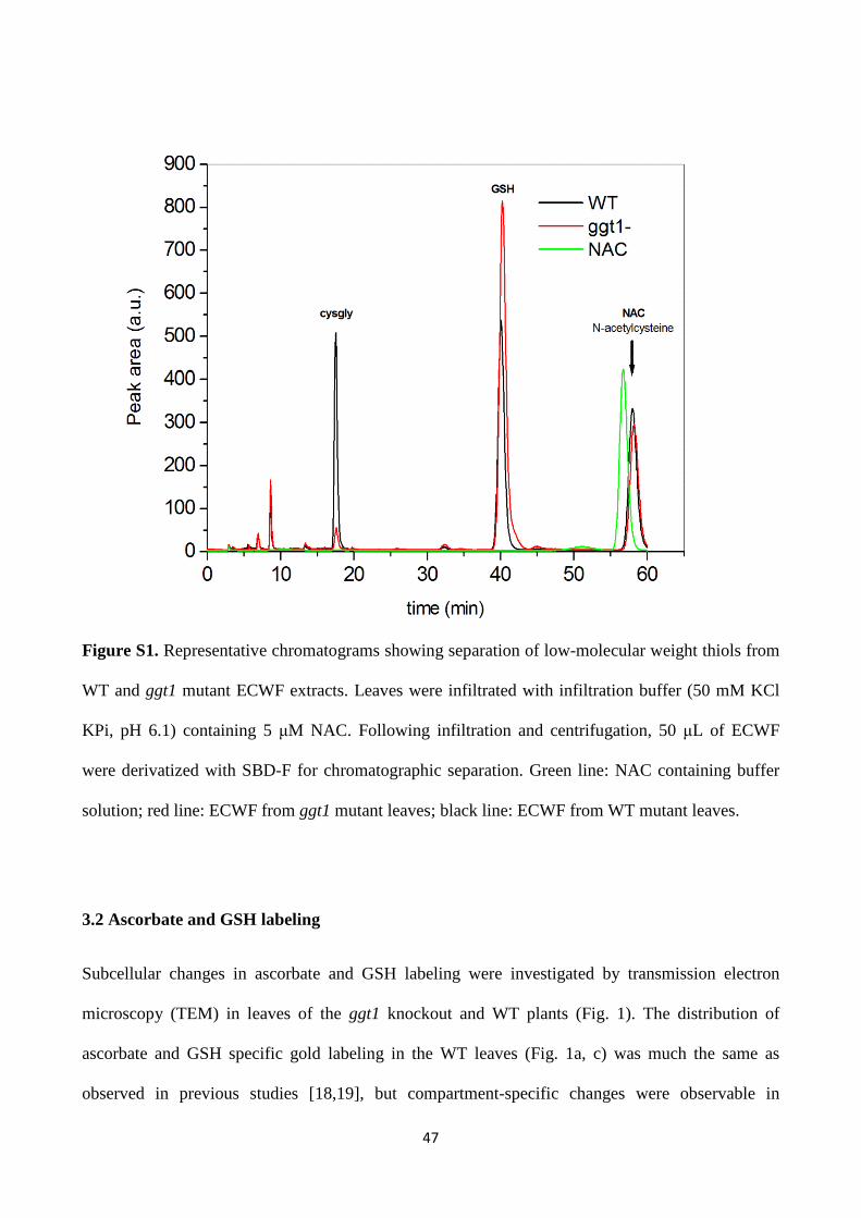
47
Figure S1. Representative chromatograms showing separation of low-molecular weight thiols from
WT and ggt1 mutant ECWF extracts. Leaves were infiltrated with infiltration buffer (50 mM KCl
KPi, pH 6.1) containing 5 µM NAC. Following infiltration and centrifugation, 50 µL of ECWF
were derivatized with SBD-F for chromatographic separation. Green line: NAC containing buffer
solution; red line: ECWF from ggt1 mutant leaves; black line: ECWF from WT mutant leaves.
3.2 Ascorbate and GSH labeling
Subcellular changes in ascorbate and GSH labeling were investigated by transmission electron
microscopy (TEM) in leaves of the ggt1 knockout and WT plants (Fig. 1). The distribution of
ascorbate and GSH specific gold labeling in the WT leaves (Fig. 1a, c) was much the same as
observed in previous studies [18,19], but compartment-specific changes were observable in

48
ggt1mutant (Fig. 1b, d). The ascorbate content was significantly higher in plastids (118%),
peroxisomes (60%) and cytosol (44%), and remained at the same levels as in WT in the other cell
compartments. The GSH content in ggt1 mutant was significantly higher in mitochondria (23%),
while remaining unchanged in nuclei and peroxisomes, but significantly lower in plastids (-62%)
and cytosol (-28%). Most strikingly, GSH-specific labeling was common in the ggt1 mutant
apoplast (Fig. 1d), but not seen in control (Fig. 1c).
Figure 1. Subcellular localization of ascorbate and GSH in leaves of the ggt1 knockout and WT
plants. Transmission electron micrographs (upper panels) and quantitative analysis (lower graphs)
show the overall distribution of gold particles bound to ascorbate (left panel) and GSH (right panel)
in leaf cells of WT (a, c) and ggt1 mutants (b, d). Values are represented as means with standard
errors and document the amounts of gold particles bound to GSH per square micrometer in different
cell compartments of mesophyll cells. Significant differences were calculated using the Mann–
Whitney U test; *, ** and *** indicate significance at the 0.05, 0.01 and 0.001 level of confidence,
49
respectively. n > 20 for peroxisomes and vacuoles and n > 60 for all other cell structures.
Abbreviations: Ns, non significance; nd, not detected; C, chloroplasts with or without starch (St);
CW, cell walls; IS, intercellular spaces; M, mitochondria; N, nuclei; Px, peroxisomes; V, vacuoles,
scale bar= 0.5µm.
3.3 2-DE analysis of carbonylated proteins
Proteins may undergo cleavage by hydroxyl radicals, and side chain modifications such as tyrosine
hydroxylations and nitrations, and methionine or cysteine oxidation (by hydroxyl radicals or
hydrogen peroxide). However, one of the most common modifications involves carbonyl formation,
which has been widely used as an indicator of oxidative damage in several organisms and has been
shown to increase in older tissues [26, 27]. The presence of oxidative damage on proteins under
conditions of oxidative stress is revealed by detecting carbonyls through the immunodetection of
coupled dinitrophenylhydrazine (DNPH). The great sensitivity of immunodetection enables the
detection of even minute amounts of modified proteins.
We characterized the oxidized proteome in leaves (Fig. 2). The 2-DE combined with Western
blotting allowed us to detect carbonylated protein spots as 2,4-dinitrophenylhydrazone (DNP)-
derivatives. There were several spots of greater intensity in the LMW range, suggesting that protein
carbonylations increased in the ggt1 knockout leaf.
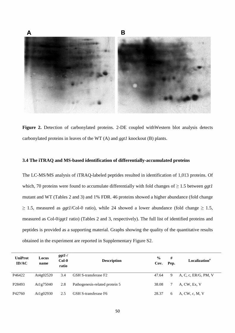
50
Figure 2. Detection of carbonylated proteins. 2-DE coupled withWestern blot analysis detects
carbonylated proteins in leaves of the WT (A) and ggt1 knockout (B) plants.
3.4 The iTRAQ and MS-based identification of differentially-accumulated proteins
The LC-MS/MS analysis of iTRAQ-labeled peptides resulted in identification of 1,013 proteins. Of
which, 70 proteins were found to accumulate differentially with fold changes of ≥ 1.5 between ggt1
mutant and WT (Tables 2 and 3) and 1% FDR. 46 proteins showed a higher abundance (fold change
≥ 1.5, measured as ggt1/Col-0 ratio), while 24 showed a lower abundance (fold change ≥ 1.5,
measured as Col-0/ggt1 ratio) (Tables 2 and 3, respectively). The full list of identified proteins and
peptides is provided as a supporting material. Graphs showing the quality of the quantitative results
obtained in the experiment are reported in Supplementary Figure S2.
UniProt ID/AC
Locus name
ggt1-/ Col-0 ratio
Description %
Cov. #
Pep. Localizationa
P46422 At4g02520 3.4 GSH S-transferase F2 47.64 9 A, C, c, ER/G, PM, V
P28493 At1g75040 2.8 Pathogenesis-related protein 5 38.08 7 A, CW, Ex, V
P42760 At1g02930 2.5 GSH S-transferase F6 28.37 6 A, CW, c, M, V
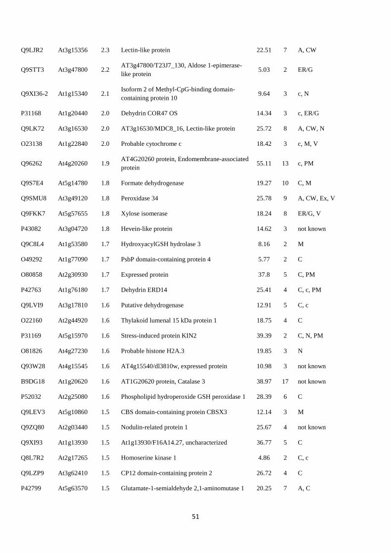
51
Q9LJR2 At3g15356 2.3 Lectin-like protein 22.51 7 A, CW
Q9STT3 At3g47800 2.2 AT3g47800/T23J7_130, Aldose 1-epimerase-like protein
5.03 2 ER/G
Q9XI36-2 At1g15340 2.1 Isoform 2 of Methyl-CpG-binding domain-containing protein 10
9.64 3 c, N
P31168 At1g20440 2.0 Dehydrin COR47 OS 14.34 3 c, ER/G
Q9LK72 At3g16530 2.0 AT3g16530/MDC8_16, Lectin-like protein 25.72 8 A, CW, N
O23138 At1g22840 2.0 Probable cytochrome c 18.42 3 c, M, V
Q96262 At4g20260 1.9 AT4G20260 protein, Endomembrane-associated protein
55.11 13 c, PM
Q9S7E4 At5g14780 1.8 Formate dehydrogenase 19.27 10 C, M
Q9SMU8 At3g49120 1.8 Peroxidase 34 25.78 9 A, CW, Ex, V
Q9FKK7 At5g57655 1.8 Xylose isomerase 18.24 8 ER/G, V
P43082 At3g04720 1.8 Hevein-like protein 14.62 3 not known
Q9C8L4 At1g53580 1.7 HydroxyacylGSH hydrolase 3 8.16 2 M
O49292 At1g77090 1.7 PsbP domain-containing protein 4 5.77 2 C
O80858 At2g30930 1.7 Expressed protein 37.8 5 C, PM
P42763 At1g76180 1.7 Dehydrin ERD14 25.41 4 C, c, PM
Q9LVI9 At3g17810 1.6 Putative dehydrogenase 12.91 5 C, c
O22160 At2g44920 1.6 Thylakoid lumenal 15 kDa protein 1 18.75 4 C
P31169 At5g15970 1.6 Stress-induced protein KIN2 39.39 2 C, N, PM
O81826 At4g27230 1.6 Probable histone H2A.3 19.85 3 N
Q93W28 At4g15545 1.6 AT4g15540/dl3810w, expressed protein 10.98 3 not known
B9DG18 At1g20620 1.6 AT1G20620 protein, Catalase 3 38.97 17 not known
P52032 At2g25080 1.6 Phospholipid hydroperoxide GSH peroxidase 1 28.39 6 C
Q9LEV3 At5g10860 1.5 CBS domain-containing protein CBSX3 12.14 3 M
Q9ZQ80 At2g03440 1.5 Nodulin-related protein 1 25.67 4 not known
Q9XI93 At1g13930 1.5 At1g13930/F16A14.27, uncharacterized 36.77 5 C
Q8L7R2 At2g17265 1.5 Homoserine kinase 1 4.86 2 C, c
Q9LZP9 At3g62410 1.5 CP12 domain-containing protein 2 26.72 4 C
P42799 At5g63570 1.5 Glutamate-1-semialdehyde 2,1-aminomutase 1 20.25 7 A, C

52
O49304 At1g23130 1.5 At1g23130/T26J12_10, Polyketide cyclase/dehydrase and lipid transport-like protein
15 2 not known
Q9SW21 At4g25050 1.5 Acyl carrier protein 20.44 3 C
F4I0N7 At1g75750 1.5 Gibberellin-regulated protein 1 34.02 4 not known
P42761 At2g30870 1.5 GSH S-transferase F10 30.23 6 CW, C, c, ER/G
Q56WK6 At1g72150 1.5 Patellin-1 39.97 25 A, C, c, ER/G, PM, V
Q9LIN0 At3g26450 1.5 Major latex protein, putative 33.55 5 not known
Q8VXZ7 At3g56310 1.5 Alpha-galactosidase 9.15 3 CW, c, V
Q9M2D8 At3g61260 1.5 Uncharacterized protein At3g61260 41.98 7 c, PM, V
O65660 At4g39730 1.5 AT4g39730/T19P19_120, Dehydration stress-induced protein
17.13 3 C, c, PM, V
Q9SVG4-2 At4g20830 1.5 Isoform 2 of Reticuline oxidase-like protein 8.33 5 A, c, CW, Ex, M, PM, V
Q9LW57 At3g23400 1.5 Probable plastid-lipid-associated protein 6 31.69 9 C, N, PM
O80852 At2g30860 1.5 GSH S-transferase F9 40.00 8 A, C, c, PM, V
Q9XFT3-2 At4g21280 1.5 Isoform 2 of Oxygen-evolving enhancer protein 3-1
43.50 10 C
Q42589 At2g38540 1.5 Non-specific lipid-transfer protein 44.07 6 A, C, CW, Ex,
O82291 At2g35490 1.5 Probable plastid-lipid-associated protein 3 9.57 3 C
Table 2.List of proteins whose content is increased in ggt1 mutants. Proteins are listed in
decreasing fold-change order, as defined by the ggt1/Col-0 ratio. The UniProtKB and TAIR
accession ID are provided. Abbreviations: Cov., coverage; Pep., peptide; A, apoplast; CW, cell
wall; C, chloroplast; c, cytosol and/or plasmodesma; ER/G, endoplasmatic reticulum/Golgi
apparatus; Ex, extracellular region; M, mitochondrion; N, nucleus; PM, plasma membrane; and V,
vacuole.
UniProt ID/AC
Locus name
Col-0/ ggt1 ratio
Description %
Cov. #
Pep. Localizationa
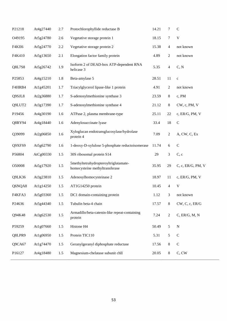
53
P21218 At4g27440 2.7 Protochlorophyllide reductase B 14.21 7 C
O49195 At5g24780 2.6 Vegetative storage protein 1 18.15 7 V
F4KII6 At5g24770 2.2 Vegetative storage protein 2 15.38 4 not known
F4K410 At5g13650 2.1 Elongation factor family protein 4.89 2 not known
Q8L7S8 At5g26742 1.9 Isoform 2 of DEAD-box ATP-dependent RNA helicase 3
5.35 4 C, N
P25853 At4g15210 1.8 Beta-amylase 5 28.51 11 c
F4HRB4 At1g45201 1.7 Triacylglycerol lipase-like 1 protein 4.91 2 not known
Q9SJL8 At2g36880 1.7 S-adenosylmethionine synthase 3 23.59 8 c, PM
Q9LUT2 At3g17390 1.7 S-adenosylmethionine synthase 4 21.12 8 CW, c, PM, V
P19456 At4g30190 1.6 ATPase 2, plasma membrane-type 25.11 22 c, ER/G, PM, V
Q8RY94 At4g18440 1.6 Adenylosuccinate lyase 33.4 18 C
Q39099 At2g06850 1.6 Xyloglucan endotransglucosylase/hydrolase protein 4
7.09 2 A, CW, C, Ex
Q9XFS9 At5g62790 1.6 1-deoxy-D-xylulose 5-phosphate reductoisomerase 11.74 6 C
P56804 AtCg00330 1.5 30S ribosomal protein S14 29 3 C, c
O50008 At5g17920 1.5 5methyltetrahydropteroyltriglutamate-homocysteine methyltransferase
35.95 29 C, c, ER/G, PM, V
Q9LK36 At3g23810 1.5 Adenosylhomocysteinase 2 18.97 11 c, ER/G, PM, V
Q6NQA8 At1g14250 1.5 AT1G14250 protein 10.45 4 V
F4KFA3 At5g03360 1.5 DC1 domain-containing protein 1.12 3 not known
P24636 At5g44340 1.5 Tubulin beta-4 chain 17.57 8 CW, C, c, ER/G
Q94K48 At3g62530 1.5 Armadillo/beta-catenin-like repeat-containing protein
7.24 2 C, ER/G, M, N
P59259 At1g07660 1.5 Histone H4 50.49 5 N
Q8LPR9 At1g06950 1.5 Protein TIC110 5.31 5 C
Q9CA67 At1g74470 1.5 Geranylgeranyl diphosphate reductase 17.56 8 C
P16127 At4g18480 1.5 Magnesium-chelatase subunit chlI 20.05 8 C, CW
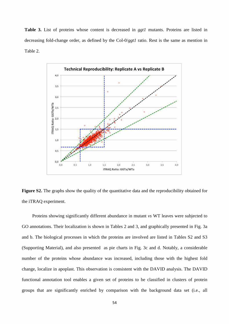
54
Table 3. List of proteins whose content is decreased in ggt1 mutants. Proteins are listed in
decreasing fold-change order, as defined by the Col-0/ggt1 ratio. Rest is the same as mention in
Table 2.
Figure S2. The graphs show the quality of the quantitative data and the reproducibility obtained for
the iTRAQ experiment.
Proteins showing significantly different abundance in mutant vs WT leaves were subjected to
GO annotations. Their localization is shown in Tables 2 and 3, and graphically presented in Fig. 3a
and b. The biological processes in which the proteins are involved are listed in Tables S2 and S3
(Supporting Material), and also presented as pie charts in Fig. 3c and d. Notably, a considerable
number of the proteins whose abundance was increased, including those with the highest fold
change, localize in apoplast. This observation is consistent with the DAVID analysis. The DAVID
functional annotation tool enables a given set of proteins to be classified in clusters of protein
groups that are significantly enriched by comparison with the background data set (i.e., all
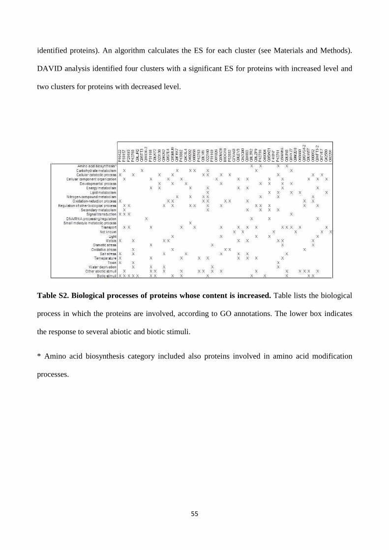
55
identified proteins). An algorithm calculates the ES for each cluster (see Materials and Methods).
DAVID analysis identified four clusters with a significant ES for proteins with increased level and
two clusters for proteins with decreased level.
Table S2. Biological processes of proteins whose content is increased. Table lists the biological
process in which the proteins are involved, according to GO annotations. The lower box indicates
the response to several abiotic and biotic stimuli.
* Amino acid biosynthesis category included also proteins involved in amino acid modification
processes.
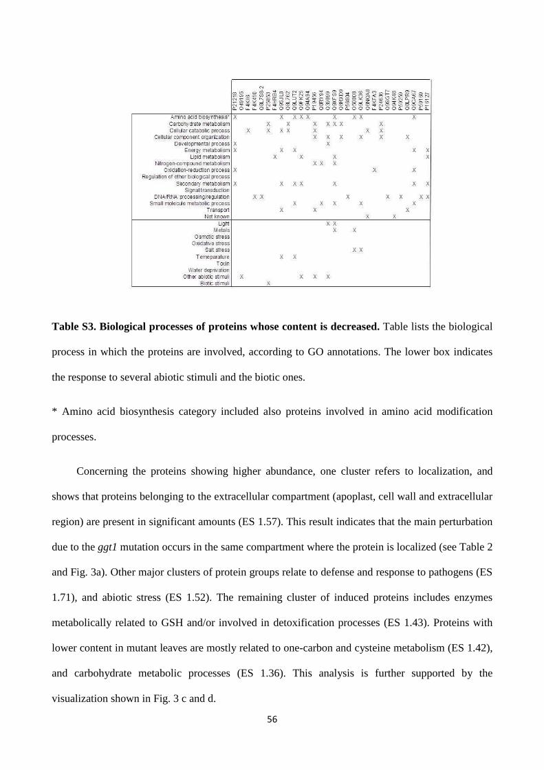
56
Table S3. Biological processes of proteins whose content is decreased. Table lists the biological
process in which the proteins are involved, according to GO annotations. The lower box indicates
the response to several abiotic stimuli and the biotic ones.
* Amino acid biosynthesis category included also proteins involved in amino acid modification
processes.
Concerning the proteins showing higher abundance, one cluster refers to localization, and
shows that proteins belonging to the extracellular compartment (apoplast, cell wall and extracellular
region) are present in significant amounts (ES 1.57). This result indicates that the main perturbation
due to the ggt1 mutation occurs in the same compartment where the protein is localized (see Table 2
and Fig. 3a). Other major clusters of protein groups relate to defense and response to pathogens (ES
1.71), and abiotic stress (ES 1.52). The remaining cluster of induced proteins includes enzymes
metabolically related to GSH and/or involved in detoxification processes (ES 1.43). Proteins with
lower content in mutant leaves are mostly related to one-carbon and cysteine metabolism (ES 1.42),
and carbohydrate metabolic processes (ES 1.36). This analysis is further supported by the
visualization shown in Fig. 3 c and d.
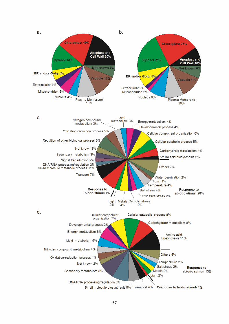
57
58
Figure 3. GO annotation of identified differential proteins. Cellular component (a, b) and functional
(c, d) classification of identified proteins with higher expression (a, c) and lower expression (b, d)
proteins in ggt1 mutant vs WT. Classification was carried out using GO database at UniProtKB
(http://www.uniprot.org/). Percentage values indicate the proportion of total number of proteins
within that category with respect to the total hits. The biological processes in which each protein is
involved are listed in Table S2 and S3 (Supplemental Material).
4. DISCUSSION
As reported in the literature and confirmed here, the ggt1knockout plants exhibit no obvious
phenotype except for early flowering and accelerated senescence. It should be noted that plants
used in this study were at the stage of fully-expanded leaf, before bolting, and had no visible signs
of senescence in the rosette. Therefore, the observed variations cannot be attributed to an age-
dependent or developmental senescent state, but rather to unknown redox signals arising from the
apoplast which trigger plant’s adaptation to environment. The implication of a redox control by the
gamma-glutamyl cycle has been highlighted in a previous work [10], in which exposure to the thiol
oxidizing drug diamide with a concomitant GGT inhibition by serine-borate resulted in net GSH
extrusion from barley roots.
In accordance with that, we show that GSH degradation in apoplast is required for intracellular
redox balance, since disrupting GGT function also results in oxidative damage to some protein
components. Consequently, there is an up-regulation of a number of enzymes involved in
antioxidative and stress response. Proteomics analysis revealed metabolic adjustments in the ggt1
mutants, which in turn affected different compartments and functions.
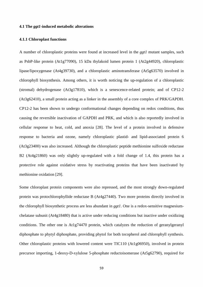
59
4.1 The ggt1-induced metabolic alterations
4.1.1 Chloroplast functions
A number of chloroplastic proteins were found at increased level in the ggt1 mutant samples, such
as PsbP-like protein (At1g77090), 15 kDa thylakoid lumen protein 1 (At2g44920), chloroplastic
lipase/lipoxygenase (At4g39730), and a chloroplastic aminotransferase (At5g63570) involved in
chlorophyll biosynthesis. Among others, it is worth noticing the up-regulation of a chloroplastic
(stromal) dehydrogenase (At3g17810), which is a senescence-related protein; and of CP12-2
(At3g62410), a small protein acting as a linker in the assembly of a core complex of PRK/GAPDH.
CP12-2 has been shown to undergo conformational changes depending on redox conditions, thus
causing the reversible inactivation of GAPDH and PRK, and which is also reportedly involved in
cellular response to heat, cold, and anoxia [28]. The level of a protein involved in defensive
response to bacteria and ozone, namely chloroplastic plastid- and lipid-associated protein 6
(At3g23400) was also increased. Although the chloroplastic peptide methionine sulfoxide reductase
B2 (At4g21860) was only slightly up-regulated with a fold change of 1.4, this protein has a
protective role against oxidative stress by reactivating proteins that have been inactivated by
methionine oxidation [29].
Some chloroplast protein components were also repressed, and the most strongly down-regulated
protein was protochlorophyllide reductase B (At4g27440). Two more proteins directly involved in
the chlorophyll biosynthetic process are less abundant in ggt1. One is a redox-sensitive magnesium-
chelatase subunit (At4g18480) that is active under reducing conditions but inactive under oxidizing
conditions. The other one is At1g74470 protein, which catalyzes the reduction of geranylgeranyl
diphosphate to phytyl diphosphate, providing phytol for both tocopherol and chlorophyll synthesis.
Other chloroplastic proteins with lowered content were TIC110 (At1g06950), involved in protein
precursor importing, 1-deoxy-D-xylulose 5-phosphate reductoisomerase (At5g62790), required for
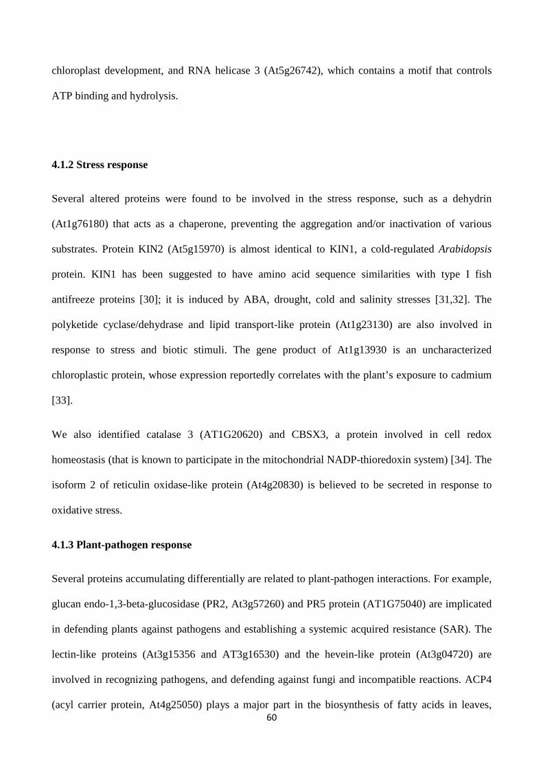
60
chloroplast development, and RNA helicase 3 (At5g26742), which contains a motif that controls
ATP binding and hydrolysis.
4.1.2 Stress response
Several altered proteins were found to be involved in the stress response, such as a dehydrin
(At1g76180) that acts as a chaperone, preventing the aggregation and/or inactivation of various
substrates. Protein KIN2 (At5g15970) is almost identical to KIN1, a cold-regulated Arabidopsis
protein. KIN1 has been suggested to have amino acid sequence similarities with type I fish
antifreeze proteins [30]; it is induced by ABA, drought, cold and salinity stresses [31,32]. The
polyketide cyclase/dehydrase and lipid transport-like protein (At1g23130) are also involved in
response to stress and biotic stimuli. The gene product of At1g13930 is an uncharacterized
chloroplastic protein, whose expression reportedly correlates with the plant’s exposure to cadmium
[33].
We also identified catalase 3 (AT1G20620) and CBSX3, a protein involved in cell redox
homeostasis (that is known to participate in the mitochondrial NADP-thioredoxin system) [34]. The
isoform 2 of reticulin oxidase-like protein (At4g20830) is believed to be secreted in response to
oxidative stress.
4.1.3 Plant-pathogen response
Several proteins accumulating differentially are related to plant-pathogen interactions. For example,
glucan endo-1,3-beta-glucosidase (PR2, At3g57260) and PR5 protein (AT1G75040) are implicated
in defending plants against pathogens and establishing a systemic acquired resistance (SAR). The
lectin-like proteins (At3g15356 and AT3g16530) and the hevein-like protein (At3g04720) are
involved in recognizing pathogens, and defending against fungi and incompatible reactions. ACP4
(acyl carrier protein, At4g25050) plays a major part in the biosynthesis of fatty acids in leaves,
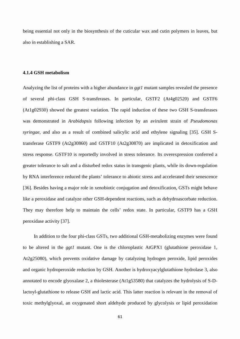
61
being essential not only in the biosynthesis of the cuticular wax and cutin polymers in leaves, but
also in establishing a SAR.
4.1.4 GSH metabolism
Analyzing the list of proteins with a higher abundance in ggt1 mutant samples revealed the presence
of several phi-class GSH S-transferases. In particular, GSTF2 (At4g02520) and GSTF6
(At1g02930) showed the greatest variation. The rapid induction of these two GSH S-transferases
was demonstrated in Arabidopsis following infection by an avirulent strain of Pseudomonas
syringae, and also as a result of combined salicylic acid and ethylene signaling [35]. GSH S-
transferase GSTF9 (At2g30860) and GSTF10 (At2g30870) are implicated in detoxification and
stress response. GSTF10 is reportedly involved in stress tolerance. Its overexpression conferred a
greater tolerance to salt and a disturbed redox status in transgenic plants, while its down-regulation
by RNA interference reduced the plants’ tolerance to abiotic stress and accelerated their senescence
[36]. Besides having a major role in xenobiotic conjugation and detoxification, GSTs might behave
like a peroxidase and catalyze other GSH-dependent reactions, such as dehydroascorbate reduction.
They may therefore help to maintain the cells’ redox state. In particular, GSTF9 has a GSH
peroxidase activity [37].
In addition to the four phi-class GSTs, two additional GSH-metabolizing enzymes were found
to be altered in the ggt1 mutant. One is the chloroplastic AtGPX1 (glutathione peroxidase 1,
At2g25080), which prevents oxidative damage by catalyzing hydrogen peroxide, lipid peroxides
and organic hydroperoxide reduction by GSH. Another is hydroxyacylglutathione hydrolase 3, also
annotated to encode glyoxalase 2, a thiolesterase (At1g53580) that catalyzes the hydrolysis of S-D-
lactoyl-glutathione to release GSH and lactic acid. This latter reaction is relevant in the removal of
toxic methylglyoxal, an oxygenated short aldehyde produced by glycolysis or lipid peroxidation
62
that accumulates in plants under environmental stresses [38]. This enzyme is located in
mitochondria, where immunocytochemical analysis reveals an increased GSH content (Fig 1).
The level of several proteins involved in cys and met biosynthesis was lower in ggt1 mutant
samples (Fig. 3d, Table S3), suggesting a slower rate of sulfur amino acid biosynthesis. This
evidence may correlate with an increase in the extracellular GSH caused by GGT mutation, acting
as a negative signal for the sulfur assimilation pathway [39,40].
4.1.5 Thiol redox homeostasis
Other proteins metabolically related to GSH and also involved in redox homeostasis are
peroxiredoxins (PRX), glutaredoxins (GRX) and thioredoxins (TRX). While several of the proteins
were identified in the MS/MS analysis, none of these components are included in Table 2 because
the relative change in their expression fell below the established threshold fold change. However, a
better look at the full list (Supplemental Table S4) shows some variations that may be relevant.
While adopting as a threshold a fold change of 1.5 represents a trade-off enabling a better screening
of statistically significant variations, from a biological perspective it may underestimate the effect
of variations in proteins with a higher coefficient of flux control over metabolism, such as
regulatory enzymes.
The fold change variation in the GRX, PRX and TRX is shown in Fig. 4. All six PRX were
up-regulated (1.2-1.4 folds) as were the eight TRX (see Supplemental Table S4), but only one of the
three GRX showed a comparable up-regulation. Together with GSH, these proteins participate in
redox homeostasis; GSH has been shown to be the preferred electron donor for PRX [41]. The
redox potentials determine whether enzymes are more reducing or more oxidizing, and differ
significantly among the various enzymes. In general, however, TRX are better disulfide bond
reducers than GRX [42].
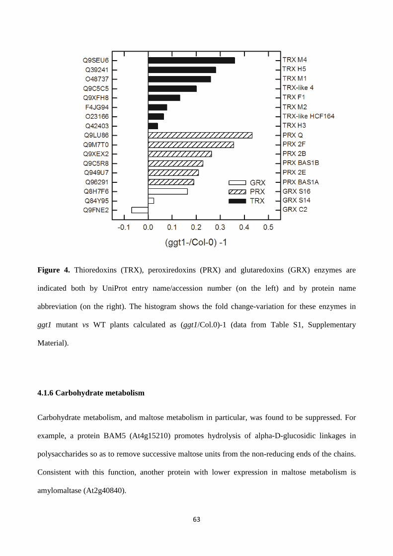
63
Figure 4. Thioredoxins (TRX), peroxiredoxins (PRX) and glutaredoxins (GRX) enzymes are
indicated both by UniProt entry name/accession number (on the left) and by protein name
abbreviation (on the right). The histogram shows the fold change-variation for these enzymes in
ggt1 mutant vs WT plants calculated as (ggt1/Col.0)-1 (data from Table S1, Supplementary
Material).
4.1.6 Carbohydrate metabolism
Carbohydrate metabolism, and maltose metabolism in particular, was found to be suppressed. For
example, a protein BAM5 (At4g15210) promotes hydrolysis of alpha-D-glucosidic linkages in
polysaccharides so as to remove successive maltose units from the non-reducing ends of the chains.
Consistent with this function, another protein with lower expression in maltose metabolism is
amylomaltase (At2g40840).
64
4.1.7 Others
Among the proteins whose level was increased in ggt1 mutants, we identified a nuclear MBD10
(At1g15340). This protein is probably a transcription regulator with a potential role in controlling
chromatin structure mediated by CpG methylation. Hormonal disturbances might explain the
alteration of GASA1 (At1g75750), a gibberellin-regulated protein. The accumulation of cytosolic
aldose 1-epimerase (At3g47800), a component of exose metabolism, cytochrome c (At1g22840) in
the mitochondrial respiratory chain, and mitochondrial formate dehydrogenase, suggests that energy
metabolism and primary processes are also affected. Formate dehydrogenase, which may also use
formyl-GSH as a substrate [43], was reportedly induced by stress [44] and ROS [45]. Formate
derived from the detoxification of formaldehyde generated by glycine decarboxylase under light can
be oxidized to generate NADH in the mitochondrial matrix.
4.2 Alteration of redox homeostasis in the ggt1 mutant: similarities with senescence, stress and
antioxidant response
Our results point to a metabolic scenario having some similarities with senescence. For example,
the strong suppression of chlorophyll biosynthesis can be gauged by the down-regulation of
protochlorophyllide reductase B. On the other hand RuBisCO expression is typically depressed in
senescence, but it was unaffected in this study. This view is further confirmed by other
observations. Comparing our protein expression list with a previous large-scale study on
senescence-associated genes [46] it was found that only some of the proteins whose content is
increased in our study (xylose isomerase, glyoxalase II, catalase 3, and the CBSX3 of unknown
function) are perhaps associated with senescence. The level of vegetative storage proteins VSP1
(At5g24780) and VSP2 (At5g24770), two sequentially expressed genes associated with senescence,
and thought to serve as a storage buffer between nitrogen losses from senescing leaves and during
grain filling [47] - strongly decreased in leaves of the ggt1 mutant. We argue that energy is needed
65
to cope with the stressful conditions induced by the ggt1 mutation, and this is diverted from storage
processes.
One more difference relates to the senescence-associated genes (SAGs). SAG2 and SAG12 have
been found to be associated with senescence [48]. In our study, SAG2 (thiol protease aleurain,
At5g60360) expression was higher, but only with a fold change of 1.3 (see supplemental list S1 and
S4), which meant that it was not included in Table 2. Surprisingly, we did not observe a change of
SAG12 (At5g45890), which is considered to be a senescence-induced marker [49]. In another
study, SAG12 was not induced by oxidative stress conditions, which is capable of triggering the
expression of other senescence-associated genes [50]. These findings point to the onset of a
complex array of metabolic adjustments in leaves of the ggt1 mutant, as seen during senescence,
and in response to biotic and abiotic stimuli, as well as other stress factors.
4.2.1 Hydrogen peroxide homeostasis in apoplast
The deployment of defense systems in the ggt1 mutants is a consequence of altered GSH
degradative metabolism in the apoplast. While GSH content in the apoplast is low (compared with
ascorbate) [51,52], our study evidenced an higher content of GSH in ggt1 mutant apoplast (see
Table 1).
The accumulation of apoplastic hydrogen peroxide as a result of ROS generation by plasma
membrane NADPH oxidases or cell wall peroxidases is a known response to various stress stimuli
and is assumed to be involved in the signaling networks leading to transcriptional reprogramming
[53]. It has been shown that ROS-generating peroxidases PRX33 and PRX34 are responsible for the
oxidative burst [50] and play an important role in Arabidopsis resistance to pathogens [54]. In the
ggt1 mutant PRX34 (At3g49120) is more abundant (1.8 fold compared with the WT), suggesting
that apoplastic glutathione, or some molecules associated with the GGT reaction, may regulate cell
wall peroxidase expression and H2O2 formation; as such, they may therefore be considered as actors
66
in apoplastic redox signalling. Thiols are candidate molecules for signal transduction because they
engage several redox reactions with target molecules [55]. It is tempting to speculate that this signal
molecule might be GSH itself, accumulating in the apoplast when GGT is disrupted. Alternatively,
it could be cys or cys-gly, downstream from the degradation process. In this regard, cys-gly is
highly reactive and highly liable to oxidation; it seems a good candidate in redox signaling, which
could interact with plasma membrane redox sensitive thiol cysteines [56,57].
4.3 Concluding remarks
A feature common to the above mentioned responses in the ggt1 mutant could be redox alterations.
Cellular redox equilibrium is maintained by a network of redox proteins and redox couples, such as
NAD and NADP, along with the antioxidant molecules ascorbate and GSH, acting synergistically
with other signaling molecules, such as salicylic acid [58,59]. The ascorbate-glutathione cycle is a
key part of the network of reactions involving enzymes and metabolites with redox properties for
the detoxification of ROS. The loss of function due to the Arabidopsis GGT1 knockout mutation
triggers highly specific changes in compartmental interplay of ascorbate and GSH in cells. These
changes accompanied remarkable rearrangements in proteome, which resemble those induced under
specific abiotic and biotic stresses. To put it simply, a sort of “alert response” is activated in ggt1
mutant leaves even in the absence of a real environmental threat, suggesting a failure in correct
redox sensing. In line with this observation, further experiments will be carried out to describe the
ggt1 mutant adaptive response under oxidative conditions, which might result in a more severe
phenotype.
Redox reactions in the extracellular space can accomplish several functions. They can participate in
building the extracellular matrix or control redox sensitive enzymes. The latter function helps cells
to adapt cell metabolism (in order to produce energy, build molecules, and defend themselves
against oxidative damage) to events occurring in the outside environment, where unfavorable
conditions may give rise to ROS with an altered redox balance and potentially detrimental effects
67
on plant functions and growth. Signals arising from the apoplast thus have a key role in plant’s
adaptation to environment [53].
Investigating the signal transduction pathway linking extracellular GSH degradation to the
intracellular redox balance is an important goal of future research with a view to finding the key
genes/proteins involved in the cross talk between the different stress responses, and to identifying
candidates for future manipulation to improve plants’ stress resistance.
ACKNOWLEDGMENTS
The authors wish to thank Prof. Massimo Ferretti (Pousada Horizonte Azul, Boipeba, Brazil) for
his valuable comments and advice, and also the Cassa di Risparmio di Padova e Rovigo (Cariparo)
for financing the purchase of the LTQ-Orbitrap XL mass spectrometer. This work was funded by the
MURST ex-60%. B.Z. was supported by the Austrian Science Fund (FWF P22988). ); G.A. was
supported by a grant from University of Padova (CPDA082784/08). The authors have no conflict
of interest to declare.
68
5. REFERENCES
[1] Scandalios, J. G., Oxidative stress and the molecular biology of antioxidant defenses, Cold
Spring Harbor Laboratory Press, Plainview, NY, USA 1997.
[2] Noctor, G., Foyer, C. H., Simultaneous measurement of foliar glutathione, gamma-
glutamylcysteine, and amino acids by high-performance liquid chromatography: comparison with
two other assay methods for glutathione. Anal. Biochem. 1998, 1, 98-110.
[3] Asada, K.,The water–water cycle in chloroplasts: scavenging of active oxygen and dissipation of
excess photons. Ann. Rev. Plant Physiol. Plant Mol. Biol. 1999, 50, 601-639.
[4] Tate, S. S., Meister A., Serine-borate complex as a transition state inhibitor of �-glutamyl
transpeptidase. Proc. Natl. Acad. Sci.USA 1978, 75, 4806-4809.
[5] Storozhenko, S., Belles-Boix, E., Babiychuk, E., Hérouart, D., et al., �-Glutamyl transpeptidase
in transgenic tobacco plants. Cellular localization, processing, and biochemical properties. Plant
Physiol. 2002, 128, 1109-1119.
[6] Grzam, A., Martin, M. N., Hell, R., Meyer, A. J., γ-Glutamyl transpeptidase GGT4 initiates
vacuolar degradation of glutathione S-conjugates in Arabidopsis. FEBS Lett. 2007, 581, 3131-3138.
[7] Martin, M. N., Saladores, P. H., Lambert, E., Hudson, A. O., Leustek, T., Localization of
members of the �-glutamyl transpeptidase family identifies sites of glutathione and glutathione S-
conjugate hydrolysis. Plant Physiol. 2007, 144, 1715-1732.
[8] Ohkama-Ohtsu, N., Radwan, S., Peterson, A., Zhao, P., et al., Characterization of the
extracellular �-glutamyl transpeptidases, GGT1 and GGT2, in Arabidopsis. Plant J. 2007a, 49,
865-877.
69
[9] Ohkama-Ohtsu, N., Zhao, P., Xiang, C., Oliver, D. J., Glutathione-conjugates in the vacuole are
degraded by γ-glutamyl transpeptidase GGT3 in Arabidopsis. Plant J. 2007b, 49, 878-888.
[10] Ferretti, M., Destro, T., Tosatto, S. C., La Rocca, N., et al., Gamma-glutamyl transferase in the
cell wall participates in extracellular glutathione salvage from the root apoplast. New Phytol. 2009,
181, 115-126.
[11] Meister, A., Tate, S. S., Griffith, O. W., Gamma-glutamyl transpeptidase. Methods Enzymol.
1981, 77, 237-253.
[12] Will, Y., Fischer, K. A., Horton, R. A., Kaetzel, R. S., Brown, M. K., et al., Gamma-
glutamyltranspeptidase-deficient knockout mice as a model to study the relationship between
glutathione status, mitochondrial function, and cellular function. Hepatology 2000, 32, 740-749.
[13] Masi, A., Ghisi, R., Ferretti, M., Measuring low-molecular-weight thiols by detecting the
fluorescence of their SBD derivatives: application to studies of diurnal and UV-B induced changes
in Zea mays L. Plant Physiol. 2002, 159, 499-507.
[14] Kushnir, S., Babiychuk, E., Kampfenkel, K., Belles-Boix, E., et al., Characterization of
Arabidopsis thaliana cDNAs that render yeasts tolerant toward the thiol-oxidizing drug diamide.
Proc. Natl. Acad. Sci.USA 1995, 92, 10580-10584.
[15] Destro, T., Prasad, D., Martignago, D., Bernet, I. L., et al., Compensatory expression and
substrate inducibility of gamma-glutamyl transferase GGT2 isoform in Arabidopsis thaliana. J.
Exp. Bot. 2011, 62, 805-814.
[16] Levitt, J., Responses of plants to environmental stresses, New York: Academic Press, 1972.
[17] Alonso, J. M., Stepanova, A. N., Leisse, T. J., Kim, C. J. et al., Genomewide insertional
mutagenesis of Arabidopsis thaliana. Science 2003, 301, 653-657.
70
[18] Zechmann, B., Müller, M., Subcellular compartmentation of glutathione in dicotyledonous
plants. Protoplasma. 2010, 246, 15-24.
[19] Zechmann, B., Stumpe, M., Mauch, F., Immunocytochemical determination of the subcellular
distribution of ascorbate in plants. Planta. 2011, 233, 1-12.
[20] Polle, A., Chakrabarti, K., Schuermann, W., Rennenberg, H., Composition and properties of
hydrogen peroxide decomposing systems in extracellular and total extracts from needles of Norway
spruce (Picea abies L., Karst). Plant Physiol. 1990, 94, 312-319.
[21] Hewitt, E. J., Dickes, G. J., Spectrophotometric measurements on ascorbic acid and their use
for the estimation of ascorbic acid and dehydroascorbic acid in plant tissues. Biochem. J. 1961, 78,
384.
[22] Ross, P. L., Huang, Y. N., Marchese, J. N., Williamson, B., et al., Multiplexed Protein
Quantitation in Saccharomyces cerevisiae Using Amine-reactive Isobaric Tagging Reagents. Mol.
Cell. Proteomics 2004, 3, 1154-1169.
[23] Köcher, T., Pichler, P., Schutzbier, M., Stingl, C. et al., High precision quantitative proteomics
using iTRAQ on an LTQ Orbitrap: a new mass spectrometric method combining the benefits of all.
J. Proteome Res. 2009, 8, 4743-4752.
[24] Dennis, G., Sherman, B. T., Hosack, D. A., Yang, et al., DAVID: Database for Annotation,
Visualization, and Integrated Discovery. Genome Biol. 2003, 4, P3.
[25] Huang, W., Sherman, B. T., Lempicki, R. A., Systematic and integrative analysis of large gene
lists using DAVID bioinformatics resources. Nat. Protoc. 2009, 4, 44-57.
[26] Nystrom, T., Role of oxidative carbonylation in protein quality control and senescence. EMBO
J. 2005, 24, 1311-1317.
71
[27] Moller, I. M., Jensen, P. E., Hansson, A., Oxidative modifications to cellular components in
plants. Annu. Rev. Plant. Biol. 2007, 58, 459-481.
[28] Singh P., Kaloudas D., Raines, C. A., Expression analysis of the Arabidopsis CP12 gene
family suggest novel roles for these proteins in roots and floral tissues. J. Exp. Bot. 2008, 59, 3975-
3985.
[29] Li C. W., Lee S.-H., Chieh P.-S., Lin C.-S., et al., Arabidopsis Root-Abundant Cytosolic
Methionine Sulfoxide Reductase B Genes MsrB7 and MsrB8 are Involved in Tolerance to
Oxidative Stress. Plant Cell Physiol. 2012, 53, 1707-1719.
[30] Kurkela, S., Franck, M., Cloning and characterization of a cold- and ABA- inducible
Arabidopsis gene. Plant. Mol. Biol. 1990, 15, 137-144.
[31] Gilmour, S. J., Artus, N. N., Thomashow, M. F., cDNA sequence analysis and expression of
two cold-regulated genes of Arabidopsis thaliana.Plant Mol Biol. 1992, 18, 13-21.
[32] Kurkela, S., Borg-Frank, M., Structure and expression of kin2, one of two cold- and ABA-
induced genes of Arabidopsis thaliana. Plant Mol. Biol. 1992, 19, 689-692.
[33] Sarry, J. E., Kuhn, L., Ducruix, C., Lafaye, A., et al., The early responses of Arabidopsis
thaliana cells to cadmium exposure explored by protein and metabolite profiling analyses.
Proteomics 2006, 6, 2180-98.
[34] Yoo, K. S., Ok, S. H., Jeong, B. C., Jung, K. W., et al., Single cystathionine β-synthase
domain-containing proteins modulate development by regulating the thioredoxin system in
Arabidopsis. Plant Cell 2011, 23, 3577-94.
[35] Lieberherr, D., Wagner, U., Dubuis, P. H., Métraux, J. P., Mauch, F., The Rapid Induction of
Glutathione S-Transferases AtGSTF2 and AtGSTF6 by Avirulent Pseudomonas syringae is the
Result of Combined Salicylic Acid and Ethylene Signaling. Plant Cell. Physiol. 2003, 44, 750-757.
72
[36] Ryu, H. Y., Kim, S. Y., Park, H. M. You, J. Y. et al., Modulations of AtGSTF10 expression
induce stress tolerance and BAK1-mediated cell death. Biochem. Biophys. Res. Commun. 2009,
379, 417-422.
[37] Nutricati, E., Miceli, A., Blando, F., De Bellis, L., Characterization of two Arabidopsis
thaliana glutathione S-transferase. Plant. Cell. Rep. 2006, 997-1005.
[38] Hoque, T. S., Uraji, M., Tuya, A., Nakamura, Y., Murata, Y., Methylglyoxal inhibits seed
germination and root elongation and up-regulates transcription of stress-responsive genes in ABA-
dependent pathway in Arabidopsis. Plant Biol. 2012, 5, 854-858.
[39] Lappartinent, A.G., Touraine, B., Demand-driven control of root ATP-sulfurylase activity and
sulfate uptake in intact canola. Plant Physiol. 1996, 177, 147-157.
[40] Lappartient, A. G., Vidmar, J. J., Leustek, T., Glass, A. D., Touraine, B., Inter-organ signaling
in plants: regulation of ATP sulfurylase and sulfate transporter genes expression in roots mediated
by phloem-translocated compound.Plant J. 1999, 18, 89-95.
[41] Finkemeier, I., Goodman, M., Lamkemeyer, P., Kandlbinder, A., et al., The mitochondrial type
II peroxiredoxin F is essential for redox homeostasis and root growth of Arabidopsis thaliana under
stress. J. Biol. Chem. 2005, 280, 12168-12180.
[42] Carmel-Harel O, Storz G., Roles of the glutathione- and thioredoxin-dependent reduction
systems in the Escherichia coli and saccharomyces cerevisiae responses to oxidative stress. Annu.
Rev. Microbiol. 2000, 54, 439-61.
[43] Uotila, L., Koivusalo, M., Purification of formaldehyde and formate dehydrogenases from pea
seeds by affinity chromatography and S-formylglutathione as the intermediate of formaldehyde
metabolism. Arch. Biochem. Biophys. 1979, 196, 33-45.
73
[44] Hourton-Cabassa C., Ambard-Bretteville F., Moreau F., Davy de Virville J., et al., Stress
induction of mitochondrial formate dehydrogenase in potato leaves. Plant Physiol. 1998, 116, 627-
635.
[45] Rhoads, D. M., Umbach, A. L., Subbaiah, C. C., Siedow, J. N., Mitochondrial reactive oxygen
species. Contribution to oxidative stress and interorganellar signaling. Plant Physiol. 2006, 141,
357-366.
[46] Gepstein, S., Sabehi, S., Carp, M. J., Hajouj, T., et al., Large-scale identification of leaf
senescence-associated genes. Plant J. 2003, 36, 629-642.
[47] Rossato L, MacDuff JH, Laine P, Le Deunff E, Ourry A., Nitrogen storage and remobilization
in Brassica napus L. during the growth cycle: effects of methyl jasmonate on nitrate uptake,
senescence, growth, and VSP accumulation.J. Exp. Bot. 2002, 53, 1131-41.
[48] Grbic, V., SAG2 and SAG12 protein expression in senescing Arabidopsis plants. Phys. Plant.
2003, 119, 263-269.
[49] Navabpour S., Morris K., Allen R., Harrison E., et al., Expression of senescence-enhanced
genes in response to oxidative stress. J. Exp. Bot. 2003, 54, 2285–2292.
[50] Bindschedler L.V., Dewdney J., Blee K.A., Stone J.M., et al., Peroxidase-dependent apoplastic
oxidative burst in Arabidopsis required for pathogen resistance. Plant. J. 2006, 47, 851–863.
[51] Luwe, M., Antioxidants in the apoplast and synplast of beech (Fagus sylvatica L.) leaves:
seasonal variations and responses to changing ozone concentrations in air. Plant Cell Environ.
1996, 19, 321-328.
[52] Pignocchi, C., Foyer, C. H., Apoplastic ascorbate metabolism and its role in the regulation of
cell signalling. Curr. Opin. Plant Biol. 2003, 6, 379-389.
74
[53] Shapiguzov A., Vainonen J.P., Wrzaczek M., Kangasjärvi J., ROS-talk - how the apoplast, the
chloroplast, and the nucleus get the message through. Front. Plant Sci. 2012, 3, 292.
[54] Daudi A, Cheng Z, O'Brien JA, Mammarella N, et al., The apoplastic oxidative burst
peroxidase in Arabidopsis is a major component of pattern-triggered immunity. Plant Cell 2012, 24,
275-87.
[55] Noctor G., Mhamdi A., Chaouch S., Han Y., Neukermans J., Marquez-Garcia B., et al.,
Glutathione in plants: an integrated overview.Plant Cell Environ. 2012,35, 454-484.
[56] Shiu S. H., Bleecker A. B., Expansion of the receptor-like kinase/Pelle gene family and
receptor-like proteins in Arabidopsis. Plant Physiol. 2003, 132, 530-543.
[57] Wrzaczek M., Brosché M., Salojärvi J., Kangasjärvi S., et al., Transcriptional regulation of the
CRK/DUF26 group of Receptor-like protein kinases by ozone and plant hormones in Arabidopsis.
BMC Plant Biol. 2010, 10, 95.
[58] Han Y., Chaouch S., Mhamdi A., Queval G., et al., Functional Analysis of Arabidopsis
Mutants Points to Novel Roles for Glutathione in Coupling H(2)O(2) to Activation of Salicylic
Acid Accumulation and Signaling. Antioxid Redox Signal. 2013, (in press).
doi:10.1089/ars.2012.5052.
[59] Foyer C.H., Noctor G., Redox Signaling in Plants. Antioxidants & Redox Signaling, 2013. (in
press) doi:10.1089/ars.2013.5278.

75
CHAPTER 2: Trentin Anna Rita , Pivato Micaela, Mehdi Syed Muhammad Muntazir, Barnabas Leonard Ebinezer, Giaretta Sabrina, Fabrega-Prats Marta, Prasad Dinesh, Arrigoni Giorgio, Masi Antonio PROTEOME READJUSTMENTS IN THE APOPLASTIC SPACE OF ARABIDOPSISTHALIANAGGT1 MUTANT
LEAVES EXPOSED TO UV-B RADIATION
Front Plant Sci. 2015 Mar 24;6:128. doi: 10.3389/fpls.2015.00128
76
Proteome readjustments in the apoplastic space of Arabidopsisthalianaggt1 mutant leaves exposed to UV-B radiation
Trentin Anna Rita1, Pivato Micaela1,2, Mehdi Syed Muhammad Muntazir1, Barnabas Leonard Ebinezer4, Giaretta Sabrina1, Fabrega-Prats Marta1, Prasad Dinesh1,3, Arrigoni Giorgio2,5, Masi Antonio1*. 1Department of DAFNAE, University of Padova (Italy) 2Proteomics Center of Padova University (Italy) 3Department of Bio-Engineering, Birla Institute of Technology (India) 4Sugarcane Breeding Institute Coimbatore (India) 5Department of Biomedical Sciences, University of Padova (Italy) Correspondence: Dr. Antonio Masi University of Padova Dept. DAFNAE Viale Universita' 16, Legnaro (PD), 35020, Italy [email protected]
77
ABSTRACT
Ultraviolet-B radiation acts as an environmental stimulus, but in high doses it has detrimental
effects on plant metabolism. Plasma membranes represent a major target for ROS generated by this
harmful radiation. Oxidative reactions occurring in the apoplastic space are counteracted by
antioxidative systems mainly involving ascorbate and, to some extent, glutathione. The occurrence
of the latter and its exact role in the extracellular space are not well documented, however. In
Arabidopsis thaliana, the gamma-glutamyl transferase isoform GGT1 bound to the cell wall takes
part in the so-called gamma-glutamyl cycle for extracellular glutathione degradation and recovery,
and may be implicated in redox sensing and balance.
In this work, oxidative conditions were imposed with UV-B and studied in redox altered ggt1
mutants. The response of ggt1 knockout Arabidopsis leaves to UV-B radiation was assessed by
investigating changes in extracellular glutathione and ascorbate content and their redox state, and in
apoplastic protein composition. Our results show that, on UV-B exposure, soluble antioxidants
respond to the oxidative conditions in both genotypes. Rearrangements occur in their apoplastic
protein composition, suggesting an involvement of H2O2, which may ultimately act as a signal.
Other important changes relating to hormonal effects, cell wall remodeling, and redox activities are
discussed. We argue that oxidative stress conditions imposed by UV-B and disruption of the
gamma-glutamyl cycle result in similar stress-induced responses, to some degree at least.
Keywords: glutathione, gamma-glutamyl-transferase, oxidative stress, iTRAQ labelling, apoplast,
ultraviolet-B radiation
Abbreviations: UV-B Ultraviolet-B radiation, ROS Reactive Oxygen Species, GGT1 gamma-
glutamyl transferase 1 isoform, H2O2Hydrogen Peroxide, iTRAQ Isobaric tags for relative and
absolute quantification, UVR8UV-B photoreceptor 8, GSHGlutathione, LC-MS-MS Liquid
Chromatography Mass Spectrometry, ECWF Extracellular washing fluid,DHADehydroascorbate,
SBD-F4-fluoro-7-sulfobenzofurazan ammonium salt fluorophore, LMWLow Molecular Weight,
HPLCHighpressure liquid chromatography, ANOVAAnalysis of variance, GLMGeneral linear
models, SDSSodium dodecyl sulfate, TEAB Triethyl ammonium bicarbonate, FDR False discovery
rates, FW Formula Weight, GSSGGlutathione disulfide / Oxidized GSH, SOD Superoxide
dismutase, PNPs Plant natriuretic peptides, GSTsGlutathione S-Transferases,GHsGlycosyl
hydrolases, PRPsPathogenesis-related proteins.
78
INTRODUCTION
The apoplast - i.e. the extraprotoplastic matrix of plant cells, including the cell wall - contains a
number of enzymatic and non-enzymatic components involved in many physiological processes and
is therefore important in the plant cell’s response to both abiotic and biotic stress (Dietz, 1997;
Agrawal et al., 2010). Being at the interface with the external environment, rapid fluctuations occur
in this compartment as a consequence of unfavorable conditions, such as salinity (Hernandez et al.,
2001), ozone (Jaspers et al., 2005) drought (Hu et al., 2005), and UV-B radiation (Pristov et al.,
2013), with consequent changes in the concentrations and redox state of its components.
Ultraviolet-B radiation (UV-B, 280-315 nm) is a component of the solar electromagnetic spectrum
reaching the Earth’s surface, which has gained attention in recent years because it has increased as a
consequence of ozone layer destruction by anthropogenic emissions.
As a component of the solar radiation reaching the leaf, UV-B also acts as an environmental
stimulus for plant growth and development. Recent literature has demonstrated the existence of the
UV-B photoreceptor 8 (UVR8), which controls the plant’s photomorphogenic response to UV-B
radiation. UVR8 promotes a signal cascade that mediates UV-B photomorphogenic responses in
order to secure plant acclimation and survival in sunlight (Rizzini et al., 2011).
While it is beneficial at low intensities (Hideget al., 2013), numerous studies have reported that
excess UV-B radiation harms plants by causing oxidative damage to cellular targets (Brosche and
Strid 2003), altering the structure and functions of the leaf epidermis, cell wall and membranes
(Pristov et al., 2013). A common consequence of many types of environmental stress in plants is a
greater abundance of some reactive oxygen species (ROS), such as superoxide, hydrogen peroxide,
hydroxyl radicals and singlet oxygen (Li and Van Staden, 1998). Increases in ROS are seen after
UV-B exposure too (Noctor et al., 2014), and result in lipid peroxidations and damage to plasma
membranes. To prevent these detrimental effects, plant cells deploy an array of non-enzymatic and
enzymatic antioxidant systems that act as biochemical barriers to counteract and deactivate ROS.
This complex interplay of several metabolites, enzymes, ROS, antioxidants and hormones gives rise
to signals that are transferred inside the cell through the plasma membrane to activate adaptive and
response mechanisms.
A major line of defense in the apoplast is represented by the antioxidant molecule ascorbate and, to
a lesser extent, glutathione. While both are involved intracellularly in the Halliwell-Asada pathway
for controlling ROS and thereby maintaining the cellular redox state and protecting the cellular
components from oxidative threat (Smirnoff and Pallanca, 1996), (Schafer and Buettner, 2001)
(Potters et al., 2010) (Saruhan et al., 2009), only ascorbate occurs in high micromolar, or even
millimolar quantities in the apoplast (Potters et al., 2010), where it can play a part in redox control.
79
The role of extracellular glutathione in the apoplastic space is controversial because it can only be
found in traces under physiological conditions, but it can rise to 2% of the total leaf glutathione
under pathogen attack (Vanacker et al., 1998a).
There have been reports, however, of the extracellular enzyme gamma-glutamyl-transferase (GGT;
E.C. 2.3.2.2) degrading GSH (Martin et al.,2007), which means that, like animals (Meister and
Anderson, 1983), plants also have a gamma-glutamyl cycle involving intracellular glutathione
biosynthesis, extrusion and extracellular degradation, with recovery of the constituent amino acids
(Ferretti et al., 2009).
These findings can explain the low levels of glutathione in the extracellular environment on the one
hand, but also raise the question of the significance of a gamma-glutamyl cycle in plants. In barley
roots, using GGT inhibitors in association with the thiol oxidizing molecule diamide resulted in a
net glutathione extrusion and accumulation in the extracellular medium (Ferretti et al., 2009). This
leads us to wonder whether a gamma-glutamyl cycle could operate as a redox sensing or redox
balancing system.
Another study (Tolin et al., 2013) characterized the leaf proteome of Arabidopsisthalianaggt1
mutant lines and showed that, even under physiological conditions, a number of antioxidant and
defense enzymes were significantly upregulated as a result of impaired extracellular GGT activity.
This also implies that GSH turnover involving apoplastic GSH degradation is needed for proper
redox sensing and/or a coordinated response to the environment. We speculated that a feedback
signal might be missing when the GGT cycle is disrupted, and this would trigger the altered
response.
To shed light on these unknown GGT functions in the plant’s adaptation to the environment, in this
work we investigated the effects of UV-B radiation as an oxidizing stress condition affecting the
apoplastic environment in wild typeArabidopsis and a previously-characterized ggt1 knockout
mutant line (Destro et al., 2011).
To improve our understanding of protein regulation, it can be helpful to use fractionation (sub-
cellular proteomics) to reduce the complexity of the total protein extract and enable the
visualization of proteins occurring in low quantities (Brunet et al., 2003).
Since apoplastic proteome analysis can afford a better understanding of the complex network of
extracellular proteins involved in plant defense (Agrawal et al., 2010), we investigated the changes
occurring in the extracellular proteome as a consequence of the null mutation and/or UV-B
treatment by means of iTRAQ labelling for relative peptide quantification and LC-MS-MS analysis.
This strategy enables an accurate and sensitive protein quantification, which is essential for
80
theidentification of apoplastic proteins in small quantities or small variations in their level of
expression.
Following extraction with the extracellular washing fluid (ECWF) technique, we also explored
ascorbate and glutathione content and their redox state in the leaf apoplastic fluids.
MATERIALS AND METHODS
Plant materials and growth conditions
Seeds of A. thaliana and a ggt1 knockout mutant line, both Columbia ecotype (Col-0), were
sterilized and incubated at 4°C in the dark for four days to synchronize germination and ensure a
uniform growth. The ggt1 knockout mutant was established in the mutant collection identified by
the Salk Institute (Alonso et al., 2003), and was obtained from the Nottingham A. thaliana Stock
Centre (http://nasc.nott.ac.uk; polymorphism SALK_080363). Seeds were sown in soil pots and
grown in a greenhouse.
For the UV-B radiation experiments, plants in the phase of maximum expansion of the rosette
(before bolting) were transferred to a climatic cell 2 days before the treatment to enable their
acclimation. The growth chamber settings were: 12/12 h light/dark cycle, 21/21°C temperature,
300 µmol m-2 s-1 photosynthetically active radiation, and 60% relative humidity. The UV-B
treatment was applied for 8 hours at the beginning of the light period. The radiation was provided
by two Philips TL40W/12 lamps with an intensity, measured on a level with the plants, of 8.3 kJ m-
2 d-1 (UVBBE, biologically effective UV-B). After the 8h UV-B treatment, leaves were immediately
harvested for ECWF andtotal leaf extraction. Following, both the infiltrate and the leaf extracts
were analysed for ascorbate content by spectrophotometric method, as described, the same day.
Aliquots of the extracts were stored in -80° for thiol measurements.
Apoplastic fluid extraction
Extracellular washing fluids (ECWF) were extracted by vacuum infiltration according to (Lohaus et
al., 2001). About 1g of fresh leaves were cut, rinsed, immersed in infiltration buffer and vacuum-
infiltrated for 10 min at 20 kPa. After infiltration, the leaves were blot-dried, weighed and placed
vertically in a 5 ml syringe. The syringes were placed in tubes and centrifuged at 200 g, 4°C for 20
min. Apoplastic fluids were collected from the bottom of the tubes. For ascorbate and thiol
extraction, 10 µl 0.1N HCl were placed at the bottom of the tubes before centrifugation to prevent
oxidation. The composition of the infiltration buffer used for the ascorbate and thiol measurements
81
was: KH2PO4 50 mM, KCl 50 mM and EDTA 2.5 mM, pH 4.5. For the GGT activity and proteomic
analyses, the infiltration buffer contained: KH2PO4 50 mM, KCl 0.2 M and PMSF 1 mM, pH 6.2.
The contamination level of the extracts obtained with the infiltration/centrifugation technique was
assessed by means of malic dehydrogenase activity measurements, and ranged between 1.6 and
2.5% among the replicate extractions (data not shown).
Total leaf extraction
Total leaf extraction for the thiol, ascorbate and DHA measurements was done using
metaphosphoric acid 1.5% and EDTA 1 mM buffer: 1 g of fresh leaves were powdered in a mortar
with liquid nitrogen and extracted in a leaves to buffer ratio of 1:4, then centrifuged at 10’000 rpm
for 10 min at 4°C. The same extraction procedure was used for total GGT activity, but using the
infiltration buffer.
Asc and DHA determination
Ascorbate and dehydroascorbate were measured by spectrophotometric analysis following the
decrease in absorbance at 265 nm according to Hewitt and Dickes 1961.
Chromatographic low-molecular-weight thiol assay
To measure total thiol concentration extracts, 50 µL of total leaf extract and ECWF were
derivatized with 4-fluoro-7-sulfobenzofurazan ammonium salt fluorophore (SBD-F) (Dojindo,
Japan). LMW thiols were separated by isocratic HPLC using the method described elsewhere(Masi
et al., 2002) with some modifications. The mobile phase was 75 mM ammonium-formiate, pH 2.9
and 3% methanol (97:3, vol/vol). For oxidized thiol quantification, samples were pre-treated with 2-
vinylpyridine according to Griffith 1980, thenbuffered to basic pH and treated with 2-vinylpyridine
for 1 hour to protect the free thiol moieties. Afterwards, the samples were washed to remove the
resulting complexes, and the remaining unreactedsamples (containing the oxidized thiols) were
derivatized and analyzed by HPLC.
GGT activity measurements
GGT activity was determined spectrophotometrically according to (Huseby and Stromme
1974). Leaf extracts were reacted in a mix of solution A (5 mM g-glutamyl-p-nitroanilide 100 mM
NaH2PO4, pH 8.0) and solution B (575 mM gly-gly in 100 mM NaH2PO4, pH 8.0) in a ratio of 10:1.
Absorbance was recorded for 1 hour at 407 nm to measure p-nitroaniline release into the assay
medium.
82
Statistical analysis
After checking for a normal distribution, data were tested with one-wayANOVA using the GLM
procedure in SAS (SAS 9.2, 2008). Data with a non-normal distribution were submitted to a
nonparametric test (Kruskal-Wallis) using XLSTAT (2014 version). In both cases, Bonferroni’s test
was used to ascertain differences between means. Significance was established at P ≤ 0.05.
Proteomic analysis
Protein in situ digestion
Proteins obtained from infiltration were quantified by bicinchoninic acid spectrophotometric assay;
50 µg of proteins were loaded in a homemade 11% SDS gel and the electrophoretic run was stopped
as soon as the protein extracts entered the runninggel. Bands were excised and washed several times
with 50 mM TEAB (triethylammonium bicarbonate) and dried under vacuum after a short
acetonitrile wash. Cysteines were reduced with 10 mM dithiothreitol (in 50 mM TEAB) for 1 hour
at 56°C, and alkylated with 55 mM iodoacetamide (in 50 mM TEAB) for 45 min at room
temperature in the dark. Gel pieces were then washed with alternate steps of TEAB and acetonitrile,
and dried. Proteins were digested in situ with sequencing grade modified trypsin (Promega,
Madison, WI, USA) at 37°C overnight (12.5 ng·µl–1 trypsin in 50mM TEAB). Peptides were
extracted with three steps of 50% acetonitrile in water. One µg of each sample was withdrawn to
check digestion efficiency using LC-MS/MS analysis, and the remaining peptide solution was dried
under vacuum.
iTRAQ labeling and peptide fractionation
Peptides were labeled with iTRAQ reagents (ABSciex) according to the manufacturer’s
instructions. They were labeled with the four iTRAQ tags using a Latin panel strategy: wt UV-B,
ggt1 UV-B, wt ctrl and ggt1 ctrl were labeled respectively with 114, 115, 116 and 117 tags in the
first replicate; 115, 116, 117, 114 tags in the second and 116, 117, 114, 115 tags in the third. Prior
to mixing the samples in a 1:1:1:1 ratio, 1 µg of each sample was analyzed separately to check label
efficiency by LC-MS/MS analysis, setting the iTRAQ labeling as a variable modification in the
database search. All the peptides were correctly identified as being iTRAQ-modified at the N-
terminus and at each lysine residue. The samples were then pooled and dried under vacuum. The
mixture of labeled samples (one per replicate) was suspended in 500 µl of buffer A (10 mM
KH2PO4, 25% acetonitrile, pH 2.9) and loaded onto a strong cation exchange cartridge (AB Sciex)
83
for fractionation according to Tolin et al., 2013. After a washing step with buffer A, the peptides
were eluted stepwise with increasing concentrations of KCl in buffer A (25, 50, 100, 200, and 350
mM). The volume of each fraction (500 µl) was reduced under vacuum, and the samples were
desalted using C18 cartridges (Sep-Pack, C18, Waters) according to the manufacturer’s instructions.
The samples were ultimately dried under vacuum and kept at -20 °C until MS analysis.
LC-MS/MS analysis, database search, and protein quantification
Samples were suspended in H2O/0.1% formic acid and analyzed by LC-MS/MS. The MS analyses
were conducted with a LTQ-Orbitrap XL mass spectrometer (Thermo Fisher Scientific, Pittsburgh,
CA, USA) coupled online with a nano-HPLC Ultimate 3000 (Dionex- Thermo Fisher Scientific).
Samples were loaded in a homemade 10 cm chromatographic column packed into a pico-frit (75
mm id, 10 mm tip, New Objectives) with C18 material (ReproSil, 300 Å, 3 µm). The LC separation
and mass spectrometer settings used for the analyses were the same as those described in Tolin et
al., 2013, and the method was as described by Köcher et al., 2009.
The raw LC-MS/MS files were analyzed using Proteome Discoverer 1.4 (Thermo Fisher Scientific),
connected to a Mascot Search Engine server (version 2.2.4, Matrix Science, London, UK). The
spectra were searched against a ARATH Uniprot protein database. Enzyme specificity was set to
trypsin with two missed cleavages, and peptide and fragment tolerance was set to 10 ppm and 0.6
Da, respectively. Methylthiocysteine, 4-plex iTRAQ at the N-terminus and Lys were set as fixed
modifications, while Met oxidation was selected as a variable modification. False discovery rates
(FDR) were calculated by the software, based on the search against the corresponding randomized
database. Only proteins identified and quantified with at least 2 unique peptides with 99%
confidence (FDR 1%) were considered as positive identifications. A 5% FDR was adopted in only
two cases (as shown in Table 2), in which the MS/MS spectra were manually inspected for
confirmation. Data were pre-filtered to exclude MS/MS spectra containing less than 5 peaks or with
a total ion count below 50. Quantification was done by normalizing the results on the median value
of all measured iTRAQ reporter ratios.
Protein expression ratios are given as: wt (UV-B/ctrl), ggt1 (UV-B/ctrl), ctrl (ggt1/wt) and UV-B
(ggt1/wt) and they are the mean value of at least 2 biological replicates. To improve the statistical
robustness of the data, all proteins were submitted to a two-tailed Z test with a confidence level of p
< 0.05. The variations were further restricted to proteins exhibiting an at least ±50% fold change in
their expression (1.5 for upregulated and 0.68 for downregulated proteins).
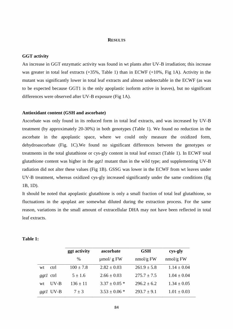
84
RESULTS
GGT activity
An increase in GGT enzymatic activity was found in wt plants after UV-B irradiation; this increase
was greater in total leaf extracts (+35%, Table 1) than in ECWF (+10%, Fig 1A). Activity in the
mutant was significantly lower in total leaf extracts and almost undetectable in the ECWF (as was
to be expected because GGT1 is the only apoplastic isoform active in leaves), but no significant
differences were observed after UV-B exposure (Fig 1A).
Antioxidant content (GSH and ascorbate)
Ascorbate was only found in its reduced form in total leaf extracts, and was increased by UV-B
treatment (by approximately 20-30%) in both genotypes (Table 1). We found no reduction in the
ascorbate in the apoplastic space, where we could only measure the oxidized form,
dehydroascorbate (Fig. 1C).We found no significant differences between the genotypes or
treatments in the total glutathione or cys-gly content in total leaf extract (Table 1). In ECWF total
glutathione content was higher in the ggt1 mutant than in the wild type; and supplementing UV-B
radiation did not alter these values (Fig 1B). GSSG was lower in the ECWF from wt leaves under
UV-B treatment, whereas oxidized cys-gly increased significantly under the same conditions (fig
1B, 1D).
It should be noted that apoplastic glutathione is only a small fraction of total leaf glutathione, so
fluctuations in the apoplast are somewhat diluted during the extraction process. For the same
reason, variations in the small amount of extracellular DHA may not have been reflected in total
leaf extracts.
Table 1:
ggt activity ascorbate GSH cys-gly
% µmol/ g FW nmol/g FW nmol/g FW
wt ctrl 100 ± 7.8 2.82 ± 0.03 261.9 ± 5.8 1.14 ± 0.04
ggt1 ctrl 5 ± 1.6 2.66 ± 0.03 275.7 ± 7.5 1.04 ± 0.04
wt UV-B 136 ± 11 3.37 ± 0.05 * 296.2 ± 6.2 1.34 ± 0.05
ggt1 UV-B 7 ± 3 3.53 ± 0.06 * 293.7 ± 9.1 1.01 ± 0.03
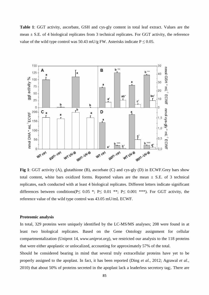
85
Table 1: GGT activity, ascorbate, GSH and cys-gly content in total leaf extract. Values are the
mean ± S.E. of 4 biological replicates from 3 technical replicates. For GGT activity, the reference
value of the wild type control was 50.43 mU/g FW. Asterisks indicate P ≤ 0.05.
Fig 1: GGT activity (A), glutathione (B), ascorbate (C) and cys-gly (D) in ECWF.Grey bars show
total content, white bars oxidized forms. Reported values are the mean ± S.E. of 3 technical
replicates, each conducted with at least 4 biological replicates. Different letters indicate significant
differences between conditions(P≤ 0.05 *; P≤ 0.01 **; P≤ 0.001 ***). For GGT activity, the
reference value of the wild type control was 43.05 mU/mL ECWF.
Proteomic analysis
In total, 329 proteins were uniquely identified by the LC-MS/MS analyses; 208 were found in at
least two biological replicates. Based on the Gene Ontology assignment for cellular
compartmentalization (Uniprot 14, www.uniprot.org), we restricted our analysis to the 118 proteins
that were either apoplastic or unlocalized, accounting for approximately 57% of the total.
Should be considered bearing in mind that several truly extracellular proteins have yet to be
properly assigned to the apoplast. In fact, it has been reported (Ding et al., 2012; Agrawal et al.,
2010) that about 50% of proteins secreted in the apoplast lack a leaderless secretory tag;. There are
86
consequently many unpredicted secretory proteins in plants, and their occurrence is often
underestimated or they are even considered improperly as contaminants. Our decision to restrict our
assignments according to the Uniprot database was therefore rigorous, but probably led to an
underestimation of the truly apoplastic proteins.
The variations considered were further restricted to proteins exhibiting an at least ±50% fold change
in expression.
Various information can be drawn from comparisons between the four experimental conditions: i)
the effect of UV-B treatment on each genotype; ii) differential apoplastic protein composition in
ggt1 vs. wt; iii) possible differences in the behavior of the ggt1 mutant and the wt under UV-
B.Comparing the two genotypes, 23 proteins were downregulated and only 3 were upregulated in
ggt1by comparison with the wt under physiological conditions (Table 2). UV-B treatment resulted
in 8 proteins being downregulated in ggt1; and in 12 being downregulated and 11 being upregulated
in the wt. When the ggt1 and wt were compared after UV-B treatment, it emerged that 9 proteins
were expressed less, and 10 were expressed more in the mutant than in the wild type. A condensed
view of all these variations is given in Table 2.
Table 2:
Accession Locus FDR WT ggt1 CTRL UV-B
nr name Description % UVB/ctrl UVB/ctrl ggt1/wt ggt1/wt
F4HR88 At1g33590 Leucine-rich repeat-containing protein 1 0.55 0.48
O81862 At4g19810 At4g19810 1 0.55
F4IAX0 At1g31690 Putative copper amine oxidase 1 0.57
Q9M5J8 At5g06870 Polygalacturonase inhibitor 2 1 0.57
Q9LMU2 At1g17860 At1g17860/F2H15_8 1 0.57 0.48
B9DGL8 At5g08370 AT5G08370 protein 1 0.58
F4HSQ4 At1g20160 Subtilisin-like serine endopeptidase-like protein 1 0.61
F4IIQ3 At2g28470 Beta-galactosidase 1 0.62
Q9ZVS4 At1g03220 Aspartyl protease-like protein 1 0.65 0.66 2.5
Q94F20 At5g25460 At5g25460 1 0.66 0.58 1.6
Q9FT97 At5g08380 Alpha-galactosidase 1 1 0.68
Q940J8 At4g19410 Pectinacetylesterase family protein 1 0.68 0.68 1.9
O49006 At3g14310 Pectinesterase/pectinesterase inhibitor 3 1 1.5 0.55
O65469 At4g23170 Putative cysteine-rich receptor-like protein
kinase 9 1 1.5
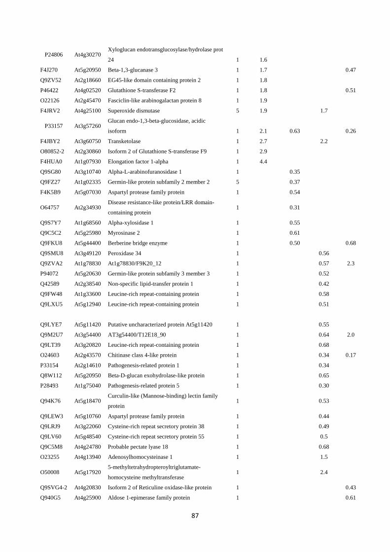
87
P24806 At4g30270 Xyloglucan endotransglucosylase/hydrolase prot
24 1 1.6
F4J270 At5g20950 Beta-1,3-glucanase 3 1 1.7 0.47
Q9ZV52 At2g18660 EG45-like domain containing protein 2 1 1.8
P46422 At4g02520 Glutathione S-transferase F2 1 1.8 0.51
O22126 At2g45470 Fasciclin-like arabinogalactan protein 8 1 1.9
F4JRV2 At4g25100 Superoxide dismutase 5 1.9 1.7
P33157 At3g57260 Glucan endo-1,3-beta-glucosidase, acidic
isoform 1 2.1 0.63 0.26
F4JBY2 At3g60750 Transketolase 1 2.7 2.2
O80852-2 At2g30860 Isoform 2 of Glutathione S-transferase F9 1 2.9
F4HUA0 At1g07930 Elongation factor 1-alpha 1 4.4
Q9SG80 At3g10740 Alpha-L-arabinofuranosidase 1 1 0.35
Q9FZ27 At1g02335 Germin-like protein subfamily 2 member 2 5 0.37
F4K5B9 At5g07030 Aspartyl protease family protein 1 0.54
O64757 At2g34930 Disease resistance-like protein/LRR domain-
containing protein 1
0.31
Q9S7Y7 At1g68560 Alpha-xylosidase 1 1 0.55
Q9C5C2 At5g25980 Myrosinase 2 1 0.61
Q9FKU8 At5g44400 Berberine bridge enzyme 1 0.50 0.68
Q9SMU8 At3g49120 Peroxidase 34 1 0.56
Q9ZVA2 At1g78830 At1g78830/F9K20_12 1 0.57 2.3
P94072 At5g20630 Germin-like protein subfamily 3 member 3 1 0.52
Q42589 At2g38540 Non-specific lipid-transfer protein 1 1 0.42
Q9FW48 At1g33600 Leucine-rich repeat-containing protein 1 0.58
Q9LXU5 At5g12940 Leucine-rich repeat-containing protein 1 0.51
Q9LYE7
At5g11420 Putative uncharacterized protein At5g11420 1 0.55
Q9M2U7 At3g54400 AT3g54400/T12E18_90 1 0.64 2.0
Q9LT39 At3g20820 Leucine-rich repeat-containing protein 1 0.68
O24603 At2g43570 Chitinase class 4-like protein 1 0.34 0.17
P33154 At2g14610 Pathogenesis-related protein 1 1 0.34
Q8W112 At5g20950 Beta-D-glucan exohydrolase-like protein 1 0.65
P28493 At1g75040 Pathogenesis-related protein 5 1 0.30
Q94K76 At5g18470 Curculin-like (Mannose-binding) lectin family
protein 1
0.53
Q9LEW3 At5g10760 Aspartyl protease family protein 1 0.44
Q9LRJ9 At3g22060 Cysteine-rich repeat secretory protein 38 1 0.49
Q9LV60 At5g48540 Cysteine-rich repeat secretory protein 55 1 0.5
Q9C5M8 At4g24780 Probable pectate lyase 18 1 0.68
O23255 At4g13940 Adenosylhomocysteinase 1 1 1.5
O50008 At5g17920 5-methyltetrahydropteroyltriglutamate-
homocysteine methyltransferase 1
2.4
Q9SVG4-2 At4g20830 Isoform 2 of Reticuline oxidase-like protein 1 0.43
Q940G5 At4g25900 Aldose 1-epimerase family protein 1 0.61
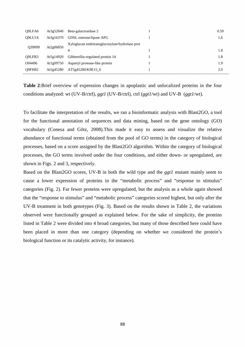
88
Q9LFA6 At3g52840 Beta-galactosidase 2 1 0.59
Q9LU14 At3g16370 GDSL esterase/lipase APG 1 1.6
Q39099 At2g06850 Xyloglucan endotransglucosylase/hydrolase prot
4 1 1.8
Q9LFR3 At5g14920 Gibberellin-regulated protein 14 1 1.8
O04496 At1g09750 Aspartyl protease-like protein 1 1.9
Q9FH82 At5g45280 AT5g45280/K9E15_6 1 2.0
Table 2:Brief overview of expression changes in apoplastic and unlocalized proteins in the four
conditions analyzed: wt (UV-B/ctrl), ggt1 (UV-B/ctrl), ctrl (ggt1/wt) and UV-B (ggt1/wt).
To facilitate the interpretation of the results, we ran a bioinformatic analysis with Blast2GO, a tool
for the functional annotation of sequences and data mining, based on the gene ontology (GO)
vocabulary (Conesa and Götz, 2008).This made it easy to assess and visualize the relative
abundance of functional terms (obtained from the pool of GO terms) in the category of biological
processes, based on a score assigned by the Blast2GO algorithm. Within the category of biological
processes, the GO terms involved under the four conditions, and either down- or upregulated, are
shown in Figs. 2 and 3, respectively.
Based on the Blast2GO scores, UV-B in both the wild type and the ggt1 mutant mainly seem to
cause a lower expression of proteins in the “metabolic process” and “response to stimulus”
categories (Fig. 2). Far fewer proteins were upregulated, but the analysis as a whole again showed
that the “response to stimulus” and “metabolic process” categories scored highest, but only after the
UV-B treatment in both genotypes (Fig. 3). Based on the results shown in Table 2, the variations
observed were functionally grouped as explained below. For the sake of simplicity, the proteins
listed in Table 2 were divided into 4 broad categories, but many of those described here could have
been placed in more than one category (depending on whether we considered the protein’s
biological function or its catalytic activity, for instance).
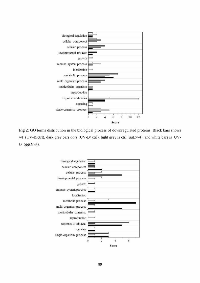
89
Fig 2: GO terms distribution in the biological process of downregulated proteins. Black bars shows
wt (UV-B/ctrl), dark grey bars ggt1 (UV-B/ ctrl), light grey is ctrl (ggt1/wt), and white bars is UV-
B (ggt1/wt).
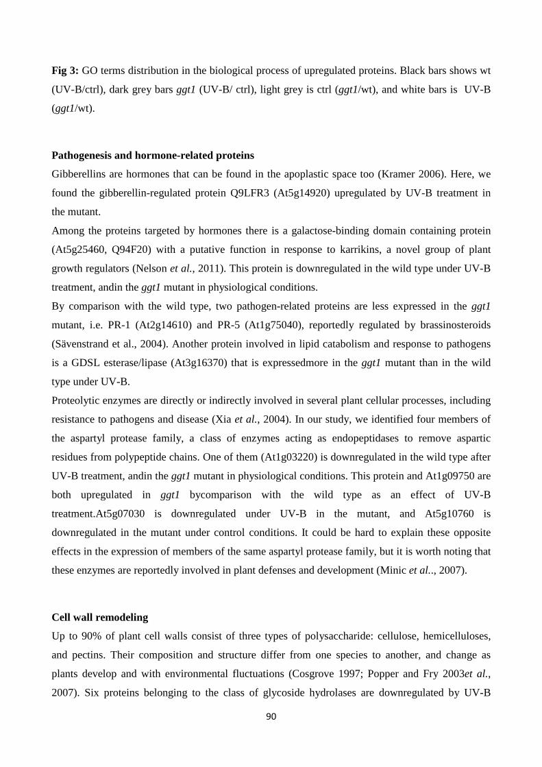
90
Fig 3: GO terms distribution in the biological process of upregulated proteins. Black bars shows wt
(UV-B/ctrl), dark grey bars ggt1 (UV-B/ ctrl), light grey is ctrl (ggt1/wt), and white bars is UV-B
(ggt1/wt).
Pathogenesis and hormone-related proteins
Gibberellins are hormones that can be found in the apoplastic space too (Kramer 2006). Here, we
found the gibberellin-regulated protein Q9LFR3 (At5g14920) upregulated by UV-B treatment in
the mutant.
Among the proteins targeted by hormones there is a galactose-binding domain containing protein
(At5g25460, Q94F20) with a putative function in response to karrikins, a novel group of plant
growth regulators (Nelson et al., 2011). This protein is downregulated in the wild type under UV-B
treatment, andin the ggt1 mutant in physiological conditions.
By comparison with the wild type, two pathogen-related proteins are less expressed in the ggt1
mutant, i.e. PR-1 (At2g14610) and PR-5 (At1g75040), reportedly regulated by brassinosteroids
(Sävenstrand et al., 2004). Another protein involved in lipid catabolism and response to pathogens
is a GDSL esterase/lipase (At3g16370) that is expressedmore in the ggt1 mutant than in the wild
type under UV-B.
Proteolytic enzymes are directly or indirectly involved in several plant cellular processes, including
resistance to pathogens and disease (Xia et al., 2004). In our study, we identified four members of
the aspartyl protease family, a class of enzymes acting as endopeptidases to remove aspartic
residues from polypeptide chains. One of them (At1g03220) is downregulated in the wild type after
UV-B treatment, andin the ggt1 mutant in physiological conditions. This protein and At1g09750 are
both upregulated in ggt1 bycomparison with the wild type as an effect of UV-B
treatment.At5g07030 is downregulated under UV-B in the mutant, and At5g10760 is
downregulated in the mutant under control conditions. It could be hard to explain these opposite
effects in the expression of members of the same aspartyl protease family, but it is worth noting that
these enzymes are reportedly involved in plant defenses and development (Minic et al.., 2007).
Cell wall remodeling
Up to 90% of plant cell walls consist of three types of polysaccharide: cellulose, hemicelluloses,
and pectins. Their composition and structure differ from one species to another, and change as
plants develop and with environmental fluctuations (Cosgrove 1997; Popper and Fry 2003et al.,
2007). Six proteins belonging to the class of glycoside hydrolases are downregulated by UV-B
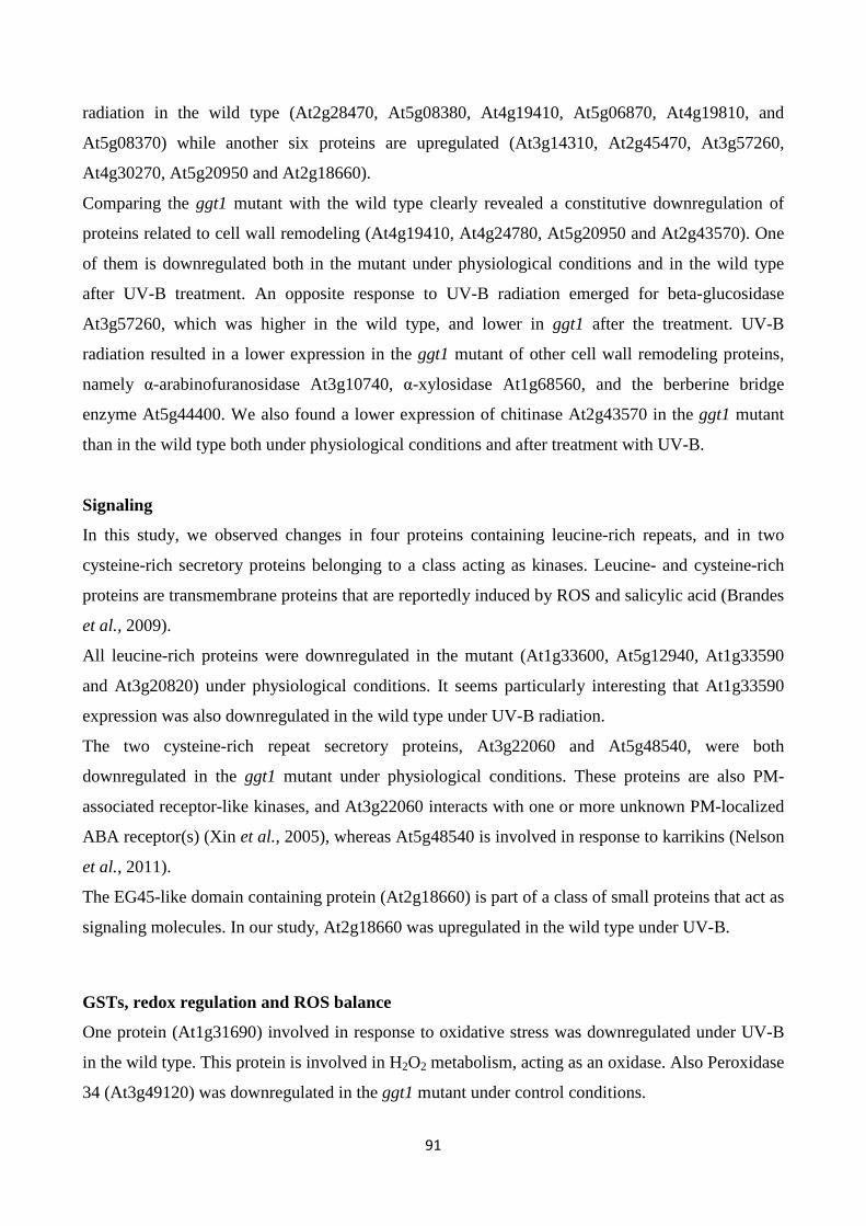
91
radiation in the wild type (At2g28470, At5g08380, At4g19410, At5g06870, At4g19810, and
At5g08370) while another six proteins are upregulated (At3g14310, At2g45470, At3g57260,
At4g30270, At5g20950 and At2g18660).
Comparing the ggt1 mutant with the wild type clearly revealed a constitutive downregulation of
proteins related to cell wall remodeling (At4g19410, At4g24780, At5g20950 and At2g43570). One
of them is downregulated both in the mutant under physiological conditions and in the wild type
after UV-B treatment. An opposite response to UV-B radiation emerged for beta-glucosidase
At3g57260, which was higher in the wild type, and lower in ggt1 after the treatment. UV-B
radiation resulted in a lower expression in the ggt1 mutant of other cell wall remodeling proteins,
namely α-arabinofuranosidase At3g10740, α-xylosidase At1g68560, and the berberine bridge
enzyme At5g44400. We also found a lower expression of chitinase At2g43570 in the ggt1 mutant
than in the wild type both under physiological conditions and after treatment with UV-B.
Signaling
In this study, we observed changes in four proteins containing leucine-rich repeats, and in two
cysteine-rich secretory proteins belonging to a class acting as kinases. Leucine- and cysteine-rich
proteins are transmembrane proteins that are reportedly induced by ROS and salicylic acid (Brandes
et al., 2009).
All leucine-rich proteins were downregulated in the mutant (At1g33600, At5g12940, At1g33590
and At3g20820) under physiological conditions. It seems particularly interesting that At1g33590
expression was also downregulated in the wild type under UV-B radiation.
The two cysteine-rich repeat secretory proteins, At3g22060 and At5g48540, were both
downregulated in the ggt1 mutant under physiological conditions. These proteins are also PM-
associated receptor-like kinases, and At3g22060 interacts with one or more unknown PM-localized
ABA receptor(s) (Xin et al., 2005), whereas At5g48540 is involved in response to karrikins (Nelson
et al., 2011).
The EG45-like domain containing protein (At2g18660) is part of a class of small proteins that act as
signaling molecules. In our study, At2g18660 was upregulated in the wild type under UV-B.
GSTs, redox regulation and ROS balance
One protein (At1g31690) involved in response to oxidative stress was downregulated under UV-B
in the wild type. This protein is involved in H2O2 metabolism, acting as an oxidase. Also Peroxidase
34 (At3g49120) was downregulated in the ggt1 mutant under control conditions.
92
Two GSTs proteins belonging to categories F2 and F9 (At4g02520 and At2g30860, respectively)
were upregulated in the wild type plants after UV-B radiation. When the two genotypes were
compared after the same treatment, the expression level was lower in the former.
Two proteins in the germin-like family (At5g20630 and At1g02335) were downregulated in ggt1,
one in physiological conditions and the other after UV-B treatment. These proteins are involved in
defending against biotic and environmental stress.
Superoxide dismutase proteins are reportedly involved in removing superoxide radicals from the
apoplast following UV-B radiation (Alscher et al., 2002). In our study, the expression of superoxide
dismutase (At4g25100) was upregulated in the wild type after UV-B radiation and in ggt1 under
physiological conditions.
DISCUSSION
GGT activity and soluble antioxidants
Following excess UV-B exposure, plants deploy a wide array of morphological and biochemical
defense mechanisms, including soluble antioxidants (Schiu 2005). The changes observed in this
study are consistent with the view that, under UV-B radiation, oxidative conditions in the apoplastic
space involve both ascorbate and glutathione, the two main soluble antioxidant molecules in plant
cells. Ascorbate in ECWF was found fully oxidized, which is consistent with the view that
oxidizing conditions prevail in the apoplastic space(Saruhan et al., 2009; Vanacker et al., 1998a;
Vanacker et al.1998b).
UV-B radiation induced an increase in apoplastic ascorbate in both genotypes, suggesting that
ascorbate is extruded as a means to counteract the artificially-imposed oxidative conditions. While
glutathione content was substantially unchanged in total leaf extracts in all the conditions tested, it
was altered in the ECWF from mutant leaves, where the effect of the ggt1 null mutation results in a
net increase in glutathione content, as a predictable effect of the reduced GGT degradation activity.
Under UV-B, the concurrent decrease in oxidized glutathione and increase in oxidized cys-gly can
be interpreted as an enhanced gamma-glutamyl transferase activity; this is supported by the
previous finding that GGT1 has a stronger preference for GSSG (Okhama-Otsu et al., 2008).
GGT activity was barely detectable in the mutant, confirming that GGT1 is the main isoform
contributing to over 90% of said activity in wild type leaves. Since the GGT2 isoform is not
expressed in leaves (Destro et al., 2011) and GGT3 is assumed to be non-functional (Martin et al.,
2007), this indicates that the activity found in the mutant represents the contribution of the
remaining vacuolar isoform GGT4.
93
The increased GGT activity following UV-B treatment in the wild type therefore suggests that the
rate of the gamma-glutamyl cycle is accelerated by this radiation. The involvement of the vacuolar
GGT4 in the degradation of glutathione conjugates, e.g. with lipoperoxides and/or other damaged
molecules, might be implicated too, but this seems unlikely since no significant increase in GGT
activity was apparent in the mutant under the same conditions.
Collectively, these novel findings thus imply that the gamma-glutamyl cycle is accelerated under
oxidative conditions imposed by ultraviolet-B radiation, and support the conviction that it is
involved in oxidative stress sensing and/or response.
Apoplastic proteome readjustments
Proteome analysis has proved a powerful tool for deciphering cell metabolism under different
perturbations and has been found useful in apoplastic studies too (Agrawal et al., 2010). Apoplastic
proteins establish a constitutive systemic defense network, with only a few of them changing under
environmental and/or biotic stress (Delaunios et al., 2014).
Two main approaches are currently adopted in plant physiology studies: the application of stress
conditions, and the use of mutants. These alternative and converging strategies may provide tools
for deciphering metabolism. In this work, oxidative conditions were imposed with UV-B and
studied in redox-altered ggt1 mutants. Subcellular fractionation and apoplastic proteome analysis
were then used to arrive at a better understanding of the rearrangements in the extracellular
compartment.
The experimental design adopted here could consequently help to describe and compare the effects
of UV-B treatment on the two genotypes, and the differences in apoplastic proteome composition
between the mutant and wild type leaves under control conditions.
In both genotypes, UV-B treatment caused a downregulation of different kinds of protein related to
cell wall biosynthesis, response to stress and proteolysis. It promptedan upregulation, but only in
the wild type, of other proteins involved in cell wall remodeling and two glutathione S-transferases,
GST-F2 and GST-F9. No proteins were found upregulated in the ggt1 mutant after UV-B (Table 2).
The hormonal changes occurring in the ggt1 mutant, with or without exposure to UV-B radiation,
were not considered in the experimental setup, and were beyond the scope of this work. Several
proteins seen here to change in expression could be targets for hormones, however. For instance,
one protein whose expression was stimulated by UV-B is reportedly a gibberellin-regulated protein
(At5g14920), suggesting that gibberellins could be implicated in the response. The expression of a
galactose-binding domain containing protein (At5g25460), which is stimulated by karrikins (a novel
group of plant growth regulators (Nelson et al., 2011), was also higher in the mutant than in the
94
wild type after UV-B. Interplay with hormones may also concern two PRPs (PR-1 and PR-5),
whose expression was lower in the ggt1 mutant under physiological conditions. A previous study
(Sävenstrand et al., 2004) had found their expression strongly reduced in brassinosteroid
metabolism mutants. It would be interesting to see whether the brassinosteroid pathway is altered in
ggt1 mutants too.
Broadly speaking, cell wall modifying proteins such as glycosyl hydrolases (GHs), peroxidases,
esterases, transglycosylases and lyases, are involved in the construction, remodeling or turnover of
cell wall components (Cosgrove, 1997; Stolle-Smits et al., 1999; Obel et al., 2002; Reiter, 2002).
Some of them may have other functions too, e.g. in the glycosylation state of target proteins (Kang
et al., 2008), which in turn could be involved in signaling processes (Minic et al., 2007).
Taking a broader look at the changes found in this category suggests that UV-B affects the
expression of some proteins in the wild type (e.g. pectine-acetylesterase and its inhibitor,
xyloglucan endotransglucosylase, beta 1,3-glucanase, beta-galactosidase, alpha-galactosidase and a
polygalacturonase inhibitor) and others in the ggt1 mutant (alpha-xylosidase, myrosinase, alpha-
arabinofuranosidase, and a berberine bridge enzyme), confirming the view that the cell walls are the
target of this radiation. Notably, these remodeling processes are affected in the ggt1 mutant not only
by UV-B treatment, but also under physiological conditions. Since cell wall structure is reportedly
altered during development and by exposure to stress (Potters et al., 2009), our findings could be
explained by the existence of a stress-like condition in the mutant, where some signals mimic the
oxidative state induced by UV-B in the wild type.
Myrosinase, a protein in the class of glycoside hydrolases, was less expressed under UV-B in the
ggt1 mutant, and this could have ecophysiological consequences because in Brassicaceae
myrosinases play a part in growth, development and defenses against microbes, as well as deterring
insects and herbivores (Rodman, 1991). The two germin-like proteins that were downregulated in
the ggt1 mutant could also be consistent with alterations in the defense systems against biotic and
environmental stress.
The expression of some other proteins was altered in opposite ways (up- or downregulated)after
UV-B exposure, depending on the genotype considered: for instance, the stress-responsive
glucanendo-1,3-beta-glucosidase was upregulated in the wild type, but downregulated in ggt1.
A group of proteins involved in response to stimuli, i.e. the leucine-rich and cysteine-rich proteins,
wasdownregulated in ggt1 in physiological conditions. Leucine-rich proteins contain a leucine-rich
repeat (LRR) motif that has revealed a central role in recognizing different pathogen-associated
molecules in the innate host defense of plants and animals (Gunawardena et al., 2011).
95
In this study, we also identified 4 aspartyl proteases that were altered under our experimental
conditions: this may mean that members of this category of enzymes related to plant defenses are
sensitive to redox variations. Aspartyl proteases are important for plant development. They have
been implicated in the ABA-dependent responsiveness to drought-induced stress (Yao et al., 2012),
and inArabidopsis agene encoding the aspartyl protease protein was found downregulated by cold
and high-salinity stress (Seki et al., 2002).
The EG45-like domain containing protein 2 (At2g18660) was upregulated in the wild type under
UV-B. This protein belongs to the category of plant natriuretic peptides (PNPs), a novel class of
small proteins showing homology with the N-terminus of expansins, though they are significantly
shorter and lack the wall-binding domain (Ludidi et al., 2002). Previous studies found PNPs
upregulated under saline and osmotic conditions (Rafudeen et al., 2003), but the effects of UV-B on
this class of peptidic signaling molecules had not been reported before.
Among the variations in apoplastic enzyme expression found in the present study, some that
particularly attracted our attention are closely related to ROS metabolism.
Superoxide anion formation is reportedly triggered by ultraviolet-B radiation (Alscher et al., 2002).
It seems noteworthy that no plant superoxide dismutase (SOD)identified to date contains a signal
peptide, but extracellular SOD activity in stressed or pathogen-infected plants has been reported in
many works (Hernández et al., 2001; Karpinska et al., 2001; Kaffarnik et al., 2009; Pechanova et
al., 2010). SODsproduce H2O2, which is degraded to H2O by ascorbate peroxidase. By removing
superoxide anions, SODs may limit the duration of the oxidative burst to an early event in plant
defense (Scheler et al., 2013; Pristov et al., 2013).
In this study, a superoxide dismutase (At4g25100) was found upregulated by UV-B radiation in
wild type leaves: this can be interpreted as the need to improve scavenging activity to remove
excess superoxide anions. Although its localization is not reported in the official databases, its
occurrence in the apoplast was noted in other studies too (Ding et al., 2012; Kwon et al., 2005).
Higher SOD levels combined with lower levels of the putative copper amine oxidase (At1g31690)
may result in higher H2O2 levels. An increased GST expression could also result in its scavenging,
however, so while it seems clear that ROS metabolism is affected by UV-B treatment in the wild
type, it is hard to draw any conclusions on hydrogen peroxide levels, and further experimentation is
needed to validate our hypothesis.
The ROS scavenging scenario in the ggt1 mutant is undoubtedly more complex. The above-
mentioned apoplastic SOD is upregulated under physiological conditions, and a peroxidase is
downregulated. These effects may be interpreted as readjustments in the redox-altered
96
ggt1background. Such readjustments may be needed to sustain a higher H2O2 level, which could act
as a signal.
Taken together, these effects may resultin higher H2O2 levels in the mutant under physiological
conditions (schematically shown in Fig. 4), whereas the rise in H2O2 in the wild type is a direct
consequence of oxidative stress conditions induced by UV-B radiation.
As a signaling molecule, H2O2 may cross membranes in a process facilitated by aquaporins (Bienert
et al., 2006), reaching internal cell compartments and the nucleus, where it can activate defense
gene expression (Mullineaux et al., 2006). If this assumption holds true, it might explain the
“constitutive alert response” effect observed in a previous proteomic analysis of total leaf extracts
from ggt1 mutant leaves (Tolin et al., 2013).
Future research is therefore needed to ascertain the level of ROS, and especially H2O2, in the
apoplast of ggt1 mutants, and the possible involvement of hormones (e.g. brassinosteroids and
gibberellins) in the response. Both H2O2 and hormones are signals arising in the apoplast that can be
transferred intracellularly and evoke the cell’s responses. For this signal transduction function we
could also consider four leucine-rich and two cysteine-rich proteins belonging to the superfamily of
receptor-like kinases (RLKs), which are associated with the plasma membrane and contain redox-
sensitive thiols,which were found at lower level in the ggt1 mutant.Disrupting of the gamma-
glutamyl cycle could result in an altered signal perception pathway.
While hormonal and redox readjustments seem to be implicated in the modified metabolism of ggt1
mutants, it remains to be seen how silencing the gamma-glutamyl transferase activity and
consequently impairing the gamma-glutamyl cycle may lead to the effects reported here. Further
experiments are needed to clarify the link between the gamma-glutamyl cycle and apoplastic redox
events.
97
Fig. 4: Schematic overview of apoplastic proteome variations in: A) wild type, induced by UV-B;
B) ggt1 genotype due to the mutation and/or to UV-B treatment.
Vertical arrows refer to stimulation (↑) or repression (↓) caused by the mutation; horizontal arrows
indicate repression (├ ) or stimulation (←) caused by UV-B treatment.
ACKNOWLEDGMENTS
The authors wish to thank Renato Millioni from the Proteomics Center of Padova University for his
kind support, and Frances Coburn for text and style improvement. The authors also wish to thank
the “Cassa di Risparmio di Padova e Rovigo” (CARIPARO) Foundation for funding the acquisition
of the LTQ-Orbitrap XL mass spectrometer.
Dinesh Prasad was granted a University of Padova fellowship for Indian researchers; MS Mehdi
was supported by an Erasmus Mundus - EMMA Fellowship. Leonard E. Barnabas was granted a
“Fondazione A. Gini” fellowship. Micaela Pivato was funded by a University of Padova research
fellowship. Marta Fàbrega-Prats was granted a fellowship for doctoral students from the
CARIPARO Foundation. This work was funded by the University of Padova - MURST ex-60%.
98
REFERENCES
Agrawal, G. K., Jwa, N. S., Lebrun, M. H., Job, D., Rakwal, R. (2010). Plant secretome: unlocking secrets of the secreted proteins. Proteomics 10, 799-827. doi: 10.1002/pmic.200900514
Alonso, J. M., Stepanova, A. N., Leisse, T. J., Kim, C. J., Chen, H., Shinn, P., Stevenson, D. K., Zimmerman, J., Barajas, P., Cheuk, R., Gadrinab, C., Heller, C., Jeske, A., Koesema, E., Meyers, C. C., Parker, H., Prednis, L., Ansari, Y., Choy, N., Deen, H., Geralt, M., Hazari, N., Hom, E., Karnes, M., Mulholland, C., Ndubaku, R., Schmidt, I., Guzman, P., Aguilar-Henonin, L., Schmid, M., Weigel, D., Carter, D. E., Marchand, T., Risseeuw, E., Brogden, D., Zeko, A., Crosby, W. L., Berry, C. C., Ecker, J. R. (2003). Genome-wide insertional mutagenesis of Arabidopsisthaliana. Science 301 (5633), 653–657. doi:10.1126/science.1086391
Alscher, R. G., Erturk, N., Health, L. S. (2002). Role of superoxide dismutases (SODs) in controlling oxidative stress in plants. J. Exp. Bot. 53, 1331–1341. doi:10.1093/jexbot/53.372.1331
Bienert, G. P., Schjoerring, J. K., Jahn T. P. (2006). Membrane transport of hydrogen peroxide. Biochimica et Biophysica Acta 1758, 994-1004
Brandes, N., Schmitt, S., Jakob, U. (2009). Thiol-based redox switches in eukaryotic proteins. Antiox. Redox Signal. 11, 997–1014. doi:10.1089/ARS.2008.2285
Brosché, M., Strid, Å., (2003). Molecular events following perception of ultraviolet-B radiation by plants. Physiol. Plant. 117, 1-10. doi: 10.1034/j.1399-3054.2003.1170101.x
Brunet, S. Thibault, P., Gagnon, E., Kearney, P., Bergeron, J. J., Desjardins, M. (2003). Organelle proteomics: looking at less to see more. Trends Cell Biol. 13, 629–638. doi:10.1016/j.tcb.2003.10.006
Cheng, Z., Zheng, Z., Huang, J., Lai, Z., Fan, B. (2009). Biosynthesis of salicylic acid in plants. Plant signal Behav. 4:6, 493-496
Conesa, A., Götz, S. (2008). Blast2GO: a comprehensive suite for functional analysis in plant genomics. Int. J. Plant Genomics 2008:619832. doi:10.1155/2008/619832
Cosgrove, D. J. (1997). Assembly and enlargement of the primary cell wall in plants. Ann. Rev. Cell Dev. Biol. 13, 171-201
99
Delaunois, B., Jeandet, P., Clément, C., Baillieul, F., Dorey, S., Cordelier, S. (2014). Uncovering plant-pathogen crosstalk through apoplastic proteomic studies. Front. Plant. Sci. 5, 249. doi:10.3389/fpls.2014.00249
Destro, T., Prasad, D., Martignago, D., Bernet, I. L., Trentin, A. R., Renu I. K., Ferretti, M., Masi, A. (2011). Compensatory expression and substrate inducibility of γ-glutamyl transferase GGT2 isoform in Arabidopsisthaliana. J. Exp. Bot. 62, 805–814
Dietz, K.J. (1997). Functions and responses of the leaf apoplast under stress. Progress in Botany 58, 221-254
Ding, Y., Wang, J., Wang, J., Stierhof, Y. D., Robinson, D. G., Jiang, L. (2012). Unconventional protein secretion. Trends in Plant Science 17, 606-615. doi:10.1016/j.tplants.2012.06.004
Ferretti, M., Destro, T., Tosatto, S. C., La Rocca, N., Rascio, N., Masi, A. (2009). Gamma-glutamyl transferase in the cell wall participates in extracellular glutathione salvage from the root apoplast. New Phytol. 181, 115–126. doi:10.1111/j.1469-8137.2008.02653.x
Griffith, O. W. (1980). Determination of glutathione and glutathione disulfide using glutathione reductase and 2-vinylpyridine. Anal. Biochem. 106, 207–212. doi:10.1016/0003-2697(80)90139-6
Gunawardena, H. P., Huang, Y.,Kenjale, R.,Wang, H., Xie, L., Chen, X. (2011). Unambiguous Characterization of Site-specific Phosphorylation of Leucine-rich Repeat Fli-I-interacting Protein 2 (LRRFIP2) in Toll-like Receptor 4 (TLR4)-mediated Signaling. J Biol Chem, 286, 10897-10910.
Henzler, T., Steudle, E. (2000). Transport and metabolic degradation of hydrogen peroxide in Chara corallina : model calculations and measurements with the pressure probe suggest transport of H2O2 across water channels. J. Exp. Bot. 51, 2053–2066
Hernandez, J. A., Ferrer, M. A., Jimenez, A., Barcelo, A. R., Sevilla, F. (2001). Antioxidant systems and O2.-/ H2O2 production in the apoplast of Pea leaves. Its relation with salt-induced necrotic lesions in minor veins. Plant Physiol. 127, 817–831. doi:10.1104/pp.010188
Hewitt, E. J., Dickes, G. J. (1961). Spectrophotometric measurements on ascorbic acid and their use for the estimation of ascorbic acid and dehydroascorbic acid in plant tissues. Biochem. J. 78, 384–391
100
Hideg, E., Jansen, M. A. K., Strid, A. (2013). UV-B exposure, ROS, and stress: inseparable companions or loosely linked associates? Trends Plant Sci. 18, 107–115. doi:10.1016/j.tplants.2012.09.003
Hu, J.F., Li, G. F., Gao, Z. H., Chen, L., Ren, H. B., Jia, W. S. (2005). Regulation of water deficit-induced abscisic acid accumulation by apoplastic ascorbic acid in maize seedlings. J. Integr. Plant Biol. 47, 1335-1344. doi: 10.1111/j.1744-7909.2005.00165.x
Huseby, N. E., Stromme, J. H. (1974). Practical points regarding routine determination of γ -glutamyl transferase (γ-GT) in serum with a kinetic method at 37°C. Scand. J. Clin. Lab. Invest. 34, 357–363
Jaspers, P., Kollist, H., Langebartels, C., Kangasjarvi, J. (2005). “Plant responses to ozone,” in Antioxidants and reactive oxygen species in plants, ed. N. Smirnoff (Oxford: Blackwell Publishing), 268-292
Kaffarnik, F. A., Jones, A. M., Rathjen, J. P.,. Peck, S. C. (2009). Effector proteins of the bacterial pathogen pseudomonas syringae alter the extracellular proteome of the host plant, Arabidopsisthaliana. Mol. Cell. Proteomics 8:145–56. doi:10.1074/mcp.M800043-MCP200
Kang, J. S., Frank, J., Kang, C. H., Kajiura, H., Vikram, M., Ueda, A., Kim, S., Bahk, J. D., Triplett, B., Fujiyama, K., Lee, S.Y., Von Schaewen, A., Koiwa, H. (2008). Salt tolerance of Arabidopsisthaliana requires maturation of N-glycosylated proteins in the Golgi apparatus. Proc. Natl. Acad. Sci. U. S. A. 105, 5933-5938. doi: 10.1073/pnas.0800237105
Karpinska, B., Karlsson, M., Schinkel, H., Streller, S., Su, K., Melzer, M., Wingsle, G. (2001). A novel superoxide dismutase with a high isoelectric point in higher plants. Expression, regulation, and protein localization. Plant Physiol. 126, 1668–1677
Köcher, T., Pichler, P., Schutzbier, M., Stingl, C., Kaul, A., Teucher, N., Hasenfuss, G., Penninger, J. M., Mechtler, K. (2009). High precision quantitative proteomics using iTRAQ on an LTQ orbitrap: a new mass spectrometric method combining the benefits of all. J. Prot. Res. 8, 4743–4752. doi:10.1021/pr900451u
Kramer, E. M. (2006). How far can a molecule of weak acid travel in the apoplast or xylem ? Plant Physiol. 141, 1233–1236. doi:10.1104/pp.106.083790.1
Kwon, H. K., Yokoyama, R., Nishitani, K. (2005).A proteomic approach to apoplastic proteins involved in cell wall regeneration in protoplasts of Arabidopsis suspension-cultured cells.Plant Cell Physiol 46, 843-857
101
Li, L., Van Staden, J. (1998). Effects of plant growth regulators on the antioxidant system in callus of two maize cultivars subjected to water stress. Plant Growth Regulation 24, 55–66
Lohaus, G., Pennewiss, K., Sattelmacher, B., Hussmann, M., Muehling, K. H. (2001). Is the infiltration-centrifugation technique appropriate for the isolation of apoplastic fluid? A critical evaluation with different plant species. Physiol. Plant. 111, 457–465. doi:10.1034/j.1399-3054.2001.1110405.x
Ludidi, N. N., Heazlewood, J. L., Seoighe, C., Irving, H. R., Gehring, C. A. J. (2002). Expansin-like molecules: novel functions derived from common domains. Mol. Evol. 54, 587-594
Martin, M. N., Saladores, P. H., Lambert, E., Hudson, A. O., Leustek, T. (2007). Localization of members of the gamma-glutamyl transpeptidase family identifies sites of glutathione and glutathione S-conjugate hydrolysis. Plant Physiol. 144, 1715–1732. doi:10.1104/pp.106.094409
Masi, A., Ghisi R., Ferretti M. (2002). Measuring low-molecular-weight thiols by detecting the fluorescence of fheir SBD-derivatives: application to studies of diurnal and UV-B induced changes in Zea Mays L. J. Plant Physiol.159, 499–507. doi:10.1078/0176-1617-00655
Meister, A., Anderson, M. E. (1983). Glutathione. Annu. Rev. Biochem. 52, 711–760. doi:10.1146/annurev.bi.52.070183.003431
Minic, Z., Jamet, E., Négroni, L., Arsene der Garabedian, P., Zivy, M., Jouanin, L. (2007). A sub-proteome of Arabidopsisthaliana mature stems trapped on Concanavalin A is enriched in cell wall glycoside hydrolases. J. Exp. Bot. 58, 2503–2512. doi:10.1093/jxb/erm082
Mullineaux, P. M., Karpinski, S., Baker, N. R. (2006). Spatial dependence for hydrogen peroxide-directed signalling in light-stressed plants. Plant Physiol 141, 346-350
Nelson, D. C., Scaffidi, A., Dun, E. A., Waters, M. T., Flematti, G. R., Dixon, K. W., Beveridge, C. A., Ghisalberti, E. L., Smith, S. M. (2011). F-Box protein MAX2 has dual roles in karrikin and strigolactone signaling in Arabidopsisthaliana. Proc. Natl. Acad. Sci. U.S.A. 108, 8897–8902. doi:10.1073/pnas.1100987108
Noctor, G., Mhamdi, A., Foyer, C. H. (2014). The roles of reactive oxygen metabolism in drought: not so cut and dried. Plant Physiol. 164, 1636-1648. doi: 10.1104/pp.113.233478
Obel, N., Porchia, A. C., Scheller, H. V. (2002). Dynamic changes in cell wall polysaccharides during wheat seedling development. Phytochemistry 60, 603-610
102
Ohkama-Ohtsu, N., Oikawa, A., Zhao, P., Xiang, C., Saito, K., Oliver, D. J. (2008). A gamma-glutamyl transpeptidase-independent pathway of glutathione catabolism to glutamate via 5-oxoproline in Arabidopsis. Plant Physiol. 148, 1603–1613. doi:10.1104/pp.108.125716
Pechanova, O., Hsu, C. Y., Adams, J. P., Pechan, T., Vandervelde, L., Drnevich, J., Jawdy, S., Adeli, A., Suttle, J. C., Lawrence, A. M., Tschaplinski, T. J., Séguin, A., Yuceer, C. (2010). Apoplast proteome reveals that extracellular matrix contributes to multistress response in Poplar. BMC Genomics 11: 674. doi:10.1186/1471-2164-11-674
Popper, Z. A., Fry, S.C. (2003). Primary cell wall composition of Bryophytes and Charophytes. Ann. Bot. 91, 1–12. doi:10.1093/aob/mcg013
Potters, G., Horemans, N., Jansen, M. K. (2010). The cellular redox state in plant stress biology a charging concept. Plant Physiol. Biochem. 48, 292–300. doi:10.1016/j.plaphy.2009.12.007
Potters, G., Pasternak, T. P, Guisez, Y, Jansen, M. A. (2009). Different stresses, similar morphogenic responses: integrating a plethora of pathways.Plant Cell Environ 32(2), 158-169. doi: 10.1111/j.1365-3040.2008.01908.x
Pristov, J. B., Jovanović, S. V., Mitrović, A., Spasojević, I. (2013). UV-irradiation provokes generation of superoxide on cell wall polygalacturonic acid. Physiol. Plant. 148, 574–581. doi:10.1111/ppl.12001
Rafudeen, S., Gxaba, G., Makgoke, G., Bradley, G., Pironcheva, G., Raitt, L., Irving H., Gehrin, C. (2003). A role for plant natriuretic peptide immuno-analogues in NaCl- and drought-stress responses. Physiol. Plant. 199, 554-562. doi: 10.1046/j.1399-3054.2003.00201.x
Reiter, W. D. (2002). Biosynthesis and properties of the plant cell wall. Curr. Opin. Plant Biol. 5, 536–542
Rizzini, L., Favory, J. J., Cloix, C., Faggionato, D., O’Hara, A., Kaiserli, E., Baumeister, R., Schäfer, E., Nagy, F., Jenkins, G. I., Ulm, R. (2011). Perception of UV-B by the Arabidopsis UVR8 protein. Science 332 (6025), 103–106. doi:10.1126/science.1200660
Rodman, J. E. (1991). A taxonomic analysis of glucosinolate-producing plants. Part 1: Phenetics, Syst. Bot. 16, 598-618
103
Saruhan, N., Terzi, R., Saglam, A., Kadioglu, A. (2009). The relationship between leaf rolling and ascorbate-glutathione cycle enzymes in apoplastic and symplastic areas of Ctenanthe Setosa subjected to drought stress. Biol. Res. 42, 315–326. doi:10.4067/S0716-97602009000300006
Sävenstrand, H., Brosché, M., Strid, A. (2004). Ultraviolet-B signalling: Arabidopsis brassinosteroid mutants are defective in UV-B regulated defence gene expression. Plant Physiol. Biochem. 42, 687–694. doi:10.1016/j.plaphy.2004.06.011
Schafer, F. Q., Buettner, G. R. (2001). Redox environment of the cell as viewed through the redox state of the glutathione disulfide/glutathione couple. Free Rad. Biol. Med. 30, 1191–1212. doi:10.1016/S0891-5849(01)00480-4
Scheler, C., Durner, J., Astier, J. (2013). Nitric oxide and reactive oxygen species in plant biotic interactions. Curr. Opin. Plant Biol. 16, 534–539. doi:10.1016/j.pbi.2013.06.020
Seki, M., Narusaka, M., Ishida, J., Nanjo, T., Fujita, M., Oono, Y., Kamiya, A., Nakajima, M., Enju, A., Sakurai, T., Satou, M., Akiyama, K., Taji, T., Yamaguchi-Shinozaki, K., Carninci, P., Kawai, J., Hayashizaki, Y., Shinozaki K. (2002). Monitoring the expression profiles of 7000 Arabidopsis genes under drought, cold and high -salinity stresses using a full-length cDNA microarray. Plant J 31, 279-292
Shiu, C.-T. (2005). Ultraviolet-B-induced oxidative stress and responses of the ascorbate-glutathione cycle in a marine macroalga Ulva Fasciata. J. Exp. Bot. 56,2851–2865. doi:10.1093/jxb/eri277
Smirnoff, N., Pallanca, J. E. (1996). Ascorbate metabolism in relation to oxidative stress. Biochem. Soc. Trans. 24, 472–478
Stolle-Smits, T., Beekhuizen, J. G., Kok, M. T., Pijnenburg, M, Recourt, K., Derksen, J., Voragen, A. G. (1999). Changes in cell wall polysaccharides of green bean pods during development. Plant Physiol. 121, 363–372
Tolin, S., Arrigoni, G., Trentin, A. R., Veljovic-Jovanovic, S., Pivato, M., Zechman, B., Masi, A. (2013). Biochemical and quantitative proteomics investigations in Arabidopsisggt1 mutant leaves reveal a role for the gamma-glutamyl cycle in plant’s adaptation to environment. Proteomics 13, 2031–2045
Vanacker, H., Carver, T. L. W., Foyer, C. H. (1998a). Pathogen-Induced Changes in the antioxidant status of the apoplast in barley leaves. Plant Physiol. 117, 1103–1114. doi:10.1104/pp.117.3.1103
104
Vanacker, H., Harbinson, J., Ruisch, J., Carver, T. L. W., Foyer, C. H. (1998b). Antioxidant defences of the apoplast. Protoplasma 205, 129-140
Xia, Y., Suzuki, H., Borevitz, J., Blount, J., Guo, Z., Patel, K., Dixon, R. A., Lamb, C. (2004). An extracellular aspartic protease functions in Arabidopsis disease resistance signaling. EMBO J. 23, 980–988. doi:10.1038/sj.emboj.7600086
Xin, Z., Zhao, Y., Zheng, Z. L., (2005). Transcriptome analysis reveals specific modulation of abscisic acid signaling by ROP10 small GTPase in Arabidopsis. Plant Physiol 139, 1350–1365. doi:10.1104/pp.105.068064.dopsis
Yao, X., Xiong, W., Ye, T., Wu, Y. (2012). Overexpression of the aspartic protease ASPG1 gene confers drought avoidance in Arabidopsis. J Exp Bot 1-15doi: 10.1093/jxb/err433
105
CHAPTER 2: Antonio Masi, Anna Rita Trentin ,Ganesh Kumar Agrawal, and Randeep Rakwal
Gamma-glutamyl cycle in plants: a bridge connecting the environment to the plant cell?
1DAFNAE, University of Padova, Legnaro, Italy
2Research Laboratory for Biotechnology and Biochemistry (RLABB), Kathmandu, GPO Box 13265, Kathmandu,
Nepal
3GRADE Academy Private Limited, Adarsh Nagar-13, Main Road, Birgunj, Nepal
4Organization for Educational Initiatives, University of Tsukuba, 1-1-1 Tennoudai, Tsukuba, Ibaraki 305-8577, Japan
5Department of Anatomy I, Showa University School of Medicine, 1-5-8 Hatanodai, Shinagawa, Tokyo 142-8555,
Japan
*Correspondence: Dr. Antonio Masi, DAFNAE, University of Padova, Legnaro, Italy
E-mail: [email protected]
Fax: +39-049-827-2929
106
APOPLAST AND REDOX COMPONENTS IN PLANTS ADAPTATION TO THE
ENVIRONMENT
Being at the interphase between the plant cell and the environment, the apoplast represents a
compartment where an extensive cross-talk occurs among different components, to generate signals
that can pass through the plasmalemma and reach the symplast (Agrawal et al., 2010). In this way,
both abiotic and biotic stress conditions evoke defensive and adaptive responses. These responses
are represented by the structural and metabolic readjustments that are driven by enzymes (proteins),
whose coordinated expression is regulated by signals and signal transduction pathways(Foyer and
Noctor 2005). For an environmental stimulus to be sensed and the response to occur, it follows that
any environmental condition, which is external to the cell, must initiate an extracellular signal.
Upon interaction of such signals with the receptors located on the plasma membrane, or their
transfer to the inner compartment, a signal transduction pathway can be initiated to readjust cell
metabolism to the new conditions. This task requires the concerted action of many players: specific
genes expression, post-transcriptional and post-translational regulation, hormones and cell
regulators.
When trying to explain the process of plant sensing and adaptation to the environment, key
questions arise: what are the signals generated by the environment? How can they evoke the
response? A widely accepted view is that many unfavourable conditions result in the appearance of
reactive oxygen species (ROS) (Pitzschkeet al., 2006). ROS are the natural consequence of a life in
an oxygen-containing atmosphere, and result from any imbalance in the electron flow in
fundamental processes such as photosynthesis and respiration. They are represented by oxygen-
containing radical species or hydrogen peroxide, H2O2, having an intrinsic reactivity with the
organic molecules which can be consequently either damaged or undergo a redox modification. The
increase of ROS in the apoplast, as a consequence of oxidative conditions, has been documented
(Mittler et al., 2004; Noctor, 2006; Potters et al., 2010). Under physiological conditions, they are
107
involved in cell wall synthesis and remodeling, e.g., the ROS are also part of a cross-talk between
host and pathogen during the infection process(Torres et al., 2006).ROS and redox modifications
thus seem to be good candidates in transferring the information related to the environment to the
cell.
GAMMA-GLUTAMYL CYCLE AND GAMMA-GLUTAMYL-TRANSFERASE S
In plants, the gamma-glutamyl cycle is a metabolic route of extra-cytosolic glutathione degradation
by gamma-glutamyl-transferase (GGT) and cys-gly dipeptidase, followed by the re-uptake of
constituent amino acids, intracellular re-synthesis and extrusion (Ferretti et al., 2009). In
Arabidopsis, a detailed description of the four GGT genes expression was obtained by GUS-
staining of transformed lines (Martin et al., 2007). GGT1 and GGT2 have high similarity and share
large sequence identity, and they both are located in the apoplast; but while GGT1 is ionically cell-
wall bound and expressed in most vascular tissues (Ferretti et al., 2009), GGT2 seems to be
preferentially associated to plasma membranes and expressed in specific tissues in seeds, flowers,
and roots.GGT3 is believed to be a non-functional, truncated sequence, whereas GGT4 has been
localised in the vacuole, where it assists in the degradation of GS-conjugates of toxic compounds
and xenobiotics (Grzam et al., 2007). The significance of GSH cycling between the extracellular
and intracellular space was addressed in an A. thaliana knockout mutant line lacking the ggt1
isoform, by comparative proteomic analyses of total leaf extracts (Tolin et al., 2013). In that study,
it was reported that the disrupture of the gamma-glutamyl cycle by ggt1silencing results in
enhanced expression of an array of antioxidant and defense enzymes, which could be collectively
described as a “constitutive alert response”.
The occurrence of glutathione in the apoplast has often been questioned in the past, but several
evidences now indicate that it is indeed there, although at low level (Zechman, 2014); it seems
puzzling, however, that a glutathione degradation activity, occurring outside the cell, can result in

108
redox alteration inside the cell. Due to its low extracellular concentration, it is unlikely that
glutathione itself acts as an antioxidant outside the cell; that function might better be fulfilled by
ascorbate, reportedly by far more abundant in the apoplast (Pignocchi and Foyer 2003), where in
any case oxidizing conditions are prevalent.
GLUTATHIONE AND GLUTATHIONE DEGRADATION ACTIVITY
All this considered, what could be then the function of glutathione and glutathione degradation
activity? Some key elements that are worth considering are: i) presence of a redox sensitive thiol
group in the molecule; ii) apoplastic ROS production as a consequence of the unfavourable
environmental conditions; iii) presence of the plasma-membrane bound receptors; iv) redox
exchange reactions occurring between the low-molecular-weight thiols and cysteines of plasma-
membrane bound proteins, acting as redox switches. In order for a molecule to act as a signal, its
concentration should be low and un-buffered, such that perturbations may induce large variations in
its pool size. The reversible conversion of reduced to oxidized form may also rapidly modify the
GSH pool. The interaction and exchange reactions of low-molecular-weight thiols and cysteines of
plasma-membrane receptors and components may secondarily amplify the signal. On the other
hand, the possibility that gamma-glutamyl cycling be implicated in the response to oxidative stress
might be inferred by some previous reports (Masi et al., 2002; Ferretti et al., 2009).
To better investigate the relationship between oxidizing stress conditions and GGT-driven
glutathione degradation, apoplastic fluid proteins were extracted from ggt1 mutant leaves following
ultraviolet B (UV-B) treatment (Trentin et al., submitted). Comparative analysis with wild-type and
control conditions, and data integration suggest that while expression of cell wall remodelling
proteins is affected by both UV-B and ggt1 silencing, the mutation itself resulted in reduced
expression of a number of plasma-membrane associated genes (cys-rich, leucine-rich secretory
proteins) which are involved in signalling and are assigned to the gene ontology category of
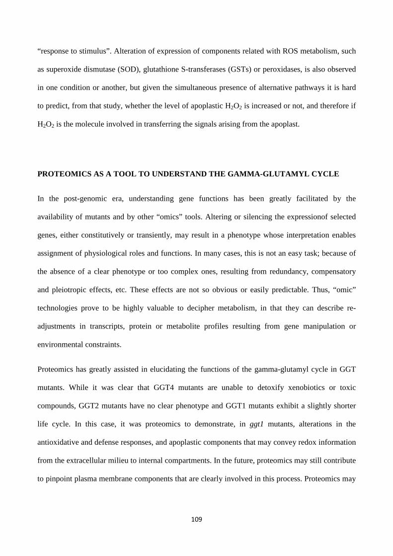
109
“response to stimulus”. Alteration of expression of components related with ROS metabolism, such
as superoxide dismutase (SOD), glutathione S-transferases (GSTs) or peroxidases, is also observed
in one condition or another, but given the simultaneous presence of alternative pathways it is hard
to predict, from that study, whether the level of apoplastic H2O2 is increased or not, and therefore if
H2O2 is the molecule involved in transferring the signals arising from the apoplast.
PROTEOMICS AS A TOOL TO UNDERSTAND THE GAMMA-GLUTAM YL CYCLE
In the post-genomic era, understanding gene functions has been greatly facilitated by the
availability of mutants and by other “omics” tools. Altering or silencing the expressionof selected
genes, either constitutively or transiently, may result in a phenotype whose interpretation enables
assignment of physiological roles and functions. In many cases, this is not an easy task; because of
the absence of a clear phenotype or too complex ones, resulting from redundancy, compensatory
and pleiotropic effects, etc. These effects are not so obvious or easily predictable. Thus, “omic”
technologies prove to be highly valuable to decipher metabolism, in that they can describe re-
adjustments in transcripts, protein or metabolite profiles resulting from gene manipulation or
environmental constraints.
Proteomics has greatly assisted in elucidating the functions of the gamma-glutamyl cycle in GGT
mutants. While it was clear that GGT4 mutants are unable to detoxify xenobiotics or toxic
compounds, GGT2 mutants have no clear phenotype and GGT1 mutants exhibit a slightly shorter
life cycle. In this case, it was proteomics to demonstrate, in ggt1 mutants, alterations in the
antioxidative and defense responses, and apoplastic components that may convey redox information
from the extracellular milieu to internal compartments. In the future, proteomics may still contribute
to pinpoint plasma membrane components that are clearly involved in this process. Proteomics may
110
also help in identifying one missing step in the gamma-glutamyl cycle, i.e., the cysteinyl-glycine
dipeptidase, whose occurrence is inferred but not demonstrated yet (Kumada et al., 2007).
CONCLUSIONS
The significance of the gamma-glutamyl cycle is not fully understood yet. Glutathione cycling
between the symplast and apoplast may represent a way to transfer redox information. Functional
genomics approaches indicate that disruption of the functional cell-wall bound GGT1 isoform
results in a constitutive alert response where anti-oxidative enzymes are up-regulated, probably as
an effect of the altered plasma membrane receptors level and the redox state. With the more general
aim of understanding how environmental challenges are perceived by plant cells, it seems therefore
important to conclusively assign a role for extracellular GGTs and the gamma-glutamyl cycle in
controlling the redox signals generated in the apoplast, which are transferred to the symplast and
activate antioxidant systems. To this end, further high-throughput and targeted proteomic
approaches will be necessary to perform and compare under the diverse stresses as indicated in
Fig.1.
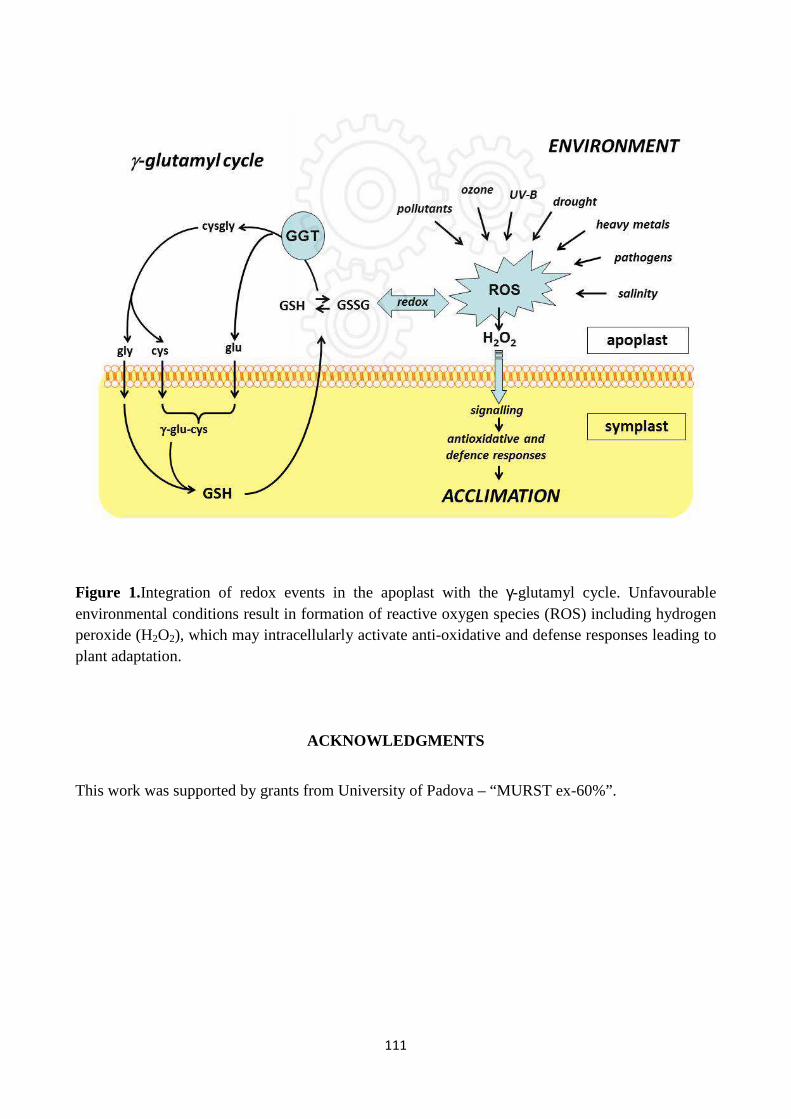
111
Figure 1.Integration of redox events in the apoplast with the γ-glutamyl cycle. Unfavourable environmental conditions result in formation of reactive oxygen species (ROS) including hydrogen peroxide (H2O2), which may intracellularly activate anti-oxidative and defense responses leading to plant adaptation.
ACKNOWLEDGMENTS
This work was supported by grants from University of Padova – “MURST ex-60%”.
112
REFERENCES
1. Agrawal G. K., Jwa N. S., Lebrun M. H., Job D., Rakwal R. (2010). Plant secretome:
unlocking secrets of the secreted proteins. Proteomics 10, 1-29.
2. Ferretti M., Destro T., Tosatto S.C., La Rocca N., Rascio N., Masi A. (2009). Gamma-
glutamyl transferase in the cell wall participates in extracellular glutathione salvage from the
root apoplast. New Phytol. 181, 115-126
3. Foyer C.H., Noctor G. (2005). Redox homeostasis and antioxidant signaling: a metabolic
interface between stress perception and physiological responses. Plant Cell 17, 1866-1875
4. Grzam, A., Martin, M. N., Hell, R., Meyer, A. J. (2007). γ-Glutamyl transpeptidase GGT4
initiates vacuolar degradation of glutathione S-conjugates in Arabidopsis. FEBS Lett. 581,
3131-3138.
5. Kumada H. O., Koizumi Y., Sekiya J. (2007). Purification and characterization of
dipeptidase hydrolyzing L-cysteinylglycine from radish cotyledon. Biosci. Biotechnol.
Biochem. 71, 3102-3104.
6. Martin M. N., Saladores P. H., Lambert E., Hudson A. O., Leustek T. (2007). Localization
of members of the γ-glutamyl transpeptidase family identifies sites of glutathione and
glutathione S-conjugate hydrolysis. Plant Physiol. 144, 1715-1732.
7. Masi A., Ghisi R., Ferretti M. (2002). Measuring low-molecular-weight thiols by detecting
the fluorescence of their SBD derivatives: application to studies of diurnal and UV-B
induced changes in Zea mays L. J. Plant Physiol. 159, 499-0507.
8. Mittler R., Vanderauwera S., Gollery M., Van Breusegem F. (2004). Reactive oxygen gene
network of plants. Trends Plant Sci. 9, 490-498.
9. Noctor G. (2006). Metabolic signaling in defence and stress: the central roles of soluble
redox couples. Plant Cell Environ. 29, 409-425.
113
10. Pignocchi C., Foyer C. H. (2003). Apoplastic ascorbate metabolism and its role in the
regulation of cell signalling. Curr. Opin. Plant Biol. 6, 379-389.
11. Pitzschke A., Forzani C., Hirt H. (2006). Reactive oxygen species signaling in plants.
Antioxid. Redox Signal. 8, 1757-1764.
12. Potters G., Horemans N., Jansen M. A. K. (2010). The cellular redox state in plant stress
biology – a charging concept. Plant Physiol. Biochem. 48, 292-300
13. Tolin S., Arrigoni G., Trentin A. R., Veljovic-Jovanovic S., Pivato M., Zechman B., Masi
A. (2013). Biochemical and quantitative proteomics investigations in Arabidopsis ggt1
mutant leaves reveal a role for the gamma-glutamyl cycle in plant's adaptation to
environment. Proteomics 13, 2031-2045.
14. Torres M. A., Jones J. D. G., Dangl J. L. (2006). Reactive oxygen species signaling in
response to pathogens. Plant Physiol. 41, 373-378.
15. Trentin A. R. et al.(submitted to Frontiers in Plant Science, revision pending).
16. Zechmann B. (2014). Compartment-specific importance of glutathione during abiotic and
biotic stress. Front. Plant Sci. 5, 566.
114
CHAPTER 3: REGULATION OF ROS HOMEOSTASIS IN APOPLASTIC SPACE BY THIOLS
ABSTRACT
Glutathione and low molecular weight thiols (cysteine and cysteinyl-glycine), with their –SH
moiety, are strongly reactive nucleophilic molecules. They have a main role in plant metabolism,
defense from biotic and abiotic stress and in redox homeostasis by contrasting excessive oxidation
states.
In plant cell the apoplast is an interphase between the environment and symplast. Environmental
oxidative stresses evoke in the apoplastic space production of reactive oxygen species (ROS)
subsequently, in some way, a signal transduction pathways is activated. This allows to signal to
pass through plasma membrane and reach the cytoplasm.
Aim of this work was to investigate the role of LMW thiols in root apoplast, by highlighting
reactions involving low molecular weight thiols, metals and enzymes related to ROS metabolism.
Our results obtained with chromatography, ICP spectrometry, EPR spectroscopy and absorption
spectroscopy analyses, and in presence of isolated cell walls, indicate that glutathione, cysteinyl-
glycine and cysteine have quenching capacity for reactive oxygen species generated in the
apoplastic space.
Our findings point to a role for LMW thiols, which are metabolically related to each other in the
gamma-glutamyl cycle, in modulating redox reactions in plant cell walls.
115
INTRODUCTION
Reactive oxygen species (ROS) generation requires an activation energy but then it continues
spontaneously (Karuppanapandianet al., 2011); in plant tissues and cells they can be produced by
photosynthesis and by various environmental stresses (Schofer et al., 2001; Rodrigues-Serrano et
al., 2006). As a consequence, ROS concentration in the apoplast varies fastly and highly due to
biotic and abiotic stresses (Hernandez et al., 2001; Jaspers et al., 2005; Hu et al., 2005; Pristov et
al., 2013).
Notably, regulation of a plethora of important cell functions occurs by oxidation/reduction reactions
in mammalian cells (Paolicchi et al., 2002) and recent evidences have shown that ROS play a key
role as a messenger in normal cell signal transduction and cell cycling (Rahantaniaina et al., 2013).
In cell wall and apoplast there are several enzymes, like oxidases and peroxidases, that produce
ROS in response to development and environmental challenge. Moreover, it is known that
superoxide has an important role in redox signalling and activates defense responses (Foyer and
Noctor 2009). Plants present many and different strategies to improve defence responses by
scavenging ROS with enzymatic and non-enzymatic processes (Sharma et al., 2012).
One of the main non-protein antioxidants in cells is glutathione, a versatile molecule with a plethora
of roles in plant metabolism and defense. It is known that GSH plays a crucial protective role in
redox homeostasis (Foyer and Noctor 2011); it reacts with ROS thus protecting thiol groups to
contrast excessive protein oxidation (Rahantaniaina et al., 2013). Glutathione’s functions depend on
cysteine’s thiol moiety, a strong nucleophilic group (Pivato et al., 2014) that allows reacting with a
wide spectrum of molecules to form not only GSSG but also GS-conjugates (Dixon and Edwards,
2010) and protein thiolation (Pivato et al., 2014). Glutathione scavenging capacity is depending on
pKa for the sulphydryl group (pKa = 8.83); when proton concentration decreases also glutathione
redox potential is reduced. This property has relevant physiological implications, since pH differs
significantly among cell compartments; consequently, also the GSH/GSSG ratio is expected to
change (Rahantaniaina et al., 2013). It is known that pH in the apoplastic space is around 4.5-5;
therefore, in theory, glutathione will be completely reduced. However, the presence of oxidizing
molecules are able to oxidize GSH to GSSG (e.g.: dehydroascorbate, ROS).
In plant tissues, GSH is mainly localised intracellularly; however, a limited amount is also found in
the apoplastic space, where it is involved in the so called gamma-glutamyl cycle. In this cycle, it is
synthetized in the cytosol, carried out to the extracellular space and cleaved by gamma-
glutamyltransferase (GGT) to produce cysteinyl-glycine (cys-gly) and glutamate (Ferretti et al.,
2009). Subsequently, cysgly is cleaved to cysteine and glycine by an unknown yet dipeptidase
116
(Ohkama-Ohtsu et al., 2008); single aminoacids can finally cross plasma membrane by means of
aminoacid transporters, and be used in the cytosol to synthesize new GSH.
The presence of these GSH-related thiols in the apoplast may result in redox reactions and an
altered redox balance in the extracellular space, due to their biochemical properties and reactivity.
Although GSH is commonly considered a major antioxidant in living cells, in the last few years it
has been provided evidence for a prooxidant role of GSH and thiols deriving from its catabolism in
animals, as a result of gamma-glutamyl transpeptidase (GGT) activity(Stark et al., 1993; Pompella
et al., 2003; Dominici et al., 2005). This novel evidence may have strong implications also in
plantbiology and physiology.
In particular, GSH, cysteine and cysteinyl-glycine promote metal ions reduction and induce
production of reactive species such as superoxide, hydrogen peroxide and other free radicals
(Paolicchi et al., 2002). Metal ions are also present in the apoplast; their concentration depends on
plant species, and they interact with cell wall components and thiols (Speisky et al., 2008, 2011;
Carrasco-Pozo et al., 2008; Aliaga et al., 2010, 2012). Notably, it is reported that redox active
transition metals can react with H2O2 and convert it into hydroxyl radical via metal-catalyzed
Fenton reaction (Karuppanapandian et al., 2011).
Two categories of enzymes located on the cell wall are mainly involved in oxidative metabolism:
peroxidases, which use H2O2 as a substrate, and superoxidase dismutases converting O2- to H2O2
and producing •OH(Higashi et al., 2015; del Río 2015).However, it has been reported that PODs
may also produce H2O2 by oxidation of different kinds of reductants as metal and phenolic
compounds (Urbañski and Berêsewicz 2000; Šukalovicet al., 2005; Kukavica et al., 2009) and in
presence of other reductants (such as NADH) PODs can produce •OH (Chen and Schoffer 1999;
Schoffer et al., 2002). Additionally, there is evidence for a superoxide radical involvement in
hydroxyl radical production (Chen and Schoffer 1999; Liszkay et al., 2004; Karkonen and Fry
2006). However, who are the naturally occurring reductants that in apoplast participate to ROS
production? This question remains still unknown (Kukavica et al., 2009).
Aim of this work is to investigate the role of LMW thiols in root apoplast, by highlighting reactions
and interactions that occur in apoplastic space between low molecular weight thiols, metals and
enzymes involved in response to oxidative metabolism.
In this study, various techniques have been used to determine structural variations occuring in the
apoplastic space due to low molecular weight thiols: HPLCchromatography, ICP spectrometry,
EPR spectroscopy and absorption spectroscopy.
117
MATERIAL AND METHODS
Plant material
Barley seeds were treated with bleach solution for 10 minutes, washed with water and then soaked
to germinate in a dark room in controlled temperature and humidity for 4 days. Seedlings were
transferred into a becker with a modified Hoagland solution (1/2 strength, as described in table 1) in
a growth chamber set as follows 12/12 h light/dark cycle, 21/21 °C temperature, 300 µmol m-2 s-1
photosynthetically active radiation and 60% relative humidity.
Chromatographic low molecular weight thiols determination
10 days plants were used for the experiments. Roots where washed 2 times with 1 mM CaCl2, dryed
and weighed. About 1 g was used for the extraction of extracellular washing fluid (ECWF)
obtained by centrifuging roots at 4°C, 200 g for 20 minutes. Total root extracts were obtained by
powdering plant material in liquid N2 with 0.1 M HCl in ratio 1:4 fresh weight : buffer(n=10
replicates). To determine low molecular weight (LMW) thiols concentration in the extracts, 50 µL
of total leaf extract and ECWF were derivatized with 4-Fluoro-7-sulfobenzofurazan ammonium salt
fluorophore (SBD-F) (Dojindo, Japan). Thiols were separated by isocratic HPLC using the method
described by Masi et al., 2002 with some modifications. The mobile phase was 75 mM NH4-
formiate, pH 2.9 and 3% methanol (97:3, vol/vol).
Metals determination
The elemental determination was conducted with ICP-OES, SPECTRO ARCOS EOP (Spectro A.I.,
Germany).
The ICP-OES was employed to determine 24 elements: Al, As, B, Ba, Ca, Cd, Co, Cr, Cu, Fe, K,
Mg, Mn, Mo, Na, Ni, P, Pb, S, Se, Sr, Tl, V and Zn. All instrument operating parameters were
optimized for nitric acid 3% solution.
Sample Preparation and Calibration Standards
The root samples were analysed after microwave closed vessel digestion (Ethos 1600 Milestone
S.r.l. Sorisole, BG, Italy) . Roots (0,35 g)were added to hydrogen peroxide and nitric acid
“suprapure” quality (Merck, Darmstadt, Germany). After microwave digestion, the so dissolved
sample was diluted with ultrapure water to a final volume of 25 ml. Cell wall fractions were
obtained by powdering roots in liquid N2and extraction with buffer (50 mM Tris-HCl pH 7.2, 50
mM NaCl, 0.05% Tween 80, 1 mM PMSF), and centrifugation at 4°C, 500 g for 20 minutes. The
pellet phase contained cell wall fraction and was digested with microware as above.Calibration
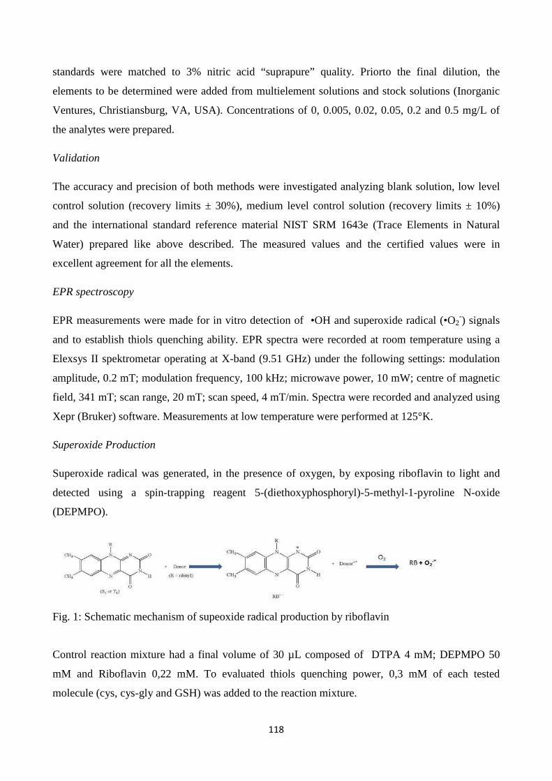
118
standards were matched to 3% nitric acid “suprapure” quality. Priorto the final dilution, the
elements to be determined were added from multielement solutions and stock solutions (Inorganic
Ventures, Christiansburg, VA, USA). Concentrations of 0, 0.005, 0.02, 0.05, 0.2 and 0.5 mg/L of
the analytes were prepared.
Validation
The accuracy and precision of both methods were investigated analyzing blank solution, low level
control solution (recovery limits ± 30%), medium level control solution (recovery limits ± 10%)
and the international standard reference material NIST SRM 1643e (Trace Elements in Natural
Water) prepared like above described. The measured values and the certified values were in
excellent agreement for all the elements.
EPR spectroscopy
EPR measurements were made for in vitro detection of •OH and superoxide radical (•O2-) signals
and to establish thiols quenching ability. EPR spectra were recorded at room temperature using a
Elexsys II spektrometar operating at X-band (9.51 GHz) under the following settings: modulation
amplitude, 0.2 mT; modulation frequency, 100 kHz; microwave power, 10 mW; centre of magnetic
field, 341 mT; scan range, 20 mT; scan speed, 4 mT/min. Spectra were recorded and analyzed using
Xepr (Bruker) software. Measurements at low temperature were performed at 125°K.
Superoxide Production
Superoxide radical was generated, in the presence of oxygen, by exposing riboflavin to light and
detected using a spin-trapping reagent 5-(diethoxyphosphoryl)-5-methyl-1-pyroline N-oxide
(DEPMPO).
Fig. 1: Schematic mechanism of supeoxide radical production by riboflavin
Control reaction mixture had a final volume of 30 µL composed of DTPA 4 mM; DEPMPO 50
mM and Riboflavin 0,22 mM. To evaluated thiols quenching power, 0,3 mM of each tested
molecule (cys, cys-gly and GSH) was added to the reaction mixture.
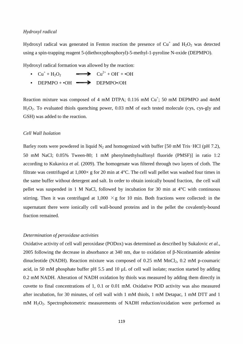
119
Hydroxyl radical
Hydroxyl radical was generated in Fenton reaction the presence of Cu+ and H2O2 was detected
using a spin-trapping reagent 5-(diethoxyphosphoryl)-5-methyl-1-pyroline N-oxide (DEPMPO).
Hydroxyl radical formation was allowed by the reaction:
• Cu+ + H2O2 Cu2+ + OH- + •OH
• DEPMPO + •OH DEPMPO•/OH
Reaction mixture was composed of 4 mM DTPA; 0.116 mM Cu+; 50 mM DEPMPO and 4mM
H2O2. To evaluated thiols quenching power, 0.03 mM of each tested molecule (cys, cys-gly and
GSH) was added to the reaction.
Cell Wall Isolation
Barley roots were powdered in liquid N2 and homogenized with buffer [50 mM Tris–HCl (pH 7.2),
50 mM NaCl; 0.05% Tween-80; 1 mM phenylmethylsulfonyl fluoride (PMSF)] in ratio 1:2
according to Kukavica et al. (2009). The homogenate was filtered through two layers of cloth. The
filtrate was centrifuged at 1,000× g for 20 min at 4°C. The cell wall pellet was washed four times in
the same buffer without detergent and salt. In order to obtain ionically bound fraction, the cell wall
pellet was suspended in 1 M NaCl, followed by incubation for 30 min at 4°C with continuous
stirring. Then it was centrifuged at 1,000 ×g for 10 min. Both fractions were collected: in the
supernatant there were ionically cell wall-bound proteins and in the pellet the covalently-bound
fraction remained.
Determination of peroxidase activities
Oxidative activity of cell wall peroxidase (PODox) was determined as described by Sukalovic et al.,
2005 following the decrease in absorbance at 340 nm, due to oxidation of β-Nicotinamide adenine
dinucleotide (NADH). Reaction mixture was composed of 0.25 mM MnCl2, 0.2 mM p-coumaric
acid, in 50 mM phosphate buffer pH 5.5 and 10 µL of cell wall isolate; reaction started by adding
0.2 mM NADH. Alteration of NADH oxidation by thiols was measured by adding them directly in
cuvette to final concentrations of 1, 0.1 or 0.01 mM. Oxidative POD activity was also measured
after incubation, for 30 minutes, of cell wall with 1 mM thiols, 1 mM Detapac, 1 mM DTT and 1
mM H2O2. Spectrophotometric measurements of NADH reduction/oxidation were performed as

120
described above except. All the assays were performed at 28°C using Shimadzu, UV-160
spectrophotometer (Japan).
RESULTS AND DISCUSSION
Thiols detection Low molecular weight thiols were measured in a total roots extract and in the extracellular washing
fluids. A representative chromatogram of thiols distribution is reported in Fig. 1 and contents are
shown in table 1.
Fig. 1: A rapresentative chromatogram of thiols in barley roots in a total extract (red line) and in the apoplast (black line).
Cys Cys-Gly γ-EC GSH total extract 6,75 ± 0,94 1,79 ± 0,50 225,73 ± 88,03 89,60 ± 11,82 nmol/g FW
apoplastic fluid 2,10 ± 1,29 2,26 ± 1,18 1,94 ± 2,15 1,33 ± 0,75 nmol/mL
ratio apoplast/total 0,31 ± 0,22 1,26 ± 0,66 0,01 0,01
Table 1: Thiols content in barley: in total roots, apoplastic space and relative distribution. Values
are mean of 8 biological replicates + standard deviation.
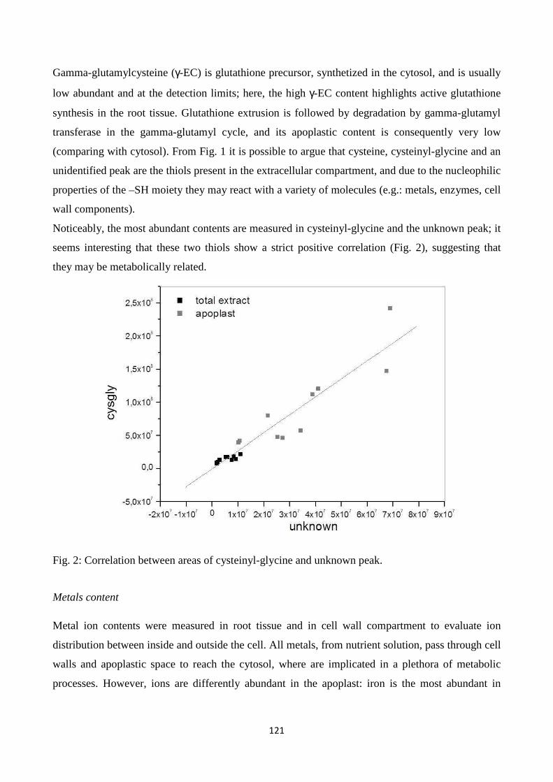
121
Gamma-glutamylcysteine (γ-EC) is glutathione precursor, synthetized in the cytosol, and is usually
low abundant and at the detection limits; here, the high γ-EC content highlights active glutathione
synthesis in the root tissue. Glutathione extrusion is followed by degradation by gamma-glutamyl
transferase in the gamma-glutamyl cycle, and its apoplastic content is consequently very low
(comparing with cytosol). From Fig. 1 it is possible to argue that cysteine, cysteinyl-glycine and an
unidentified peak are the thiols present in the extracellular compartment, and due to the nucleophilic
properties of the –SH moiety they may react with a variety of molecules (e.g.: metals, enzymes, cell
wall components).
Noticeably, the most abundant contents are measured in cysteinyl-glycine and the unknown peak; it
seems interesting that these two thiols show a strict positive correlation (Fig. 2), suggesting that
they may be metabolically related.
Fig. 2: Correlation between areas of cysteinyl-glycine and unknown peak.
Metals content
Metal ion contents were measured in root tissue and in cell wall compartment to evaluate ion
distribution between inside and outside the cell. All metals, from nutrient solution, pass through cell
walls and apoplastic space to reach the cytosol, where are implicated in a plethora of metabolic
processes. However, ions are differently abundant in the apoplast: iron is the most abundant in
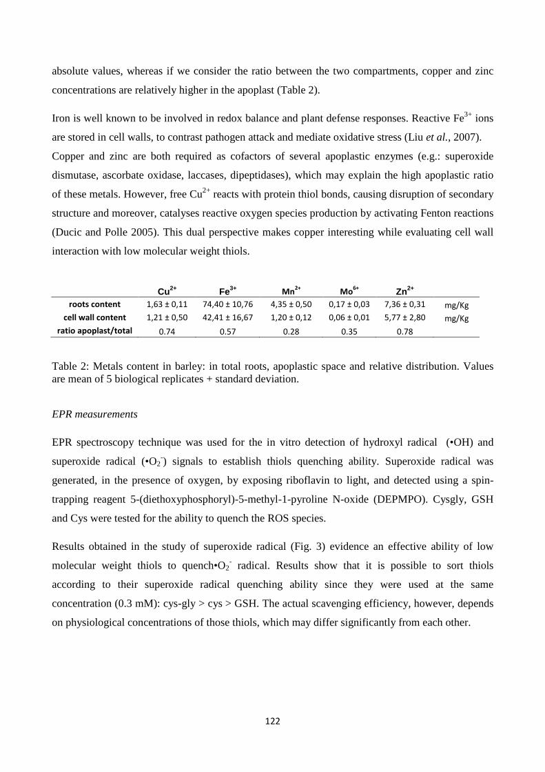
122
absolute values, whereas if we consider the ratio between the two compartments, copper and zinc
concentrations are relatively higher in the apoplast (Table 2).
Iron is well known to be involved in redox balance and plant defense responses. Reactive Fe3+ ions
are stored in cell walls, to contrast pathogen attack and mediate oxidative stress (Liu et al., 2007).
Copper and zinc are both required as cofactors of several apoplastic enzymes (e.g.: superoxide
dismutase, ascorbate oxidase, laccases, dipeptidases), which may explain the high apoplastic ratio
of these metals. However, free Cu2+ reacts with protein thiol bonds, causing disruption of secondary
structure and moreover, catalyses reactive oxygen species production by activating Fenton reactions
(Ducic and Polle 2005). This dual perspective makes copper interesting while evaluating cell wall
interaction with low molecular weight thiols.
Cu2+ Fe3+ Mn2+
Mo6+
Zn2+
roots content 1,63 ± 0,11 74,40 ± 10,76 4,35 ± 0,50 0,17 ± 0,03 7,36 ± 0,31 mg/Kg
cell wall content 1,21 ± 0,50 42,41 ± 16,67 1,20 ± 0,12 0,06 ± 0,01 5,77 ± 2,80 mg/Kg
ratio apoplast/total 0.74 0.57 0.28 0.35 0.78
Table 2: Metals content in barley: in total roots, apoplastic space and relative distribution. Values are mean of 5 biological replicates + standard deviation.
EPR measurements
EPR spectroscopy technique was used for the in vitro detection of hydroxyl radical (•OH) and
superoxide radical (•O2-) signals to establish thiols quenching ability. Superoxide radical was
generated, in the presence of oxygen, by exposing riboflavin to light, and detected using a spin-
trapping reagent 5-(diethoxyphosphoryl)-5-methyl-1-pyroline N-oxide (DEPMPO). Cysgly, GSH
and Cys were tested for the ability to quench the ROS species.
Results obtained in the study of superoxide radical (Fig. 3) evidence an effective ability of low
molecular weight thiols to quench•O2- radical. Results show that it is possible to sort thiols
according to their superoxide radical quenching ability since they were used at the same
concentration (0.3 mM): cys-gly > cys > GSH. The actual scavenging efficiency, however, depends
on physiological concentrations of those thiols, which may differ significantly from each other.
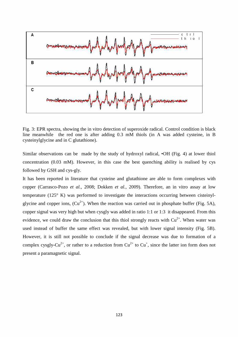
123
Fig. 3: EPR spectra, showing the in vitro detection of superoxide radical. Control condition is black line meanwhile the red one is after adding 0.3 mM thiols (in A was added cysteine, in B cysteinylglycine and in C glutathione).
Similar observations can be made by the study of hydroxyl radical, •OH (Fig. 4) at lower thiol
concentration (0.03 mM). However, in this case the best quenching ability is realised by cys
followed by GSH and cys-gly.
It has been reported in literature that cysteine and glutathione are able to form complexes with
copper (Carrasco-Pozo et al., 2008; Dokken et al., 2009). Therefore, an in vitro assay at low
temperature (125° K) was performed to investigate the interactions occurring between cisteinyl-
glycine and copper ions, (Cu2+). When the reaction was carried out in phosphate buffer (Fig. 5A),
copper signal was very high but when cysgly was added in ratio 1:1 or 1:3 it disappeared. From this
evidence, we could draw the conclusion that this thiol strongly reacts with Cu2+. When water was
used instead of buffer the same effect was revealed, but with lower signal intensity (Fig. 5B).
However, it is still not possible to conclude if the signal decrease was due to formation of a
complex cysgly-Cu2+, or rather to a reduction from Cu2+ to Cu+, since the latter ion form does not
present a paramagnetic signal.
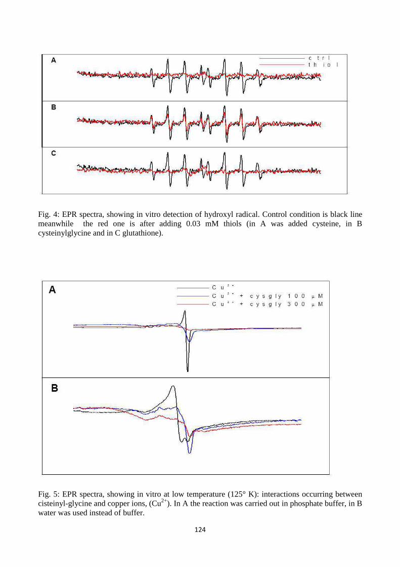
124
Fig. 4: EPR spectra, showing in vitro detection of hydroxyl radical. Control condition is black line meanwhile the red one is after adding 0.03 mM thiols (in A was added cysteine, in B cysteinylglycine and in C glutathione).
Fig. 5: EPR spectra, showing in vitro at low temperature (125° K): interactions occurring between cisteinyl-glycine and copper ions, (Cu2+). In A the reaction was carried out in phosphate buffer, in B water was used instead of buffer.
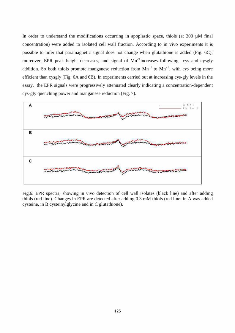
125
In order to understand the modifications occurring in apoplastic space, thiols (at 300 µM final
concentration) were added to isolated cell wall fraction. According to in vivo experiments it is
possible to infer that paramagnetic signal does not change when glutathione is added (Fig. 6C);
moreover, EPR peak height decreases, and signal of Mn2+increases following cys and cysgly
addition. So both thiols promote manganese reduction from Mn3+ to Mn2+, with cys being more
efficient than cysgly (Fig. 6A and 6B). In experiments carried out at increasing cys-gly levels in the
essay, the EPR signals were progressively attenuated clearly indicating a concentration-dependent
cys-gly quenching power and manganese reduction (Fig. 7).
Fig.6: EPR spectra, showing in vivo detection of cell wall isolates (black line) and after adding thiols (red line). Changes in EPR are detected after adding 0.3 mM thiols (red line: in A was added cysteine, in B cysteinylglycine and in C glutathione).
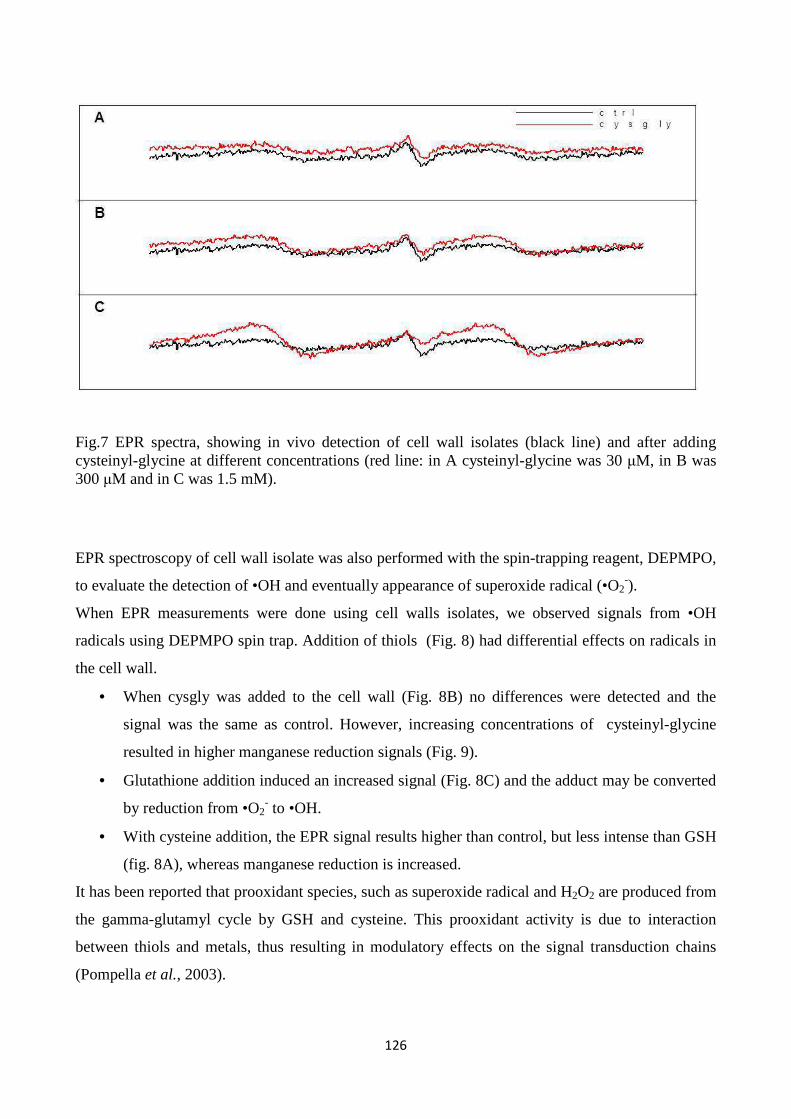
126
Fig.7 EPR spectra, showing in vivo detection of cell wall isolates (black line) and after adding cysteinyl-glycine at different concentrations (red line: in A cysteinyl-glycine was 30 µM, in B was 300 µM and in C was 1.5 mM).
EPR spectroscopy of cell wall isolate was also performed with the spin-trapping reagent, DEPMPO,
to evaluate the detection of •OH and eventually appearance of superoxide radical (•O2-).
When EPR measurements were done using cell walls isolates, we observed signals from •OH
radicals using DEPMPO spin trap. Addition of thiols (Fig. 8) had differential effects on radicals in
the cell wall.
• When cysgly was added to the cell wall (Fig. 8B) no differences were detected and the
signal was the same as control. However, increasing concentrations of cysteinyl-glycine
resulted in higher manganese reduction signals (Fig. 9).
• Glutathione addition induced an increased signal (Fig. 8C) and the adduct may be converted
by reduction from •O2- to •OH.
• With cysteine addition, the EPR signal results higher than control, but less intense than GSH
(fig. 8A), whereas manganese reduction is increased.
It has been reported that prooxidant species, such as superoxide radical and H2O2 are produced from
the gamma-glutamyl cycle by GSH and cysteine. This prooxidant activity is due to interaction
between thiols and metals, thus resulting in modulatory effects on the signal transduction chains
(Pompella et al., 2003).
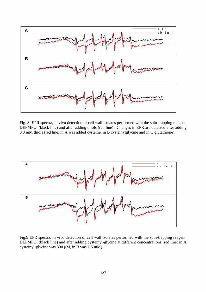
127
Fig. 8: EPR spectra, in vivo detection of cell wall isolates performed with the spin-trapping reagent, DEPMPO, (black line) and after adding thiols (red line) . Changes in EPR are detected after adding 0.3 mM thiols (red line: in A was added cysteine, in B cysteinylglycine and in C glutathione).
Fig.9 EPR spectra, in vivo detection of cell wall isolates performed with the spin-trapping reagent, DEPMPO, (black line) and after adding cysteinyl-glycine at different concentrations (red line: in A cysteinyl-glycine was 300 µM, in B was 1.5 mM).
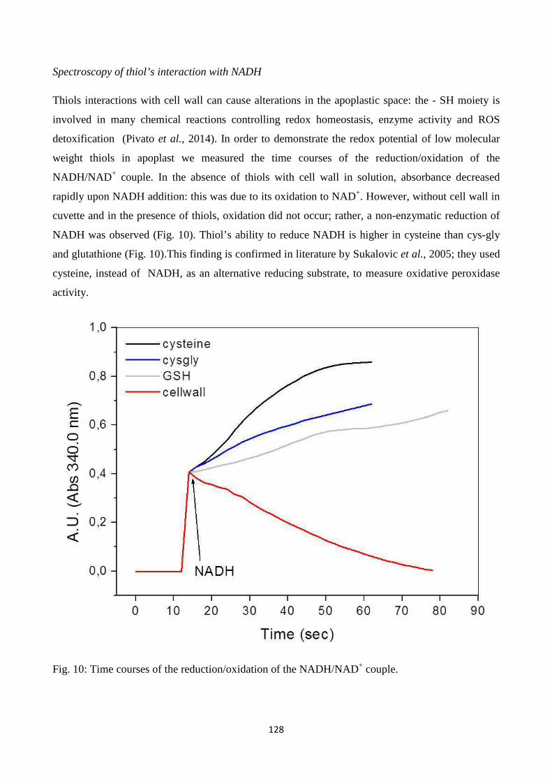
128
Spectroscopy of thiol’s interaction with NADH
Thiols interactions with cell wall can cause alterations in the apoplastic space: the - SH moiety is
involved in many chemical reactions controlling redox homeostasis, enzyme activity and ROS
detoxification (Pivato et al., 2014). In order to demonstrate the redox potential of low molecular
weight thiols in apoplast we measured the time courses of the reduction/oxidation of the
NADH/NAD+ couple. In the absence of thiols with cell wall in solution, absorbance decreased
rapidly upon NADH addition: this was due to its oxidation to NAD+. However, without cell wall in
cuvette and in the presence of thiols, oxidation did not occur; rather, a non-enzymatic reduction of
NADH was observed (Fig. 10). Thiol’s ability to reduce NADH is higher in cysteine than cys-gly
and glutathione (Fig. 10).This finding is confirmed in literature by Sukalovic et al., 2005; they used
cysteine, instead of NADH, as an alternative reducing substrate, to measure oxidative peroxidase
activity.
Fig. 10: Time courses of the reduction/oxidation of the NADH/NAD+ couple.
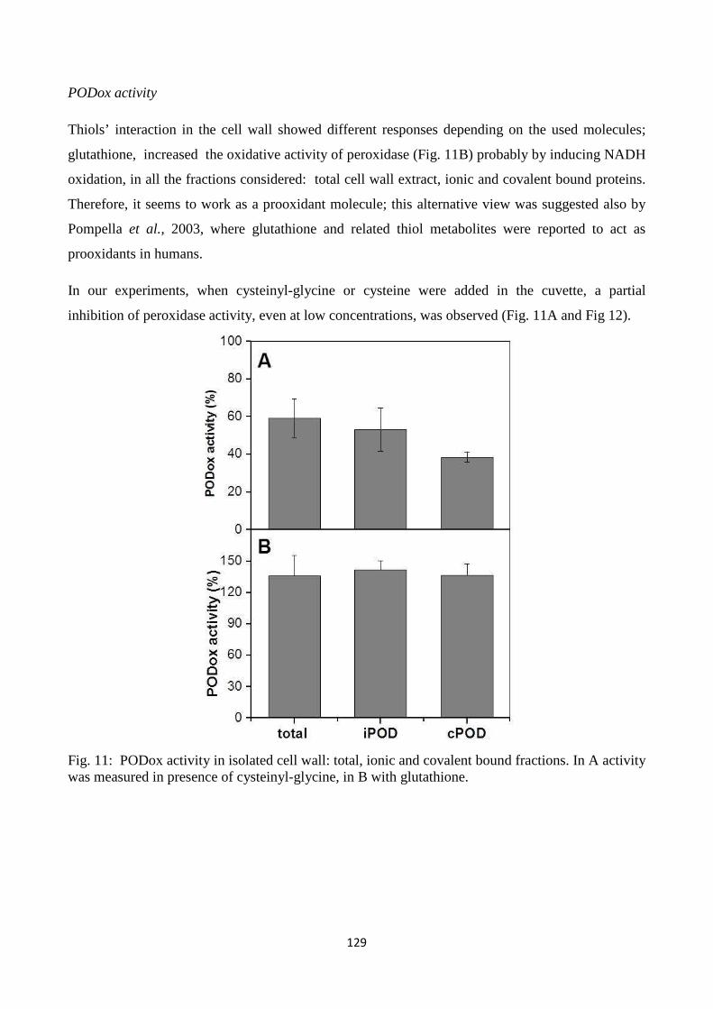
129
PODox activity
Thiols’ interaction in the cell wall showed different responses depending on the used molecules;
glutathione, increased the oxidative activity of peroxidase (Fig. 11B) probably by inducing NADH
oxidation, in all the fractions considered: total cell wall extract, ionic and covalent bound proteins.
Therefore, it seems to work as a prooxidant molecule; this alternative view was suggested also by
Pompella et al., 2003, where glutathione and related thiol metabolites were reported to act as
prooxidants in humans.
In our experiments, when cysteinyl-glycine or cysteine were added in the cuvette, a partial
inhibition of peroxidase activity, even at low concentrations, was observed (Fig. 11A and Fig 12).
Fig. 11: PODox activity in isolated cell wall: total, ionic and covalent bound fractions. In A activity was measured in presence of cysteinyl-glycine, in B with glutathione.
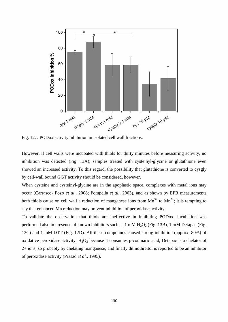
130
Fig. 12: : PODox activity inhibition in isolated cell wall fractions. However, if cell walls were incubated with thiols for thirty minutes before measuring activity, no
inhibition was detected (Fig. 13A); samples treated with cysteinyl-glycine or glutathione even
showed an increased activity. To this regard, the possibility that glutathione is converted to cysgly
by cell-wall bound GGT activity should be considered, however.
When cysteine and cysteinyl-glycine are in the apoplastic space, complexes with metal ions may
occur (Carrasco- Pozo et al., 2008; Pompella et al., 2003), and as shown by EPR measurements
both thiols cause on cell wall a reduction of manganese ions from Mn3+ to Mn2+; it is tempting to
say that enhanced Mn reduction may prevent inhibition of peroxidase activity.
To validate the observation that thiols are ineffective in inhibiting PODox, incubation was
performed also in presence of known inhibitors such as 1 mM H2O2 (Fig. 13B), 1 mM Detapac (Fig.
13C) and 1 mM DTT (Fig. 12D). All these compounds caused strong inhibition (approx. 80%) of
oxidative peroxidase activity: H2O2 because it consumes p-coumaric acid; Detapac is a chelator of
2+ ions, so probably by chelating manganese; and finally dithiothreitol is reported to be an inhibitor
of peroxidase activity (Prasad et al., 1995).
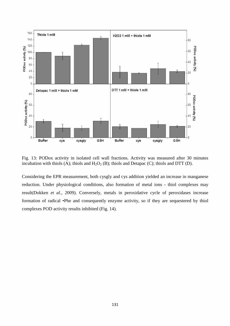
131
Fig. 13: PODox activity in isolated cell wall fractions. Activity was measured after 30 minutes incubation with thiols (A); thiols and H2O2 (B); thiols and Detapac (C); thiols and DTT (D).
Considering the EPR measurement, both cysgly and cys addition yielded an increase in manganese
reduction. Under physiological conditions, also formation of metal ions - thiol complexes may
result(Dokken et al., 2009). Conversely, metals in peroxidative cycle of peroxidases increase
formation of radical •Phe and consequently enzyme activity, so if they are sequestered by thiol
complexes POD activity results inhibited (Fig. 14).
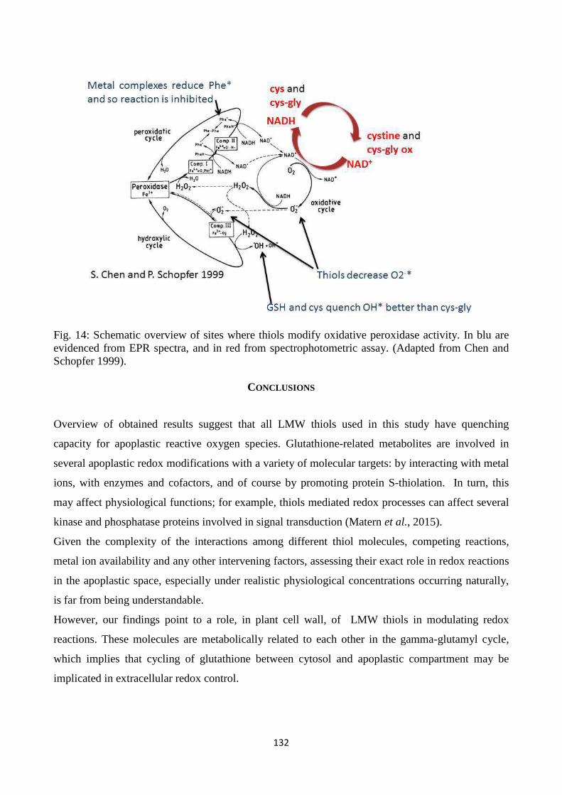
132
Fig. 14: Schematic overview of sites where thiols modify oxidative peroxidase activity. In blu are evidenced from EPR spectra, and in red from spectrophotometric assay. (Adapted from Chen and Schopfer 1999).
CONCLUSIONS
Overview of obtained results suggest that all LMW thiols used in this study have quenching
capacity for apoplastic reactive oxygen species. Glutathione-related metabolites are involved in
several apoplastic redox modifications with a variety of molecular targets: by interacting with metal
ions, with enzymes and cofactors, and of course by promoting protein S-thiolation. In turn, this
may affect physiological functions; for example, thiols mediated redox processes can affect several
kinase and phosphatase proteins involved in signal transduction (Matern et al., 2015).
Given the complexity of the interactions among different thiol molecules, competing reactions,
metal ion availability and any other intervening factors, assessing their exact role in redox reactions
in the apoplastic space, especially under realistic physiological concentrations occurring naturally,
is far from being understandable.
However, our findings point to a role, in plant cell wall, of LMW thiols in modulating redox
reactions. These molecules are metabolically related to each other in the gamma-glutamyl cycle,
which implies that cycling of glutathione between cytosol and apoplastic compartment may be
implicated in extracellular redox control.
133
REFERENCES
Aliaga M.E., López-Alarcón C., Barriga G., Olea-Azar C., Speisky H. (2010). “Redox-active
complexes formed during the interaction between glutathione and mercury and/or copper ions”.J
Inorg Biochem 104(10):1084-90. doi: 10.1016/j.jinorgbio.2010.06.006.
Aliaga M.E., López-Alarcón C., García-Río L., Martín-Pastor M., Speisky H. (2012). “Redox-
changes associated with the glutathione-dependent ability of the Cu(II)-GSSG complex to generate
superoxide”.Bioorg Med Chem 1;20(9):2869-76. doi: 10.1016/j.bmc.2012.03.027.
Carrasco-Pozo C., Aliaga M.E., Olea-Azar C., Speisky H. (2008). “Double edge redox-implications
for the interaction between endogenous thiols and copper ions: In vitro studies”. Bioorg Med Chem
15;16(22):9795-803. doi: 10.1016/j.bmc.2008.09.068.
Chen S. and SchopferP. (1999). “Hydroxyl-radical production in physiological reactionsA novel
function of peroxidase”.Eur J Biochem 260 pp. 726-735.
del Río L.A. (2015). “ROS and RNS in plant physiology: an overview”. J Exp Bot 66 (10): 2827-
2837. doi: 10.1093/jxb/erv099.
Dixon D.P., Edwards R.(2010).“GlutathioneS-transferases”.InThe Arabidopsis Book. American
Society of Plant Biologists, Rockville, MD, doi/10.1199:tab.0131.
Dokken K.M., Parsons J.G., McClure J., and Gardea-Torresdey J.L. (2009). Synthesis and structural
analysis of copper(II) cysteine complexes Inorg Chim Acta362 pp. 395–401.
Dominici S., Paolicchi A., Corti A., Maellaro E., Pompella A. (2005). “Prooxidant reactions
promoted by soluble and cell-bound gamma-glutamyltransferase activity”. Methods Enzymol 401
pp. 484–501.
Ducic T. and Polle A. (2005). “Transport and detoxification of manganese and copper in plants”.
Braz J Plant Physiol17 pp. 103-112.
Ferretti M., Destro T., Tosatto S.C.E., La Rocca N., Rascio N., Masi A. (2009). “Gamma-glutamyl
transferase in the cell wall participates in extracellular glutathione salvage from the root apoplast”.
New Phytol 181 pp. 115–126.
134
Foyer C.H. and Noctor G. (2009). “Redox Regulation in Photosynthetic Organisms: Signaling,
Acclimation, and Practical Implications”. Antioxidants & RedoxSignaling 11(4) pp. 861-905.
doi:10.1089/ars.2008.2177.
Foyer C.H., Noctor G. (2011). “Ascorbate and Glutathione: the heart of the redox hub”. Plant
Physiol155 pp. 2-18.
Hernandez J.A., Ferrer M.A., Jimenez A., Barcelo A.R., Sevilla F. (2001). “Antioxidant systems
and O2.-/ H2O2 production in the apoplast of Pea leaves. Its relation with salt-induced necrotic
lesions in minor veins”. Plant Physiol 127 pp. 817–831.
HigashiY., TakechiK., TakanoH., Takio S. (2015). “Maintenance of Normal Stress Tolerance in the
Moss Physcomitrella patens Lacking Chloroplastic CuZn-Superoxide Dismutase”.AJPS
Vol.06 No.05 Article ID:54601,10 pages doi: 10.4236/ajps.2015.65064
Hu J.F., Li G.F., Gao Z.H., Chen L., Ren H B., Jia W.S. (2005). “Regulation of water deficit-
induced abscisic acid accumulation by apoplastic ascorbic acid in maize seedlings”. J Integr Plant
Biol47 pp. 1335-1344.
Jaspers, P., Kollist, H., Langebartels, C., Kangasjarvi, J. (2005). “Plant responses to ozone,” in
Antioxidants and reactive oxygen species in plants, ed. N. Smirnoff (Oxford: Blackwell Publishing)
pp. 268-292.
Karkonen A. and Fry S.C. ( 2006 ).“Effect of ascorbate and its oxidation products on H2O2
production in cell-suspension cultures of Picea abies and in the absence of cells”. J Exp Bot 57 pp.
1633 – 1644 .
Karuppanapandian T., Moon J.C., Kim C., Manoharan K., Kim W. (2011). “Reactive oxygen
species in plants: their generation, signal transduction, and scavenging mechanisms”. AJCS5(6) pp.
709-725.
Kukavica B., Mojovic´ M., Vucˇinic´Ž., Maksimovic´ V., Takahama U.and Veljovic´ Jovanovic´ S.
(2009).“Generation of Hydroxyl Radical in Isolated Pea Root Cell Wall, and the Role of Cell Wall-
Bound Peroxidase, Mn-SOD and Phenolics in Their Production”. Plant Cell Physiol50(2) pp. 304–
317 doi:10.1093/pcp/pcn199.
135
Liszkay A., van der Zalm A.and Schopfer P. ( 2004 ). “Production of reactive oxygen intermediates
(O 2–・ H2O2and ・OH) by maize rootsand their role in wall loosening and elongation growth”.
Plant Physiol136 pp. 3114 – 3123.
Liu G., Greenshields D.L., Sammynaiken R., Hirji R.N., Selvaraj G., Wei Y. (2007). “Targeted
alterations in iron homeostasis underlie plant defense responses”. J Cell Sci 15;120 pp. 596-605.
Masi, A., Ghisi, R., Ferretti, M., (2002). “Measuring low-molecular-weight thiols by detecting the
fluorescence of their SBD derivatives: application to studies of diurnal and UV-B induced changes
in Zea mays L”. Plant Physiol159 pp. 499-507.
Matern S., Peskan-BerghoeferT., GromesR., Vazquez Kiesel R., and Rausch T. (2015). “Imposed
glutathione-mediated redox switch modulates the tobacco wound-induced protein kinase and
salicylic acid-induced protein kinase activation state and impacts on defence against”. J Exp Bot
doi: 10.1093/jxb/eru546.
Ohkama-Ohtsu N., Oikawa A., Zhao P., Xiang C., Saito K., Oliver D. J. (2008). “A gamma-
glutamyl transpeptidase-independent pathway of glutathione catabolism to glutamate via 5-
oxoproline in Arabidopsis”. Plant Physiol 148 pp. 1603–1613.
Paolicchi A, Dominici S, Pieri L, Maellaro E, Pompella A.(2002). “Glutathione catabolism as a
signaling mechanism”. Biochem Pharmacol 64(5–6) pp. 1027–35. Epub 2002/09/06.
Pivato M., Fabrega-Prats M., Masi A. (2014). “Low-molecular-weight thiols in plants: Functional
and analytical implications”.Arch Biochem Biophys 560 pp. 83–99.
Pompella A., Visvikis A., Paolicchi A., De Tata V., Casini A.F. (2003). “The changing faces of
glutathione, a cellular protagonist”. Biochem Pharmacol 66(8) pp. 1499–1503.
Prasad T.K., Anderson M.D., and Stewart C.R. (1995).“Localization and Characterization of
Peroxidases in the Mitochondria of Chilling-Acclimated Maize Seedlings”.Plant Physiol 108
pp.1597-1605.
Pristov J.B., Jovanović S.V., Mitrović A., Spasojević I. (2013). “UV-irradiation provokes
generation of superoxide on cell wall polygalacturonic acid”. Physiol Plant 148 pp. 574–581.
Rahantaniaina M.S., Tuzet A., Mhamdi A., Noctor G. (2013). “Missing links in understanding
redox signaling via thiol/disulfide modulation: how is glutathione oxidized in plants?”Front Plant
Sci 4:477 10.3389/fpls.2013.00477
136
Rodriguez-Serano M. , Romero-Puertas M., Zabalza A., CorpasF.J., Gomez M., Del Rio L.A. et al .
( 2006 ). “Cadmium effect on oxidative metabolism of pea ( Pisum sativum L.) roots. Imaging of
reactive oxygen species and nitric oxide accumulation in vivo”. Plant Cell Environ 29 pp. 1532 –
1544.
Schopfer P., Liszkay A., Bechtold M, Frahy G. and Wagner A. ( 2002 ). “Evidence that hydroxyl
radicals mediate auxin-induced extension growth”. Planta 214 pp. 821 – 828 .
Schopfer P., Plachy C. and Frahry G. ( 2001 ). “Release of reactive oxygen intermediates
(superoxide radicals, hydrogen peroxide, and hydroxyl radicals) and peroxidase in germinating
radish seeds controlled by light, gibberellin, and abscisic acid”. Plant Physiol 125 pp. 1591 – 1602.
SharmaP.,Bhushan Jha A., Dubey R.S., and Pessarakli M. (2012).“Reactive Oxygen Species,
Oxidative Damage, and Antioxidative Defense Mechanism in Plants under Stressful Conditions”.J
Bot Article ID 217037.
Speisky H., Gómez M., Carrasco-Pozo C., Pastene E., Lopez-Alarcón C., Olea-Azar C. (2008).
“Cu(I)-glutathione complex: a potential source of superoxide radicals generation”. Bioorg Med
Chem 1;16(13):6568-74. doi: 10.1016/j.bmc.2008.05.026.
Speisky H., López-Alarcón C., Olea-Azar C., Sandoval-Acuña C., Aliaga M.E. (2011). “Role of
superoxide anions in the redox changes affecting the physiologically occurring cu(i)-glutathione
complex”. Bioinorg Chem Appl 2011:674149. doi: 10.1155/2011/674149.
Stark A.A, Zeiger E, Pagano D.A. (1993).”Glutathione metabolism by γ-glutamyl transpeptidase
leads to lipid peroxidation: characterization of the system and relevance to
hepatocarcinogenesis”.Carcinogenesis 14 pp. 183–189.
Šukalovic´ V.H.T., Vuletic´ M. and Vucˇinic´ Ž. ( 2005 ). “The role of p -coumaric acid in
oxidative and peroxidative cycle of the ionically bound peroxidase of the maize root cell wall”.
Plant Sci 168 pp. 931 – 938.
Urbañski N.K. and Berêsewicz A. (2000). “Generation of •OH initiated by interaction of Fe2+ and
Cu+with dioxygen; comparison with the Fenton chemistry”. Acta Biochimica Polonica 47 pp. 951-
962.

137
CONCLUSIONS
138
CONCLUSIONS
Glutathione is a primary non-proteic antioxidantinvolved in a network of redox reactions for the
detoxification of ROS.Its synthesis in the cytosol and degradation in the extracellular space are part
of the so called gamma-glutamyl cycle. In this cycle, extracellular glutathione is cleaved by gamma-
glutamyltransferase (GGT) to produce cysteinyl-glycine (cys-gly) and glutamate. The significance
of the gamma-glutamyl cycle in plants is not fully understood yet, however, glutathione cycling
between the symplast and apoplast may represent a way to transfer redox information.
• The loss of function due to the Arabidopsis ggt1 knockout mutation triggers highly specific
rearrangements in proteome, which resemble those induced under specific abiotic and biotic
stresses. A sort of “alert response” is activated in ggt1 mutant leaves even in the absence of
a real environmental threat, suggesting a failure in correct redox sensing.Probably, this can
be due to an effect of the altered plasma membrane receptors level and the redox state.
(Tolin et al., 2013)
• Oxidative conditions (imposed with UV-B) and apoplastic proteome analysis were used to
arrive at a better understanding of the rearrangements in the extracellular compartment. The
ggt1 mutant scenario is likely toresult in higher H2O2 levels under physiological conditions,
whereas the rise in H2O2 in the wild type is a direct consequence of oxidative stress
conditions induced by UV-B radiation. As a signaling molecule, H2O2 may cross
membranes in a process facilitated by aquaporins, reaching internal cell compartments and
the nucleus, where it can activate defense gene expression.This assumption might explain
the “constitutive alert response” effect observed in the previous proteomic analysis of total
leaf extracts from ggt1 mutant leaves.(Trentin et al., 2015)
• LMW thiols are metabolically related to each other in the gamma-glutamyl cycle, which
implies that cycling of glutathione between cytosol and apoplastic compartment may be
implicated in extracellular redox control. In physiological conditions, they are involved in
several apoplastic redox modifications, by interacting with a number of molecules and metal
ions. (Trentin et al., in preparation)

139
LIST OF PUBBLICATIONS
140
L IST OF PUBLICATIONS
A.R. Trentin, G. Arrigoni, Bernd Zechmann A. Masi (2012). “Proteomic analysis of ggt1 mutants
indicates the involvement of gamma glutamyl cycle in plant cell redox homeostasis”. VII ItPA
National Congress Viterbo, June 12-15, 2012.
A.R. Trentin, D. Prasad, G. Arrigoni A. Masi (2012). “Functional genomic studi by RNAi
tecnology and proteomic analisys of Arabidopsis mutants indicates the involvement of gamma-
glutamyl transferases in cysteine delivery to the seeds”. VII ItPA National Congress Viterbo, June
12-15, 2012.
S. Tolin, G. Arrigoni, A.R. Trentin, S. Veljovic-Jovanovic, M. Pivato, B. Zechman and A. Masi
(2013).“Biochemical and quantitative proteomics investigations in Arabidopsis ggt1 mutant leaves
reveal a role for the gamma-glutamyl cycle in plant’s adaptation to environment” Proteomics, 13,
2031–2045.
A.R. Trentin, S. Tolin, M. Pivato, S. Giaretta, M. Fabrega-Prats, M. Syed Muhammad Muntazir, G.
Arrigoni, D. Prasad, A.Masi “Functions of the Gamma-Glutamyl Cycle in Plant Metabolism
Explored by Proteomic Analysis of Arabidopsis Mutants” VIII ItPA National Congress Padua, June
18th-21st, 2013.
A.R. Trentin, M. Pivato, S. Giaretta, M.Fabrega-Prats, G. Arrigoni, D. Prasad, A. Masi “ggt-
assisted glutathione degradation is functional to apoplastic redox control and seed storage proteins
accumulation” 11° International POG conference Reactive Oxygen and nitrogen species in plants
Warsaw, 17-19 luglio 2013.
Trentin, A.R., Pivato, M., Ebinezer, L.B., Giaretta, S., Prasad, D., Fabrega-Prats, M., Masi, A
(2014). “Role of apoplastic GGT1 in plant’s response to UV-B radiation” The final network
conference of COST Action FA0906, UV4Growth. Bled, 30 March, 2 April.
Trentin, A.R., Pivato, M., Giaretta, S., Fabrega-Pratz, M., Ebinezer, L.B., Mehdi, M.S., Masi, A.
(2014) “Involvement of extracellular glutathione degradation in the response to UV-B radiation in
A. thaliana wild-type and ggt1 mutant”. 9th International Workshop: Sulfur Metabolism in Plants,
14-17 April
Fabrega-Prats M., Trentin A.R., Masi A., Dall’Acqua S. (2014). "Identification of low molecular
weight thiols in plants by fluorescence derivatization and LC-MS/MS" 20th International Mass
Spectrometry Conference (IMSC). Geneva, Switzerland, 24-29 August
141
Trentin A.R., Pivato M., Mehdi M.S., Barnabas L.E., Giaretta S., Fabrega-Prats M., Prasad D., Masi
A. (2015). Proteome readjustments in the apoplastic space of Arabidopsis thalianaggt1 mutant
leaves exposed to UV-B radiation. Front Plant Sci. 2015 Mar 24;6:128. doi:
10.3389/fpls.2015.00128.
Masi A., Trentin A.R., Agrawal, G.K., Rakwal R. (2015). “Gamma-glutamyl cycle in plants: a
bridge connecting the environment to the plant cell?” Front. Plant Sci., 16 April 2015 |
http://dx.doi.org/10.3389/fpls.2015.00252.
Trentin A.R., Sharma N., Giaretta S., Fabrega-Prats M., , Arrigoni G., Masi A. (2015). “gamma-
glutamyl cycle and oxidative stress: investigations on apoplastic proteome composition in ggt1
mutants” 12° International POG conference Reactive Oxygen and nitrogen species in plants
Verona, 24-26 giugno 2015.


![Medical Ozone Reduces the Risk of γ-Glutamyl Transferase ... · Previously, ozone’s protective effects against liver damage such as MTX-induced hepatotoxicity in rats [9], CCl](https://static.fdocument.org/doc/165x107/606bd1351d0ec53c2b5c31f0/medical-ozone-reduces-the-risk-of-glutamyl-transferase-previously-ozoneas.jpg)