
γλώσσες
Σελίδες
Νομικός

1259
Heart failure (HF) is currently one of the leading causes of morbidity and mortality worldwide, with myocardial
infarction (MI) being the most common cause.1,2 The loss of cardiac function after acute MI drives specific cardiac remod-eling and hypertrophy processes, aiming at preserving cardiac output.3 However, adequate growth of capillaries and arterioles is necessary to support muscle growth in the surviving myo-cardium.4 Several lines of evidence have proven that angio-genesis is inadequate in the failing heart, thus contributing to
maladaptive left ventricular (LV) remodeling and promoting the transition from adaptive cardiac hypertrophy to LV dila-tion and dysfunction.5,6 Thus, stimulation of cardiac angiogen-esis is considered a promising tool in post-MI therapy.
Clinical Perspective on p 1267
During the past decades, large bodies of evidence have shown that β-blocker therapy reduces HF-related morbid-ity and mortality.7,8 Overall, the success of β-blockers in HF
Original Article
© 2013 American Heart Association, Inc.
Circ Heart Fail is available at http://circheartfailure.ahajournals.org DOI: 10.1161/CIRCHEARTFAILURE.113.000329
Background—Impaired angiogenesis in the post-myocardial infarction heart contributes to the progression to heart failure. The inhibition of vascular endothelial growth factor (VEGF) signaling has been shown to be crucial for the transition from compensatory hypertrophy to cardiac failure. Importantly, β-adrenergic receptor blocker therapy has been also shown to improve myocardial perfusion by enhancing neoangiogenesis in the failing heart.
Methods and Results—Eight weeks from surgically induced myocardial infarction, heart failure rats were randomized to receive bisoprolol (B) or vehicle. At the end of a 10-week treatment period, echocardiography revealed reduced cardiac diameters and improved cardiac function in B-treated compared with vehicle-treated rats. Moreover, B treatment was associated with increased cardiac angiogenesis and in vivo coronary perfusion and reduced cardiac fibrosis. Importantly, 2 weeks after B treatment was started, increased cardiac VEGF expression and Akt and endothelial NO synthase activation were observed by comparing B-treated with drug-untreated failing hearts. To test whether the proangiogenic effects of B act via activation of VEGF pathway, rats were intravenously injected with adenoviral vector encoding a decoy VEGF receptor (Ad-Flk) or a control adenovirus (Ad-C), at the start of the treatment with B. After 10 weeks, histological analysis revealed reduced capillary and coronary perfusion in B-treated plus Ad-Flk rats compared with B-treated plus Ad-C rats. Moreover, VEGF inhibition counteracted the positive effects of B on cardiac function and remodeling.
Conclusions—β-Blockade promotes cardiac angiogenesis in heart failure via activation of VEGF signaling pathway. β-Blocker–induced enhancement of cardiac angiogenesis is essential for the favorable effects of this therapy on cardiac function and remodeling. (Circ Heart Fail. 2013;6:1259-1267.)
Key Words: adrenergic β-1 receptor antagonists ◼ angiogenesis ◼ heart failure ◼ vascular endothelial growth factor A
Received March 12, 2013; accepted September 9, 2013.From the Division of Cardiology, “Salvatore Maugeri” Foundation–IRCCS–Institute of Telese Terme (BN), Telese Terme (BN), Italy (G.R., C.Z., N.F.);
Department of Translational Medical Sciences (G.R., A.C., D.L., C.d.L., K.K., G.P., V.P., N.F., G.D.F., D.L.) and Department of Advanced Biomedical Sciences (O.S., A.A., A.R., P.P.F., B.T.), Federico II University, Naples, Italy; and Center for Translational Medicine, Temple University, Philadelphia, PA (G.R., A.C., W.J.K., D.L.).
*Drs Rengo, Cannavo, and Liccardo contributed equally to this article.The online-only Data Supplement is available at http://circheartfailure.ahajournals.org/lookup/suppl/doi:10.1161/CIRCHEARTFAILURE.
113.000329/-/DC1. Correspondence to Giuseppe Rengo, MD, PhD, Division of Cardiology, “Salvatore Maugeri” Foundation–IRCCS–Institute of Telese Terme (BN), Italy
Via Bagni Vecchi 1, 82037 Telese Terme (BN), Italy. E-mail [email protected] or Grazia Daniela Femminella, MD, Department of Translational Medical Sciences, University of Naples Federico II, Via Sergio Pansini 5, 80131, Naples, Italy. E-mail [email protected]
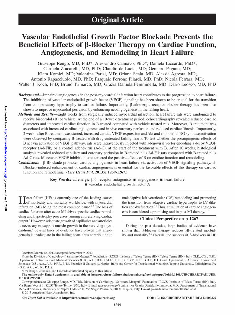
Vascular Endothelial Growth Factor Blockade Prevents the Beneficial Effects of β-Blocker Therapy on Cardiac Function,
Angiogenesis, and Remodeling in Heart FailureGiuseppe Rengo, MD, PhD*; Alessandro Cannavo, PhD*; Daniela Liccardo, PhD*;
Carmela Zincarelli, MD, PhD; Claudio de Lucia, MD; Gennaro Pagano, MD; Klara Komici, MD; Valentina Parisi, MD; Oriana Scala, MD; Alessia Agresta, MD;
Antonio Rapacciuolo, MD, PhD; Pasquale Perrone Filardi, MD, PhD; Nicola Ferrara, MD; Walter J. Koch, PhD; Bruno Trimarco, MD; Grazia Daniela Femminella, MD; Dario Leosco, MD, PhD
by guest on June 1, 2018http://circheartfailure.ahajournals.org/
Dow
nloaded from
by guest on June 1, 2018http://circheartfailure.ahajournals.org/
Dow
nloaded from
by guest on June 1, 2018http://circheartfailure.ahajournals.org/
Dow
nloaded from
by guest on June 1, 2018http://circheartfailure.ahajournals.org/
Dow
nloaded from
by guest on June 1, 2018http://circheartfailure.ahajournals.org/
Dow
nloaded from
by guest on June 1, 2018http://circheartfailure.ahajournals.org/
Dow
nloaded from
1260 Circ Heart Fail November 2013
treatment is attributed, at least in part, to their ability to block the continuous increased adrenergic overdrive present in the failing human heart.7–9 Moreover, β-adrenergic receptor (βAR) blockade has been proven to exert several additional therapeu-tic effects, including reduced oxygen consumption, improved cardiac reverse remodeling, blunted apoptosis, inhibited βAR internalization, and reduced risk of arrhythmias.9–12
Importantly, β-blocker therapy has been also shown to improve myocardial perfusion by enhancing neoangiogenesis in the failing heart.13–15 This latter phenomenon seems to be related to β-blocker–dependent heart rate reduction (HRR) that has been demonstrated to enhance coronary reserve as well as capillary and arteriolar growth in normal16 and in the post-MI hearts.17,18 Moreover, it has been demonstrated that HRR is able to activate vascular endothelial growth factor (VEGF)–dependent angiogenic pathway.16
However, to the best of our knowledge, there are no stud-ies investigating the effects of β-blocker therapy on VEGF proangiogenic signaling pathway in HF. Thus, in an experi-mental model of chronic HF, we demonstrate that bisoprolol (B) treatment induces cardiac VEGF upregulation and that prevention of β-blocker–dependent VEGF induction abro-gates the proangiogenic effect of β-blocker. Moreover, we prove that angiogenesis induced by β-blocker plays a crucial role to avoid the transition from compensatory to maladaptive hypertrophy, thus representing an essential mechanism for the therapeutic effects of β-blocker.
MethodsExperimental Groups and Pharmacological Treatment ProtocolsSeventy-nine Sprague-Dawley male rats (300 g) entered the study: sham-operated (n=12) and rats with surgically induced MI (n=67) by permanent ligation of the left anterior descending coronary artery as previously described.19 At 8 weeks post-MI, HF rats were random-ized to the following treatment groups: (1) placebo (drinking water; n=10); (2) B (10 mg/kg per day in drinking water; n=12); (3) pla-cebo plus intravenous injection of Adenovirus (Ad) vectors encoding Flk1-Fc (AdFlk), a potent angiogenesis inhibitor that acts as a decoy VEGF receptor20 (n=10); (4) placebo plus intravenous injection of Ad encoding for the control Fc fragment (AdCTR)18 (n=10); (5) B plus intravenous injection of AdFlk (n=13); and (6) B plus intravenous injection of Ad-control (n=12). We injected 4×10¹⁰ plaque-forming units of AdFlk or AdCTR into the jugular vein of rats at 8 weeks post-MI (when also B treatment was started). Treatment period was of 10 weeks for all groups. All animal care and experimental protocols were approved by the Ethics Committee for the Use of Animals in Research of our institution.
EchocardiographyEchocardiography was performed 8 weeks after surgery (after ran-domization to treatments) and repeated at the end of the study (18 weeks after MI) in anesthetized (1.5% isoflurane; v/v) rats with a Vevo770 (VisualSonics) echocardiograph, as previously described.21
Myocardial Perfusion StudiesMyocardial perfusion was determined using 15 μm fluorescent mi-crospheres (Triton Inc.), as previously described.22 Cardiac and blood samples were processed for microspheres determination. Total myocardial blood flow and coronary conductance (coronary blood flow normalized by corresponding perfusion pressure) were measured at basal condition and after maximal coronary dilation by
dipyridamole (6 mg/kg per minute IV). Coronary flow reserve was calculated as maximal coronary conductance divided by basal coro-nary conductance.
Measurement of Infarct SizeInfarct size was examined in all experimental groups at the end of the study period. Briefly, hearts were frozen in liquid nitrogen and sectioned from apex to base into 2-mm slices. To delineate the in-farct size, sections were incubated in 1% (wt/vol) triphenyltetrazo-lium chloride (Sigma) in PBS (pH, 7.4) at room temperature for 15 minutes. For each section, the infarct size of the LV was calculated from enlarged digital photos using SigmaScan version 5.0 software, as described previously.23
RNA Isolation and Real-Time Reverse Transcription Polymerase Chain ReactionCardiac total RNA isolations, reverse transcription to cDNA, and quantitative real-time reverse transcription polymerase chain reaction were performed as previously described.24
HistologyCapillary density and arteriolar length density were measured as pre-viously described.22 Briefly, LV specimens were fixed in 4% form-aldehyde and embedded in paraffin. After deparaffinization and rehydration, 4-μm-thick sections were prepared, mounted on glass slides. Capillary density and arteriolar length density were evaluated in 5 randomly selected LV sections in either anterior or lateral wall (border zones) at ≈1 mm from the edge of scar tissue and in the lateral wall, far from the infarcted area (remote). Capillaries (5–10 μm thick) were detected by Lectin Bandeiraea simplicifolia I staining. Arterioles (<50 μm thick) were identified by immunofluorescence using anti-smooth muscle α-actin antibody as previously described.22 Arteriolar length density was calculated with the following formula: Length den-sity (mm/mm3)=(Σa/b)/N·N/A, where a and b represent long and short axes, respectively, of individual arterioles, N is total number of arterio-lar profiles, and A is the total area in which arterioles were measured.
Cardiac fibrosis has been evaluated by picro-sirius staining. Briefly, after deparaffinization and rehydration, 4-μm-thick sections were prepared, mounted on glass slides and stained with 1% Sirius red in picric acid (Carlo Erba Laboratories, Italy) to detect interstitial fibrosis. All the sections were examined with a microscope (Leitz, DIAPLAN), and images were acquired with a digital camera (Digital JVC, TK-C1380).
Cardiomyocyte Size MeasurementCardiomyocyte surface area was determined from sections of the LV myocardium from hearts of all study groups. Sections were stained with wheat germ agglutinin coupled to Alexa Flour 488 (1:100; Invitrogen, W11261). Surface areas of cardiomyocytes were mea-sured in 10 randomly selected fields from each individual heart sam-ple (5 hearts per group) using Software ImageJ.
Immunoblotting and VEGF/Akt/Endothelial NO Synthase MeasurementLV samples were lysed in an radio immunoprecipitation assay (RIPA) buffer with protease and phosphatase inhibitors cocktail (Roche). Measurements of cardiac VEGF, Akt, serin473-phospho-Akt (pAkt), endothelial NO synthase (eNOS), and serin1177-phospho-eNOS (p-eNOS) protein levels were performed using specific primary antibodies (Ab-VEGF, Santacruz; Ab-Akt, Santacruz; Ab-pAkt, Upstate; Ab-eNOS, Upstate; Ab p-eNOS, Upstate). Secondary anti-bodies were purchased from Immunoreagent Inc. Bands were visu-alized by enhanced chemiluminescence (Millipore) according to the manufacturer’s instructions and were quantified using densitometry (Chemidoc, Biorad). Each experiment and densitometric quantifica-tion was separately repeated ≥3 times.
by guest on June 1, 2018http://circheartfailure.ahajournals.org/
Dow
nloaded from
Rengo et al β-Blocker–Induced Angiogenesis in Heart Failure 1261
StatisticsNormally distributed, continuous variables were compared by 1-way ANOVA, followed by a Bonferroni post hoc analysis or by repeated measures ANOVA, as appropriate. Data are reported as means±SEM. Statistical significance was accepted at P<0.05.
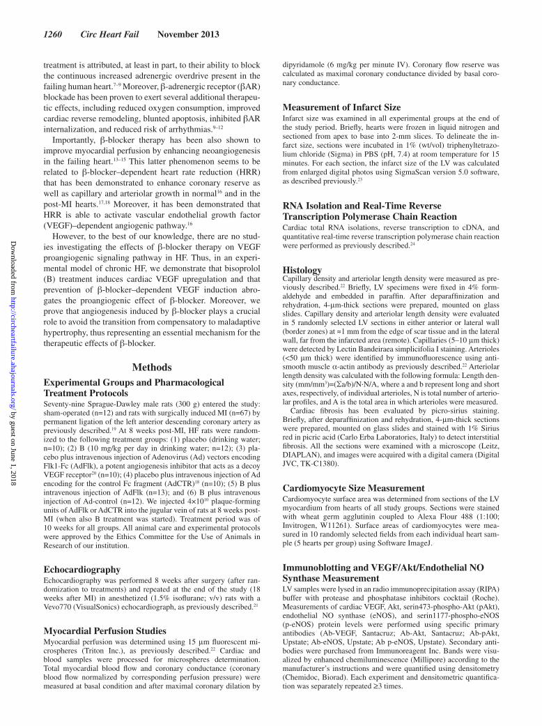
ResultsEffects of B on In Vivo Cardiac FunctionEchocardiography performed 8 weeks after MI induction revealed that LV ejection fraction and internal diameter at diastole were not statistically different among HF groups before placebo or B treatment initiation (Figure I in the online-only Data Supplement). Ejection fraction was signifi-cantly decreased, and LV diastolic diameter was significantly increased in both HF groups compared with sham, demon-strating a similar degree of HF (Figure I in the online-only Data Supplement). At the end of the study period (18 weeks post-MI), the 2 HF groups still had worse cardiac function compared with sham (Figure 1A and Table 1). As expected, although no differences in infarct size were observed among HF groups (Table 1), 10 weeks of B treatment positively affected both cardiac contractility and LV geometry com-pared with HF control group (Figure 1B and Table 1). Impor-tantly, HF B-treated rats showed a significant reduction in HR compared with sham and HF control groups (Figure 1C). The heart weight/body weight ratio was significantly increased
in control HF group compared with sham rats, consistent with an HF phenotype; importantly, B treatment resulted in a reduction of heart weight/body weight ratio compared with HF control (Table 1).
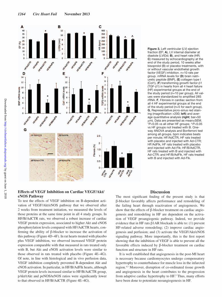
Figure 1. Left ventricular (LV) ejection fraction (EF, as %; A), LV internal diam-eter at diastole (LVIDd, as millimeters; B), and heart rate (HR, as beats per minute [bpm]; C) measured by echocardiography at the end of the study period, 10 weeks after bisoprolol (B) or placebo treatments. n=12 rats per group. mRNA levels for (D) brain natriuretic peptide (BNP), (E) collagen type I (Col1), (F) transforming growth factor-β1 (TGF-β1) in hearts from sham, heart failure (HF), and HF rats treated with B (HF/B) groups at the end of the study period (n=10 per group). All values were standardized to amplified 28S rRNA. F, Fibrosis in cardiac section from all 3 experimental groups at the end of the study period (n=5 for each group). G, Representative panels of picro-sirius red staining (magnification ×200; left) and average quantitative analysis (right; bar=50 μm). Data are presented as mean±SEM. *P<0.05 vs sham; #P<0.05 vs HF; ^P<0.05 vs sham and HF. One-way ANOVA analysis with Bonferroni test among all groups.
Table 1. Physical and Echocardiographic Data of Sham-Operated and HF Rats at the End of the Study Period
Sham HF/Control HF/Bisoprolol
Physical data
BW, kg 0.471±0.013 0.469±0.014 0.454±0.015
HW, g 1.16±0.04 1.41±0.01* 1.24±0.03*†
HW/BW, g/kg 2.48±0.08 3.01±0.09* 2.73±0.08*†
Echocardiography
HR, beats per minute 321.5±15.9 324.8±7.1 268.8±7.7**
LVEF, % 64.8±0.7 27.9±1.9* 36.7±1.4*†
LVIDd, mm 8.5±0.2 11.0±0.4* 9.9±0.2*†
LVIDs, mm 5.4±0.1 9.4±0.4* 8.1±0.2*†
LVAWDd, mm 1.72±0.05 1.54±0.10* 1.46±0.07*
LVPWDd, mm 1.72±0.07 1.91±0.10* 2.22±0.08*†
Infarct size, % … 46.3±4.2 44.8±3.1
ANOVA analysis and Bonferroni test were used among all 3 groups. Data are presented as mean±SEM. BW indicates body weight; HF, heart failure; HR, heart rate; HW, heart weight; LVAWDd, LV anterior wall diameter at diastole; LVEF, left ventricular ejection fraction; LVIDd, LV internal diameter at diastole; LVIDs, LVID at systole; and LVPWDd, LV posterior wall diameter at diastole.
*P<0.05 vs sham; †P<0.05 vs HF/control, **P<0.05 vs sham and HF/control.
by guest on June 1, 2018http://circheartfailure.ahajournals.org/
Dow
nloaded from
1262 Circ Heart Fail November 2013
Effects of β-Blocker Therapy on Cardiac Remodeling Gene ProfileAt the end of the study period, we evaluated cardiac gene expression patterns related to ventricular remodeling in our experimental groups. As a marker of HF, we investigated expression of the mRNA for brain natriuretic peptide in the LV and found this to be significantly increased in the HF control group, whereas in B-treated animals we observed values similar to sham (Figure 1D). We further examined, as markers of remodeling and fibrosis, cardiac mRNA lev-els of collagen type I and transforming growth factor-β1 (Figure 1E and 1F) and found mRNA levels of both of these markers significantly elevated in HF control rats compared with sham controls; but both were markedly reduced in the B-treated HF rats. Consistently, picro-sirius red staining for cardiac fibrosis performed at the end of the study period showed markedly increased fibrosis in HF control rat hearts compared with B-treated rat hearts. As expected, no fibrosis was detectable in sham-operated rat hearts (Figure 1G). Of note, cardiomyocytes surface area was increased in HF con-trol group compared with sham, as expected. Importantly,
B treatment resulted in an slight but significant increase in cardiomyocytes size compared with HF control (Figure IIA in the online-only Data Supplement).
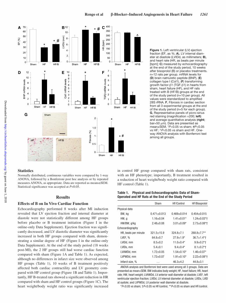
Effects of B on Cardiac Angiogenesis and PerfusionAs expected, HF control rats showed a marked capillary density rarefaction in both LV border and remote zones compared with sham (Figure 2A). Ten weeks of B treat-ment resulted in a significant increase of capillary density compared with HF controls (Figure 2A). Consistent with capillary density data, arteriolar length density was dra-matically reduced in the LV border and remote zones in HF control group compared with sham (Figure 2A, Figure IIIA in the online-only Data Supplement). Interestingly, a sig-nificant growth of arterioles was found in LV border and remote zones after 10 weeks of B treatment (Figure 2A, Fig-ure IIIA in the online-only Data Supplement). Importantly, myocardial blood flow and coronary conductance were sig-nificantly reduced after maximal vasodilation in HF con-trol group compared with sham rats (Figure 2B and 2C and
Figure 2. A, Effects of bisoprolol (B) on cardiac capillary and arteriolar network. Left, Representative images of Lectin Bandeiraea simplicifolia I (BS-I) staining of capillaries and of arterioles stained with antibodies against smooth muscle α-actinin in cardiac section obtained from sham, heart failure (HF) rats, and HF rats treated with B (HF/B) at the end of the study period in the lateral wall far from the infarcted area (remote; mag-nification ×200; bar=50 μm). Right, Bar graphs show data on capillary counts (capillary to myocytes ratio) and arteriolar length density in either left ventricular border or remote zones in all study groups at the end of the study period (n=5 rats per group and 5 sections per animal). Total myocardial blood flow (MBF; B), coronary conductance (C), and coronary flow reserve (CFR; D) in sham, HF, and HF/B rats at the end of the study period (n=10 rats per group). Cardiac protein expression of (E) vascu-lar endothelial growth factor (VEGF), (F) Akt and serin473-phospho(p)-Akt, and (G) endothelial NO synthase (eNOS) and Ser1177-phospho(p)-eNOS in sham, HF, and HF/B hearts at the end of the study period. The expression of glycer-aldehyde-3-phosphate dehydrogenase (GAPDH) was used as an internal control to normalize VEGF protein levels. pAkt to total-Akt (tAkt) ratio and p-eNOS to eNOS ratio indicated, respectively, the levels of Akt and eNOS phosphorylation in the heart (n=5 hearts per group). Data are presented as mean±SEM. *P<0.05 vs sham; #P<0.05 vs HF. One-way ANOVA analysis with Bonferroni correction in D to G and repeated measures ANOVA with Bonferroni correction in A to C.
by guest on June 1, 2018http://circheartfailure.ahajournals.org/
Dow
nloaded from
Rengo et al β-Blocker–Induced Angiogenesis in Heart Failure 1263
Table I in the online-only Data Supplement). B improved myocardial perfusion and reduced coronary vascular resis-tances in HF/B rats compared with HF control. Accordingly, coronary reserve, which showed a ≈3-fold decrease in HF control rats compared with sham, significantly increased after β-blocker treatment, although final values remained still lower than in sham (Figure 2D and Table I in the online-only Data Supplement).
Effects of B on Cardiac VEGF/Akt/eNOS PathwayInterestingly, 2 weeks after the start of treatments, in HF control hearts we observed a significant reduction of cardiac VEGF protein levels compared with sham. This finding was associated to a reduced Akt activation and eNOS phosphory-lation compared with sham, as reflected by phospho-Akt/total-Akt (pAkt/tAkt) and peNOS/teNOS ratios, respectively. Surprisingly, 2 weeks of B treatment induced an increase of cardiac VEGF protein expression, associated to Akt activation and eNOS phosphorylation, even at higher values to those observed in sham hearts (Figure 2E–2G).
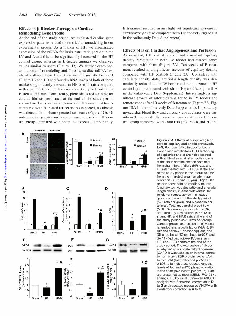
Effects of VEGF Inhibition on B-Induced Improvement of Cardiac FunctionTo test the in vivo pathophysiological relevance of B-depen-dent cardiac VEGF upregulation and Akt and eNOS activation on cardiac function, angiogenesis, and remodeling, HF rats at 8 weeks post-MI were treated with placebo or B and were also injected intravenously with an adenoviral vector encoding for the ligand-binding domain of VEGF receptor 2 (Flk1) fused to murine IgG2a Fc (Ad-Flk), a potent VEGF inhibitor, or with a control adenovirus that encodes only for the murine IgG2a Fc (Ad-CTR).18 Thus, we obtain 4 study groups: (1) HF rats treated with placebo and injected with Ad-CTR (HF/AdCTR); (2) HF rats treated with placebo and injected with Ad-Flk (HF/AdFlk); (3) HF rats treated with B and injected with Ad-CTR (HF/B/AdCTR); (4) HF rats treated with B and injected with Ad-Flk (HF/B/AdFlk). Importantly, no statistical differences have been found between HF rats treated with placebo and HF/AdCTR rats, as well as between HF rats treated with B and HF/B/AdCTR rats, for all echocardiographic, histologi-cal, and molecular parameters. This observation indicates that, as expected, Ad-CTR does not affect cardiac function, remod-eling, and angiogenesis. At the end of the study period (10 weeks of treatment), cardiac function has been assessed by echocardiography in all 4 study groups. As expected, in HF/B/AdCTR rats we observed a significant reduction of LV inter-nal diameter and a significant increase of LV ejection frac-tion percentage compared with HF/AdCTR (Figure 3A). HF/AdFlk showed similar levels of both LV dilation and contrac-tility compared with HF/AdCTR. Surprisingly, the positive effects of B on cardiac function and remodeling were com-pletely abolished when VEGF was inhibited. In fact, HF/B/AdFlk rats showed an increased LV internal diameter and decreased LV ejection fraction when compared with HF/B/AdCTR, at values that were similar to those measured in HF/AdCTR (Figure 3B). Importantly, no differences in infarct size were observed among all 4 HF groups (Table 2). As expected, HF/B/AdCTR and HF/B/AdFlk rats showed similar
reduction in HR compared with groups not treated with B (Figure 3C), demonstrating that Ad-Flk administration did not affect B-dependent HRR. Consistently, heart weight/body weight ratio was similar between HF groups, with the only exception of HF/B/AdCTR group in which it was significantly lower (Table 2).
Effects of VEGF Inhibition on B-Dependent Amelioration of Cardiac Remodeling Gene ProfileAt the end of the study period, in HF/B/AdCTR hearts we observed a marked reduction of brain natriuretic peptide, collagen type I, and transforming growth factor-β1 mRNAs compared with HF/AdCTR, as assessed by reverse tran-scription polymerase chain reaction (Figure 3D–3F). In HF/AdFlk rats, cardiac levels of these gene expression patterns were similar to those observed in HF/AdCTR. Importantly, when VEGF was inhibited, B completely failed to reduce the mRNA levels of all the 3 genes investigated, with lev-els indistinguishable from those observed in rats not treated with β-blocker (Figure 3D–3F). Consistently, we observed a significant reduction of fibrosis in HF/B/AdCTR hearts com-pared with HF/AdCTR (Figure 3). In HF/AdFlk rats, car-diac fibrosis was similar to that observed in HF/AdCTR rats. More important, when B treatment was associated to VEGF inhibition, the ability of β-blocker to reduce cardiac fibrosis was completely lost, with levels of cardiac fibrosis similar to those observed in rats not treated with B (Figure 3G). Of note, B treatment in HF/B/AdCRT group resulted in an slight but significant increase in cardiomyocytes size compared with HF/AdCTR group (Figure IIB in the online-only Data Supplement). Importantly, when B treatment was associated to VEGF inhibition, the ability of β-blocker to increase car-diomyocytes surface area was completely prevented (Figure IIB in the online-only Data Supplement).
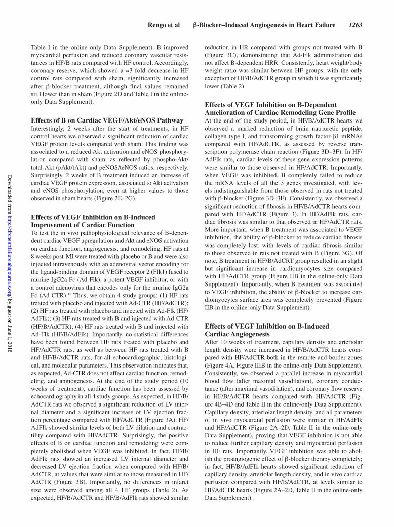
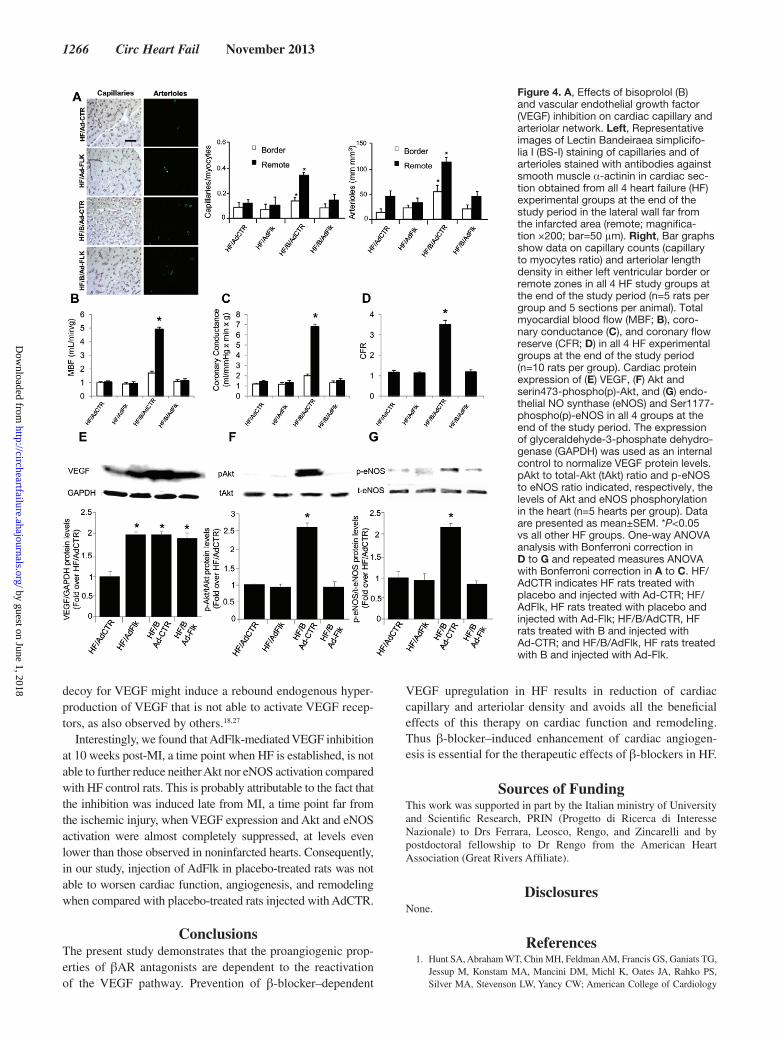
Effects of VEGF Inhibition on B-Induced Cardiac AngiogenesisAfter 10 weeks of treatment, capillary density and arteriolar length density were increased in HF/B/AdCTR hearts com-pared with HF/AdCTR both in the remote and border zones (Figure 4A, Figure IIIB in the online-only Data Supplement). Consistently, we observed a parallel increase in myocardial blood flow (after maximal vasodilation), coronary conduc-tance (after maximal vasodilation), and coronary flow reserve in HF/B/AdCTR hearts compared with HF/AdCTR (Fig-ure 4B–4D and Table II in the online-only Data Supplement). Capillary density, arteriolar length density, and all parameters of in vivo myocardial perfusion were similar in HF/AdFlk and HF/AdCTR (Figure 2A–2D, Table II in the online-only Data Supplement), proving that VEGF inhibition is not able to reduce further capillary density and myocardial perfusion in HF rats. Importantly, VEGF inhibition was able to abol-ish the proangiogenic effect of β-blocker therapy completely; in fact, HF/B/AdFlk hearts showed significant reduction of capillary density, arteriolar length density, and in vivo cardiac perfusion compared with HF/B/AdCTR, at levels similar to HF/AdCTR hearts (Figure 2A–2D, Table II in the online-only Data Supplement).
by guest on June 1, 2018http://circheartfailure.ahajournals.org/
Dow
nloaded from
1264 Circ Heart Fail November 2013
Effects of VEGF Inhibition on Cardiac VEGF/Akt/eNOS PathwayTo test the effects of VEGF inhibition on B-dependent acti-vation of VEGF/Akt/eNOS pathway that we observed after 2 weeks from treatment initiation, we measured the levels of those proteins at the same time point in all 4 study groups. In HF/B/AdCTR rats, we observed a robust increase of cardiac VEGF protein expression, associated to higher Akt and eNOS phosphorylation levels compared with HF/AdCTR hearts, con-firming the ability of β-blocker to increase the activation of this pathway (Figure 4D–4F). In rat hearts treated with placebo plus VEGF inhibition, we observed increased VEGF protein expression comparable with that measured in rats treated only with B, but Akt and eNOS activation levels were similar to those observed in rats treated with placebo (Figure 4E–4G). Of note, in line with histological and in vivo perfusion data, VEGF inhibition completely prevented B-dependent Akt and eNOS activation. In particular, in HF/B/AdFlk hearts, although VEGF protein levels increased similar to HF/B/AdCTR group, pAkt/tAkt and peNOS/teNOS ratios were significantly lower to that observed in HF/B/AdCTR (Figure 4E–4G).
DiscussionThe most significant finding of the present study is that β-blocker favorably affects performance and remodeling of the failing heart through reactivation of angiogenesis. We show that the effects of β-blocker treatment on cardiac angio-genesis and remodeling in HF are dependent on the activa-tion of VEGF proangiogenic pathway. Indeed, we provide evidence that in HF rats β1AR blockade is able to: (1) prevent HF-related adverse remodeling; (2) improve cardiac angio-genesis and perfusion; and (3) activate the VEGF/Akt/eNOS signaling pathway. More importantly, this is the first report showing that the inhibition of VEGF is able to prevent all the favorable effects induced by β-blocker treatment on cardiac function and structure in HF rats.
It is well established that angiogenesis in the post-MI heart is necessary because cardiomyocytes undergo compensatory hypertrophy to counterbalance for muscle loss in the infarcted region.4,5 Moreover, disruption of co-ordinated tissue growth and angiogenesis in the heart contributes to the progression from adaptive cardiac hypertrophy to HF.5 Thus, many efforts have been done to potentiate neoangiogenesis in HF.
Figure 3. Left ventricular (LV) ejection fraction (EF; A), LV internal diameter at diastole (LVIDd; B), and heart rate (HR; C) measured by echocardiography at the end of the study period, 10 weeks after bisoprolol (B) or placebo treatments, with or without vascular endothelial growth factor (VEGF) inhibition. n=10 rats per group. mRNA levels for (D) brain natri-uretic peptide (BNP), (E) collagen type I (Col1), (F) transforming growth factor-β1 (TGF-β1) in hearts from all 4 heart failure (HF) experimental groups at the end of the study period (n=10 per group). All val-ues were standardized to amplified 28S rRNA. F, Fibrosis in cardiac section from all 4 HF experimental groups at the end of the study period (n=5 for each group). G, Representative picro-sirius red stain-ing (magnification ×200; left) and aver-age quantitative analysis (right; bar=50 μm). Data are presented as mean±SEM. *P<0.05 vs all other HF groups; ^P<0.05 vs HF groups not treated with B. One-way ANOVA analysis and Bonferroni test among all groups. bpm indicates beats per minute; HF/AdCTR, HF rats treated with placebo and injected with Ad-CTR; HF/AdFlk, HF rats treated with placebo and injected with Ad-Flk; HF/B/AdCTR, HF rats treated with B and injected with Ad-CTR; and HF/B/AdFlk, HF rats treated with B and injected with Ad-Flk.
by guest on June 1, 2018http://circheartfailure.ahajournals.org/
Dow
nloaded from
Rengo et al β-Blocker–Induced Angiogenesis in Heart Failure 1265
Previous studies have demonstrated that bradycardia enhances coronary reserve, capillary density, and arteriolar growth in the postinfarcted heart17,18 and that bradycardia-induced cardiac angiogenesis is dependent on VEGF.16 It has been hypothesized that HRR, prolonging diastole, is able to enhance diastolic filling and stretch myocytes and capillaries, thus increasing cardiac VEGF expression.16 Here, we report that activation of VEGF proangiogenic pathway underlies B-induced angiogenesis in HF. Of course, β-blockers have negative chronotropic effects, and previous reports have shown that these drugs are able to increase cardiac angiogen-esis in post-MI animal models,13,14 thus we cannot exclude that in our study increased angiogenesis in B-treated animals is mainly dependent on HRR induced by this drug. Thus, whether bradycardia or other biological effects of β-blockers are detrimental for the induction of VEGF-dependent angio-genesis is still unclear. Future studies will be needed to evalu-ate separately HR-dependent and HR-independent effects of β-blockers treatment on VEGF-dependent angiogenesis and subsequent restoration of cardiac function and remodeling. However, we offer the first demonstration that B is able to activate the VEGF/Akt/eNOS signaling pathway in HF, and to test the pathophysiological relevance of this mechanism, we administered HF B-treated rats with an adenoviral vector encoding for a VEGF decoy receptor (AdFlk), that is known to act as a potent VEGF inhibitor.20 Surprisingly, we found that VEGF inhibition prevented not only B-induced cardiac angiogenesis, but, unexpectedly, it also neutralized the favor-able effects of β-blocker therapy on cardiac function and remodeling. Indeed, HF rats treated with B and VEGF inhi-bition showed no significant differences compared with HF/
AdCTR rats in terms of cardiac chamber dilation and con-tractility, as well as for cardiac remodeling genes expression, indicating that B-induced VEGF upregulation is essential in co-ordinating adequate vascular and myocardial growth in post-MI failing hearts. Moreover, our data indicate that 10 weeks of B treatment resulted in a slight but significant increase in cardiomyocytes size compared with HF control group. This finding is in line with the proangiogenic effect of β-blocker treatment, which is known to be essential to sup-port co-ordinated vessel and cardiomyocytes growth. The discrepancy between decreased heart weight/body weight ratio and increased cardiomyocytes size observed in HF rats treated with β-blocker compared with HF control rats could be explainable by the increased cardiac size (LV chamber dilation) and fibrosis observed in HF control compared with HF/B group. Importantly, the effects of B treatment on car-diomyocytes size is in line with the echocardiographic data showing that posterior wall thickness is increased in hearts treated with β-blocker compared with control. Taken together, these results support the notion that the imbalance between myocyte growth and coronary angiogenesis plays a critical role in cardiac function20,22 and demonstrates that β-blocker–dependent activation of cardiac angiogenesis is a crucial part of the mechanism of action of this drug class.
Moreover, our study is consistent with previous observa-tions from our group and others that have identified in VEGF/Akt/eNOS pathway reactivation a potential target to promote co-ordinated angiogenesis in HF.20,22,25 Previously, we have shown that exercise training, independently from HRR, is able to induce VEGF upregulation and Akt activation in the post-ischemic heart22 and to improve age-dependent VEGF down-regulation and angiogenesis responses to hindlimb ischemia.26 Moreover, β2AR overexpression in the failing heart is able to enhance neoangiogenesis, inducing a sustained and co-ordi-nated Akt and VEGF activation.21 The involvement of β2AR in the neoangiogenic processes in response to ischemia prompted us to use in this study a selective β1AR blocker, in order to not interfere with β2AR. However, further studies are needed to demonstrate potential differences in proangiogenic properties between unselective β-blockers and selective β1AR blockers.
Notably, in the present study, VEGF/Akt/eNOS pathway was strongly suppressed in HF control rats compared with sham animals at 10 weeks post-MI. These molecular changes were associated with maladaptive cardiac remodeling and severe cardiac dysfunction. Importantly, in our model, we demonstrate that B-dependent VEGF/Akt/eNOS pathway activation, which occurs within 2 weeks from treatment ini-tiation, is crucial for the proangiogenic effects of β-blocker treatment. Moreover, we think that the switch on of this path-way promotes growth of new vessels and thus the transition from a maladaptive to an adaptive, angiogenesis-dependent LV remodeling, which attenuates the negative LV remodeling observed in placebo-treated animals late from MI.
In rat hearts treated with placebo plus VEGF inhibition, we observed increased VEGF protein expression comparable with that measured in rats treated with B, but Akt and eNOS activa-tion levels were similar to those observed in rats treated with placebo (Figure 4). This apparent inconsistency is explainable by considering the mechanism of action of Flk that acting as a
Table 2. Physical and Echocardiographic Data of HF Rats at the End of the Study Period
HF/AdCTR HF/AdFlk HF/B/AdCTR HF/B/AdFlk
Physical data
BW, kg 0.457±0.019 0.468±0.017 0.460±0.016 0.459±0.013
HW, g 1.50±0.06 1.43±0.04 1.22±0.09* 1.40±0.08
HW/BW, g/kg 3.29±0.15 3.06±0.11 2.76±0.09* 3.05±0.12
Echocardiography
HR, beats per minute
334.4±13.2 317.0±13,2 274.0±4.1† 276.5±11.3†
LVEF, % 27.3±0.9 24.3±1.8 36.1±0.7* 26.0±1.6
LVIDd, mm 10.7±0.3 11.0±0.2 9.8±0.2* 10.9±0.2
LVIDs, mm 9.3±0.3 9.7±0.3 7.9±0.2* 9.7±0.1
LVAWDd, mm 1.37±0.11 1.42±0.08 1.51±0.08 1.40±0.15
LVPWDd, mm 1.87±0.21 2.05±0.08 2.35±0.10* 2.07±0.12
Infarct size, % 47.6±5.2 45.3±3.8 48.1±4.6 44.1±4.2
ANOVA analysis and Bonferroni test were used among all 4 groups. Data are presented as mean±SEM. BW indicates body weight; HF, heart failure; HF/AdCTR, HF rats treated with placebo and injected with Ad-CTR; HF/AdFlk, HF rats treated with placebo and injected with Ad-Flk; HF/B/AdCTR, HF rats treated with bisoprolol (B) and injected with Ad-CTR; HF/B/AdFlk, HF rats treated with B and injected with Ad-Flk; HR, heart rate; HW, heart weight; LVEF, left ventricular ejection fraction; LVAWDd, LV anterior wall diameter at diastole; LVIDd, LV internal diameter at diastole; LVIDs, LVID at systole; and LVPWDd, LV posterior wall diameter at diastole.
*P<0.05 vs all other groups; †P<0.05 vs HF/AdCTR and Ad/AdFlk.
by guest on June 1, 2018http://circheartfailure.ahajournals.org/
Dow
nloaded from
1266 Circ Heart Fail November 2013
decoy for VEGF might induce a rebound endogenous hyper-production of VEGF that is not able to activate VEGF recep-tors, as also observed by others.18,27
Interestingly, we found that AdFlk-mediated VEGF inhibition at 10 weeks post-MI, a time point when HF is established, is not able to further reduce neither Akt nor eNOS activation compared with HF control rats. This is probably attributable to the fact that the inhibition was induced late from MI, a time point far from the ischemic injury, when VEGF expression and Akt and eNOS activation were almost completely suppressed, at levels even lower than those observed in noninfarcted hearts. Consequently, in our study, injection of AdFlk in placebo-treated rats was not able to worsen cardiac function, angiogenesis, and remodeling when compared with placebo-treated rats injected with AdCTR.
ConclusionsThe present study demonstrates that the proangiogenic prop-erties of βAR antagonists are dependent to the reactivation of the VEGF pathway. Prevention of β-blocker–dependent
VEGF upregulation in HF results in reduction of cardiac capillary and arteriolar density and avoids all the beneficial effects of this therapy on cardiac function and remodeling. Thus β-blocker–induced enhancement of cardiac angiogen-esis is essential for the therapeutic effects of β-blockers in HF.
Sources of FundingThis work was supported in part by the Italian ministry of University and Scientific Research, PRIN (Progetto di Ricerca di Interesse Nazionale) to Drs Ferrara, Leosco, Rengo, and Zincarelli and by postdoctoral fellowship to Dr Rengo from the American Heart Association (Great Rivers Affiliate).
DisclosuresNone.
References 1. Hunt SA, Abraham WT, Chin MH, Feldman AM, Francis GS, Ganiats TG,
Jessup M, Konstam MA, Mancini DM, Michl K, Oates JA, Rahko PS, Silver MA, Stevenson LW, Yancy CW; American College of Cardiology
Figure 4. A, Effects of bisoprolol (B) and vascular endothelial growth factor (VEGF) inhibition on cardiac capillary and arteriolar network. Left, Representative images of Lectin Bandeiraea simplicifo-lia I (BS-I) staining of capillaries and of arterioles stained with antibodies against smooth muscle α-actinin in cardiac sec-tion obtained from all 4 heart failure (HF) experimental groups at the end of the study period in the lateral wall far from the infarcted area (remote; magnifica-tion ×200; bar=50 μm). Right, Bar graphs show data on capillary counts (capillary to myocytes ratio) and arteriolar length density in either left ventricular border or remote zones in all 4 HF study groups at the end of the study period (n=5 rats per group and 5 sections per animal). Total myocardial blood flow (MBF; B), coro-nary conductance (C), and coronary flow reserve (CFR; D) in all 4 HF experimental groups at the end of the study period (n=10 rats per group). Cardiac protein expression of (E) VEGF, (F) Akt and serin473-phospho(p)-Akt, and (G) endo-thelial NO synthase (eNOS) and Ser1177-phospho(p)-eNOS in all 4 groups at the end of the study period. The expression of glyceraldehyde-3-phosphate dehydro-genase (GAPDH) was used as an internal control to normalize VEGF protein levels. pAkt to total-Akt (tAkt) ratio and p-eNOS to eNOS ratio indicated, respectively, the levels of Akt and eNOS phosphorylation in the heart (n=5 hearts per group). Data are presented as mean±SEM. *P<0.05 vs all other HF groups. One-way ANOVA analysis with Bonferroni correction in D to G and repeated measures ANOVA with Bonferroni correction in A to C. HF/AdCTR indicates HF rats treated with placebo and injected with Ad-CTR; HF/AdFlk, HF rats treated with placebo and injected with Ad-Flk; HF/B/AdCTR, HF rats treated with B and injected with Ad-CTR; and HF/B/AdFlk, HF rats treated with B and injected with Ad-Flk.
by guest on June 1, 2018http://circheartfailure.ahajournals.org/
Dow
nloaded from
Rengo et al β-Blocker–Induced Angiogenesis in Heart Failure 1267
Foundation; American Heart Association. 2009 Focused update incorporat-ed into the ACC/AHA 2005 Guidelines for the Diagnosis and Management of Heart Failure in Adults A Report of the American College of Cardiology Foundation/American Heart Association Task Force on Practice Guidelines Developed in Collaboration With the International Society for Heart and Lung Transplantation. J Am Coll Cardiol. 2009;53:e1–e90.
2. Rengo F, Leosco D, Iacovoni A, Rengo G, Golino L, Borgia F, De Lisa G, Beneduce F, Senni M. Epidemiology and risk factors for heart failure in the elderly. Ital Heart J. 2004;5(suppl 10):9S–16S.
3. McMurray JJ, Pfeffer MA. Heart failure. Lancet. 2005;365:1877–1889. 4. Anversa P, Beghi C, Kikkawa Y, Olivetti G. Myocardial infarction in
rats. Infarct size, myocyte hypertrophy, and capillary growth. Circ Res. 1986;58:26–37.
5. Shiojima I, Sato K, Izumiya Y, Schiekofer S, Ito M, Liao R, Colucci WS, Walsh K. Disruption of coordinated cardiac hypertrophy and an-giogenesis contributes to the transition to heart failure. J Clin Invest. 2005;115:2108–2118.
6. Frey N, Olson EN. Cardiac hypertrophy: the good, the bad, and the ugly. Annu Rev Physiol. 2003;65:45–79.
7. Bristow MR. beta-adrenergic receptor blockade in chronic heart failure. Circulation. 2000;101:558–569.
8. Foody JM, Farrell MH, Krumholz HM. beta-Blocker therapy in heart failure: scientific review. JAMA. 2002;287:883–889.
9. Rengo G, Lymperopoulos A, Leosco D, Koch WJ. GRK2 as a novel gene therapy target in heart failure. J Mol Cell Cardiol. 2011;50:785–792.
10. Rengo G, Lymperopoulos A, Koch WJ. Future g protein-coupled recep-tor targets for treatment of heart failure. Curr Treat Options Cardiovasc Med. 2009;11:328–338.
11. Rengo G, Perrone-Filardi P, Femminella GD, Liccardo D, Zincarelli C, de Lucia C, Pagano G, Marsico F, Lymperopoulos A, Leosco D. Targeting the β-adrenergic receptor system through G-protein-coupled receptor kinase 2: a new paradigm for therapy and prognostic evaluation in heart failure: from bench to bedside. Circ Heart Fail. 2012;5:385–391.
12. Lymperopoulos A, Rengo G, Koch WJ. Adrenal adrenoceptors in heart fail-ure: fine-tuning cardiac stimulation. Trends Mol Med. 2007;13:503–511.
13. Dedkov EI, Christensen LP, Weiss RM, Tomanek RJ. Reduction of heart rate by chronic beta1-adrenoceptor blockade promotes growth of arteri-oles and preserves coronary perfusion reserve in postinfarcted heart. Am J Physiol Heart Circ Physiol. 2005;288:H2684–H2693.
14. Ulu N, Henning RH, Goris M, Schoemaker RG, van Gilst WH. Effects of ivabradine and metoprolol on cardiac angiogenesis and endothelial dys-function in rats with heart failure. J Cardiovasc Pharmacol. 2009;53:9–17.
15. Christensen LP, Zhang RL, Zheng W, Campanelli JJ, Dedkov EI, Weiss RM, Tomanek RJ. Postmyocardial infarction remodeling and coronary reserve: effects of ivabradine and beta blockade therapy. Am J Physiol Heart Circ Physiol. 2009;297:H322–H330.
16. Zheng W, Brown MD, Brock TA, Bjercke RJ, Tomanek RJ. Bradycardia-induced coronary angiogenesis is dependent on vascular endothelial growth factor. Circ Res. 1999;85:192–198.
17. Lei L, Zhou R, Zheng W, Christensen LP, Weiss RM, Tomanek RJ. Bradycardia induces angiogenesis, increases coronary reserve, and pre-serves function of the postinfarcted heart. Circulation. 2004;110:796–802.
18. Lamping KG, Zheng W, Xing D, Christensen LP, Martins J, Tomanek RJ. Bradycardia stimulates vascular growth during gradual coronary occlu-sion. Arterioscler Thromb Vasc Biol. 2005;25:2122–2127.
19. Rengo G, Lymperopoulos A, Zincarelli C, Femminella G, Liccardo D, Pagano G, de Lucia C, Cannavo A, Gargiulo P, Ferrara N, Perrone Filardi P, Koch W, Leosco D. Blockade of β-adrenoceptors restores the GRK2-mediated adrenal α(2) -adrenoceptor-catecholamine production axis in heart failure. Br J Pharmacol. 2012;166:2430–2440.
20. Shiojima I, Sato K, Izumiya Y, Schiekofer S, Ito M, Liao R, Colucci WS, Walsh K. Disruption of coordinated cardiac hypertrophy and an-giogenesis contributes to the transition to heart failure. J Clin Invest. 2005;115:2108–2118.
21. Rengo G, Zincarelli C, Femminella GD, Liccardo D, Pagano G, de Lucia C, Altobelli GG, Cimini V, Ruggiero D, Perrone-Filardi P, Gao E, Ferrara N, Lymperopoulos A, Koch WJ, Leosco D. Myocardial β(2) -adrenocep-tor gene delivery promotes coordinated cardiac adaptive remodelling and angiogenesis in heart failure. Br J Pharmacol. 2012;166:2348–2361.
22. Leosco D, Rengo G, Iaccarino G, Golino L, Marchese M, Fortunato F, Zincarelli C, Sanzari E, Ciccarelli M, Galasso G, Altobelli GG, Conti V, Matrone G, Cimini V, Ferrara N, Filippelli A, Koch WJ, Rengo F. Exercise promotes angiogenesis and improves beta-adrenergic recep-tor signalling in the post-ischaemic failing rat heart. Cardiovasc Res. 2008;78:385–394.
23. Lymperopoulos A, Rengo G, Zincarelli C, Kim J, Koch WJ. Adrenal beta-arrestin 1 inhibition in vivo attenuates post-myocardial infarction progression to heart failure and adverse remodeling via reduction of cir-culating aldosterone levels. J Am Coll Cardiol. 2011;57:356–365.
24. Rengo G, Leosco D, Zincarelli C, Marchese M, Corbi G, Liccardo D, Filippelli A, Ferrara N, Lisanti MP, Koch WJ, Lymperopoulos A. Adrenal GRK2 lowering is an underlying mechanism for the beneficial sympa-thetic effects of exercise training in heart failure. Am J Physiol Heart Circ Physiol. 2010;298:H2032–H2038.
25. Carmeliet P. Mechanisms of angiogenesis and arteriogenesis. Nat Med. 2000;6:389–395.
26. Leosco D, Rengo G, Iaccarino G, Sanzari E, Golino L, De Lisa G, Zincarelli C, Fortunato F, Ciccarelli M, Cimini V, Altobelli GG, Piscione F, Galasso G, Trimarco B, Koch WJ, Rengo F. Prior exercise improves age-dependent vascular endothelial growth factor downregulation and angiogenesis responses to hind-limb ischemia in old rats. J Gerontol A Biol Sci Med Sci. 2007;62:471–480.
27. Izumiya Y, Shiojima I, Sato K, Sawyer DB, Colucci WS, Walsh K. Vascular endothelial growth factor blockade promotes the transition from compensatory cardiac hypertrophy to failure in response to pres-sure overload. Hypertension. 2006;47:887–893.
CLINICAL PERSPECTIVEHeart failure represents one of the leading causes of mortality worldwide. β-Blocker therapy represents one of the mainstay of therapeutic armamentarium for its ability to improve survival and to reduce hospitalization in patients with heart failure. This class of drug is known to protect the failing heart from the noxious effects of increased adrenergic drive, but the precise mechanisms by which β-blocker confers cardioprotection are not completely elucidated. Importantly, β-blocker therapy has also been shown to improve cardiac angiogenesis and myocardial perfusion, and this latter effect represents a key step to support the adequate growth of vascular network and to permit cardiomyocytes growth in the failing myocardium. In fact, angiogenesis is inadequate in the failing heart, thus contributing to the transition from adaptive cardiac hypertrophy to mal-adaptive left ventricular remodeling. In this context, the present study demonstrates for the first time that β-blocker therapy is able to activate the vascular endothelial growth factor/Akt/endothelial NO synthase signaling pathway, that is necessary to support co-ordinated tissue growth and angiogenesis in the heart, thus preventing the progression from adaptive cardiac hypertrophy to left ventricular dilation and dysfunction. Importantly, prevention of β-blocker–dependent angiogenesis, via vascular endothelial growth factor inhibition, avoids all the beneficial effects of β-blocker therapy on cardiac function and structure and results in reduction of cardiac vascular network and myocardial perfusion. These results indicate that β-blocker–induced stimulation of cardiac angiogenesis represents a relevant therapeutic effect of β-blockers in heart failure and open a new scenario for future studies testing whether the benefits of β-blocker are reduced in patients with heart failure undergoing antiangiogenic therapies.
by guest on June 1, 2018http://circheartfailure.ahajournals.org/
Dow
nloaded from
Daniela Femminella and Dario LeoscoRapacciuolo, Pasquale Perrone Filardi, Nicola Ferrara, Walter J. Koch, Bruno Trimarco, Grazia
Gennaro Pagano, Klara Komici, Valentina Parisi, Oriana Scala, Alessia Agresta, Antonio Giuseppe Rengo, Alessandro Cannavo, Daniela Liccardo, Carmela Zincarelli, Claudio de Lucia,
Therapy on Cardiac Function, Angiogenesis, and Remodeling in Heart Failure-BlockerβVascular Endothelial Growth Factor Blockade Prevents the Beneficial Effects of
Print ISSN: 1941-3289. Online ISSN: 1941-3297 Copyright © 2013 American Heart Association, Inc. All rights reserved.
75231is published by the American Heart Association, 7272 Greenville Avenue, Dallas, TXCirculation: Heart Failure
doi: 10.1161/CIRCHEARTFAILURE.113.0003292013;6:1259-1267; originally published online September 12, 2013;Circ Heart Fail.
http://circheartfailure.ahajournals.org/content/6/6/1259World Wide Web at:
The online version of this article, along with updated information and services, is located on the
http://circheartfailure.ahajournals.org/content/suppl/2013/11/05/CIRCHEARTFAILURE.113.000329.DC1Data Supplement (unedited) at:
http://circheartfailure.ahajournals.org//subscriptions/
is online at: Circulation: Heart Failure Information about subscribing to Subscriptions:
http://www.lww.com/reprints Information about reprints can be found online at: Reprints:
document. Permissions and Rights Question and Answer about this process is available in the
located, click Request Permissions in the middle column of the Web page under Services. Further information isthe Editorial Office. Once the online version of the published article for which permission is being requested
can be obtained via RightsLink, a service of the Copyright Clearance Center, notCirculation: Heart Failurein Requests for permissions to reproduce figures, tables, or portions of articles originally publishedPermissions:
by guest on June 1, 2018http://circheartfailure.ahajournals.org/
Dow
nloaded from
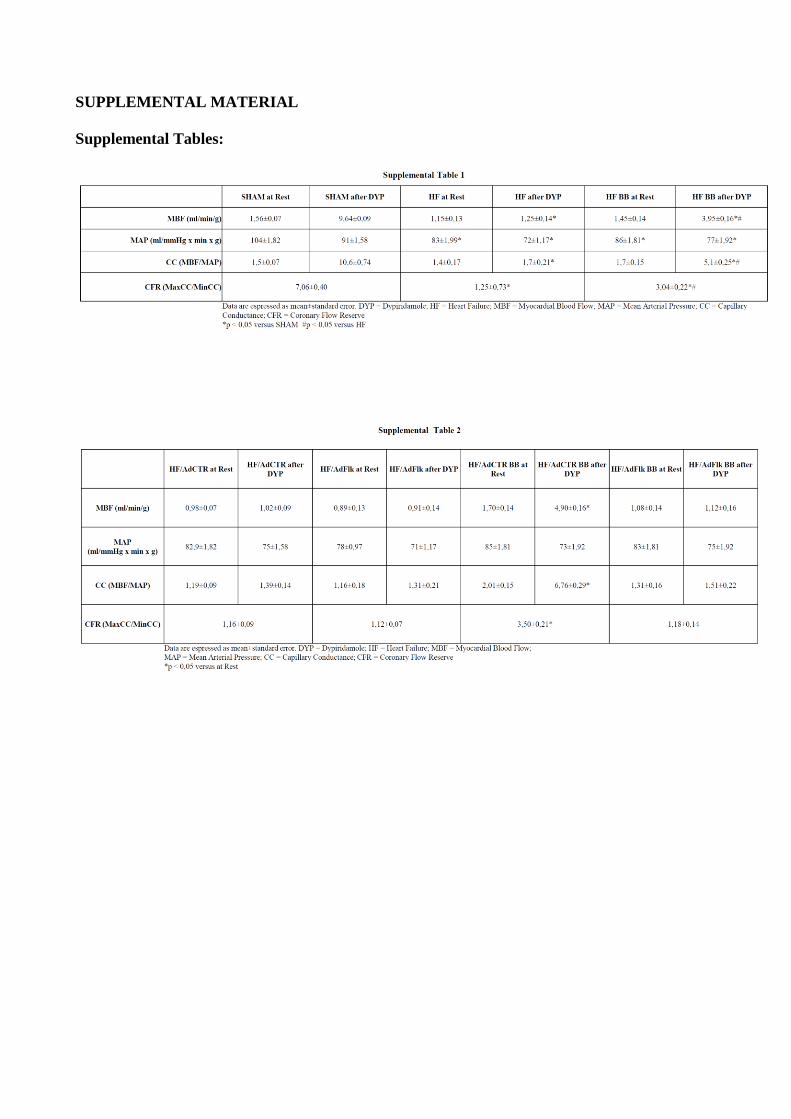
SUPPLEMENTAL MATERIAL
Supplemental Tables:
Supplemental Figures:
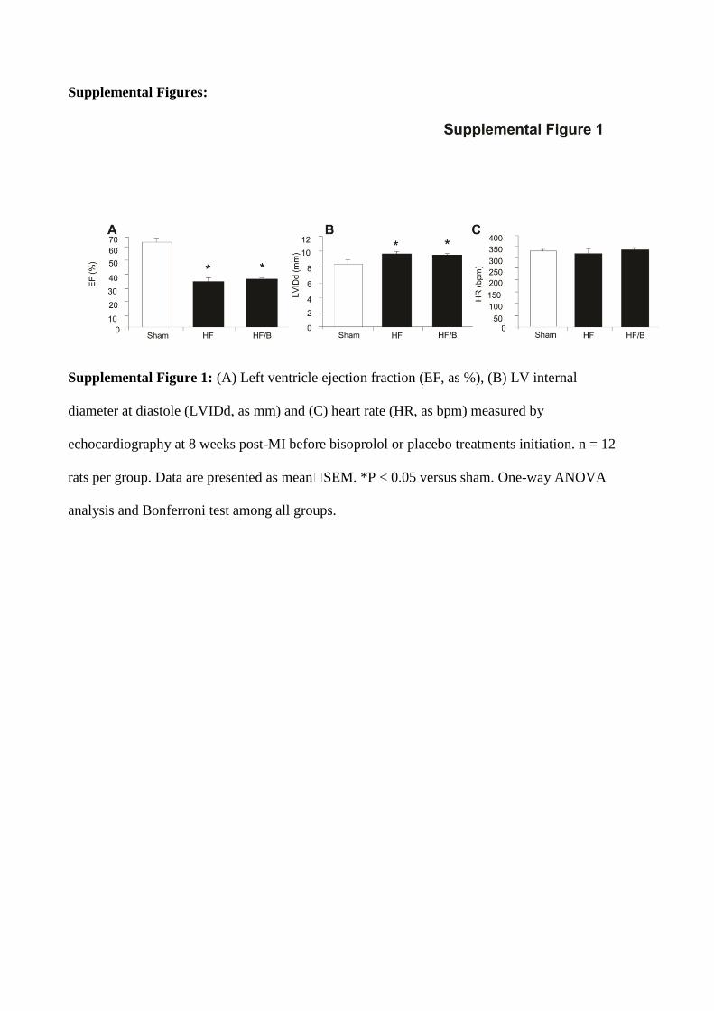
Supplemental Figure 1: (A) Left ventricle ejection fraction (EF, as %), (B) LV internal
diameter at diastole (LVIDd, as mm) and (C) heart rate (HR, as bpm) measured by
echocardiography at 8 weeks post-MI before bisoprolol or placebo treatments initiation. n = 12
rats per group. Data are presented as mean�SEM. *P < 0.05 versus sham. One-way ANOVA
analysis and Bonferroni test among all groups.
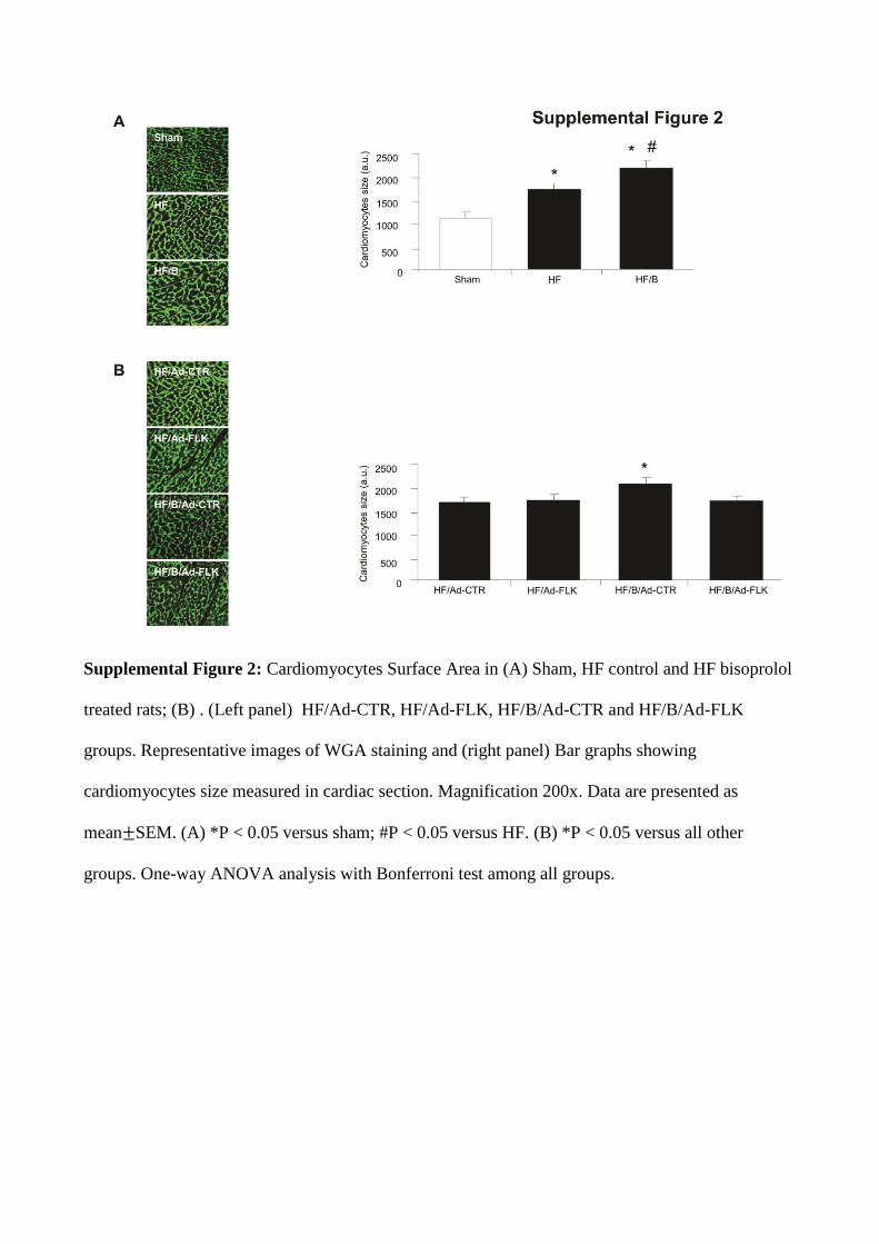
Supplemental Figure 2: Cardiomyocytes Surface Area in (A) Sham, HF control and HF bisoprolol
treated rats; (B) . (Left panel) HF/Ad-CTR, HF/Ad-FLK, HF/B/Ad-CTR and HF/B/Ad-FLK
groups. Representative images of WGA staining and (right panel) Bar graphs showing
cardiomyocytes size measured in cardiac section. Magnification 200x. Data are presented as
mean SEM. (A) *P < 0.05 versus sham; #P < 0.05 versus HF. (B) *P < 0.05 versus all other
groups. One-way ANOVA analysis with Bonferroni test among all groups.
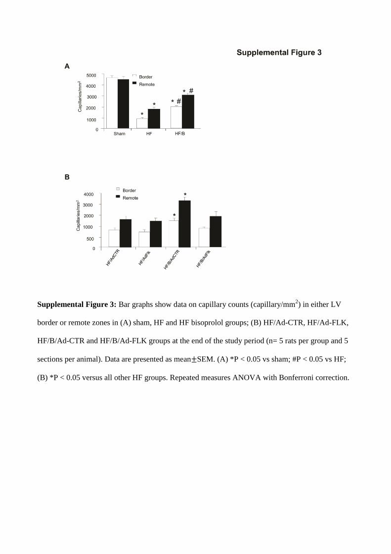
Supplemental Figure 3: Bar graphs show data on capillary counts (capillary/mm2) in either LV
border or remote zones in (A) sham, HF and HF bisoprolol groups; (B) HF/Ad-CTR, HF/Ad-FLK,
HF/B/Ad-CTR and HF/B/Ad-FLK groups at the end of the study period (n= 5 rats per group and 5
sections per animal). Data are presented as mean SEM. (A) *P < 0.05 vs sham; #P < 0.05 vs HF;
(B) *P < 0.05 versus all other HF groups. Repeated measures ANOVA with Bonferroni correction.
Top Related