A STUDY ON ANALYSIS OF FETAL HEART RATE … filea study on analysis of fetal heart rate...
39
A STUDY ON ANALYSIS OF FETAL HEART RATE ABNORMALITIES COMPARING EPIDURAL ANALGESIA ROPIVACAINE 0.2% PLUS FENTANYL 2 μg/ML WITH BUPIVACAINE 0.1% PLUS FENTANYL 2 μg/ML DURING LABOUR BY DR MARY ANGELINE PETER Dissertation Submitted In Partial Fulfillment of the Requirement for The Degree Of Master Of Medicine (ANESTHESIOLOGY) UNIVERSITI SAINS MALAYSIA 2014
Transcript of A STUDY ON ANALYSIS OF FETAL HEART RATE … filea study on analysis of fetal heart rate...
A STUDY ON ANALYSIS OF FETAL HEART RATE ABNORMALITIES COMPARING EPIDURAL ANALGESIA
ROPIVACAINE 0.2% PLUS FENTANYL 2 μg/ML WITH BUPIVACAINE 0.1% PLUS FENTANYL 2 μg/ML DURING
LABOUR
BY
DR MARY ANGELINE PETER
Dissertation Submitted In Partial Fulfillment of the Requirement for The Degree Of Master Of Medicine
(ANESTHESIOLOGY)
UNIVERSITI SAINS MALAYSIA
2014

ii
ACKNOWLEDGEMENTS
First and foremost I would like to thank God the Almighty for his blessings and graces
throughout my life and making it possible for me to come thus far. May the good Lord continue
to guard and guide us in my course and in life.
My utmost appreciation to my supervisor, Dr Gnandev Phutane for his tolerance, guidance,
supervision and unfailing support to facilitate my work during the course of my study. My
appreciation to my Co-Supervisor Dr Laila Mukmin. My sincere gratitude to the Dean of HUSM,
School of Medicine, Prof Dr Ahmad Sukari Halim and Asst Dean Prof Madya Abd Razak
Sulaiman. The Head of Department of Anesthesia and Intensive Care, Prof.Madya (Dr) Shamsul
Kamalrujan Hassan for his support and assistance throughout my course here in HUSM.
To my dearest parents,I thank God for you. I wouldn’t be where I am today without your love,
support and sacrifice, the pillar of my strength. To my husband, thank you from the bottom of
my heart for your unwavering love, support and understanding. My siblings who have been there
for me through thick and thin, god bless you.
Finally, I would like to acknowledge all my lecturers, colleagues and staffs in the Department of
Anesthesiology and Intensive Care, School of Medical Science, Universiti Sains Malaysia for
their excellent support and cooperation. To The Head of Department of Obstetrics and
Gynecology Prof. Madya.Dr Shah Reza Johan Noor, medical officers, house officers and the
staff nurses of the Labour Room HUSM, my gratitude for your assistance throughout the course
of my study.
iii
TABLE OF CONTENTS
Pages
ACKNOWLEDGEMENT ii
TABLE OF CONTENTS iii
LIST OF TABLES vii
LIST OF FIGURES viii
LIST OF ABBREVIATIONS x
ABSTRAK xi
ABTRACT xiii
CHAPTER 1: INTRODUCTION 1
CHAPTER 2: LITERATURE REVIEW 5
2.1 Foetal Heart Rate 5
2.2 Foetal Heart Rate Monitoring 5
2.3 Baseline Foetal Heart Rate 8
2.4 Foetal Heart Rate Variability 9
2.5 Acceleration 10
2.6 Deceleration 10
2.6.1 Early Deceleration 11
2.6.2 Late Deceleration 11
2.6.3 Variable Deceleration 12
2.6.4 Prolonged Deceleration/Bradycardia 13
2.7 Characteristic of CTG 14

iv
2.8 Medications that effect the foetal heart rate 14
2.9 Labour pain 16
2.9.1 Physiology of pain in labour 16
2.9.2 Acute Pain Pathway 17
2.9.3 Labour pain during first stage 20
2.9.4 Labour pain during second stage 21
2.10 Neuraxial analgesia in labour 22
2.10.1 Anatomy of the Lumbar spine 24
2.10.2 Risks and complications of epidural analgesia 29
2.11 Local Anesthesia 30
2.11.1 History of Local Anesthetic 30
2.11.2 Structure Activity Relationship 31
2.11.3 Mechanism of Action of Local Anesthesia 32
2.11.4 Minimum Concentration 33
2.11.5 Pharmakokinetics 34
2.11.6 Bupivacaine 36
2.11.7 Ropivacaine 37
2.11.8 Side effects, systemic toxicity and their management 38
CHAPTER 3: OBJECTIVES 42
3.1 General objective 42
3.2 Specific objective 42
3.3 Study hypotheses 43
CHAPTER 4: METHODOLOGY 44

v
4.1 Study design 44
4.2 Study period 44
4.3 Study setting 44
4.4 Study population 45
4.4.1 Inclusion criteria 45
4.4.2 Exclusion criteria 45
4.5 Sample size calculation 46
4.6 Sampling method 47
4.7 Study protocol 47
4.8 Flow chart of the study 50
4.9 Data entry and statistical analysis 51
CHAPTER 5: RESULTS 53
5.1 Demographic data 53
5.2 Measurement of outcomes and contributing factors 56
5.2.1 Mode of delivery 56
5.2.2 Neonatal outcome 58
5.2.3 Maternal outcome 63
CHAPTER 6: DISCUSSION 64
6.1 Introduction 64
6.2 Demographic characteristics
6.3 Outcomes and the contributing factors
67
68
6.3.1 Foetal heart rate 68
6.3.2 Mode of delivery 71
vi
6.3.3 Maternal outcome 74
6.3.4 Neonatal outcome 76
CHAPTER 7: CONCLUSION 77
LIMITATIONS 78
RECOMMENDATION 79
REFERENCES 80
APPENDICES 86
Appendix A : Data Collection Sheet 86
Appendix B : Study information Sheet and Consent Form (Malay) 90
Appendix C : Study information Sheet and Consent Form (English) 101
vii
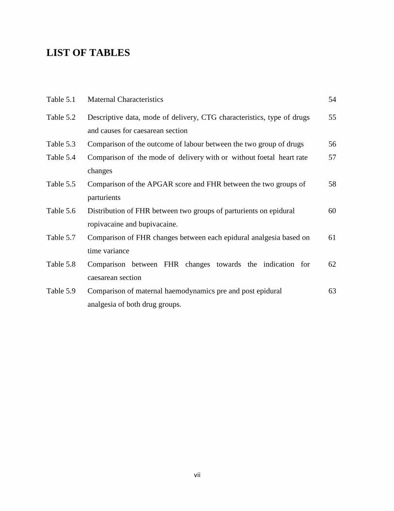
LIST OF TABLES
Table 5.1 Maternal Characteristics 54
Table 5.2 Descriptive data, mode of delivery, CTG characteristics, type of drugs
and causes for caesarean section
55
Table 5.3 Comparison of the outcome of labour between the two group of drugs 56
Table 5.4 Comparison of the mode of delivery with or without foetal heart rate
changes
57
Table 5.5 Comparison of the APGAR score and FHR between the two groups of
parturients
58
Table 5.6 Distribution of FHR between two groups of parturients on epidural
ropivacaine and bupivacaine.
60
Table 5.7 Comparison of FHR changes between each epidural analgesia based on
time variance
61
Table 5.8
Comparison between FHR changes towards the indication for
caesarean section
62
Table 5.9 Comparison of maternal haemodynamics pre and post epidural
analgesia of both drug groups.
63
viii
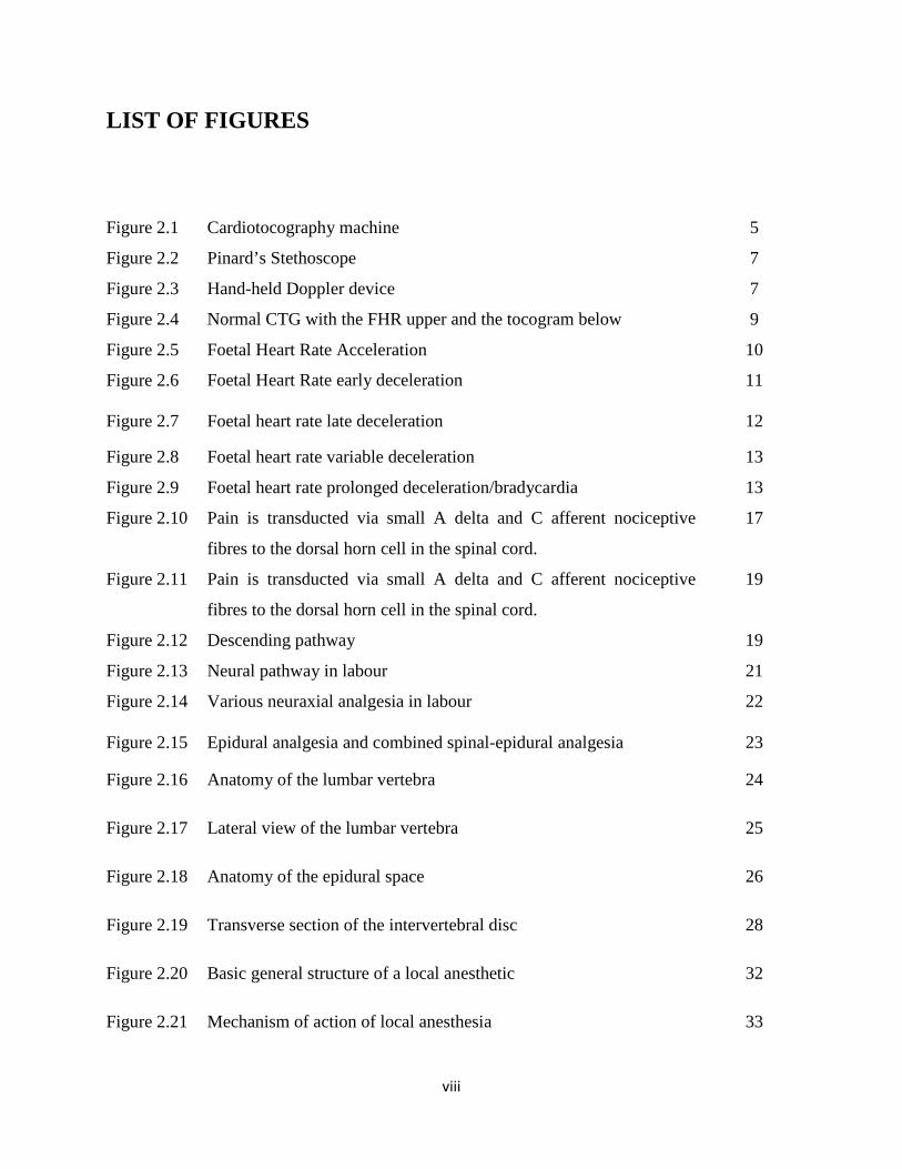
LIST OF FIGURES
Figure 2.1 Cardiotocography machine 5
Figure 2.2 Pinard’s Stethoscope 7
Figure 2.3 Hand-held Doppler device 7
Figure 2.4 Normal CTG with the FHR upper and the tocogram below 9
Figure 2.5 Foetal Heart Rate Acceleration 10
Figure 2.6 Foetal Heart Rate early deceleration 11
Figure 2.7 Foetal heart rate late deceleration 12
Figure 2.8 Foetal heart rate variable deceleration 13
Figure 2.9 Foetal heart rate prolonged deceleration/bradycardia 13
Figure 2.10 Pain is transducted via small A delta and C afferent nociceptive
fibres to the dorsal horn cell in the spinal cord.
17
Figure 2.11 Pain is transducted via small A delta and C afferent nociceptive
fibres to the dorsal horn cell in the spinal cord.
19
Figure 2.12 Descending pathway 19
Figure 2.13 Neural pathway in labour 21
Figure 2.14 Various neuraxial analgesia in labour 22
Figure 2.15 Epidural analgesia and combined spinal-epidural analgesia 23
Figure 2.16 Anatomy of the lumbar vertebra 24
Figure 2.17 Lateral view of the lumbar vertebra 25
Figure 2.18 Anatomy of the epidural space 26
Figure 2.19 Transverse section of the intervertebral disc 28
Figure 2.20 Basic general structure of a local anesthetic 32
Figure 2.21 Mechanism of action of local anesthesia 33

ix
Figure 2.22 CNS toxicity of local anesthesia 39
x
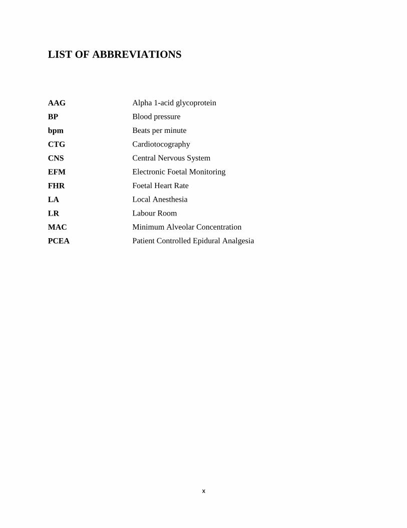
LIST OF ABBREVIATIONS
AAG Alpha 1-acid glycoprotein
BP Blood pressure
bpm Beats per minute
CTG Cardiotocography
CNS Central Nervous System
EFM Electronic Foetal Monitoring
FHR Foetal Heart Rate
LA Local Anesthesia
LR Labour Room
MAC Minimum Alveolar Concentration
PCEA Patient Controlled Epidural Analgesia

xi
ABSTRAK
Tajuk: Kajian tentang kelainan analisa denyutan jantung janin membandingkan ubat tahan
sakit epidural ropivacaine 0.2% ditambah fentanyl 2ug/ml dengan bupivacaine 0.1% ditambah
fentanyl 2ug/ml semasa bersalin.
Latar Belakang dan Objektif: Ubat tahan sakit epidural melegakan rasa sakit bersalin secara
efektif dan ramai ibu bersalin memilih teknik tersebut. Keselamatan janin dalam kandungan
semasa bersalin penting dan kebanyakan pembiusan separuh badan sewaktu bersalin
mempengaruhi denyutan jantung janin. Pelbagai kajian telah dilakukan untuk membandingkan
kepekatan ubat bius setempat serta teknik pembiusan separuh yang berlainan untuk menganalisa
kesan terhadap kadar denyutan jantung janin dan kesan terhadap cara melahirkan anak, kesan
terhadap kestabilan ibu serta bayi. Jadi, kajian ini bertujuan untuk menganalisa kadar denyutan
jantung semasa menggunakan dua kepekatan berlainan bagi ubat tahan sakit epidural dan kesan
terhadap cara melahirkan bayi, kestabilan ibu serta keadaan bayi semasa lahir.
Keputusan: Berdasarkan cara melahirkan anak, ibu yang bersalin secara spontan bagi
kumpulan yang menggunakan ropivacain adalah seramai 48 orang (71.6%) dengan bupivacaine
pula adalah seramai 49 orang (83.1%). Bagi kelahiran melalui Caesarean pula kumpulan
ropivacain adalah seramai 15 orang (22.4%) dan bagi bupivacaine pula ialah seramai 10 orang
(16.9%). Perbandingan di antara kedua-dua kumpulan ubat bius setempat menunjukkan tiada

xii
perbezaan yang bermakna dalam cara melahirkan anak. Seramai 115 ibu bersalin menunjukkan
kadar denyutan jantung bayi yang normal di mana 96 orang (83.5%) dari mereka melahirkan
secara spontan, 2 orang (1.7%) melahirkan secara instrumental dan selebihnya iaitu 17 orang
(14.8%) menjalani pembedahan Caesarean. Dalam pada itu 11 ibu bersalin menunjukkan kadar
denyutan jantung janin yang tidak normal, di mana 1 orang (9.1%) melahirkan secara spontan, 2
orang (18.2%) melahirkan secara instrumental dan seramai 8 orang (72.7%) secara pembedahan
Caesarean. Ibu bersalin yang menunjukkan CTG yang tidak normal mempunyai peratusan yang
lebih tinggi untuk bersalin secara pembedahan Caesarean. Seramai 27 ibu yang bersalin secara
pembedahan Caesarean di mana seramai 18 orang menunjukkan CTG yang normal, 4 orang
(22.2%) mengalami kelahiran tersekat, 2 orang (11.1%) mengalami kelemasan janin dan
selebihnya 12 orang (66.7%) mengalami proses kelahiran yang lambat. Ibu yang menunjukkan
CTG yang tidak normal, seramai seorang (11.1%) mengalami kelahiran tersekat manakala 8
orang (88.9%) mengalami kelemasan janin dengan nilai p=<0.001 yang bermakna. Tiada
perbezaan bermakna dilihat dari segi kestabilan ibu serta bayi semasa lahir.
Kesimpulan: Berdasarkan kajian ini, ubat tahan sakit secara epidural sewaktu bersalin
menggunakan ropivacain 0.2% dan bupivacain 0.1% dengan fentanyl 2ug/ml di dapati tidak
memberikan perubahan dalam denyutan jantung janin. Terdapat peningkatan kadar pembedahan
Caesarean disebab oleh CTG yang tidak normal dari kumpulan ropivacain diakibatkan
kelemasan janin. Tiada perubahan dalam kestabilan ibu dan bayi yang dilahirkan.
xiii
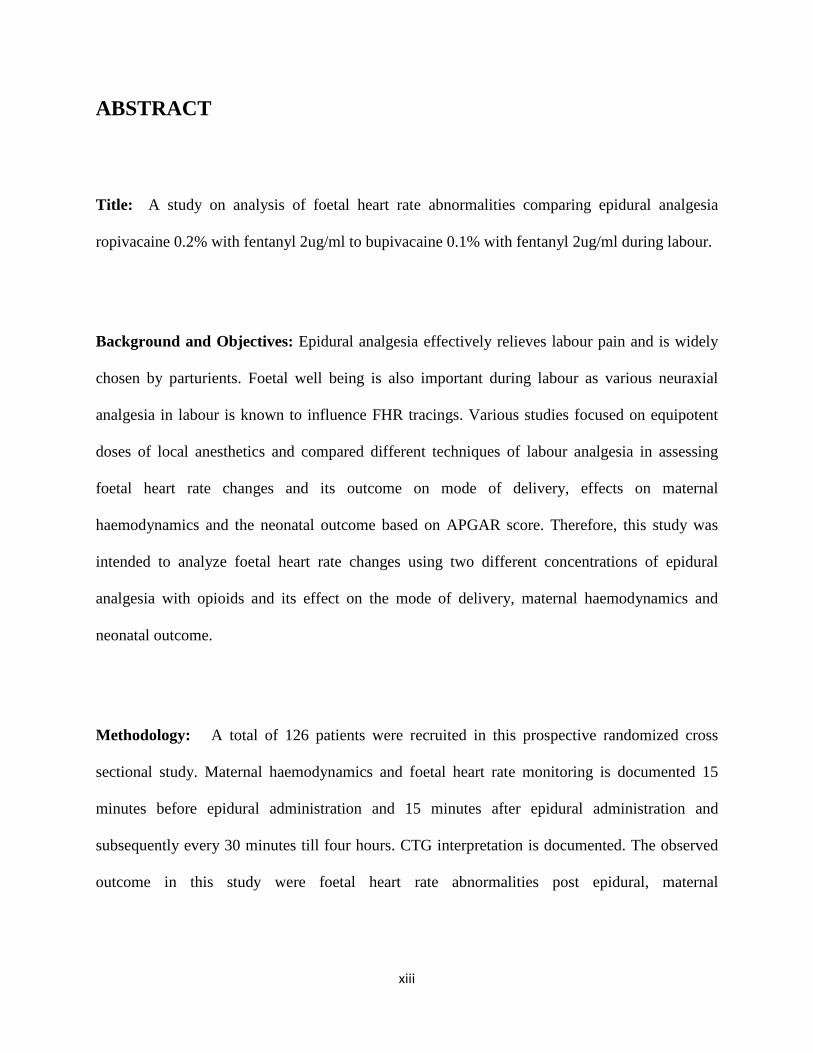
ABSTRACT
Title: A study on analysis of foetal heart rate abnormalities comparing epidural analgesia
ropivacaine 0.2% with fentanyl 2ug/ml to bupivacaine 0.1% with fentanyl 2ug/ml during labour.
Background and Objectives: Epidural analgesia effectively relieves labour pain and is widely
chosen by parturients. Foetal well being is also important during labour as various neuraxial
analgesia in labour is known to influence FHR tracings. Various studies focused on equipotent
doses of local anesthetics and compared different techniques of labour analgesia in assessing
foetal heart rate changes and its outcome on mode of delivery, effects on maternal
haemodynamics and the neonatal outcome based on APGAR score. Therefore, this study was
intended to analyze foetal heart rate changes using two different concentrations of epidural
analgesia with opioids and its effect on the mode of delivery, maternal haemodynamics and
neonatal outcome.
Methodology: A total of 126 patients were recruited in this prospective randomized cross
sectional study. Maternal haemodynamics and foetal heart rate monitoring is documented 15
minutes before epidural administration and 15 minutes after epidural administration and
subsequently every 30 minutes till four hours. CTG interpretation is documented. The observed
outcome in this study were foetal heart rate abnormalities post epidural, maternal
xiv
haemodynamics before and after epidural analgesia in labour, mode of delivery and the neonatal
APGAR score.
Result: Based on the mode of delivery between both the groups of epidural analgesia
ropivacaine and bupivacaine, there were 48 (71.6%) in the ropivacaine group and 49 (83.1%) in
the bupivacaine group who delivered spontaneously. 4 (6%) only delivered via instrumental in
the ropivacaine group. Via caesarean section 15 (22.4%) in the ropivacaine group and 10
(16.9%) in the bupivacaine group. There were no significant changes in the mode of delivery
between both the groups. 115 parturients had normal FHR, 96(83.5%) delivered spontaneously, 2
(1.7%) had instrumental delivery, 17 (14.8%) delivered via caesarean section. Whereas 11
patients had abnormal FHR, 1 (9.1%) delivered spontaneously, 2 (18.2%) via instrumental
delivery and 8 (72.7%) via caesarean delivery. A higher percentage in caesarean delivery with
abnormal CTG with a significant p value <0.001. Parturients for caesarean section were 27, 18
had normal CTG out of which 4 (22.2%) for secondary arrest, 2 (11.1%) for acute foetal distress,
12 (66.7%) for poor progress. 9 parturients had abnormal CTG, out of which 1 (11.1%) for
secondary arrest, 8 (88.9%) for acute foetal distress with a significant p value=<0.001.There
were no significant difference seen in maternal outcome and neonatal outcome.
Conclusion: This study revealed that with epidural analgesia in labour using ropivacaine 0.2%
and bupivacaine 0.1% with fentanyl 2ug/ml, there were no foetal heart rate changes. There were
increased risk for caesarean delivery with abnormal CTG in the ropivacaine group due to acute
foetal distress. There were no changes in the maternal and neonatal outcome.

xv
1
CHAPTER 1
INTRODUCTION
Foetal well being is the most important aspect during labour.To determine foetal well-
being, one of the ways is by recording the foetal heart rate (FHR). Loss of beat to beat
variability and deceleration patterns are known to be associated with foetal distress.
Gynaecological factors, maternal and foetal factors, but also anaesthesiological factors
can influence these FHR tracings. Decelerations and foetal bradycardia have been
described after all types of effective labour analgesia such as (epidural,spinal,and
combined spinal epidural(CSE) and intravenous opioids.(Nicole Maria Anna Adela
Engel, 2011).
Epidural analgesia effectively relieves labour pain and is now widely chosen by
parturients. In the US the use of neuraxial analgesia for labour increased to 77% in
2001 from 21% in 1981 and a little over 33% of parturients chose neuraxial analgesia
for childbirth in UK from 2008-2009.(Cambic and Wong)2010. The additional use of
opioids increases the effect of local anesthetic allowing the use of minimal
concentration of local anesthetic, reducing the side effects such as hypotension and
motor block. The minimum effective doses are used to avoid interference with the
progression of labour and permit ambulation, however transient foetal heart rate(FHR)
changes may occasionally follow with the use of any technique.(Capogna, 2001)

2
The beneficial effects of epidural analgesia may be offset by the detrimental effects
such as prolonged labour, higher incidence of instrumental delivery and Caesarean
section, maternal factor such as (hypotension,epidural haematoma and epidural
abscess) and also towards the neonatal outcome. Although numerous studies have been
done on the effects of epidural analgesia the results are contradictory.
(Cambic and Wong)2010 conducted a study and the evidence of neuraxial analgesia on
labour outcome shows that neuraxial analgesia does not increase the risk of caesarean
delivery .Conflicting evidence but overall it suggests increased rate of instrumental
vaginal delivery in women receiving neuraxial labour analgesia.This was mainly
affected by multiple factors, such as the degree of analgesia during second stage of
labour, local anaesthetic concentration, method of epidural analgesia maintenance,
technique and obstetric factors. There were no difference in the duration of first stage
of labour and effective neuraxial analgesia increases the duration of second stage of
labour.
A systemic review was done by (Leighton and Halpern, 2002) and their findings
showed that parturients preferred epidural analgesia compared to parenteral opioids and
they were more comfortable and satisfied,The length of first stage and second stage of
labour were prolonged and the total incidence of instrumental delivery was higher in
the epidural group. Neonatal outcome between the two groups showed no difference or
more so favoured epidural analgesia. There were no significant trend toward an
increase in the total number of caesarean delivery compared to the previous systemic
review.(Halpern et al., 1998).
3
In relation to dose related epidural analgesia there were numerous studies comparing
bupivacaine and ropivacaine for labour analgesia. A randomized double blinded study
by(Lee et al., 2004) comparing epidural infusions of ropivacaine 0.1% with 2ug/ml of
fentanyl with bupivacaine 0.1% with 2ug/ml Fentanyl focused on the obstetric outcome
where the mode of delivery was similar between the two groups. Parturients who
delivered vaginally, the duration of the first stage of labour was shorter in the
ropivacaine group compared with the bupivacaine group. Incidence of maternal and
neonatal outcome were similar between both the groups.
Another study was conducted by (Halpern and Walsh, 2003) which was a meta analysis
also concluded that both ropivacaine and bupivacaine provided excellent labour
analgesia and there is no significant difference between the two drugs with regards to
neonatal, spontaneous vaginal delivery or any other obstetrical oucome. A randomized
double blinded study(Fischer et al., 2000) compared the administration of 0.1%
ropivacaine and 0.5 ug/ml sufentanil with bupivacaine 0.1% and 0.5ug/ml sufentanil
via a PCEA device. Outcome of this study produced effective pain relief in both the
groups but maternal satisfaction was greater with the bupivacaine group. Ropivacaine
drug was used more than bupivacaine during the second stage of labour, concluding
that ropivacaine was less potent compared to bupivacaine and also due to the reduction
in motor impairment.
Many studies were done on neuraxial analgesia in labour with different techniques,
comparisons of equipotent concentrations of local anesthesia, comparisons with
parenteral analgesia in labour with and without opioids and all these studies were
4
focused on the outcomes of maternal, neonatal and mode of delivery. There a few
studies which has focused on the foetal wellbeing post neuraxial analgesia. A study on
the incidence and clinical significance of foetal heart rate changes using intrathecal
sufentanil or epidural bupivacaine for labour analgesia(Nielsen et al., 1996),there were
no significant difference in foetal heart rate between these two groups and also to the
neonatal outcome.
A review on the effect of epidural analgesia on the foetal heart rate(Capogna, 2001)
concluded that epidural or spinal analgesia in the absence of maternal hypotension or
uterine hypotonus causes minimal changes in FHR. Even if there were it is transient
and should not produce maternal or foetal morbidity. A recent French study (Korb et
al.) analyzed foetal heart rate abnormalities occurring within one hour after laying of
epidural analgesia using 0.1% levobupivacaine and sufentanil 0.2ug/ml. The results
showed an increase in obstetric intervention in the abnormal foetal heart rate group and
an absence of impact in the neonate state.
The importance of this study is to characterize abnormalities in the foetal heart rate by
using two different concentration of epidural analgesia and its effect on the mode of
delivery and neonatal outcome. The aim of this study is to characterize the
abnormalities in FHR with ropivacaine 0.2%+ 2ug fentanyl and bupivacaine 0.1% +
2ug fentanyl, and its outcome on the mode of delivery and neonatal status. It attempts
to address the potencies of different concentrations of the local anesthetic towards
foetal heart rate abnormality during epidural analgesia in labour.
5
CHAPTER 2
LITERATURE REVIEW
2.1 Foetal Heart Rate
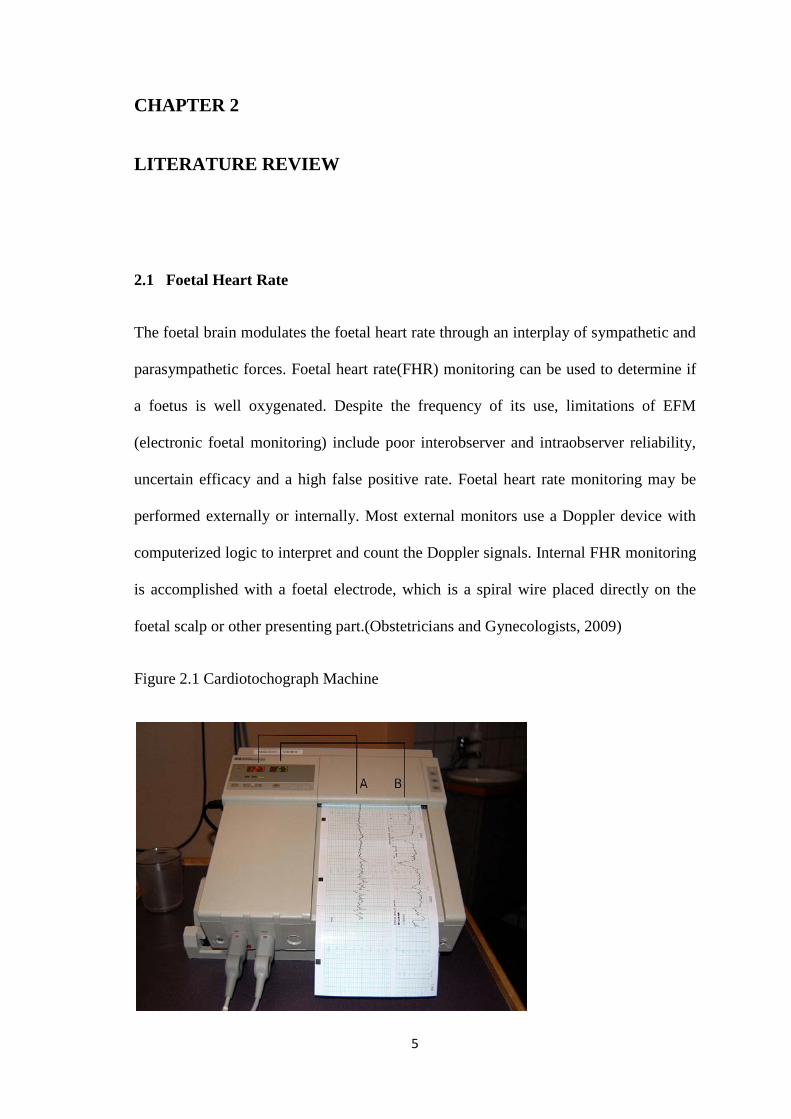
The foetal brain modulates the foetal heart rate through an interplay of sympathetic and
parasympathetic forces. Foetal heart rate(FHR) monitoring can be used to determine if
a foetus is well oxygenated. Despite the frequency of its use, limitations of EFM
(electronic foetal monitoring) include poor interobserver and intraobserver reliability,
uncertain efficacy and a high false positive rate. Foetal heart rate monitoring may be
performed externally or internally. Most external monitors use a Doppler device with
computerized logic to interpret and count the Doppler signals. Internal FHR monitoring
is accomplished with a foetal electrode, which is a spiral wire placed directly on the
foetal scalp or other presenting part.(Obstetricians and Gynecologists, 2009)
Figure 2.1 Cardiotochograph Machine
6
Foetal heart sounds were said to be first detected by Marsac in the 1600’s. Killian
proposed that foetal heart rate could be used to determine foetal well being in the
1600’s. This thought went unnoticed until 1818 when Mayor and Kergaradec described
the method of auscultating foetal heart sounds by placing the ear next to the maternal
abdomen. Kergaradec also suggested that foetal heart sounds could be used to
determine foetal life and multiple pregnancy and if it would be possible to assess foetal
abnormalities from variations in the foetal heart rate. Evory Kennedy, an English
physician, published guidelines for foetal distress and recommended auscultation of the
foetal heart rate as a tool of intrapartum monitoring in 1833. By 1893, Von Winkel
developed a criteria for foetal distress that remained unchanged until the arrival of
electronic fetal monitoring.(Alfirevic et al., 2006)
Since then, many methods of listening to the foetal heart have been developed and
introduced into maternity care, with the goal of improving outcomes for neonates and
easing the heartache for mothers and families when a baby dies or suffers long-term
disability. For now, monitoring the foetal heart during labour, by one method or
another, has become a routine part of care during labour, although access to such care
are different across the world.(Alfirevic et al., 2006)
2.2 Foetal Heart Rate Monitoring
Methods of monitoring the foetal heart rate
The foetal heart rate can be monitored either intermittently (at regular intervals during
labour) or continuously (recording the foetal heart rate throughout labour, stopping
only briefly, e.g. for visits to the toilet) as follows.
7
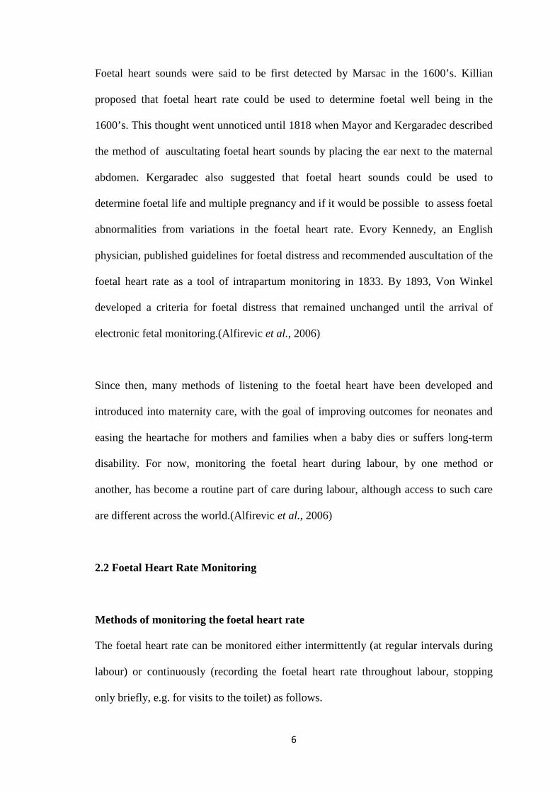
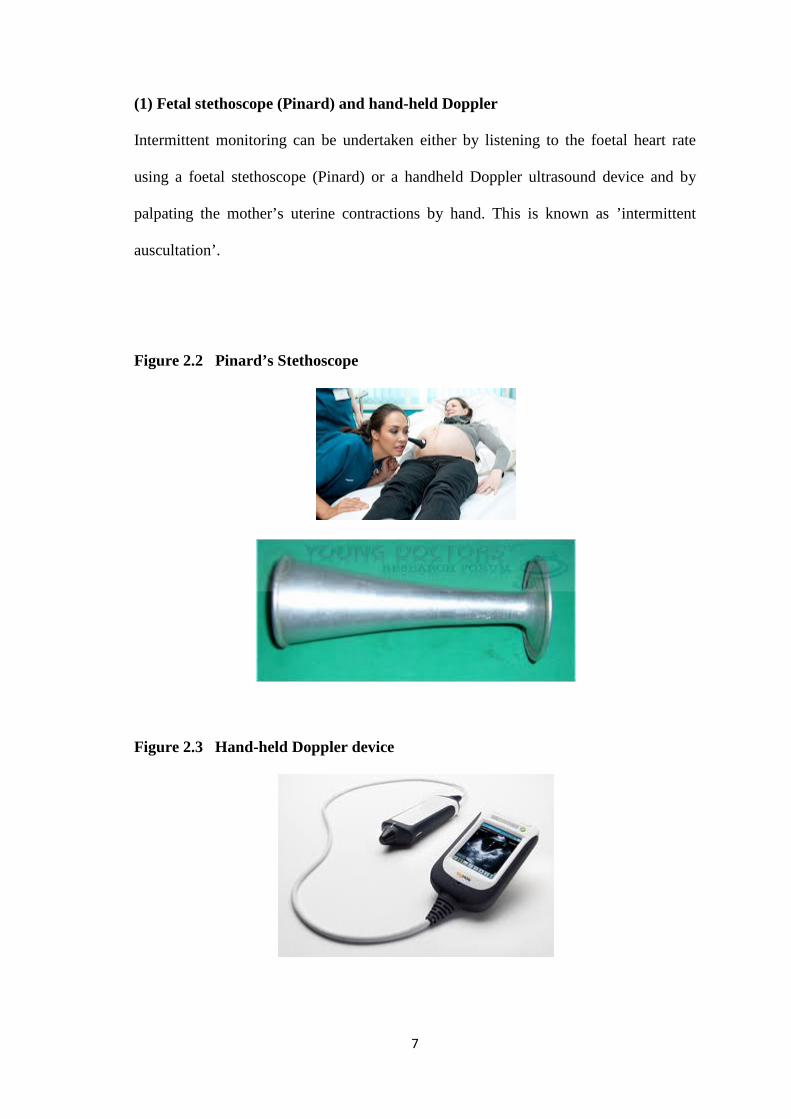
(1) Fetal stethoscope (Pinard) and hand-held Doppler
Intermittent monitoring can be undertaken either by listening to the foetal heart rate
using a foetal stethoscope (Pinard) or a handheld Doppler ultrasound device and by
palpating the mother’s uterine contractions by hand. This is known as ’intermittent
auscultation’.
Figure 2.2 Pinard’s Stethoscope
Figure 2.3 Hand-held Doppler device
8
(2) Cardiotocograph (CTG)
Cardiotocograph electronically records the foetal heart rate and the mother’s uterine
contractions on a paper trace. External CTG: This is obtained by using a Doppler
ultrasound transducer to monitor the foetal heart rate and a pressure transducer to
monitor uterine contractions, both are connected to a recording machine. Internal
CTG: Monitors the foetal heart rate with the CTG machine by attaching an electrode
directly to the baby’s presenting part usually its head. It is a form of continuous
monitoring and requires a ruptured amniotic sac (either spontaneously or artificially)
and a scalp electrode (clip) attached to the foetal head.(Alfirevic et al., 2006).
CTG monitoring and a systematic approach to CTG analysis may enable anaesthetists
to better understand why obstetricians make specific clinical decisions. This
understanding may aid communication and timely delivery especially when the foetus
is considered at high risk.(ATOTW, 2013).Parturients receiving regional analgesia in
labour, requires continuous electronic fetal monitoring for at least 30 minutes during
establishment of regional analgesia and after administration of a further bolus of local
anaesthetic agent. In most UK centres, continuous CTG monitoring is performed after
the insertion of a labour epidural.(ATOTW,2013)
2.3 Baseline Fetal Heart Rate
The normal baseline foetal heart rate is defined as 110 – 160 bpm. Foetal bradycardia is
a baseline rate of <110 bpm. Foetal tachycardia is a baseline rate of >160 bpm. Many
foetal baseline bradycardias have no identifiable cause but may occur as a result of:
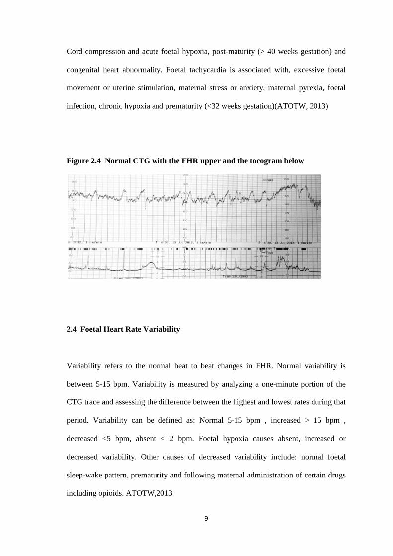
9
Cord compression and acute foetal hypoxia, post-maturity (> 40 weeks gestation) and
congenital heart abnormality. Foetal tachycardia is associated with, excessive foetal
movement or uterine stimulation, maternal stress or anxiety, maternal pyrexia, foetal
infection, chronic hypoxia and prematurity (<32 weeks gestation)(ATOTW, 2013)
Figure 2.4 Normal CTG with the FHR upper and the tocogram below
2.4 Foetal Heart Rate Variability
Variability refers to the normal beat to beat changes in FHR. Normal variability is
between 5-15 bpm. Variability is measured by analyzing a one-minute portion of the
CTG trace and assessing the difference between the highest and lowest rates during that
period. Variability can be defined as: Normal 5-15 bpm , increased > 15 bpm ,
decreased <5 bpm, absent < 2 bpm. Foetal hypoxia causes absent, increased or
decreased variability. Other causes of decreased variability include: normal foetal
sleep-wake pattern, prematurity and following maternal administration of certain drugs
including opioids. ATOTW,2013
10
2.5 Acceleration
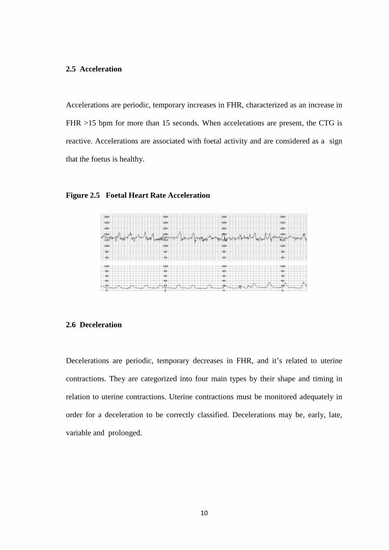
Accelerations are periodic, temporary increases in FHR, characterized as an increase in
FHR >15 bpm for more than 15 seconds. When accelerations are present, the CTG is
reactive. Accelerations are associated with foetal activity and are considered as a sign
that the foetus is healthy.
Figure 2.5 Foetal Heart Rate Acceleration
2.6 Deceleration
Decelerations are periodic, temporary decreases in FHR, and it’s related to uterine
contractions. They are categorized into four main types by their shape and timing in
relation to uterine contractions. Uterine contractions must be monitored adequately in
order for a deceleration to be correctly classified. Decelerations may be, early, late,
variable and prolonged.
11
2.6.1 Early Deceleration
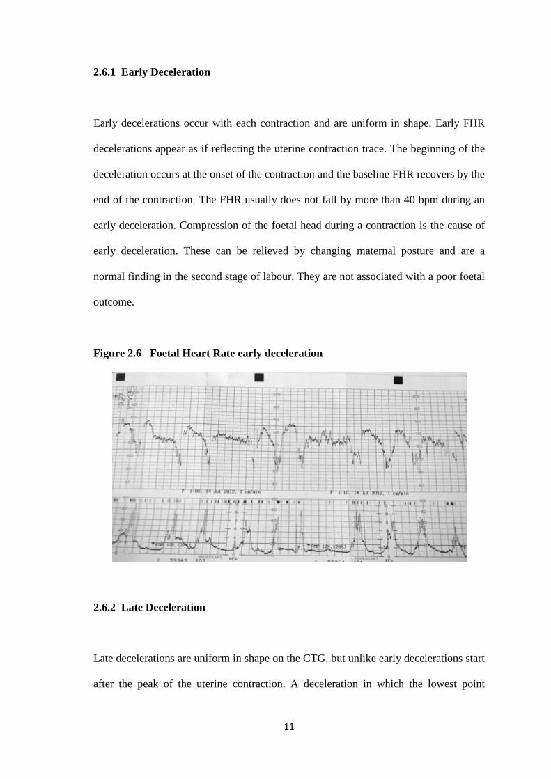
Early decelerations occur with each contraction and are uniform in shape. Early FHR
decelerations appear as if reflecting the uterine contraction trace. The beginning of the
deceleration occurs at the onset of the contraction and the baseline FHR recovers by the
end of the contraction. The FHR usually does not fall by more than 40 bpm during an
early deceleration. Compression of the foetal head during a contraction is the cause of
early deceleration. These can be relieved by changing maternal posture and are a
normal finding in the second stage of labour. They are not associated with a poor foetal
outcome.
Figure 2.6 Foetal Heart Rate early deceleration
2.6.2 Late Deceleration
Late decelerations are uniform in shape on the CTG, but unlike early decelerations start
after the peak of the uterine contraction. A deceleration in which the lowest point
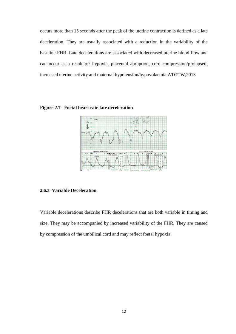
12
occurs more than 15 seconds after the peak of the uterine contraction is defined as a late
deceleration. They are usually associated with a reduction in the variability of the
baseline FHR. Late decelerations are associated with decreased uterine blood flow and
can occur as a result of: hypoxia, placental abruption, cord compression/prolapsed,
increased uterine activity and maternal hypotension/hypovolaemia.ATOTW,2013
Figure 2.7 Foetal heart rate late deceleration
2.6.3 Variable Deceleration
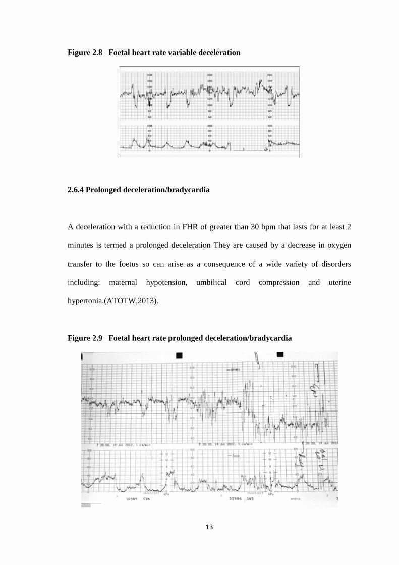
Variable decelerations describe FHR decelerations that are both variable in timing and
size. They may be accompanied by increased variability of the FHR. They are caused
by compression of the umbilical cord and may reflect foetal hypoxia.
13
Figure 2.8 Foetal heart rate variable deceleration
2.6.4 Prolonged deceleration/bradycardia
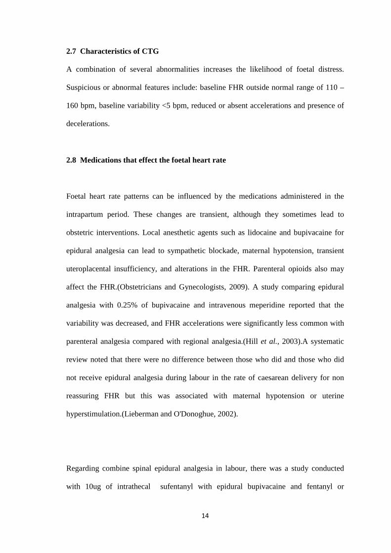
A deceleration with a reduction in FHR of greater than 30 bpm that lasts for at least 2
minutes is termed a prolonged deceleration They are caused by a decrease in oxygen
transfer to the foetus so can arise as a consequence of a wide variety of disorders
including: maternal hypotension, umbilical cord compression and uterine
hypertonia.(ATOTW,2013).
Figure 2.9 Foetal heart rate prolonged deceleration/bradycardia
14
2.7 Characteristics of CTG
A combination of several abnormalities increases the likelihood of foetal distress.
Suspicious or abnormal features include: baseline FHR outside normal range of 110 –
160 bpm, baseline variability <5 bpm, reduced or absent accelerations and presence of
decelerations.
2.8 Medications that effect the foetal heart rate
Foetal heart rate patterns can be influenced by the medications administered in the
intrapartum period. These changes are transient, although they sometimes lead to
obstetric interventions. Local anesthetic agents such as lidocaine and bupivacaine for
epidural analgesia can lead to sympathetic blockade, maternal hypotension, transient
uteroplacental insufficiency, and alterations in the FHR. Parenteral opioids also may
affect the FHR.(Obstetricians and Gynecologists, 2009). A study comparing epidural
analgesia with 0.25% of bupivacaine and intravenous meperidine reported that the
variability was decreased, and FHR accelerations were significantly less common with
parenteral analgesia compared with regional analgesia.(Hill et al., 2003).A systematic
review noted that there were no difference between those who did and those who did
not receive epidural analgesia during labour in the rate of caesarean delivery for non
reassuring FHR but this was associated with maternal hypotension or uterine
hyperstimulation.(Lieberman and O'Donoghue, 2002).
Regarding combine spinal epidural analgesia in labour, there was a study conducted
with 10ug of intrathecal sufentanyl with epidural bupivacaine and fentanyl or
15
intravenous meperidine(opioid). The results showed a significantly higher rate of
bradycardia and caesarean delivery for abnormal FHR in the CSE group however the
neonatal outcome were not significantly different between the two groups.(Gambling
et al., 1998).Another study comparing combine spinal analgesia with epidural
analgesia, noted that FHR abnormalities were more common in the combine spinal
epidural group.(Trial et al.)
There are also other drugs which can affect foetal heart rate such as butorphanol which
causes a transient sinusoidal FHR pattern with a slight increased mean heart rate
compared with meperidine.(HATJIS and MEIS, 1986). Cocaine causes decreased long
term variability in the foetal heart rate.(Forman and Gandhi, 1991). Corticosteroids
causes decrease in FHR variability and this happens with beta-methasone and not with
dexamethasone. It causes abolishment of diurnal foetal rhythm and this effect usually
occurs more than 29 weeks of gestation.(Rotmensch et al., 1999). Magnesium sulphate
causes a significant decrease in short term variability and it inhibits the increase in
accelerations with advancing gestational age.(Hallak et al., 1999). Terbutaline causes
an increase in baseline FHR and incidence of foetal tachycardia.(Roth et al., 1990).
There are other factors which causes changes in foetal heart rate such as maternal
position as a cause of severe FHR changes after epidural bupivacaine labour analgesia.
This is due to occult aortocaval compression.(Preston et al., 1993). Maternal
hypotension too has been associated to foetal bradycardia after epidural or spinal
analgesia.Uterine hypertonus has been associated with foetal bradycardia after
induction of labour analgesia such as CSE compared to epidural.(Landau et al., 2009)
16
2.9 Labour Pain
For most women labour causes severe pain, it is similar to that caused by complex
regional pain syndrome. The American College of Obstetricians and Gynecologists and
the American Society of Anesthesiologists (ASA) stated that, “There is no other
circumstance where it is considered acceptable for an individual to experience
untreated severe pain, amenable to safe intervention, while under a physician’s
care.(Obstet Gynecol,2004). Without a medical contraindication, maternal request is a
sufficient medical indication for pain relief during labour. Although severe pain is not
life-threatening in healthy parturient women, it can have neuropsychological
consequences. Postnatal depression may be more common when analgesia is not
used.(Hiltunen et al., 2004).And pain during labour has been correlated with the
development of post-traumatic stress disorder.(Soet et al., 2003).
2.9.1 Physiology of pain in labour
Labour pain is caused by uterine contractions and cervical dilatation and is transmitted
through visceral afferent(sympathetic) nerves entering the spinal cord from T10
through L1. Later in labour, perineal stretching transmits painful stimuli through the
pudendal nerve and sacral nerves S2 through S4. The maternal stress response can lead
to increased release of corticotropin, cortisol, norepinephrine, β-endorphins, and
epinephrine. Epinephrine can have relaxant effects on the uterus that may prolong
labor.(Hawkins, 2010).
17
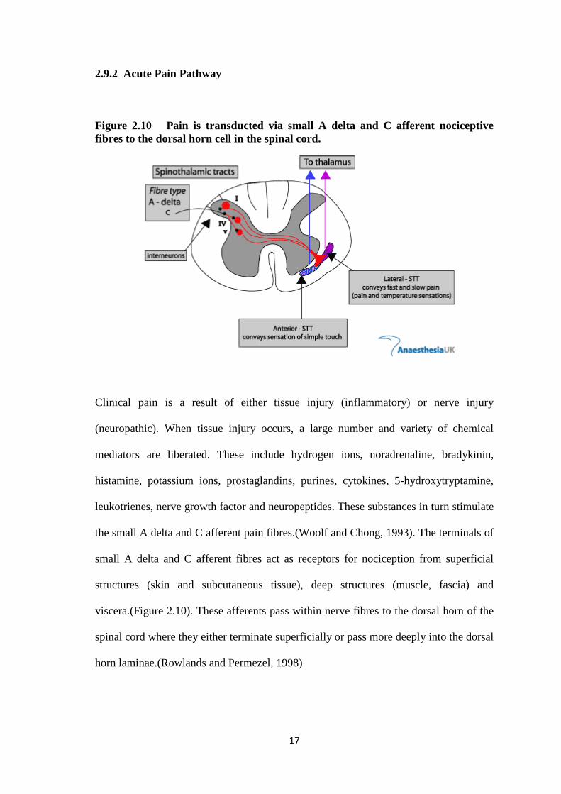
2.9.2 Acute Pain Pathway
Figure 2.10 Pain is transducted via small A delta and C afferent nociceptive fibres to the dorsal horn cell in the spinal cord.
Clinical pain is a result of either tissue injury (inflammatory) or nerve injury
(neuropathic). When tissue injury occurs, a large number and variety of chemical
mediators are liberated. These include hydrogen ions, noradrenaline, bradykinin,
histamine, potassium ions, prostaglandins, purines, cytokines, 5-hydroxytryptamine,
leukotrienes, nerve growth factor and neuropeptides. These substances in turn stimulate
the small A delta and C afferent pain fibres.(Woolf and Chong, 1993). The terminals of
small A delta and C afferent fibres act as receptors for nociception from superficial
structures (skin and subcutaneous tissue), deep structures (muscle, fascia) and
viscera.(Figure 2.10). These afferents pass within nerve fibres to the dorsal horn of the
spinal cord where they either terminate superficially or pass more deeply into the dorsal
horn laminae.(Rowlands and Permezel, 1998)
18
Action potentials generated within the dorsal cell neurone may participate in local
spinal reflexes in which anterior and anterolateral horn cells stimulate skeletal muscle
and sympathetic outflow. The action potential is also relayed centrally via the
spinothalamic tract (Figure 2.11), from which the impulse is modulated successively by
the reticular formation in the brainstem (integrative function), thalamus (level of
arousal), hypothalamus and limbic systems (attention, mood and motivation).
Nociception is modulated at the level of the dorsal horn by descending spinal tracts,
which receive input from higher centres and the limbic system via stimulation of
structures in the midbrain. This phenomenon is called 'stimulation-produced analgesia'.
The type of nociceptive input appears to determine the descending tract stimulated and
thus the neurotransmitter response.(Rowlands and Permezel, 1998).
Mechanical nociception activates the synthesis of β-endorphin which stimulates
supraspinal opioid receptors and results in spinal release of enkephalins. Other
endogenous opioids are known to be involved in the spinal modulation of nociception;
proenkephalin is the precursor for met-enkephalin and other enkephalins which act by
interaction with 'δ-opioid' receptors and dynorphin derived from prodynorphin
interacts with 'κ-opioid' receptors.(Tseng et al., 1995). Following the occupation of the
opioid receptors on the dorsal horn neurones, the anti-nociceptive effect is produced by
attenuation of the primary afferent nociceptive input, by inhibition of propagation of
the action potential along the dorsal horn cell or by reducing the release of excitatory
neurotransmitter substances from primary afferent terminals.
19
Figure 2.11 Ascending pathways
Figure 2.12 Descending pathway
20
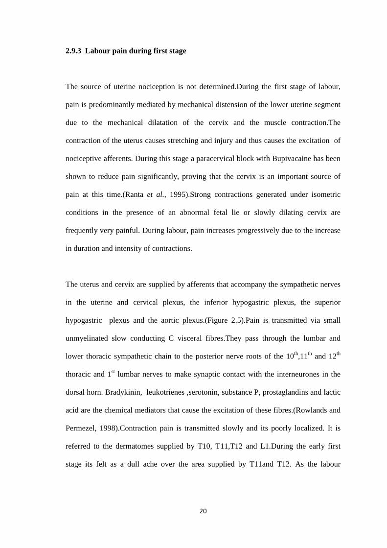
2.9.3 Labour pain during first stage
The source of uterine nociception is not determined.During the first stage of labour,
pain is predominantly mediated by mechanical distension of the lower uterine segment
due to the mechanical dilatation of the cervix and the muscle contraction.The
contraction of the uterus causes stretching and injury and thus causes the excitation of
nociceptive afferents. During this stage a paracervical block with Bupivacaine has been
shown to reduce pain significantly, proving that the cervix is an important source of
pain at this time.(Ranta et al., 1995).Strong contractions generated under isometric
conditions in the presence of an abnormal fetal lie or slowly dilating cervix are
frequently very painful. During labour, pain increases progressively due to the increase
in duration and intensity of contractions.
The uterus and cervix are supplied by afferents that accompany the sympathetic nerves
in the uterine and cervical plexus, the inferior hypogastric plexus, the superior
hypogastric plexus and the aortic plexus.(Figure 2.5).Pain is transmitted via small
unmyelinated slow conducting C visceral fibres.They pass through the lumbar and
lower thoracic sympathetic chain to the posterior nerve roots of the 10th,11th and 12th
thoracic and 1st lumbar nerves to make synaptic contact with the interneurones in the
dorsal horn. Bradykinin, leukotrienes ,serotonin, substance P, prostaglandins and lactic
acid are the chemical mediators that cause the excitation of these fibres.(Rowlands and
Permezel, 1998).Contraction pain is transmitted slowly and its poorly localized. It is
referred to the dermatomes supplied by T10, T11,T12 and L1.During the early first
stage its felt as a dull ache over the area supplied by T11and T12. As the labour
21
progresses, pain increases and becomes more severe and is referred to the abdomen,
lower lumbar and upper sacrum supplied by T10 and L1.
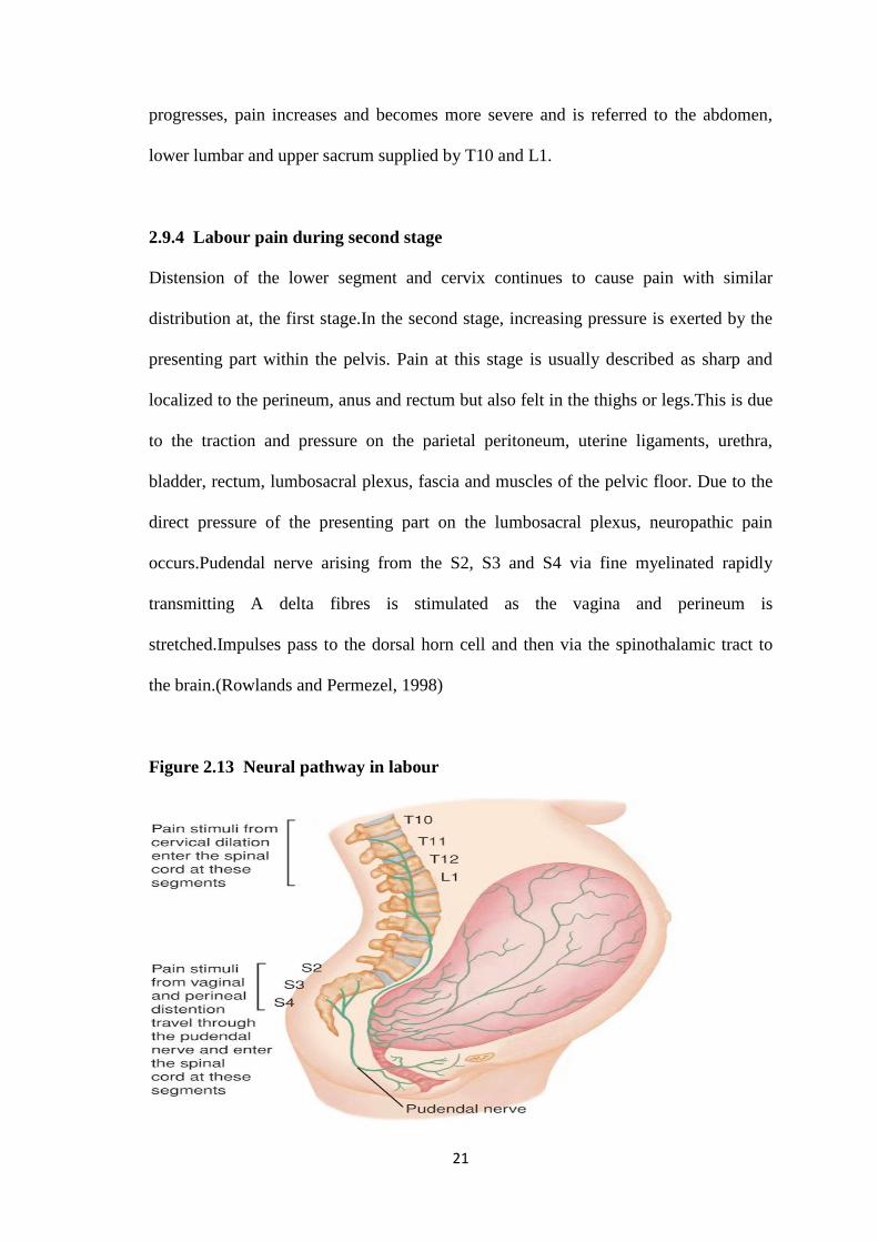
2.9.4 Labour pain during second stage
Distension of the lower segment and cervix continues to cause pain with similar
distribution at, the first stage.In the second stage, increasing pressure is exerted by the
presenting part within the pelvis. Pain at this stage is usually described as sharp and
localized to the perineum, anus and rectum but also felt in the thighs or legs.This is due
to the traction and pressure on the parietal peritoneum, uterine ligaments, urethra,
bladder, rectum, lumbosacral plexus, fascia and muscles of the pelvic floor. Due to the
direct pressure of the presenting part on the lumbosacral plexus, neuropathic pain
occurs.Pudendal nerve arising from the S2, S3 and S4 via fine myelinated rapidly
transmitting A delta fibres is stimulated as the vagina and perineum is
stretched.Impulses pass to the dorsal horn cell and then via the spinothalamic tract to
the brain.(Rowlands and Permezel, 1998)
Figure 2.13 Neural pathway in labour
22
Segmental and suprasegmental reflex responses from the pain of labour may effect
respiratory, cardiovascular, gastrointestinal, urinary and neuroendocrine function.These
responses are mediated by pain and its evidenced by the fact that they can be prevented
or abolished by central neural blockade.(Rowlands and Permezel, 1998).
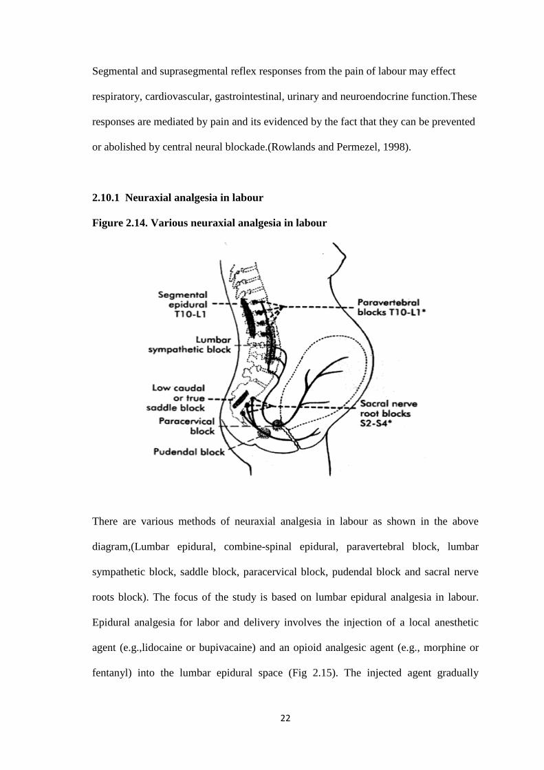
2.10.1 Neuraxial analgesia in labour
Figure 2.14. Various neuraxial analgesia in labour
There are various methods of neuraxial analgesia in labour as shown in the above
diagram,(Lumbar epidural, combine-spinal epidural, paravertebral block, lumbar
sympathetic block, saddle block, paracervical block, pudendal block and sacral nerve
roots block). The focus of the study is based on lumbar epidural analgesia in labour.
Epidural analgesia for labor and delivery involves the injection of a local anesthetic
agent (e.g.,lidocaine or bupivacaine) and an opioid analgesic agent (e.g., morphine or
fentanyl) into the lumbar epidural space (Fig 2.15). The injected agent gradually
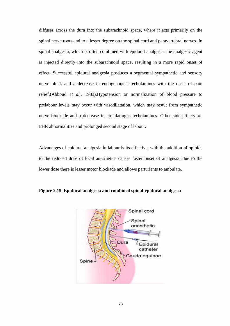
23
diffuses across the dura into the subarachnoid space, where it acts primarily on the
spinal nerve roots and to a lesser degree on the spinal cord and paravertebral nerves. In
spinal analgesia, which is often combined with epidural analgesia, the analgesic agent
is injected directly into the subarachnoid space, resulting in a more rapid onset of
effect. Successful epidural analgesia produces a segmental sympathetic and sensory
nerve block and a decrease in endogenous catecholamines with the onset of pain
relief.(Abboud et al., 1983).Hypotension or normalization of blood pressure to
prelabour levels may occur with vasodilatation, which may result from sympathetic
nerve blockade and a decrease in circulating catecholamines. Other side effects are
FHR abnormalities and prolonged second stage of labour.
Advantages of epidural analgesia in labour is its effective, with the addition of opioids
to the reduced dose of local anesthetics causes faster onset of analgesia, due to the
lower dose there is lesser motor blockade and allows parturients to ambulate.
Figure 2.15 Epidural analgesia and combined spinal-epidural analgesia
24
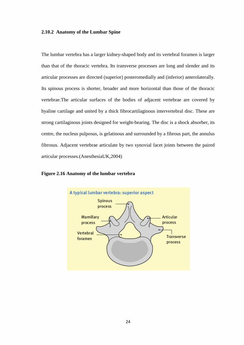
2.10.2 Anatomy of the Lumbar Spine
The lumbar vertebra has a larger kidney-shaped body and its vertebral foramen is larger
than that of the thoracic vertebra. Its transverse processes are long and slender and its
articular processes are directed (superior) posteromedially and (inferior) anterolaterally.
Its spinous process is shorter, broader and more horizontal than those of the thoracic
vertebrae.The articular surfaces of the bodies of adjacent vertebrae are covered by
hyaline cartilage and united by a thick fibrocartilaginous intervertebral disc. These are
strong cartilaginous joints designed for weight-bearing. The disc is a shock absorber, its
centre, the nucleus pulposus, is gelatinous and surrounded by a fibrous part, the annulus
fibrosus. Adjacent vertebrae articulate by two synovial facet joints between the paired
articular processes.(AnesthesiaUK,2004)
Figure 2.16 Anatomy of the lumbar vertebra