


γλώσσες
Σελίδες
Νομικός

MESTRADO EM ODONTOLOGIA
RENAN DALLA SOARES
ANÁLISE IN VITRO DA ADESÃO E PROLIFERAÇÃO DE OSTEOBLASTOS SOBRE A SUPERFÍCIE DE ZIRCÔNIA
TRATADA POR LASER DE Er,Cr:YSGG (ERBIUM-CHROMIUM: YTTRIUM, SCANDIUM, GALLIUM , GARNET)
Guarulhos 2012
1
RENAN DALLA SOARES
ANÁLISE IN VITRO DA ADESÃO E PROLIFERAÇÃO DE OSTEOBLASTOS SOBRE A SUPERFÍCIE DA ZIRCÔNIA
TRATADA POR LASER DE Er,Cr:YSGG (ERBIUM-CHROMIUM: YTTRIUM, SCANDIUM, GALLIUM , GARNET)
Dissertação apresentada à Universidade Guarulhos para obtenção do título de Mestre
em Odontologia. Área de Concentração: Implantodontia. Orientador: Prof. Dr. José Augusto Rodrigues Co-orientadora: Profa. Dra. Alessandra Cassoni
Guarulhos 2012
2
FICHA CATALOGRÁFICA
3
DEDICATÓRIA
Dedico este trabalho à minha esposa
Danielle e às minhas filhas Estella e Alice,
amores da minha vida, que me dão força e
coragem para prosseguir.
Dedico este trabalho ao meu pai Sebastião Tamburini Soares (in memoriam), a minha
mãe Iara Dalla Soares e aos meus irmão
Denis, Luciana e Allan que me
acompanham por toda a vida.
4
AGRADECIMENTOS
Ao Prof. Dr. José Augusto Rodrigues, que me deu a honra de tê-lo como
orientador, pela amizade e paciência, cuja dedicação o faz um exemplo de
profissional.
À Profa. Dra. Alessandra Cassoni Ferreira que, com entusiasmo, sempre
acreditou no sucesso do trabalho.
Ao Prof. Dr. Jamil Awad Shibli, por ter me recebido como aluno no curso de
mestrado e compartilhado comigo seus conhecimentos, ideias e filosofias. Muito
obrigado.
À Profa. Dra. Marta Bastos, pela sua inestimável ajuda científica e
metodológica, que permitiu um melhor desenvolvimento deste trabalho. Muito
obrigado.
A todos os professores do Curso de Mestrado em Odontologia da
Universidade Guarulhos, pela amizade e pelo estímulo na busca pelo conhecimento.
À Profa. Dra. Maria Christina Brunetti, um exemplo de pessoa e uma
profissional admirável, que por todos estes anos confia em mim, nesta parceria pela
docência em Periodontia e me honra com sua inestimável amizade. Muito obrigado.
Ao Prof. Dr. Paulo Francisco Cesar, da Universidade de São Paulo, pela
amizade e por disponibilizar a zircônia para a realização deste trabalho.
Ao Prof. Alberto Blay, por ter me recebido tão bem em sua clínica e pelo
suporte no uso do laser de Er,Cr:YSGG.
À Profa. Dra. Ariadne Cruz e à Profa. Dra. Claudia O. Simões, da
Universidade Federal de Santa Catarina, pelo total suporte técnico e científico na
realização da cultura dos osteoblastos.
Aos colegas de mestrado, Alexandre Dottore, Fabiano Zanardo, José Della
Pasqua Neto, Karen Bechara, Pérsio Vasconcelos e Ricardo Vecchiatti, e do
doutorado, Carlos Eduardo Pena, José Divino, Luis Braga e Ronaldo Viotti, pelo
companheirismo e amizade.
À equipe de Periodontia do Senac-SP, Profa. Cristiane Prazeres, Profa.
5
Renata Morceli Campos e Profa. Dra. Teresa Morais, pela amizade e pela luta na
realização de um excelente curso de Especialização em Periodontia.
A todos os funcionários do Senac-SP, em especial os diretores clínicos Prof.
Dr. Flávio Zoega e Profa. Dra. Lílian Cabral, pelo carinho e compreensão
nos momentos em que me ausentei para me dedicar ao Mestrado e,
também, pela ajuda com a infra estrutura do Centro Universitário Senac-
SP.
À Universidade Guarulhos, pela oportunidade dada na obtenção do título de
Mestre.
Aos funcionários do Curso de Odontologia da Universidade Guarulhos pela
dedicação e apoio.
A toda minha família, em especial minha esposa Danielle, minha filhinha
Estella e minha mãe Iara, pela compreensão nos momentos que não estive
presente. Muito obrigado.
6
RESUMO
Em função da inerente resistência, biocompatibilidade e cor semelhante ao dente, a zircônia tem potencial para tornar-se uma alternativa como material para implantes dentários. O objetivo do presente estudo foi investigar a adesão e proliferação de osteoblastos sobre a superfície da zircônia tetragonal policristal estabilizada por ítrium (Y-TZP) irradiada por laser de Er,Cr:YSGG. Quarenta e oito blocos de zircônia Y-TZP (Vita In-Cenam YZ) com dimensões de 5x5x2mm foram divididos, aleatoriamente, em quatro grupos para a irradiação com laser: G1= controle (sem irradiação); G2= 1,5W; G3= 3,0W; G4= 5,0W. Cada bloco foi tratado por 30s com laser de Er,Cr:YSGG (Waterlase, Biolase Technologies Inc), em modo focado, com taxa de repetição de 20Hz, com refrigeração de água e ar. As amostras foram limpas em cuba ultrassônica por 10 min em solução de acetona, álcool etílico e água destilada, e foram esterilizadas em autoclave por 20 min, a 121ºC sob 2 ATM. Doze blocos de cada grupo foram inseridos em uma placa de 96 poços. Células osteoblásticas do tipo MC3T3-E1 Subclone 4 (ATCC, Manassas, VA, USA) foram cultivadas em meio de cultura MEM α-modificado. As células foram tripsinizadas e semeadas na concentração de 1x10⁴cels/ml. O meio de cultura foi trocado após o primeiro dia. Ao terceiro e sétimo dia, a proliferação celular foi mensurada pelo método colorimétrico de avaliação 3-[4,5- dimethylthiazol-2yl]-2,5 diphenyl tetrazolium bromide (MTT test, Sigma-Aldrich, St. Louis, MO, USA). As células foram fixadas em glutaraldeido e preparadas para microscopia eletrônica de varredura (MEV) para avaliação da adesão e morfologia celulares. Após três dias de incubação, a avaliação pelo método MTT mostrou que as células MC3T3-E1 aderiram e se proliferaram em todos os grupos. Comparado ao G1 (controle), no G2 houve uma proliferação 1,4% maior (p>0,05), no G3 houve uma proliferação 3,1% maior (p<0,05) e no G4 houve uma proliferação 4,5% maior (p<0,01) (ANOVA, Newman-Keuls). Após 7 dias de incubação, não houve diferença estatisticamente significativa entre os grupos (ANOVA, Newman-Keuls). As imagens obtidas nas análises do MEV demonstraram que, em todas as amostras, os osteoblastos MC3T3-E1 aderiram e se proliferaram. Portanto, a irradiação com laser de Er,Cr:YSGG pode ter contribuído para uma maior proliferação celular sobre as superfícies de Y-TZP. No entanto, após sete dias, a proliferação celular foi similar. Palavras-Chave: Laser de Er;Cr:YSGG; osteoblastos; zircônia; tratamento de superfície; implantes dentários; cultura de células.
7
ABSTRACT Because of its inherent strength, biocompatibility, and tooth-like color, zirconia ceramics have the potential to become an alternative to titanium as dental implant material. The aim of the present study was investigate the adhesion and proliferation of osteoblasts on yttrium-stabilized tetragonal zirconia polycrystal (Y-TZP) surface after Er,Cr:YSGG laser irradiation treatment. Forty-eigth blocks of zirconia Y-TZP (Vita In-Ceram YZ) with 5x5x2mm were randomized in four groups to laser irradiation: G1= control (no laser irradiation); G2= 1.5W; G3= 3.0W; G4= 5.0W. Each block was treated for 30s with Er,Cr:YSGG laser (Waterlase, Biolase Technologies Inc), in focused mode, repetition rate fixed on 20 Hz, with air-water irrigation. The specimens were ultrasonically cleaned for 10 min in acetone, ethyl alcohol, and distilled water, and were sterilized for 20 min at 121°C under 2 ATM. Twelve blocks of each group were inserted in a 96-well plate. Osteoblast cells MC3T3-E1 Subclone 4 (ATCC, Manassas, VA, USA) were cultured in α-Modified Minimal Essential medium, and when reached confluence they were trypsinated and seeded onto the treated surfaces at a plate density of 1x10⁴cells/ml. The media were exchanged after 1-day. At the third and seventh day, cell proliferation was measured by the 3-[4,5- dimethylthiazol-2yl]-2,5 diphenyl tetrazolium bromide (MTT test, Sigma-Aldrich, St. Louis, MO, USA) colorimetric assay methods. Cells were fixed with glutaraldehyde and prepared to scanning electron microscope (SEM) to observe adhesion and morphology. After three days of incubation the MTT assay showed adhesion and proliferation of MC3T3-E1 cells in all groups. Compared to Group-1 (control), the Group-2 had a proliferation 1.4% higher (p>0.05), the Group-3 had a proliferation 3.1% higher (p<0.05) and Group-4 had a proliferation 4.5% higher (p<0.01). After seven days there was no difference between the groups (ANOVA, Newman-Keuls). SEM images demonstrated that MC3T3-E1 on all the plates appeared to attach and spread well. Er,Cr:YSGG laser irradiation may have contributed to the greater and earlier cell spreading that was observed with the Y-TZP surface, however after seven days cell proliferation were similar in both surface. Key words: Er;Cr:YSGG laser; osteoblasts; zircônia; surface treatment; dental implants; cell culture.
8
SUMÁRIO
1. INTRODUÇÃO E JUSTIFICATIVA ......................................................................... 9
1.1. OSSEOINTEGRAÇÃO DOS IMPLANTES DENTÁRIOS ............................................. 9
1.2. USO DA ZIRCÔNIA COMO BIOMATERIAL ........................................................... 11
1.3. TRATAMENTO DE SUPERFÍCIE DA ZIRCÔNIA E O USO DO LASER DE ALTA
POTÊNCIA ............................................................................................................ 13
1.4 ADESÃO CELULAR À SUPERFÍCIE DE IMPLANTES: ESTUDOS IN VITRO ................ 16
2. PROPOSIÇÃO ..................................................................................................... 19
3. DESENVOLVIMENTO .......................................................................................... 20
IN VITRO BEHAVIOR OF OSTEOBLASTS ON ZIRCONIA AFTER ER,CR:YSGG-LASER
IRRADIATION ........................................................................................................ 21
4. CONCLUSÕES .................................................................................................... 35
5. REFERÊNCIAS .................................................................................................... 36
9
1. INTRODUÇÃO E JUSTIFICATIVA
A reposição de dentes perdidos por implantes de titânio em pacientes parcial
ou completamente edêntulos, tem se tornado uma modalidade de terapia
restauradora bem documentada e aceita cientificamente (Steinemann, 1998;
Albrektsson et al., 2008).
Os implantes têm demonstrado uma alta taxa de sucesso, de
aproximadamente 98% em mandíbula e 95% em maxila após um período superior a
10 anos (Esposito et al., 1998). Esse sucesso é devido a obtenção de uma
adequada e estável integração do implante de titânio ao tecido ósseo. A
osseointegração é a conexão direta (estrutural e funcional) entre o osso neoformado
e a superfície do implante, sobre a ação de carga funcional, sem a interposição de
tecido fibroso (Albrektsson et al., 1981).
1.1. OSSEOINTEGRAÇÃO DOS IMPLANTES DENTÁRIOS
Em 1981, Albrektsson e colaboradores descreveram seis fatores que podem
influenciar na osseointegração: a qualidade óssea, a técnica cirúrgica, as condições
da instalação do implante, o material do implante, o desenho do implante, e as
propriedades da superfície do implante (Albrektsson et al., 1981).
A quantidade e a qualidade óssea são dois fatores primordiais que
determinarão o nível de conexão entre implante e tecido ósseo. De fato, volume e
densidade óssea suficientes são os fatores-chave para o sucesso do tratamento
com implantes dentários (Esposito et al., 1998). No entanto, em diversas situações,
as condições ósseas não se encontram favoráveis e modificações de técnicas
cirúrgicas, melhorias na composição do material de confecção e modificações no
desenho macro e microscópico dos implantes, podem ajudar na estabilidade e na
longevidade dos tratamentos (Marković et al., 2011).
Diversos autores têm estudado os fatores relacionados às propriedades das
superfícies, como rugosidade, topografia e composição química (Keselowsky et al.,
2004; Conserva et al., 2010; Magnani et al., 2003). Da mesma forma, tem se
desenvolvido diversos métodos capazes de alterar tais características das
superfícies dos implantes com o objetivo de, não apenas aumentar o contato
10
ossoimplante, como também de diminuir o tempo em que este íntimo contato ocorre
(Shibli et al., 2010; Shibli et al., 2007; Ferguson et al., 2006; Shalabi et al., 2006).
A sensibilidade do citoesqueleto dos osteoblastos em se organizar de acordo
com a estrutura da superfície parece ser alta quando apresenta um nível
intermediário de rugosidade, e baixa em relação às superfícies lisas ou
extremamente rugosas (Lange et al., 2002).
No entanto, a morfologia ideal da superfície dos implantes ainda é
desconhecida (Butz et al., 2011). As propriedades topográficas das superfícies dos
implantes têm demonstrado um papel fundamental no comportamento celular.
Diversos biomateriais e métodos de modificação de superfície têm sido introduzidos
para melhorar os níveis de contato ossoimplante e reduzir o tempo de cicatrização
no processo de osseointegração (Ko et al., 2007). Utilizam-se técnicas por adição ou
subtração para alterar a topografia das superfícies dos implantes, como oxidação,
jateamento de partículas, condicionamento com ácidos ou a combinação dessas
técnicas (Sykaras et al., 2000), e preparadas por laser (Wong et al., 1995; Shibli et
al., 2010-b).
O objetivo destas alterações nas superfícies dos implantes é de promover
uma osteogênese peri-implantar superior quando comparada a aquela que ocorre ao
redor de implantes com superfícies convencionais, conhecidas por lisas
(Wennerberg e Albrektsson, 2009). Atualmente, está bem esclarecido que
microrrugosidades na superfície do titânio promovem a retenção das fibrinas do
coágulo sanguíneo, o qual permite a migração de células osteogênicas para a
superfície do implante, promovendo uma mais rápida produção da matriz
extracelular e sua mineralização pelos osteoblastos (Zhu et al., 2004).
Além disso, implantes com superfícies microrrugosas apresentam uma maior
porcentagem de contato ossoimplante e clinicamente requerem maiores forças de
contra torque para quebrar a ancoragem, quando comparados a implantes de
superfície lisa (Shalabi et al., 2006).
Associada à rugosidade da superfície dos implantes, a hidrofilia, ou seja, a
maior afinidade por líquidos, também tem um forte efeito no comportamento das
células, pois promove uma maior e mais rápida interação do coágulo sanguíneo com
os biomateriais favorecendo a adesão e proliferação celular (Ferguson et al., 2006;
Hao et al., 2004).
11
1.2. USO DA ZIRCÔNIA COMO BIOMATERIAL
Os implantes dentários e pilares protéticos são usualmente fabricados em
titânio comercialmente puro, principalmente devido sua bem documentada
biocompatibilidade e suas propriedades mecânicas (Albrektsson et al., 2008).
Todavia, a principal desvantagem do uso do titânio como um biomaterial é
sua cor cinza escuro que pode transparecer na peça protética. Desta forma,
condições desfavoráveis de tecido mole ou retrações da mucosa peri-implantar,
podem levar a um problema estético gengival, especialmente quando dentes
superiores e anteriores estão envolvidos (Heydecke et al., 1999; Kohal et al., 2006).
Bressan e colaboradores (2011), avaliaram a influência do material do pilar
protético sobre a cor da mucosa peri-implantar em restaurações implanto
suportadas. Compararam os materiais titânio, zircônia e ouro, e verificaram que o
pilar de titânio modificou a cor da mucosa peri-implantar, significativamente, mais do
que os outros materiais estudados (Bressan et al., 2011).
Um dos maiores objetivos da implantologia moderna é o aprimoramento de
técnicas e dispositivos, para que esses se integrem de forma rápida e eficiente ao
tecido ósseo, agregando função e estética ao tratamento reabilitador (Depprich et
al., 2008; Puleo e Thomas, 2006).
Nesse contexto, a utilização da zircônia tetragonal estabilizada por ítrio (Y-
TZP) ou da zircônia parcialmente estabilizada por ítrio (Y-PSZ) vêm se tornando
cada vez mais popular na área da implantologia, principalmente devido à sua boa
biocompatibilidade e resultados estéticos favoráveis (Kohal et al., 2006). Sua cor é
similar à cor de um dente natural e, portanto, extremamente útil em regiões
anteriores, em que a obtenção de estética é crítica (Ahmad et al., 1998).
Em um estudo clínico de quatro anos, a zircônia demonstrou estabilidade
como pilar protético de coroas unitárias em pré molares e dentes anteriores, com
uma reação extremamente favorável dos tecidos peri-implantares moles e duros
(Glauser et al., 2004). Em implantes de zircônia de corpo único foi notada uma boa
resposta dos tecidos moles, semelhante à encontrada nos implantes de titânio
(Kohal et al., 2004; Kohal et al., 2008).
12
Outra vantagem da zircônia é a baixa formação de biofilme, a qual reduz o
risco de processos inflamatórios e alterações nos tecidos moles adjacentes
(Akagawa et al., 1998).
Além da estética e biocompatibilidade, as propriedades mecânicas da
zircônia, incluindo alta resistência flexural (900 a 1.200 MPa), alta dureza (1.200
Vickers) e adequado módulo Weibull (10 a 12) fazem desta, um material ainda mais
interessante que o titânio para ser utilizado como implante osseointegravel (Piconi et
al., 1998; Bachle et al., 2007).
A zircônia é radiopaca e claramente visível em radiografias (Ahmad et al.,
1998). Além disso, possui uma característica de autoproteção, pois quando
submetida ao estresse e consequente formação de trinca, a transformação local da
fase tetragonal para a fase monoclínica resulta em uma expansão volumétrica local
contendo a propagação da trinca. A transformação absorve parte da energia
necessária para a propagação da trinca, ocorrendo um aumento da tenacidade à
fratura. (Hisbergues et al., 2009). Estudos clínicos com pilares protéticos em zircônia
mostraram que, em períodos de 4 e 6 anos de observação, estes pilares obtiveram
índice de sucesso de 100% (Glauser et al., 2004; Glauser et al., 2004-b).
Apesar destes benefícios, a zircônia apresenta algumas limitações. Sob
condições de carregamento cíclico, principalmente na cavidade oral, ocorre a fadiga
hidrolítica relacionada ao tempo e incidência de trincas (Tinschert et al., 2007). Em
particular, as mudanças de fases microestruturais que ocorrem quando a zircônia é
submetida ao estresse, podem reduzir o tempo de vida das restaurações dentárias
(Tinschert et al., 2007). Além disso, relatos de casos, como o recente estudo de
Osman e colaboradores (2012), mostrou que implantes de zircônia podem fraturar
durante a instalação em regiões de maior densidade óssea. Estes autores
concluíram que os implantes de zircônia devem ter características de design
específicas e passar por rígido controle de qualidade durante sua manufatura, e
teorizam que modificações nas técnicas e no protocolo cirúrgico podem ser
necessárias (Osman et al., 2012). Já o trabalho de Gahlert e colaboradores (2011),
avaliou 13 implantes de zircônia de corpo único (Z-Look3) fraturados, de um total de
170 implantes inseridos e sob carregamento, por um período de 36,75 ± 5,34 meses.
Dos 13 implantes fraturados, 12 tinham diâmetro de 3,25 mm e um tinha diâmetro de
4 mm. Todos os implantes fraturados estavam localizados na região anterior da
13
maxila ou mandíbula. Os autores concluíram que implantes de zircônia de diâmetro
reduzido (3,25 mm) não podem ser recomendados para uso clinico e, melhorias do
material cerâmico, assim como, modificações no desenho geométrico do implante
devem ser providenciadas, para reduzir as taxas de fraturas (Gahlert et al., 2012).
Estudos com modelos animais mostraram resultados promissores para a
osseointegração de implantes de zircônia sobre condições de carregamento e
cargas oclusais ou não, em oito mandíbulas (Akagawa et al., 1998) e seis maxilas de
macacos (Kohal et al., 2004), e nenhuma perda de implante foi relatada.
Por meio de uma revisão sistemática, Andreiotelli e colaboradores (2009)
encontraram apenas três estudos clínicos retrospectivos de 1 a 1,8 anos, utilizando
implantes de zircônia em humanos, nos quais somente as taxas de sobrevivência de
84 a 98% destes implantes foram relatadas. Esses autores questionaram a
qualidade da osseointegração e consideraram que o suporte científico para o uso
destes implantes, em humanos, ainda é muito pobre (Andreiotelli et al., 2009). Um
destes estudos clínicos foi o de Oliva e colaboradores (2007), apresentando uma
taxa de sucesso de 98% de 100 implantes instalados em 36 pacientes, pelo período
de um ano de acompanhamento (Oliva et al., 2007).
Em outro estudo, Oliva e colaboradores (2010) demonstraram taxas de
sucesso de 95% em cinco anos de acompanhamento clínico de 831 implantes de
zircônia realizados em 378 pacientes, o que indica que a reposição de dentes com
esse tipo de implante é uma alternativa viável (Oliva et al., 2010).
1.3. TRATAMENTO DE SUPERFÍCIE DA ZIRCÔNIA E O USO DO LASER DE ALTA
POTÊNCIA
Da mesma forma que os implantes de titânio podem ter a osseointegração
melhorada quando sua superfície é tratada, em modelos animais os implantes de
zircônia com diferentes tipos de modificação de superfície têm mostrado um padrão
cicatricial com um forte contato entre o tecido ósseo e implante após a inserção
(Akagawa et al., 1998; Kohal et al., 2004; Sennerby et al., 2005; Gahlert et al., 2009).
Em recente estudo, foi demostrado que após 8 e 12 semanas da instalação,
os implantes de zircônia de superfície lisa apresentaram um valor de torque de
remoção (RTV) estatisticamente menor que implantes jateados por óxido de
14
zircônia, e menor que implantes de titânio jateados com óxido de zircônia e
condicionados com ácido (Gahlert et al., 2009).
Implantes de zircônia de corpo único, com três diferentes tratamentos de
superfície foram desenvolvidos para o estudo de Oliva e colaboradores (2010), e
estes implantes apresentaram taxa de sucesso de 95% em 5 anos (Oliva et al.,
2010).
Em estudos in vitro, têm sido demonstrado que o tratamento da superfície da
zircônia por ablação a laser de alta potência pode promover alterações na
microestrutura, produzir controlada rugosidade e melhorar as características
hidrofílicas da zircônia o que, subsequentemente, melhora a adesão, diferenciação e
proliferação dos osteoblastos e fibroblastos humanos (Hao et al., 2004; Hao e
Laurence, 2004).
Hao e colaboradores, em 2004, utilizaram o laser de dióxido de carbono
(CO2) e promoveram alterações na superfície de uma zircônia parcialmente
estabilizada por magnésio (MgO-PSZ). As superfícies tratadas e não tratadas foram
comparadas e concluiu-se que o laser de CO2 aumenta a rugosidade da superfície, a
presença de oxigênio (O2) e a energia de superfície, gerando um aumento nas
características de molhabilidade da MgO-PSZ e um aumento na adesão de
osteoblastos (Hao et al., 2004).
Um estudo realizado por Stübinger e colaboradores (2008) comparou os
efeitos do laser de Erbium: Yttrium, Aluminium, Garnet (Er:YAG), do laser de CO2
e do laser de diodo sobre a superfície de discos de Y-TZP. Em diferentes
intensidades o laser de CO2 produziu efeitos indesejáveis nas superfícies como
microfraturas e derretimento do material. Por outro lado o laser de Er:YAG e o laser
de diodo não foram capazes de promover qualquer alteração na superfície da
zircônia (Stübinger et al., 2008).
O laser de neodymium: yttrium, aluminium, garnet (Nd:YAG) também foi
utilizado em diferentes intensidades sobre a superfície de MgO-PSZ (Hao e
Lawrence, 2006) e sobre a superfície de Y-TZP (Noda et al., 2010) e também
produziu microfraturas e derretimento do material. Estes efeitos são indesejáveis
pois fragilizam a zircônia.
Delgado-Ruíz e colaboradores (2011) avaliaram os efeitos da ablação
ultrarrápida por laser de femtossegundos sobre a superfície de implantes dentários
15
de zircônia. Os autores relataram que o laser de femtossegundos promove uma
micro texturização na superfície da zircônia, aumenta a rugosidade e remove
contaminantes incorporados nos estágios de fabricação do implante, melhorando
suas características de biocompatibilidade. Além disso, as análises por difração de
Raios X e por espectrometria mostraram que as áreas ao redor da microtextura
promovida pelo laser não exibem a fase de transformação da zircônia, como
resultado de um eventual superaquecimento. Portanto, o laser de femtossegundos
não fragilizou a zircônia (Delgado-Ruíz et al., 2011).
Não foram encontrados, na literatura, estudos sobre a ação do laser de erbium-chromium: yttrium, scandium, gallium, garnet (Er;Cr:YSGG) sobre a
superfície da zircônia. Sobre titânio, foi demostrada sua eficiência na remoção do
biofilme (Schwarz et al., 2006) e outros contaminantes orgânicos (Miller, 2004).
Azzeh (2008), em um relato de caso clínico, avaliou um paciente do gênero
masculino, 28 anos, não fumante, portador de peri-implantite, no qual foi realizado
terapia regenerativa cirúrgica utilizando enxerto ósseo e membrana reabsorvível
associados a laser de Er,Cr:YSGG. O paciente apresentava antes do tratamento
uma recessão gengival ao redor do implante em região de incisivo central esquerdo,
após os exames clínicos e radiográficos foi observado que havia uma recessão de 2
mm e uma profundidade de sondagem de 7 mm, sangramento a sondagem e
supuração, além de mobilidade grau I. O tratamento foi realizado utilizando o laser
de Er;Cr:YSGG em vários parâmetros para abrir o retalho, remover os tecidos de
granulação, perfurar o osso e limpar a superfície do implante. O paciente foi avaliado
aos 3, 6, 12 e 18 meses após o tratamento. Após 18 meses observou-se uma
profundidade de sondagem de 2mm, redução de 1mm de recessão, ausência de
sangramento, supuração e mobilidade, e boa formação óssea (Azzeh M, 2008).
Romanos e colaboradores, em 2006, utilizaram o laser de Er;Cr:YSGG a
1,25W de potência, com refrigeração, sobre discos de titânio com diferentes
tratamentos de superfície: maquinado, com cobertura de hidroxiapatita (HA), jateado
e com cobertura de plasma spray de titânio. Realizaram cultura de osteoblastos
sobre os discos de titânio e, após 3 dias, foram examinados por microscopia
eletrônica de varredura. As imagens demonstraram que houve adesão e
crescimento celular em todas as amostras (Romanos et al., 2006).
16
1.4 ADESÃO CELULAR À SUPERFÍCIE DE IMPLANTES: ESTUDOS IN VITRO
Recentemente, experimentos com cultura de células têm se destacado por
promoverem um melhor entendimento e controle sobre as interações interfaciais
diretas entre as células e as superfícies dos biomateriais. Em particular, culturas de
osteoblastos têm sido frequentemente usadas para avaliar o efeito das modificações
de superfícies dos implantes dentários sobre o metabolismo e comportamento
celular (Romanos et al., 2006).
Dentre os eventos que levam à integração de um implante e,
consequentemente, ao sucesso ou falha deste dispositivo, a interface implante-
tecido desempenha um papel primordial e os osteoblastos, que recobrem a
superfície do implante logo após sua instalação, são as células cruciais que regulam
a resposta tecidual óssea sobre a superfície do biomaterial (Depprich et al., 2008).
Biomateriais osseointegráveis devem ter uma resposta celular favorável em
termos de ligação, adesão, proliferação e diferenciação celulares. As características
da superfície como topografia, propriedades químicas ou energia de superfície,
desempenham um papel importante na adesão de osteoblastos em biomateriais,
especialmente para as fases iniciais, pois este estágio influenciará a capacidade da
célula em se proliferar e se diferenciar (Anselme K, 2000).
As respostas celulares sobre os biomateriais têm início na adsorção proteica
e em seguida pela fase de ligação celular, a qual envolve ligações físico-químicas
entre células e materiais por forças iônicas e forças de Van der Waals. A fase de
adesão celular envolve muitas proteínas e moléculas, componentes da matriz
extracelular, inicialmente mediada pelas integrinas. A integrina β1 é conhecida como
a principal subunidade das integrinas envolvidas na adesão de osteoblastos em
biomateriais e a adesão celular inicial é mediada por diversas proteínas de ligação
(Ko et al., 2007).
A diferenciação osteoblástica geralmente implica na atividade de fosfatase
alcalina e de proteínas específicas como osteocalcina, osteopontina e colágeno tipo-
1. No osso, a matriz extracelular é composta por diversas proteínas, como colágeno,
osteonectina e outras glicoproteínas. Osteonectina é uma glicoproteína fosforilada
secretada pela matriz extracelular do tecido ósseo. Está envolvida na ligação de íons
17
Ca2+ e hidroxiapatita durante o processo de mineralização e é um importante
marcador e modulador destes mecanismos (Roach, 1994).
No entanto, a topografia da superfície pode influenciar no espraiamento e
crescimento celular, especialmente na fase inicial da proliferação celular. Enquanto
sabe-se que a rugosidade da superfície afeta significativamente as interações entre
titânio e tecido mineralizado, informações sobre o papel da topografia da superfície
da zircônia na resposta dos osteoblastos e do tecido mineralizado são raros
(Depprich et al., 2008).
Bächle e colaboradores (2007) avaliaram a adesão de células tipo
osteoblasto humano em amostras de zircônia com superfície polida; jateada;
jateada e atacada por ácido, e compararam com a tradicional superfície SLA (sigla
de termo em inglês, cuja superfície é jateada com partículas de sílica e atacada por
ácido) em titânio. Seus resultados mostraram que, independente do tipo de
tratamento de superfície, houve boa adesão e proliferação celular, indicando que a
zircônia pode ser um bom material para a confecção de implantes osseointegráveis
(Bächle et al., 2007).
Wang e colaboradores (2011) avaliaram a biocompatibilidade de substratos
de titânio com cobertura de zircônia, depositada por sistema de spray de plasma.
Três tipos de superfícies foram desenvolvidas: uma lisa (rugosidade = 0,09 ± 0,03
µm) e duas com rugosidade similar (6,61 ± 0,50 e 6,59 ± 0,58 µm), porém uma
dessas superfícies, em alta magnificação, apresentou nano-grãos de 30 a 50 nm.
Portanto, os três tipos de cobertura de zircônia apresentavam as mesmas
composições químicas, mas diferentes topografias de superfície. Foi realizada
cultura de osteoblastos in vitro sobre as superfícies. As superfícies rugosas
apresentaram uma maior adesão e proliferação celular que a superfície lisa,
comprovando a afinidade e sensibilidade maior dos osteoblastos por uma topografia
de superfície rugosa. No entanto, a superfície com topografia nano-estruturada
apresentou uma maior e mais rápida taxa de proliferação celular do que a sem
partículas nanométricas. Uma vez que a adesão e proliferação celular inicia-se
através da interação da superfície com proteínas em escala nanométrica, é possível
que uma superfície com topografia de nano partículas possa influenciar a
capacidade dos aminoácidos de promover uma maior e mais rápida adesão celular,
comparada às superfícies convencionais (Wang et al., 2011).
18
A adesão e proliferação de osteoblastos sobre superfícies de zircônia (Y-
TZP e NanoZr) foram avaliadas por Yamashita e colaboradores (2009) e foram
comparadas com superfícies de titânio. Os biomateriais foram preparados com dois
tipos de topografia: superfícies lisas (Ra=0,24µm) e rugosas (Ra=1,04µm), em que o
comportamento de osteoblastos MC3T3-E1 demonstrou que as superfícies rugosas
promovem uma maior bioestimulação, aumentando a adesão e proliferação celular
quando comparado às superfícies lisas, independentemente do tipo de material em
que os implantes foram produzidos (Yamashita et al., 2009).
Esta mesma conclusão, a de que a rugosidade da superfície é primordial na
adesão e proliferação celular, também foi demonstrada pelo trabalho de Conserva e
colaboradores (2010). Compararam, ainda, dois tipos celulares: células pré-
osteoblastos SaOS-2 e células mesenquimais indiferenciadas humanas, não
observando diferenças entre elas (Conserva et al., 2010).
Segundo Pae e colaboradores (2011), os tratamentos que promovem
determinada rugosidade nas superfícies dos implantes, podem melhorar a adesão
inicial dos osteoblastos e sua habilidade em se diferenciar e se proliferar, e
desempenham um papel importante no fenômeno da osseointegração. Obtendo-se
uma osseointegração mais rápida e mais forte, possibilitam uma melhor ancoragem
e um tempo de cicatrização mais curto para o carregamento protético/funcional (Pae
et al., 2011).

19
2. PROPOSIÇÃO
O objetivo desse estudo foi avaliar a adesão e proliferação de osteoblastos
sobre a superfície de zircônia irradiada por laser de Er,Cr:YSGG.
20
3. DESENVOLVIMENTO
Essa dissertação, ANÁLISE IN VITRO DA ADESÃO E PROLIFERAÇÃO DE OSTEOBLASTOS SOBRE A SUPERFÍCIE DA ZIRCÔNIA MODIFICADA POR LASER DE Er,Cr:YSGG (ERBIUM-CHROMIUM: YTTRIUM, SCANDIUM, GALLIUM, GARNET), gerou o artigo IN VITRO BEHAVIOR OF OSTEOBLASTS ON ZIRCONIA AFTER ER,CR:YSGG-LASER IRRADIATION, que será submetido para
avaliação no periódico CLINICAL ORAL IMPLANTS RESEARCH.
21
IN VITRO BEHAVIOR OF OSTEOBLASTS ON ZIRCONIA AFTER ER,CR:YSGG-LASER
IRRADIATION
Renan Dalla Soares – DDS, Department of Implantology, Guarulhos University, Guarulhos, SP, Brazil
José Augusto Rodrigues - Department of Restorative Dentistry, Guarulhos University, Guarulhos, SP, Brazil
Alessandra Cassoni - Department of Restorative Dentistry, Guarulhos University, Guarulhos, SP, Brazil
Ariadne Cruz – DDS, MSc, PhD, Masters of Science Program in Clinical Dentistry, Positivo University, Curitiba, Brazil.
Claudia O. Simões - DDS, MSc, PhD, Universidade Federal De Sant Caterina, Florianópolis, Brazil,
Alberto Blay - DDS, MSc, Prived Practice.
Paulo Francisco Cesar - DDS, MSc, PhD, Department of Dental Materials, Faculty of Dentistry - Univesity of São Paulo (USP), São Paulo, Brazil
Jamil Awad Shibli - DDS, MSc, PhD, Department of Implantology, Guarulhos University, Guarulhos, SP, Brazil
Key words: Laser, Er;Cr:YSGG, osteoblasts, zirconia, surface treatment, dental
implants, cell culture.
Abstract Objective: Because of its biocompatibility, strength, and tooth-like color, zirconia
material have became an alternative as dental implant to titanium material. The aim
of the present study was investigate the adhesion and proliferation of osteoblasts on
yttrium-stabilized tetragonal zirconia polycrystal (Y-TZP) surface after Er,Cr:YSGG
laser irradiation treatment.
Materials and Methods: Blocks of zirconia Y-TZP were randomized in four groups to
laser irradiation: G1= control (no laser irradiation); G2= 1.5W; G3= 3.0W; G4= 5.0W.
Each block was treated with Er,Cr:YSGG laser, with air-water irrigation. Blocks of
each group were inserted in a 96-well plate. Osteoblast cells MC3T3-E1 were
22
cultured on the treated surfaces at a plate density of 1x10⁴cells/ml. At the third and
seventh day cell proliferation was measured by the 3-[4,5-dimethylthiazol-2yl]-2,5
diphenyl tetrazolium bromide (MTT) colorimetric assay methods. The specimens
were prepared to scanning electron microscope (SEM) to observe adhesion and
morphology of cells.
Results: After three days of incubation the MTT assay showed adhesion and
proliferation of MC3T3-E1 cells in all groups. Compared to Group-1 (control), the
Group-2 had a proliferation 1.4% higher (p>0.05), the Group-3 had a proliferation
3.1% higher (p<0.05) and Group-4 had a proliferation 4,5% higher (p<0.01). After
seven days there was no difference between the groups (ANOVA, Newman-Keuls).
SEM images demonstrated that MC3T3-E1 on all the plates appeared to attach and
spread well.
Conclusion: Er,Cr:YSGG laser irradiation may have contributed to the greater and
earlier cell spreading that was observed with the Y-TZP surface, however after seven
days cell proliferation were similar in both surface.
Introduction With the increase of elderly population, clinical applications of dental implants
are expanding (Buser et al. 1997; Bächle et al. 2007). Titanium has been the largely
biomaterial of choice employed in dental implantology (Albrektsson et al. 2008).
However, the esthetic demands of patients are increasing regarding tooth
replacement, especially in the anterior regions and all-ceramic materials are
frequently requested for the reconstruction of dental implants, including abutments
and crowns, to improve the esthetic result (Kohal et al. 2006).
Zirconia (zirconium dioxide, ZrO2) is a bio-inert non-resorbable metal oxide
with tooth-like color. This material offers improved esthetics results of dental implants
(Depprich et al. 2008). Since 2004, all zirconia implants have been distributed and
certified on the market (Kohal et al. 2006). The question arising about the choice of
titanium use and zirconia (Y-TZP) slowly raises importance (Hisbergues et al. 2009).
Zirconia implants present mechanical favorable properties as high strength,
hardness, wear resistance, resistance to corrosion, modulus of elasticity similar to
steel, coefficient of thermal expansion similar to iron, and elevated fracture
toughness, all of interest for biomedical sciences (Piconi et al. 1998; Hisbergues et
23
al. 2009). Also, zirconia implants restored with the ceramic crowns fulfill the
biomechanical requirements for anterior teeth (Glauser et al., 2004; Glauser et al.,
2004-b). Tissues around zirconia healing under a lower rate of inflammation-
associated processes than titanium (Degidi et al. 2006). Their physical and chemical
properties seem to be satisfactory to improve the peri-implant soft tissue
compatibility, which is an essential factor in implant success (Akagawa et al. 1998;
Kohal et al. 2004). Zirconia (ZrO2) does not present any cytotoxic effect to hard
tissues, is able to interact with osteoblasts by intimate contacts, and makes the cells
capable of elaborating the extracellular matrix by synthesizing various essential and
structural proteins (Torricelli et al. 2001).
Depprich et al. (2008) showed that primary bovine osteoblasts are able to
attach, proliferate and differentiate on zirconia surfaces in vitro, suggesting that this
ceramic material may also have beneficial effects on biocompatibility and
osseointegration when used in patients.
However, the interaction of the cells with the material surface is
fundamentally relevant and contributes to osseointegration. Also, the components of
cell adhesion are influenced by the surface roughness of the materials, which is able
to moderate proliferation, differentiation, matrix production, and calcification of
osteoblasts cells (Ko et al., 2007). In addition, surface roughness has an important
role in implant success and prognosis. The quality of the first phase of cell–implant
interaction will influence the cell proliferation and differentiation. It has been
demonstrated that surface modifications of titanium including apatite coating, etching,
or anodization can improve initial cell responses (Conserva et al. 2010; Magnani et
al. 2003). Thus, it may be necessary to modify the surface of zirconia for the further
improvement of initial cell responses (Yamashita et al. 2009).
To obtain a rapid and specific modification of organic and inorganic materials,
laser surface processing has aroused growing interest and has been proven to be a
controllable and flexible technique for modifying the surface properties of
biomaterials (Stübinger et al. 2008). Generally, the laser interaction results in heating
followed by cooling of surface material. Hao et al. (2004) showed that an increased
surface energy of zirconia (MgOPSZ) bioceramic after CO2 laser treatment resulted
in higher initial cell attachment and enhanced cell growth of human fetal osteoblast
cells.
24
Therefore, the aim of the present study was to investigate the adhesion and
proliferation of osteoblasts on yttrium-stabilized tetragonal zirconia polycrystal (Y-
TZP) surface after Er,Cr:YSGG (Erbium-Chromium: Yttrium, Scandium, Gallium,
Garnet) laser irradiation treatment.
Materials and Methods
Material and laser processing Forty-eight blocks of yttrium partially stabilized tetragonal zirconia polycrystal
Y-TZP (Vita In-Ceram® YZ, Vita-Zahnfabrik, H. Raute GmbH & Co., Bad Säckingen,
Germany) were used in this study. This ceramic was composed by 92% ZrO2, 5%
Y2O3, HfO2<3%, and Al2O3, SiO2<1%. Blocks with dimensions of 5x5x2mm were cut
from pre-sintered blocks using a low-speed cutting machine (Isomet 1000 Precision
Saw; Buehler Ltd, Lake Bluff, IL, USA) under water cooling. They were sintered in a
furnace (VITA ZYrcomat; VITA Zahnfabrik, Bad Säckingen,Germany) at 1,530°C for
7.5h in accordance with the manufacturer’s instructions.
The blocks were randomized in four groups to Er,Cr:YSGG laser irradiation
(WaterlaseTM, Biolase Technology Inc., Irvine, CA, USA; wavelength of 2,780 nm)
with different intensities: G1= control (no laser irradiation); G2= 1.5W; G3= 3.0W;
G4= 5.0W. Each block was treated for 30s, in focused mode with a G6-type tip,
repetition rate fixed on 20 Hz, with air (65%) and water (55%) refrigeration. The beam
was aligned to be perpendicular to the target area, and was applied at a 1-mm
distance to the surfaces with short movements. Additional specimens of each group
was irradiated and evaluated at scanning electron microscopy (SEM).
Surface characterization analysis After irradiation, the additional specimens of each group were coated with
gold sputter (SCD 050; Bal-Tec RG, Balzers, Liechtenstein, Germany) and the
surface morphology was observed on SEM (XL30 FEG; Philips, Eindhoven, The
Netherlands) with magnification of 1000x.
25
Cell Culture and Cell Seeding The specimens were ultrasonically cleaned for 10 min in acetone, ethyl
alcohol, and distilled water, and were sterilized in autoclave for 20 min at 121°C
under 2 atm. Twelve blocks of each group were inserted in a 96-well plates. MC3T3-
E1 Subclone 4 cells (ATCC, Manassas, VA, USA), an immortalized line derived from
mouse tissue, were cultured in α-modified minimum essential medium (α-MEM,
Nutricell, SP, Brazil) supplemented with 10% fetal bovine serum (FBS) (Invitrogen,
CA, USA) and 1% antibiotic/antifungic (PSA) (Cultilab, SP, Brazil) under standard
conditions (at 37ºC in 5% CO2). Cells from passage 13 were used in this study. The
cells remain in culture until 100% of confluence. They were trypsinized, clean with α-
MEM and the cell concentration was established in 1x10⁴cells/ml. Afterwards, the
cells were incubated with different treated surfaces in triplicate, for 3 and 7 days at
37ºC in 5% CO2, with a daily change of culture medium. The cell proliferation was
measured by MTT colorimetric assay, and SEM evaluated cell density, at 3 and 7
days.
MTT Colorimetric Assay Methods MC3T3-E1 cell proliferation after 3 and 7 days was measured by the 3-[4,5-
dimethylthiazol-2yl]-2,5 diphenyl tetrazolium bromide (MTT test, Sigma-Aldrich, St.
Louis, MO, USA) colorimetric assay methods. The wells were washed with
phosphate buffered saline (PBS) and twenty microliters of MTT solution (5 mg/mL)
was added in each well. After 3 h of incubation at 37ºC, at 5% CO2, the supernatant
was removed and 150 ml of dimethyl sulfoxide (DMSO, Sigma-Aldrich, St. Louis,
MO, USA) was added to each well. The absorbance of this colored solution in each
well, representing the number of viable cells, was measured using a
spectrophotometer (EL800, Bio-Tek, Winooski, VT, USA) at a wavelength of 540 nm.
Cellular morphology The MC3T3-E1 cells cultured on all groups zirconia surfaces, for 3 and 7
days were observed by SEM. Cells were rinsed with PBS and fixed in 4%
glutaraldehyde solution for 3 hours. The samples were washed with PBS and
dehydrated in ascending concentration of ethanol (30%, 50%, 70%, 80%, 90%, and
26
100%). The dehydrated samples were subjected to critical point dried (CPD-030,
Balzers AG, Liechtenstein, Germany), and then coated with gold sputter (SCD 050;
Bal-Tec RG, Balzers, Liechtenstein, Germany) for SEM observation and
photographed at 1000X of magnification to qualitatively evaluate cellular morphology.
Statistical Analysis Data relative to MTT assay at 3 and 7 days were tested with non-parametric
one-way analysis of variance (ANOVA) with differences assessed using the Student
Newman–Keuls post hoc test and a confidence interval of 95% was adopted. All
analyses were performed using GraphPad InStat 3 software (Graph Pad Software
Inc, CA, USA). Statistical differences were considered significant when p< 0.05.
Results
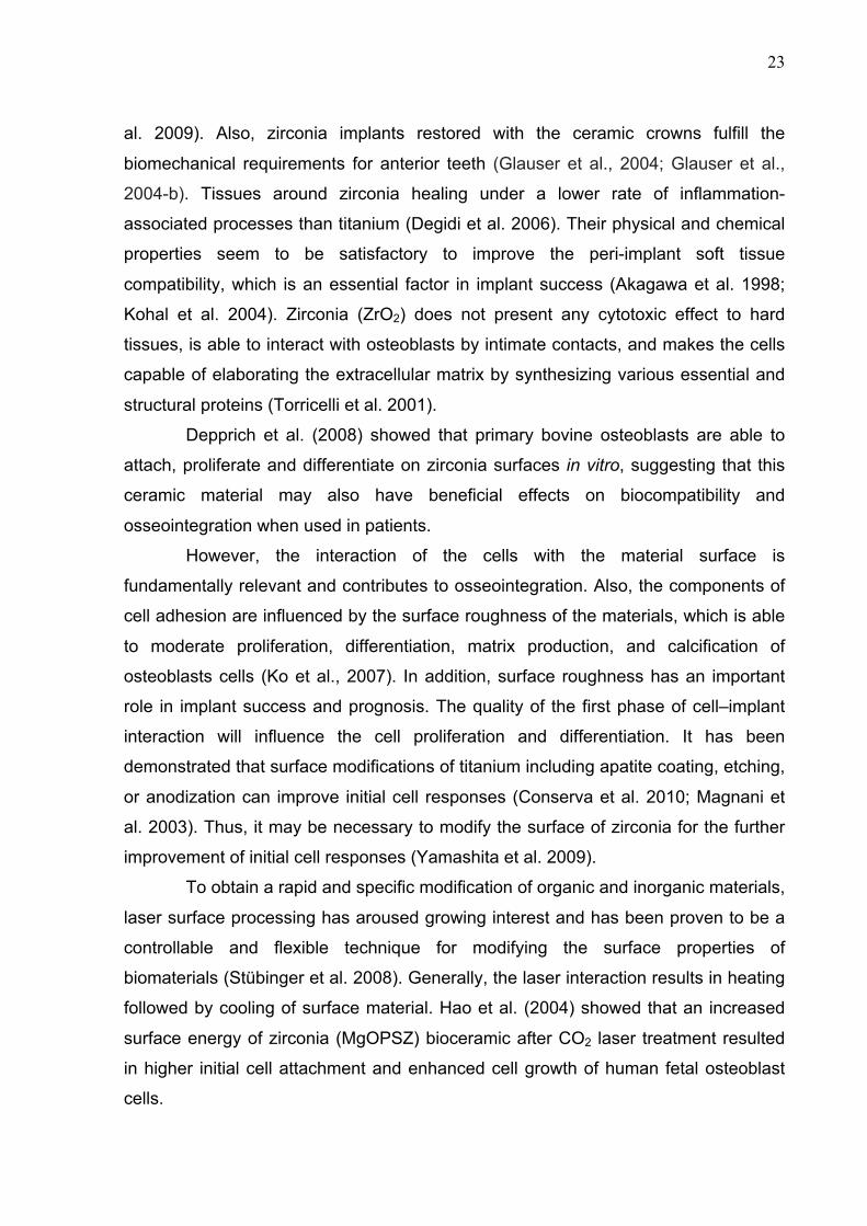
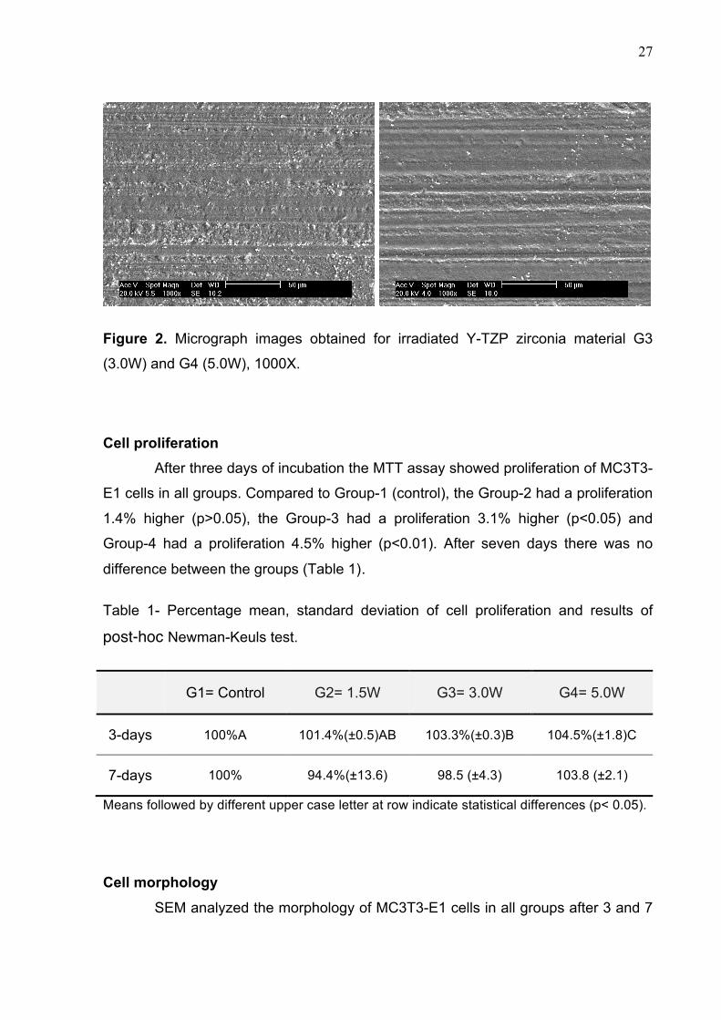
Surface characterization analysis SEM examination showed that there was no significant difference in surface
morphology between all parameter laser treated zirconia and the control group (Fig.
1 and 2).
Figure 1. Micrograph images obtained for irradiated Y-TZP zirconia material G1
(control) and G2 (1.5W), 1000X.
27
Figure 2. Micrograph images obtained for irradiated Y-TZP zirconia material G3
(3.0W) and G4 (5.0W), 1000X.
Cell proliferation After three days of incubation the MTT assay showed proliferation of MC3T3-
E1 cells in all groups. Compared to Group-1 (control), the Group-2 had a proliferation
1.4% higher (p>0.05), the Group-3 had a proliferation 3.1% higher (p<0.05) and
Group-4 had a proliferation 4.5% higher (p<0.01). After seven days there was no
difference between the groups (Table 1).
Table 1- Percentage mean, standard deviation of cell proliferation and results of
post-hoc Newman-Keuls test.
G1= Control G2= 1.5W G3= 3.0W G4= 5.0W
3-days 100%A 101.4%(±0.5)AB 103.3%(±0.3)B 104.5%(±1.8)C
7-days 100% 94.4%(±13.6) 98.5 (±4.3) 103.8 (±2.1)
Means followed by different upper case letter at row indicate statistical differences (p< 0.05).
Cell morphology SEM analyzed the morphology of MC3T3-E1 cells in all groups after 3 and 7
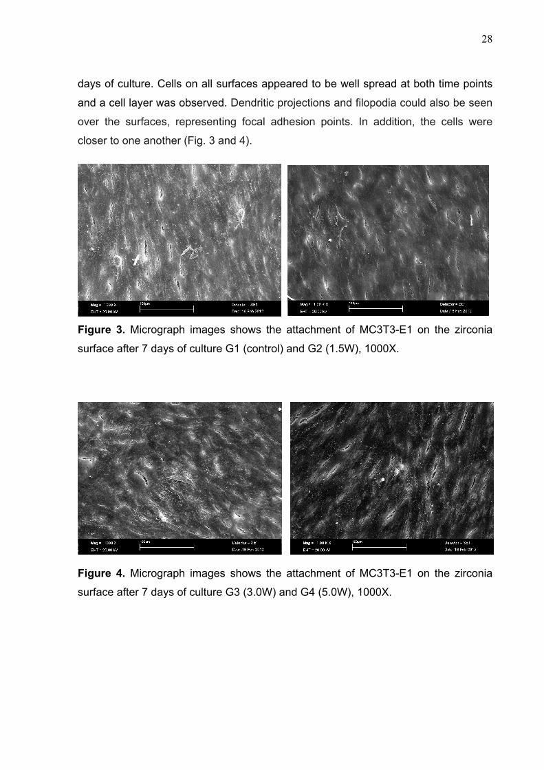
28
days of culture. Cells on all surfaces appeared to be well spread at both time points
and a cell layer was observed. Dendritic projections and filopodia could also be seen
over the surfaces, representing focal adhesion points. In addition, the cells were
closer to one another (Fig. 3 and 4).
Figure 3. Micrograph images shows the attachment of MC3T3-E1 on the zirconia
surface after 7 days of culture G1 (control) and G2 (1.5W), 1000X.
Figure 4. Micrograph images shows the attachment of MC3T3-E1 on the zirconia
surface after 7 days of culture G3 (3.0W) and G4 (5.0W), 1000X.
29
Discussion
A parameter of major importance for the clinical success of dental implants is
the establishment of a direct contact between the implant and the surrounding bone.
Implant chemistry and surface topography is thought to influence the implant-bone
response. Since bone is the essential structure of implants integration,
biocompatibility tests using constitutive elements of this tissue, like osteoblasts cells,
revealed to be important to predict the clinical success (Hisbergues et al. 2009).
This study evaluated the behavior of osteoblast cells on Y-TZP zirconia
surface treated with Er,Cr:YSGG laser. Since the attachment phase occurs rapidly
and involves short-term events like physico-chemical linkages between cells and
materials involving ionic forces and van der Waals forces (Bachle et al., 2007), the
specimens of the present study were harvested at the third and seventh days for
analysis.
At the third day, more osteoblasts cells proliferations were observed on the
Y-TZP zirconia surface in the G4. This group was irradiated with the highest laser
dose (5.0W). By the other side, the osteoblasts cells population in the G2 that was
irradiated with the lowest dose (1.5W) proliferated similarly to unlased control group.
However, G2 showed intermediary results and did not differ from G3 (3.0W), which
showed osteoblasts cells population higher than control group. Also, G3 showed
lesser osteoblasts cells proliferation than G4.
Comparing the cells response to laser doses irradiation, groups G2, G3, and
G4 showed respectively a 1.4%, 3.1%, and 4.5% higher osteoblasts population than
control group, this way it can be supposed that higher the laser dose higher the
osteoblast cells proliferation at this stage of earlier attachment. Yamashita et al.
(2009) also showed that zirconia improves cell proliferation significantly during the
first days of culture, but it does not improve attachment and adhesion strength
(Yamashita et al 2009).
However, at the seventh day there were no differences in the viable
osteoblasts population among the experimental groups and control group. It can be
supposed that Er,Cr:YSGG laser improves cell proliferation significantly during the
three first days of culture facilitating osteoblast cells adhesion to zirconia but after all
surface adhesion the cell reach a similar proliferation rate. Although, the laser
30
treatment may improve the quality of the first phase of cell-material interaction which
will improve the cell capacity to differentiate and may favors osseointegration.
Furthermore, it was suggest that the surface roughness of zirconia
specimens is an important factor to accelerate the initial attachment of osteoblasts,
similar to titanium and the expression of integrin α5 and β1 was enhanced for rough
surface specimens of zircônia (Yamashita et al 2009). Also, initial cell adhesion on
the material surface occurs through mechanical interlocking, which can produce
beneficial mechanical interlocking at the initial adhesion stage and aid cell adhesion.
Bächle et al (2007), using discs with different surface roughening of Y-TZP on CAL-
72 osteoblast-like cells, could demonstrate a change in proliferation after three days
in relation with the surface.
However, by SEM analysis the Er,Cr:YSGG laser treated zirconia showed
similar morphology to control group. The desired effect of laser energy in a material
is the absorption of the laser energy which the conversion of light energy into heat.
The heat will act on the substrate producing chemical, mechanical or physical
alterations. This effect may also be modified by the elapsed time between the energy
pulse, which permits the releasing of the heat and cooling, for subsequent energy
and heating (Hao et al., 2004). Moreover, the absorption of laser energy by zirconia
might be compromised since these ceramics are water-free materials and have a
white, opaque color (Subası & Inan, 2011), consequentially there is the need to
employ high energy densities to produce effects.
Hao et al. (2005) analyzed the effect of CO2 laser-modified zirconia on
human fetal osteoblasts cell adhesion and could demonstrate a better adhesion in
vitro after laser treatment, most likely because of a change in the wettability
characteristics of the Y-TZP. However, the surface also showed topography
alterations with pit and fissures that could have impaired mechanical properties.
Subası & Inan (2011) treated Y-TZP zirconia with 400 mJ Er:YAG laser and
observed rougher surfaces than the control group. By SEM analysis Er:YAG laser
slightly changed the surface morphology of zirconia with the formation of rare pits
when compared to the control group.
To solve the problem of laser absorption by Y-TZP zirconia, Cavalcanti et al.
(2009) coated with graphite powder the material for improve the absorption of laser
energy on the target surface. They used three different Er:YAG laser parameters
31
(200, 400, and 600 mJ) on zirconia and suggested that the lower energy intensity
(200 mJ) might be a potential method of rough Y-TZP zirconia surface.
However, SEM micrographs showed no morphological differences among Y-
TZP zirconia surface Er,Cr:YSGG laser irradiated besides the increase of laser dose
from 1.5W, 3.0W to 5.0W and observations of osteoblast-like cell morphology
showed good adhesion and spreading of the cells on the various surfaces, this way
the biocompatibility of an implant material depends on its chemical, physical, and
structural properties (Bachle et al. 2007), it seems that Er,Cr:YSGG influence the cell
response to Y-TZP zirconia surface by means of chemical or physical alteration.
Conclusions
Within the limitation of the present study, the following conclusions can be drawn:
• the surfaces of Y-TZP zirconia investigated allow cell adhesion and proliferation
and, therefore, the possibility of use as a biomaterial for dental implant.
• Er,Cr:YSGG laser irradiation may have contributed to the greater and earlier cell
spreading that was observed with the Y-TZP surface, however after seven days cell
proliferation were similar in both surface.
Acknowledgment
The authors of this study would like to thank Dr. Kitagima and Dr. Tanaka
(NAP/MEPA – College of Agriculture “Luiz de Queiroz” – University of Sao Paulo) for
SEM equipment support.
References
Akagawa, Y., Hosokawa, R., Sato Y., Kamayama, K. (1998) Comparison between
freestanding and tooth-connected partially stabilized zirconia implants after two
years' function in monkeys: a clinical and histologic study. Journal of Prosthetic
Dentistry 80(5): 551-8.
32
Albrektsson, T., Sennerby, L., Wennerberg, A. (2008) State of the art of oral
implants. Periodontology 2000 47: 15-26.
Bächle, M., Butz, F., Hübner, U., Bakalinis, E., Kohal, R.J. (2007) Behavior of CAL72
osteoblast-like cells cultured on zirconia ceramics with different surface topographies.
Clinical Oral Implants Research 18(1): 53-9.
Buser, D., Mericske-Stern, R., Bernard, J.P., Behneke, A., Behneke, N., Hirt, H.P.,
Belser, U.C. & Lang, N.P. (1997) Long-term evaluation of non-submerged ITI
implants. Part 1: 8-year life table analysis of a prospective multi-center study with
2359 implants. Clinical Oral Implants Research 8: 161–172.
Cavalcanti, A.N., Pilecki, P., Foxton, R.M., Watson, T.F., Oliveira, M.T., Gianinni, M.,
Marchi, G.M. (2009) Evaluation of the surface roughness and morphologic features
of Y-TZP ceramics after different surface treatments. Photomedicine and Laser
Surgery 27: 473–479.
Conserva, E., Lanuti, A., Menini, M. (2010) Cell behavior related to implant surfaces
with different microstructure and chemical composition: an in vitro analysis.
International Journal of Oral and Maxillofacial Implants 25(6): 1099-107.
Degidi, M., Artese, L., Scarano, A., Perrotti, V., Gehrke, P., Piattelli, A. (2006)
Inflammatory infiltrate, microvessel density, nitric oxide synthase expression,
vascular endothelial growth factor expression, and proliferative activity in peri-implant
soft tissues around titanium and zirconium oxide healing caps. Journal of
Periodontology 77(1): 73-80.
Depprich, R., Ommerborn, M., Zipprich, H., Naujoks, C., Handschel, J., Wiesmann,
H.P., Kübler, N.R., Meyer, U. (2008) Behavior of osteoblastic cells cultured on
titanium and structured zirconia surfaces. Head & Face Medicine 8: 4-29.
33
Glauser, R., Sailer, I., Wohlwend, A., Studer, S., Schibli, M., Schärer, P. (2004)
Experimental zirconia abutments for implant-supported single-tooth restorations in
esthetically demanding regions: 4-year results of a prospective clinical study. The
International Journal of Prosthodontics 17(3): 285-90.
Glauser, R., Wohlwend, A., Studer, S. (2004) Application of zirconia abutments on
single-tooth implants in the maxillary esthetic zone. A 6-year clinical and radiographic
follow-up report. Applied Osseointegration Research 4: 41–45 (b).
Hao, L., Lawrence, J., Lim, G.C., Zheng, H.Y. (2004) Examination of CO2 laser-
induced rapid solidification structures on magnesia partially stabilised zirconia and
the effects thereof on wettability characteristics. Optics and Lasers in Engineering 42: 355–374.
Hao, L., Lawrence, J., Chian, K.S. (2005) Osteoblast cell adhesion on a laser
modified zirconia based bioceramic. Journal of Materials Science: Materials in
Medicine 16(8): 719-26
Hisbergues, M., Vendeville, S., Vendeville, P. (2009) Zirconia: Established facts and
perspectives for a biomaterial in dental implantology. Journal of Biomedical Materials
Research Part B Applied Biomaterials 88(2): 519-29.
Ko, H.C., Han, J.S., Bächle, M., Jang, J.H., Shin, S.W., Kim, D.J. (2007) Initial
osteoblast-like cell response to pure titanium and zirconia/alumina ceramics. Dental
Materials 23(11): 1349-55.
Kohal, R.J., Weng, D., Bächle, M. & Strub, J.R. (2004) Loaded custom-made zirconia
and titanium implants show similar osseointegration: an animal experiment. Journal
of Periodontology 75: 1260–1266.
34
Kohal, R.J., Klaus, G., Strub, J.R. (2006) Zirconia-implant-supported all-ceramic
crowns withstand long-term load: a pilot investigation. Clinical Oral Implants
Research 17(5): 565-71.
Magnani, A., Priamo, A., Pasqui, D., Barbucci, R. (2003) Cell behavior on chemically
microstructured surfaces. Materials Science and Engineering: C 23.
Piconi, C., Burger, W., Richter, H.G., Cittadini, A., Maccauro, G., Covacci, V.,
Bruzzese, N., Ricci, G.A., Marmo, E. (1998) Y-TZP ceramics for artificial joint
replacements. Biomaterials 19(16): 1489-94.
Stübinger, S., Homann, F., Etter, C., Miskiewicz, M., Wieland, M., Sader, R. (2008)
Effect of Er:YAG, CO(2) and diode laser irradiation on surface properties of zirconia
endosseous dental implants. Lasers in Surgery and Medicine 40(3): 223-8.
Subaşı, M.G., Inan, O. (2011) Evaluation of the topographical surface changes and
roughness of zirconia after different surface treatments. Lasers in Medical Science
24.
Torricelli, P., Verne´, E., Brovarone, C.V., Appendino, P., Rustichelli, F., Krajewski,
A., Ravaglioli, A., Pierini, G., Fini, M., Giavaresi, G., Giardino, R. (2001) Biological
glass coating on ceramic materials: In vitro evaluation using primary osteoblast
cultures from healthy and osteopenic rat bone. Biomaterials 22: 2535–2543.
Yamashita, D., Machigashira, M., Miyamoto, M., Takeuchi, H., Noguchi, K., Izumi, Y.,
Ban, S. Effect of surface roughness on initial responses of osteoblast-like cells on
two types of zirconia. Dental Materials Journal 28(4): 461-70.
35
4. CONCLUSÕES
As superfícies de zircônia, investigadas neste estudo, permitiram a adesão e
proliferação de osteoblastos, indicando que a cerâmica Y-TZP é um biomaterial
promissor na confecção de implantes dentários.
A irradiação com laser de Er,Cr:YSGG contribuiu para uma maior e mais
precoce adesão e proliferação celular sobre as superfícies de Y-TZP. No entanto,
após sete dias, a proliferação celular foi similar sobre as superfícies irradiadas e não
irradiadas.
36
5. REFERÊNCIAS
Ahmad I. Yttrium-partially stabilized zirconium dioxide posts: an approach to restoring
coronally compromised nonvital teeth. Int J Periodontics Restorative Dent 1998 Oct;
18 (5): 454-65.
Akagawa Y, Hosokawa R, Sato Y, Kamayama K. Comparison between freestanding
and tooth-connected partially stabilized zirconia implants after two years' function in
monkeys: a clinical and histologic study. J Prosthet Dent 1998 Nov; 80 (5): 551-8.
Albrektsson T, Branemark PI, Hansson HA, Lindstrom J. Osseointegrated titanium
implants. Acta Orthop Scand 1981; 52: 155–170.
Albrektsson T, Sennerby L, Wennerberg A. State of the art of oral implants.
Periodontol 2000. 2008; 47: 15-26.
Andreiotelli M, Wenz HJ, Kohal RJ. Are ceramic implants a viable alternative to
titanium implants? A systematic literature review. Clin Oral Implants Res 2009 Sep;
20 Suppl 4: 32-47.
Anselme K. Osteoblast adhesion on biomaterials. Biomaterials 2000 Apr; 21 (7): 667-
81. Review.
Azzeh MM. Er, Cr: YSGG Laser-assisted surgical treatment of peri-implantitis with 1-
year reentry and 18-month follow-up. J Periodontol 2008; 79: 2000-2005.
Bächle M, Butz F, Hübner U, Bakalinis E, Kohal RJ. Behavior of CAL72 osteoblast-
like cells cultured on zirconia ceramics with different surface topographies. Clin Oral
Implants Res 2007 Feb; 18 (1): 53-9.
Bressan E, Paniz G, Lops D, Corazza B, Romeo E, Favero G. Influence of abutment
material on the gingival color of implant-supported all-ceramic restorations: a
37
prospective multicenter study. Clin Oral Implants Res 2011 Jun; 22 (6): 631-7.
Butz F, Ogawa T, Nishimura I. Interfacial shear strength of endosseous implants. Int
J Oral Maxillofac Implants 2011 Jul-Aug; 26 (4): 746-51.
Conserva E, Lanuti A, Menini M. Cell behavior related to implant surfaces with
different microstructure and chemical composition: an in vitro analysis. Int J Oral
Maxillofac Implants 2010 Nov-Dec; 25 (6): 1099-107.
Delgado-Ruíz RA, Calvo-Guirado JL, Moreno P, Guardia J, Gomez-Moreno G, Mate-
Sánchez JE, Ramirez-Fernández P, Chiva F. Femtosecond laser microstructuring of
zirconia dental implants. J Biomed Mater Res B Appl Biomater 2011 Jan; 96 (1): 91-
100.
Depprich R, Ommerborn M, Zipprich H, Naujoks C, Handschel J, Wiesmann HP,
Kübler NR, Meyer U. Behavior of osteoblastic cells cultured on titanium and
structured zirconia surfaces. Head Face Med 2008 Dec; 8: 4-29.
Esposito M, Hirsch JM, Lekholm U, Thomsen P. Biological factors contributing to
failures of osseointegrated oral implants. (II). Etiopathogenesis. Eur J Oral Sci 1998
Jun; 106 (3): 721-64.
Ferguson SJ, Broggini N, Wieland M, de Wild M, Rupp F, Geis-Gerstorfer J, Cochran
DL, Buser D. Biomechanical evaluation of the interfacial strength of a chemically
modified sandblasted and acid-etched titanium surface. J Biomed Mater Res A 2006
Aug; 78 (2): 291-7.
Gahlert M, Röhling S, Wieland M, Sprecher CM, Kniha H, Milz S. Osseointegration of
zirconia and titanium dental implants: a histological and histomorphometrical study in
the maxilla of pigs. Clin Oral Implants Res 2009 Nov; 20 (11): 1247-53.
Gahlert M, Burtscher D, Grunert I, Kniha H, Steinhauser E. Failure analysis of
38
fractured dental zirconia implants. Clin Oral Implants Res 2012 Mar; 23 (3): 287-93.
Glauser R, Sailer I, Wohlwend A, Studer S, Schibli M, Schärer P. Experimental
zirconia abutments for implant-supported single-tooth restorations in esthetically
demanding regions: 4-year results of a prospective clinical study. Int J Prosthodont.
2004 May-Jun; 17 (3): 285-90.
Glauser R, Wohlwend A, Studer S. Application of zirconia abutments on single-tooth
implants in the maxillary esthetic zone. A 6-year clinical and radiographic follow-up
report. Applied Osseointegration Research 2004-b; 4: 41–45.
Hao L, Lawrence J, Lim GC, Zheng HY. Examination of CO2 laser-induced rapid
solidification structures on magnesia partially stabilized zirconia and the effects
thereof on wettability characteristics. Optics and Lasers in Engineering 2004; 42:
355–374
Hao L, Lawrence J. On the role of CO2 laser treatment in the human serum albumin
and human plasma fibronectin adsorption on zirconia (MGO-PSZ) bioceramic
surface. J Biomed Mater Res A. 2004 Jun 15; 69 (4): 748-56.
Hao L, Lawrence J, Chian KS. Osteoblast cell adhesion on a laser modified zirconia
based bioceramic. J Mater Sci Mater Med 2005 Aug; 16 (8): 719-26
Hao L, Lawrence J. Effects of Nd:YAG laser treatment on the wettability
characteristics of a zirconia-based bioceramic. Optics and Lasers in Engineering
2006; 44: 803–814.
Heydecke G, Kohal R, Gläser R. Optimal esthetics in single-tooth replacement with
the Re-Implant system: a case report. Int J Prosthodont. 1999 Mar-Apr; 12 (2): 184-9.
Hisbergues M, Vendeville S, Vendeville P. Zirconia: Established facts and
perspectives for a biomaterial in dental implantology. J Biomed Mater Res B Appl
Biomater 2009 Feb; 88 (2): 519-29.
39
Keselowsky BG, Collard DM, García AJ. Surface chemistry modulates focal adhesion
composition and signaling through changes in integrin binding. Biomaterials 2004
Dec; 25 (28): 5947-54.
Ko HC, Han JS, Bächle M, Jang JH, Shin SW, Kim DJ. Initial osteoblast-like cell
response to pure titanium and zirconia/alumina ceramics. Dent Mater 2007 Nov; 23
(11): 1349-55.
Kohal, R.J., Weng, D., Bächle, M. & Strub, J.R. Loaded custom-made zirconia and
titanium implants show similar osseointegration: an animal experiment. Journal of
Periodontology 2004; 75: 1260–1266.
Kohal RJ, Klaus G, Strub JR. Zirconia-implant-supported all-ceramic crowns
withstand long-term load: a pilot investigation. Clin Oral Implants Res 2006 Oct; 17
(5): 565-71.
Kohal RJ, Att W, Bächle M, Butz F. Ceramic abutments and ceramic oral implants. An update. Periodontology 2000. 2008; 47: 224-43.
Lange R, Lüthen F, Beck U, Rychly J, Baumann A, Nebe B. Cell-extracellular matrix
interaction and physico-chemical characteristics of titanium surfaces depend on the
roughness of the material. Biomol Eng. 2002 Aug; 19 (2-6): 255-61.
Magnani A, Priamo A, Pasqui D, Barbucci R. Cell behavior on chemically
microstructured surfaces. Mater Sci Eng C 2003; 23.
Marković A, Calvo-Guirado JL, Lazić Z, Gómez-Moreno G, Calasan D, Guardia J,
Colic S, Aguilar-Salvatierra A, Gačić B, Delgado-Ruiz R, Janjić B, Mišić T. Evaluation
of Primary Stability of Self-Tapping and Non-Self-Tapping Dental Implants. A 12-
Week Clinical Study. Clin Implant Dent Relat Res 2011; Dec 15.
40
Miller RJ. Treatment of the contaminated implant surface using the Er;Cr:YSGG
laser. Implant Dentistry 2004; 13 (2).
Noda M, Okuda Y, Tsuruki J, Minesaki Y, Takenouchi Y, Ban S. Surface damages of
zirconia by Nd:YAG dental laser irradiation. Dent Mater J 2010 Oct 14; 29 (5): 536-
41. Epub 2010 Sep 18.
Oliva J, Oliva X, Oliva JD. One-year follow-up of first consecutive 100 zirconia dental
implants in humans: a comparison of 2 different rough surfaces. Int J Oral Maxillofac
Implants 2007 May-Jun; 22 (3): 430-5.
Oliva J, Oliva X, Oliva JD. Five-year success rate of 831 consecutively placed
Zirconia dental implants in humans: a comparison of three different rough surfaces.
Int J Oral Maxillofac Implants 2010 Mar-Apr; 25 (2): 336-44.
Osman RB, Ma S, Duncan W, De Silva RK, Siddiqi A, Swain MV. Fractured zirconia
implants and related implant designs: scanning electron microscopy analysis. Clin
Oral Implants Res 2012 Jan 26. (Epub ahead of print)
Pae A, Kim SS, Kim HS, Woo YH. Osteoblast-like cell attachment and proliferation
on turned, blasted, and anodized titanium surfaces. Int J Oral Maxillofac Implants
2011 May-Jun; 26 (3): 475-81.
Piconi C, Burger W, Richter HG, Cittadini A, Maccauro G, Covacci V, Bruzzese N,
Ricci GA, Marmo E. Y-TZP ceramics for artificial joint replacements. Biomaterials
1998 Aug; 19 (16): 1489-94.
Puleo DA, Thomas MV. Implant surfaces. Dent Clin North Am 2006 Jul; 50 (3): 323-
38.
Roach HI. Why does bone matrix contain non-collagenous proteins? The possible
roles of osteocalcin, osteonectin, osteopontin and bone sialoprotein in bone
mineralisation and resorption. Cell Biol Int 1994 Jun; 18 (6): 617-28
41
Romanos G, Crespi R, Barone A, Covani U. Osteoblast attachment on titanium disks
after laser irradiation. Int J Oral Maxillofac Implants 2006 Mar-Apr; 21 (2): 232-6.
Sennerby L, Dasmah A, Larsson B, Iverhed M. Bone tissue responses to surface-
modified zirconia implants: A histomorphometric and removal torque study in the
rabbit. Clin Implant Dent Relat Res 2005; 7 Suppl 1: S13-20.
Shalabi MM, Gortemaker A, Van't Hof MA, Jansen JA, Creugers NH. Implant surface
roughness and bone healing: a systematic review. J Dent Res 2006 Jun; 85 (6): 496-
500.
Shibli JA, Grassi S, Piattelli A, Pecora GE, Ferrari DS, Onuma T, d'Avila S, Coelho
PG, Barros R, Iezzi G. Histomorphometric evaluation of bioceramic molecular
impregnated and dual acid-etched implant surfaces in the human posterior maxilla.
Clin Implant Dent Relat Res 2010 Dec; 12 (4): 281-8.
Shibli JA, Mangano C, D'avila S, Piattelli A, Pecora GE, Mangano F, Onuma T,
Cardoso LA, Ferrari DS, Aguiar KC, Iezzi G. Influence of direct laser fabrication
implant topography on type IV bone: a histomorphometric study in humans. J Biomed
Mater Res A 2010-b May; 93 (2): 607-14.
Shibli JA, Grassi S, de Figueiredo LC, Feres M, Iezzi G, Piattelli A. Human peri-
implant bone response to turned and oxidized titanium implants inserted and
retrieved after 2 months. Implant Dent 2007 Sep; 16 (3): 252-9.
Schwarz F, Nuesry E, Bieling K, Herten M, Becker J. Influence of an erbium,
chromium-doped yttrium, scandium, gallium, and garnet (Er,Cr:YSGG) laser on the
reestablishment of the biocompatibility of contaminated titanium implant surfaces. J
Periodontol 2006 Nov; 77 (11): 1820-7.
Steinemann SG. Titanium--the material of choice? Periodontol 2000. 1998 Jun; 17:
7-21.
42
Stübinger S, Homann F, Etter C, Miskiewicz M, Wieland M, Sader R. Effect of
Er:YAG, CO(2) and diode laser irradiation on surface properties of zirconia
endosseous dental implants. Lasers Surg Med 2008 Mar; 40 (3): 223-8.
Sykaras N, Iacopino AM, Marker VA, Triplett RG, Woody RD. Implant materials,
designs, and surface topographies: their effect on osseointegration. A literature
review. Int J Oral Maxillofac Implants 2000 Sep-Oct; 15 (5): 675-90.
Tinschert J, Natt G, Mohrbotter N, Spiekermann H, Schulze KA. Lifetime of alumina-
and zirconia ceramics used for crown and bridge restorations. J Biomed Mater Res B
Appl Biomater 2007; 80: 317-21.
Wang G, Liu X, Zreiqat H, Ding C. Enhanced effects of nano-scale topography on the
bioactivity and osteoblast behaviors of micron rough ZrO2 coatings. Colloids Surf B
Biointerfaces 2011 Sep 1; 86 (2): 267-74.
Wennerberg A, Albrektsson T. Effects of titanium surface topography on bone
integration: a systematic review. Clin Oral Implants Res 2009 Sep; 20 Suppl 4: 172-
84.
Wong M, Eulenberger J, Schenk R, Hunzinker E. Effect of surface topology on the
ossointegration of implant material in trabecular bone. Biomaterials 1995; 29: 1567-
1575.
Yamashita D, Machigashira M, Miyamoto M, Takeuchi H, Noguchi K, Izumi Y, Ban S.
Effect of surface roughness on initial responses of osteoblast-like cells on two types
of zirconia. Dent Mater J 2009 Jul; 28 (4): 461-70.
Zhu X, Chen J, Scheideler L, Reichl R, Geis-Gerstorfer J. Effects of topography and
composition of titanium surface oxides on osteoblast responses. Biomaterials 2004
Aug; 25 (18): 4087-103.
Top Related