Yun-Hee Youm HHS Public Access Kim Y. Nguyen Ryan … in LPS primed and NLRP3 agonist treated...
19
Ketone body β-hydroxybutyrate blocks the NLRP3 inflammasome-mediated inflammatory disease Yun-Hee Youm 1,* , Kim Y. Nguyen 1,* , Ryan W. Grant 2 , Emily L. Goldberg 1 , Monica Bodogai 3 , Dongin Kim 4 , Dominic D'Agostino 5 , Noah Planavsky 6 , Christopher Lupfer 7 , Thirumala D. Kanneganti 7 , Seokwon Kang 8 , Tamas L. Horvath 1 , Tarek M. Fahmy 4 , Peter A. Crawford 9 , Arya Biragyn 3 , Emad Alnemri 8 , and Vishwa Deep Dixit 1,10 1 Section of Comparative Medicine and Program on Integrative Cell Signaling and Neurobiology of Metabolism, Yale School of Medicine 2 Department of Nutrition Sciences, Purdue University 3 Laboratory of Molecular Biology and Immunology, National Institute on Aging, National Institutes of Health (NIH), Baltimore, MD 4 Department of Biomedical Engineering Yale University 5 Department of Molecular Pharmacology and Physiology, University of South Florida, Tampa 6 Department of Geology and Geophysics, Yale University 7 Department of Immunology, St. Jude Children's Hospital, Memphis, TN 8 Department of Biochemistry and Molecular Biology, Thomas Jefferson University, Philadelphia PA 9 Diabetes and Obesity Research Center, Sanford-Burnham Medical Research Institute, Orlando, FL 10 Department of Immunobiology, Yale School of Medicine, New Haven, CT06520 Abstract Ketone bodies , β-hydroxybutyrate (BHB) and acetoacetate support mammalian survival during states of energy deficit by serving as alternative source of ATP 1 . BHB levels are elevated during starvation, high-intensity exercise or by the low carbohydrate ketogenic diet 2 . Prolonged caloric restriction or fasting reduces inflammation as immune system adapts to low glucose supply and Address and Correspondence to: Vishwa Deep Dixit, Ph.D, Section of Comparative Medicine and Department of Immunobiology, Yale School of Medicine, 310 Cedar St, New Haven CT06520, [email protected], Phone: 203-785-2525, Fax: 203-785-7499. * Authors contributed equally to this work. Author contributions: Y.H.Y. and K.Y.N . contributed equally to the study, They designed and conducted majority of in vitro and all of in vivo experiments, analyzed and interpreted the data and participated in writing the manuscript. R.W.G. participated in design and conduct of inflammasome activation experiments. ELG performed ASC speck and neutrophil assays. MB and AB performed the human monocytes experiments. DK and TMF synthesized the BHB-nanolipogels and conducted control experiments to determine the dose response. DDA formulated the ketone diester diet. NP conducted the ICP-MS experiments to determine K + efflux. CL and TDK conducted the F.tularensis and S. typhimurium infections experiments. TH designed the experiments and provided essential reagents for experiments involving mitochondrial ROS and UCP2. PAC generated the macrophage specific Scot deficient mice and contributed in experiment design. SK and EA designed and conducted the ASC oligomerization experiments. VDD conceived and supervised the project, interpreted the data and wrote the manuscript. HHS Public Access Author manuscript Nat Med. Author manuscript; available in PMC 2015 September 01. Published in final edited form as: Nat Med. 2015 March ; 21(3): 263–269. doi:10.1038/nm.3804. Author Manuscript Author Manuscript Author Manuscript Author Manuscript
Transcript of Yun-Hee Youm HHS Public Access Kim Y. Nguyen Ryan … in LPS primed and NLRP3 agonist treated...
Ketone body β-hydroxybutyrate blocks the NLRP3 inflammasome-mediated inflammatory disease
Yun-Hee Youm1,*, Kim Y. Nguyen1,*, Ryan W. Grant2, Emily L. Goldberg1, Monica Bodogai3, Dongin Kim4, Dominic D'Agostino5, Noah Planavsky6, Christopher Lupfer7, Thirumala D. Kanneganti7, Seokwon Kang8, Tamas L. Horvath1, Tarek M. Fahmy4, Peter A. Crawford9, Arya Biragyn3, Emad Alnemri8, and Vishwa Deep Dixit1,10
1Section of Comparative Medicine and Program on Integrative Cell Signaling and Neurobiology of Metabolism, Yale School of Medicine
2Department of Nutrition Sciences, Purdue University
3Laboratory of Molecular Biology and Immunology, National Institute on Aging, National Institutes of Health (NIH), Baltimore, MD
4Department of Biomedical Engineering Yale University
5Department of Molecular Pharmacology and Physiology, University of South Florida, Tampa
6Department of Geology and Geophysics, Yale University
7Department of Immunology, St. Jude Children's Hospital, Memphis, TN
8Department of Biochemistry and Molecular Biology, Thomas Jefferson University, Philadelphia PA
9Diabetes and Obesity Research Center, Sanford-Burnham Medical Research Institute, Orlando, FL
10Department of Immunobiology, Yale School of Medicine, New Haven, CT06520
Abstract
Ketone bodies , β-hydroxybutyrate (BHB) and acetoacetate support mammalian survival during
states of energy deficit by serving as alternative source of ATP1. BHB levels are elevated during
starvation, high-intensity exercise or by the low carbohydrate ketogenic diet2. Prolonged caloric
restriction or fasting reduces inflammation as immune system adapts to low glucose supply and
Address and Correspondence to: Vishwa Deep Dixit, Ph.D, Section of Comparative Medicine and Department of Immunobiology, Yale School of Medicine, 310 Cedar St, New Haven CT06520, [email protected], Phone: 203-785-2525, Fax: 203-785-7499.*Authors contributed equally to this work.
Author contributions: Y.H.Y. and K.Y.N . contributed equally to the study, They designed and conducted majority of in vitro and all of in vivo experiments, analyzed and interpreted the data and participated in writing the manuscript. R.W.G. participated in design and conduct of inflammasome activation experiments. ELG performed ASC speck and neutrophil assays. MB and AB performed the human monocytes experiments. DK and TMF synthesized the BHB-nanolipogels and conducted control experiments to determine the dose response. DDA formulated the ketone diester diet. NP conducted the ICP-MS experiments to determine K+ efflux. CL and TDK conducted the F.tularensis and S. typhimurium infections experiments. TH designed the experiments and provided essential reagents for experiments involving mitochondrial ROS and UCP2. PAC generated the macrophage specific Scot deficient mice and contributed in experiment design. SK and EA designed and conducted the ASC oligomerization experiments. VDD conceived and supervised the project, interpreted the data and wrote the manuscript.
HHS Public AccessAuthor manuscriptNat Med. Author manuscript; available in PMC 2015 September 01.
Published in final edited form as:Nat Med. 2015 March ; 21(3): 263–269. doi:10.1038/nm.3804.
Author M
anuscriptA
uthor Manuscript
Author M
anuscriptA
uthor Manuscript
energy metabolism switches towards mitochondrial fatty acid oxidation, ketogenesis and
ketolysis2-6. However, role of ketones bodies in regulation of innate immune response is
unknown. We report that BHB, but neither acetoacetate nor structurally-related short chain fatty
acids, butyrate and acetate, suppresses activation of the NLRP3 inflammasome in response to
several structurally unrelated NLRP3 activators, without impacting NLRC4, AIM2 or non-
canonical caspase-11 inflammasome activation. Mechanistically, BHB inhibits NLRP3
inflammasome by preventing K+ efflux and reducing ASC oligomerization and speck formation.
The inhibitory effects of BHB on NLRP3 were not dependent on chirality or classical starvation
regulated mechanisms like AMPK, reactive oxygen species (ROS), autophagy or glycolytic
inhibition. BHB blocked NLRP3 inflammasome without undergoing oxidation in TCA cycle,
independently of uncoupling protein-2 (UCP2), Sirt2, receptor Gpr109a and inhibition of NLRP3
did not correlate with magnitude of histone acetylation in macrophages. BHB reduced the NLRP3
inflammasome mediated IL-1β and IL-18 production in human monocytes. In vivo, BHB
attenuates caspase-1 activation and IL-1β secretion in mouse models of NLRP3-mediated diseases
like Muckle-Wells Syndrome (MWS), Familial Cold Autoinflammatory syndrome (FCAS) and
urate crystal induce body cavity inflammation. Taken together, these findings suggest that the anti-
inflammatory effects of caloric restriction or ketogenic diets may be mechanistically linked to
BHB-mediated inhibition of the NLRP3 inflammasome, and point to the potential use of
interventions that elevate circulating BHB against NLRP3-mediated proinflammatory diseases.
The NLRP3 inflammasome controls the activation of caspase-1 and the release of the pro-
inflammatory cytokines IL-1β and IL-18 in macrophages7-11. It is an important innate
immune sensor that can get activated in response to structurally diverse damage-associated
molecular patterns (DAMPs) such as toxins8, ATP8, excess glucose7, ceramides12,
amyloids13,14 , urate15 and cholesterol crystals16. Ablation of NLRP3 attenuates type 2
diabetes12, 14,17, atherosclerosis16, multiple sclerosis18, Alzheimer's disease14, age-related
functional decline19, bone loss19 and gout15. Thus, identification of endogenous
mechanisms that control NLRP3 inflammasome deactivation may provide insights into the
control of several chronic diseases. Although it is known that immune-metabolic
interactions via inhibition of glycolysis dampen pro-inflammatory responses6, it is not
known whether alternate metabolic fuels such as ketones that are produced during energy
deficits impact the innate immune response.
The ketone bodies β-hydroxybutyrate (BHB) and acetoacetate (AcAc) are produced in the
liver and serve as alternative energy sources for the brain, heart, and skeletal muscle in
mammals during nutrient deprivation and adherence to low carbohydrate diets1,2.
Circulating levels of BHB can increase up to 6-8 mM upon prolonged fasting as liver
glycogen stores get utilized1,2. While these nutritional states are associated with altered
immune cell function, it is unknown whether ketone bodies serve as immune effectors. To
test whether BHB impacts inflammasome activation, we treated LPS-primed mouse bone
marrow derived macrophages (BMDMs) with the NLRP3 activator ATP along with BHB
for 60 minutes and measured caspase-1 activation using a Western blot that detects the
enzymatically-active p20 subunit. BHB dose-dependently inhibited ATP-induced caspase-1
cleavage into p20 and processing of biologically active p17 form of IL-1β at concentrations
similar to elevations in BHB induced by strenuous exercise or 2 days of fasting1,2 (Fig.1a
Youm et al. Page 2
Nat Med. Author manuscript; available in PMC 2015 September 01.
Author M
anuscriptA
uthor Manuscript
Author M
anuscriptA
uthor Manuscript
and Supplementary Fig. 1a). Ketone body acetoacetate (AcAc) and the microbiota-derived
short chain fatty acids (SCFAs), butyrate and acetate, that are structurally related to BHB
did not affect ATP-induced NLRP3 activation (Fig. 1b). We sought to determine whether
BHB specifically targets ATP-induced inflammasome activation or common signaling
mechanisms in response to structurally diverse NLRP3 activators. BHB, but not butyrate,
inhibited monosodium urate (MSU) crystal or particulate matter-induced caspase-1
activation (Fig. 1c and Supplementary Fig. 1b). Furthermore, BHB blocked inflammasome
activation by five additional NLRP3 activators nigericin, silica particles (Supplementary
Fig. 1b), lipotoxic fatty acids palmitate, ceramides and sphingosine (Fig. 1d). BHB also
inhibited processing of IL-1β in response to the TLR4 pathogen associated molecular pattern
(PAMP) agonist lipid A, the TLR1/2 ligand Pam3-CSK4 and the TLR2 agonist lipoteichoic
acid (LTA) (Fig. 1e).
We next investigated the specificity of BHB to NLRP3 as compared to other
inflammasomes. The BMDMs were infected with either Francisella tularensis to activate
the AIM2 or Salmonella typhimurium to activate the NLRC4 inflammasome. BHB did not
inhibit either AIM2 inflammasome induced IL-1β activation (Fig. 1f) or NLRC4-mediated
caspase-1 cleavage (Fig. 1g). Given inflammasome can also be activated by LPS through
caspase-11 activation independently of TLR420,21, we also evaluated the non-canonical
inflammasome pathway. Our data indicate that neither butyrate nor BHB blocks caspase-11
activation (Supplementary Fig. 1c).These results suggest that BHB acts on a central
common signalling pathway specific to the NLRP3 inflammasome in response to PAMPs
and a wide array of pro-inflammatory DAMPs.
Prolonged fasting and subsequent increases in circulating BHB are linked to a reduction in
oxidative stress22, increased AMPK activity 23 and autophagy24. Furthermore, all these
mechanisms have also been implicated in regulating the NLRP3 inflammasome8. Consistent
with recent data25, ROS damage via rotenone or hydrogen peroxide (Fig. 2a, Supplementary
Fig. 2a, b) was not sufficient to induce caspase-1 cleavage and did not abrogate the
suppressive effects of BHB on ATP-induced NLRP3 inflammasome activation. Caspase-1
activation was induced by LPS priming alone in macrophages deficient in the autophagy
regulator Atg5 (Fig. 2b, Supplementary Fig. 2a). However, absence of Atg5 did not alter the
inhibitory effects of BHB on the inflammasome (Fig. 2b, Supplementary Fig. 2a).
Consistent with these findings, the autophagy inhibitor 3-methyladenine (3-MA) and the
proteasome blocker epoxomicin did not abrogate BHB's suppressive effects on ATP-induced
NLRP3 inflammasome activation (Fig. 2c, Supplementary Fig. 2a). The activation of AMPK
using AICAR (Fig. 2d) and inhibition of glycolysis with 2-deoxy glucose did not mimic the
effects of BHB on inhibition of NLRP3 inflammasome (Supplementary Fig. 3a).
Furthermore, inhibition of AMPK via compound C did not abrogate BHB's inhibitory
effects on NLRP3-mediated caspase-1 activation. (Fig. 2d and Supplementary Figure 2a, c,
Supplementary Figure 3a). BHB also did not impair the viability of BMDMs and at a
concentration of 10 mM increased cellular proliferation (Fig. 2e).
It has been suggested that BHB can act as signaling molecule by binding the G protein
coupled receptor GPR109a26 or by serving as a histone deacetylase (HDAC) inhibitor22.
Inhibition of HDACs using trichostatin A (TSA) did not did not impact inflammasome
Youm et al. Page 3
Nat Med. Author manuscript; available in PMC 2015 September 01.
Author M
anuscriptA
uthor Manuscript
Author M
anuscriptA
uthor Manuscript
activation in LPS primed and NLRP3 agonist treated macrophages (Fig. 2f, Supplementary
Fig. 2a despite induction of H3 acetylation by BHB in macrophages (Supplementary Fig.
3b). To understand the role of GPR109a in BHB's effects on macrophages, we used niacin, a
GPR109a ligand that has been reported to inhibit colonic inflammation 27. We found that,
unlike BHB, niacin did not block the NLRP3 inflammasome activation (Fig. 2f,
Supplementary Fig. 2a).The effects of BHB on the inflammasome were not altered in
Gpr109a-deficient BMDM (Fig. 2f, g, Supplementary Fig. 2a), and neither butyrate nor
acetoacetate altered NLRP3 inflammasome activity in Gpr109a-sufficient or –deficient cells
(Fig. 2g, Supplementary Fig. 2a). BHB is a chiral compound and its enantiomer, (S)-BHB,
does not enter TCA cycle but binds Gpr109a with high affinity26. The enantiomer (S)-BHB
exhibits similar effects on the inflammasome as D-(BHB) and does not require Gpr109a to
block NLRP3 (Fig. 2h).
Compared to fatty acids, oxidation of BHB is energetically more efficient as all reducing
equivalents generated by ketone oxidation are delivered through NADH to complex-I within
the mitochondrial electron transport chain2. Furthermore, ketone oxidation increases the
redox span between complex-I and complex-III by keeping mitochondrial ubiquinone
oxidized28. We next asked whether BHB oxidation, entry into the TCA cycle or reduced
mitochondrial stress mediate its effects on the inflammasome. Macrophages express
ketogenic and ketolytic enzymes Acat1, Bdh1, Bdh2 and Hmgcl (Supplementary Figure. 4a,
b, and c). In terms of macrophage polarization, classically activated M1 macrophages show
reduction expression of Acat1, Bdh1, Bdh2 and Hmgcl as compared to M2 macrophages,
suggesting ketones may affect macrophage polarization (Supplementary Figure. 4b). In
addition, LPS induced protein expression of the ketolytic enzyme, Scot (succinyl-CoA:3-
oxoacid CoA transferase , encoded by Oxct) and the ketogenic enzyme Hmgcl in BMDMs
(Supplementary Figure. 4c). However, the TCA cycle entry inhibitor aminoxyacetate (AOA)
did inhibit the effects of BHBon the inflammasome (Supplementary Figure. 4d), and the
enantiomer (S)- BHB that does not enter the TCA cycle efficiently blocked NLRP3
inflammasome activation (Supplementary Figure. 4e). To directly assess the role of BHB
oxidation, we specifically deleted the ketolytic mitochondrial enzyme Scot27 in
macrophages (Fig. 3a, Supplementary Figure 4f). These experiments confirmed that TCA
intermediates generated through ketone body oxidation in macrophage mitochondria do not
mediate the suppressive effects of BHB on the NLRP3 inflammasome (Fig. 3a,
Supplementary Figure 4f).
The NAD dependent deacetylase Sirt2 regulates acetylation of α-tubulin, which controls
microtubule-driven apposition of Nlrp3 and Asc29. Inhibition of Sirt2 by the small molecule
AGK2 activates the Nlrp3 inflammasome and supplementation with NAD+ lowers IL-1β
secretion from macrophages29. Inhibition of Sirt2 by AGK2 or ablation of Sirt2 or
uncoupling protein 2 (UCP2) did not abrogate the effects of BHB on the inflammasome, and
addition of NAD+ did not block caspase-1 activation in response to LPS and ATP (Fig. 3b-d,
Supplementary Figure 4f). These findings rule out a major role for mitochondrial ROS in the
effects of ketone bodies on the inflammasome.
BHB is a strongly anionic endogenous molecule2 and exerts anti-epileptic effects by
reducing neuronal excitability via regulation of intracellular potassium cations30. Consistent
Youm et al. Page 4
Nat Med. Author manuscript; available in PMC 2015 September 01.
Author M
anuscriptA
uthor Manuscript
Author M
anuscriptA
uthor Manuscript
with recent studies that demonstrate K+ efflux triggers NLRP3 inflammasome
activation8, 25, BHB prevented the decline in intracellular K+ in response to incubation with
the NLRP3 activators ATP, MSU and ceramides (Fig. 3e, f, g and Supplementary Figure
5a). Furthermore, the NLRP3-dependent ASC nucleation-induced polymerization or
oligomerization is considered a common mechanism of NLRP3 inflammasome
activation31,32. BHB prevents ATP-induced ASC oligomerization (Fig. 3h) and speck
formation (Fig. 3i). Our data suggest that BHB blocks NLRP3 inflammasome activation by
controlling an unknown upstream event that reduces K+ efflux from macrophages and by
inhibiting ASC polymerization, speck formation and assembly of the inflammasome.
Next we investigated whether delivery of BHB can inhibit the NLRP3 inflammasome in
human monocytes and mouse models of NLRP3-driven inflammation in vivo. BHB dose-
dependently inhibited IL-1β and IL-18 (Fig. 4a) secretion in LPS-stimulated human
monocytes without significantly affecting the TNFα production (Supplementary Figure 5b).
In vivo administration of BHB is insufficient to achieve sustained high serum concentration
due to rapid clearance1,2,30. To reduce clearance BHB was complexed with nanolipogels
(nLGs) to improve its bioavailability.33 BHB nLGs inhibited NLRP3 inflammasome
activation in macrophages (Fig. 4b). In mice the NLRP3 inflammasome was activated
following intraperitoneal (i.p.) injection of MSU crystal, resulting in the influx of
neutrophils into the peritoneum and increased secretion of IL-1β 4 h after injection34.
Compared to mice given nLGs alone, BHB nLGs reduced neutrophil infiltration into the
peritoneum (Fig. 4c and Supplementary Figure 5c, 5d and 5e) without directly impairing
neutrophil migration (Supplementary Figure 5f) suggesting direct effects in vivo on NLRP3-
driven neutrophil influx. Peritoneal cells from MSU-injected mice treated with BHB-nLGs
produced less IL-1β (Fig. 4d) and the concentration of IL-1β in the serum was reduced
following BHB nLG treatment (Fig. 4e).
Missense mutations in NLRP3 cause systemic inflammatory diseases like Muckle Wells
Syndrome and Familial Cold Autoinflammatory syndrome (FCAS) that are characterized by
overproduction of IL-1β and IL-1835. We tested the efficiency of BHB in knockin mice that
mimic the human MWS and FCAS due to gain of function mutation A350V and L351P in
the NLRP3 which renders inflammasome constitutively active without the requirement of
NLRP3 ligands 35. As described before35, in FCAS mouse model, the tamoxifen-induced
Cre recombinase-mediated excision of floxed neomycin cassette induces the expression of
activating NLRP3L351P mutation and inflammasome activation in macrophages
(Supplementary Fig. 6a).
Treatment of BMDMs of MWS (NLRPA350V) (Fig. 4f, Supplementary Fig. 5g) and FCAS
(NLRPL351P) (Fig. 4g, h Supplementary Fig. 5g) mice with BHB-nLGs led dose
dependently inhibited constitutive NLRP3 inflammasome activation. When complexed with
nLGs, D-BHB inhibited inflammasome activation in FCAS macrophages at lower dose
(Supplementary Fig. 6b, c). BHB directly prevents the ASC oligomerization in macrophages
upon Cre mediated induction of NLRP3L351P by tamoxifen treatment in vitro. To model a
ketogenic diet and elevate BHB levels in vivo, the FCAS mice were fed 1,3-butanediol
ketone diesters (KD) for one week prior to induction of missense NLRP3 mutation. The KD
in mice increases the serum BHB levels to fasting levels of 0.75-1mM. As reported
Youm et al. Page 5
Nat Med. Author manuscript; available in PMC 2015 September 01.
Author M
anuscriptA
uthor Manuscript
Author M
anuscriptA
uthor Manuscript
previously, FCAS mice (Nlrp3L351P Cre+) develop severe neutrophila35 in the peritoneum 3
days after the induction of mutant NLRP3. Compared to chow fed FCAS mice, the KD
treatment protects the these animals from neutrophilia (Fig.4i) and hyperglycemia
(Supplementary Fig. 6d), without any effects on the infiltration of CD11b+F4/80+ peritoneal
macrophages (Supplementary Fig. 6e). Furthermore, the KD diet did not impact the overall
frequency of splenic T cells, macrophages or neutrophils (Supplementary Figure 7a,b,c,d).
Given FCAS is caspase-1 dependent and IL-1β and IL-18 do not impact all of the observed
pathology35, ketogenic diets that elevate BHB may improve therapeutic outcome in patients
by inhibiting the inflammasome.
These findings suggest that a fasting or exercise -induced metabolite, BHB, inhibits the
NLRP3 inflammasome in macrophages independently of binding to surface Gpr109a
receptors or mitochondrial oxidation, which may avoid competition for receptor occupancy
and a requirement of ATP generation. Thus, in states of extreme energy deficit such as
starvation, metabolic signals like BHB can dampen innate immune responses, sparing ATP
for functioning of ketone-dependent organs such as the brain and heart (Supplementary
Figure 8a). These findings provide insight into immunological functions of metabolic
signals such as BHB and suggest that dietary or pharmacological approaches to elevate
BHB, without inducing the generalized starvation response, holds promise in reducing the
severity of multiple NLRP3 mediated chronic inflammatory diseases.
Online Methods
Mice and animal care
The global Nlrp3-/-, Gpr109a-/-, Ucp2-/- and Sirt2-/- knockout mice have been described
before12,19,20. Oxct1fl/fl 28 and Atg5 fl/fl mice were crossed with LysM-Cre (B6.129P2-Lyz2tm1(cre)Ifo/J) animals for macrophage specific gene ablations. NLRP3L351P gain of
function Familial Cold Autoinflammatory Syndrome (FCAS) and NLRP3A350V Muckle-
Wells Syndrome (MWS) knockin mutations have been described before32,33. Briefly, the
Nlrp3L351PneoR/+ and Nlrp3A350VPneoR/+ mutation was conditionally activated by breeding
these animals with tamoxifen-inducible Cre mice (B6.Cg-Tg(CAG-cre/Esr1*)5Amc/J) or in
vitro by treating cells with 4-hydroxy tamoxifen. Mice were fed 1,3-butanediol ketone
diesters (KD) (B84785, Sigma )for one week after weaning and injected with tamoxifen for
3 days and analysed. The WT littermates and mutant cohorts were housed with a 12-hour
light/12- hour dark cycle at 22°C. The mice were multi-housed and were either fed ad
libitum normal chow diet consisting of 4.5% fat (5002; LabDiet) or ad libitum normal chow
diet mixed in with 20% 1,3-butanediol ketone diesters and aged in the specific-pathogen free
barrier facility in ventilated cage racks that delivers HEPA filtered air to each cage with free
access to sterile water through a hydropac system. Sentinel mice in our animal rooms were
negative for currently tested standard murine pathogens (Ectromelia, EDIM, LCMV,
Mycoplasma pulmonis, MHV, MNV, MPV, MVM, PVM, REO3, TMEV and Sendai virus)
at various times while the studies were performed (RADIL, Research Animal Diagnostic
Laboratory, Columbia, MO). All experiments and animal use were conducted in compliance
with the National Institutes of Health Guide for the Care and Use of Laboratory Animals and
Youm et al. Page 6
Nat Med. Author manuscript; available in PMC 2015 September 01.
Author M
anuscriptA
uthor Manuscript
Author M
anuscriptA
uthor Manuscript
were approved by the Institutional Animal Care and Use Committee at Yale and Washington
University.
Human monocytes—CD14+ monocytes were sorted from cryopreserved peripheral
blood monocuclear cells using the Human Monocyte Isolation Kit II from Miltenyi
(130-091-153). A total of 6 healthy subjects, (67 yr female, 31 yr female, 44 yr female, 67 yr
male, 31 yr male and 35 yr male) were used for monocyte isolations. Monocytes were
seeded in 6 well plates at a concentration of 3 million/mL of RPMI1640 media (11875119,
Life Technologies) with 10% FCS (10437028, Life Technologies) and antibiotic/
antimycotic mixture (10378-016 , Life Technologies) and stimulated with LPS (62325,
Sigma) for 4 h. The supernatants were used to measure IL-1β, IL-18 and TNF-α using the
Human IL-1β, IL-18 and TNF-α Platinum ELISA from Ebioscience (BMS 267/2,
BMS2034, BMS224). Human peripheral blood was collected by the Health Apheresis Unit
and the Clinical Core Laboratory, the National Institute on Aging-Intramural Research
Program, under Human Subject Protocol # 2003054 and Tissue Procurement Protocol #
2003-071, consent was obtained.
Cell culture
All steps were performed using sterile technique. Femurs were collected in RPMI
(22400105, Life Technologies) + 10% FBS (R10; Omega Scientific). Both ends of the femur
were then cut and the femur was flushed with R10. The bone marrow was centrifuged at 450
g for 5 min, the supernatant was decanted and red blood cells were lysed using ACK lysis
buffer (118-156-101, Quality Biological). After neutralization with R10, bone marrow cells
were centrifuged, resuspended in 10 ml of R10 and placed into a 6 well plate. Non-adherent
cells were collected the following morning. The non-adherent cells were resuspended at
4×10⁶ cells/ml in media consisting of 10 ml supernatant of non-adherent cells, 7.2 ml L929
conditioned media, 6.8 ml R10 and MCSF (10 ng/ml; 416-ML, R&D Systems,). An
additional 2 ml of fresh media was added 4 d after isolation. Non-adherent cells were
collected on day 7, separated by density gradient separation using Fico/Lite (I40650, Atlanta
Biologicals) and mononuclear cells were collected. Cells were rinsed twice with Dulbecco's
PBS + 2% FBS, and resuspended at 1×10⁶ cells/ml. Cells were treated with ultrapure LPS
(L6529-1mg, Sigma) alone or in combination with 5 mM ATP (1A7699-1G, Sigma) or 200
μM palmitate-BSA (P9767, Sigma). The BMDMs were also primed with ultrapure lipid A
(10ug/ml; tlrl-mpla, Invivogen), lipoteichoic acid (10ug/ml; tlrl-pslta, Invivogen), or Pam3-
CSK4 (10ug/ml; (tlrl-pms, Invivogen) for 4 hours and stimulated with various NLRP3
activators (ATP 5mM-1hr, MSU 250 ug/ml- 5hr; tlrl-msu, Invivogen, Silica 200 ug/ml -5hr;
tlrl-sio,Invivogen, palmitate-BSA 200uM-24hr; P9767, Sigma), Ceramide C6 80ug/ml-6hr;
62525-10, Cayman, and sphingosine 40uM-1hr; 62570, Cayman) together with D-BHB or
S-BHB (54920, 54925, Sigma) at indicated concentration and time. The cell supernatants
and cell lysates were collected after BHB treatment and analyzed for caspase-1(1:250,
Genentech) and IL-1β (1:1000, GTX74034, Genetex).
Salmonella typhimurium (SL1344, Dr. Monack-Stanford) and Francisella tularensis (U112,
Dr. Teale –UTSA) was grown overnight and then subcultured to mid-Log phase. BMDMs
were infected with 1 multiplicity of infection S. typhimurium and treated 1h after infection
Youm et al. Page 7
Nat Med. Author manuscript; available in PMC 2015 September 01.
Author M
anuscriptA
uthor Manuscript
Author M
anuscriptA
uthor Manuscript
with 0, 1, 5, or 10 mM of BHB. Cell supernatants and cell lysates were collected 3h after
treatment (4h after infection).
ASC oligomerization and ASC speck formation
ASC oligomerization blotting was performed as described previously32. BMDM were plated
on chamber slides and allowed to attach overnight. The following day cells were primed
with LPS and treated with ATP in the presence or absence of BHB (10 mM). Cells were
fixed with 4% paraformaldehyde followed by ASC (ADI-905-173-100, Enzo Lifesciences)
and DAPI (D9542, Sigma) staining. ASC specks were quantified using ImageJ software. At
least 5 distinct fields were analyzed and a minimum of 550 cells from each treatment
condition were quantified.
Neutrophil chemotaxis assay
Neutrophils were isolated from mouse bone marrow (19762, Stemcell Technologies) and
2×10⁵ cells were plated in 3um transwell 96-well plates. Untreated cells had just RPMI
(+10% FBS) in the bottom chamber. To induce chemotaxis, LPS+ATP-stimulated
macrophage-conditioned media (CM) was diluted 1:1 with RPMI with or without BHB (10
mM) as indicated. Cells were incubated at 37°C for 90 minutes and cells that passed through
the membrane to the lower chamber were counted with a hemocytometer.
Western Blot analysis
The BMDM cell lysates were prepared using RIPA buffer and immediately snap frozen in
liquid nitrogen. Samples were vortexed every 10 mins for 1 hour. Samples were then
centrifuged at 14,000g for 15 min, the supernatant was collected and the protein
concentration was determined using the DC Protein Assay (Bio-RAD). The immunoblot
analysis was performed as described previously12. Antibodies to Caspase 1 (1:250, 4B4.2.1
Genentech), IL-1β (GTX74034, Genetex), Caspase 11(NB120-10454, Clone 17D9, Novus
Bio), Scot (12175-1-AP, Proteintech), Asc (ADI-905-173-100, Enzo Life Science),
Acetylated Histone H3 ( 06-942, LYS9, EMD Millipore), HMGCL (NBP1-58026, Novus
Bio), and β-Actin (4967L, Cell Signaling) were used at dilutions specified by the
manufacturer. The immune complexes were visualized by incubation with horseradish
peroxidase-conjugated anti-rat (PI31470, Pierce) or anti-rabbit secondary antibody
(PI31460). Immunoreactive bands were visualized by enhanced chemiluminescence
(PI32209, Pierce). Densitometry analysis was performed using the Image J Gel Analysis
tool, where gel background was also removed individually for each band.
Gene expression analysis
Total RNA was extracted using the Trizol method and transferred to the Qiagen RNeasy
mini kit and purified according to the manufactures instruction. On the columns, DNA
digestion was performed to remove DNA by following the RNase-Free DNase Set's
manufacturer's instructions (79254, Qiagen). Synthesis of cDNA and Q-PCR was performed
as described previously (15). The primer pairs used for real-time PCR:
Bdh1 (Forward: GCTTCCTTGTATTTGCTGGC , Reverse:
TTCTCCACCTCTTCACTGTTG, Probe: TGGATGGTTCTCAGTCGGTCACTCT), Bdh2
Youm et al. Page 8
Nat Med. Author manuscript; available in PMC 2015 September 01.
Author M
anuscriptA
uthor Manuscript
Author M
anuscriptA
uthor Manuscript
(Forward: TCTCAATGAATCTCAACGTCCG, Reverse: ATCTGTTCTCCACCCCTTTG,
Probe: ATCAACATGTCGTCTGTGGCCTCC), Acat1 (forward:
GGCTGCTGCAGGAAGTAAGA, Reverse: ATCCCTGCCTTCTCAATGGC), Hmgcl
(Forward: CAGGTGAAGATCGTGGAAGTC, Reverse: TGGGAGAAACAAAGCTGGTG
and Gapdh (Forward: TCA ACA GCA ACT CCC ACT CTT CCA, Reverse: ACC CTG
TTG CTG TAG CCG TAT TCA).
BHB nanolipogel (nLG) generation and treatment
nLG is a nanoparticle that combines the advantages of both liposomes and polymer-based
particles and it can provide means for delivery of two or more pharmaceutical agents at
different rates, especially agents with different chemical properties and or molecular
weights. nLGs were fabricated by remotely loading liposomes with BHB and cross-linkable
poly(ethylene glycol) oligomers. To prepare liposomes, a molar ratio mixture of 2:1:0.1
phosphatidylcholine:cholesterol:DSPE-PEG(2000)-COOH (8400151P, 700000P, 880125P,
Avanti Polar Lipids) in chloroform was evaporated under a nitrogen gas stream and then
lyophilized after extrusion. Lyophilized liposomes were rehydrated with the aqueous BHB-
cyclodextrin-Irgacure-PEG mixture. Vigorous mixing was applied for 30 minutes. The
liposomes were then cross-linked under a 430 W UV lamp with UVA light (315–400 nm
transmission filter) for 8 minutes on ice to form the nLGs, rinsed with PBS, and pelleted by
ultracentrifugation. nLGs were stored at −20°C until use.
Mice were treated with BHB nLG (i.p 125mg/kg/bw). 24 hours later mice were challenged
with MSU (i.p 3mg in 200ul) and given a second treatment of BHB nLG (i.p.125mg/kg/bw).
4 hours after MSU challenge, mice were sacrificed. We isolated peritoneum-infiltrating cell
by performing peritoneal lavage with PBS. Lavage fluid containing cells was centrifuged at
1500rpm for 5 min. The supernatant was decanted, and the pellet was resuspended in ACK
lysis buffer to lyse the red blood cells. We neutralized the reaction with RPMI (22400105,
Life Technologies) + 10% FBS (R10; Omega Scientific) and filtered the cells through a
100micron filter. We pelleted the cells again by centrifuging at 1500rpm for 5min and
resuspended cells in 1ml RMPI to count with a hemocytometer. Cells were stained for
CD45(clone 30-F11;ebioscience), Ly6C(clone AL-21;ebioscience), and Gr1 (clone
RB6-8C5;ebioscience) and analyzed using FACS Calibur. All the FACS data was analyzed
by post collection compensation using FlowJO (Treestar Inc) software.
BMDM cell proliferation
Proliferation of BMDMs in response to BHB treatment was analyzed using the MTT assay
(CGD1-1KT, Sigma) according to manufacturer's instructions.
Intracellular K+ measurement
BMDM were incubated with Asante Potassium Green 1 (APG-1) (3604, TEFLABS) which
is a fluorescent indicator with a Kd for measuring cytosolic K+ concentration. Its non-
ratiometric large fluorescence dynamic range allows sensing of small changes in the
concentration of K+. Optimal excitation was recorded at 517 nm (SpectraMay M5,
Molecular Devices). K+ was also measured in BMDMs using an ElementXR or Agilent
Youm et al. Page 9
Nat Med. Author manuscript; available in PMC 2015 September 01.
Author M
anuscriptA
uthor Manuscript
Author M
anuscriptA
uthor Manuscript
7700 Inductively Coupled Mass Spectrometry (ICP-MS) as described previously25. For
error analysis samples and standard solutions were run in duplicate.
Enzyme-linked immunosorbent assay (ELISA)
Peritoneal cells were incubated overnight in RPMI (22400105, Life Technologies) + 10%
FBS (R10; Omega Scientific) +Antibiotic (15240062, Life Technolgies) mixture. The
supernatants were analyzed for IL-1β by the Ebioscience Mouse IL-1β Platinum Elisa kit
(BMS6002TWO) The sera from mice were collected and stored at -80°C and used to
quantify the concentration of IL-1β in serum using the Ebioscience Mouse IL-1β Platinum
Elisa kit (BMS6002TWO) ELISAs were read on the Tecan Infinite M200 i-control.
Statistical Analyses
A two-tailed Student's t test was used to examine differences between genotypes or
treatments with a P < 0.05 considered statistically significant. The results are expressed as
the mean ± SEM. The differences between means and the effects of treatments were
determined by one-way ANOVA using Tukey's test using Sigma Stat software, which
protects the significance (p < 0.05) of all pair combinations.
Supplementary Material
Refer to Web version on PubMed Central for supplementary material.
Acknowledgments
We thank M. Koch, Y. Kui, P. Chang, D. Albarado for technical assistance and V.M Dixit (Genentech Inc) and R. Medzhitov (Yale School of Medicine) for helpful discussions and providing knockout mice. MB and AB are supported by the National Institute on Aging -Intramural Research Program. VDD is supported in part by the grants from National Institutes of Health (AG043608, AG31797, DK090556 and AI105097).
References
1. Newman JC, Verdin E. Ketone bodies as signaling metabolites. Trends Endocrinol Metab. 2014; 25:42–52. [PubMed: 24140022]
2. Cotter DG, Schugar RC, Crawford PA. Ketone body metabolism and cardiovascular disease. Am J Physiol Heart Circ Physiol. 2013; 304:H1060–1076. [PubMed: 23396451]
3. Shido O, Nagasaka T, Watanabe T. Blunted febrile response to intravenous endotoxin in starved rats. J Appl Physiol. 1989; 67:963–969. [PubMed: 2793726]
4. Johnson JB, et al. Alternate day calorie restriction improves clinical findings and reduces markers of oxidative stress and inflammation in overweight adults with moderate asthma. Free Radic Biol Med. 2007; 42:665–674. [PubMed: 17291990]
5. Mercken EM, et al. Calorie restriction in humans inhibits the PI3K/AKT pathway and induces a younger transcription profile. Aging Cell. 2013; 12:645–51. [PubMed: 23601134]
6. McGettrick AF, O'Neill LA. How metabolism generates signals during innate immunity and inflammation. J Biol Chem. 2013; 288:22893–22288. [PubMed: 23798679]
7. Martinon F, Mayor A, Tschopp J. Annu Rev Immunol. 2009; 27:229. [PubMed: 19302040]
8. Lamkanfi M, Dixit VM. Mechanisms and functions of inflammasomes. Cell. 2014; 157:1013–1022. [PubMed: 24855941]
9. Wen H, Miao EA, Ting JP. Mechanisms of NOD-like receptor-associated inflammasome activation. Immunity. 2013; 39:432–441. [PubMed: 24054327]
Youm et al. Page 10
Nat Med. Author manuscript; available in PMC 2015 September 01.
Author M
anuscriptA
uthor Manuscript
Author M
anuscriptA
uthor Manuscript
10. Latz E, Xiao TS, Stutz A. Activation and regulation of the inflammasomes. Nat Rev Immunol. 2013; 13:397–411. [PubMed: 23702978]
11. Franchi L, Eigenbrod T, Muñoz-Planillo R, Nuñez G. The inflammasome: a caspase-1-activation platform that regulates immune responses and disease pathogenesis. Nat Immunol. 2009; 10:241–247. [PubMed: 19221555]
12. Vandanmagsar B, et al. The NLRP3 inflammasome instigates obesity-induced inflammation and insulin resistance. Nat Med. 2011; 17:179–188. [PubMed: 21217695]
13. Masters SL, et al. Activation of the NLRP3 inflammasome by islet amyloid polypeptide provides a mechanism for enhanced IL-1β in type 2 diabetes. Nat Immunol. 2010; 11:897–904. [PubMed: 20835230]
14. Heneka MT, et al. NLRP3 is activated in Alzheimer's disease and contributes to pathology in APP/PS1 mice. Nature. 2013; 493:674–678. [PubMed: 23254930]
15. Martinon F, Pétrilli V, Mayor A, Tardivel A, Tschopp J. Gout-associated uric acid crystals activate the NALP3 inflammasome. Nature. 2006; 440:237–241. [PubMed: 16407889]
16. Duewell P, et al. NLRP3 inflammasomes are required for atherogenesis and activated by cholesterol crystals. Nature. 2010; 464:1357–1361. [PubMed: 20428172]
17. Wen H, et al. Fatty acid-induced NLRP3-ASC inflammasome activation interferes with insulin signaling. Nat Immunol. 2011; 12:408–15. [PubMed: 21478880]
18. Shaw PJ, et al. Cutting edge: critical role for PYCARD/ASC in the development of experimental autoimmune encephalomyelitis. J Immunol. 2010; 184:4610–4614. [PubMed: 20368281]
19. Youm YH, et al. Canonical Nlrp3 inflammasome links systemic low-grade inflammation to functional decline in aging. Cell Metab. 2013; 18:519–532. [PubMed: 24093676]
20. Kayagaki N, et al. Noncanonical inflammasome activation by intracellular LPS independent of TLR4. Science. 2013; 341:1246–1249. [PubMed: 23887873]
21. Hagar JA, Powell DA, Aachoui Y, Ernst RK, Miao EA. Cytoplasmic LPS activates caspase-11: implications in TLR4-independent endotoxic shock. Science. Sep.2013 341:1250–1253. [PubMed: 24031018]
22. Shimazu T, et al. Suppression of oxidative stress by β-hydroxybutyrate, an endogenous histone deacetylase inhibitor. Science. 2013; 339:211–214. [PubMed: 23223453]
23. Laeger T, Pöhland R, Metges CC, Kuhla B. The ketone body β-hydroxybutyric acid influences agouti-related peptide expression via AMP-activated protein kinase in hypothalamic GT1-7 cells. J Endocrinol. 2012; 213:193–203. [PubMed: 22357971]
24. Finn PF, Dice JF. Ketone bodies stimulate chaperone-mediated autophagy. J Biol Chem. 2005; 280:25864–25870. [PubMed: 15883160]
25. Muñoz-Planillo R, et al. K+ efflux is the common trigger of NLRP3 inflammasome activation by bacterial toxins and particulate matter. Immunity. 2013; 38:1142–1153. [PubMed: 23809161]
26. Taggart AK, et al. (D)-beta-Hydroxybutyrate inhibits adipocyte lipolysis via the nicotinic acid receptor PUMA-G. J Biol Chem. 2005; 280:26649–26652. [PubMed: 15929991]
27. Singh N, et al. Activation of Gpr109a, receptor for niacin and the commensal metabolite butyrate, suppresses colonic inflammation and carcinogenesis. Immunity. 2014; 40:128–139. [PubMed: 24412617]
28. Cotter DG, Ercal B, d'Avignon DA, Dietzen DJ, Crawford PA. Impact of peripheral ketolytic deficiency on hepatic ketogenesis and gluconeogenesis during the transition to birth. J Biol Chem. 2013; 288:19739–19749. [PubMed: 23689508]
29. Misawa T, et al. Microtubule-driven spatial arrangement of mitochondria promotes activation of the NLRP3 inflammasome. Nat Immunol. 2013 May; 14(5):454–60. [PubMed: 23502856]
30. Lutas A, Yellen G. The ketogenic diet: metabolic influences on brain excitability and epilepsy. Trends Neurosci. 2013; 36:32–40. [PubMed: 23228828]
31. Lu A, et al. Unified polymerization mechanism for the assembly of ASC-dependent inflammasomes. Cell. 2014; 156:1193–1206. [PubMed: 24630722]
32. Yu JW, Wu J, Zhang Z, Datta P, Ibrahimi I, Taniguchi S, Sagara J, Fernandes-Alnemri T, Alnemri ES. Cryopyrin and pyrin activate caspase-1, but not NF-kappaB, via ASC oligomerization. Cell Death Differ. 2006; 13:236–249. [PubMed: 16037825]
Youm et al. Page 11
Nat Med. Author manuscript; available in PMC 2015 September 01.
Author M
anuscriptA
uthor Manuscript
Author M
anuscriptA
uthor Manuscript
33. Demento SL, Siefert AL, Bandyopadhyay A, Sharp FA, Fahmy TM. Pathogen-associated molecular patterns on biomaterials: a paradigm for engineering new vaccines. Trends Biotechnol. 2011; 29:294–306. [PubMed: 21459467]
34. Martinon F, Pétrilli V, Mayor A, Tardivel A, Tschopp J. Gout-associated uric acid crystals activate the NALP3 inflammasome. Nature. 440:237–241. [PubMed: 16407889]
35. Brydges SD, et al. Inflammasome-mediated disease animal models reveal roles for innate but not adaptive immunity. Immunity. 2009; 30:875–87. [PubMed: 19501000]
Youm et al. Page 12
Nat Med. Author manuscript; available in PMC 2015 September 01.
Author M
anuscriptA
uthor Manuscript
Author M
anuscriptA
uthor Manuscript
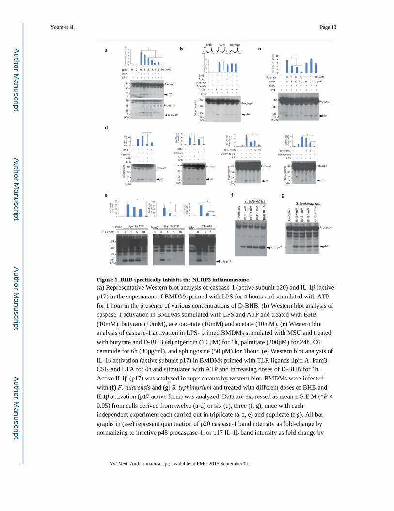
Figure 1. BHB specifically inhibits the NLRP3 inflammasome(a) Representative Western blot analysis of caspase-1 (active subunit p20) and IL-1β (active
p17) in the supernatant of BMDMs primed with LPS for 4 hours and stimulated with ATP
for 1 hour in the presence of various concentrations of D-BHB. (b) Western blot analysis of
caspase-1 activation in BMDMs stimulated with LPS and ATP and treated with BHB
(10mM), butyrate (10mM), acetoacetate (10mM) and acetate (10mM). (c) Western blot
analysis of caspase-1 activation in LPS- primed BMDMs stimulated with MSU and treated
with butyrate and D-BHB (d) nigericin (10 μM) for 1h, palmitate (200μM) for 24h, C6
ceramide for 6h (80μg/ml), and sphingosine (50 μM) for 1hour. (e) Western blot analysis of
IL-1β activation (active subunit p17) in BMDMs primed with TLR ligands lipid A, Pam3-
CSK and LTA for 4h and stimulated with ATP and increasing doses of D-BHB for 1h.
Active IL1β (p17) was analysed in supernatants by western blot. BMDMs were infected
with (f) F. tularensis and (g) S. typhimurium and treated with different doses of BHB and
IL1β activation (p17 active form) was analyzed. Data are expressed as mean ± S.E.M (*P <
0.05) from cells derived from twelve (a-d) or six (e), three (f, g), mice with each
independent experiment each carried out in triplicate (a-d, e) and duplicate (f g). All bar
graphs in (a-e) represent quantitation of p20 caspase-1 band intensity as fold-change by
normalizing to inactive p48 procaspase-1, or p17 IL-1β band intensity as fold change by
Youm et al. Page 13
Nat Med. Author manuscript; available in PMC 2015 September 01.
Author M
anuscriptA
uthor Manuscript
Author M
anuscriptA
uthor Manuscript
normalizing to inactive p37 pro-IL-1β. The differences between means and the effects of
treatments were determined by one-way ANOVA using Tukey's test.
Youm et al. Page 14
Nat Med. Author manuscript; available in PMC 2015 September 01.
Author M
anuscriptA
uthor Manuscript
Author M
anuscriptA
uthor Manuscript
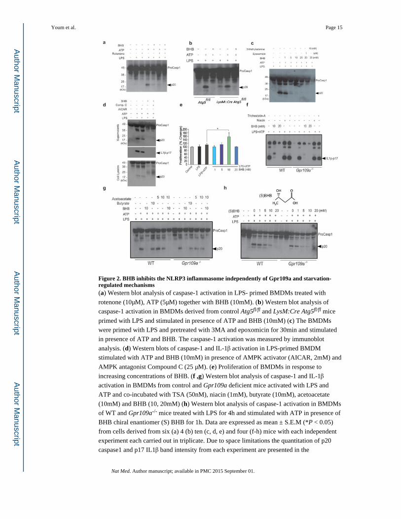
Figure 2. BHB inhibits the NLRP3 inflammasome independently of Gpr109a and starvation-regulated mechanisms(a) Western blot analysis of caspase-1 activation in LPS- primed BMDMs treated with
rotenone (10μM), ATP (5μM) together with BHB (10mM). (b) Western blot analysis of
caspase-1 activation in BMDMs derived from control Atg5fl/fl and LysM:Cre Atg5fl/fl mice
primed with LPS and stimulated in presence of ATP and BHB (10mM) (c) The BMDMs
were primed with LPS and pretreated with 3MA and epoxomicin for 30min and stimulated
in presence of ATP and BHB. The caspase-1 activation was measured by immunoblot
analysis. (d) Western blots of caspase-1 and IL-1β activation in LPS-primed BMDM
stimulated with ATP and BHB (10mM) in presence of AMPK activator (AICAR, 2mM) and
AMPK antagonist Compound C (25 μM). (e) Proliferation of BMDMs in response to
increasing concentrations of BHB. (f ,g) Western blot analysis of caspase-1 and IL-1β
activation in BMDMs from control and Gpr109a deficient mice activated with LPS and
ATP and co-incubated with TSA (50nM), niacin (1mM), butyrate (10mM), acetoacetate
(10mM) and BHB (10, 20mM) (h) Western blot analysis of caspase-1 activation in BMDMs
of WT and Gpr109a-/- mice treated with LPS for 4h and stimulated with ATP in presence of
BHB chiral enantiomer (S) BHB for 1h. Data are expressed as mean ± S.E.M (*P < 0.05)
from cells derived from six (a) 4 (b) ten (c, d, e) and four (f-h) mice with each independent
experiment each carried out in triplicate. Due to space limitations the quantitation of p20
caspase1 and p17 IL1β band intensity from each experiment are presented in the
Youm et al. Page 15
Nat Med. Author manuscript; available in PMC 2015 September 01.
Author M
anuscriptA
uthor Manuscript
Author M
anuscriptA
uthor Manuscript
Supplementary Fig. 2A. The differences between means and the effects of treatments were
determined by one-way ANOVA using Tukey's test.
Youm et al. Page 16
Nat Med. Author manuscript; available in PMC 2015 September 01.
Author M
anuscriptA
uthor Manuscript
Author M
anuscriptA
uthor Manuscript
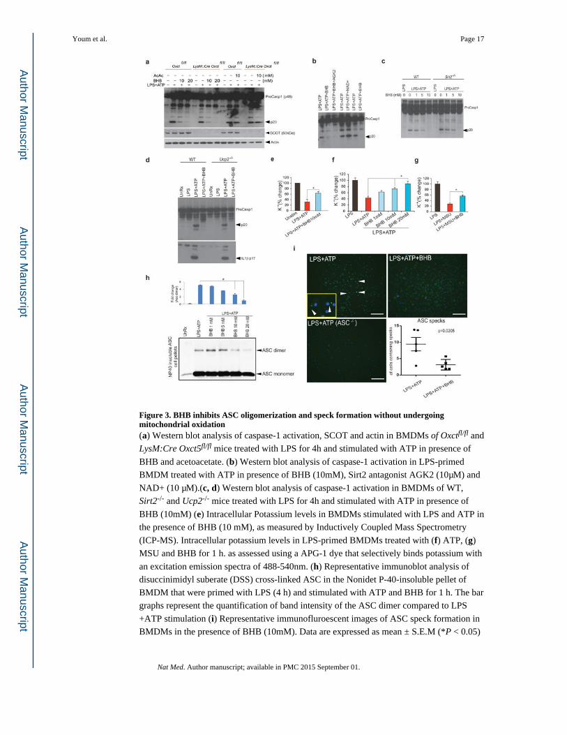
Figure 3. BHB inhibits ASC oligomerization and speck formation without undergoing mitochondrial oxidation(a) Western blot analysis of caspase-1 activation, SCOT and actin in BMDMs of Oxctfl/fl and
LysM:Cre Oxct5fl/fl mice treated with LPS for 4h and stimulated with ATP in presence of
BHB and acetoacetate. (b) Western blot analysis of caspase-1 activation in LPS-primed
BMDM treated with ATP in presence of BHB (10mM), Sirt2 antagonist AGK2 (10μM) and
NAD+ (10 μM).(c, d) Western blot analysis of caspase-1 activation in BMDMs of WT,
Sirt2-/- and Ucp2-/- mice treated with LPS for 4h and stimulated with ATP in presence of
BHB (10mM) (e) Intracellular Potassium levels in BMDMs stimulated with LPS and ATP in
the presence of BHB (10 mM), as measured by Inductively Coupled Mass Spectrometry
(ICP-MS). Intracellular potassium levels in LPS-primed BMDMs treated with (f) ATP, (g)
MSU and BHB for 1 h. as assessed using a APG-1 dye that selectively binds potassium with
an excitation emission spectra of 488-540nm. (h) Representative immunoblot analysis of
disuccinimidyl suberate (DSS) cross-linked ASC in the Nonidet P-40-insoluble pellet of
BMDM that were primed with LPS (4 h) and stimulated with ATP and BHB for 1 h. The bar
graphs represent the quantification of band intensity of the ASC dimer compared to LPS
+ATP stimulation (i) Representative immunofluroescent images of ASC speck formation in
BMDMs in the presence of BHB (10mM). Data are expressed as mean ± S.E.M (*P < 0.05)
Youm et al. Page 17
Nat Med. Author manuscript; available in PMC 2015 September 01.
Author M
anuscriptA
uthor Manuscript
Author M
anuscriptA
uthor Manuscript
from cells derived from five (a) six (b,d), eight (e-g) and four (h) mice with each
independent experiment carried out in triplicate. The differences between means and the
effects of treatments were determined by one-way ANOVA using Tukey's test.(i) Data are
shown as mean ± SEM and are representative of two independent experiments. Statistical
differences were calculated by student's t-test.
Youm et al. Page 18
Nat Med. Author manuscript; available in PMC 2015 September 01.
Author M
anuscriptA
uthor Manuscript
Author M
anuscriptA
uthor Manuscript
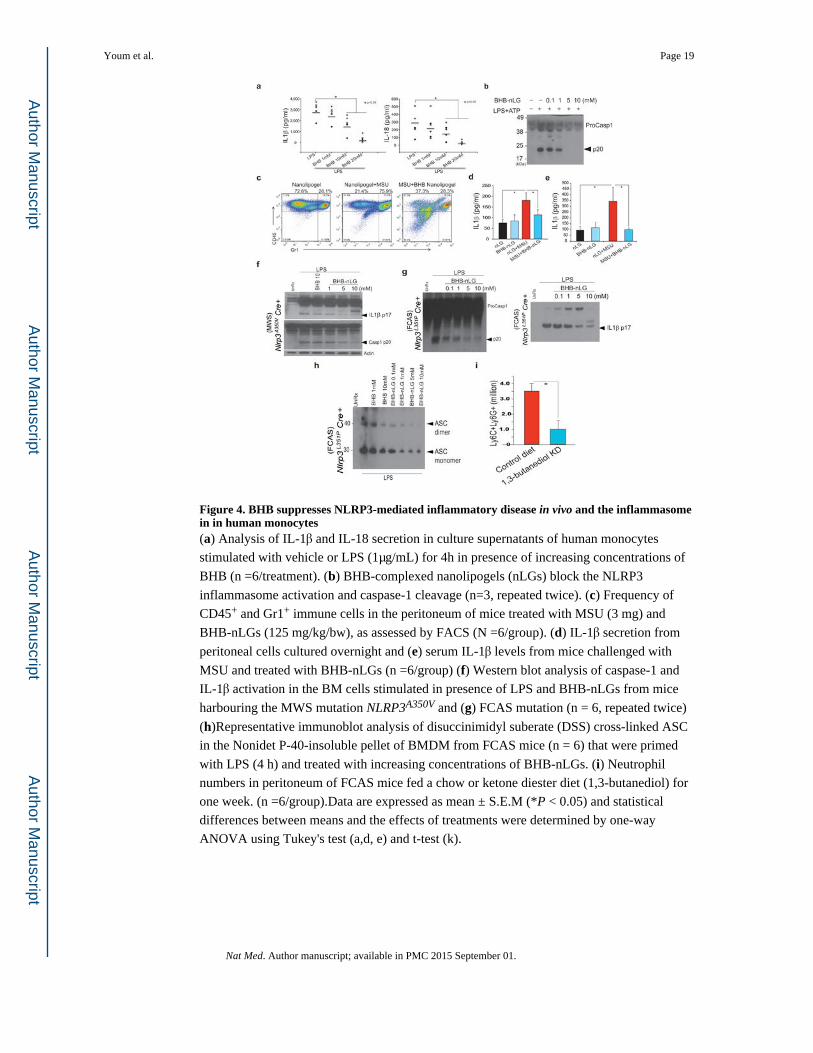
Figure 4. BHB suppresses NLRP3-mediated inflammatory disease in vivo and the inflammasome in in human monocytes(a) Analysis of IL-1β and IL-18 secretion in culture supernatants of human monocytes
stimulated with vehicle or LPS (1μg/mL) for 4h in presence of increasing concentrations of
BHB (n =6/treatment). (b) BHB-complexed nanolipogels (nLGs) block the NLRP3
inflammasome activation and caspase-1 cleavage (n=3, repeated twice). (c) Frequency of
CD45+ and Gr1+ immune cells in the peritoneum of mice treated with MSU (3 mg) and
BHB-nLGs (125 mg/kg/bw), as assessed by FACS (N =6/group). (d) IL-1β secretion from
peritoneal cells cultured overnight and (e) serum IL-1β levels from mice challenged with
MSU and treated with BHB-nLGs (n =6/group) (f) Western blot analysis of caspase-1 and
IL-1β activation in the BM cells stimulated in presence of LPS and BHB-nLGs from mice
harbouring the MWS mutation NLRP3A350V and (g) FCAS mutation (n = 6, repeated twice)
(h)Representative immunoblot analysis of disuccinimidyl suberate (DSS) cross-linked ASC
in the Nonidet P-40-insoluble pellet of BMDM from FCAS mice (n = 6) that were primed
with LPS (4 h) and treated with increasing concentrations of BHB-nLGs. (i) Neutrophil
numbers in peritoneum of FCAS mice fed a chow or ketone diester diet (1,3-butanediol) for
one week. (n =6/group).Data are expressed as mean ± S.E.M (*P < 0.05) and statistical
differences between means and the effects of treatments were determined by one-way
ANOVA using Tukey's test (a,d, e) and t-test (k).
Youm et al. Page 19
Nat Med. Author manuscript; available in PMC 2015 September 01.
Author M
anuscriptA
uthor Manuscript
Author M
anuscriptA
uthor Manuscript
![Set 6: Relativitybackground.uchicago.edu/~whu/Courses/Ast305_10/ast305_06.pdf · 2011-12-10 · Special Relativity. x=ct x'=ct' O [unprimed frame] O' [primed frame] v emission at](https://static.fdocument.org/doc/165x107/5ec4fe7fbe92464bde029ce6/set-6-whucoursesast30510ast30506pdf-2011-12-10-special-relativity-xct.jpg)