X. ΓΡΑΪΔΗΣ Επμβαικός καριολόος, FSCAI · Crusade 6. Valet 7. ......
61
X. ΓΡΑΪΔΗΣ Επεμβατικός καρδιολόγος, FSCAI Kλινική Euromedica-Κυανούς Σταυρός, Θεσσαλονίκη Χρόνιες Ολικές Αποφράξεις: Ορθόδρομη προσπέλαση 7ο ΣΥΝΕΔΡΙΟ ΕΠΕΜΒΑΤΙΚΗΣ ΚΑΡΔΙΟΛΟΓΙΑΣ & ΗΛΕΚΤΡΟΦΥΣΙΟΛΟΓΙΑΣ 9 - 11 Οκτωβρίου 2014, Makedonia Palace ,Θεσσαλονίκη.
Transcript of X. ΓΡΑΪΔΗΣ Επμβαικός καριολόος, FSCAI · Crusade 6. Valet 7. ......
X. ΓΡΑΪΔΗΣΕπεμβατικός καρδιολόγος, FSCAI
Kλινική Euromedica-Κυανούς Σταυρός, Θεσσαλονίκη
Χρόνιες Ολικές Αποφράξεις: Ορθόδρομη προσπέλαση
7ο ΣΥΝΕΔΡΙΟ ΕΠΕΜΒΑΤΙΚΗΣ ΚΑΡΔΙΟΛΟΓΙΑΣ & ΗΛΕΚΤΡΟΦΥΣΙΟΛΟΓΙΑΣ 9 - 11 Οκτωβρίου 2014, Makedonia Palace ,Θεσσαλονίκη.
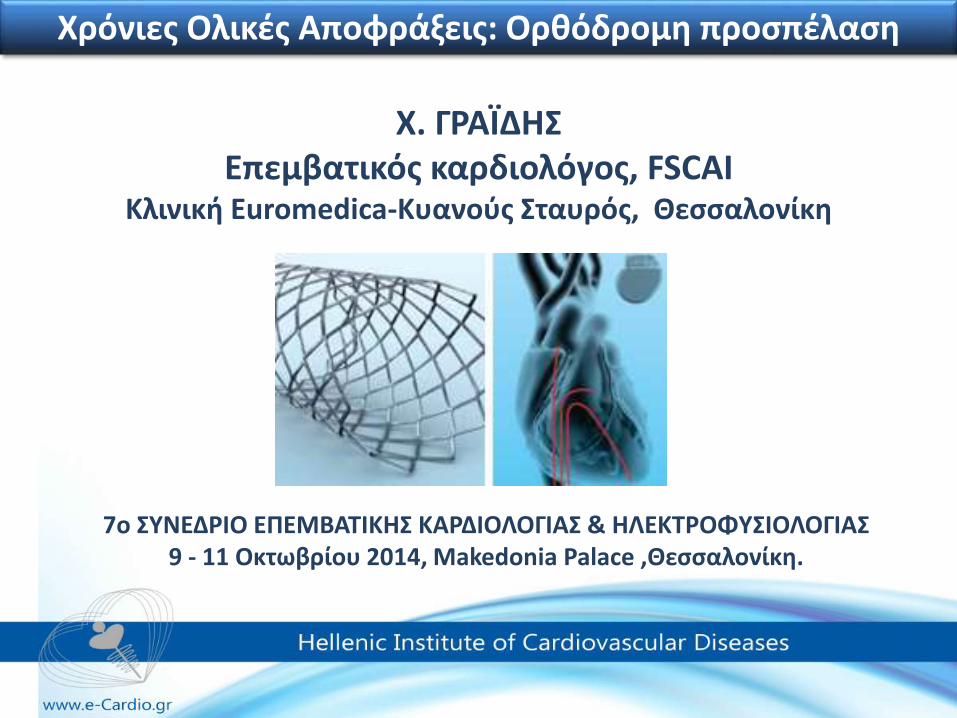
Conquering the Final Frontier
“If you know the enemy and know yourself, you need not fear the result of a hundred battles. If you know yourself but not the enemy, for every victory gained you will also suffer a defeat. If you know neither the enemy nor yourself, you will succumb in every battle”
Know the enemy and ……..

In ideal circumstances, CTO PCI should be a planned procedure and not performed ad hoc.
Anatomy Dictates Strategy and Device Selection
Optimal angiographic technique may be the most important preparatory and procedural
determinant for success in CTO PCI
Spend time examining diagnostic films & decide on:
•Approach ,vascular access, guide shape & size
•dedicated equipment availability
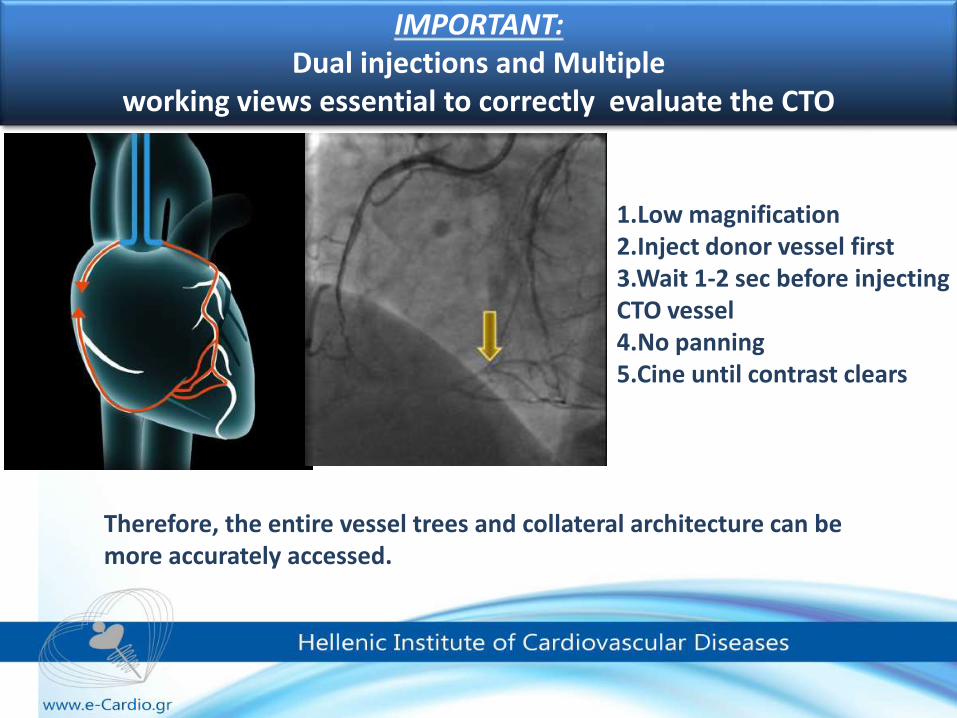
IMPORTANT: Dual injections and Multiple
working views essential to correctly evaluate the CTO
Therefore, the entire vessel trees and collateral architecture can be more accurately accessed.
1.Low magnification2.Inject donor vessel first 3.Wait 1-2 sec before injecting CTO vessel 4.No panning 5.Cine until contrast clears

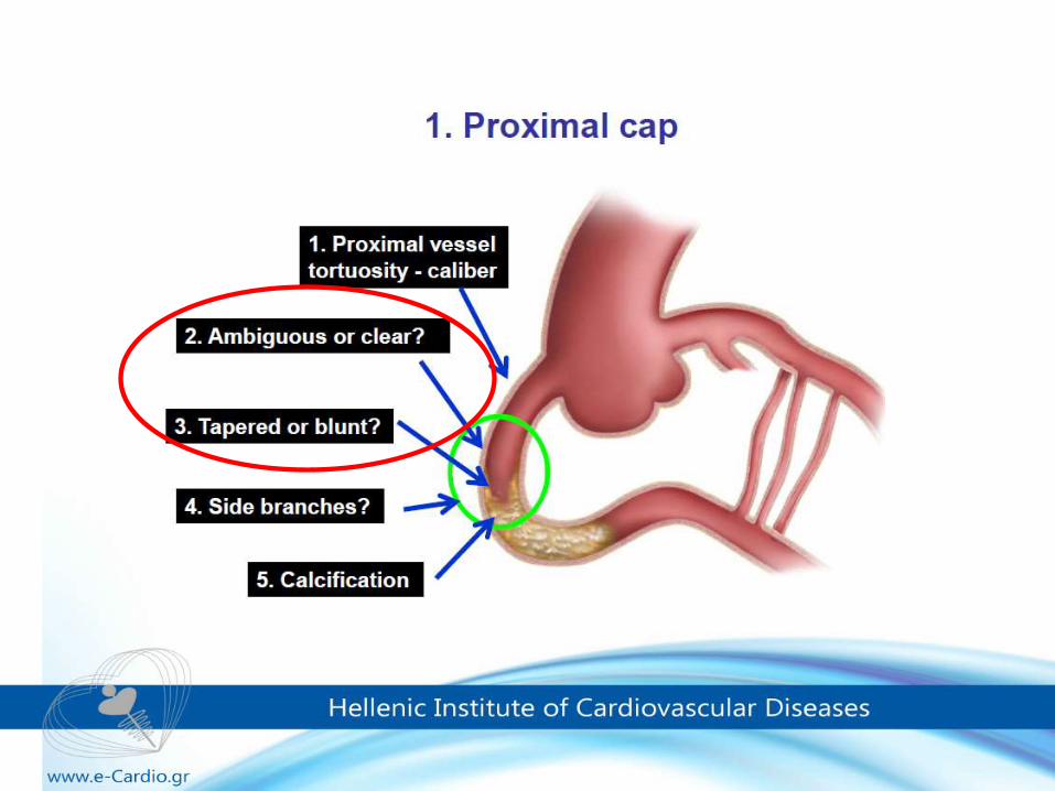
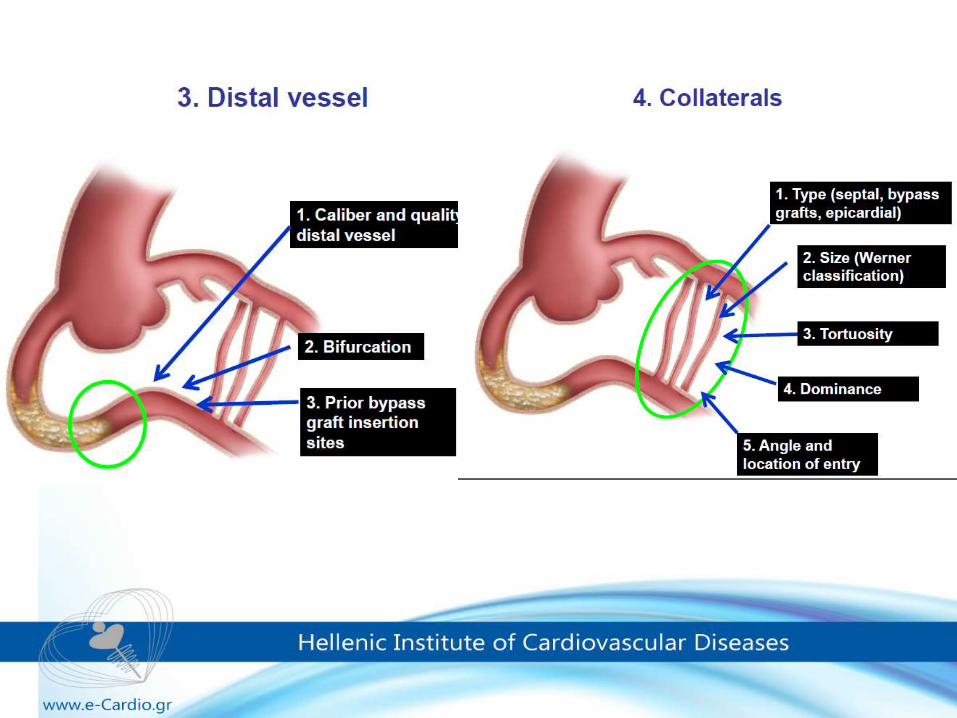
The CTO angiograms should be carefully reviewed, often by single frame-by-frame analysis, to better understand:
1. the nature of proximal and distal caps, 2. the calcification and suspected
angulation in the CTO segment and target vessel,
3. lesion length,4. collateral vessel pattern and anatomy,5. relationships with side branches, and6. donor and CTO vessel characteristics for
catheter selection.
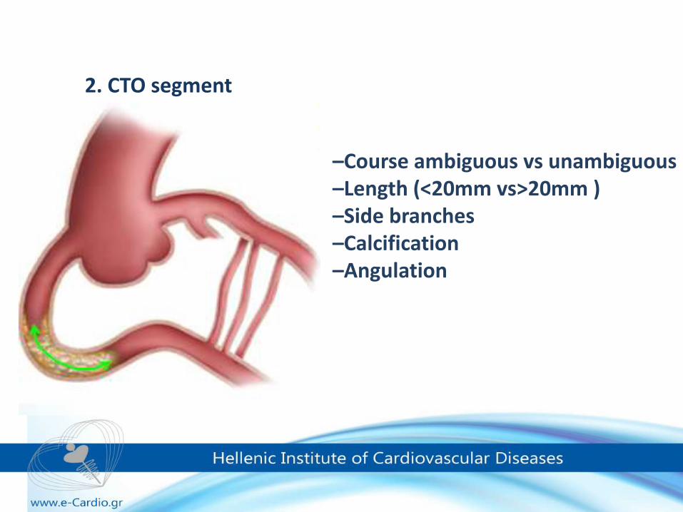
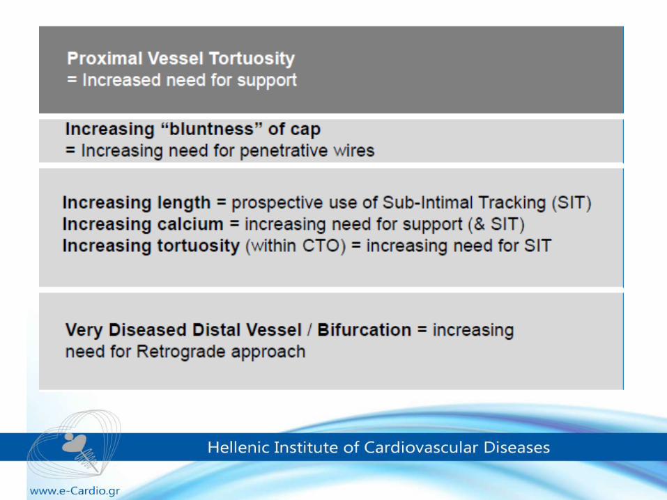
–Course ambiguous vs unambiguous–Length (<20mm vs>20mm )–Side branches–Calcification–Angulation
2. CTO segment
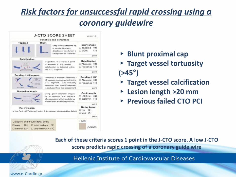
▸ Blunt proximal cap▸ Target vessel tortuosity(>45°)▸ Target vessel calcification▸ Lesion length >20 mm▸ Previous failed CTO PCI
Risk factors for unsuccessful rapid crossing using a coronary guidewire
Each of these criteria scores 1 point in the J-CTO score. A low J-CTO score predicts rapid crossing of a coronary guide wire
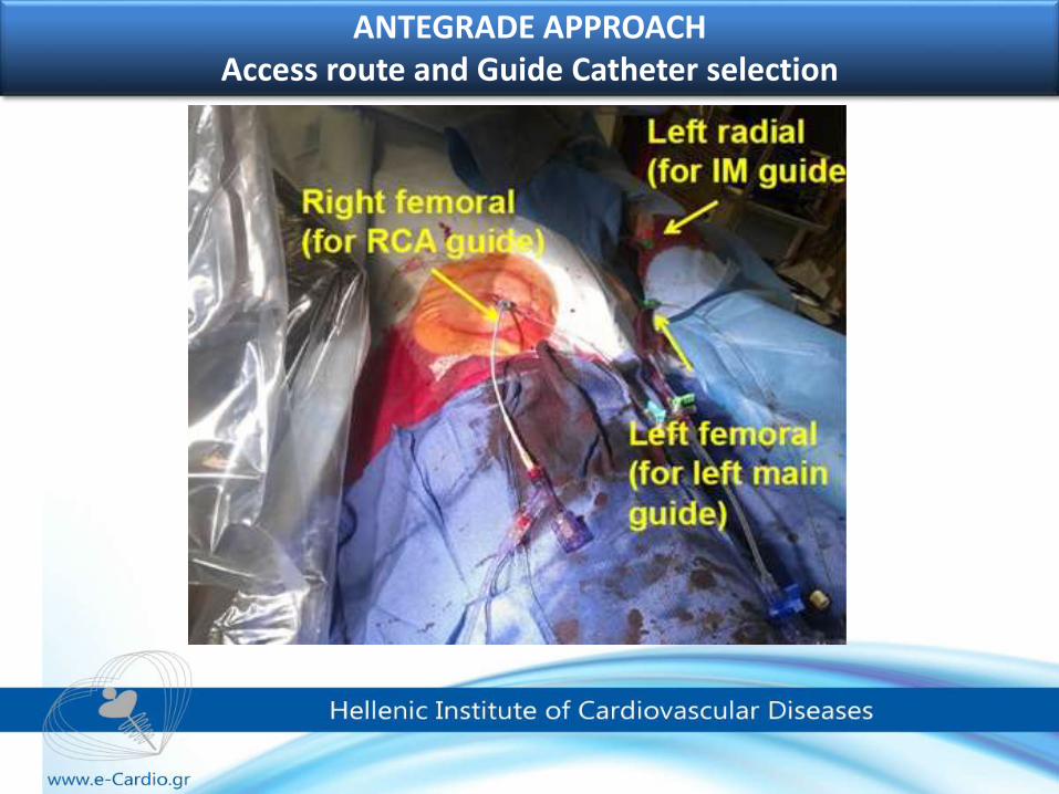
ANTEGRADE APPROACHAccess route and Guide Catheter selection
ANTEGRADE APPROACHAccess route and Guide Catheter selection
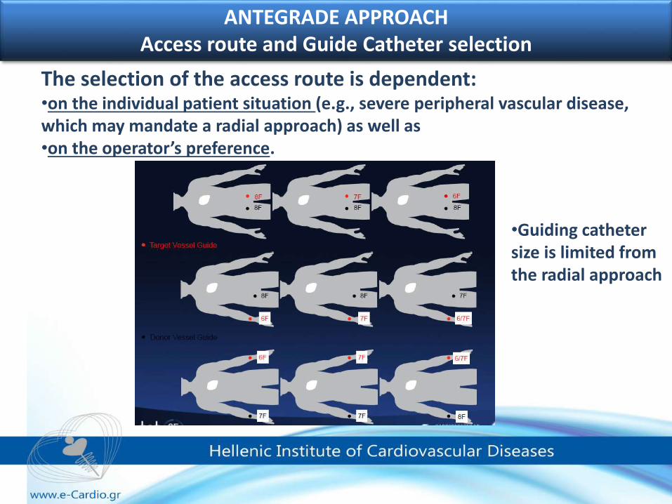
The selection of the access route is dependent: •on the individual patient situation (e.g., severe peripheral vascular disease, which may mandate a radial approach) as well as •on the operator’s preference.
•Guiding catheter size is limited from the radial approach
ANTEGRADE APPROACHAccess route and Guide Catheter selection
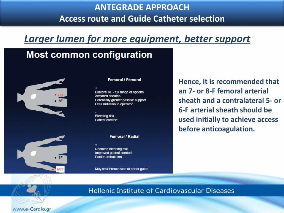
Hence, it is recommended that an 7- or 8-F femoral arterial sheath and a contralateral 5- or 6-F arterial sheath should be used initially to achieve access before anticoagulation.
Larger lumen for more equipment, better support
Access route and Guide Catheter selection
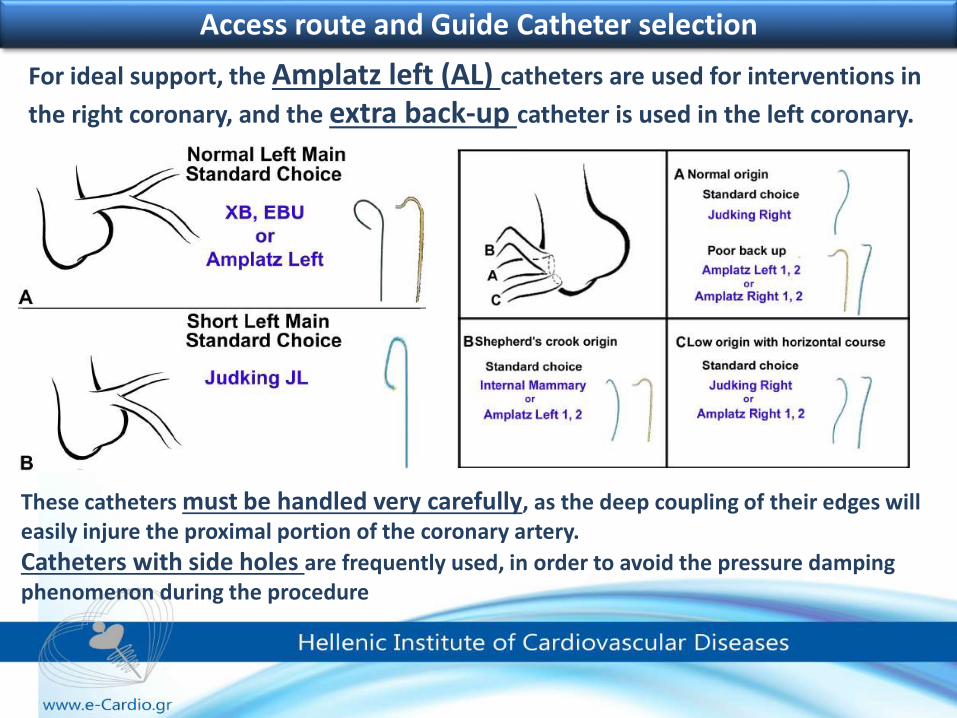
These catheters must be handled very carefully, as the deep coupling of their edges will easily injure the proximal portion of the coronary artery.
Catheters with side holes are frequently used, in order to avoid the pressure damping phenomenon during the procedure
For ideal support, the Amplatz left (AL) catheters are used for interventions in
the right coronary, and the extra back-up catheter is used in the left coronary.
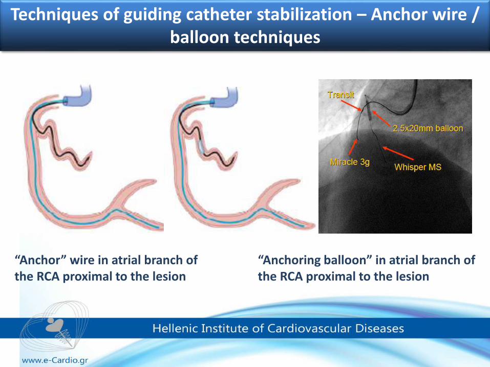
Techniques of guiding catheter stabilization – Anchor wire / balloon techniques
“Anchoring balloon” in atrial branch of the RCA proximal to the lesion
“Anchor” wire in atrial branch of the RCA proximal to the lesion
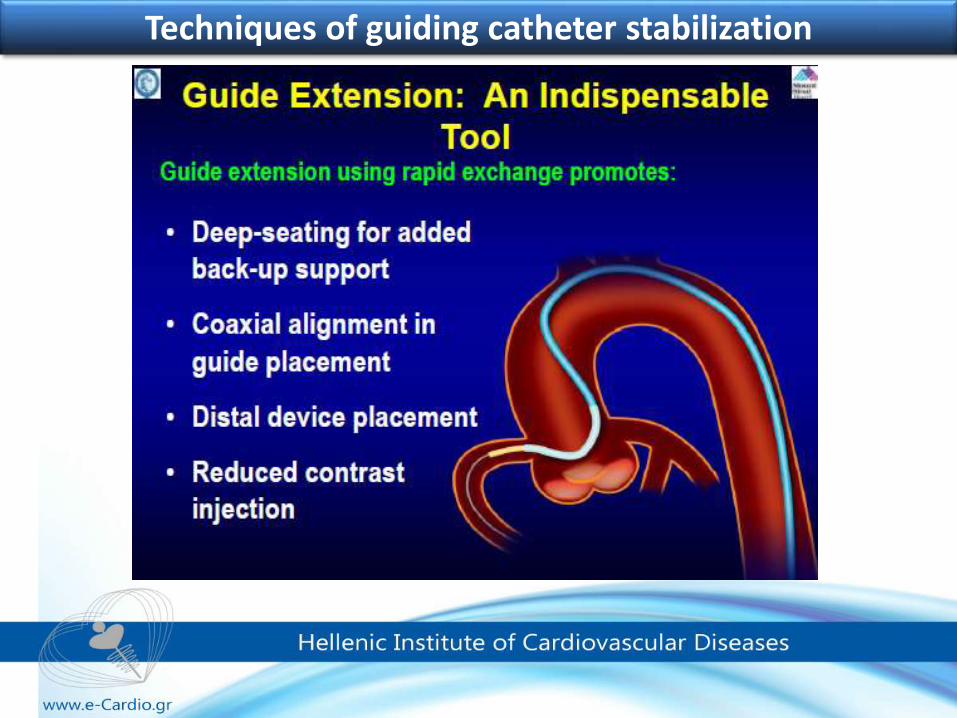
Techniques of guiding catheter stabilization
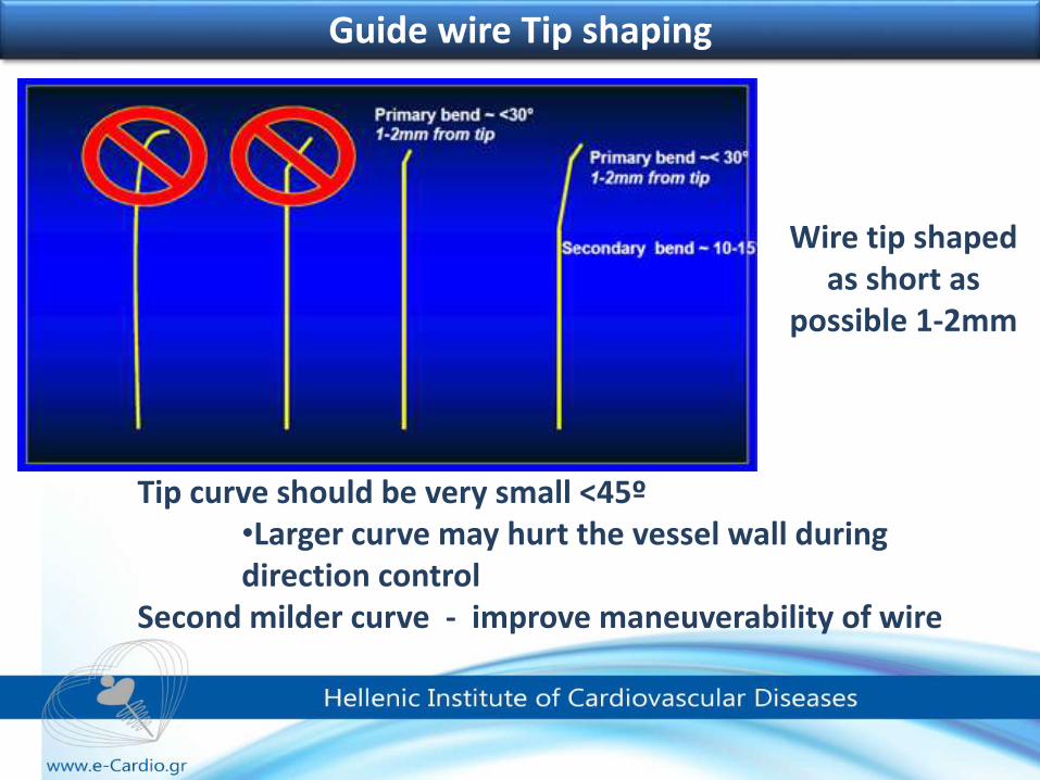
Tip curve should be very small <45º•Larger curve may hurt the vessel wall during direction control
Second milder curve - improve maneuverability of wire
Wire tip shaped as short as
possible 1-2mm
Guide wire Tip shaping
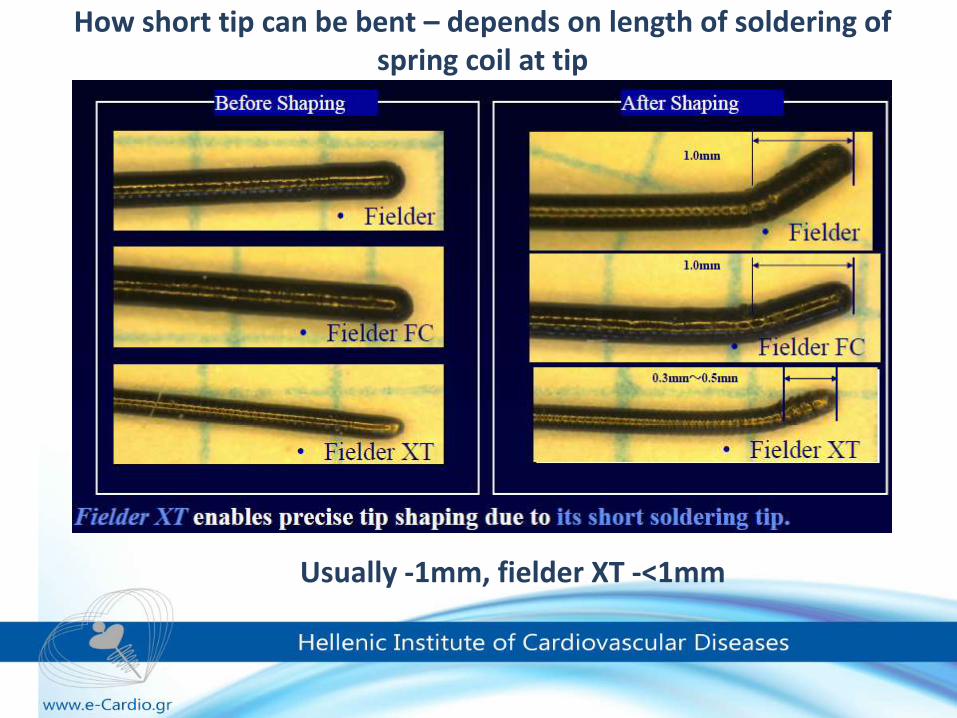
How short tip can be bent – depends on length of soldering of spring coil at tip
Usually -1mm, fielder XT -<1mm
Guide wire Tip shaping
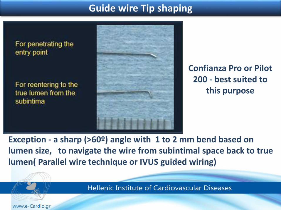
Exception - a sharp (>60º) angle with 1 to 2 mm bend based on lumen size, to navigate the wire from subintimal space back to true lumen( Parallel wire technique or IVUS guided wiring)
Confianza Pro or Pilot 200 - best suited to
this purpose
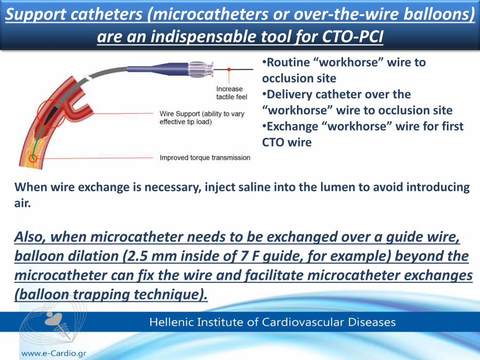
Support catheters (microcatheters or over-the-wire balloons) are an indispensable tool for CTO-PCI
•Routine “workhorse” wire to occlusion site•Delivery catheter over the “workhorse” wire to occlusion site•Exchange “workhorse” wire for first CTO wire
When wire exchange is necessary, inject saline into the lumen to avoid introducing air.
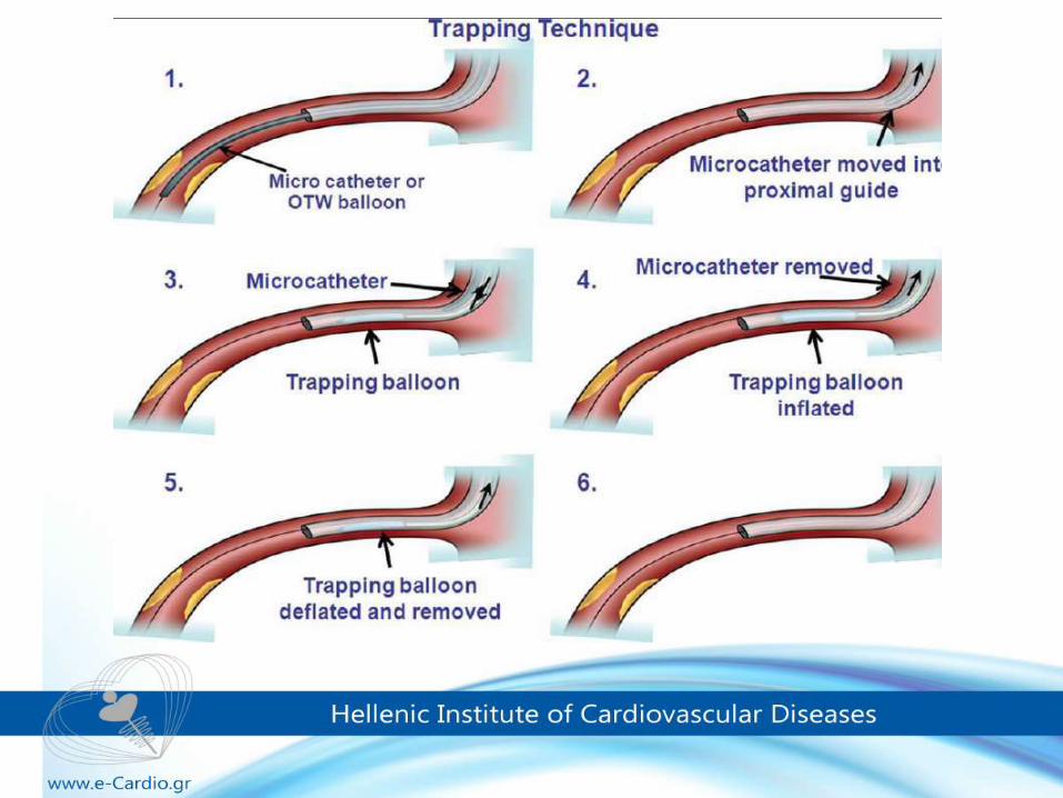
Also, when microcatheter needs to be exchanged over a guide wire, balloon dilation (2.5 mm inside of 7 F guide, for example) beyond the microcatheter can fix the wire and facilitate microcatheter exchanges (balloon trapping technique).
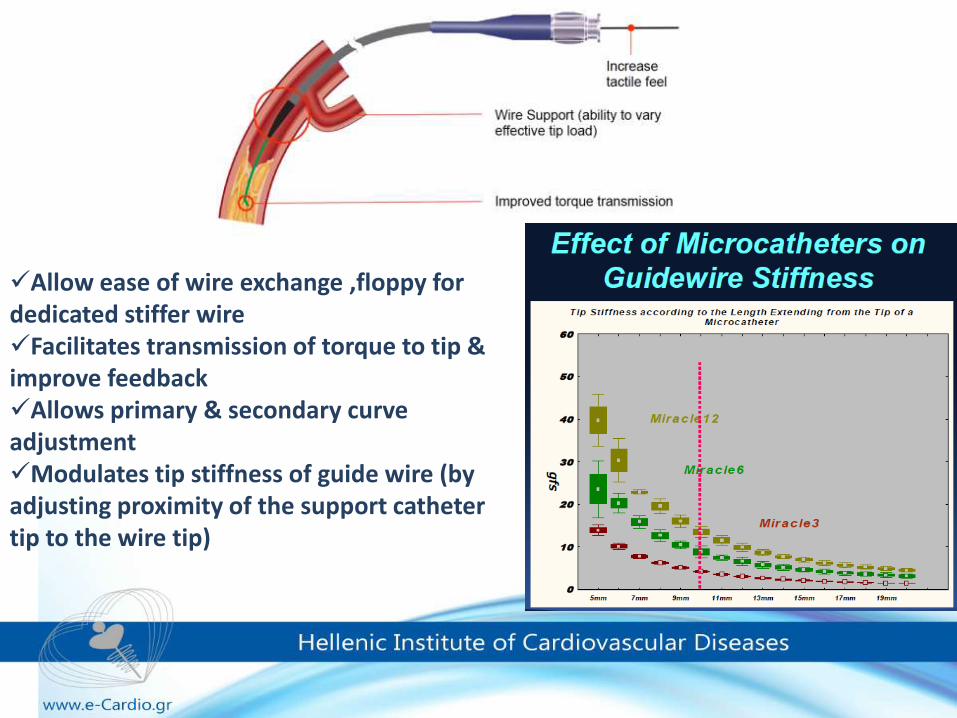
Allow ease of wire exchange ,floppy for dedicated stiffer wire Facilitates transmission of torque to tip & improve feedback Allows primary & secondary curve adjustment Modulates tip stiffness of guide wire (by adjusting proximity of the support catheter tip to the wire tip)
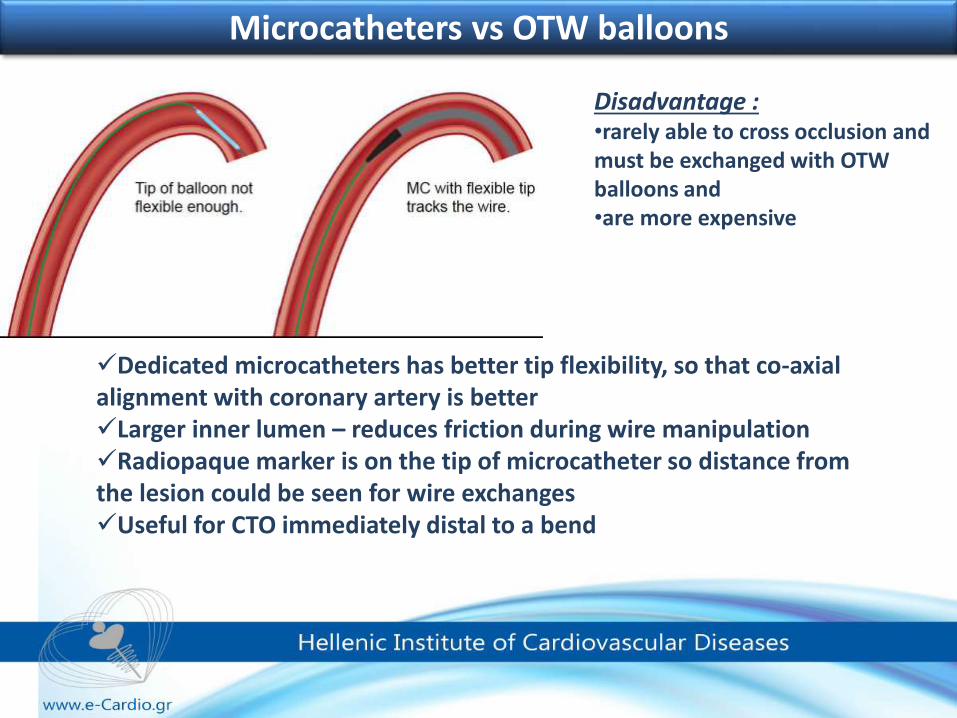
Microcatheters vs OTW balloons
Dedicated microcatheters has better tip flexibility, so that co-axial alignment with coronary artery is better Larger inner lumen – reduces friction during wire manipulationRadiopaque marker is on the tip of microcatheter so distance from the lesion could be seen for wire exchangesUseful for CTO immediately distal to a bend
Disadvantage : •rarely able to cross occlusion and must be exchanged with OTW balloons and •are more expensive
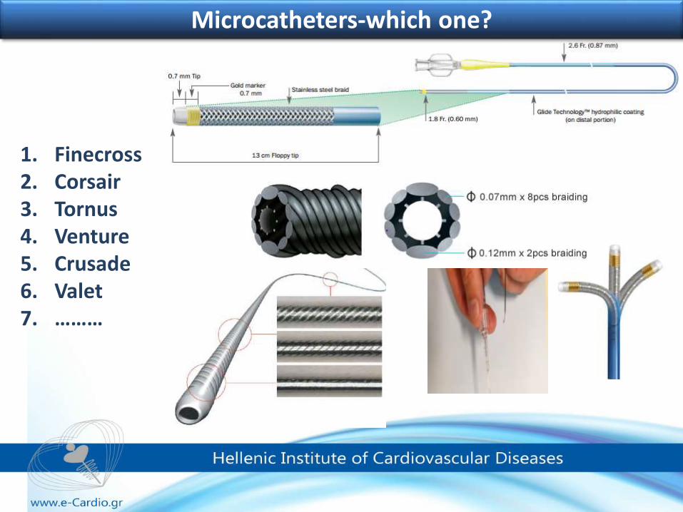
1. Finecross2. Corsair3. Tornus4. Venture5. Crusade6. Valet7. ………
Microcatheters-which one?
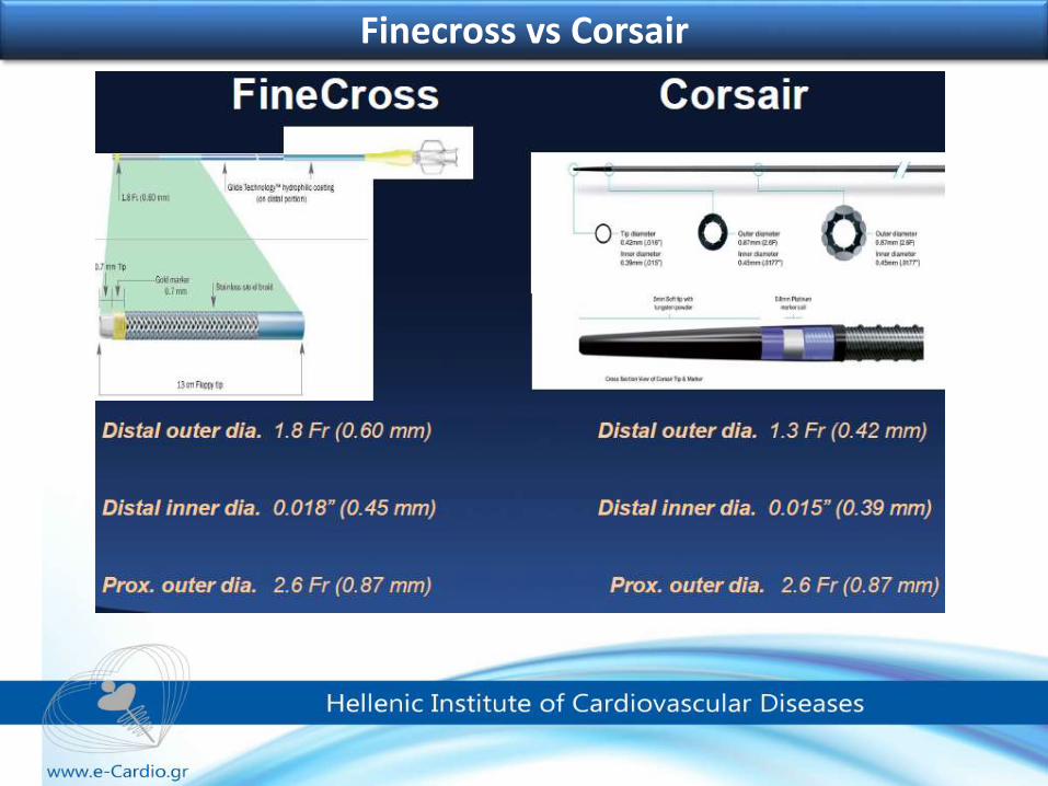
Finecross vs Corsair
Finecross vs Corsair
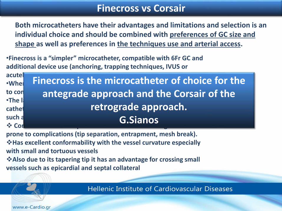
Both microcatheters have their advantages and limitations and selection is an individual choice and should be combined with preferences of GC size and shape as well as preferences in the techniques use and arterial access.
•Finecross is a “simpler” microcatheter, compatible with 6Fr GC and additional device use (anchoring, trapping techniques, IVUS or acutely in case of complications……)•When failing to be advanced ,antegrade or retrograde, is less prone to complications ( such as trapping, breaking, tip separation…..).•The lack of tapering at the very tip of the Finecross makes the catheter less eligible for crossing very small and tortuous vessels such as the epicardial collaterals. Corsair is a more complex structure and when failing is more prone to complications (tip separation, entrapment, mesh break). Has excellent conformability with the vessel curvature especially with small and tortuous vessels Also due to its tapering tip it has an advantage for crossing small vessels such as epicardial and septal collateral
Finecross is the microcatheter of choice for the antegrade approach and the Corsair of the
retrograde approach. G.Sianos
In the antegrade wire procedure, plaque tracking should be
described as either intimal or subintimal.
In addition, antegrade intimal plaque tracking has 2 techniques:
• loose tissue tracking and
•intentional intimal plaque tracking.
Antegrade wire procedure
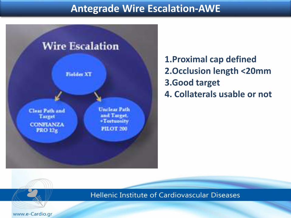
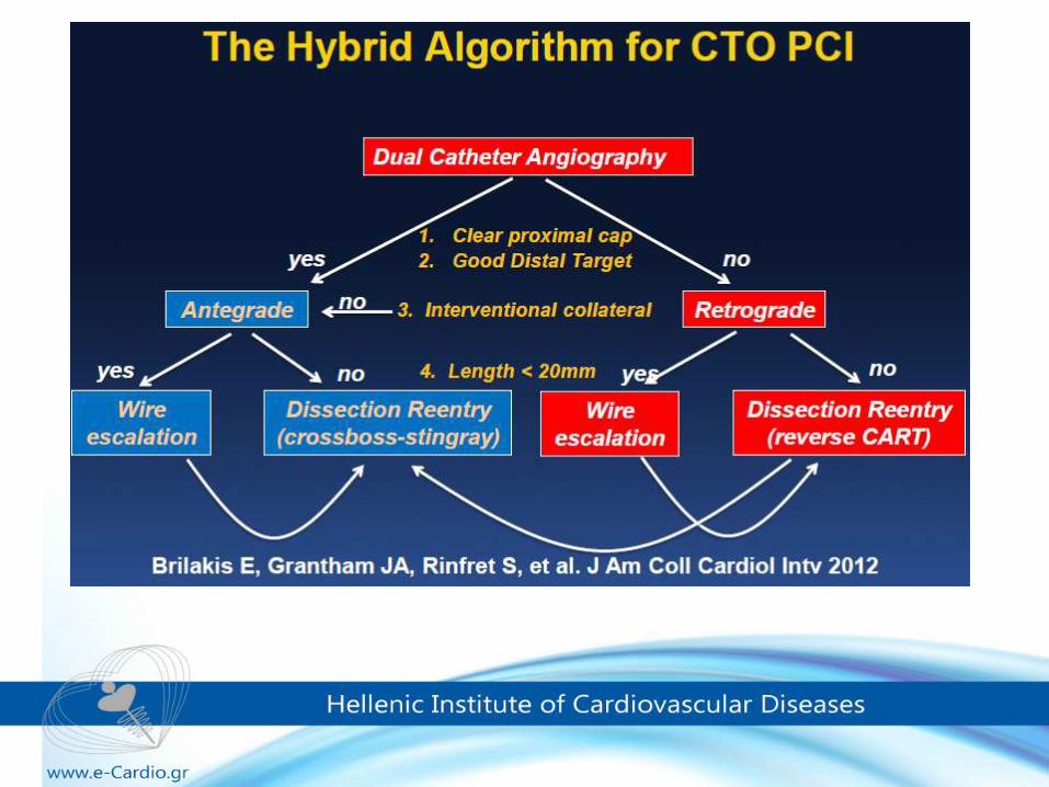
1.Proximal cap defined2.Occlusion length <20mm3.Good target4. Collaterals usable or not
Antegrade Wire Escalation-AWE
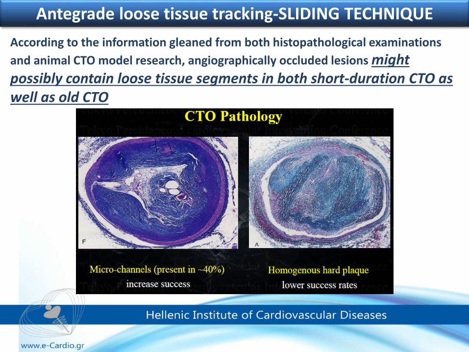
According to the information gleaned from both histopathological examinations
and animal CTO model research, angiographically occluded lesions might possibly contain loose tissue segments in both short-duration CTO as well as old CTO
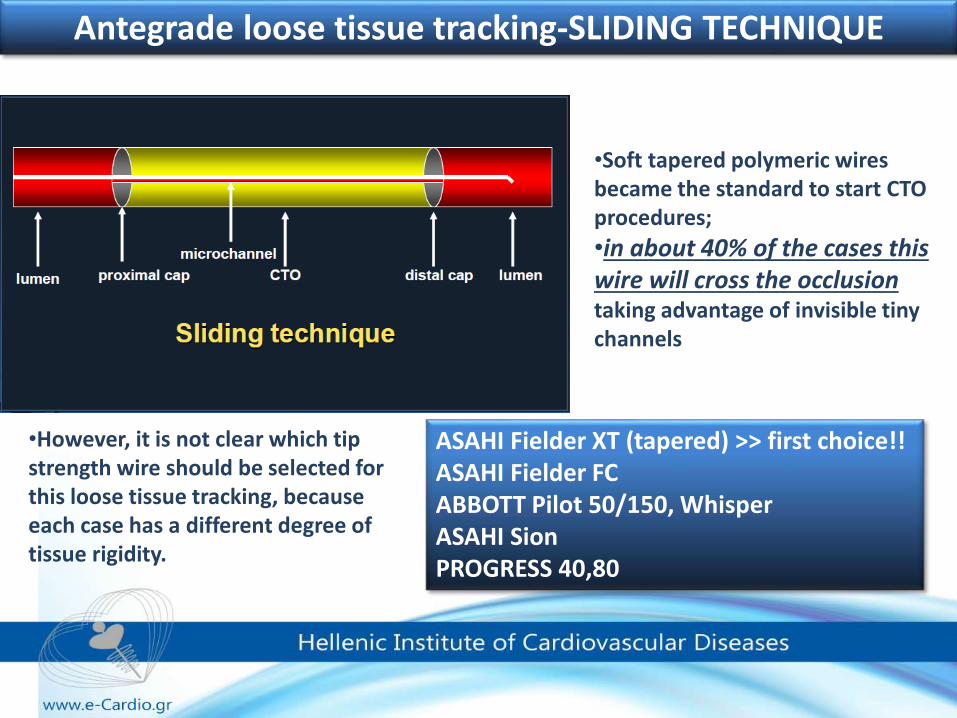
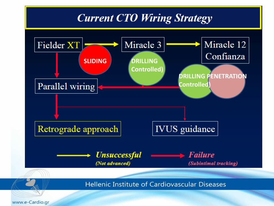
Antegrade loose tissue tracking-SLIDING TECHNIQUE
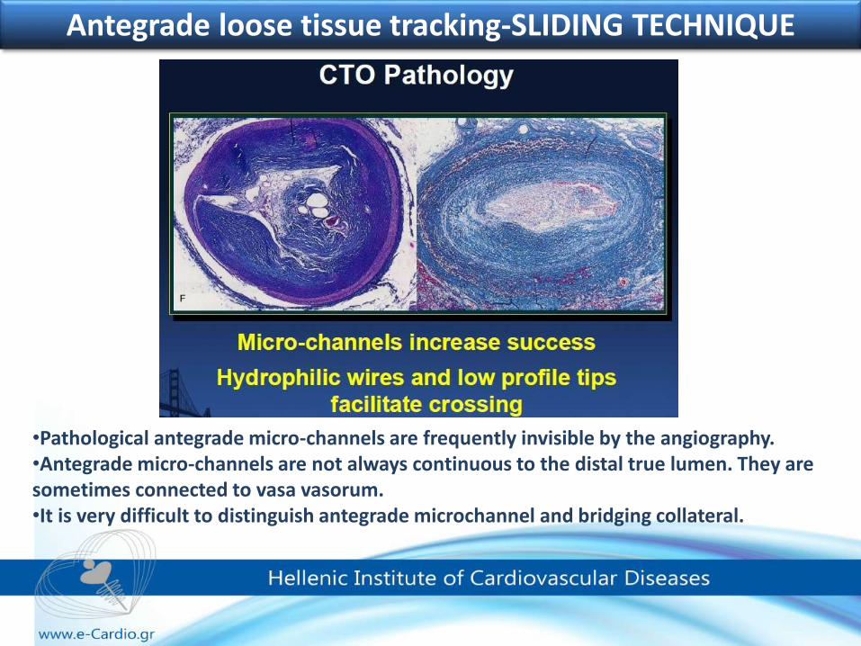
•Pathological antegrade micro-channels are frequently invisible by the angiography.•Antegrade micro-channels are not always continuous to the distal true lumen. They are sometimes connected to vasa vasorum.•It is very difficult to distinguish antegrade microchannel and bridging collateral.
Antegrade loose tissue tracking-SLIDING TECHNIQUE
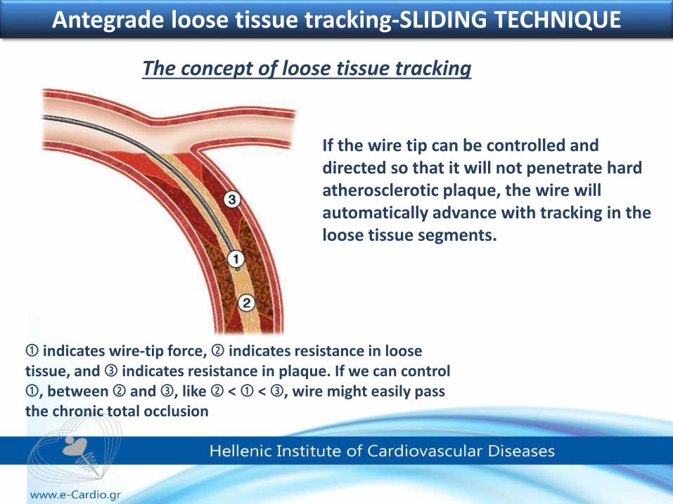
➀ indicates wire-tip force, ➁ indicates resistance in loose tissue, and ➂ indicates resistance in plaque. If we can control ➀, between ➁ and ➂, like ➁ < ➀ < ➂, wire might easily pass the chronic total occlusion
Antegrade loose tissue tracking-SLIDING TECHNIQUE
If the wire tip can be controlled and directed so that it will not penetrate hard atherosclerotic plaque, the wire will automatically advance with tracking in the loose tissue segments.
The concept of loose tissue tracking
Antegrade loose tissue tracking-SLIDING TECHNIQUE
•However, it is not clear which tip strength wire should be selected for this loose tissue tracking, because each case has a different degree of tissue rigidity.
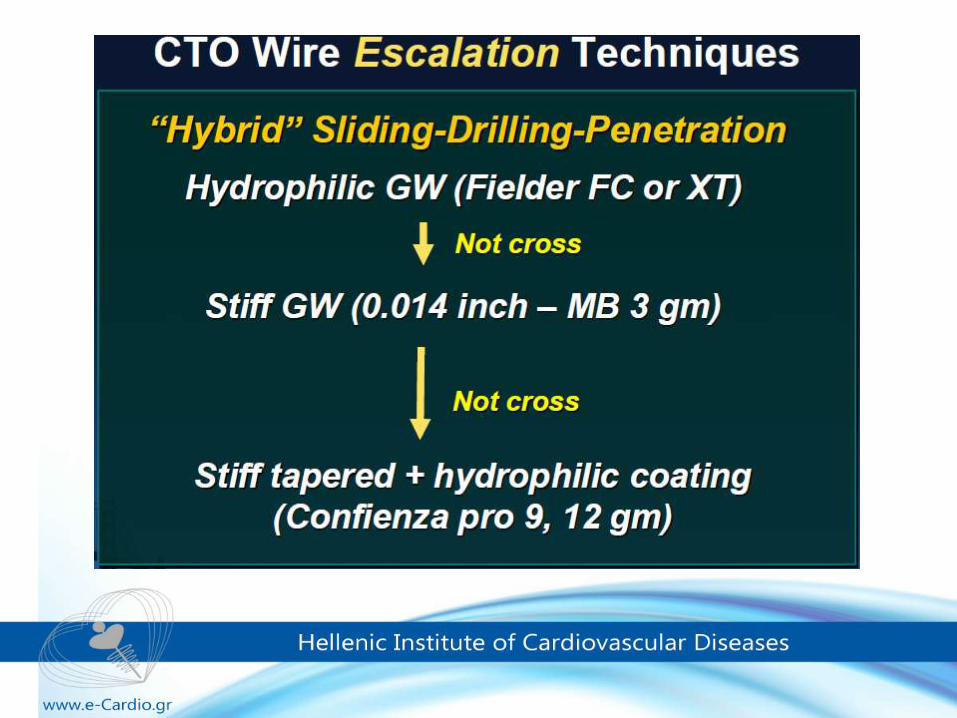
ASAHI Fielder XT (tapered) >> first choice!!ASAHI Fielder FCABBOTT Pilot 50/150, WhisperASAHI SionPROGRESS 40,80
•Soft tapered polymeric wires became the standard to start CTO procedures;
•in about 40% of the cases this wire will cross the occlusion taking advantage of invisible tiny channels
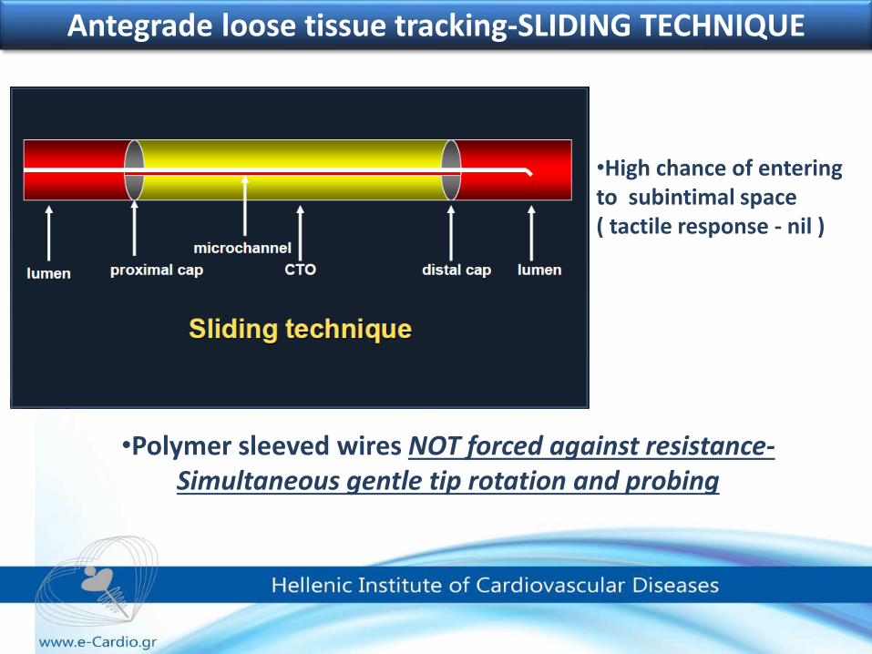
Antegrade loose tissue tracking-SLIDING TECHNIQUE
•High chance of entering to subintimal space ( tactile response - nil )
•Polymer sleeved wires NOT forced against resistance-Simultaneous gentle tip rotation and probing
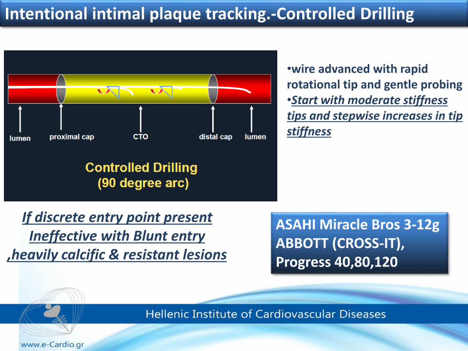
Intentional intimal plaque tracking.-Controlled Drilling
•wire advanced with rapid rotational tip and gentle probing•Start with moderate stiffness tips and stepwise increases in tip stiffness
If discrete entry point present Ineffective with Blunt entry
,heavily calcific & resistant lesions
ASAHI Miracle Bros 3-12gABBOTT (CROSS-IT),Progress 40,80,120
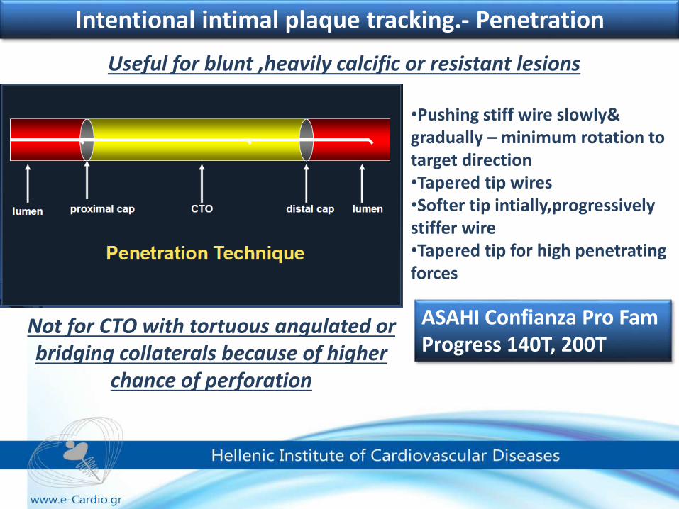
Intentional intimal plaque tracking.- Penetration
Not for CTO with tortuous angulated or bridging collaterals because of higher
chance of perforation
ASAHI Confianza Pro FamProgress 140T, 200T
•Pushing stiff wire slowly& gradually – minimum rotation to target direction•Tapered tip wires •Softer tip intially,progressivelystiffer wire•Tapered tip for high penetrating forces
Useful for blunt ,heavily calcific or resistant lesions
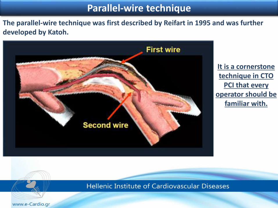
The parallel-wire technique was first described by Reifart in 1995 and was further developed by Katoh.
Parallel-wire technique
It is a cornerstone technique in CTO
PCI that every operator should be
familiar with.
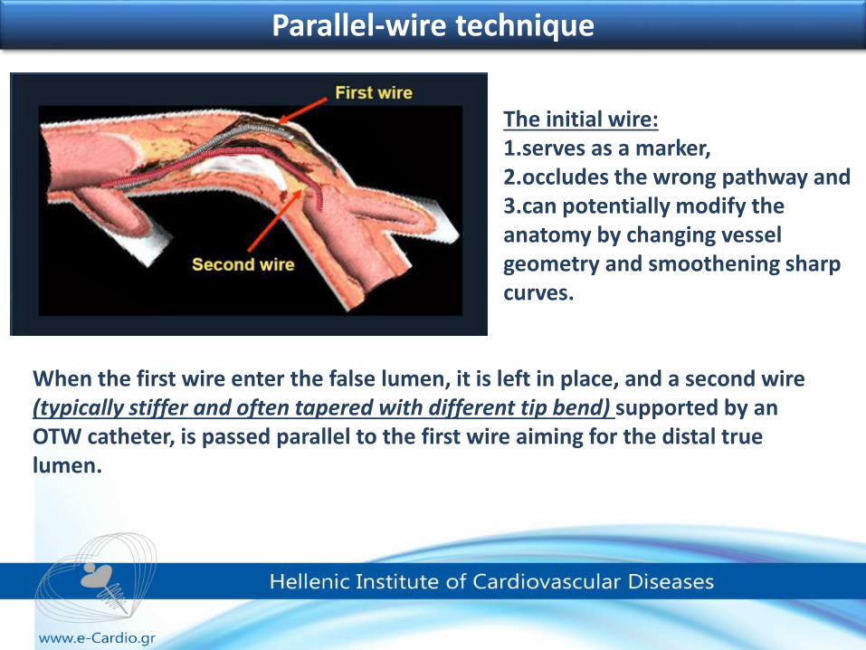
When the first wire enter the false lumen, it is left in place, and a second wire (typically stiffer and often tapered with different tip bend) supported by an OTW catheter, is passed parallel to the first wire aiming for the distal true lumen.
Parallel-wire technique
The initial wire: 1.serves as a marker, 2.occludes the wrong pathway and 3.can potentially modify the anatomy by changing vessel geometry and smoothening sharp curves.
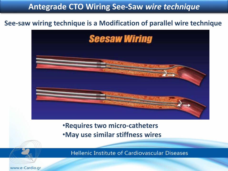
Antegrade CTO Wiring See-Saw wire technique
See-saw wiring technique is a Modification of parallel wire technique
•Requires two micro-catheters•May use similar stiffness wires
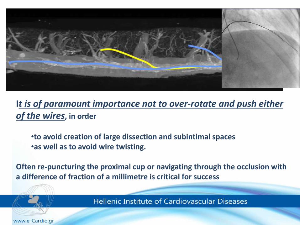
It is of paramount importance not to over-rotate and push either of the wires, in order
•to avoid creation of large dissection and subintimal spaces •as well as to avoid wire twisting.
Often re-puncturing the proximal cup or navigating through the occlusion with a difference of fraction of a millimetre is critical for success
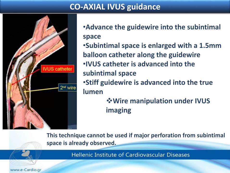
•Advance the guidewire into the subintimalspace•Subintimal space is enlarged with a 1.5mm balloon catheter along the guidewire•IVUS catheter is advanced into the subintimal space•Stiff guidewire is advanced into the true lumen
Wire manipulation under IVUS imaging
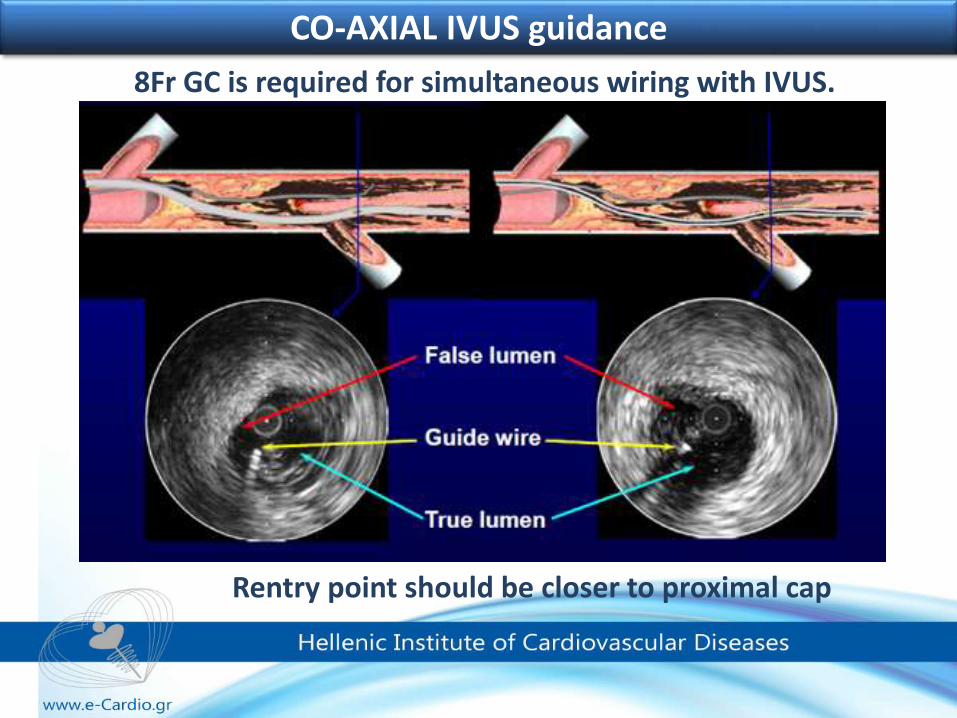
CO-AXIAL IVUS guidance
This technique cannot be used if major perforation from subintimalspace is already observed.
CO-AXIAL IVUS guidance
8Fr GC is required for simultaneous wiring with IVUS.
Rentry point should be closer to proximal cap
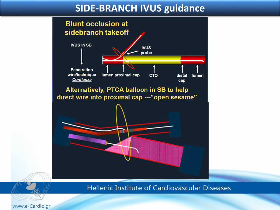
SIDE-BRANCH IVUS guidance
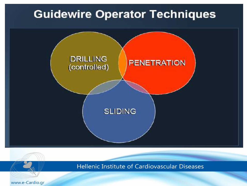
SLIDING DRILLINGControlled)
PENETRATIONDRILLINGControlled)
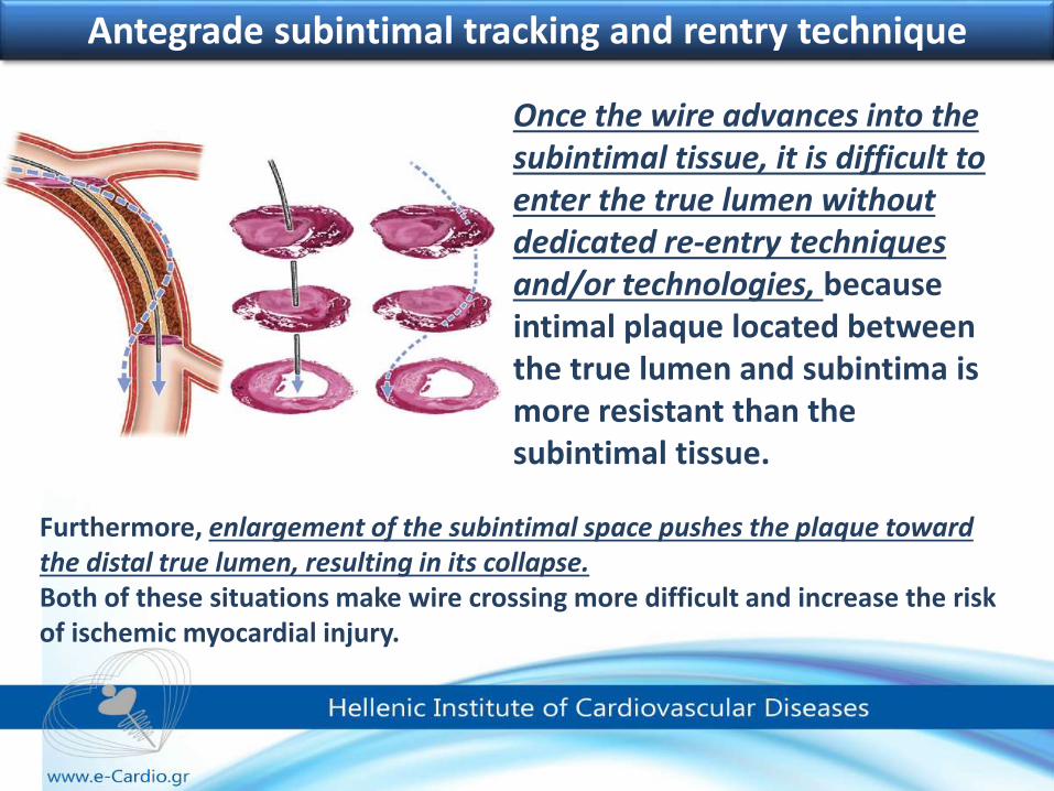
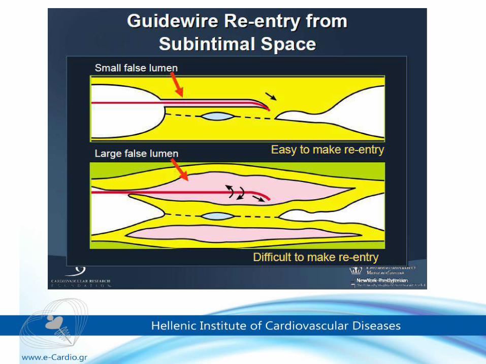
Antegrade subintimal tracking and rentry technique
Once the wire advances into the subintimal tissue, it is difficult to enter the true lumen without dedicated re-entry techniques and/or technologies, because intimal plaque located between the true lumen and subintima is more resistant than the subintimal tissue.
Furthermore, enlargement of the subintimal space pushes the plaque toward the distal true lumen, resulting in its collapse. Both of these situations make wire crossing more difficult and increase the risk of ischemic myocardial injury.
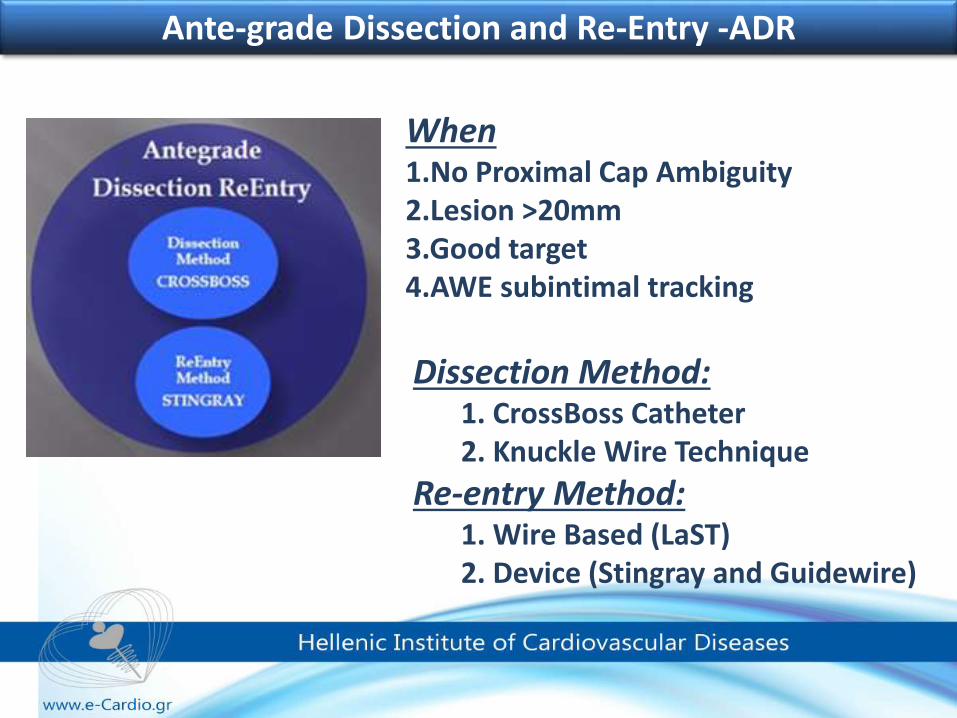
When1.No Proximal Cap Ambiguity 2.Lesion >20mm 3.Good target 4.AWE subintimal tracking
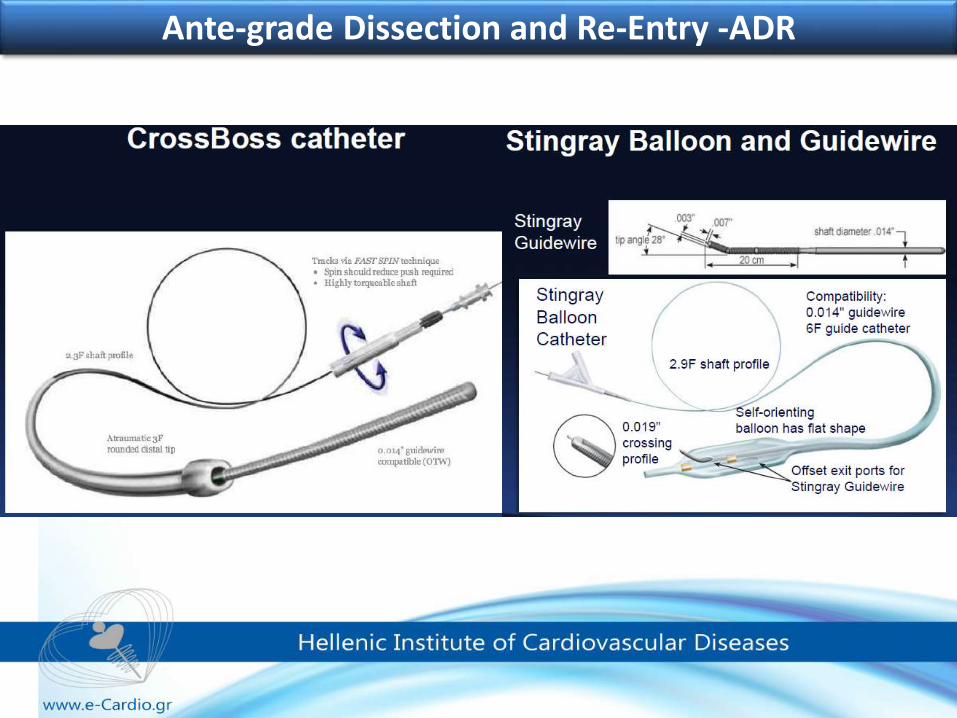
Dissection Method: 1. CrossBoss Catheter 2. Knuckle Wire Technique
Re-entry Method: 1. Wire Based (LaST) 2. Device (Stingray and Guidewire)
Ante-grade Dissection and Re-Entry -ADR
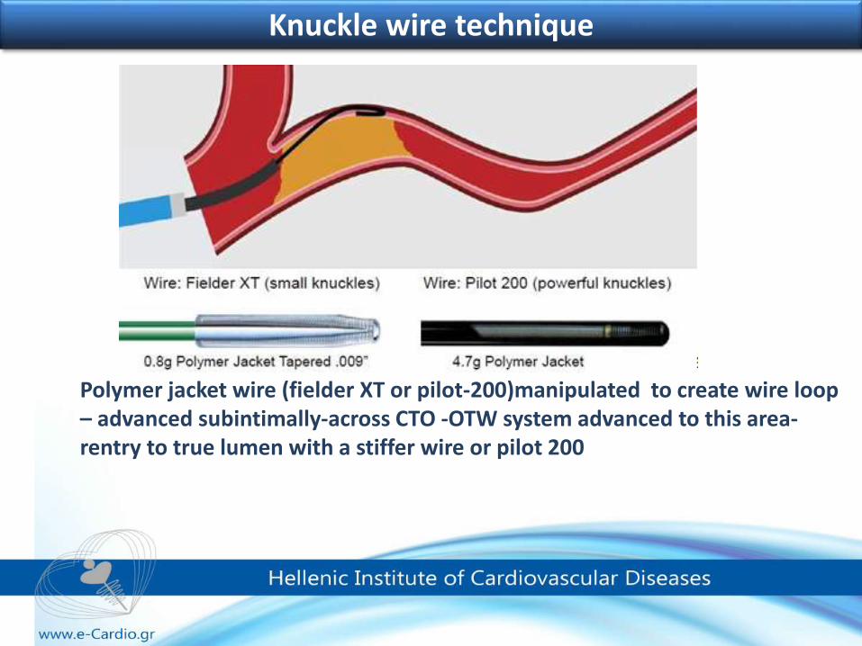
Knuckle wire technique
Polymer jacket wire (fielder XT or pilot-200)manipulated to create wire loop – advanced subintimally-across CTO -OTW system advanced to this area-rentry to true lumen with a stiffer wire or pilot 200
Ante-grade Dissection and Re-Entry -ADR
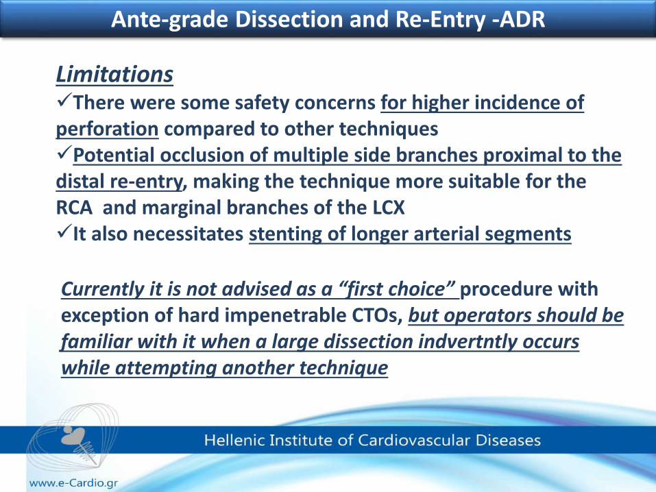
LimitationsThere were some safety concerns for higher incidence of perforation compared to other techniquesPotential occlusion of multiple side branches proximal to the distal re-entry, making the technique more suitable for the RCA and marginal branches of the LCXIt also necessitates stenting of longer arterial segments
Ante-grade Dissection and Re-Entry -ADR
Currently it is not advised as a “first choice” procedure with exception of hard impenetrable CTOs, but operators should be familiar with it when a large dissection indvertntly occurs while attempting another technique
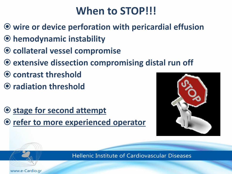
When to STOP!!!
wire or device perforation with pericardial effusion
hemodynamic instability
collateral vessel compromise
extensive dissection compromising distal run off
contrast threshold
radiation threshold
stage for second attempt
refer to more experienced operator
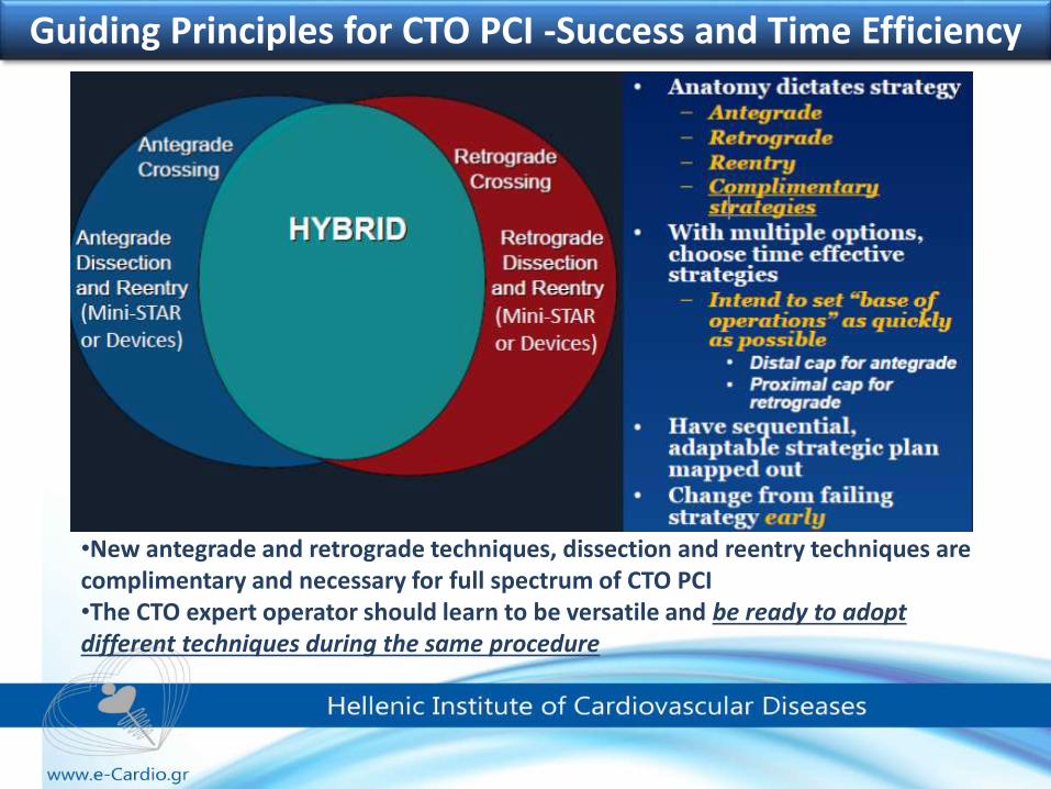
Guiding Principles for CTO PCI -Success and Time Efficiency
•New antegrade and retrograde techniques, dissection and reentry techniques are complimentary and necessary for full spectrum of CTO PCI •The CTO expert operator should learn to be versatile and be ready to adopt different techniques during the same procedure