EFA6 proteins regulate lumen formation through α-actinin 1 · 2018-02-07 · His–EFA6A (Fig....
15
RESEARCH ARTICLE EFA6 proteins regulate lumen formation through α-actinin 1 Julie Milanini 1 , Racha Fayad 1 , Mariagrazia Partisani 1 , Patrick Lecine 2, *, Jean-Paul Borg 2 , Michel Franco 1 and Fre ́ de ́ ric Luton 1, ‡ ABSTRACT A key step of epithelial morphogenesis is the creation of the lumen. Luminogenesis by hollowing proceeds through the fusion of apical vesicles at cell–cell contacts. The small nascent lumens grow through extension, coalescence and enlargement, coordinated with cell division, to give rise to a single central lumen. Here, by using MDCK cells grown in 3D-culture, we show that EFA6A (also known as PSD) participates in luminogenesis. EFA6A recruits α-actinin 1 (ACTN1) through direct binding. In polarized cells, ACTN1 was found to be enriched at the tight junction where it acts as a primary effector of EFA6A for normal luminogenesis. Both proteins are essential for the lumen extension and enlargement, where they mediate their effect by regulating the cortical acto-myosin contractility. Finally, ACTN1 was also found to act as an effector for the isoform EFA6B (also known as PSD4) in the human mammary tumoral MCF7 cell line. EFA6B restored the glandular morphology of this tumoral cell line in an ACTN1-dependent manner. Thus, we identified new regulators of cyst luminogenesis essential for the proper maturation of a newly- formed lumen into a single central lumen. KEY WORDS: Epithelium, Lumen, EFA6, ACTN1, Contractility INTRODUCTION During organogenesis, the coordinated establishment of the apico- basal polarity with the de novo formation of an apical luminal space is fundamental to the emergence of the different types of epithelia. In adult organisms, the aptitude of the internal organs, which are lined with epithelial tissues, to ensure specific functions relies on the preservation of these characteristics. The failure in doing so is associated with a large variety of diseases (Blasky et al., 2015; Datta et al., 2011; Sigurbjörnsdóttir et al., 2014). In particular, these features are often compromised in carcinomas and tumors are formed of non-polarized cell aggregates incapable of collectively organizing a lumen (Martin-Belmonte and Perez-Moreno, 2011; Tanos and Rodriguez-Boulan, 2008; Wang et al., 2012). However, compelling tumoral cells to maintain their normal epithelial phenotype can help them override the power of oncogenes (DuFort et al., 2011; Weaver et al., 1997). Thus, it is important to decipher the molecular programs that instruct epithelial cells to collectively organize around lumens in order to maintain their physiological homeostasis. For most epithelial tissues, the de novo formation of a lumen is generated by hollowing. In this process, apical vesicles are delivered to a focal point of the cell–cell contact named the apical membrane initiation site (AMIS) to give rise to the apical plasma membrane and a facing hollow cavity. The nascent lumen appears first as a closed elongated space named the pre-apical patch (PAP), which is limited by tight junctions. The PAP will then open and expand through a combination of events: the delivery of vesicular membranes, the repulsion of the apposed membrane by highly charged molecules, the increase of hydrostatic pressure, the coalescence of mini-lumens and, eventually, expansion through cell division. This process is closely synchronized with a profound rearrangement of the actin cytoskeleton into discrete structures essential for the attachment of structural and signaling apical proteins. These proteins will then yield a scaffold to shape the lumen and form an acto-myosin ring in support of the circumferential apical junctional complexes (AJCs), which are made of adherens junctions (AJs) and the apical tight junctions (TJs) that outline the luminal space (Datta et al., 2011; Sigurbjörnsdóttir et al., 2014). The organization and functions of the acto-myosin ring attached to the AJ have been extensively studied (Arnold et al., 2017; Braga, 2016; Grikscheit and Grosse, 2016; Lecuit and Yap, 2015); however, far less is known about the actin cytoskeleton associated to the TJ. Nevertheless, it is likely that both structures are somehow intermingled within the so-called apical perijunctional acto- myosin ring (PAMR) (Ebrahim et al., 2013; Sluysmans et al., 2017). The PAMR is described as a sarcomeric-like belt made of F- actin bundles containing myosin-II, which confers contractile properties, and bundling proteins, such as the non-muscle α- actinins, which stiffen the structure. The balance of both activities is believed to determine the flexibility of this belt, its mechanosensitivity and the tension forces exerted on the cell surface (Foley and Young, 2014; Martin and Goldstein, 2014; Murrell et al., 2015; Röper, 2015). The existence of a central apical acto-myosin network with radial contractility has also been reported (Coravos and Martin, 2016). The α-actinin (ACTN) family comprises four members, the muscle ACTN2 and ACTN3, and the non-muscle ACTN1 and ACTN4, which are expressed in most other cell types. They share a common primary structure with a N-terminal actin-binding domain (ABD) and a C-terminal calmodulin-like domain (CAMD) separated by a central repeat of four spectrin-like domains (spectrin repeats domain; SRD). ACTN molecules form antiparallel dimers through their rigid SRD allowing for the cross- linking of actin filaments by the ABD positioned on either end. In comparison with the filamin proteins, which orthogonally cross- link actin filaments, non-muscle ACTNs form linear F-actin bundles, which increase the stiffness (Jahed et al., 2014; Stossel et al., 2001). They are believed to contribute to myosin-II-driven contractility by facilitating force transmission (Le Clainche and Received 3 August 2017; Accepted 11 December 2017 1 Université Co ̂ te d’Azur, CNRS, Institut de Pharmacologie Molé culaire et Cellulaire (IPMC), Valbonne, F-06560, France. 2 Centre de Recherche en Cancé rologie de Marseille (CRCM), ‘Cell Polarity, Cell Signalling and Cancer’, Equipe Labellisé e Ligue Contre le Cancer, Inserm U1068, Marseille, F-13009, France; CNRS, UMR7258, Marseille, F-13009, France; Institut Paoli-Calmettes, Marseille, F-13009, France; Aix-Marseille University, UM105, Marseille, F-13284, France. *Present address: BIOASTER, Lyon, F-69007, France. ‡ Author for correspondence ([email protected]) J.M., 0000-0002-5861-2830; J.-P.B., 0000-0001-8418-3382; M.F., 0000-0003- 1853-8661; F.L., 0000-0001-6868-4654 1 © 2018. Published by The Company of Biologists Ltd | Journal of Cell Science (2018) 131, jcs209361. doi:10.1242/jcs.209361 Journal of Cell Science
Transcript of EFA6 proteins regulate lumen formation through α-actinin 1 · 2018-02-07 · His–EFA6A (Fig....

RESEARCH ARTICLE
EFA6 proteins regulate lumen formation through α-actinin 1Julie Milanini1, Racha Fayad1, Mariagrazia Partisani1, Patrick Lecine2,*, Jean-Paul Borg2, Michel Franco1 andFrederic Luton1,‡
ABSTRACTA key step of epithelial morphogenesis is the creation of the lumen.Luminogenesis by hollowing proceeds through the fusion of apicalvesicles at cell–cell contacts. The small nascent lumens grow throughextension, coalescence and enlargement, coordinated with celldivision, to give rise to a single central lumen. Here, by usingMDCK cells grown in 3D-culture, we show that EFA6A (also known asPSD) participates in luminogenesis. EFA6A recruits α-actinin 1(ACTN1) through direct binding. In polarized cells, ACTN1 was foundto be enriched at the tight junction where it acts as a primary effector ofEFA6A for normal luminogenesis. Both proteins are essential for thelumen extension and enlargement, where they mediate their effect byregulating the cortical acto-myosin contractility. Finally, ACTN1 wasalso found to act as an effector for the isoform EFA6B (also known asPSD4) in the human mammary tumoral MCF7 cell line. EFA6Brestored the glandular morphology of this tumoral cell line in anACTN1-dependent manner. Thus, we identified new regulators ofcyst luminogenesis essential for the proper maturation of a newly-formed lumen into a single central lumen.
KEY WORDS: Epithelium, Lumen, EFA6, ACTN1, Contractility
INTRODUCTIONDuring organogenesis, the coordinated establishment of the apico-basal polarity with the de novo formation of an apical luminal spaceis fundamental to the emergence of the different types of epithelia.In adult organisms, the aptitude of the internal organs, which arelined with epithelial tissues, to ensure specific functions relies on thepreservation of these characteristics. The failure in doing so isassociated with a large variety of diseases (Blasky et al., 2015; Dattaet al., 2011; Sigurbjörnsdóttir et al., 2014). In particular, thesefeatures are often compromised in carcinomas and tumors areformed of non-polarized cell aggregates incapable of collectivelyorganizing a lumen (Martin-Belmonte and Perez-Moreno, 2011;Tanos and Rodriguez-Boulan, 2008; Wang et al., 2012). However,compelling tumoral cells to maintain their normal epithelialphenotype can help them override the power of oncogenes(DuFort et al., 2011; Weaver et al., 1997). Thus, it is importantto decipher the molecular programs that instruct epithelial cells to
collectively organize around lumens in order to maintain theirphysiological homeostasis.
For most epithelial tissues, the de novo formation of a lumen isgenerated by hollowing. In this process, apical vesicles are deliveredto a focal point of the cell–cell contact named the apical membraneinitiation site (AMIS) to give rise to the apical plasma membraneand a facing hollow cavity. The nascent lumen appears first as aclosed elongated space named the pre-apical patch (PAP), which islimited by tight junctions. The PAP will then open and expandthrough a combination of events: the delivery of vesicularmembranes, the repulsion of the apposed membrane by highlycharged molecules, the increase of hydrostatic pressure, thecoalescence of mini-lumens and, eventually, expansion throughcell division. This process is closely synchronized with a profoundrearrangement of the actin cytoskeleton into discrete structuresessential for the attachment of structural and signaling apicalproteins. These proteins will then yield a scaffold to shape the lumenand form an acto-myosin ring in support of the circumferentialapical junctional complexes (AJCs), which are made of adherensjunctions (AJs) and the apical tight junctions (TJs) that outline theluminal space (Datta et al., 2011; Sigurbjörnsdóttir et al., 2014). Theorganization and functions of the acto-myosin ring attached to theAJ have been extensively studied (Arnold et al., 2017; Braga, 2016;Grikscheit and Grosse, 2016; Lecuit and Yap, 2015); however, farless is known about the actin cytoskeleton associated to the TJ.Nevertheless, it is likely that both structures are somehowintermingled within the so-called apical perijunctional acto-myosin ring (PAMR) (Ebrahim et al., 2013; Sluysmans et al.,2017). The PAMR is described as a sarcomeric-like belt made of F-actin bundles containing myosin-II, which confers contractileproperties, and bundling proteins, such as the non-muscle α-actinins, which stiffen the structure. The balance of both activities isbelieved to determine the flexibility of this belt, itsmechanosensitivity and the tension forces exerted on the cellsurface (Foley and Young, 2014; Martin and Goldstein, 2014;Murrell et al., 2015; Röper, 2015). The existence of a central apicalacto-myosin network with radial contractility has also been reported(Coravos and Martin, 2016).
The α-actinin (ACTN) family comprises four members, themuscle ACTN2 and ACTN3, and the non-muscle ACTN1 andACTN4, which are expressed in most other cell types. They share acommon primary structure with a N-terminal actin-binding domain(ABD) and a C-terminal calmodulin-like domain (CAMD)separated by a central repeat of four spectrin-like domains(spectrin repeats domain; SRD). ACTN molecules formantiparallel dimers through their rigid SRD allowing for the cross-linking of actin filaments by the ABD positioned on either end. Incomparison with the filamin proteins, which orthogonally cross-link actin filaments, non-muscle ACTNs form linear F-actinbundles, which increase the stiffness (Jahed et al., 2014; Stosselet al., 2001). They are believed to contribute to myosin-II-drivencontractility by facilitating force transmission (Le Clainche andReceived 3 August 2017; Accepted 11 December 2017
1Universite Cote d’Azur, CNRS, Institut de Pharmacologie Moleculaire et Cellulaire(IPMC), Valbonne, F-06560, France. 2Centre de Recherche en Cancerologie deMarseille (CRCM), ‘Cell Polarity, Cell Signalling and Cancer’, Equipe LabelliseeLigue Contre le Cancer, Inserm U1068, Marseille, F-13009, France; CNRS,UMR7258, Marseille, F-13009, France; Institut Paoli-Calmettes, Marseille, F-13009,France; Aix-Marseille University, UM105, Marseille, F-13284, France.*Present address: BIOASTER, Lyon, F-69007, France.
‡Author for correspondence ([email protected])
J.M., 0000-0002-5861-2830; J.-P.B., 0000-0001-8418-3382; M.F., 0000-0003-1853-8661; F.L., 0000-0001-6868-4654
1
© 2018. Published by The Company of Biologists Ltd | Journal of Cell Science (2018) 131, jcs209361. doi:10.1242/jcs.209361
Journal
ofCe
llScience

Carlier, 2008). ACTNs also act as a mechanical linker between actinfilaments and cell–cell and cell–extracellular matrix (ECM)adhesion complexes. Besides its structural roles, ACTNs couldalso serve to couple actin nucleation to assembly at cell–cellcontacts (Tang and Brieher, 2012) and contribute to the maturationof cell–ECM focal adhesion by transmitting mechanical forces(Iskratsch et al., 2014; Jahed et al., 2014; Parsons et al., 2010; Yeet al., 2014). Both ACTN1 and ACTN4 were found at the apicalacto-myosin ring in association with the AJ (Honda et al., 1998;Tang and Brieher, 2012), whereas a few studies suggested a linkwith the TJ (Chen et al., 2006; Geiger et al., 1979; Nakatsuji et al.,2008). Nevertheless, the repertoire of ACTN molecules associatedwith the various F-actin structures is poorly defined and is madeeven more complex by the existence of ACTN1–ACTN4heterodimers (Foley and Young, 2013). Moreover, it is notcompletely clear how ACTNs are recruited to cell–cell contacts.Thus, much remains to be discovered about the role of ACTNs at theAJC as well as their role during luminogenesis.Small G-proteins of the Rho family and their partners are key
regulators in the assembly and maintenance of the PAMR. Inparticular, RhoA contributes to the constitution of the contractileapical acto-myosin array through the localization and activation ofthe formin proteins and the activation of the motor protein myosin-IIby the Rho-associated coiled-coil kinase effectors ROCK1 andROCK2 (hereafter denoted ROCK) (Arnold et al., 2017; Quiros andNusrat, 2014; Sluysmans et al., 2017; Takeichi, 2014). Tensionforces are necessary for the establishment and functioning of theAJC, as they support the changes in cell shape occurring duringepithelial morphogenesis (Coravos et al., 2017; Lecuit and Yap,2015; Takeichi, 2014). However, how contractility and its regulatorsare impacting epithelial cell luminogenesis is an issue of ongoingdebate.Another small G-protein that regulates the cortical cytoskeleton is
the ADP ribosylation factor 6 (Arf6). It plays a pivotal role in a widevariety of cellular events including cell surface trafficking,phagocytosis, cell–cell adhesion, and tumor cell migration andinvasion (D’Souza-Schorey and Chavrier, 2006; Gillingham andMunro, 2007; Jaworski, 2007; Sabe et al., 2009; Schweitzer et al.,2011). In epithelial cells, Arf6 was initially shown to regulatevesicular trafficking to the apical pole of the cell (Altschuler et al.,1999) and later to impact the turnover of the AJ (Palacios et al.,2001, 2002, 2005), the establishment of the TJ (Klein et al., 2008;Luton et al., 2004), cyst morphogenesis (Tushir et al., 2010) andHGF-induced tubulogenesis (Tushir and D’Souza-Schorey, 2007).Consistent with these observations, the exchange factor for Arf6(EFA6; also known as PSD) was found to be enriched at the apicalpole and at the TJ in fully polarized MDCK cells (Luton et al.,2004). During early epithelial polarization, EFA6 is recruited to thecell–cell contact in a manner that is dependent on E-cadherinengagement, where it contributes to the formation of the TJ bystabilizing the apical acto-myosin ring (Théard et al., 2010).Expression of EFA6B (also known as PSD4) in the mammarytumoral MCF7 cell line restored a normal glandular phenotype, withthe formation of lumens delineated by TJs (Zangari et al., 2014).Conversely, knockdown of EFA6B expression drives variousmammary cell lines into epithelial-to-mesenchymal transition(Zangari et al., 2014). In breast cancer patients, the loss ofEF6AB expression is associated with the claudin-low subtypecharacterized by the loss of expression of all the TJ components anda poor prognosis (Zangari et al., 2014).The EFA6 family consists of four isoforms (EFA6A–EFA6D;
EFA6C is also known as PSD2, and EFA6D as PSD3) sharing a
general structure that comprises a variable N-terminal domain, acatalytic Sec7 domain bearing the nucleotide exchange activity, aPH domain responsible for their plasma membrane localization anda conserved C-terminal region involved in actin cytoskeletonrearrangement (Derrien et al., 2002; Franco et al., 1999; Sakagami,2008; Sironi et al., 2009). In a previous study, we found thatmutations that abolish the nucleotide exchange activity or delete theC-terminal domain abrogated the stimulatory effects of EFA6,indicating that both Arf6 activation and the C-terminal domainare necessary for epithelial polarization (Luton et al., 2004).In addition, complementation experiments demonstrated a finelytuned cooperation between the two signaling pathways associatedwith the activated Arf6 and with the EFA6 C-terminus (Klein et al.,2008).
In this study, we aimed to determine the signaling pathwayassociated to EFA6 C-terminus that contributes to its action onepithelial morphogenesis. Our data showed that: (1) ACTN1 is adirect partner of EFA6AC-terminal domain, (2) EFA6A is a crucialregulator of luminogenesis for which ACTN1 is the major effector,(3) together, they stimulate the formation and enlargement of asingle lumen with a proper round shape, (4) they act by regulatingcortical acto-myosin contractility, and finally (5) ACTN1 is also apartner of the EFA6B isoform in the promotion of lumen formationin the mammary tumoral cells MCF7.
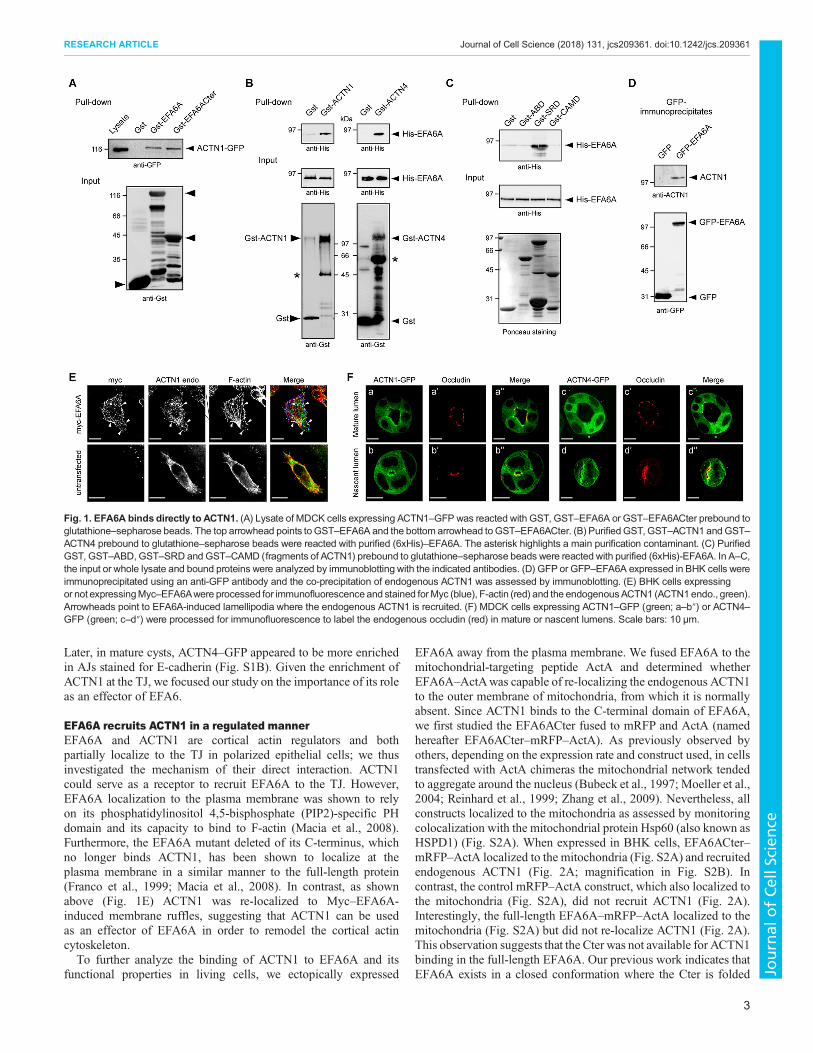
RESULTSEFA6A binds directly to ACTN1Looking for functional partners of EFA6A in epithelial cells, weperformed a two-hybrid screen of an epithelial library using the C-terminus (Cter) of EFA6A as a bait, and identified ACTN1 as themajor interacting protein. A similar result was found by Sakagamiet al. in a previous screen using a neuronal library (Sakagami et al.,2007). A pulldown assay was used to confirm that the Cter ofEFA6A could bind exogenously expressed ACTN1–GFP (Fig. 1A).We then further characterized this interaction by assessing whetherthe two proteins could bind directly. We found that bothGST–ACTN1 and GST–ACTN4 could pulldown full-lengthHis–EFA6A (Fig. 1B), and that the central SRD of ACTN1 bindsdirectly to His–EFA6A (Fig. 1C). In BHK cells, GFP–EFA6Aco-immunoprecipitated endogenous ACTN1 (Fig. 1D), and Myc–EFA6A re-localized the endogenous ACTN1 to F-actin-enrichedlamellipodia as well as to the cell surface microspikes induced byMyc–EFA6A (Fig. 1E; Derrien et al., 2002; Macia et al., 2008). Weconclude that, in vitro, ACTN and EFA6A can bind directly throughtheir respective spectrin and C-terminal domains, and that, in vivo,EFA6A can recruit the endogenous ACTN1 to cortical F-actinstructures. In agreement with our previous reports (Luton et al.,2004; Théard et al., 2010; Zangari et al., 2014), during lumenformation mRFP–EFA6A was transiently found to be enriched atthe AMIS and the opened PAP before its expression at the apicalsurface decreased to the low level found in mature cyst (Fig. S1A).We also examined the localization of ACTN1 and ACTN4in polarized cysts formed by MDCK cells. ACTN1–GFP wasdiffused within the cytoplasm and enriched at the apex of cell-celljunctions (Fig. S1A). Colocalization of ACTN1–GFP with occludin(Fig. 1Fa–a″) indicated its accumulation at the TJ. In contrast,ACTN4–GFP was not consistently observed at the TJ at above thelevel of the GFP control (Fig. S1A; Fig. 1Fc–c″). In addition,ACTN1–GFPwas found to be enriched at the nascent lumen formedin between two cells and its surrounding TJ (Fig. 1Fb–b″), whereasACTN4–GFP was found on the newly formed luminal membraneand also was enriched along the cell–cell contact (Fig. 1Fd–d″).
2
RESEARCH ARTICLE Journal of Cell Science (2018) 131, jcs209361. doi:10.1242/jcs.209361
Journal
ofCe
llScience
Later, in mature cysts, ACTN4–GFP appeared to be more enrichedin AJs stained for E-cadherin (Fig. S1B). Given the enrichment ofACTN1 at the TJ, we focused our study on the importance of its roleas an effector of EFA6.
EFA6A recruits ACTN1 in a regulated mannerEFA6A and ACTN1 are cortical actin regulators and bothpartially localize to the TJ in polarized epithelial cells; we thusinvestigated the mechanism of their direct interaction. ACTN1could serve as a receptor to recruit EFA6A to the TJ. However,EFA6A localization to the plasma membrane was shown to relyon its phosphatidylinositol 4,5-bisphosphate (PIP2)-specific PHdomain and its capacity to bind to F-actin (Macia et al., 2008).Furthermore, the EFA6A mutant deleted of its C-terminus, whichno longer binds ACTN1, has been shown to localize at theplasma membrane in a similar manner to the full-length protein(Franco et al., 1999; Macia et al., 2008). In contrast, as shownabove (Fig. 1E) ACTN1 was re-localized to Myc–EFA6A-induced membrane ruffles, suggesting that ACTN1 can be usedas an effector of EFA6A in order to remodel the cortical actincytoskeleton.To further analyze the binding of ACTN1 to EFA6A and its
functional properties in living cells, we ectopically expressed
EFA6A away from the plasma membrane. We fused EFA6A to themitochondrial-targeting peptide ActA and determined whetherEFA6A–ActAwas capable of re-localizing the endogenous ACTN1to the outer membrane of mitochondria, from which it is normallyabsent. Since ACTN1 binds to the C-terminal domain of EFA6A,we first studied the EFA6ACter fused to mRFP and ActA (namedhereafter EFA6ACter–mRFP–ActA). As previously observed byothers, depending on the expression rate and construct used, in cellstransfected with ActA chimeras the mitochondrial network tendedto aggregate around the nucleus (Bubeck et al., 1997; Moeller et al.,2004; Reinhard et al., 1999; Zhang et al., 2009). Nevertheless, allconstructs localized to the mitochondria as assessed by monitoringcolocalization with the mitochondrial protein Hsp60 (also known asHSPD1) (Fig. S2A). When expressed in BHK cells, EFA6ACter–mRFP–ActA localized to the mitochondria (Fig. S2A) and recruitedendogenous ACTN1 (Fig. 2A; magnification in Fig. S2B). Incontrast, the control mRFP–ActA construct, which also localized tothe mitochondria (Fig. S2A), did not recruit ACTN1 (Fig. 2A).Interestingly, the full-length EFA6A–mRFP–ActA localized to themitochondria (Fig. S2A) but did not re-localize ACTN1 (Fig. 2A).This observation suggests that the Cter was not available for ACTN1binding in the full-length EFA6A. Our previous work indicates thatEFA6A exists in a closed conformation where the Cter is folded
Fig. 1. EFA6A binds directly to ACTN1. (A) Lysate of MDCK cells expressing ACTN1–GFP was reacted with GST, GST–EFA6A or GST–EFA6ACter prebound toglutathione–sepharose beads. The top arrowhead points to GST–EFA6A and the bottom arrowhead toGST–EFA6ACter. (B) PurifiedGST, GST–ACTN1 andGST–ACTN4 prebound to glutathione–sepharose beads were reacted with purified (6xHis)–EFA6A. The asterisk highlights a main purification contaminant. (C) PurifiedGST, GST–ABD, GST–SRD and GST–CAMD (fragments of ACTN1) prebound to glutathione–sepharose beads were reacted with purified (6xHis)-EFA6A. In A–C,the input or whole lysate and bound proteins were analyzed by immunoblotting with the indicated antibodies. (D) GFP or GFP–EFA6A expressed in BHK cells wereimmunoprecipitated using an anti-GFP antibody and the co-precipitation of endogenous ACTN1 was assessed by immunoblotting. (E) BHK cells expressingor not expressingMyc–EFA6Awere processed for immunofluorescence and stained forMyc (blue), F-actin (red) and the endogenousACTN1 (ACTN1endo., green).Arrowheads point to EFA6A-induced lamellipodia where the endogenous ACTN1 is recruited. (F) MDCK cells expressing ACTN1–GFP (green; a–b″) or ACTN4–GFP (green; c–d″) were processed for immunofluorescence to label the endogenous occludin (red) in mature or nascent lumens. Scale bars: 10 µm.
3
RESEARCH ARTICLE Journal of Cell Science (2018) 131, jcs209361. doi:10.1242/jcs.209361
Journal
ofCe
llScience

back onto the PH domain, preventing its association with theC-terminus of β-arrestin1 (denoted β-arrestin1Cter), anotherEFA6ACter ligand (Macia et al., 2012). To test whether theEFA6A–mRFP–ActA was in a locked conformation, we co-expressed β-arrestin1Cter–GFP with EFA6ACter–mRFP–ActA orthe full-length EFA6A–mRFP–ActA constructs. Similar to whatwas observed with ACTN1, β-arrestin1Cter–GFP could only bind toEFA6ACter–mRFP–ActA (Fig. 2B) indicating that the binding of
ACTN1 to EFA6A is regulated, and requires the release of itsC-terminal domain.
Some F-actin staining was observed colocalized together withEFA6ACter–mRFP–ActA and the endogenous ACTN1 at themitochondria (Fig. 2A; magnification in Fig. S2B). When the cellswere treated with Latrunculin A, F-actin was absent from themitochondria whereas the endogenous ACTN1 was still efficientlyrecruited by EFA6Cter–ActA (Fig. 2C). Thus, independently of the
Fig. 2. EFA6A recruits ACTN1 in a regulated manner. (A) BHK cells expressing EFA6ACter–mRFP–ActA, mRFP–ActA and EFA6A–mRFP–ActA wereprocessed for immunofluorescence to label the endogenous ACTN1 and F-actin. In merge images, mRFP is colored red, ACTN1 blue and F-actin green. (B) BHKcells co-expressing β-arrestin1Cter–GFP (green) with EFA6ACter–mRFP–ActA (red) or with the full-length EFA6A–mRFP–ActA (red) were processed forimmunofluorescence to label the endogenous F-actin (blue). (C) BHK cells expressing EFA6ACter–mRFP–ActA were exposed to Latrunculin A (2 µM) for 2 h.The cells were processed for immunofluorescence to label the endogenous ACTN1 (green) and F-actin (blue). Scale bars: 10 µm.
4
RESEARCH ARTICLE Journal of Cell Science (2018) 131, jcs209361. doi:10.1242/jcs.209361
Journal
ofCe
llScience
presence of F-actin, EFA6A can directly recruit ACTN1, which inturn might function as an effector to organize EFA6A-regulatedactin-based structures.
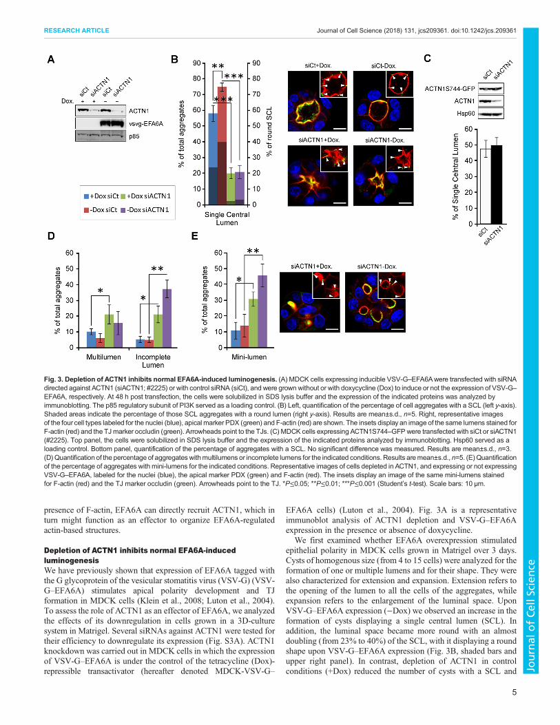
Depletion of ACTN1 inhibits normal EFA6A-inducedluminogenesisWe have previously shown that expression of EFA6A tagged withthe G glycoprotein of the vesicular stomatitis virus (VSV-G) (VSV-G–EFA6A) stimulates apical polarity development and TJformation in MDCK cells (Klein et al., 2008; Luton et al., 2004).To assess the role of ACTN1 as an effector of EFA6A, we analyzedthe effects of its downregulation in cells grown in a 3D-culturesystem in Matrigel. Several siRNAs against ACTN1 were tested fortheir efficiency to downregulate its expression (Fig. S3A). ACTN1knockdown was carried out in MDCK cells in which the expressionof VSV-G–EFA6A is under the control of the tetracycline (Dox)-repressible transactivator (hereafter denoted MDCK-VSV-G–
EFA6A cells) (Luton et al., 2004). Fig. 3A is a representativeimmunoblot analysis of ACTN1 depletion and VSV-G–EFA6Aexpression in the presence or absence of doxycycline.
We first examined whether EFA6A overexpression stimulatedepithelial polarity in MDCK cells grown in Matrigel over 3 days.Cysts of homogenous size (from 4 to 15 cells) were analyzed for theformation of one or multiple lumens and for their shape. They werealso characterized for extension and expansion. Extension refers tothe opening of the lumen to all the cells of the aggregates, whileexpansion refers to the enlargement of the luminal space. UponVSV-G–EFA6A expression (−Dox) we observed an increase in theformation of cysts displaying a single central lumen (SCL). Inaddition, the luminal space became more round with an almostdoubling (from 23% to 40%) of the SCL, with it displaying a roundshape upon VSV-G–EFA6A expression (Fig. 3B, shaded bars andupper right panel). In contrast, depletion of ACTN1 in controlconditions (+Dox) reduced the number of cysts with a SCL and
Fig. 3. Depletion of ACTN1 inhibits normal EFA6A-induced luminogenesis. (A) MDCK cells expressing inducible VSV-G–EFA6Awere transfected with siRNAdirected against ACTN1 (siACTN1; #2225) or with control siRNA (siCt), and were grown without or with doxycycline (Dox) to induce or not the expression of VSV-G–EFA6A, respectively. At 48 h post transfection, the cells were solubilized in SDS lysis buffer and the expression of the indicated proteins was analyzed byimmunoblotting. The p85 regulatory subunit of PI3K served as a loading control. (B) Left, quantification of the percentage of cell aggregates with a SCL (left y-axis).Shaded areas indicate the percentage of those SCL aggregates with a round lumen (right y-axis). Results are mean±s.d., n=5. Right, representative imagesof the four cell types labeled for the nuclei (blue), apical marker PDX (green) and F-actin (red) are shown. The insets display an image of the same lumens stained forF-actin (red) and the TJmarker occludin (green). Arrowheads point to the TJs. (C) MDCK cells expressing ACTN1S744–GFPwere transfected with siCt or siACTN1(#2225). Top panel, the cells were solubilized in SDS lysis buffer and the expression of the indicated proteins analyzed by immunoblotting. Hsp60 served as aloading control. Bottom panel, quantification of the percentage of aggregates with a SCL. No significant difference was measured. Results are mean±s.d., n=3.(D)Quantification of the percentage of aggregateswithmultilumens or incomplete lumens for the indicated conditions. Results aremean±s.d., n=5. (E)Quantificationof the percentage of aggregates with mini-lumens for the indicated conditions. Representative images of cells depleted in ACTN1, and expressing or not expressingVSV-G–EFA6A, labeled for the nuclei (blue), the apical marker PDX (green) and F-actin (red). The insets display an image of the same mini-lumens stainedfor F-actin (red) and the TJ marker occludin (green). Arrowheads point to the TJ. *P≤0.05; **P≤0.01; ***P≤0.001 (Student’s t-test). Scale bars: 10 µm.
5
RESEARCH ARTICLE Journal of Cell Science (2018) 131, jcs209361. doi:10.1242/jcs.209361
Journal
ofCe
llScience

severely altered the shape of the lumen such that the lumens adoptedan ‘octopus-like’ shape with a small central opening from whichclosed or barely opened luminal extensions reached in between thecells (Fig. 3B, shaded bars and lower left panel). Depletion ofACTN1in cells expressing VSV-G–EFA6A also impaired enlargement of thelumens, although the opening was more visible (Fig. 3B, shaded barsand bottom right panel). Thus, ACTN1 is required for the stimulatoryeffects found for EFA6A on SCL formation and on the rounding ofthe luminal space of multicellular cysts. However, in all conditions,the cells remained well polarized as indicated by the correctlocalization of the apical podocalyxin (PDX; also known asPODXL) and basolateral (E-cadherin) markers (Fig. S3B), and bythe basal positioning of the nuclei, the general F-actin organizationand the apical assembly of the TJ (Fig. 3B). To confirm that thephenotypes induced by the siRNA against ACTN1 (siACTN1) weredue to ACTN1 depletion we carried out a rescue experiment. MDCKcells expressing anACTN1–GFPwhich is insensitive to the siACTN1(ACTN1S744–GFP) did not display any defect in lumen formationupon depletion of the endogenous ACTN1 (Fig. 3C).The loss of cysts with SCLs in the ACTN1-depleted cells was
counterbalanced by an increase of cell aggregates with multiplelumens and others with incomplete extension to all of the cells(Fig. 3D, see Materials and Methods). In both cases, the lumensdisplayed the distorted octopus-like shape. The extension defect wasfurther reflected by a strong increase in the number of aggregatesdisplaying multiple lumens that only opened in between two cells(hereafter called mini-lumens) that were often blocked at the PAPstage with no visible opening (Fig. 3E), which suggests that ACTN1depletion might favor the early formation of an initial lumen. VSV-G–EFA6A expression did not significantly alter the phenotypicchanges imposed upon ACTN1-depletion; however, it stimulatedthe enlargement and rounding of the mini-lumens blocked at thetwo-cell stage (Fig. 3E, right panel). Thus, as opposed towhat is seenfor mature lumens, EFA6A can stimulate the volumetric enlargementof nascent mini-lumens in an ACTN1-independent manner. Takentogether, these observations suggest that ACTN1 is dispensable forthe formation of the initial lumen in between two cells but is requiredlater on for its extension and enlargement (see Discussion section forfurther comments). In summary, depletion of ACTN1 blocked boththe effects of VSV-G–EFA6A on luminogenesis, that is, theformation of fully extended SCL and its expansion as regular roundlumen. This suggests that ACTN1 acts as an effector downstream ofEFA6A that is important for luminogenesis.
ACTN1 acts as an effector of EFA6A to promote normalluminogenesisIf EFA6A controls luminogenesis and ACTN1 acts as an effector,then EFA6A depletion should hamper luminogenesis, and whencombined with ACTN1 depletion there should have no additionaleffect. MDCK-VSV-G–EFA6A cells grown in the absence orpresence of doxycycline were submitted to siRNA against EFA6A(siEFA6A)-, siACTN1- or simultaneous siEFA6A- and siACTN1-mediated depletion. The efficient knockdown of the indicatedproteins and the induction of the expression of VSV-G–EFA6Awere analyzed by immunoblotting (Fig. 4A; Fig. S4).Depletion of the endogenous EFA6A was accompanied by a
reduction in the amount of cell aggregates with a SCLdemonstrating that EFA6A is required for luminogenesis(Fig. 4B). Expression of the exogenous human VSV-G–EFA6A,which is insensitive to the canine-specific siRNA, rescued thenormal phenotype, thus controlling for the specificity of EFA6Aknockdown (Fig. 4B). As shown in Fig. 3, ACTN1 depletion
hampered luminogenesis and blocked the VSV-G–EFA6Astimulation. However, ACTN1 depletion did not have anyadditional effect when the endogenous EFA6A was knockeddown (Fig. 4B). In addition, the exogenous expression of VSV-G–EFA6A in the double knockdown cells could not rescue the normalphenotype (Fig. 4B). In addition, we analyzed the consequences onthe lumen formation of expressing a mutant of EFA6A deleted of itsC-terminus (EFA6AΔC), which contains the ACTN1-binding site.Expression of VSV-G–EFA6AΔC impaired normal luminogenesisand caused the formation of lumens with an octopus-like shape(Fig. 4C). Although the C-terminus of EFA6A likely interacts withother molecules, it is important to note that its truncation generatedlumens with shapes that were similar to those observed in ACTN1-depleted cells. Taken together, these observations indicate thatEFA6A mediates luminogenesis in MDCK cells and that ACTN1 isa crucial effector for this process.
ACTN1 is an effector of EFA6B for luminogenesis induction inMCF7 breast cancer cellsWe have reported that the EFA6B isoform is an antagonist of breastcancer development (Zangari et al., 2014). Tumoral MCF7 cellsgrown in 3D-culture systems form compact aggregates with nolumen (Han et al., 2010; Kenny et al., 2007). The exogenousexpression of VSV-G–EFA6B in the tumoral MCF7 cells restoresan epithelial phenotype characterized by the appearance ofaggregates with extended lumens (although not often a SCL),delineated by functional TJs (Zangari et al., 2014). Thus, we askedwhether ACTN1 was required downstream of EFA6B to contributeto luminogenesis in MCF7 cells.
We first verified that ACTN1 could also bind to the EFA6Bisoform (Fig. S5). Next, we analyzed the effect of ACTN1 depletionin both control MCF7 and MCF7-VSV-G–EFA6B cells grown inMatrigel. An immunoblot analysis demonstrated the efficientknockdown of ACTN1 in both cell lines (Fig. 5A). We quantifiedthe number of aggregates with extended lumens opened to at leastfour cells, as opposed to mini-lumens opened in between only twocells. As previously reported, MCF7 control cells do not form cystswith lumens (Han et al., 2010; Kenny et al., 2007; Zangari et al.,2014), whereas exogenous expression of VSV-G–EFA6B inducedthe formation of cysts with extended lumens (Fig. 5B; Zangari et al.,2014). We observed that ACTN1 depletion blocked the formation ofextended lumens induced by EFA6B indicating that, in MCF7 cells,ACTN1 is also a crucial effector of EFA6B-induced lumenformation (Fig. 5B). In addition, although control MCF7 cellaggregates did not form extended lumens, in ∼20% of theaggregates one or several mini-lumens were observed (Fig. 5C,D).Upon ACTN1 depletion, the number of these mini-lumensincreased in both the VSV-G–EFA6B-expressing and non-expressing MCF7 cell lines. However, in VSV-G–EFA6B-expressing cells aggregates the luminal space was enlarged whileinMCF7 controls cells themini-lumenswere seen as PAPs (Fig. 5C,D).Thus, similar to what we had observed in MDCK cells, ACTN1depletion facilitates the initial lumen formation in between two cells butthen hampers both extension and enlargement.
EFA6AandACTN1control apical contractility contributing toluminogenesisThe PAMR and ventral stress fibers (SFs) are sarcomere-like actin-based structures capable of contractility that have been implicated inepithelial morphogenesis. Contractile actin bundles are formed byaligned fibers cross-linked by a periodic distribution of ACTNs thatalternates with myosin-II (Burridge andWittchen, 2013; Sluysmans
6
RESEARCH ARTICLE Journal of Cell Science (2018) 131, jcs209361. doi:10.1242/jcs.209361
Journal
ofCe
llScience

et al., 2017). RhoA and its effectors (ROCK and mDia1) have beenrecognized as major regulators of the contractile properties of theacto-myosin cytoskeletons (Arnold et al., 2017; Quiros and Nusrat,2014; Sluysmans et al., 2017; Takeichi, 2014). Thus, we tested thepossibility that EFA6A and ACTN1 regulate luminogenesis bycontributing to the PAMR contractility. First, we used an antibodydirected against the phosphorylated regulatory myosin light chain 2(pMLC, also known as MYL2) as a proxy to evaluate the tensionforces in response to modulating EFA6A and ACTN1 expression.Fig. 6B shows a representative immunoblot analysis of ACTN1depletion and VSV-G–EFA6A expression in the presence orabsence of doxycycline. In control cells, the pMLC stainingappeared as small intracellular dots, as well as larger dots located inproximity to the apical membrane. Under conditions of EFA6A orACTN1 depletion, this apical staining disappeared and the pMLC
distribution became more diffuse. Upon VSV-G–EFA6Aexpression the proportion of apical pMLC was increased and itsstaining was evenly distributed all around the luminal cortex. InACTN1-depleted cells, the VSV-G–EFA6A-induced apicalaccumulation of pMLC was abrogated (Fig. 6A). These resultssuggest that EFA6A can modulate the apical tension forces in anACTN1-dependent manner.
We investigated the contribution of ACTN1 by first analyzing theeffect of its depletion on the RhoA-stimulated ventral SFs inMDCKcells by expressing the N-terminal Myc-tagged constitutively activemutant of RhoA (Myc–RhoAV14) under the control of theinducible Tet-off system. The levels of expression of the proteinsin different conditions were analyzed by immunoblotting (Fig. 6C).We monitored the Myc–RhoAV14-induced SF contractility byimmunofluorescence. We looked at the F-actin organization and the
Fig. 4. ACTN1 acts as an effector of EFA6A to promote normal luminogenesis. (A) MDCK cells expressing inducible VSV-G–EFA6Awere transfected with siRNAcontrol (siCt), siRNA directed against ACTN1 (siACTN1; #2225) or EFA6A (siEFA6A; #2661), or both siACTN1 and siEFA6A, and then grown without or withdoxycycline (Dox) to induce or not the expression of VSV-G–EFA6A, respectively. At 48 h post transfection the cells were solubilized in SDS lysis buffer and theexpression of the indicated proteins analyzed by immunoblotting. Hsp60 served as a loading control. (B) Quantification of the percentage of cell aggregates with a SCL.Results aremean±s.d., n=4. (C)MDCKcells expressing inducibleVSV-G–EFA6AΔCwere grownwith orwithout Dox. Top panel, at 48 h post transfection the cellsweresolubilized in SDS lysis buffer and the expression of VSV-G–EFA6AΔC analyzed by immunoblotting. Hsp60 served as a loading control. Middle panel, representativeimages of cell aggregates grown with or without Dox labeled for the nuclei (blue), apical marker PDX (green) and F-actin (red). Bottom panel. quantification of thepercentage of cell aggregates with a SCL. Results are mean±s.d., n=3. *P≤0.05; **P≤0.01; ***P≤0.001; N.S., not significant (Student’s t-test). Scale bars: 10 µm.
7
RESEARCH ARTICLE Journal of Cell Science (2018) 131, jcs209361. doi:10.1242/jcs.209361
Journal
ofCe
llScience

contractility status by following the distribution of pMLC andpaxillin, a marker of the focal adhesions (Fig. 6D). As expected, theexpression of Myc–RhoAV14 (−Dox) stimulated the formation ofSFs that appeared as thick bundles of parallel F-actin going acrossthe whole cell cluster (Fig. 6Db). In Myc–RhoAV14-expressingcells, pMLC was redistributed all along the SFs (Fig. 6Db′) while incontrol cells pMLC was enriched at the periphery of the cell clusterand excluded from cell–cell contacts (Fig. 6Da′). Concomitantly,paxillin was re-localized to the periphery of the cell cluster mostlydecorating the focal adhesions at the extremity of the SF (Fig. 6Db″).As previously observed by others (Oakes et al., 2012), depletion ofACTN1 in control cells (+Dox) led to the loss of bundles orshortening of radial F-actin bundles resembling transverse arcs(Fig. 6Dc). These structures were stained for pMLC, which remainedabsent from cell–cell contacts (Fig. 6Dc′). The paxillin signalincreased but was still homogeneously distributed within each cell ofthe cluster similar to what was seen in control cells (Fig. 6Dc″).Depletion of ACTN1 blocked the effects of Myc–RhoAV14, leadingto cells displaying a phenotype that was comparable to that of thecontrol cells with respect to all three markers (Fig. 6Dd–d″). We
conclude that ACTN1 can balance the contractility status of RhoA-dependent SFs in MDCK cells.
Interfering with the RhoA–ROCK–myosin-II contractilitypathway has been shown to alter luminogenesis in MDCK cells(Ferrari et al., 2008; Kim et al., 2015; Rodríguez-Fraticelli et al.,2012). In particular, under permissive conditions, theconstitutively active RhoAV14 mutant blocks the initial step oflumen formation (Ferrari et al., 2008). We thus asked whetherACTN1 depletion could prevent the Myc–RhoAV14 inhibitoryeffect on luminogenesis. In our 3D cell culture conditions, Myc–RhoAV14 expression abrogated lumen formation. The cellsbecame round and loosely attached, forming aggregates almostexclusively without lumens and that displayed an invertedpolarity, as judged by the peripheral localization of the apicalPDX marker (Fig. 6E,F). ACTN1 depletion partially rescued theformation of lumens (Fig. 6E). We observed aggregates withmultilumens or incomplete lumen formation, essentially as mini-lumens (Fig. 6E). The cells adopted a more cuboidal shape andformed more-compact aggregates (Fig. 6F). These results suggestthat the knockdown of ACTN1 acts to reduce the strong
Fig. 5. ACTN1 is an effector of EFA6B that acts to induce luminogenesis in the MCF7 breast cancer cell line. (A) Control and VSV-G–EFA6B-expressingMCF7 cells were transfected with siRNA control (siCt) or siRNA directed against ACTN1 (siACTN1). At 48 h post transfection the cells were solubilized inSDS lysis buffer and the expression of the indicated proteins analyzed by immunoblotting. Actin served as a loading control. (B) Quantification of the percentage ofthe indicated cell aggregates with extended lumens. (C) Quantification of the percentage of the indicated cell aggregates with mini-lumens. Results in B and Care mean±s.d., n=3. *P≤0.05; ***P≤0.001 (Student’s t-test). (D) Representative images of cells depleted or not for ACTN1 and expressing or not expressingVSV-G–EFA6B. The cell aggregates were processed for immunofluorescence by labeling cytoskeletal F-actin with fluorescent phalloidin. Arrowheads point toPAPs. Scale bars: 10 µm.
8
RESEARCH ARTICLE Journal of Cell Science (2018) 131, jcs209361. doi:10.1242/jcs.209361
Journal
ofCe
llScience

Fig. 6. EFA6A and ACTN1 control apical contractility, thereby contributing to luminogenesis. (A,B) MDCK cells expressing inducible VSV-G–EFA6Aweretransfected with siRNA control (siCt), or siRNA directed against ACTN1 (siACTN1; #2225) or EFA6A (siEFA6A; #2661) and then grown without or with doxycycline(Dox) to induce or not the expression of VSV-G–EFA6A, respectively. (A) Representative images of the aggregates labeled for pMLC, F-actin and the nuclei. Toppanels, pMLC staining alone. Bottom panels show the corresponding merged images; pMLC is colored in green, F-actin in red and the nuclei in blue. Scale bars: 10µm. (B) At 48 h post transfection the cells were solubilized in SDS lysis buffer and the expression of the indicated proteins analyzed by immunoblotting. Hsp60 servedas a loading control. (C) MDCK cells expressing inducible Myc–RhoAV14 were transfected with siCt or siACTN1 (#2225) and grown with or without doxycycline. At48 h post transfection the cells were solubilized in SDS lysis buffer and the expression of the indicated proteins was analyzed by immunoblotting. Hsp60 served as aloading control. (D) MDCK cells expressing inducible Myc–RhoAV14 were transfected with siCt or siACTN1 (#2225) and grown on coverslips with or withoutdoxycycline. At 48 h post transfection the cells were processed for immunofluorescence and labeled for F-actin (a–d), pMLC (a′–d′) and the focal adhesion proteinpaxillin (a″–d″). Scale bars: 20 µm. (E) MDCK cells expressing inducible Myc-RhoAV14 were transfected with siCt or siACTN1 (#2225), and grown with or withoutdoxycycline inMatrigel.Quantification of the indicated cell aggregates forSCLs,multilumens and incomplete lumens or no lumen is reported in the bar graph.Resultsaremean±s.d.,n=3. **P≤0.01; ***P≤0.001 (Student’s t-test). (F)Representative imagesMDCKcellswith inducibleMyc–RhoAV14depletedor not for ACTN1 (#2225)and grown for 4 days inMatrigel with or without doxycycline. The cell aggregateswere processed for immunofluorescence and labeled for the nuclei (blue), the apicalmarker PDX (green) and the F-actin (red). Arrowheads point to PAPs and mini-lumens. Scale bars: 10 µm.
9
RESEARCH ARTICLE Journal of Cell Science (2018) 131, jcs209361. doi:10.1242/jcs.209361
Journal
ofCe
llScience
contractility imposed by Myc–RhoAV14, thus allowing theinitiation of lumen formation. This is in agreement withprevious studies that show that reduction of the myosin-IIcontractility stimulates the initial early step of lumen opening atthe two-cell stage (Ferrari et al., 2008; Rodríguez-Fraticelli et al.,2012). However, the extension and enlargement of the lumens,which we demonstrate to be dependent on ACTN1, were both stillcompromised upon reduction of contractility induced by ACTN1depletion. Thus, our findings support a model whereby the initialstage of the lumen formation is facilitated by the absence of ACTN1,followed by lumen maturation, which is ACTN1 dependent.Taken together, our data suggest that EFA6A and its effector
ACTN1 contribute to SCL formation through the regulation of thecell surface tension forces within the cell aggregate.
DISCUSSIONIn this study, we aimed to define the role of EFA6, a regulator ofepithelial polarization, in lumen formation by using a 3D epithelialcell culture system. Given the importance of the reorganization ofthe actin cytoskeleton during cystogenesis, we were particularlyinterested in deciphering the signaling pathway associated with theEFA6 C-terminal domain that is capable of remodeling the corticalactin independently of EFA6 GEF activity.Searching for partners of EFA6ACter, we find that EFA6A binds
directly to the non-muscular ACTN member ACTN1. Wecharacterized this interaction in living cells by using amitochondrial-targeting system, and observed that it is likelyregulated by the structural conformation of the EFA6AC-terminus.This observation confirms our previous results obtained withanother EFA6Cter ligand, β-arrestin1 (Macia et al., 2012), andimplies the existence of a common regulatory mechanism wherebybinding to EFA6ACter requires an opening signal. A possiblecandidate for mediating this signal is Arf6-GTP, the product ofEFA6A Sec7-dependent nucleotide exchange activity. ARNO (alsoknown as CYTH2), another Sec7 family guanine nucleotideexchange factor for Arf1, was shown to adopt an auto-inhibitedconformation where its short C-terminus downstream of thePH domain interferes with the catalytic Sec7 domain (DiNittoet al., 2007). Arf6-GTP binds to the PH domain of ARNO andstimulates, in synergy with plasma membrane lipids, the nucleotideexchange activity of ARNO on Arf1 (Cohen et al., 2007b; Stalderet al., 2011). We found that Arf6-GTP binds to the PH-C-terminalregion of EFA6 (Padovani et al., 2014). Thus, by analogy withARNO, one could speculate that Arf6-GTP, by binding to the PHdomain of EFA6, could release the C-terminus and allow forACTN1 binding.We had previously reported that EFA6A is present at and
regulates the TJ (Luton et al., 2004; Théard et al., 2010), where wenow find ACTN1. Our results support the hypothesis that it isEFA6A that recruits ACTN1. The re-localization experimentsperformed in the presence of Latrunculin B indicate that the bindingbetween EFA6A and ACTN1 is independent of F-actin. However,F-actin is found around the mitochondria together withEFA6ACter–ActA and ACTN1 suggesting that the EFA6A andACTN1 couple could organize actin filament structures or evencontribute to their nucleation, as proposed for ACTN4 at the AJ(Tang and Brieher, 2012). ARNO was also found to bind directly toACTN1, through its C-terminal extremity, to modulate neuriteextension, suggesting that ACTNs could be a general player in theArf-regulated actin cytoskeleton (Torii et al., 2012).Since EFA6A recruits ACTN1, we hypothesized that ACTN1
was acting as its effector. In the past, studying monolayers of
MDCK cells, we had found that overexpression of EFA6Aaccelerates the general program of epithelial polarization,including the assembly of functional TJs (Klein et al., 2008;Luton et al., 2004). Part of the contribution of EFA6A has beenattributed to its ability to stabilize the PAMR. Here, by using a 3Dcell culture system together with siRNA-mediated depletion, weshow that EFA6A is necessary for normal luminogenesis in MDCKcells. Conversely, overexpression of EFA6A increased theproportion of cysts with a SCL and stimulated their enlargement.Thus, we asked whether ACTN1 was acting as an effector totransduce some of the effects of EFA6A on luminogenesis. Indeed,we observed that depletion of ACTN1 blocked the stimulatory effectof EFA6A on both the formation and enlargement of the round SCL.In further support of this idea, depletion of ACTN1 had noadditional disruptive impact on cells knocked down for EFA6A.This addition, expression of a mutant of EFA6A deleted of itsC-terminus, which contains the ACTN1-binding site, impairednormal luminogenesis. These results demonstrate that ACTN1 is acrucial effector of EFA6A whose function is to promote normalluminogenesis.
To understand the roles of ACTN1, we analyzed in detail thedefects induced upon its depletion. In the absence of ACTN1, therewas an increase in cysts with multiple lumens, mostly as mini-lumens formed between two cells, or cysts with lumens that failed toextend to all cells. We propose that, in the absence of ACTN1, theinitial fusion event to form a PAP is facilitated; however, thesubsequent coalescence and extension of lumens to all cells ofthe aggregate is precluded. In addition, the volumetric growth ofthe lumens is impaired. In the ACTN1 knockdown cells, the mini-lumens appeared to be essentially blocked at the PAP stage and, inlarger cysts, the extended lumens and SCL adopted an octopus-likeshape rather than a nice round hollow. Interestingly, in both MDCKand MCF7 cells, in the absence of ACTN1, EFA6 overexpression iscapable of rescuing enlargement of the mini-lumens. However, atlater stages EFA6A overexpression cannot rescue the enlargementbecause ACTN1 is required as an effector downstream of EFA6.This implies that the enlargement of the luminal space relies ondifferent molecular machinery at different stages along the processof lumen formation. Thus, ACTN1 is dispensable at the initial stageto create the nascent lumens, but it is essential downstreamof EFA6A at later stages for the coalescence, extension andenlargement of the lumens.
ACTN1 is an actin-bundling protein known to regulate thecontractility and stiffness of acto-myosin. It competes with myosin-II to bind F-actin in order to maintain a complementary periodicity,which results in linear F-actin bundles with contractile properties.The ratio and distribution of these two proteins determines theoverall contractility and rigidity of F-actin cytoskeletons, includingthat of the PARM (Arnold et al., 2017; Ferrari et al., 2008;Sluysmans et al., 2017). Thus, ACTN1 could act by regulatingtension forces at the surface of the developing lumen. Severalobservations support of this hypothesis. First, EFA6A and ACTN1depletion reduced the amount of apical pMLC, whereas EFA6Aexpression stimulated its accumulation at the apical pole in anACTN1-dependent manner. Second, upon ACTN1 depletion inboth MDCK and MCF7 cells, we observed two defects: (1) astimulatory effect on the initial formation of lumens as indicated bythe rapid accumulation of multiple mini-lumens and, (2) at laterstages, a defect of maturation (coalescence and extension) that leadsto large cysts with several lumens or incomplete lumens. Theseresults are consistent with a role for ACTN1 on contractility orstiffness. Indeed, several independent studies have shown that
10
RESEARCH ARTICLE Journal of Cell Science (2018) 131, jcs209361. doi:10.1242/jcs.209361
Journal
ofCe
llScience

decreasing contractility through treatment with blebbistatin, ROCKinhibitors, or downregulation of LKB1 (also known as STK11), anupstream regulator of RhoA, stimulates the initial formation of alumen at the two-cell stage (Cohen et al., 2007a; Ferrari et al., 2008;Rodríguez-Fraticelli et al., 2012; Taniguchi et al., 2015). Lessappreciated is the impact of contractility at later stages. We foundthat similar to what is seen upon ACTN1 depletion, blebbistatinleads to large cysts with multiple lumens (our unpublished results).A similar observation was made by reducing contractility throughdepletion of LKB1, which first stimulates the initial formation oflumens but then prevents their coalescence into a SCL (Rodríguez-Fraticelli et al., 2012). Third, supplemental evidence for the role ofACTN1 on contractility came from its effects in RhoAV14-expressing cells. The small G protein RhoA, through its actionon the apical PAMR, is a general regulator of the assembly andmaintenance of the AJ (Lecuit and Yap, 2015) and TJ (Terry et al.,2010). We showed that ACTN1-depletion could counterbalancethe RhoAV14-mediated contractility. We also found thatACTN1-depletion rescued the initial formation of lumensinhibited by RhoAV14 expression. Taken together, these resultsindicate that luminogenesis is regulated by acto-myosincontractility and stiffness, which should be kept low for theinitial formation of a mini-lumen, and then be increased to allowfor their coalescence and extension to form a SCL in largermulticellular aggregates.The implementation of tension forces relies on molecular pickets,
which are usually transmembrane proteins that anchor the acto-myosin structures. In polarized epithelial cells, the AJ and TJ areanchor points for the PAMR. It is noteworthy that neither EFA6Anor ACTN1 appeared to be essential for the assembly andpositioning of the AJ and TJ, nor the establishment andmaintenance of the asymmetry along the apico-basal axis. Thus,EFA6A and its effector ACTN1 modulate the contractility byaffecting the activity and/or organization of the apical acto-myosincytoskeleton and not the anchoring junctional complexes.Not all the functions of ACTN1 appeared to be mediated through
regulating contractility. ACTN1 depletion prevented the luminalenlargement, while the inhibition of contractility by inhibitingmyosin-II or LKB1 depletion (Rodríguez-Fraticelli et al., 2012) didnot affect the luminal enlargement. Enlargement has been proposedto depend on apical membrane transport involving the delivery ofhighly charged molecules, hydrostatic pressure mediated by ionchannels and coalescence of multilumens. In ACTN1-depletedcells, the surface of the apical membrane appears large enough toaccommodate a bigger luminal volume. In addition, the highlycharged PDX protein is properly delivered and coalescence is notrelevant when considering SCLs with an octopus-like shape.However, ACTN1 could help to retain polycystins, which areimplicated in intercellular mechanotransduction (Li et al.,2005); Wilson, 2001), as well as ion channels. In fact, many ionchannels have been shown to bind or to require ACTNs in order forthem to be retained at the cell surface (Cukovic et al., 2001; Luet al., 2009; Maruoka et al., 2000; Sadeghi et al., 2002; Schnizleret al., 2009; Wyszynski et al., 1997; Ziane et al., 2010). EFA6Awas also shown to bind the ion channels TWIK1 and Kir3.4(Decressac et al., 2004; Gong et al., 2007). Furthermore, it has beenreported that ACTN4 regulates the outwards water transport in aprocess called regulatory volume decrease in response to osmoticswelling (Ando-Akatsuka et al., 2012). In summary, together withEFA6A, the recruitment of ACTN1 to the apical surface might helpstabilize ion channels regulating fluid influx and consequentlylumen enlargement.
Alternatively, ACTN1 could serve to organize a non-contractilescaffold to help support the spherical architecture of the luminalmembrane. To do so, ACTN1 could participate by bundling F-actinfilaments into rigid structures and/or could serve to link or evennucleate actin filaments at the AJ or TJ to anchor the luminal actincytoskeleton. Such a role has been proposed for ACTN4 at the levelof the AJ (Tang and Brieher, 2012).
Finally, we examined the role of ACTN1 in the human tumoralmammary cell lineMCF7.When grown in 3D culture, these cells failto assemble TJ, form a lumen or polarize (Han et al., 2010; Kennyet al., 2007; Zangari et al., 2014). Only a small fraction of cellaggregates display one or several mini-lumens. As shown in the past,EFA6B expression stimulated the formation of extended lumens andrestored an epithelial-like phenotype. So, in contrast to MDCK cells,the formation of extended lumens in MCF7 is strictly dependent onthe expression of VSV-G–EFA6B. Thus, these cells make a goodmodel to study the role of ACTN1 downstream of EFA6B. Indeed,ACTN1 depletion totally blocked lumen formation, demonstratingthat ACTN1 is a crucial effector for EFA6B in the induction ofluminogenesis. Furthermore, ACTN1-depletion in wild-type MCF7cells stimulated the formation of multiple mini-lumens that wereblocked at the PAP stage. EFA6B expression did not rescue theformation of extended lumens but did enlarge their volume. Thus, inagreement with our observations in MDCK cells, ACTN1 depletionfavors the initial formation of lumens in between two cells butprevents their coalescence and enlargement.
In conclusion, we show that ACTN1 is an effector of EFA6A andEFA6B to promote luminogenesis in normal and tumoral cellmodels. We propose a scenario whereby, at the onset ofluminogenesis, the acto-myosin contractility and rigidity must bekept low to relax the sub-membranous actin cytoskeleton wherevesicle fusion occurs at the AMIS. At this stage, EFA6 proteins havea stimulatory effect on the formation and the enlargement of themini-lumens suggesting that it acts through an effector other thanACTN1; perhaps through Arf6, which has already been shown to beinvolved in luminogensis (Tushir et al., 2010; Zangari et al., 2014).At a later stage, EFA6 proteins recruit ACTN1 to contribute to thecoalescence of the mini-lumens, their extension to neighboringcells and enlargement.We do not exclude the possibility that EFA6proteins could recruit other effectors to mediate its effects.Nevertheless, ACTN1 is certainly a primary effector that mightact by modulating the contractility of the acto-myosin ring andthereby mediating its effects on coalescence and extension (byhelping to pull the lumens together and breaking through thejunctional complexes). Given these results, the EFA6A–ACTN1pathway might be important for tubulogenesis occurring throughthe cord hollowing mechanism, where multiple mini-lumensare formed along the tubular structure and subsequently fuseto coalesce in a single lumen, in a ROCK–myosin II-dependentmanner (Bernascone et al., 2017; Kim et al., 2015;Sigurbjörnsdóttir et al., 2014).
In summary, we identified and characterized the role of two newregulators of luminogenesis: EFA6A and ACTN1. ACTN1 behavesas an effector to transduce the stimulating effect of EFA6A on theformation of a single and well-expanded central lumen byfacilitating the extension and enlargement stages. The EFA6A–ACTN1 couple acts, at least in part, by balancing the contractility ofthe cortical acto-myosin cytoskeleton. In the tumoral MCF7 cells,this pathway has the capacity to restore the apico-basal polarization,TJ assembly and collective cellular organization into a cyst with acentral lumen pointing to new directions for cancer researchand therapy.
11
RESEARCH ARTICLE Journal of Cell Science (2018) 131, jcs209361. doi:10.1242/jcs.209361
Journal
ofCe
llScience

MATERIALS AND METHODSCells, media and transfectionBHK cells were grown in Glasgow’s minimum essential medium (GMEM;Invitrogen, Paris, France) supplemented with 5% heat decomplemented fetalcalf serum (FCS; Biowest-Abcys, Nuaillé, France) and penicillin-streptomycin (Invitrogen). Transient transfection was performed bylipofection using JETPEI (Polyplus Transfection, Illkirck, France).MDCK clone II cells expressing VSV-G–EFA6A, VSV-G–EFA6AΔC(Luton et al., 2004) or Myc-RhoAV14 (Jou and Nelson, 1998) under thecontrol of the tetracycline-repressible transactivator were grown in MEM(Sigma-Aldrich, Saint-Quentin-Fallavier, France), 5% decomplementedFCS (Biowest-Abcys), penicillin-streptomycin and 20 ng/ml doxycycline.Expression of EFA6A and RhoAV14 was induced upon removal ofdoxycycline. Plasmids and siRNA transfections were performed bynucleofection (Nucleofector™; Lonza, Köln, Germany). For stableexpression of ACTN1–GFP, ACTN4–GFP and ACTN1S744–GFP, thecells were selected with geneticin (Invitrogen) 2 days after transfection.MCF7 and MCF7-VSV-G–EFA6B (Zangari et al., 2014) were grown inDulbecco’s modified Eagle’s medium (DMEM; Sigma-Aldrich) containing10% decomplemented FCS (Hyclone™, GE Healthcare, France),supplemented with insulin, transferrin, selenium, glutamine, sodiumpyruvate,MEMnon-essential amino-acids and penicillin-streptomycin (allfrom Invitrogen). Transient transfection was performed by nucleofection.For 3D cell culture, 104 cells were mixed with 20 µl of 5 mg/ml Matrigel(BDBiosciences, Le Pont de Claix, France) deposited as a drop on a 12 mmglass coverslip. The BHK and MCF7 cells were obtained from the ATCCand authenticated by short tandem repeat profiling by the vendor. Theparental MDCK II cell line was from Dr Keith Mostov (University ofCalifornia, San Francisco, USA). MDCK and derived cell lines were testedfor species specificity. Newly thawed cells from frozen stocks were testedfor absence of mycoplasma contamination and used for 10 passages beforereplacement.
Antibodies and reagentsRabbit polyclonal sera specific for EFA6B (HPA034722; Sigma-Aldrich), occludin (71-1500, Invitrogen), phosphorylated (Thr18/Ser19)myosin light chain 2 (3674, Cell Signaling Technology, Leiden, TheNetherlands), the PI3K p85 regulatory subunit (ABS234, Millipore,Molsheim, France), Hsp60 (Ab46798, Abcam, Paris, France), GST (27-4577-01, GE Healthcare) were used. Mouse monoclonal antibodiesspecific for gp135/podocalyxin (3B8; gift from George Ojakian, StateUniversity of New York Downstate Medical Center), the VSV-G tag(P5D4; Roche Diagnostics, Mannheim, Germany), actin (AC-40; Sigma-Aldrich), E-cadherin (36; Invitrogen), paxillin (610052, BDBiosciences), the 6× histidine tag (HIS1; Sigma-Aldrich), the Myc tag(9E10 and rat hybridoma 3F10; Roche Diagnostics), GFP (7.1, RocheDiagnostics), ACTN1 (BM75.2; Sigma-Aldrich and H-2; Santa CruzBiotechnology, Heidelberg, Germany) were used. The rabbit polyclonalanti-EFA6A was as described elsewhere (Sakagami et al., 2007). Thehorseradish peroxidase (HRP)-coupled and fluorescent probes(secondary antibodies, phalloidin and DAPI) were from JacksonImmunoResearch Labs (Suffolk, UK) and Molecular Probes (Eugene,Oregon, USA), respectively. Full details of antibodies, includingdilutions used are given in Table S1. All other reagents and chemicalswere from Sigma-Aldrich.
DNA constructs and siRNAConstructs for the expression of the following proteins have been describedelsewhere: GFP–EFA6A (Decressac et al., 2004), mRFP–EFA6A (Théardet al., 2010), GST–EFA6A, GST–EFA6ACter (Macia et al., 2008), (6xhis)–EFA6A (Macia et al., 2008), βarrestin1Cter–GFP (Scott et al., 2002),the GST–ABD [amino acids (aa) 1–269], GST–SRD (aa 218–749) andGST–CAMD (aa 713–887) fragments of ACTN1 (Fraley et al., 2003),ACTN1–GFP (Rajfur et al., 2002), GST–ACTN4 (Khurana et al., 2011),ACTN4–HA and ACTN4–GFP (Michaud et al., 2006). The GST–ACTN1construct was prepared by PCR amplification of full-length ACTN1 frompEGFP-ACTN1 and cloned into EcoR1 and Xho1 sites of the pGEX4T1using the following primers:5′-GATCGATCGAATTCATGGACCA-
TTATGATTCTCAG-3′ and 5′-TGTATCACTCGAGTTAGAGGTCACT-CTCGCCGTAC-3′. The siRNA #2225 insensitive ACTN1–GFP was ge-nerated by introducing silent mutations at positions 2236, 2237 and 2238(aa Ser744) using the QuikChange mutagenesis kit (Agilent Technologies,Courtabœuf, France) using the following primers 5′-GCCAAGGGCATC-TCGCAGGAGCAGATGAATG-3′ and 5′-CATTCATCTGCTCCTGCG-AGATGCCCTTGGC-3′. The resulting plasmid was termed ACTN1S744–GFP. Full-length EFA6A and EFA6ACter were cloned into mRFP-N1-ActA (Benjamin et al., 2010) by PCR amplification at the EcoRI and SacIIsites. Primers used to amplify and clone the full-length EFA6A were 5′-GATCGATCGAATTCGCCGCCACCATGCCTCTCAAGTCACCTGTG-3′ and 5′-GATCGATCCCGCGGGGGCTTCCGCCGCCCACTGCC-3′,and those for the EFA6ACter fragment were 5′-GATCGATCGAATTCG-CCGCCACCATGTTCTCTGCGCCCCCCTTCC-3′ and 5′-GATCGAT-CCCGCGGGGGCTTCCGCCGCCCACTGCC-3′. The resulting plasmidswere termed EFA6A–mRFP–ActA and EFA6ACter–mRFP–ActA.
The specific and control siRNAs were designed and obtained fromEurogentec (Angers, France) and Sigma-Aldrich. The silencing efficiencyof several siRNAs per target was assessed by immunoblotting (Figs S3 andS4). Knockdown of canine EFA6A and ACTN1 expression in MDCK cellswas carried out using the siRNA #2661 5′-CCUAUCAGAGGCGGAGC-UA-3′ or #1440 5′-CUCUUUCAGUUGUGUGUUU-3′ and siRNA #22255′-GCAUCAGCCAGGAGCAAAU-3′ or #594 5′-GGACGACCCACUC-ACAAAU-3′, respectively. Knockdown of human ACTN1 expression inMCF7 cells was carried out using the siRNA #1789 5′-CCUCAGGAGAUCAAUGGCAAA-3′. Silencing specificity was verifiedwith control siRNAs, rescue experiments (Fig. 3C and Fig. 4B) andadditional independent siRNA (Fig. S4).
Recombinant proteins, pull-down assay andimmunoprecipitationThe induction and purification of GST constructs with glutathione–sepharose CL-4B beads (GE Healthcare) was as previously described(Macia et al., 2008). The N-terminal 6×his–EFA6Awas purified on Ni-NTAcolumns according to the manufacturer’s instructions (Qiagen, Courtabœuf,France). For the GST-EFA6A pull-down from cell lysates, MDCK-ACTN1–GFP cells were lysed at 4°C in 0.5% Nonidet P-40, 20 mMHepes pH 7.4, 125 mM NaCl, 1 mM phenylmethylsulphonyl fluoride(PMSF) and a cocktail of protease inhibitors (Complete™, RocheDiagnostics). The cleared lysates were incubated for 4 h at 4°C with1.5 µM of the indicated GST-fused proteins and 30 µl of glutathione-sepharose CL-4B beads. After three washes in lysis buffer, the beads wereboiled in Laemmli buffer, submitted to SDS-PAGE and the proteinsrevealed by immunoblotting. For the GST pull-down of purified His–EFA6A, 10 µM of the indicated GST fusion proteins and 10 µM of His–EFA6Awere incubated together for 2 h at 4°C, and the experiments carriedon as described above. For all GST pulldown experiments, an aliquot of themixture was analyzed by SDS-PAGE followed by immunoblotting toestimate the total amount of the added proteins. For immunoprecipitation,cells were solubilized in ice-cold Triton X-100 lysis buffer (1% Triton X-100, 150 mM NaCl, 5 mM EDTA, 20 mM Triethanolamine-HCl pH 8.1and 1 mM PMSF). After centrifugation for 20 min at 16,000 g at 4°C, thesupernatants were pre-cleared at 4°C for 10 min, centrifuged for 10 min at16,000 g and combined with protein A–sepharose and the indicatedantibody overnight at 4°C. The beads were then washed three times inwashing buffer (1% Triton X-100, 0.2% SDS, 150 mMNaCl, 5 mMEDTA,8% sucrose, 1 mM PMSF and the cocktail of protease inhibitors) andwashed once in washing buffer without detergent. The immunoprecipitateswere then resuspended and boiled for 5 min in Laemmli buffer before SDS-PAGE and immunoblot analysis.
ImmunoblottingSamples were resolved on SDS-PAGE and proteins transferred onto anitrocellulose membrane.Membrane blocking (30 min at room temperature)and antibody dilutions were performed in PBS with 5% non-fat dried milk.The membranes were incubated overnight at 4°C with the indicatedantibody. The proteins were revealed by chemiluminescence (ECL,Amersham, GE Healthcare) using secondary antibodies directly coupled
12
RESEARCH ARTICLE Journal of Cell Science (2018) 131, jcs209361. doi:10.1242/jcs.209361
Journal
ofCe
llScience

to HRP. The membranes were analyzed with the luminescent imageanalyzer LAS-3000 (Fujifilm, Saint-Quentin-en-Yvelines, France).
ImmunofluorescenceCells were fixed in 4% PFA, the samples processed as previously described(Luton et al., 2004) and imaged on a confocal microscope (Leica TCS-SP5,Nanterre, France and Zeiss LSM780, Marly-le-Roi, France). Images wereprocessed for presentation using NIH Image and Adobe® Photoshop® CS2software.
Quantification of luminogenesisUnless specified, all quantifications were from cysts of 4–15 cells obtainedafter 48 to 72 h growth in Matrigel. Experiments were repeated at least threetimes in triplicate and a minimum of 100 cysts per experimental replicatewere analyzed. Each cell aggregate was scanned by using confocalmicroscopy to analyze the shape of the lumens and determine thepresence of a single lumen or multiple lumens. Cysts were classifiedwithin five categories: cysts with a single central opened lumen (SCL), twoor more lumens regardless of their shape (multilumen), with an openedlumen but that was not extended to all cells (incomplete lumen), with anoptically closed lumen (otherwise named pre-apical patch, PAP), and withno lumen. Within the SCL class, we discriminated those lumens that werewell enlarged from those that were barely opened and not expandeddisplaying an ‘octopus-like’ shape (see Fig. 3B). Note, that cysts withdividing cells or with lumen-containing cells and/or nuclei were notincluded. In addition, a separate quantification was performed for a lumenopened only in between two cells (mini-lumen) as they fell in themultilumen, incomplete and optically closed lumen categories (see Fig. 3E).Enlargement refers to the increase of the luminal volume. Whenenlargement was compromised there was an octopus-like lumen.Extension refers to the opening of a lumen (in general starting in-betweentwo cells) to all the cells of the cell aggregate or coalescence of multiplesmall lumens to eventually form a single lumen. When extension wascompromised it led to an incomplete lumen and/or to the presence ofmultiple lumens including mini-lumens (see Fig. 3D,E).
StatisticsThe experiments were performed at least three times in triplicate, and datafrom all experiments were combined. Values are mean±s.d. Statisticalsignificance was calculated with a two-tailed Student’s t-test. Non-significant difference (N.S.) are P>0.05; *P≤0.05; **P≤0.01; ***P≤0.001.
AcknowledgementsWe thank Drs Hiroyuki Sakagami (Kitasato University, Japan) for the EFA6A-specificanti-serum, James Nelson (Stanford University, USA) for the MDCK-RhoAV14 cellsand the ActA–mRFP plasmid, Dr Jeffery Greenwood (Oregon State University, USA)for the plasmids encoding the three fragments of GST–ACTN1 and GFP–ACTN1, DrChrisKennedy (University ofOttawa,Canada) for plasmids encodingHA–ACTN4 andGFP–ACTN4, and Dr Hung-Ying Kao (Case Western Reserve University, USA) forplasmid encoding GST–ACTN4. We are grateful to K. L. Singer for critical reading ofthe manuscript.
Competing interestsThe authors declare no competing or financial interests.
Author contributionsConceptualization: J.M., F.L.; Methodology: J.M., R.F., M.P., P.L., F.L.; Validation:J.M., M.P., P.L., F.L.; Formal analysis: J.M., R.F., M.P., P.L., J.-P.B., M.F., F.L.;Investigation: J.M., F.L.; Data curation: J.M., F.L.; Writing - original draft: J.M., F.L.;Writing - review & editing: J.M., R.F., M.F.; Visualization: M.F., F.L.; Supervision:J.-P.B., M.F., F.L.; Project administration: F.L.; Funding acquisition: J.-P.B., M.F., F.L.
FundingThis work was supported by Fondation ARC pour la Recherche sur le Cancer, andAgence Nationale de la Recherche (ANR) through the ‘Investments for the Future’LABEX SIGNALIFE (program reference ANR-11-LABX-0028-01). R.F. was therecipient of a PhD fellowship from the Labex SIGNALIFE program. J.-P.B. is fundedby La Ligue Contre le Cancer, Canceropole PACA and SIRIC (INCa-DGOS-Inserm6038). J.-P.B. is a scholar of Institut Universitaire de France.
Supplementary informationSupplementary information available online athttp://jcs.biologists.org/lookup/doi/10.1242/jcs.209361.supplemental
ReferencesAltschuler, Y., Liu, S.-H., Katz, L., Tang, K., Hardy, S., Brodsky, F., Apodaca, G.
and Mostov, K. (1999). ADP-ribosylation factor 6 and endocytosis at the apicalsurface of Madin-Darby canine kidney cells. J. Cell Biol. 147, 7-12.
Ando-Akatsuka, Y., Shimizu, T., Numata, T. and Okada, Y. (2012). Involvementsof the ABC protein ABCF2 and alpha-actinin-4 in regulation of cell volume andanion channels in human epithelial cells. J. Cell. Physiol. 227, 3498-3510.
Arnold, T. R., Stephenson, R. E. and Miller, A. L. (2017). Rho GTPases andactomyosin: partners in regulating epithelial cell-cell junction structure andfunction. Exp. Cell Res. 358, 20-30.
Benjamin, J. M., Kwiatkowski, A. V., Yang, C., Korobova, F., Pokutta, S.,Svitkina, T., Weis,W. I. and Nelson,W. J. (2010). AlphaE-catenin regulates actindynamics independently of cadherin-mediated cell-cell adhesion. J. Cell Biol. 189,339-352.
Bernascone, I., Hachimi, M. and Martin-Belmonte, F. (2017). Signaling networksin epithelial tube formation. Cold Spring Harb. Perspect. Biol. 9, pii: a027946.
Blasky, A. J., Mangan, A. and Prekeris, R. (2015). Polarized protein transport andlumen formation during epithelial tissue morphogenesis. Annu. Rev. Cell Dev.Biol. 31, 575-591.
Braga, V. (2016). Spatial integration of E-cadherin adhesion, signalling and theepithelial cytoskeleton. Curr. Opin. Cell Biol. 42, 138-145.
Bubeck, P., Pistor, S., Wehland, J. and Jockusch, B. M. (1997). Ligandrecruitment by vinculin domains in transfected cells. J. Cell Sci. 110, 1361-1371.
Burridge, K. andWittchen, E. S. (2013). The tensionmounts: stress fibers as force-generating mechanotransducers. J. Cell Biol. 200, 9-19.
Chen, V. C., Li, X., Perreault, H. and Nagy, J. I. (2006). Interaction of zonulaoccludens-1 (ZO-1) with alpha-actinin-4: application of functional proteomics foridentification of PDZ domain-associated proteins. J. ProteomeRes. 5, 2123-2134.
Cohen, D., Tian, Y. and Musch, A. (2007a). Par1b promotes hepatic-type lumenpolarity in Madin Darby canine kidney cells via myosin II- and E-cadherin-dependent signaling. Mol. Biol. Cell 18, 2203-2215.
Cohen, L. A., Honda, A., Varnai, P., Brown, F. D., Balla, T. and Donaldson, J. G.(2007b). Active Arf6 recruits ARNO/cytohesin GEFs to the PM by binding their PHdomains. Mol. Biol. Cell 18, 2244-2253.
Coravos, J. S. and Martin, A. C. (2016). Apical sarcomere-like actomyosincontracts nonmuscle Drosophila epithelial cells. Dev. Cell 39, 346-358.
Coravos, J. S., Mason, F. M. andMartin, A. C. (2017). Actomyosin pulsing in tissueintegrity maintenance during morphogenesis. Trends Cell Biol. 27, 276-283.
Cukovic, D., Lu, G.W.-K.,Wible, B., Steele, D. F. and Fedida, D. (2001). A discreteamino terminal domain of Kv1.5 and Kv1.4 potassium channels interacts with thespectrin repeats of alpha-actinin-2. FEBS Lett. 498, 87-92.
D’Souza-Schorey, C. and Chavrier, P. (2006). ARF proteins: roles in membranetraffic and beyond. Nat. Rev. Mol. Cell Biol. 7, 347-358.
Datta, A., Bryant, D. M. and Mostov, K. E. (2011). Molecular regulation of lumenmorphogenesis. Curr. Biol. 21, R126-R136.
Decressac, S., Franco, M., Bendahhou, S., Warth, R., Knauer, S., Barhanin, J.,Lazdunski, M. and Lesage, F. (2004). ARF6-dependent interaction of the TWIK1K+ channel with EFA6, a GDP/GTP exchange factor for ARF6. EMBO Rep. 5,1171-1175.
Derrien, V., Couillault, C., Franco, M., Martineau, S., Montcourrier, P.,Houlgatte, R. and Chavrier, P. (2002). A conserved C-terminal domain ofEFA6-family ARF6-guanine nucleotide exchange factors induces lengthening ofmicrovilli-like membrane protrusions. J. Cell Sci. 115, 2867-2879.
DiNitto, J. P., Delprato, A., Gabe Lee, M.-T., Cronin, T. C., Huang, S., Guilherme,A., Czech, M. P. and Lambright, D. G. (2007). Structural basis andmechanism ofautoregulation in 3-phosphoinositide-dependent Grp1 family Arf GTPaseexchange factors. Mol. Cell 28, 569-583.
DuFort, C. C., Paszek, M. J. and Weaver, V. M. (2011). Balancing forces:architectural control of mechanotransduction. Nat. Rev. Mol. Cell Biol. 12,308-319.
Ebrahim, S., Fujita, T., Millis, B. A., Kozin, E., Ma, X., Kawamoto, S., Baird, M. A.,Davidson, M., Yonemura, S., Hisa, Y. et al. (2013). NMII forms a contractiletranscellular sarcomeric network to regulate apical cell junctions and tissuegeometry. Curr. Biol. 23, 731-736.
Ferrari, A., Veligodskiy, A., Berge, U., Lucas, M. S. and Kroschewski, R. (2008).ROCK-mediated contractility, tight junctions and channels contribute to theconversion of a preapical patch into apical surface during isochoric lumeninitiation. J. Cell Sci. 121, 3649-3663.
Foley, K. S. and Young, P. W. (2013). An analysis of splicing, actin-bindingproperties, heterodimerization and molecular interactions of the non-musclealpha-actinins. Biochem. J. 452, 477-488.
Foley, K. S. and Young, P. W. (2014). The non-muscle functions of actinins: anupdate. Biochem. J. 459, 1-13.
Franco, M., Peters, P. J., Boretto, J., van Donselaar, E., Neri, A., D’Souza-Schorey, C. and Chavrier, P. (1999). EFA6, a sec7 domain-containing exchange
13
RESEARCH ARTICLE Journal of Cell Science (2018) 131, jcs209361. doi:10.1242/jcs.209361
Journal
ofCe
llScience

factor for ARF6, coordinates membrane recycling and actin cytoskeletonorganization. EMBO J. 18, 1480-1491.
Fraley, T. S., Tran, T. C., Corgan, A. M., Nash, C. A., Hao, J., Critchley, D. R. andGreenwood, J. A. (2003). Phosphoinositide binding inhibits alpha-actininbundling activity. J. Biol. Chem. 278, 24039-24045.
Geiger, B., Tokuyasu, K. T. and Singer, S. J. (1979). Immunocytochemicallocalization of alpha-actinin in intestinal epithelial cells. Proc. Natl. Acad. Sci. USA76, 2833-2837.
Gillingham, A. K. and Munro, S. (2007). The small G proteins of the Arf family andtheir regulators. Annu. Rev. Cell Dev. Biol. 23, 579-611.
Gong, Q., Weide, M., Huntsman, C., Xu, Z., Jan, L. Y. and Ma, D. (2007).Identification and characterization of a new class of trafficking motifs for controllingclathrin-independent internalization and recycling. J. Biol. Chem. 282,13087-13097.
Grikscheit, K. and Grosse, R. (2016). Formins at the Junction. Trends Biochem.Sci. 41, 148-159.
Han, J., Chang, H., Giricz, O., Lee, G. Y., Baehner, F. L., Gray, J. W., Bissell,M. J., Kenny, P. A. and Parvin, B. (2010). Molecular predictors of 3Dmorphogenesis by breast cancer cell lines in 3D culture. PLoS Comput. Biol. 6,e1000684.
Honda, K., Yamada, T., Endo, R., Ino, Y., Gotoh, M., Tsuda, H., Yamada, Y.,Chiba, H. and Hirohashi, S. (1998). Actinin-4, a novel actin-bundling proteinassociated with cell motility and cancer invasion. J. Cell Biol. 140, 1383-1393.
Iskratsch, T., Wolfenson, H. and Sheetz, M. P. (2014). Appreciating force andshape – the rise of mechanotransduction in cell biology. Nat. Rev. Mol. Cell Biol.15, 825-833.
Jahed, Z., Shams, H., Mehrbod, M. and Mofrad, M. R. K. (2014).Mechanotransduction pathways linking the extracellular matrix to the nucleus.Int. Rev. Cell Mol. Biol. 310, 171-220.
Jaworski, J. (2007). ARF6 in the nervous system. Eur. J. Cell Biol. 86, 513-524.Jou, T.-S. andNelson,W. J. (1998). Effects of regulated expression ofmutant RhoAand Rac1 small GTPases on the development of epithelial (MDCK) cell polarity.J. Cell Biol. 142, 85-100.
Kenny, P. A., Lee, G. Y., Myers, C. A., Neve, R. M., Semeiks, J. R., Spellman,P. T., Lorenz, K., Lee, E. H., Barcellos-Hoff, M. H., Petersen, O.W. et al. (2007).The morphologies of breast cancer cell lines in three-dimensional assayscorrelate with their profiles of gene expression. Mol. Oncol. 1, 84-96.
Khurana, S., Chakraborty, S., Cheng, X., Su, Y. T. andKao, H. Y. (2011). The actin-binding protein, actinin alpha 4 (ACTN4), is a nuclear receptor coactivator thatpromotes proliferation ofMCF-7 breast cancer cells. J. Biol. Chem. 286, 1850-1859.
Kim, M., Shewan, A. M., Ewald, A. J., Werb, Z. and Mostov, K. E. (2015).p114RhoGEF governs cell motility and lumen formation during tubulogenesisthrough a ROCK-myosin-II pathway. J. Cell Sci. 128, 4317-4327.
Klein, S., Partisani, M., Franco, M. and Luton, F. (2008). EFA6 facilitates theassembly of the tight junction by coordinating an Arf6-dependent and-independent pathway. J. Biol. Chem. 283, 30129-30138.
Le Clainche, C. and Carlier, M.-F. (2008). Regulation of actin assembly associatedwith protrusion and adhesion in cell migration. Physiol. Rev. 88, 489-513.
Lecuit, T. and Yap, A. S. (2015). E-cadherin junctions as active mechanicalintegrators in tissue dynamics. Nat. Cell Biol. 17, 533-539.
Li, Q., Montalbetti, N., Shen, P. Y., Dai, X.-Q., Cheeseman, C. I., Karpinski, E.,Wu, G., Cantiello, H. F. and Chen, X.-Z. (2005). Alpha-actinin associates withpolycystin-2 and regulates its channel activity. Hum. Mol. Genet. 14, 1587-1603.
Lu, L., Timofeyev, V., Li, N., Rafizadeh, S., Singapuri, A., Harris, T. R. andChiamvimonvat, N. (2009). Alpha-actinin2 cytoskeletal protein is required for thefunctional membrane localization of a Ca2+-activated K+ channel (SK2 channel).Proc. Natl. Acad. Sci. USA 106, 18402-18407.
Luton, F., Klein, S., Chauvin, J. P., Le Bivic, A., Bourgoin, S., Franco, M. andChardin, P. (2004). EFA6, exchange factor for ARF6, regulates the actincytoskeleton and associated tight junction in response to E-cadherinengagement. Mol. Biol. Cell 15, 1134-1145.
Macia, E., Partisani, M., Favard, C., Mortier, E., Zimmermann, P., Carlier, M.-F.,Gounon, P., Luton, F. and Franco, M. (2008). The pleckstrin homology domainof the Arf6-specific exchange factor EFA6 localizes to the plasma membrane byinteracting with phosphatidylinositol 4,5-bisphosphate and F-actin. J. Biol. Chem.283, 19836-19844.
Macia, E., Partisani, M., Paleotti, O., Luton, F. and Franco, M. (2012). Arf6negatively controls the rapid recycling of the beta2 adrenergic receptor. J. Cell Sci.125, 4026-4035.
Martin, A. C. and Goldstein, B. (2014). Apical constriction: themes and variationson a cellular mechanism driving morphogenesis. Development 141, 1987-1998.
Martin-Belmonte, F. and Perez-Moreno, M. (2011). Epithelial cell polarity, stemcells and cancer. Nat. Rev. Cancer 12, 23-38.
Maruoka, N. D., Steele, D. F., Au, B. P.-Y., Dan, P., Zhang, X., Moore, E. D.W. andFedida, D. (2000). alpha-actinin-2 couples to cardiac Kv1.5 channels, regulatingcurrent density and channel localization in HEK cells. FEBS Lett. 473, 188-194.
Michaud, J. L., Chaisson, K. M., Parks, R. J. and Kennedy, C. R. (2006). FSGS-associated alpha-actinin-4 (K256E) impairs cytoskeletal dynamics in podocytes.Kidney Int. 70, 1054-1061.
Moeller, M. J., Soofi, A., Braun, G. S., Li, X., Watzl, C., Kriz, W. and Holzman,L. B. (2004). Protocadherin FAT1 binds Ena/VASP proteins and is necessary foractin dynamics and cell polarization. EMBO J. 23, 3769-3779.
Murrell, M., Oakes, P. W., Lenz, M. and Gardel, M. L. (2015). Forcing cells intoshape: the mechanics of actomyosin contractility. Nat. Rev. Mol. Cell Biol. 16,486-498.
Nakatsuji, H., Nishimura, N., Yamamura, R., Kanayama, H. O. and Sasaki, T.(2008). Involvement of actinin-4 in the recruitment of JRAB/MICAL-L2 to cell-celljunctions and the formation of functional tight junctions. Mol. Cell. Biol. 28,3324-3335.
Oakes, P. W., Beckham, Y., Stricker, J. and Gardel, M. L. (2012). Tension isrequired but not sufficient for focal adhesion maturation without a stress fibertemplate. J. Cell Biol. 196, 363-374.
Padovani, D., Folly-Klan, M., Labarde, A., Boulakirba, S., Campanacci, V.,Franco, M., Zeghouf, M. and Cherfils, J. (2014). EFA6 controls Arf1 and Arf6activation through a negative feedback loop. Proc. Natl. Acad. Sci. USA 111,12378-12383.
Palacios, F., Price, L., Schweitzer, J., Collard, J. G. and D’Souza-Schorey, C.(2001). An essential role for ARF6-regulated membrane traffic in adherensjunction turnover and epithelial cell migration. EMBO J. 20, 4973-4986.
Palacios, F., Schweitzer, J. K., Boshans, R. L. and D’Souza-Schorey, C. (2002).ARF6-GTP recruits Nm23-H1 to facilitate dynamin-mediated endocytosis duringadherens junctions disassembly. Nat. Cell Biol. 4, 929-936.
Palacios, F., Tushir, J. S., Fujita, Y. and D’Souza-Schorey, C. (2005). Lysosomaltargeting of E-cadherin: a unique mechanism for the down-regulation of cell-celladhesion during epithelial to mesenchymal transitions. Mol. Cell. Biol. 25,389-402.
Parsons, J. T., Horwitz, A. R. and Schwartz, M. A. (2010). Cell adhesion:integrating cytoskeletal dynamics and cellular tension.Nat. Rev.Mol. Cell Biol. 11,633-643.
Quiros, M. and Nusrat, A. (2014). RhoGTPases, actomyosin signaling andregulation of the epithelial Apical Junctional Complex. Semin. Cell Dev. Biol. 36,194-203.
Rajfur, Z., Roy, P., Otey, C., Romer, L. and Jacobson, K. (2002). Dissecting thelink between stress fibres and focal adhesions by CALI with EGFP fusion proteins.Nat. Cell Biol. 4, 286-293.
Reinhard, M., Zumbrunn, J., Jaquemar, D., Kuhn, M., Walter, U. and Trueb, B.(1999). An alpha-actinin binding site of zyxin is essential for subcellular zyxinlocalization and alpha-actinin recruitment. J. Biol. Chem. 274, 13410-13418.
Rodrıguez-Fraticelli, A. E., Auzan, M., Alonso, M. A., Bornens, M. and Martın-Belmonte, F. (2012). Cell confinement controls centrosome positioning andlumen initiation during epithelial morphogenesis. J. Cell Biol. 198, 1011-1023.
Roper, K. (2015). Integration of cell-cell adhesion and contractile actomyosin activityduring morphogenesis. Curr. Top. Dev. Biol. 112, 103-127.
Sabe, H., Hashimoto, S., Morishige, M., Ogawa, E., Hashimoto, A., Nam, J.-M.,Miura, K., Yano, H. and Onodera, Y. (2009). The EGFR-GEP100-Arf6-AMAP1signaling pathway specific to breast cancer invasion and metastasis. Traffic 10,982-993.
Sadeghi, A., Doyle, A. D. and Johnson, B. D. (2002). Regulation of the cardiac L-type Ca2+ channel by the actin-binding proteins alpha-actinin and dystrophin.Am. J. Physiol. Cell Physiol. 282, C1502-C1511.
Sakagami, H. (2008). The EFA6 family: guanine nucleotide exchange factors forADP ribosylation factor 6 at neuronal synapses. Tohoku J. Exp. Med. 214,191-198.
Sakagami, H., Honma, T., Sukegawa, J., Owada, Y., Yanagisawa, T. and Kondo,H. (2007). Somatodendritic localization of EFA6A, a guanine nucleotide exchangefactor for ADP-ribosylation factor 6, and its possible interaction with alpha-actininin dendritic spines. Eur. J. Neurosci. 25, 618-628.
Schnizler, M. K., Schnizler, K., Zha, X. M., Hall, D. D., Wemmie, J. A., Hell, J. W.and Welsh, M. J. (2009). The cytoskeletal protein alpha-actinin regulates acid-sensing ion channel 1a through a C-terminal interaction. J. Biol. Chem. 284,2697-2705.
Schweitzer, J. K., Sedgwick, A. E. and D’Souza-Schorey, C. (2011). ARF6-mediated endocytic recycling impacts cell movement, cell division and lipidhomeostasis. Semin. Cell Dev. Biol. 22, 39-47.
Scott, M. G., Benmerah, A., Muntaner, O. and Marullo, S. (2002). Recruitment ofactivated G protein-coupled receptors to pre-existing clathrin-coated pits in livingcells. J. Biol. Chem. 277, 3552-3559.
Sigurbjornsdottir, S., Mathew, R. and Leptin, M. (2014). Molecular mechanismsof de novo lumen formation. Nat. Rev. Mol. Cell Biol. 15, 665-676.
Sironi, C., Teesalu, T., Muggia, A., Fontana, G., Marino, F., Savaresi, S. andTalarico, D. (2009). EFA6A encodes two isoforms with distinct biological activitiesin neuronal cells. J. Cell Sci. 122, 2108-2118.
Sluysmans, S., Vasileva, E., Spadaro, D., Shah, J., Rouaud, F. and Citi, S.(2017). The role of apical cell-cell junctions and associated cytoskeleton inmechanotransduction. Biol. Cell 109, 139-161.
Stalder, D., Barelli, H., Gautier, R., Macia, E., Jackson, C. L. and Antonny, B.(2011). Kinetic studies of the Arf activator Arno on model membranes in thepresence of Arf effectors suggest control by a positive feedback loop. J. Biol.Chem. 286, 3873-3883.
14
RESEARCH ARTICLE Journal of Cell Science (2018) 131, jcs209361. doi:10.1242/jcs.209361
Journal
ofCe
llScience

Stossel, T. P., Condeelis, J., Cooley, L., Hartwig, J. H., Noegel, A., Schleicher,M. and Shapiro, S. S. (2001). Filamins as integrators of cell mechanics andsignalling. Nat. Rev. Mol. Cell Biol. 2, 138-145.
Takeichi, M. (2014). Dynamic contacts: rearranging adherens junctions to driveepithelial remodelling. Nat. Rev. Mol. Cell Biol. 15, 397-410.
Tang, V. W. and Brieher, W. M. (2012). alpha-Actinin-4/FSGS1 is required for Arp2/3-dependent actin assembly at the adherens junction. J. Cell Biol. 196, 115-130.
Taniguchi, K., Shao, Y., Townshend, R. F., Tsai, Y.-H., DeLong, C. J., Lopez,S. A., Gayen, S., Freddo, A. M., Chue, D. J., Thomas, D. J. et al. (2015). Lumenformation is an intrinsic property of isolated human pluripotent stem cells. StemCell Reports 5, 954-962.
Tanos, B. and Rodriguez-Boulan, E. (2008). The epithelial polarity program:machineries involved and their hijacking by cancer. Oncogene 27, 6939-6957.
Terry, S., Nie, M., Matter, K. and Balda, M. S. (2010). Rho signaling and tightjunction functions. Physiology (Bethesda) 25, 16-26.
Theard, D., Labarrade, F., Partisani, M., Milanini, J., Sakagami, H., Fon, E. A.,Wood, S. A., Franco, M. and Luton, F. (2010). USP9x-mediated deubiquitinationof EFA6 regulates de novo tight junction assembly. EMBO J. 29, 1499-1509.
Torii, T., Miyamoto, Y., Nakamura, K., Maeda, M., Yamauchi, J. and Tanoue, A.(2012). Arf6 guanine-nucleotide exchange factor, cytohesin-2, interacts withactinin-1 to regulate neurite extension. Cell. Signal. 24, 1872-1882.
Tushir, J. S. and D’Souza-Schorey, C. (2007). ARF6-dependent activation of ERKand Rac1 modulates epithelial tubule development. EMBO J. 26, 1806-1819.
Tushir, J. S., Clancy, J., Warren, A., Wrobel, C., Brugge, J. S. and D’Souza-Schorey, C. (2010). Unregulated ARF6 activation in epithelial cysts generates
hyperactive signaling endosomes and disrupts morphogenesis.Mol. Biol. Cell 21,2355-2366.
Wang, C.-C., Jamal, L. and Janes, K. A. (2012). Normal morphogenesis ofepithelial tissues and progression of epithelial tumors. Wiley Interdiscip. Rev.Syst. Biol. Med. 4, 51-78.
Weaver, V. M., Petersen, O. W., Wang, F., Larabell, C. A., Briand, P., Damsky, C.and Bissell, M. J. (1997). Reversion of the malignant phenotype of human breastcells in three-dimensional culture and in vivo by integrin blocking antibodies.J. Cell Biol. 137, 231-245.
Wilson, P. D. (2001). Polycystin: new aspects of structure, function, and regulation.J. Am. Soc. Nephrol. 12, 834-845.
Wyszynski, M., Lin, J., Rao, A., Nigh, E., Beggs, A. H., Craig, A. M. and Sheng,M. (1997). Competitive binding of alpha-actinin and calmodulin to the NMDAreceptor. Nature 385, 439-442.
Ye, N., Verma, D., Meng, F., Davidson,M.W., Suffoletto, K. andHua, S. Z. (2014).Direct observation of alpha-actinin tension and recruitment at focal adhesionsduring contact growth. Exp. Cell Res. 327, 57-67.
Zangari, J., Partisani, M., Bertucci, F., Milanini, J., Bidaut, G., Berruyer-Pouyet,C., Finetti, P., Long, E., Brau, F., Cabaud, O. et al. (2014). EFA6B antagonizesbreast cancer. Cancer Res. 74, 5493-5506.
Zhang, L., Li, L., Liu, H., Borowitz, J. L. and Isom, G. E. (2009). BNIP3 mediatescell death by different pathways following localization to endoplasmic reticulumand mitochondrion. FASEB J. 23, 3405-3414.
Ziane, R., Huang, H., Moghadaszadeh, B., Beggs, A. H., Levesque, G. andChahine, M. (2010). Cell membrane expression of cardiac sodium channelNa(v)1.5 is modulated by alpha-actinin-2 interaction. Biochemistry 49, 166-178.
15
RESEARCH ARTICLE Journal of Cell Science (2018) 131, jcs209361. doi:10.1242/jcs.209361
Journal
ofCe
llScience