Università degli Studi di Milano Scuola di Dottorato in ... · Notch receptor...
126
Università degli Studi di Milano Scuola di Dottorato in Medicina Molecolare Curriculum di Oncologia Molecolare Settore Disciplinare Patologia Generale MED/04 Ciclo XXV TESI DI DOTTORATO DI RICERCA NOTCH PRODUCES A DEREGULATION OF CXCR4/SDF-1α CHEMOKINE SIGNALING IN MULTIPLE MYELOMA CELLS Dottorando: Luana APICELLA Matricola: R08624 Direttore della Scuola: Ch.mo Prof. Mario Clerici Tutore: Ch.ma Prof. Raffaella Chiaramonte Tesi svolta presso Laboratorio di Patologia Generale Dipartimento di Scienze della Salute Ospedale San Paolo - Milano Anno Accademico 2011/2012
Transcript of Università degli Studi di Milano Scuola di Dottorato in ... · Notch receptor...
Università degli Studi di Milano Scuola di Dottorato in Medicina Molecolare
Curriculum di Oncologia Molecolare
Settore Disciplinare Patologia Generale MED/04
Ciclo XXV
TESI DI DOTTORATO DI RICERCA
NOTCH PRODUCES A DEREGULATION OF CXCR4/SDF-1α CHEMOKINE SIGNALING IN MULTIPLE
MYELOMA CELLS
Dottorando: Luana APICELLA
Matricola: R08624
Direttore della Scuola: Ch.mo Prof. Mario Clerici
Tutore: Ch.ma Prof. Raffaella Chiaramonte
Tesi svolta presso Laboratorio di Patologia Generale
Dipartimento di Scienze della Salute Ospedale San Paolo - Milano
Anno Accademico 2011/2012
I
ABSTRACT Notch deregulation occurs in several solid and hematopoietic tumors. Recently, Notch receptor oncogenic role has been shown to be critical in multiple myeloma (MM) which frequently displays over-expression of the Notch ligand, Jagged2. MM is a malignant disorder in which the tumor microenvironment plays a critical role: in this contest, Ig-secreting plasma cells accumulate in the bone marrow where they interacts with stroma and BM cells. The cross-talk between MM cells and BM milieu activates signaling such as chemokines and their receptors (CRs) pathways that mediate growth, survival and migration of MM cells, cell-adhesion-mediated drug resistance (CAM-DR) and finally bone lesions trough hyper-stimulation of osteoclasts (OCLs) activity. In our study we took advantage of a panel of MM and bone marrow stromal (BMSC) cell lines and investigated the effects of the Notch signaling withdrawal on MM cell and several chemokine systems. Inhibition of Notch activity, obtained by treatment with gamma-secretase inhibitor (GSI) or Jagged 1 and 2 knock-down indicated that Notch down-regulation hampers MM cell growth, arresting cell cycle progression and inducing increase of apoptosis. Moreover the effects of Notch inhibition on the expression of a number of CRs and correspondent ligands which display a relevant role in MM were investigated: mRNA and protein expression of CXCR4 and SDF-1 were under Notch control. Functional consequences of Notch inhibition were analyzed: GSI XII inhibits SDF1-dependent chemotaxis and proliferation of MM cells. Afterwards, the role of Notch in the MM cells relationship with the BM microenvironment was investigated trough co-culture assays. Our results show that Notch is able to control the cross-talk between MM and BMSCs trough the modulation of SDF-1 and other soluble factors produced by stroma, initiating in this way, a surviving loop. Thus, Notch pathway is able to modulate the MM cell proliferation, apoptosis and migration by directly deregulating the CXCR4/SDF-1 axis activity and the cross-talk between MM cells and BMSCs.
II
INTRODUCTION 1. THE NOTCH SIGNALING PATHWAY…..……………………………………..page 1
1.1 NOTCH RECEPTORS 1.2 NOTCH LIGANDS 1.3 NOTCH MATURATION, ACTIVATION AND SIGNAL TRANSDUCTION
1.3.1 Notch maturation 1.3.2 Notch activation 1.3.3 Notch signal transduction
1.4 NOTCH TARGET GENES 1.5 NON-CANONICAL NOTCH PATHWAYS
2. THE ROLE OF NOTCH IN PHYSIOLOGICAL AND PATHOLOGICAL SYSTEMS……………………………………………………………..page 13
2.1 NOTCH IN PHYSIOLOGICAL PROCESSES 2.2 NOTCH AND CANCER
2.2.1 Notch as oncogene 2.2.2 Notch as tumor suppressor
3. MULTIPLE MYELOMA………………….………………………………………..……..page 22
3.1 DIAGNOSIS 3.2 GENETIC ARCHITECTURE AND DISEASE PROGRESSION 3.3 CELLULAR ORIGINS OF MULTIPLE MYELOMA 3.4 THE BONE MARROW MICROENVIRONMENT IN MULTIPLE MYELOMA
3.4.1 Osteoclastogenesis 3.4.2 The adhesion molecules 3.4.3 Soluble factors and their receptors
3.5 MM THERAPY 3.6 DRUG-RESISTANCE MECHANISMS IN MULTIPLE MYELOMA 3.7 NOTCH AND MULTIPLE MYELOMA
4. CHEMOKINE SYSTEM………………….………………………………………..……...page 40
4.1 STRUCTURE AND FUNCTION OF CHEMOKINES AND CHEMOKINE RECEPTORS 4.2 CHEMOKINE SYSTEM AND CANCER 4.3 THE CXCR4/SDF-1 AXIS
4.3.1 The CXCR4/SDF-1 pathway in cancer
III
4.3.1.1 Role of CXCR4/SDF-1 pathway in MM
MATERIALS AND METHODS ……………………………..……....page 53 1. CELL CULTURES
1.1 Single cultures 1.2 Co-culture of MM/BMSC lines
2. CELLS COUNT 3. TREATMENTS
3.1 Notch inhibition GSI-XII-mediated 3.2 CXCR4 inhibition AMD3100-mediated 3.3 SDF-1 inhibition trough neutralizing antibody 3.4 Exogen SDF-1 treatment
4. GENE EXPRESSION ANALYSIS 4.1 RNA isolation 4.2 RNA quantification 4.3 Reverse transcription 4.4 PCR (Polymerase Chain Reaction) 4.5 Electrophoresis 4.6 Quantitative PCR
5. FLOW CYTOMETRY ANALYSIS 5.1 Detection of apoptosis 5.2 Cell cycle assay 5.3 CXCR4 cell surface staining 5.4 SDF-1 intracellular staining 5.5 ICN intracellular staining 5.6 PKH plasma membrane staining
6. ELISA ASSAY 7. MTT ASSAY 8. CHEMOTAXIS ASSAY 9. REVERSAL OF GSI XII-DEPENDENT NOTCH INHIBITION BY CHEMOKINE SDF-1α. 10. TRANSFECTIONS AND PLASMIDS 11. RNA INTERFERENCE 12. JAGGED1-2 KNOCK-DOWN AND DETECTION OF APOPTOSIS IN CO-CULTURED CELLS. 13. STATISTICAL ANALYSIS
AIMS ………………………………………………………………….……………………..……....page 68
IV
RESULTS…………………………………………………….……………………..………....page 69 1. NOTCH BLOCKADE INHIBITS MM CELL LINES PROLIFERATION AND VIABILITY BY AFFECTING CELL CYCLE PROGRESSION AND APOPTOSIS 2. NOTCH REGULATES SEVERAL CHEMOKINE RECEPTORS AND THEIR LIGANDS IN MM CELL LINES 3. NOTCH INHIBITION PRODUCES A DOWN-REGULATION OF CXCR4 AND ITS LIGAND SDF-1 AT MRNA AND PROTEIN LEVELS 4. NOTCH PATHWAY INHIBITION HAMPERS CXCR4-DRIVEN CHEMOTAXIS 5. SDF-1Α INDUCES A GROWTH INCREASE IN MM CELL LINES 6. CXCR4/SDF-1 ARE PROLIFERATIVE EFFECTORS DOWNSTREAM NOTCH PATHWAY 7. NOTCH1 OVER-EXPRESSION INCREASES CXCR4 PROTEIN LEVEL ON SURFACE OF OPM-2 CELL LINE 8. JAGGED 1-2 SILENCING REGULATES MM CELL LINES PROLIFERATION AND APOPTOSIS 9. JAGGED 1-2 SILENCING HAMPERS CXCR4/SDF-1 EXPRESSION IN MM CELL LINE 10. MM CELLS STIMULATE THE PRODUCTION OF SOLUBLE FACTORS IN BMSC TROUGH NOTCH SIGNALING 11. JAGGED 1 AND 2 INDUCE IN MM CELLS INTRINSIC SURVIVAL MECHANISM WHICH IS NOT DEPENDENT BY BMSC
DISCUSSION………………………………………….…………...…………..………....page 94 CONCLUSIONS……………..…………………….…………...…………..………....page 100 REFERENCES…………..………….……………….…………...…………..………....page 101
1
INTRODUCTION
1. THE NOTCH SIGNALING PATHWAY Notch genes encode evolutionarily conserved transmembrane bound receptors (Fleming, 1998). Notch was initially identified and studied for yielding a ‘notched’ wing phenotype in the fruit fly Drosophila Melanogaster (Dexter, 1914; Morgan, 1917) due to a haploinsufficency of the Notch gene. The precise numbers of Notch paralogues differ between specie: there are two Notch receptors in Caenorhabditis elegans (LIN-12 and GLP-1), one in Drosophila melanogaster (Notch) and four Notch receptors in mammals (Notch1–4) located on chromosomes 9q34, 1p13-p11, 19p13.2-p13.1, and 6p21.3, respectively (Yeh, 2003) which display both redundant and uniques functions. The pathway has since been implicated in development of several different tissues and organisms. The Notch pathway regulates cell fate decisions during embryonic development by facilitating short-range signalling between neighbouring cells that are in physical contact; in mammals, Notch plays a critical role in the regulation of neurogenesis, gliogenesis, myogenesis, vasculogenesis, hematopoiesis and development of the epidermis. in a context-dependent manner, in fact Notch signalling coordinates a wide range of fundamental processes and cellular programs including proliferation, apoptosis, migration, growth, and differentiation (Artavanis-Tsakonas, 1999; Greenwald, 1998; Kopan, 1996; Kopan, 2009). Because of its broad involvement in all these process, mutations or deregulation of Notch receptors and/or ligands are associated with the onset of various diseases, including solid cancers (breast, ovarian, prostate, cervical, skin, pancreas and liver cancer, neuroblastoma), T-cell leukemia, and multiple myeloma .
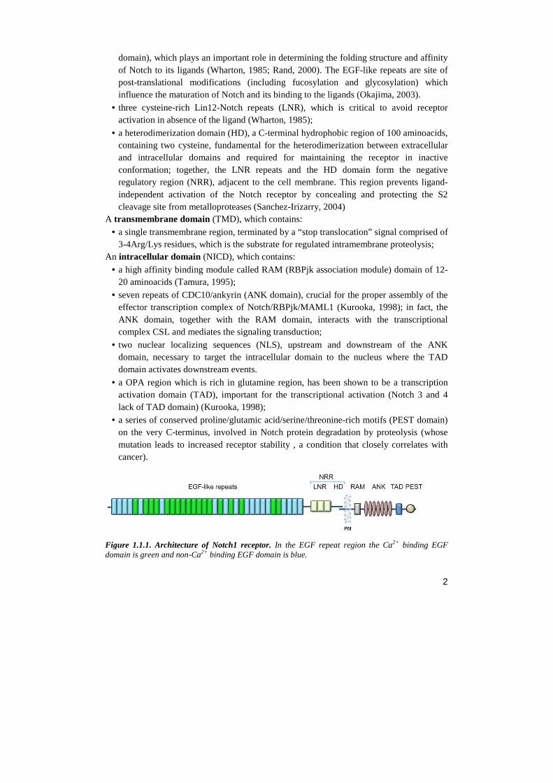
1.1 NOTCH RECEPTORS Notch receptors are single pass type I transmembrane proteins. The mature form of Notch on the cell surface is a large heterodimer, held together by non-covalent calcium-dependent interactions through the heterodimerization domain (HD). The structure of the receptor (fig. 1.1.1) comprises three domains, in which different regions are associated with different functions (Chillakuri, 2012): An extracellular domain (NECD), which contains:
• At the N-terminus, a variable number of tandem Epidermal Growth Factor (EGF)-like repeats (ELR) in mammals (36 in Notch-1 and Notch-2; 34 in Notch-3, and 29 in Notch-4) mediate positive interactions with ligand presented by neighboring cells (repeats 11–12) and also mediate inhibitory interactions with ligand co-expressed in the same cell (repeats 24–29) (Rebay, 1991). Many EGF repeats bind calcium (cbEGF-like
2
domain), which plays an important role in determining the folding structure and affinity of Notch to its ligands (Wharton, 1985; Rand, 2000). The EGF-like repeats are site of post-translational modifications (including fucosylation and glycosylation) which influence the maturation of Notch and its binding to the ligands (Okajima, 2003).
• three cysteine-rich Lin12-Notch repeats (LNR), which is critical to avoid receptor activation in absence of the ligand (Wharton, 1985);
• a heterodimerization domain (HD), a C-terminal hydrophobic region of 100 aminoacids, containing two cysteine, fundamental for the heterodimerization between extracellular and intracellular domains and required for maintaining the receptor in inactive conformation; together, the LNR repeats and the HD domain form the negative regulatory region (NRR), adjacent to the cell membrane. This region prevents ligand-independent activation of the Notch receptor by concealing and protecting the S2 cleavage site from metalloproteases (Sanchez-Irizarry, 2004)
A transmembrane domain (TMD), which contains:
• a single transmembrane region, terminated by a “stop translocation” signal comprised of 3-4Arg/Lys residues, which is the substrate for regulated intramembrane proteolysis;
An intracellular domain (NICD), which contains:
• a high affinity binding module called RAM (RBPjk association module) domain of 12-20 aminoacids (Tamura, 1995);
• seven repeats of CDC10/ankyrin (ANK domain), crucial for the proper assembly of the effector transcription complex of Notch/RBPjk/MAML1 (Kurooka, 1998); in fact, the ANK domain, together with the RAM domain, interacts with the transcriptional complex CSL and mediates the signaling transduction;
• two nuclear localizing sequences (NLS), upstream and downstream of the ANK domain, necessary to target the intracellular domain to the nucleus where the TAD domain activates downstream events.
• a OPA region which is rich in glutamine region, has been shown to be a transcription activation domain (TAD), important for the transcriptional activation (Notch 3 and 4 lack of TAD domain) (Kurooka, 1998);
• a series of conserved proline/glutamic acid/serine/threonine-rich motifs (PEST domain) on the very C-terminus, involved in Notch protein degradation by proteolysis (whose mutation leads to increased receptor stability , a condition that closely correlates with cancer).
Figure 1.1.1. Architecture of Notch1 receptor. In the EGF repeat region the Ca2+ binding EGF domain is green and non-Ca2+ binding EGF domain is blue.
3
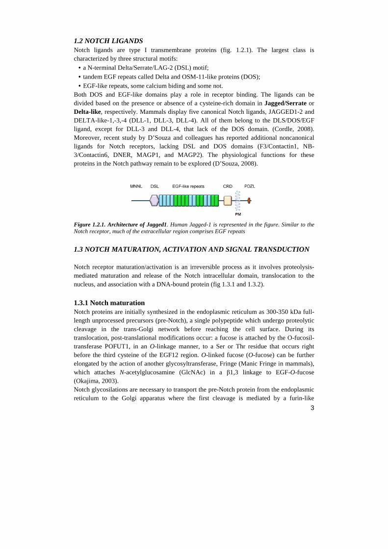
1.2 NOTCH LIGANDS Notch ligands are type I transmembrane proteins (fig. 1.2.1). The largest class is characterized by three structural motifs:
• a N-terminal Delta/Serrate/LAG-2 (DSL) motif;
• tandem EGF repeats called Delta and OSM-11-like proteins (DOS);
• EGF-like repeats, some calcium biding and some not. Both DOS and EGF-like domains play a role in receptor binding. The ligands can be divided based on the presence or absence of a cysteine-rich domain in Jagged/Serrate or Delta-like, respectively. Mammals display five canonical Notch ligands, JAGGED1-2 and DELTA-like-1,-3,-4 (DLL-1, DLL-3, DLL-4). All of them belong to the DLS/DOS/EGF ligand, except for DLL-3 and DLL-4, that lack of the DOS domain. (Cordle, 2008). Moreover, recent study by D’Souza and colleagues has reported additional noncanonical ligands for Notch receptors, lacking DSL and DOS domains (F3/Contactin1, NB-3/Contactin6, DNER, MAGP1, and MAGP2). The physiological functions for these proteins in the Notch pathway remain to be explored (D’Souza, 2008).
Figure 1.2.1. Architecture of Jagged1. Human Jagged-1 is represented in the figure. Similar to the Notch receptor, much of the extracellular region comprises EGF repeats
1.3 NOTCH MATURATION, ACTIVATION AND SIGNAL TRANSDUCTION Notch receptor maturation/activation is an irreversible process as it involves proteolysis-mediated maturation and release of the Notch intracellular domain, translocation to the nucleus, and association with a DNA-bound protein (fig 1.3.1 and 1.3.2).
1.3.1 Notch maturation Notch proteins are initially synthesized in the endoplasmic reticulum as 300-350 kDa full-length unprocessed precursors (pre-Notch), a single polypeptide which undergo proteolytic cleavage in the trans-Golgi network before reaching the cell surface. During its translocation, post-translational modifications occur: a fucose is attached by the O-fucosil-transferase POFUT1, in an O-linkage manner, to a Ser or Thr residue that occurs right before the third cysteine of the EGF12 region. O-linked fucose (O-fucose) can be further elongated by the action of another glycosyltransferase, Fringe (Manic Fringe in mammals), which attaches N-acetylglucosamine (GlcNAc) in a β1,3 linkage to EGF-O-fucose (Okajima, 2003). Notch glycosilations are necessary to transport the pre-Notch protein from the endoplasmic reticulum to the Golgi apparatus where the first cleavage is mediated by a furin-like

4
convertase and occurs within HD domain at a site referred to as the S1 cleavage site (at 70 amino acids from the transmembrane domain), converting the pre-Notch polypeptide into the heterodimer NECD/NTMIC (Notch-extracellular domain/Notch transmembrane and intracellular domain) (Blaumueller, 1997; Logeat, 1998). The two subunits resulting from this process are brought to the plasma membrane as one heterodimer, held together by non-covalent calcium-dependent interactions. Post-translational modifications of Notch can modulate Notch ligand interactions since Fringe enzyme is expressed only in a subset of cells and this seems to influence the Notch activation. In cells expressing Fringe, Notch ligands show distinct preferences: Delta-like prefers Fringe-modified Notch, whereas Serrate-like would much rather bind unmodified Notch. These preferences are the basis for Notch hyperactivation at boundaries between Fringe expressing and -nonexpressing territories, but the relative importance of each site glycosylated and the molecular basis for this regulation is unknown (Okajima, 2003).
1.3.2 Notch activation The Notch signaling is a cell-to-cell communication pathway that is activated when Notch ligand on the sending cell bind to Notch receptor on the receiving cell (Schroeter 1998). The binding of ligand on Notch receptor triggers conformational modifications in the Notch protein which cause a sequence of proteolytic cleavages terminating in Notch trans-activation (Brown, 2000; Mumm, 2000). This process is characterize by two steps:
• Following ligand binding, Notch signaling is initiated when endocytosis of the ligand–NECD complex induces unfolding of the juxtamembrane negative control region (NRR). In particular, the DSL ligand epsin-mediated endocytosis is triggered by monoubiquitination of the intracellular domain mediated by the E3 ubiquitin ligases Neuralized (which preferentially recognizes Delta ligands) and Mindbomb (which recognizes Serrate/Jagged). The resulting conformational change in NRR exposes site 2 (S2) in Notch for the first activating cleavage allowing access by ADAM/TACE (A disintegrin and metalloprotease/tumor-necrosis-factor α converting enzyme) metalloprotease. The S2 cleavage occurs within the extracellular domain, approximately 12 amino acids before the transmembrane domain at a site referred to as the S2 cleavage site (Brou, 2000; Mumm, 2000). This is a key regulatory step in Notch activation, but some ambiguity still exists regarding the enzymes that mediate the cleavage: while ADAM17/TACE seems to be the main metalloprotease able to cleave Notch receptors in vitro (Brou, 2000), animal models point to ADAM10/Kuzbanian metalloprotease for this essential function in vivo (van Tetering, 2009, Lieber, 2002). ADAM proteases leaves a short-lived fragment anchored to the plasma membrane, called NEXT (Notch extracellular truncation).
• NEXT becomes the substrate for the last cleavage: Notch intracellular fragment is recognized by the inactive aminopeptidase domain of Nicastrin (NCT), which transfers NEXT to the active site of γ-secretase which operate the cleavage within the
5
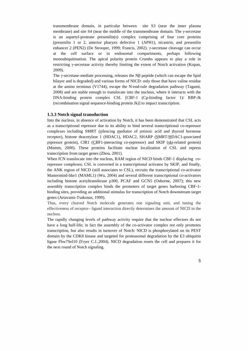
transmembrane domain, in particular between site S3 (near the inner plasma membrane) and site S4 (near the middle of the transmembrane domain. The γ-secretase is an aspartyl-protease presenilin(s) complex comprising of four core proteins (presenilin 1 or 2, anterior pharynx defective 1 (APH1), nicastrin, and presenilin enhancer 2 (PEN2) (De Strooper, 1999; Francis, 2002). γ-secretase cleavage can occur at the cell surface or in endosomal compartments, perhaps following monoubiquitination. The apical polarity protein Crumbs appears to play a role in restricting γ-secretase activity thereby limiting the extent of Notch activation (Kopan, 2009). The γ-secretase-mediate processing, releases the Nβ peptide (which can escape the lipid bilayer and is degraded) and various forms of NICD: only those that have valine residue at the amino terminus (V1744), escape the N-end-rule degradation pathway (Tagami, 2008) and are stable enough to translocate into the nucleus, where it interacts with the DNA-binding protein complex CSL [CBF-1 (Cp-binding factor 1)/ RBP-Jk (recombination signal sequence-binding protein Jk)] to impact transcription.
1.3.3 Notch signal transduction Into the nucleus, in absence of activation by Notch, it has been demonstrated that CSL acts as a transcriptional repressor due to its ability to bind several transcriptional co-repressor complexes including SMRT (silencing mediator of retinoic acid and thyroid hormone receptor), histone deacetylase 1 (HDAC1), HDAC2, SHARP (SMRT/HDAC1-associated repressor protein), CIR1 (CBF1-interacting co-repressor) and SKIP (ski-related protein) (Mumm, 2000). These proteins facilitate nuclear localization of CSL and repress transcription from target genes (Zhou, 2001). When ICN translocate into the nucleus, RAM region of NICD binds CBF-1 displacing co-repressor complexes; CSL is converted in a transcriptional activator by SKIP, and finally, the ANK region of NICD (still associates to CSL), recruits the transcriptional co-activator Mastermind-like1 (MAML1) (Wu, 2004) and several different transcriptional co-activators including histone acetyltransferase p300, PCAF and GCN5 (Osborne, 2007); this new assembly transcription complex binds the promoters of target genes harboring CBF-1-binding sites, providing an additional stimulus for transcription of Notch downstream target genes (Artavanis-Tsakonas, 1999). Thus, every cleaved Notch molecule generates one signaling unit, and tuning the effectiveness of receptor– ligand interaction directly determines the amount of NICD in the nucleus. The rapidly changing levels of pathway activity require that the nuclear effectors do not have a long half-life; in fact the assembly of the co-activator complex not only promotes transcription, but also results in turnover of Notch: NICD is phosphorylated on its PEST domain by the CDK8 kinase and targeted for proteasomal degradation by the E3 ubiquitin ligase Fbw7Sel10 (Fryer C.J.,2004). NICD degradation resets the cell and prepares it for the next round of Notch signaling.
6
Figure 1.3.1. Schematic representation of the proteolytic cascade upstream Notch activation.
HD
7
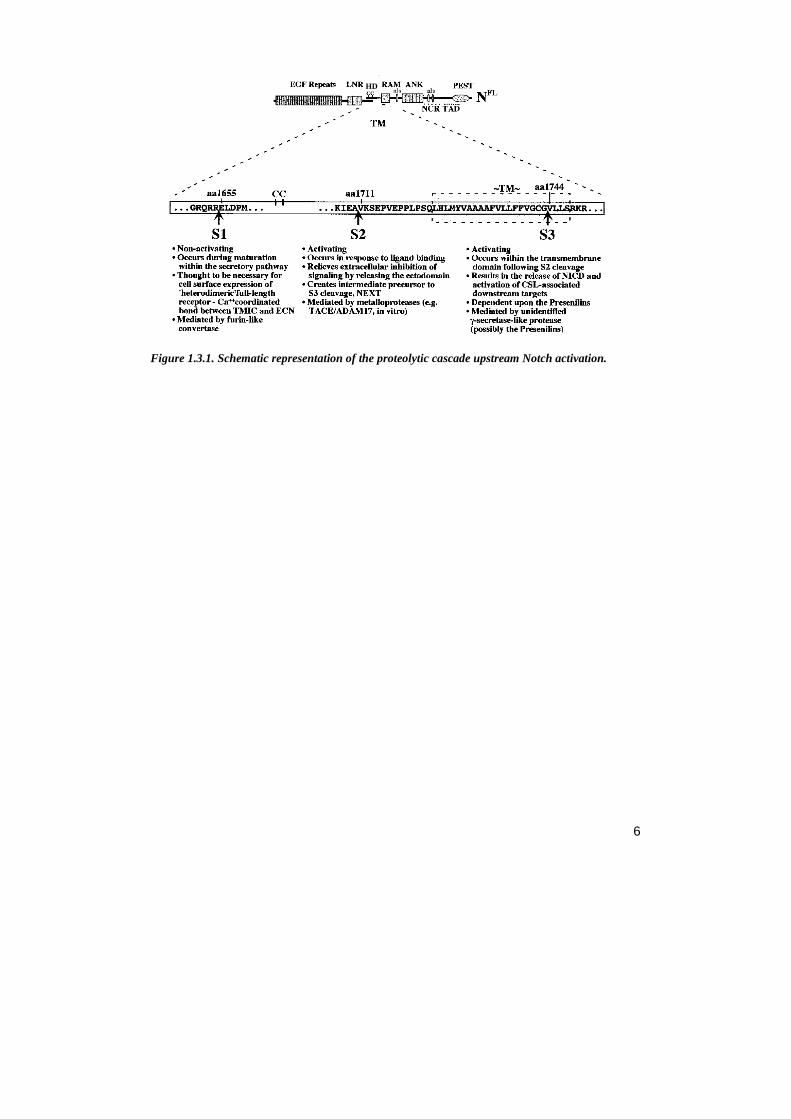
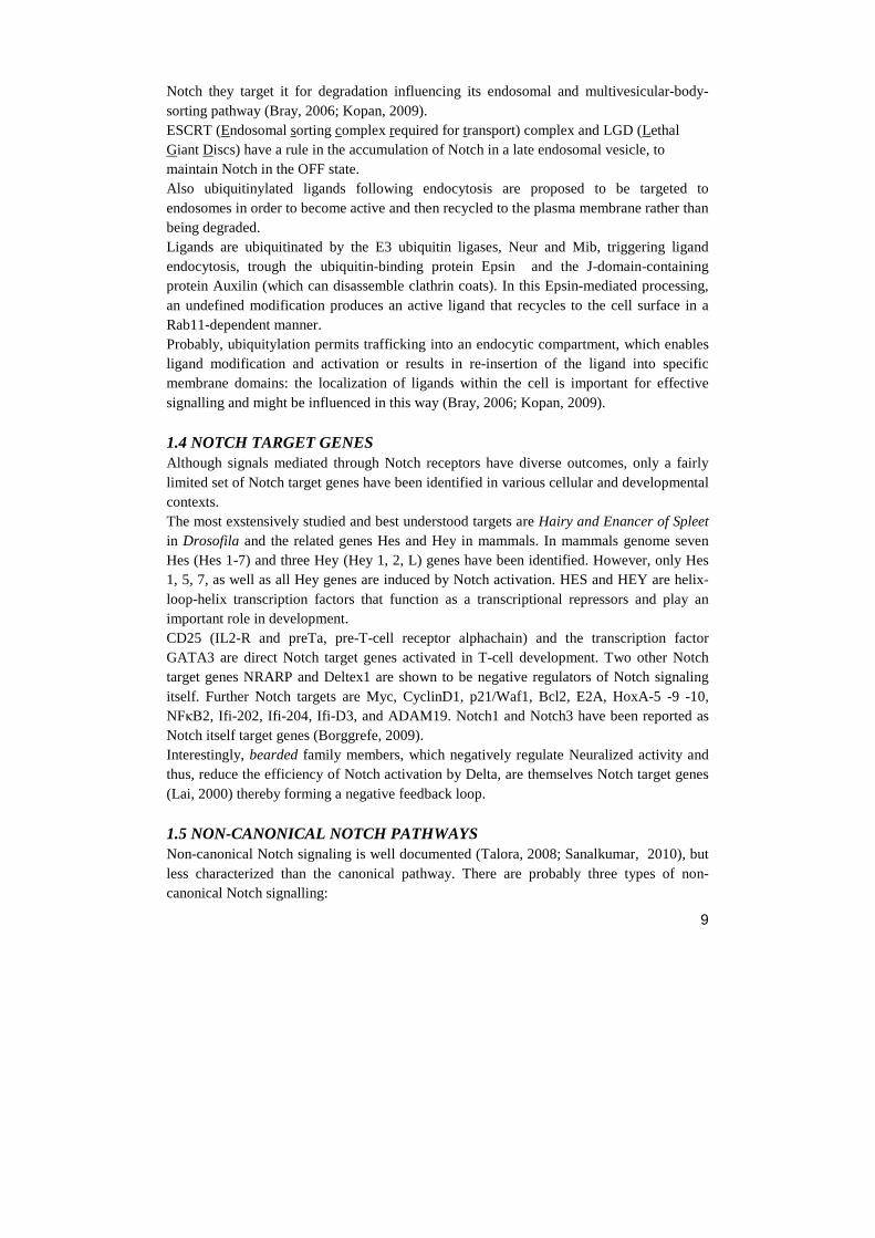
Figure 1.3.2. Notch canonical pathway. After translation, Notch receptor pre-protein is glycosylated by Ofut, essential enzyme for the production of a functional receptor. PC5/furin protease produces the mature receptor cleaving Notch at site 1 (S1). Then, Notch reaches the cell surface as a heterodimer, held together by noncovalent interactions. The glycosyltransferase Fringe extends the O-fucose, thereby altering the ability of specific ligands to activate Notch. The Notch receptor is activated by a ligand presented by a neighboring cell. Endocytosis and membrane trafficking regulate ligand and receptor availability at the cell surface. Ligand endocytosis promotes a conformational change in the Notch receptor. Such conformational change exposes site 2 (S2) in Notch for cleavage by ADAM metalloproteases. S2 cleavage generates the NEXT fragment (membrane-anchored Notch extracellular truncation), a substrate for the γ-secretase complex. γ-secretase cleaves NEXT progressively from site 3 (S3) to site 4 (S4) releasing the Notch intracellular domain (NICD) and Nβ peptide. γ-Secretase cleavage can occur at the cell surface or in endosomal compartments, but cleavage at the membrane favors the production of a more stable form of NICD. NICD then enters the nucleus where it associates with the CSL (CBF1/RBPjκ/Su(H)/Lag-1) complex. In the absence of NICD, CSL associates with corepressor (Co-R) proteins and histone deacetylases (HDACs). NICD binding facilitates displacement of transcriptional repressors. Then, Mastermind (MAM) coactivator recognizes the NICD/CSL interface, and this triprotein complex recruits additional coactivators (Co-A) to activate transcription. In addition to trans-activating Notch–ligand complexes, the receptor can also form cis-inhibitory complexes when binding occurs between Notch and ligand expressed on the same cell surface. Cis-inhibition serves to limit the zone of Notch activity and thus determine whether a cell will signal (the ligand is more abundant than Notch) or receive (Notch is more abundant than the ligand) (Sprinzak, 2010). Alternatively, in
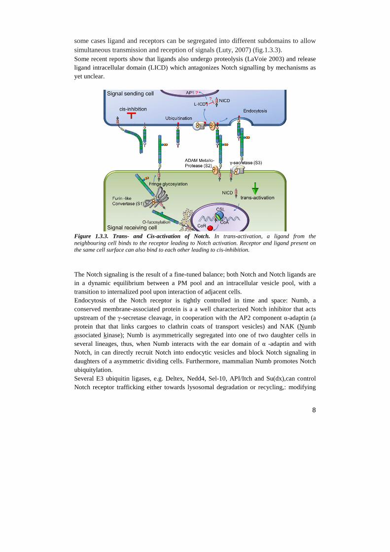
8
some cases ligand and receptors can be segregated into different subdomains to allow simultaneous transmission and reception of signals (Luty, 2007) (fig.1.3.3). Some recent reports show that ligands also undergo proteolysis (LaVoie 2003) and release ligand intracellular domain (LICD) which antagonizes Notch signalling by mechanisms as yet unclear.
Figure 1.3.3. Trans- and Cis-activation of Notch. In trans-activation, a ligand from the neighbouring cell binds to the receptor leading to Notch activation. Receptor and ligand present on the same cell surface can also bind to each other leading to cis-inhibition. The Notch signaling is the result of a fine-tuned balance; both Notch and Notch ligands are in a dynamic equilibrium between a PM pool and an intracellular vesicle pool, with a transition to internalized pool upon interaction of adjacent cells. Endocytosis of the Notch receptor is tightly controlled in time and space: Numb, a conserved membrane-associated protein is a a well characterized Notch inhibitor that acts upstream of the γ-secretase cleavage, in cooperation with the AP2 component α-adaptin (a protein that that links cargoes to clathrin coats of transport vesicles) and NAK (Numb associated kinase); Numb is asymmetrically segregated into one of two daughter cells in several lineages, thus, when Numb interacts with the ear domain of α -adaptin and with Notch, in can directly recruit Notch into endocytic vesicles and block Notch signaling in daughters of a asymmetric dividing cells. Furthermore, mammalian Numb promotes Notch ubiquitylation. Several E3 ubiquitin ligases, e.g. Deltex, Nedd4, Sel-10, API/Itch and Su(dx),can control Notch receptor trafficking either towards lysosomal degradation or recycling,: modifying
9
Notch they target it for degradation influencing its endosomal and multivesicular-body-sorting pathway (Bray, 2006; Kopan, 2009). ESCRT (Endosomal sorting complex required for transport) complex and LGD (Lethal Giant Discs) have a rule in the accumulation of Notch in a late endosomal vesicle, to maintain Notch in the OFF state. Also ubiquitinylated ligands following endocytosis are proposed to be targeted to endosomes in order to become active and then recycled to the plasma membrane rather than being degraded. Ligands are ubiquitinated by the E3 ubiquitin ligases, Neur and Mib, triggering ligand endocytosis, trough the ubiquitin-binding protein Epsin and the J-domain-containing protein Auxilin (which can disassemble clathrin coats). In this Epsin-mediated processing, an undefined modification produces an active ligand that recycles to the cell surface in a Rab11-dependent manner. Probably, ubiquitylation permits trafficking into an endocytic compartment, which enables ligand modification and activation or results in re-insertion of the ligand into specific membrane domains: the localization of ligands within the cell is important for effective signalling and might be influenced in this way (Bray, 2006; Kopan, 2009).
1.4 NOTCH TARGET GENES Although signals mediated through Notch receptors have diverse outcomes, only a fairly limited set of Notch target genes have been identified in various cellular and developmental contexts. The most exstensively studied and best understood targets are Hairy and Enancer of Spleet in Drosofila and the related genes Hes and Hey in mammals. In mammals genome seven Hes (Hes 1-7) and three Hey (Hey 1, 2, L) genes have been identified. However, only Hes 1, 5, 7, as well as all Hey genes are induced by Notch activation. HES and HEY are helix-loop-helix transcription factors that function as a transcriptional repressors and play an important role in development. CD25 (IL2-R and preTa, pre-T-cell receptor alphachain) and the transcription factor GATA3 are direct Notch target genes activated in T-cell development. Two other Notch target genes NRARP and Deltex1 are shown to be negative regulators of Notch signaling itself. Further Notch targets are Myc, CyclinD1, p21/Waf1, Bcl2, E2A, HoxA-5 -9 -10, NFκB2, Ifi-202, Ifi-204, Ifi-D3, and ADAM19. Notch1 and Notch3 have been reported as Notch itself target genes (Borggrefe, 2009). Interestingly, bearded family members, which negatively regulate Neuralized activity and thus, reduce the efficiency of Notch activation by Delta, are themselves Notch target genes (Lai, 2000) thereby forming a negative feedback loop.
1.5 NON-CANONICAL NOTCH PATHWAYS Non-canonical Notch signaling is well documented (Talora, 2008; Sanalkumar, 2010), but less characterized than the canonical pathway. There are probably three types of non-canonical Notch signalling:
10
Type I involves Notch ligation and translocation of activation signals independent of CBF1 (NICD-dependent / CBF1-independent); Type II involves Notch activity in a S3 cleavage-independent manner (NICD- and CBF1-independent); Type III involves CBF1-dependent gene activation without receptor cleavage and NICD release (Sanalkumar, 2010). Several signalling pathways are involved, including Hedgehog, Jak/STAT, RTK, TGF, Wnt, PI3/Akt, mTor/Akt, JNK, MEK/ERK, and NFκB (Talora, 2008; Sanalkumar, 2010). Non-canonical Notch signalling seems important for maintenance of lineage-restricted hematopoietic progenitors, and several of the mediators involved in this signaling are in addition important in leukemogenesis as well as regulation of cellular immune responses. The non-canonical pathway thus represents a point of crosstalk between other intracellular signaling pathways. Interactions between Notch and the Wnt pathway have been best characterized, but other interactions with various pathways have also been described.
• Notch/Wnt/ β-catenin signaling: Wnt signalling is mediated through the downstream β-catenin. The Wnt and Notch pathways seem to act in synergy for example to maintain the stem cell pool. The crosstalk between these two pathways seems to occur both at transcriptional level and at protein interaction level. Members of the Wnt pathway regulate the expression of established Notch target genes since the inhibition of Wnt signalling affects the expression of both Wnt and Notch target genes as well as the expression of Notch1. Another example of crosstalk between these two pathways in the stem cell niche is the induced expression of Notch ligands by activated β-catenin in stromal cells which thereby induce-Notch-mediated intracellular signalling in adjacent cells. Also Notch is able to interact directly and inactivate the β-catenin complex, but this signaling is in equilibrium with the Notch inhibition Wnt-mediated (Blank, 2008; Staal, 2010; Trowbridge, 2006; Reya, 2003; Yamane, 2001; Hayward, 2005) (fig. 1.5.1).
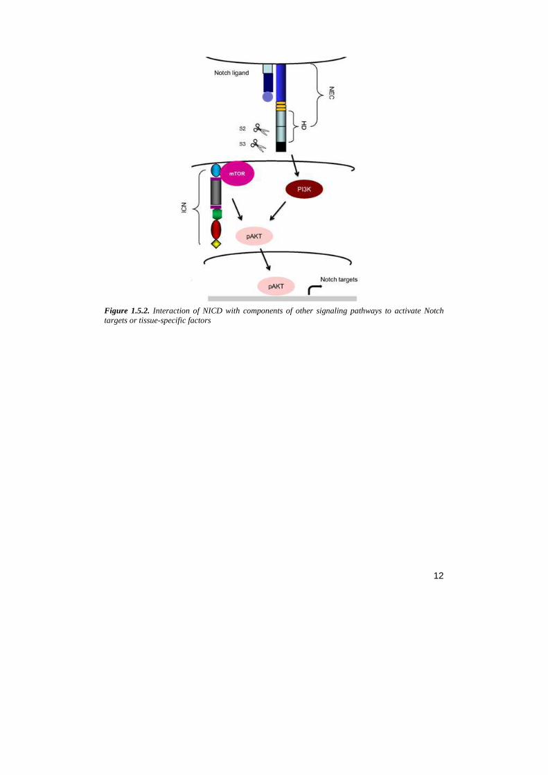
• Notch/mTor/AKT signaling : Akt is a key downstream target in the antiapoptotic pathway activated by Notch. Nuclear functions of ICN is shown to be not essential for this pathway which is independent by the transcription factor, CBF1. NIC activity is initiated by a membrane-anchored form of ICN that converges on the kinase mammalian target of rapamycin (mTOR) and the substrate-defining protein rapamycin independent companion of mTOR (Rictor), triggering the activation of the kinase Akt/PKB and consequently cell survival (Perumalsamy, 2009) (fig. 1.5.2).
• Notch/NF-κB signaling: several reports have proposed direct interactions of Notch1-IC with NF-κB subunits (Wang, 2001; Espinosa, 2003; Oakley, 2003) and very recent work has demonstrated that ICN interacts with NF-κB and competes with the IkBα protein, enhancing the retention of Nfkb1 and Rel in the nucleus (Shin, 2006). Notch1 also regulates the NF-κB pathway by inducing the expression of Relb and Nfkb2 and by a direct interaction of Notch1 with the IKK complex which stimulates the activity of IKK (Vilimas, 2007). In addition, activation of NF-kB may also be mediated by
11
Notch3: Notch3 seems to be a direct transcriptional target of Notch1 (Palomero, 2006; Vilimas, 2007) and recent work shows that Notch3 can activate the NF-κB pathway by phosphorylating IKKα homodimers, which in turn activate the noncanonical p52-Relb NF-κB pathway (Vacca, 2006). Thus, in principle, Notch-1 could activate the NF-κB canonical signaling by activating the IKKα/β/γ signalosome and facilitating the nuclear retention of NF-κB heterodimers, and the noncanonical pathway by inducing the expression of Relb and Nfkb2 and activating IKKα homodimers via Notch-3.
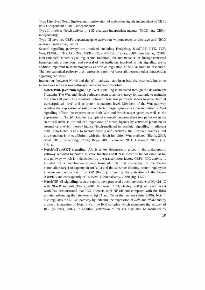
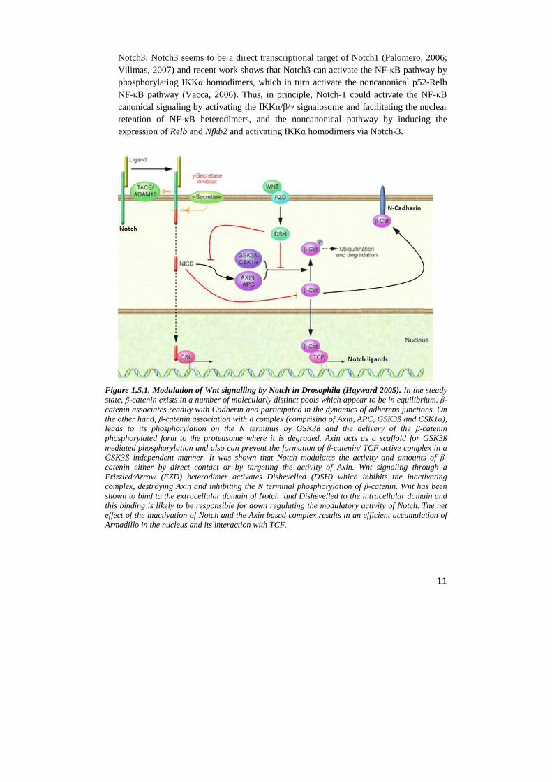
Figure 1.5.1. Modulation of Wnt signalling by Notch in Drosophila (Hayward 2005). In the steady state, β-catenin exists in a number of molecularly distinct pools which appear to be in equilibrium. β-catenin associates readily with Cadherin and participated in the dynamics of adherens junctions. On the other hand, β-catenin association with a complex (comprising of Axin, APC, GSK3ß and CSK1α), leads to its phosphorylation on the N terminus by GSK3ß and the delivery of the β-catenin phosphorylated form to the proteasome where it is degraded. Axin acts as a scaffold for GSK3ß mediated phosphorylation and also can prevent the formation of β-catenin/ TCF active complex in a GSK3ß independent manner. It was shown that Notch modulates the activity and amounts of β-catenin either by direct contact or by targeting the activity of Axin. Wnt signaling through a Frizzled/Arrow (FZD) heterodimer activates Dishevelled (DSH) which inhibits the inactivating complex, destroying Axin and inhibiting the N terminal phosphorylation of β-catenin. Wnt has been shown to bind to the extracellular domain of Notch and Dishevelled to the intracellular domain and this binding is likely to be responsible for down regulating the modulatory activity of Notch. The net effect of the inactivation of Notch and the Axin based complex results in an efficient accumulation of Armadillo in the nucleus and its interaction with TCF.
12
Figure 1.5.2. Interaction of NICD with components of other signaling pathways to activate Notch targets or tissue-specific factors
13
2. THE ROLE OF NOTCH IN PHYSIOLOGICAL AND PATHOLOGICAL SYSTEMS
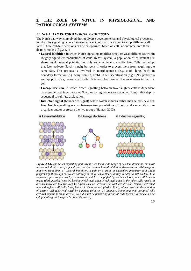
2.1 NOTCH IN PHYSIOLOGICAL PROCESSES The Notch pathway is involved during diverse developmental and physiological processes, in which its signaling occurs between adjacent cells to direct them to adopt different cell fates. These cell-fate decisions can be categorized, based on cellular outcome, into three distinct models (fig.2.1.1):
• Lateral inhibition in which Notch signaling amplifies small or weak differences within roughly equivalent populations of cells. In this system, a population of equivalent cell share developmental potential but only some achieve a specific fate. Cells that adopt that fate, activate Notch in neighbor cells in order to prevent them from acquiring the same fate. This process is involved in morphogenesis (e.g. tooth, lung, hair), in boundary formation (e.g. wing, somtes, limb), in cell specification (e.g. CNS, pancreas) and apoptosis (e.g. neural crest cells). It is not clear how a difference arises in the first cell.
• Lineage decision, in which Notch signalling between two daughter cells is dependent on asymmetrical inheritance of Notch or its regulators (for example, Numb); this step is sequential to cell fate assignation.
• Inductive signal (boundaries signal) where Notch induces rather then selects new cell fate: Notch signalling occurs between two populations of cells and can establish an organizer and/or segregate the two groups (Haines, 2003).
Figure 2.1.1. The Notch signalling pathway is used for a wide range of cell-fate decisions, but most instances fall into one of a few distinct modes, such as lateral inhibition, decisions on cell-lineage or inductive signalling. a | Lateral inhibition: a pair or a group of equivalent precursor cells (light purple) signal through the Notch pathway to inhibit each other's ability to adopt a distinct fate. In a sequential process (shown by the arrows), which is amplified by feedback loops, one cell in each group (dark purple) 'wins' by lacking Notch activation. Notch activation in the other cells results in an alternative cell fate (yellow). b | Asymmetric cell divisions: at each cell division, Notch is activated in one daughter cell (solid lines) but not in the other cell (dashed lines), which results in the adoption of distinct cell fates (indicated by different colours). c | Inductive signalling: one group of cells (yellow) signals (orange arrows) to a distinct neighbouring group of cells (green) to induce a new cell fate along the interface between them (red).
14
Notch receptors and ligands are widely expressed during organogenesis in mammalian embryos, and studies of spontaneous or induced mutants demonstrate that Notch signaling regulates cell lineage decisions in tissues derived from all three primary germ layers: endoderm (e.g. pancreas), mesoderm (skeleton,mammary gland, vasculature, and hematopoietic cells), and ectoderm (neuronal lineages). Some developing tissues express several different receptors and ligands, whereas others express a single receptor–ligand pair. Although some Notch receptors appear to have genetically redundant functions in some developmental contexts (e.g. N1 and N4 in vasculogenesis) (Krebs, 2000), others have unique and essential functions as revealed by the severe disruption of embryogenesis that results from loss-of-function mutations. In the following paragraph are reported the main physiological processes in which Notch is involved: Notch signaling in embryogenesis The Notch signaling pathway plays an important role in cell fate determination during embryonic development. Notch signaling is required in the regulation of embryo polarity and during left-right asymmetry determination in vertebrates. Notch signaling is central to somitogenesis and in the maintenance of somite borders. Recent studies hypothesized that the primary function of Notch signaling does not act on an individual cell, but coordinates cell clocks and keep them synchronized (Austin, 1987; Levin, 2005; Conlon, 1995). Notch signaling in central nervous system development and function The Notch signaling pathway was mainly found to be critical for neural progenitor cell (NPC) maintenance and self-renewal as well as cell fate specification. In recent years, other functions of the Notch pathway have also been found, including glial cell specification, neurites development as well as learning and memory. In gliogenesis, Notch appears to have an instructive role which can directly promote the differentiation of many glial cell subtypes For example, activation of Notch signaling in the retina favors the generation of Muller glia cells at the expense of neurons, whereas reduced Notch signaling induces production of ganglion cells, causing a reduction in the number of Muller glia. In addition to developmental functions, Notch proteins and ligands are expressed in cells of the adult nervous system, suggesting a role in CNS plasticity throughout life. Adult mice heterozygous for mutations in either Notch1 or Cbf1 have deficits in spatial learning and memory (Furukawa, 2000; Scheer, 2001; Redmond, 2000; Costa, 2003; Bolo´s, 2007). Notch signaling in cardiovascular development The Notch signaling pathway is a critical component of cardiovascular formation and morphogenesis in both development and disease. It is required for the selection of endothelial tip and stalk cells during sprouting angiogenesis. Notch signal pathway plays a crucial role in at least three cardiac development processes: Atrioventricular canal development (in the boundary formation between the AV canal and the chamber myocardium), myocardial development as well as cardiac outflow tract (OFT) development. Notch may regulate this process by activating matrix metalloproteinase2
15
(MMP2) expression, or by inhibiting vascular endothelial (VE)-cadherin expression in the AV canal endocardium while suppressing the VEGF pathway via VEGFR2. The downstream effector of Notch signaling, HEY2, was also demonstrated to be important in regulating ventricular development by its expression in the interventricular septum and the endocardial cells of the cardiac cushions (Kume, 2012; Niessen, 2008; Kokubo, 2007; Timmerman, 2004; Nemir, 2006) Notch signaling in angiogenesis Endothelial cells use the Notch signaling pathway to coordinate cellular behaviors during the blood vessel sprouting that occurs in angiogenesis. Activation of Notch takes place primarily in “connector” cells and cells that line patent stable blood vessels through direct interaction with the Notch ligand, Delta-like ligand 4 (Dll4), which is expressed in the endothelial tip cells. VEGF signaling, which is an important factor for migration and proliferation of endothelial cells, can be downregulated in cells with activated Notch signaling by lowering the levels of VEGF receptor transcript. Notch signaling may be used to control the sprouting pattern of blood vessels during angiogenesis. When cells within a patent vessel are exposed to VEGF signaling, only a restricted number of them initiate the angiogenic process. VEGF is able to induce Dll4 expression. In turn, Dll4 expressing cells down-regulate VEGF receptors in neighboring cells through activation of Notch, thereby preventing their migration into the developing sprout. Similarly, during the sprouting process itself, the migratory behavior of connector cells must be limited to retain a patent connection to the original blood vessel (Hellstrom, 2007; Lobov, 2007; Siekmann, 2007). Notch signaling in pancreatic development The formation of the pancreas from endoderm begins in early development. The expression of elements of the Notch signaling pathway have been found in the developing pancreas, suggesting Notch signaling is important in pancreatic development. Evidence suggests Notch signaling regulates the progressive recruitment of endocrine cell types from a common precursor, acting through two possible mechanisms. One is the “lateral inhibition,” which could explain the dispersed distribution off endocrine cells within pancreatic epithelium. A second mechanism is “suppressive maintenance,” which explains the role of Notch signaling in pancreas differentiation (Apelqvist, 1999; Lammert, 2000; Jensen, 2000) Notch signaling and intestinal development The role of Notch signaling in the regulation of gut development has been indicated in several reports. Transcriptional analysis and gain of function experiments revealed that Notch signaling targets Hes1 in the intestine and regulates a binary cell fate decision between adsorptive and secretory cell fates (Crosnier, 2005) Notch signaling and bone development Early in vitro studies have found the Notch signaling pathway functions as down-regulator in osteoclastogenesis and osteoblastogenesis (Yamada, 2003). Notch1 is expressed in the mesenchymal condensation area and subsequently in the hypertrophic chondrocytes during chondrogenesis. Overexpression of Notch signaling inhibits bone morphogenetic protein2-
16
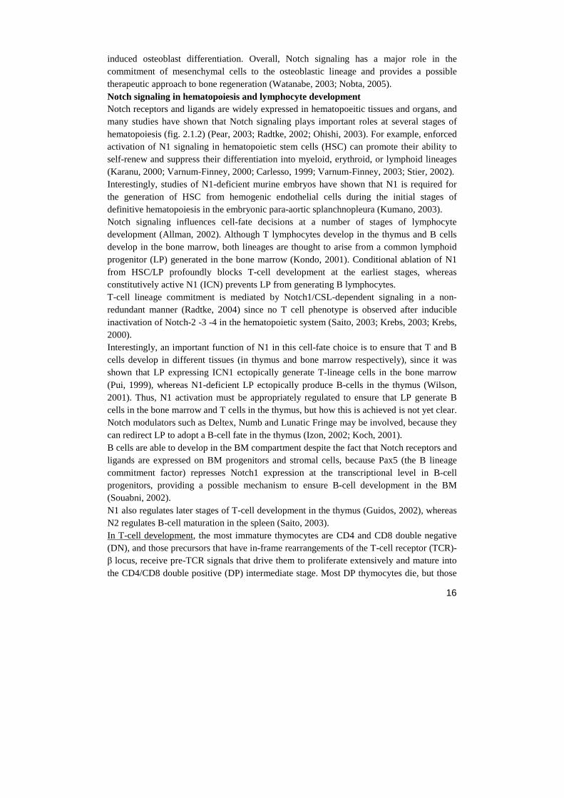
induced osteoblast differentiation. Overall, Notch signaling has a major role in the commitment of mesenchymal cells to the osteoblastic lineage and provides a possible therapeutic approach to bone regeneration (Watanabe, 2003; Nobta, 2005). Notch signaling in hematopoiesis and lymphocyte development Notch receptors and ligands are widely expressed in hematopoeitic tissues and organs, and many studies have shown that Notch signaling plays important roles at several stages of hematopoiesis (fig. 2.1.2) (Pear, 2003; Radtke, 2002; Ohishi, 2003). For example, enforced activation of N1 signaling in hematopoietic stem cells (HSC) can promote their ability to self-renew and suppress their differentiation into myeloid, erythroid, or lymphoid lineages (Karanu, 2000; Varnum-Finney, 2000; Carlesso, 1999; Varnum-Finney, 2003; Stier, 2002). Interestingly, studies of N1-deficient murine embryos have shown that N1 is required for the generation of HSC from hemogenic endothelial cells during the initial stages of definitive hematopoiesis in the embryonic para-aortic splanchnopleura (Kumano, 2003). Notch signaling influences cell-fate decisions at a number of stages of lymphocyte development (Allman, 2002). Although T lymphocytes develop in the thymus and B cells develop in the bone marrow, both lineages are thought to arise from a common lymphoid progenitor (LP) generated in the bone marrow (Kondo, 2001). Conditional ablation of N1 from HSC/LP profoundly blocks T-cell development at the earliest stages, whereas constitutively active N1 (ICN) prevents LP from generating B lymphocytes. T-cell lineage commitment is mediated by Notch1/CSL-dependent signaling in a non-redundant manner (Radtke, 2004) since no T cell phenotype is observed after inducible inactivation of Notch-2 -3 -4 in the hematopoietic system (Saito, 2003; Krebs, 2003; Krebs, 2000). Interestingly, an important function of N1 in this cell-fate choice is to ensure that T and B cells develop in different tissues (in thymus and bone marrow respectively), since it was shown that LP expressing ICN1 ectopically generate T-lineage cells in the bone marrow (Pui, 1999), whereas N1-deficient LP ectopically produce B-cells in the thymus (Wilson, 2001). Thus, N1 activation must be appropriately regulated to ensure that LP generate B cells in the bone marrow and T cells in the thymus, but how this is achieved is not yet clear. Notch modulators such as Deltex, Numb and Lunatic Fringe may be involved, because they can redirect LP to adopt a B-cell fate in the thymus (Izon, 2002; Koch, 2001). B cells are able to develop in the BM compartment despite the fact that Notch receptors and ligands are expressed on BM progenitors and stromal cells, because Pax5 (the B lineage commitment factor) represses Notch1 expression at the transcriptional level in B-cell progenitors, providing a possible mechanism to ensure B-cell development in the BM (Souabni, 2002). N1 also regulates later stages of T-cell development in the thymus (Guidos, 2002), whereas N2 regulates B-cell maturation in the spleen (Saito, 2003). In T-cell development, the most immature thymocytes are CD4 and CD8 double negative (DN), and those precursors that have in-frame rearrangements of the T-cell receptor (TCR)-β locus, receive pre-TCR signals that drive them to proliferate extensively and mature into the CD4/CD8 double positive (DP) intermediate stage. Most DP thymocytes die, but those
17
that express an αβ-TCR complex with appropriate ligand specificity are positively selected
to mature into CD4+ or CD8+ T cells. It is unclear whether Notch signaling influences γδ -T cell development. N1 activation crucially regulates either the expression or function of the pre-TCR (Wolfer, 2002), and culture of pre-TCR-expressing thymocytes with Dll-1-expressing cells induces their proliferation and maturation in vitro (Huang ,2003). Although N1 is not essential for CD4 or CD8 T-cell development (Wolfer, 2001), but a number of studies have supported a role for N1 in CD4-/CD8-lineage commitment and the maturation/survival of CD4 and CD8 cells (Robey, 1996; Deftos, 2000; Fowlkes, 2002). Most Notch ligands are expressed in the thymus, but which ones are essential for T-cell commitment and maturation are not yet clear. The final intrathymic cell fate decision is made by αβ-T-cells as CD4+ CD8+ (DP) thymocytes migrate to the periphery where they must choose to adopt either a CD4+ T helper- or a CD8+ cytotoxic-T-cell fate (Deftos, 2000; Robey, 1996; Izon, 2001; Deftos, 1998; Fowlkes, 2002). Notch1 seems to directly regulate expression of eomesodermin which is a transcriptional regulator in CD8+ cytotoxic T cells (Cho, 2009); in T-h1 cell fate, DLL ligands (DLL1 and/or DLL4) seem to promote Th1 and inhibit Th2 differentiation (Radtke, 2010), while in T-h2 seems to be involved trough the Th2-specific transcription factor Gata3 which is a Notch target gene (Jurynczyk, 2008). But the additional molecular events in this differentiations have not been characterized In B-cell development, expression levels of Notch2 increase with B-cell maturation and are highest in splenic B-cells suggesting a role for Notch signalling in peripheral B-cell development and/or function, the Notch2 gene induces maturation of a particular splenic B-cell subset located on the margin of the B cell follicle at the blood–lymphoid interface, known as marginal zone B (MZB) cells (Saito, 2003). MZB cells respond to blood-borne viral and bacterial agents. Their rapid activation and differentiation into antibody-secreting plasma cells helps to bridge the gap between innate and adaptive immunity, the latter of which is mainly effected by follicular B-cells (FoB) (Lopes-Carvalho, 2004; Pillai, 2005). In FoB cells, Notch pathway is not active because of the presence of MINT factor: MINT is a negative modulator of Notch signalling and promotes FoB cells development by interacting with RBP-J, thereby inhibiting Notch–RBP-J-binding. MINT is more abundantly expressed in FoB cells compared to MZB cells, in fact MINT-deficient mice show an increase in MZB cell numbers with a concomitant reduction of FoB cells. These reciprocal phenotypes have led to the suggestion that Notch signalling influences the commitment of a bi-potential splenic B cell progenitor that has to choose between the MZB and FoB cell lineages. Identical MZB cell phenotypes have been observed in conditional gene-targeted mice for Notch2 and CSL indicating that Delta1-mediated Notch2/CSL signalling specifies MZB cell lineage commitment in a non-redundant fashion in vivo. Dendritic cells (DCs) were suggested to mediate Notch2 signaling on B cell progenitors based on the fact that DCs expressing Delta1 are found in close proximity to MZB cells at the margins of B cell follicles (Kuroda, 2003)
18
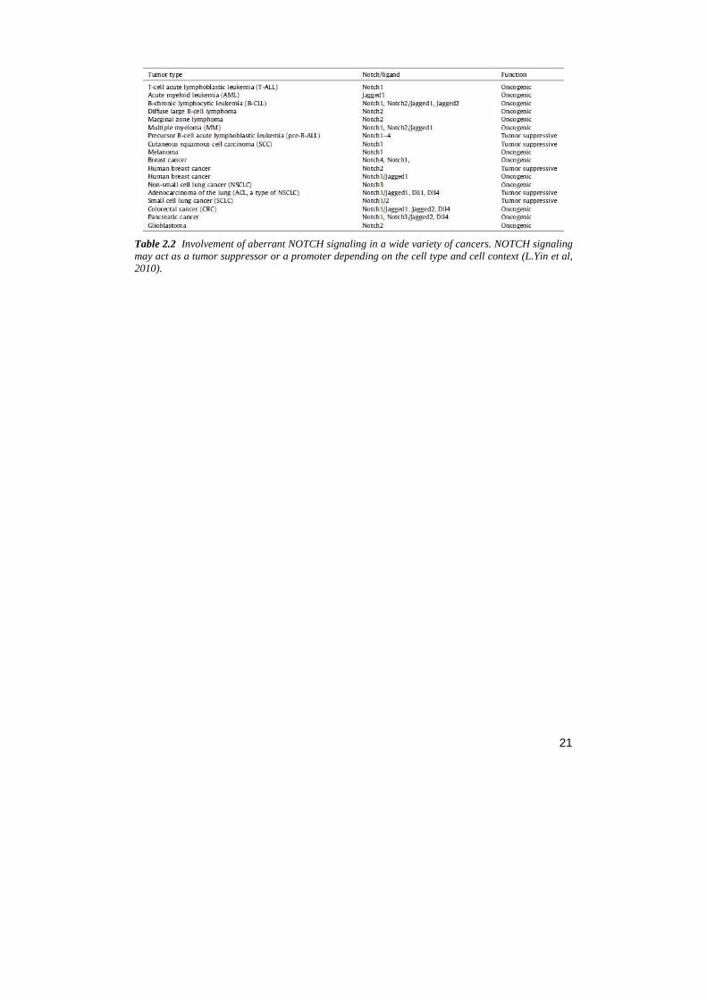
Figure 2.1.2. Notch signaling in lymphopoiesis. (Radtke, 2004) Bone marrow (BM) HSCs are maintained through selfrenewal in stem cell niches in close contact with stromal and/or other hematopoietic cells. Jagged1 (J1)–Notch1 (N1) interactions may influence the process of self-renewal. After commitment to the lymphoid lineage, early lymphocyte precursors (ELP) continue differentiation into either B or T cells. In the bone marrow, Notch (N) signalling must be ‘off’ to allow pro-B cells to progress through pre-B I and pre-B II stages to immature B cells (Imm B). After migration to the periphery, interaction of Delta1 (D1) with Notch2 (N2):CSL induces transitional B cells (Trans B) to become MZB cells. In contrast, Mint induces transitional B cells to become FoB cells. In the thymus, the early thymus precursor (ETP) requires a Notch1 (N1):CSL signal to develop into pro-T cells; otherwise, B lineage development occurs by default. This signal is mediated through Delta1. Pro-T cells then require Notch1 signals to efficiently develop into pre-T cells of the αβ�lineage and to undergo successful pre-TCR mediated signaling. It is unclear whether Notch signaling influences γδ�T cell development. Double-positive (DP) thymocytes mature into conventional CD4 or CD8 T cells and then migrate to the periphery, where CD4 T cells undergo further differentiation into TH1 or TH2 cells. This latter lineage split may be influenced by D1:Notch3 (N3) signaling. CD8 T cells undergo further differentiation into cytotoxic-T cell. Regulatory CD25+ CD4 T cells (CD25+TR) develop in the thymus from DP T cells, possibly through N3 signaling. 2.2 NOTCH AND CANCER Given the range of processes that require normal Notch signaling, it is not surprising to find that a number of human diseases and cancer are caused by mutation in components of the Notch pathway and/or in the deregulation of Notch signaling. Consequences of disruption of proper Notch signaling are very diverse (table 2.2).
19
2.2.1 Notch as oncogene Notch deregulation is involved both in solid tumors as breast cancer, skin cancer, neuroblastomas, prostate cancer and cervical cancer (Allenspach, 2002), and in non-solid malignancies, such as leukemia (Weng, 2004) and multiple myeloma (Jundt, 2004). From 90’s to nowadays Notch signaling aberrations have been shown to be linked with several hematological malignancies such as T-cell acute lymphoblastic leukemia (T-ALL), acute myeloid leukemia (AML), lymphoma and MM. The main oncogenic role of Notch can be found in T-ALL, an aggressive neoplasm of immature T-cells. In human leukemia, Notch 1 activation was initially demonstrated in T-ALL harboring the translocation (7;9)(q34;q34.3), a rare chromosomal translocation identified in less than 1% of T-ALL cases. As a result of this rearrangement, a truncated Notch-1 gene is juxtaposed next to the T-cell receptor β locus, leading to the ligand-independent aberrant expression of a constitutively active form of Notch-1 (Koch, 2007). This translocation is rare in T-ALL patients (less than 1%), but approximately 60% of T-ALL cases display activating Notch mutations (Weng, 2004). The majority of mutations are located in the HD (between exons 26 and 27), in the extracellular juxta-membrane (JME) region (exon 28)and PEST (exon 34) domains. HD mutations are typically single amino acid substitutions and small in-frame deletions and insertions that induce ligand-independent activation of Notch, leading to constitutive activation of the Notch signaling pathway (Malecki, 2006). PEST mutations encodes premature stop codons and lead to generation of truncated forms of Notch lacking the PEST domain, resulting in an increased level of active Notch due to its impaired proteasomal degradation (Weng, 2004). The HD and PEST domain mutations were found in trans in 26% and 12.5%, respectively, and in cis in 17.7% of cases examined. These mutant forms of Notch have been demonstrated to increase Notch transcriptional activity in vitro. Mutations in the JME region consist of tandem duplications that cause the expansions of the extracellular juxtamembrane region, leading to increase distance of the NNR-HD complex from the membrane, allowing ligand-independent proteolytic processing of S2 (Sulis, 2008). Given the causative role of Notch-1 mutations in T-ALL, a large number of studies focused on the analysis of Notch mutational status in this malignancy. All the reported mutations in T-ALL affected the Notch-1 isoform, while Notch-2,-3 and -4 were not found to be altered (Lee, 2007). The main way in which abnormal Notch1 activity drives T-ALL is activation of Myc and CyclinD as well as inhibition of p53: all of them promote oncogenesis through increased proliferation, survival and genomic instability. The role of Notch signaling in AML is less clear than in T-ALL. Activating mutations of Notch have been reported but they seems to be a rare event (Palomero, 2006). Chiaramonte and colleagues demonstrate that AML primary sample show high levels of Jagged-1 expression, despite low Notch-1 pathway activation (Chiaramonte, 2005), thus suggesting a Notch-independent pathway driven directly by the Jagged-1 ligand (Ascano, 2003). Regarding B-cell malignancies, Notch deregulation has been detected in Hodgkin’s lymphoma, large B-cell lymphoma, Burkitt’s lymphoma, B-cell chronic lymphocytic leukemia, diffuse large B-cell lymphoma, primary effusion lymphomas associated with Kaposi’s sarcoma herpes virus infection and in Multiple Myeloma (Mirandola, 2011a).
20
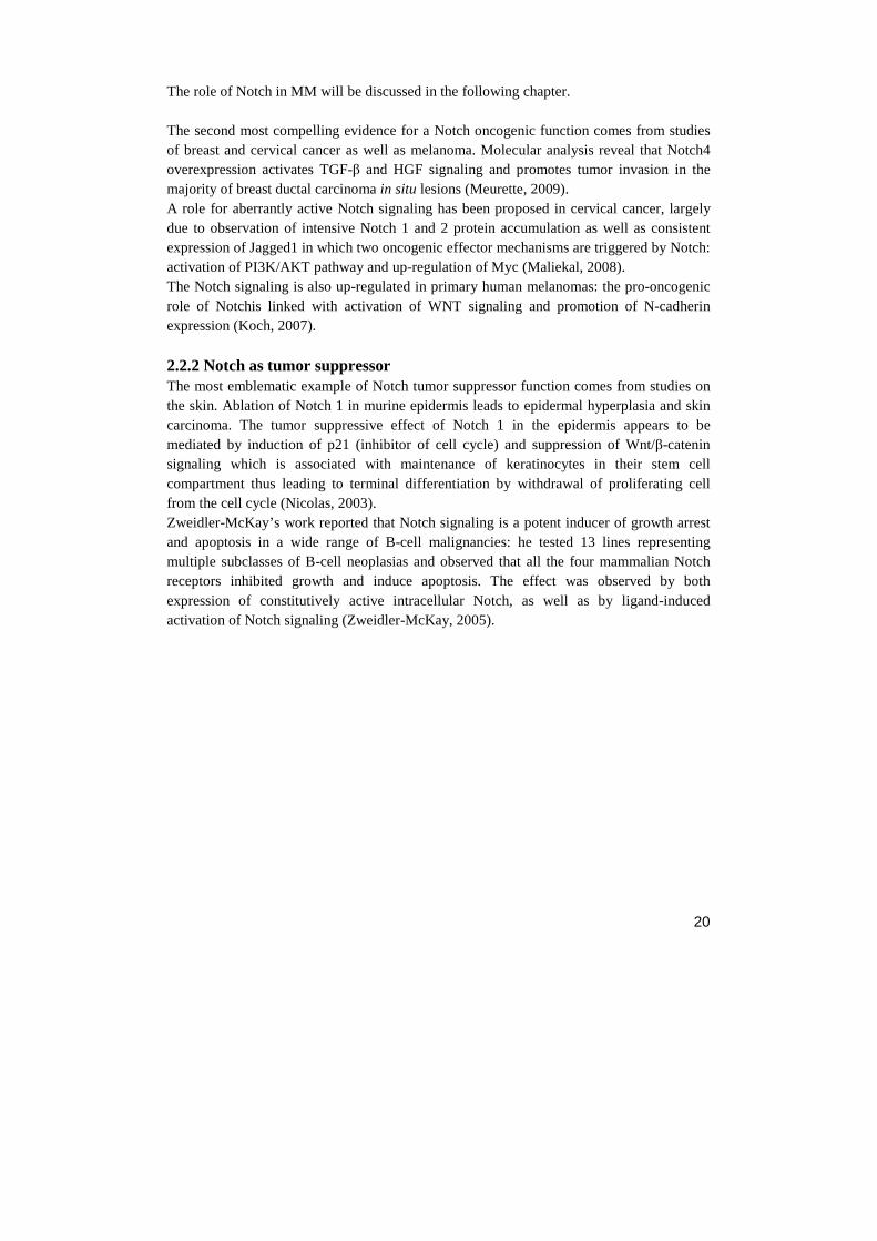
The role of Notch in MM will be discussed in the following chapter. The second most compelling evidence for a Notch oncogenic function comes from studies of breast and cervical cancer as well as melanoma. Molecular analysis reveal that Notch4 overexpression activates TGF-β and HGF signaling and promotes tumor invasion in the majority of breast ductal carcinoma in situ lesions (Meurette, 2009). A role for aberrantly active Notch signaling has been proposed in cervical cancer, largely due to observation of intensive Notch 1 and 2 protein accumulation as well as consistent expression of Jagged1 in which two oncogenic effector mechanisms are triggered by Notch: activation of PI3K/AKT pathway and up-regulation of Myc (Maliekal, 2008). The Notch signaling is also up-regulated in primary human melanomas: the pro-oncogenic role of Notchis linked with activation of WNT signaling and promotion of N-cadherin expression (Koch, 2007).
2.2.2 Notch as tumor suppressor The most emblematic example of Notch tumor suppressor function comes from studies on the skin. Ablation of Notch 1 in murine epidermis leads to epidermal hyperplasia and skin carcinoma. The tumor suppressive effect of Notch 1 in the epidermis appears to be mediated by induction of p21 (inhibitor of cell cycle) and suppression of Wnt/β-catenin signaling which is associated with maintenance of keratinocytes in their stem cell compartment thus leading to terminal differentiation by withdrawal of proliferating cell from the cell cycle (Nicolas, 2003). Zweidler-McKay’s work reported that Notch signaling is a potent inducer of growth arrest and apoptosis in a wide range of B-cell malignancies: he tested 13 lines representing multiple subclasses of B-cell neoplasias and observed that all the four mammalian Notch receptors inhibited growth and induce apoptosis. The effect was observed by both expression of constitutively active intracellular Notch, as well as by ligand-induced activation of Notch signaling (Zweidler-McKay, 2005).
21
Table 2.2 Involvement of aberrant NOTCH signaling in a wide variety of cancers. NOTCH signaling may act as a tumor suppressor or a promoter depending on the cell type and cell context (L.Yin et al, 2010).
22
3. MULTIPLE MYELOMA
Multiple myeloma is a neoplastic plasma-cell disorder that is characterized by clonal proliferation of malignant plasma cells (PCs) in the bone marrow (BM) microenvironment, monoclonal protein in the blood or urine and associated organ dysfunction. It belongs to a group of related paraproteinaemias, namely diseases that produce an immunoglobulin from a single clone that is present at high levels in the serum. They include multiple myeloma (MM), monoclonal gammopathy of undetermined significance (MGUS) and Waldenstrom’s macroglobulinaemia (WM) (Morgan, 2012). MM accounts for 1% of all cancers and about 10% of all hematologic malignancies (Rajkumar, 2012). The American Cancer Society estimates that this year 21.700 new cases (12.190 in men and 9.510 in women) will be diagnosed in the United States, and that 10.710 deaths will occur in 2012 as result of MM (ACS, 2012). The median age at diagnosis is about 65 years (Kyle, 2004a) and is slightly more common in men than in women and is twice as common in African-Americans compared to Caucasians (Landgren, 2009). The presence of somatic hypermutations of the immunoglobulin variable region genes in myeloma plasma cells suggests that malignant transformation occurs in a B cell that has traversed the germinal centers of lymph nodes. However, the hypoproliferative nature of myeloma has led to the hypothesis that the bulk of the tumor arises from a transformed B cell with the capacity for both self-renewal and production of terminally differentiated progeny (Harousseau, 2004). Almost all patient with MM evolve from the asymptomatic premalignant stage of MGUS, which affects at least 3% of adults older than 50 years (Weiss, 2009). Moreover, in some cases, MM arises from another asymptomatic but more advanced premalignant stage, referred as smoldering multiple myeloma (SMM). The risk to progress from SMM to MM was 10% per year in the first 5 years, 3% per year for the next 5 years and 1% per year for the last 10 years, reaching a cumulative probability of progression of 75% at 15 years (Kyle, 2007).
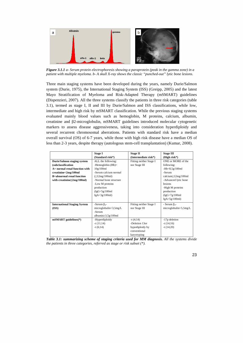
3.1 DIAGNOSIS The diagnosis of MM requires at least 10% or more clonal plasma cell on bone marrow examination or a biopsy proven plasmacytoma and evidence of end-organ damage (hypercalcemia, renal insufficiency, anemia or bone lesions) that is ascribed to the underlying plasma cells disorder. When MM is clinically suspected, patients should be tested for the presence monoclonal proteins (M proteins) through a series of test, such as serum protein electrophoresis (fig. 3.1.1), serum immunofixation and serum-free light chain (FLC) assay (Rajkumar, 2012).
23
Figure 3.1.1 a- Serum protein electrophoresis showing a paraprotein (peak in the gamma zone) in a patient with multiple myeloma. b- A skull X-ray shows the classic “punched-out” lytic bone lesions. Three main staging systems have been developed during the years, namely Durie/Salmon system (Durie, 1975), the International Staging System (ISS) (Greipp, 2005) and the latest Mayo Stratification of Myeloma and Risk-Adapted Therapy (mSMART) guidelines (Dispenzieri, 2007). All the three systems classify the patients in three risk categories (table 3.1), termed as stage I, II and III by Durie/Salmon and ISS classifications, while low, intermediate and high risk by mSMART classification. While the previous staging systems evaluated mainly blood values such as hemoglobin, M proteins, calcium, albumin, creatinine and β2-microglobulin, mSMART guidelines introduced molecular cytogenetic markers to assess disease aggressiveness, taking into consideration hyperdiploidy and several recurrent chromosomal aberrations. Patients with standard risk have a median overall survival (OS) of 6-7 years, while those with high risk disease have a median OS of less than 2-3 years, despite therapy (autologous stem-cell transplantation) (Kumar, 2008).
Stage I (Standard risk*)
Stage II (Intermediate risk*)
Stage III (High risk*)
Durie/Salmon staging system (subclassification A= normal renal function with creatinine<2mg/100ml B=abnormal renal function with creatinine≥2mg/100ml)
ALL the following: -Hemoglobin (Hb)> 10g/100ml -Serum calcium normal (≤12mg/100ml) -Normal bone structure -Low M proteins production (IgG<5g/100ml IgA<3g/100ml)
Fitting neither Stage I nor Stage III
ONE or MORE of the following: -Hb<8,5g/100ml -Serum calcium≥12mg/100ml -Advanced lytic bone lesions -High M proteins production (IgG>7g/100ml IgA>5g/100ml)
International Staging System (ISS)
-Serum β2-microglobulin<3,5mg/L -Serum albumin≥3,5g/100ml
Fitting neither Stage I nor Stage III
- Serum β2-microglobulin>5,5mg/L
mSMART guidelines(*) -Hyperdiploidy -t (11;14) -t (6;14)
-t (4;14) -Deletion 13or hypodiploidy by conventional karyotyping
-17p deletion -t (14;16) -t (14;20)
Table 3.1: summarizing scheme of staging criteria used for MM diagnosis. All the systems divide the patients in three categories, referred as stage or risk subset (*).
24
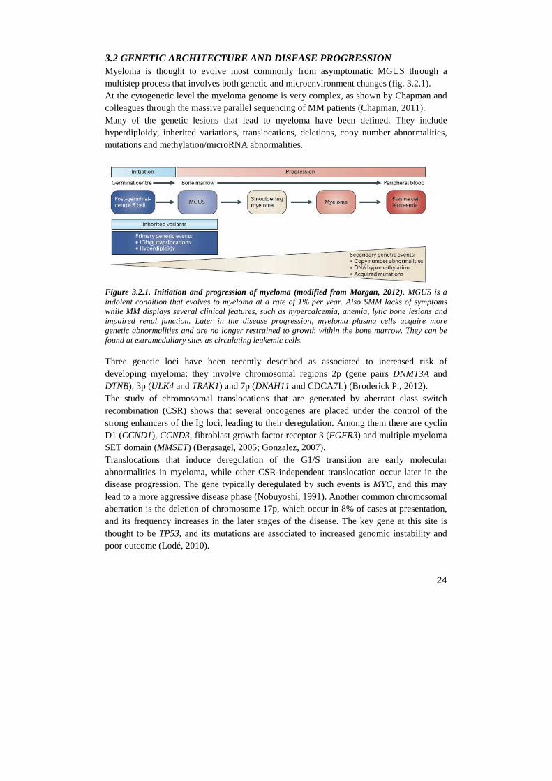
3.2 GENETIC ARCHITECTURE AND DISEASE PROGRESSION Myeloma is thought to evolve most commonly from asymptomatic MGUS through a multistep process that involves both genetic and microenvironment changes (fig. 3.2.1). At the cytogenetic level the myeloma genome is very complex, as shown by Chapman and colleagues through the massive parallel sequencing of MM patients (Chapman, 2011). Many of the genetic lesions that lead to myeloma have been defined. They include hyperdiploidy, inherited variations, translocations, deletions, copy number abnormalities, mutations and methylation/microRNA abnormalities.
Figure 3.2.1. Initiation and progression of myeloma (modified from Morgan, 2012). MGUS is a indolent condition that evolves to myeloma at a rate of 1% per year. Also SMM lacks of symptoms while MM displays several clinical features, such as hypercalcemia, anemia, lytic bone lesions and impaired renal function. Later in the disease progression, myeloma plasma cells acquire more genetic abnormalities and are no longer restrained to growth within the bone marrow. They can be found at extramedullary sites as circulating leukemic cells.
Three genetic loci have been recently described as associated to increased risk of developing myeloma: they involve chromosomal regions 2p (gene pairs DNMT3A and DTNB), 3p (ULK4 and TRAK1) and 7p (DNAH11 and CDCA7L) (Broderick P., 2012). The study of chromosomal translocations that are generated by aberrant class switch recombination (CSR) shows that several oncogenes are placed under the control of the strong enhancers of the Ig loci, leading to their deregulation. Among them there are cyclin D1 (CCND1), CCND3, fibroblast growth factor receptor 3 (FGFR3) and multiple myeloma SET domain (MMSET) (Bergsagel, 2005; Gonzalez, 2007). Translocations that induce deregulation of the G1/S transition are early molecular abnormalities in myeloma, while other CSR-independent translocation occur later in the disease progression. The gene typically deregulated by such events is MYC, and this may lead to a more aggressive disease phase (Nobuyoshi, 1991). Another common chromosomal aberration is the deletion of chromosome 17p, which occur in 8% of cases at presentation, and its frequency increases in the later stages of the disease. The key gene at this site is thought to be TP53, and its mutations are associated to increased genomic instability and poor outcome (Lodé, 2010).
25
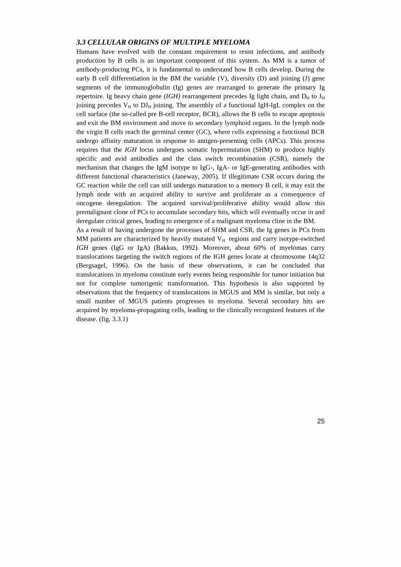
3.3 CELLULAR ORIGINS OF MULTIPLE MYELOMA Humans have evolved with the constant requirement to resist infections, and antibody production by B cells is an important component of this system. As MM is a tumor of antibody-producing PCs, it is fundamental to understand how B cells develop. During the early B cell differentiation in the BM the variable (V), diversity (D) and joining (J) gene segments of the immunoglobulin (Ig) genes are rearranged to generate the primary Ig repertoire. Ig heavy chain gene (IGH) rearrangement precedes Ig light chain, and DH to JH joining precedes VH to DJH joining. The assembly of a functional IgH-IgL complex on the cell surface (the so-called pre B-cell receptor, BCR), allows the B cells to escape apoptosis and exit the BM environment and move to secondary lymphoid organs. In the lymph node the virgin B cells reach the germinal center (GC), where cells expressing a functional BCR undergo affinity maturation in response to antigen-presenting cells (APCs). This process requires that the IGH locus undergoes somatic hypermutation (SHM) to produce highly specific and avid antibodies and the class switch recombination (CSR), namely the mechanism that changes the IgM isotype to IgG-, IgA- or IgE-generating antibodies with different functional characteristics (Janeway, 2005). If illegitimate CSR occurs during the GC reaction while the cell can still undergo maturation to a memory B cell, it may exit the lymph node with an acquired ability to survive and proliferate as a consequence of oncogene deregulation. The acquired survival/proliferative ability would allow this premalignant clone of PCs to accumulate secondary hits, which will eventually occur in and deregulate critical genes, leading to emergence of a malignant myeloma cline in the BM. As a result of having undergone the processes of SHM and CSR, the Ig genes in PCs from MM patients are characterized by heavily mutated VH regions and carry isotype-switched IGH genes (IgG or IgA) (Bakkus, 1992). Moreover, about 60% of myelomas carry translocations targeting the switch regions of the IGH genes locate at chromosome 14q32 (Bergsagel, 1996). On the basis of these observations, it can be concluded that translocations in myeloma constitute early events being responsible for tumor initiation but not for complete tumorigenic transformation. This hypothesis is also supported by observations that the frequency of translocations in MGUS and MM is similar, but only a small number of MGUS patients progresses to myeloma. Several secondary hits are acquired by myeloma-propagating cells, leading to the clinically recognized features of the disease. (fig. 3.3.1)
26
Figure 3.3.1. MM genesis hypothesis (Gonzalez, 2007).
3.4 THE BONE MARROW MICROENVIRONMENT IN MULTIPLE MYELOMA The close interaction between malignant cells and the local microenvironment where they reside is a feature that MM shares with a broad spectrum of solid tumors and hematological neoplasias. The bone marrow microenvironment consists of cellular and non-cellular elements. Cell components include hematopoietic stem cells (HSCs), progenitor cells, immune cells, erythrocytes, BM fibroblast-like stromal cells (BMSCs), vascular endothelial cells, osteoclasts and osteoblasts (fig. 3.4.1). The non-cellular elements are represented by extracellular matrix (ECM) proteins, such as fibronectin, collagen, laminin and osteopontin.
27
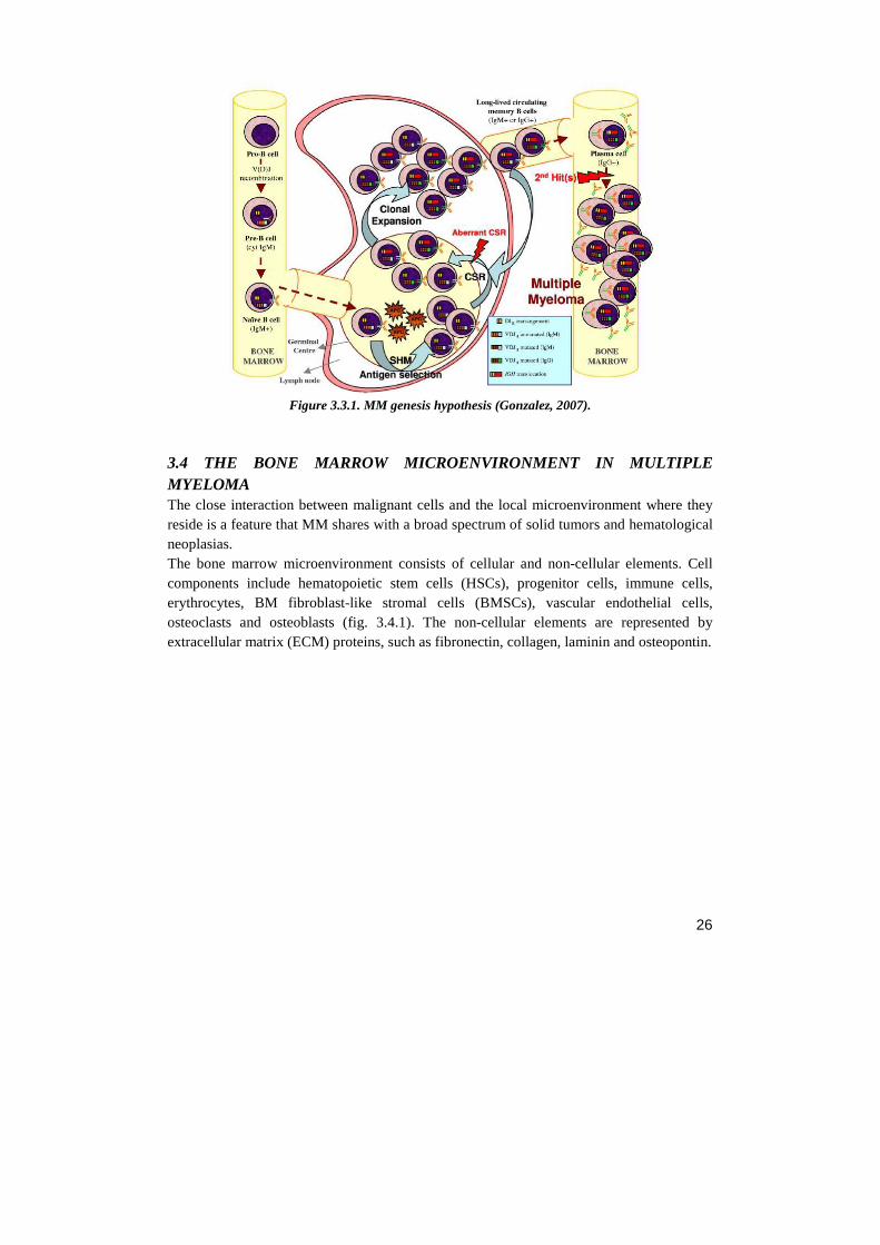
Figure 3.4.1. Interaction between malignant plasma cells and bone marrow in MM (Palumbo, 2011). The bone marrow niche represent a crowded stage in which the myeloma propagating cells have the main role in disease development and are supported by several secondary actors in disease progression. As part of the interaction between plasma cells and stromal cells, adhesion is mediated by cell-adhesion molecules, such as vascular-cell adhesion molecule 1 (VCAM1) and integrin alpha 4 (VLA-4). This interaction increases the production of growth factors, such as interleukin- 6 and vascular endothelial growth factor (VEGF), which stimulates both plasma cells and angiogenesis. The increased osteoclast activity is due to an imbalance in the ratio between receptor activator of nuclear factor κB (RANK) and osteoprotegerin (OPG) as a result of enhanced production of RANK ligand (RANKL) and reduced production of OPG. Osteoblast activity is also suppressed by the production of dickkopf homolog 1 (DKK1) by plasma cells. Moreover, plasma cells can inhibit a key transcription factor for osteoblasts, runt-related transcription factor 2, causing a reduction in differentiation from precursors to mature osteoblasts. The adhesion of plasma cells to stromal cells up-regulates many cytokines with angiogenic activity, in particular interleukin-6 and VEGF. Osteoclasts that are activated by stromal cells can also sustain angiogenesis by secreting osteopontin. Chromosomal abnormalities can cause overproduction of receptors on myeloma cells. The 1q21 amplification causes an increase in interleukin-6 receptor and consequently an increase in growth mediated by interleukin-6. CCR1 denotes chemokine receptor 1, CD40L (or CD40LG) CD40 ligand, FGFR3 fibroblast growth factor receptor 3, HGF hepatocyte growth factor, ICAM1 intercellular adhesion molecule 1, IGF1 insulin-like growth factor 1, MIP1α macrophage inflammatory protein 1 α, MUC1 cell-surface–associated mucin 1, and NF-κB nuclear factor κB.
Once myeloma cells are within the bone marrow, they localize in close proximity to stromal cells, forming specialized tumor niche that support plasma cells survival. The direct interaction of MM cells with BM microenvironment cells in fact, activate signaling
28
pathway mediating growth, survival, drug resistance and the migration of MM cells (Hideshima, 2002a), as well as osteoclastogenesis (Roodman, 2006), angiogenesis (Ribatti, 2006) and secretion of several soluble factors, such as interleukin 6 (IL-6) (Chauhan, 1996), vascular endothelial growth factor (VEGF) (Podar K., 2001), stromal cell-derived factor 1 (SDF-1) (Hideshima, 2002b) and insulin-like growth factor (IGF1) (Mitsiades, 2004). Both homotypic and heterotypic adhesion of MM cells to either BMSCs or ECM are mediated through several adhesion molecules, i.e. CD44, very late antigen 4 (VLA-4), VLA-5, intracellular adhesion molecule (ICAM-1), NCAM, syndecan 1 and MPC-1. Excessive activation of Notch pathway has been described in MM, resulting in increased secretion of MM plasma cell survival factors IL-6 and VEGF (Houde, 2004). The Notch role in MM will be deeply discussed afterwards. In the following paragraphs is reported a focus on three key elements of the interaction between myeloma and BM niche: the osteoclastogenesis, the adhesion molecules and the soluble factors and their receptors.
3.4.1 Osteoclastogenesis The cellular interplay between MM cells and BM microenvironment mediates the formation of bone lesions. MM growth is associated with increased numbers of osteoclasts and suppression of osteoblastogenesis in areas adjacent to tumor foci. These effects are frequently described to establish a “vicious cycle” between tumor cells and surrounding environment: myeloma induces osteoclastogenesis and osteoclasts induce myeloma growth (Sezer, 2009). The molecular mechanisms by which myeloma cells stimulates osteoclasts activity are multifactorial and involve osteoclasts differentiation and survival factors that are produced by microenvironmental cells and myeloma cells. Several osteoclastogenic factors have been described to be involved in MM-induced osteoclasts activity: receptor activator of NF-κB ligand (RANKL), inflammatory protein-1 alpha (MIP-1α) , SDF-1α, IL-3, IL-6 and TNFα. RANKL is a member of the tumor necrosis factor superfamily and is produced mainly by osteoblastic lineage cells and stromal cells. Its receptor, RANK, is expressed on the surface of osteoclasts precursors and mature osteoclasts. RANKL indices differentiation, formation, fusion and survival of preosteoclasts. Osteoprotegerin (OPG) is a decoy receptor antagonist for RANKL, mainly secreted by osteoblastic lineage and stromal cells. MM cells induce stromal cells to upregulate RANKL and to downregulate OPG (Giuliani, 2001). A balanced RANKL/OPG ratio is essential for normal bone turn over: Qiang and colleagues demonstrated that myeloma cell production of Wnt antagonist dickkopf 1 (DKK1) abrogates the canonical Wnt signaling to commit immature cells to osteoblastogenesis, ultimately increasing RANKL/OPG ratios, resulting in activation of osteoclasts and bone resorption (Qiang, 2008). MIP-1α belongs to the RANTES family of chemokines and is chemotactic for osteoclasts precursors and promotes osteoclastogenesis by increasing production of RANKL and IL-6 (Choi, 2001). In addition to osteoclastogenic factor produced by MM cells, it has been
29
reported that myeloma cells form themselves multinucleated cells capable of bone resorption (Silvestris, 2009). SDF-1α is directly responsible for chemotactic recruitment, development and survival of human osteoclasts (Wright, 2005). Moreover, elevated serum levels of SDF-1α are associated with osteolytic bone lesions and increased osteoclasts activity in MM patients (Zannettino, 2005). Interestingly, multiple myeloma cell–osteoclast interactions produces the up-regulation of the enzyme Chondroitin synthase 1 (CHSY1), involved in the synthesis of chondroitin sulfate which plays structural roles in cartilage and bone; CHSY1, induces Notch signalling and survival of multiple myeloma cells, and therefore represents a novel therapeutic target (Yin, 2005). As mentioned above, osteoclastogenesis and osteoblastogenesis in the normal bone are finely balanced, but this equilibrium is disrupted in MM: mesenchymal cells (MSCs) isolated from MM patients are genetically and phenotypically abnormal, and have impaired osteogenic potential (Corre, 2007).
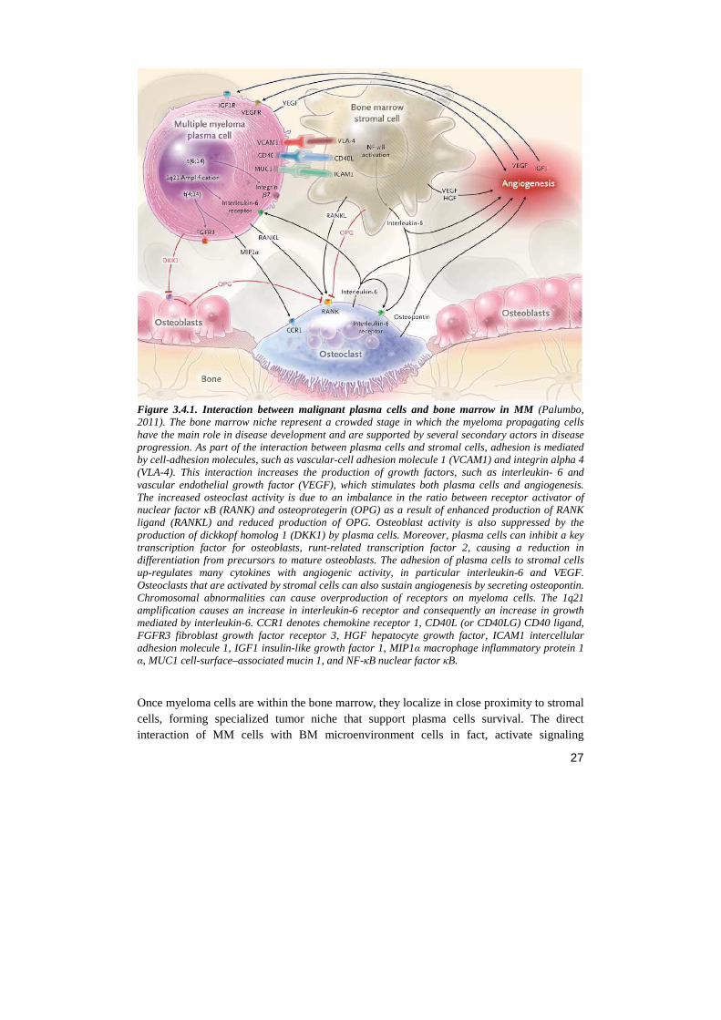
3.4.2 The adhesion molecules Adhesion molecules on MM cells were identified about two decades ago, and specific role in their adhesive interaction with the ECM were attributed to integrins (Uchiyama, 1992). MM cells exhibit preferred adhesion to several ECM constituents, including laminin, collagens and fibronectin (FN), via β1 integrin-mediated adhesion. Integrins are heterodimeric cell surface receptors that mediate adhesion to the ECM and immunoglobulin superfamily molecules. At least 24 distinct integrins heterodimers are formed by the combination of 18 α-subunits and 8 β-subunits. They are essentially expressed by all cell types, including cancer cells (Desgrollier, 2010). A wide range of integrins is expressed by MM cell lines and primary MM cells, but about their specific functional roles still little is known. The best characterized are α4, α5, αv and the β1 subunits. The predominant cellular receptor for FN is α5β1 integrin, also called VLA-5 or CD49e, which is expressed by normal PCs and in the initial stages of MM. Conversely, with the disease progression and on extramedullary MM cells there is a significant down-regulation of this integrin (Pellat-Deceunynck, 1995). In contrast with the monogamy of the interactions between integrin α5β1 and FN, the α4 subunit can form a heterodimer with β1 subunit and bind FN or vascular cell adhesion molecule 1 (VCAM-1), or pair with the β7 subunit to bind mucosal addressin call adhesion molecule (MAdCAM-1). Unlike the expression pattern of α5β1 integrin, α4β1 integrin (also referred as VLA-4 or CD49d) is expressed by all plasma cells, both normal and malignant (Pals, 2007), and it was found to be over-expressed in drug-resistant MM cells (Damiano, 1999). The β7 subunit can pair with αE subunit to mediate the adhesion of MM cells to BM stromal cells, via E-cadherin binding. The activity of α4β1 integrin is regulated both by ligand binding and by conformational changes induced by inside-out signaling (Chigaev, 2009). MM derived cell lines express α4β1 integrin, albeit al low/moderate activation status, and their cell-surface levels can be up-regulated by cytokines, e.g. TNFα (Hideshima, 2001) (fig. 3.4.2).
30
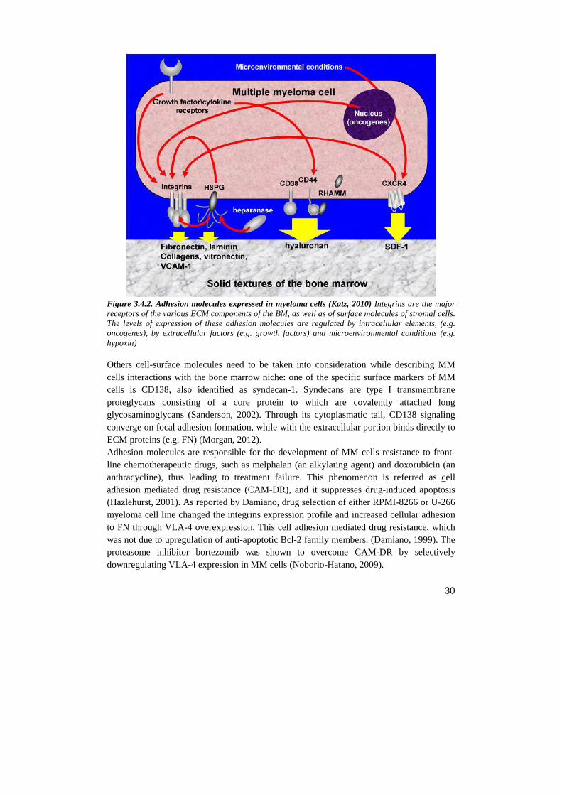
Figure 3.4.2. Adhesion molecules expressed in myeloma cells (Katz, 2010) Integrins are the major receptors of the various ECM components of the BM, as well as of surface molecules of stromal cells. The levels of expression of these adhesion molecules are regulated by intracellular elements, (e.g. oncogenes), by extracellular factors (e.g. growth factors) and microenvironmental conditions (e.g. hypoxia)
Others cell-surface molecules need to be taken into consideration while describing MM cells interactions with the bone marrow niche: one of the specific surface markers of MM cells is CD138, also identified as syndecan-1. Syndecans are type I transmembrane proteglycans consisting of a core protein to which are covalently attached long glycosaminoglycans (Sanderson, 2002). Through its cytoplasmatic tail, CD138 signaling converge on focal adhesion formation, while with the extracellular portion binds directly to ECM proteins (e.g. FN) (Morgan, 2012). Adhesion molecules are responsible for the development of MM cells resistance to front-line chemotherapeutic drugs, such as melphalan (an alkylating agent) and doxorubicin (an anthracycline), thus leading to treatment failure. This phenomenon is referred as cell adhesion mediated drug resistance (CAM-DR), and it suppresses drug-induced apoptosis (Hazlehurst, 2001). As reported by Damiano, drug selection of either RPMI-8266 or U-266 myeloma cell line changed the integrins expression profile and increased cellular adhesion to FN through VLA-4 overexpression. This cell adhesion mediated drug resistance, which was not due to upregulation of anti-apoptotic Bcl-2 family members. (Damiano, 1999). The proteasome inhibitor bortezomib was shown to overcome CAM-DR by selectively downregulating VLA-4 expression in MM cells (Noborio-Hatano, 2009).
31
3.4.3 Soluble factors and their receptors Since MM mainly progresses in the bone marrow, signals from this microenvironment play a critical role in the maintaining plasma cell growth, survival, migration, drug resistance and angiogenesis Reciprocal interactions between PCs and BM cells, namely HSCs, stromal cells, osteoblasts, osteoclasts, vascular endothelial cells and immune cells are mediated by an array of cytokines and receptors. PCs in the BM secrete tumor necrosis factor-α (TNFα), transforming growth factor-β (TGF-β), VEGF, Angiopoietin-1, FGF-2 and matrix metalloproteases (MMPs). Moreover, the cell-cell interactions mediated by adhesion molecules between PCs and BM cell trigger transcription and secretion by the latter of cytokines, such as IL-6, VEGF, SDF-1 (CXCL12), MCP-1 (CCL2) Hepatocyte growth factor-scatter factor (HGF-SF) and IGF-1 (Ribatti, 2006; Hideshima, 2001; Barillé, 1997; Dankbar, 2000; Ferlin, 2000; Alsayed, 2007). The first cytokine described that placed the focus on BMSCs-MM interplay was probably IL-6: in 90’s it was known that IL-6 induces in vitro growth of freshly isolated MM cells and that MM cells express the IL-6 receptor (IL-6R). Moreover, several MM cell lines have been described to be responsive and produce IL-6, thus leading to hypothesize an autocrine pattern. In the same years, many studies showed that BMSCs are the major source of IL-6 and that, although all human MM-derived cell lines express IL-6R mRNA, only a subset express IL-6 mRNA. In 1996, Chauhan and colleagues finally showed that adhesion of MM cell lines to BMSCs and BMSC lines resulted in significant increase in IL-6 secretion by BMSCs, thus supporting tumor growth. Noteworthy, through gene reporter assays, they also indicate involvement of NF-κB in regulation of IL-6 transcription triggered in BMSCs (Chauhan, 1996). Various soluble factors have been shown to mediate IL-6 secretion by BMSCs or MM cells, e.g. IL-1α, IL-1β, TNFα and VEGF. In MM, VEGF is expressed and secreted by tumor cells as well as BMSCs. It induces proliferation through Raf-1-MEK-extracellular-signal-regulated protein kinase (ERK) pathway, it triggers migration of human MM cells through a protein kinase C (PKC)-dependent cascade (Podar, 2001) and it stimulates the expression of IL-6 by microvascular endothelial cells and BMSCs (Dankbar, 2000). TNFα is known to be a potent mediator of inflammation and bone resorption expressed by BMSCs and PCs from myeloma patients. Several studies confirmed a central role for this cytokine in the growth and survival of MM cells in the BM milieu, given that TNFα induces proliferation expression of ICAM-1, VCAM-1 and VLA-4 and MAPK/ERK activation in MM cells, while IL-6 secretion, NF-κB activation and expression of ICAM-1 and VCAM-1 in BMSCs (Hideshima, 2001). HGF-SF is a pleiotropic cytokine that induces complex biological responses in target cells, including motility and growth. Its biological effects are transduced via the transmembrane tyrosine kinase Met, while syndecan-1 (CD138) strongly promotes HGF-induced signaling through Met, thereby acting as a co-receptor (Derksen, 2002). MM cell lines and BM plasma cells express both HGF-SF and its receptor Met (Borset, 1996). MM is a tumor with a high capacity to destroy the bone matrix thanks to matrix metalloproteases (MMPs) expression (Barillé, 1997). MMPs are a family of zinc-dependent
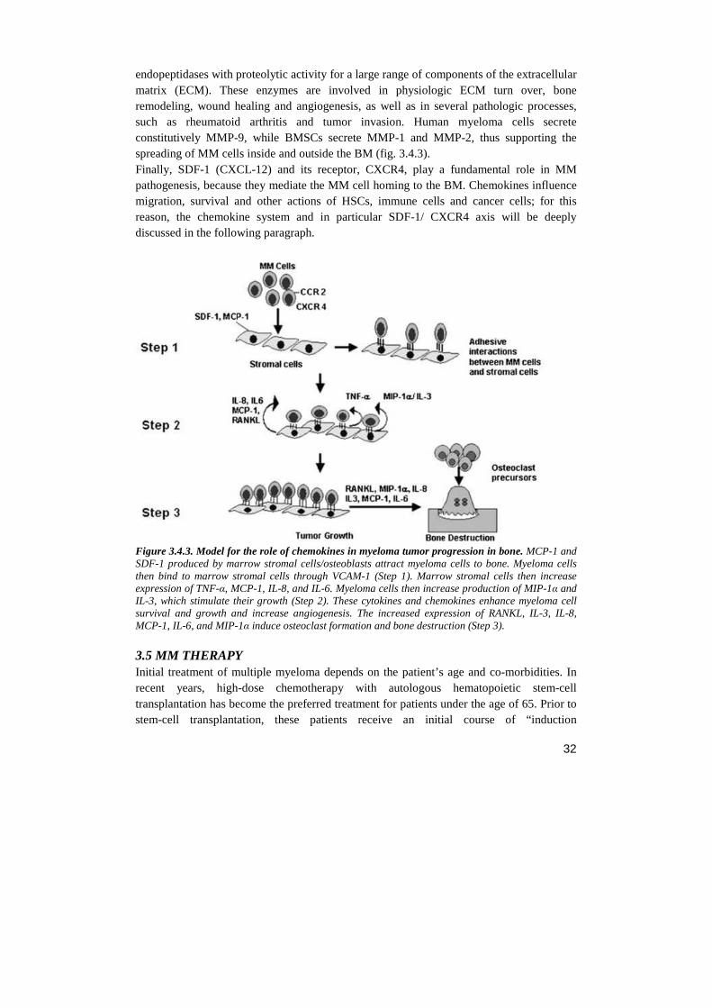
32
endopeptidases with proteolytic activity for a large range of components of the extracellular matrix (ECM). These enzymes are involved in physiologic ECM turn over, bone remodeling, wound healing and angiogenesis, as well as in several pathologic processes, such as rheumatoid arthritis and tumor invasion. Human myeloma cells secrete constitutively MMP-9, while BMSCs secrete MMP-1 and MMP-2, thus supporting the spreading of MM cells inside and outside the BM (fig. 3.4.3). Finally, SDF-1 (CXCL-12) and its receptor, CXCR4, play a fundamental role in MM pathogenesis, because they mediate the MM cell homing to the BM. Chemokines influence migration, survival and other actions of HSCs, immune cells and cancer cells; for this reason, the chemokine system and in particular SDF-1/ CXCR4 axis will be deeply discussed in the following paragraph.
Figure 3.4.3. Model for the role of chemokines in myeloma tumor progression in bone. MCP-1 and SDF-1 produced by marrow stromal cells/osteoblasts attract myeloma cells to bone. Myeloma cells then bind to marrow stromal cells through VCAM-1 (Step 1). Marrow stromal cells then increase expression of TNF-α, MCP-1, IL-8, and IL-6. Myeloma cells then increase production of MIP-1α and IL-3, which stimulate their growth (Step 2). These cytokines and chemokines enhance myeloma cell survival and growth and increase angiogenesis. The increased expression of RANKL, IL-3, IL-8, MCP-1, IL-6, and MIP-1α induce osteoclast formation and bone destruction (Step 3).
3.5 MM THERAPY Initial treatment of multiple myeloma depends on the patient’s age and co-morbidities. In recent years, high-dose chemotherapy with autologous hematopoietic stem-cell transplantation has become the preferred treatment for patients under the age of 65. Prior to stem-cell transplantation, these patients receive an initial course of “induction
33
chemotherapy” comprising of cyclophosphamide and a white blood cell stimulating drug: it was shown that chemotherapy treatment induces hematopoietic stem-cell migration from the BM to the peripheral blood vessels; then blood-forming stem cells are removed from the patient's blood by a process called leukapheresis and are preserved. After the high-dose chemotherapy, which is toxic for BM, stem cells are given back to the patient to reconstitute its BM. It is not curative, but does prolong overall survival and complete remission. Also allogeneic stem cell transplantation, of a healthy person’s stem cells into the affected patient, has the potential for a cure, but is only available to a small percentage of patients. The MM patient can receive treatment with a variety of agents, including chemotherapy, corticosteroids, immunomodulating agents, proteasome inhibitor, or a combination thereof. Novel biologically based treatments target not only the MM cell, but also MM cell-host interactions and the BM microenvironment (fig. 3.5.1). Category of drugs used in MM therapy: Immunomodulatory Drugs Thalidomide is a derivative of glutamic acid. Thalidomide’s mechanism of action in MM is not fully understood. Proposed mechanism(s) include the inhibition of TNF-α production, prevention of free-radical-mediated DNA damage, suppression of angiogenesis by blocking the angiogenic growth factors basic fibroblast growth factor (bFGF) and/or VEGF, induction of apoptosis or G1 growth arrest in drug resistant MM cells and modulation the binding of MM cells to BMSCs trough the alteration of cellular adhesion molecules expression. Thalidomide and its derivatives also block the induction of cytokine (such as IGF1, IL-6 and VEGF) secretion that is triggered by MM cell binding to BMSCs, and augment natural-killer-cell and T-cell activity against myeloma cells by stimulating their proliferation and the secretion of interleukin 2 and interferon-γ. Finally, Thalidomide may also inhibit the activity NF-κB and the enzymes cyclo-oxygenase -1 and -2. Side effects - most importantly, constipation, somnolence, teratogenicity and neuropathy - are typically dose dependent but they were not observed in novel immunomodulatory drugs, such as Lenalidomide. Proteasome Inhibitors Bortezomib is a first-in-class proteasome inhibitor. Bortezomib targets the 26S proteasome, a multicatalytic proteinase complex involved in degradation of cyclin and cyclin-dependent kinase inhibitor (CKI) proteins, thereby regulating cell-cycle progression. Bortezomib inhibits NF-κB activation and nuclear translocation by protecting from 26S degradation its inhibitor IκBα, a protein that is constitutively bound to cytosolic NF-κB. In fact, degradation of IκBα by proteasome activates NF-κB , which up-regulates transcription of proteins that promote cell survival and growth, modulates MM cell-adhesion-induced cytokine transcription and secretion in BMSCs, decreases apoptosis susceptibility, influences the expression of adhesion molecules on BMSCs/ MM cells and their related binding, and induces drug resistance in myeloma cells.
34
Bortezomib also induces apoptosis through caspase-8 and -9 activation, inhibits IL-6 and BMSC–MM cell adherence-induced p42/p44 MAPK phosphorylation and proliferation in MM cells Bortezomib not only targets the myeloma cell, but also acts in the bone marrow microenvironment by inhibiting the binding of myeloma cells to bone marrow stromal cells and bone marrow-triggered angiogenesis. Alkylating agents Melphalan is an alkylating drug that acts adding an alkyl group to DNA, inducing DNA damage and duplication arrest. Since cancer cells grow faster than normal cells, they die off more quickly, reducing the number of cancerous cells. Unfortunately, the alkylating antineoplastic agent doesn't discriminate between healthy cells and cancerous ones giving these drugs significant side effects. Cyclophosphamide is a nitrogen mustard alkylating agent, the main effect of cyclophosphamide is due to its metabolite phosphoramide mustard which forms DNA crosslinks both between and within DNA strands at guanine N-7 positions (known as interstrand and intrastrand crosslinkages, respectively). This is irreversible and leads to cell death. Cyclophosphamide also decreases the immune system's response to various diseases and conditions Corticosteroids Dexamethasone, prednisone, prednisolone and other synthetic steroids are useful in myeloma treatment because they act as anti-inflammatory and immunosuppressant drugs and can stop white blood cells from traveling to areas where cancerous myeloma cells are causing damage. This decreases the amount of swelling or inflammation in those areas and relieves associated pain and pressure. Recent studies show that in high doses, dexamethasone can kill myeloma cells. Anthracycline antibiotics Doxorubicin works by interacting with DNA by intercalation and inhibition of macromolecular biosynthesis. This inhibits the progression of the enzyme topoisomerase II, which relaxes supercoils in DNA for transcription. Doxorubicin stabilizes the topoisomerase II complex after it has broken the DNA chain for replication, preventing the DNA double helix from being resealed and thereby stopping the process of replication. Topoisomerase inhibitors Mitoxantrone is an anthracenedione antineoplastic agent. It is a type II topoisomerase inhibitor and it disrupt DNA synthesis and DNA repair in both healthy and cancer cells trough intercalation. It also acs as an immuno-modulator that inhibits T and B-cell activity. Mitotic inhibitor Vincristine is a mitotic inhibitor drug that acts binding to tubulin dimers and inhibiting assembly of microtubule structures. Disruption of the microtubules arrests mitosis in metaphase. Therefore, the mitotic inhibitors affect all rapidly dividing cell types including cancer cells, but also healthy cells.
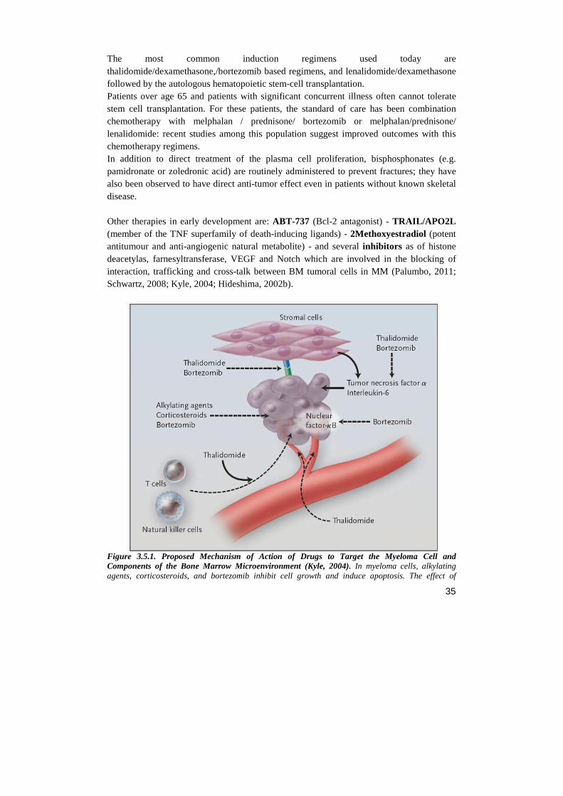
35
The most common induction regimens used today are thalidomide/dexamethasone,/bortezomib based regimens, and lenalidomide/dexamethasone followed by the autologous hematopoietic stem-cell transplantation. Patients over age 65 and patients with significant concurrent illness often cannot tolerate stem cell transplantation. For these patients, the standard of care has been combination chemotherapy with melphalan / prednisone/ bortezomib or melphalan/prednisone/ lenalidomide: recent studies among this population suggest improved outcomes with this chemotherapy regimens. In addition to direct treatment of the plasma cell proliferation, bisphosphonates (e.g. pamidronate or zoledronic acid) are routinely administered to prevent fractures; they have also been observed to have direct anti-tumor effect even in patients without known skeletal disease. Other therapies in early development are: ABT-737 (Bcl-2 antagonist) - TRAIL/APO2L (member of the TNF superfamily of death-inducing ligands) - 2Methoxyestradiol (potent antitumour and anti-angiogenic natural metabolite) - and several inhibitors as of histone deacetylas, farnesyltransferase, VEGF and Notch which are involved in the blocking of interaction, trafficking and cross-talk between BM tumoral cells in MM (Palumbo, 2011; Schwartz, 2008; Kyle, 2004; Hideshima, 2002b).
Figure 3.5.1. Proposed Mechanism of Action of Drugs to Target the Myeloma Cell and Components of the Bone Marrow Microenvironment (Kyle, 2004). In myeloma cells, alkylating agents, corticosteroids, and bortezomib inhibit cell growth and induce apoptosis. The effect of
36
bortezomib on myeloma cells is mediated in part by the inhibition of nuclear factor-kB. Thalidomide and bortezomib inhibit the interaction between myeloma cells and stromal cells as well as the production of cytokines such as tumor necrosis factor a and interleukin-6. Thalidomide inhibits angiogenesis and stimulates the immunesurveillance properties of T cells and natural killer cells. Solid arrows indicate stimulation or secretion and dashed arrows inhibition. 3.6 DRUG-RESISTANCE MECHANISMS IN MULTIPLE MYELOMA Multiple myeloma cells manifest intrinsic genetic mechanisms of drug resistance, (owing, for example, to p53 mutations), or can acquire resistance following exposure to conventional chemotherapeutic treatment (for example, through overexpression of the P-glycoprotein that can confer multidrug resistance following exposure to alkylating agents or anthracyclines). In addition, binding of multiple myeloma cells to extracellular matrix proteins induces cell-adhesion-mediated drug resistance (CAMDR) to conventional chemotherapy (for example, adhesion of tumour cells to fibronectin triggers upregulation of p27 and induces nuclear factor-κB (NFκB) activation leading to CAMDR). Moreover, multiple myeloma cells in the bone marrow, both by virtue of cell–cell contact with accessory cells (bone marrow stromal cells (BMSCs), osteoclasts, osteoblasts and endothelial cells) and secretion of growth factors (TGF-β) further induce transcription and secretion of cytokines, which in turn confer drug resistance (for example, interleukin 6 secretion by BMSCs, osteoclasts and endothelial cells abrogates the apoptosis triggered by dexamethasone). Although the precise mechanism is still to be clarified, new therapeutic agents, such as bortezomib, can overcome intrinsic drug resistance, as well as CAMDR and the protective effects of cytokines, and induce multiple myeloma cell cytotoxicity in the bone marrow milieu. These agents can also overcome clinical drug resistance to conventional and high-dose chemotherapies. IL-6 confers resistance to dexamethasone through the activation of JAK/STAT signalling and the upregulation of the antiapoptotic proteins BCL-XL and myeloid cell leukaemia sequence-1(MCL1). In addition, IL-6 activates SRC-homology tyrosine phosphatase 2 (SHP2), which blocks dexamethasone induced activation of RAFTK and apoptosis. Both IL-6 and IGF1 inhibit the drug-induced apoptosis of MM cells through PI3K/AKT signalling and NF-κB activation, with the downstream induction of intracellular inhibitors of apoptosis (IAPs) including FLICE inhibitory protein (FLIP), survivin, cellular inhibitor of apoptosis-2 (cIAP2), A1/BFL1 and X-linked inhibitor of apoptosis protein (XIAP) (Hideshima, 2007).
3.7 NOTCH AND MULTIPLE MYELOMA Notch deregulation is involved in several malignancies behaving as either an oncogene or a tumor suppressor, depending upon cellular context. Notch signaling deregulation characterizes different hematopoietic malignancies. Among these, multiple myeloma (MM) is associated to a deregulation of Notch signaling driven by the overexpression of its ligand Jagged2 by tumor cells. Recently, several studies focused on the role of Notch pathway in MM. The pathological nature of MM lesions makes it a peculiar hematological tumor, strongly depending on the interaction with the microenvironment. Evidences so far
37
available indicate a very complex picture, which involves Notch in the regulation of the interactions between MM plasma cells and the bone marrow niche. As mentioned above, Notch activation is tightly controlled during hematopoietic lineage differentiation and under physiologic conditions, hematopoietic stem cells carry Notch receptors and receive signals from Notch ligands expressed by BM stromal cells, providing signals for stem cell self-renewal, survival and differentiation. This delicate mechanism is exploited by malignant plasma cells which establish complex interactions with BM stromal cells through direct cell-cell contact and secretion of soluble mediators (fig. 3.7.1), thus leading to MM cell proliferation, chemoresistance and bone disease. Despite major advances in the treatment of MM in recent years, it still remains largely incurable and the failure of the current therapeutic strategies is mainly due to the MM cells’ ability to deregulate the complex BM microenvironment. Notch receptors (Notch 1, 2 and 3) are expressed on primary MM cells, BMSCs and osteoclasts (OCL), while Notch ligands (Jagged-1 and Jagged-2) are expressed on MM, BMSCs and macrophages, thus they are able to activate Notch signaling through homotypic as well as heterotypic interactions in MM cells. In these deregulated interaction, Jagged-2 plays a central role as its over-expression was observed in MM patients. Jagged-2 overexpression is caused by epigenetic deregulations including promoter hypomethylation (Houde, 2004) constitutive core promoter acetylation, due to reduced levels of the SMRT co-repressor (Ghoshal, 2009). Jagged-2 deregulation can occur at post-transcriptional level, involving the over-expression of Jagged-2-specific ubiquitin-ligase Skeleotrophin (Takeuchi, 2005). Jagged-2 overexpression is an early event; it is present in MGUS patients as well as in MM ones, whereas it is absent in non-MM samples. Noticeably, Jagged-2 expression levels increase with disease stage (Houde, 2004). MM cells can autonomously activate Notch signaling through homotypic interactions since they simultaneously express Notch-1, -2 and -3 receptors and their ligands; but, although Notch ligands can be detected on MM cells, they are abundantly expressed by stromal cells and macrophages (Fukushima, 2008); consequently, these BM-residing cells can activate Notch signaling in MM cells through heterotypic interactions . As reported by Houde et. al, also Jagged-expressing MM cells can activate Notch signal in stromal cells, leading to increased secretion of IL-6, VEGF and IGF-1 by the stromal cells (Houde, 2004). Indeed, the co-culture of MM cell line with DLL-1+ stromal cells promotes MM clonogenic growth in vitro and also accelerates MM development in vivo (Xu, 2012). Noteworthy, mammalian Notch-1 signaling promotes β1 integrins activation, thus modulating the main adhesion molecules that mediate myeloma-stromal cells interplay (Hodkinson, 2007). Besides the adhesion molecules, Notch pathway also controls the expression and functions of several chemokine receptors, such as CXCR4 in MM (Mirandola, 2011b). In the last few years, the importance of Notch signaling in osteoblasts (OBs) and osteoclasts (OCs) has emerged: Notch overexpression blocks the maturation of OB precursors by
38
opposing canonical Wnt/ β-catenin signaling (Zanotti, 2008). On the other side, a study reported that Notch-2 and Jagged-1 are up-regulated in OC precursors during RANKL-induced osteoclastogenesis and that Notch-2 modulates the RANK signaling in association with NF-κB (Fukishima, 2008). Interestingly, in OCs-MM cells co-cultures, Notch downstream target gene Hes-1 was found upregulated in OCs, as well as the tartrate resistant acid phosphatase-5 gene (TRAP), which correlates with OC function and serves as a specific marker of OC activity (Schwarzer, 2008). Taken together, these evidences suggest that the increased Notch signaling contributes to MM cell-dependent activation of OC through direct cell–cell contact. The outcomes of Notch activation on tumor MM cells are apoptosis inhibition (Nefedova, 2004; Schwarzer, 2008) and decreased sensitivity to chemotherapeutics (Nefedova, 2004); for this reason, inhibition of this pathway was proposed as an emerging strategy for cancer treatment. As described above, Notch activation requires two consecutive proteolytic cleavage steps, followed by the active NICD translocation to the nucleus. So far, blocking the final intra-membraneous cleavage mediated by the γ-secretase complex has been shown as a successful strategy. Nefedova and colleagues first reported that the pharmacologic inhibition of Notch signaling may enhance the effect of chemotherapy in MM: gamma-secretase inhibitors (GSI) treatment induces apoptosis of MM cells in vitro, and enhances drug sensitivity in vivo (Nefedova, 2008a; Nefedova, 2008b). MM cells reside primarily in the BM, where they interact with components of the BM microenvironment, including stromal cells. During the last 20 years, increasing evidences supported the idea that the interplay between tumor cells and BM, through the BM niche, has profound effects on growth, survival and chemo-sensitivity of malignant cells. As claimed by the previous paragraphs, Notch pathway is among the main factors that mediate this interaction.
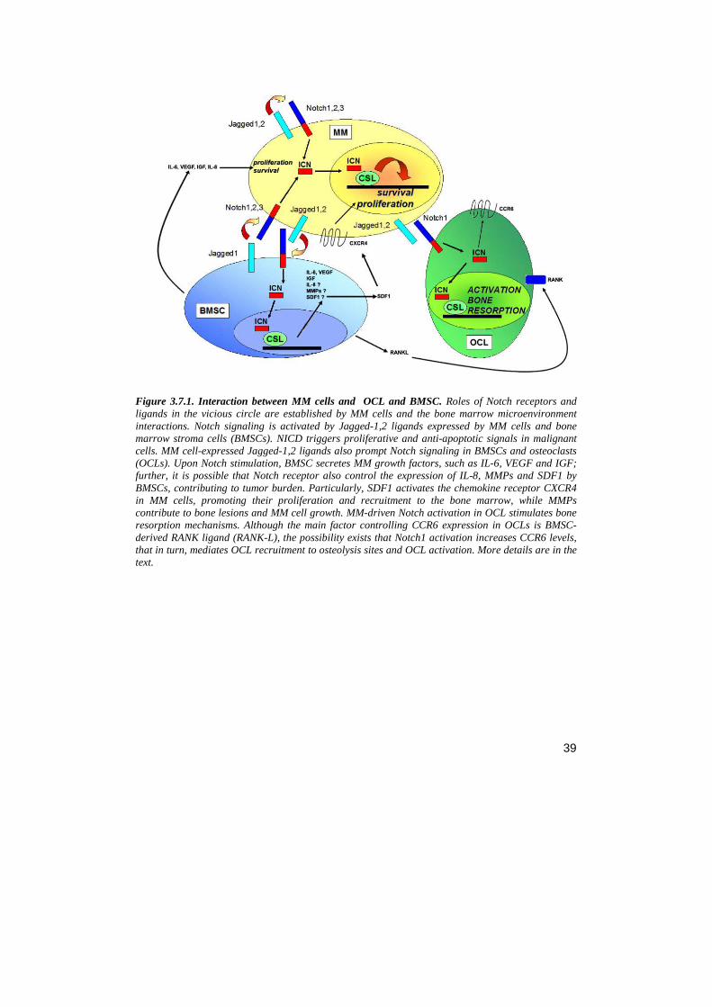
39
Figure 3.7.1. Interaction between MM cells and OCL and BMSC. Roles of Notch receptors and ligands in the vicious circle are established by MM cells and the bone marrow microenvironment interactions. Notch signaling is activated by Jagged-1,2 ligands expressed by MM cells and bone marrow stroma cells (BMSCs). NICD triggers proliferative and anti-apoptotic signals in malignant cells. MM cell-expressed Jagged-1,2 ligands also prompt Notch signaling in BMSCs and osteoclasts (OCLs). Upon Notch stimulation, BMSC secretes MM growth factors, such as IL-6, VEGF and IGF; further, it is possible that Notch receptor also control the expression of IL-8, MMPs and SDF1 by BMSCs, contributing to tumor burden. Particularly, SDF1 activates the chemokine receptor CXCR4 in MM cells, promoting their proliferation and recruitment to the bone marrow, while MMPs contribute to bone lesions and MM cell growth. MM-driven Notch activation in OCL stimulates bone resorption mechanisms. Although the main factor controlling CCR6 expression in OCLs is BMSC-derived RANK ligand (RANK-L), the possibility exists that Notch1 activation increases CCR6 levels, that in turn, mediates OCL recruitment to osteolysis sites and OCL activation. More details are in the text.
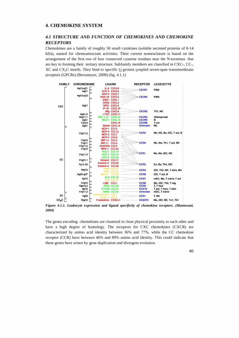
40
4. CHEMOKINE SYSTEM
4.1 STRUCTURE AND FUNCTION OF CHEMOKINES AND CHEMOKINE RECEPTORS Chemokines are a family of roughly 50 small cytokines (soluble secreted proteins of 8-14 kDa), named for chemoattractant activities. Their current nomenclature is based on the arrangement of the first two of four conserved cysteine residues near the N-terminus that are key to forming their tertiary structure. Subfamily members are classified in CXC-, CC-, XC and CX3C motifs. They bind to specific G-protein coupled seven-span transmembrane receptors (GPCRs) (Broxmeyer, 2008) (fig. 4.1.1)
Figure 4.1.1. Leukocyte expression and ligand specificity of chemokine receptors. (Mantovani, 2004)
The genes encoding chemokines are clustered in close physical proximity to each other and have a high degree of homology. The receptors for CXC chemokines (CXCR) are characterized by amino acid identity between 36% and 77%, while the CC chemokine receptor (CCR) have between 46% and 89% amino acid identity. This could indicate that these genes have arisen by gene duplication and divergent evolution.
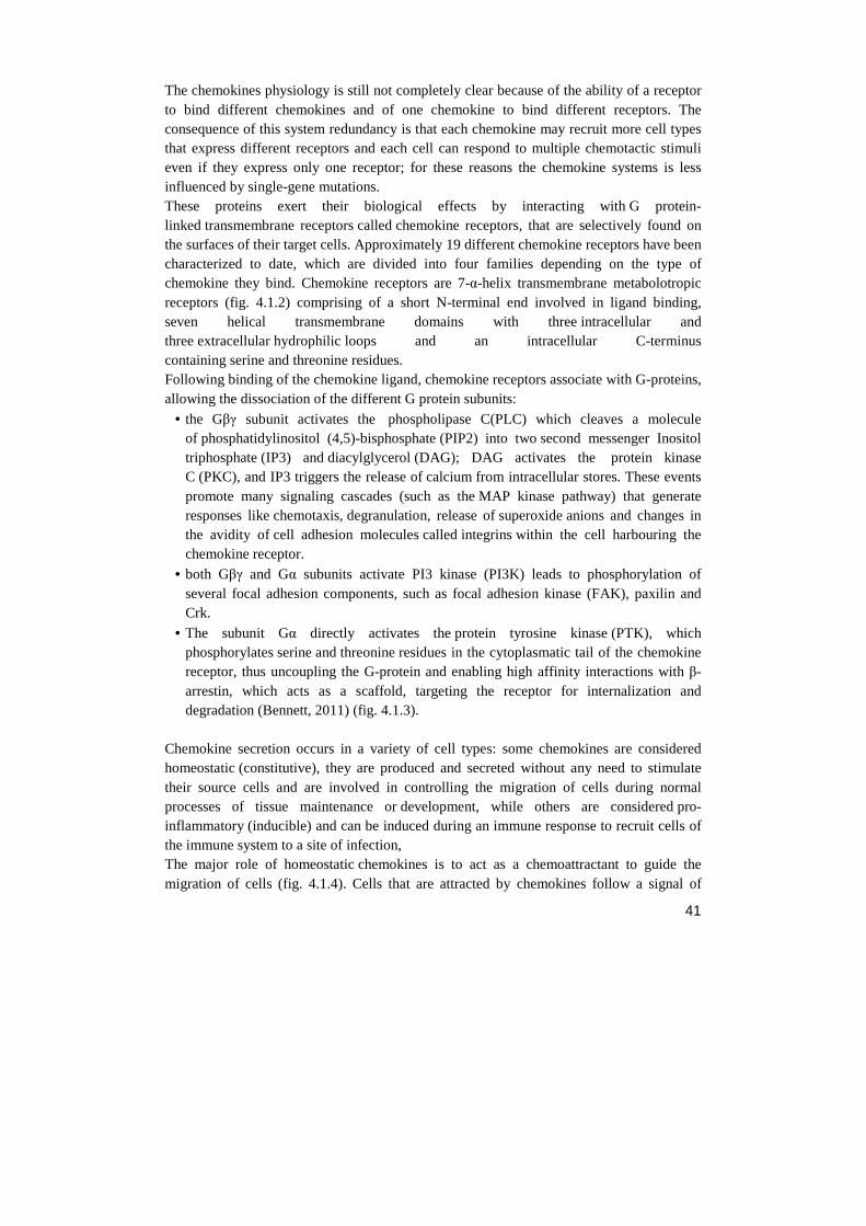
41
The chemokines physiology is still not completely clear because of the ability of a receptor to bind different chemokines and of one chemokine to bind different receptors. The consequence of this system redundancy is that each chemokine may recruit more cell types that express different receptors and each cell can respond to multiple chemotactic stimuli even if they express only one receptor; for these reasons the chemokine systems is less influenced by single-gene mutations. These proteins exert their biological effects by interacting with G protein-linked transmembrane receptors called chemokine receptors, that are selectively found on the surfaces of their target cells. Approximately 19 different chemokine receptors have been characterized to date, which are divided into four families depending on the type of chemokine they bind. Chemokine receptors are 7-α-helix transmembrane metabolotropic receptors (fig. 4.1.2) comprising of a short N-terminal end involved in ligand binding, seven helical transmembrane domains with three intracellular and three extracellular hydrophilic loops and an intracellular C-terminus containing serine and threonine residues. Following binding of the chemokine ligand, chemokine receptors associate with G-proteins, allowing the dissociation of the different G protein subunits:
• the Gβγ subunit activates the phospholipase C(PLC) which cleaves a molecule of phosphatidylinositol (4,5)-bisphosphate (PIP2) into two second messenger Inositol triphosphate (IP3) and diacylglycerol (DAG); DAG activates the protein kinase C (PKC), and IP3 triggers the release of calcium from intracellular stores. These events promote many signaling cascades (such as the MAP kinase pathway) that generate responses like chemotaxis, degranulation, release of superoxide anions and changes in the avidity of cell adhesion molecules called integrins within the cell harbouring the chemokine receptor.
• both Gβγ and Gα subunits activate PI3 kinase (PI3K) leads to phosphorylation of several focal adhesion components, such as focal adhesion kinase (FAK), paxilin and Crk.
• The subunit Gα directly activates the protein tyrosine kinase (PTK), which phosphorylates serine and threonine residues in the cytoplasmatic tail of the chemokine receptor, thus uncoupling the G-protein and enabling high affinity interactions with β-arrestin, which acts as a scaffold, targeting the receptor for internalization and degradation (Bennett, 2011) (fig. 4.1.3).
Chemokine secretion occurs in a variety of cell types: some chemokines are considered homeostatic (constitutive), they are produced and secreted without any need to stimulate their source cells and are involved in controlling the migration of cells during normal processes of tissue maintenance or development, while others are considered pro-inflammatory (inducible) and can be induced during an immune response to recruit cells of the immune system to a site of infection, The major role of homeostatic chemokines is to act as a chemoattractant to guide the migration of cells (fig. 4.1.4). Cells that are attracted by chemokines follow a signal of
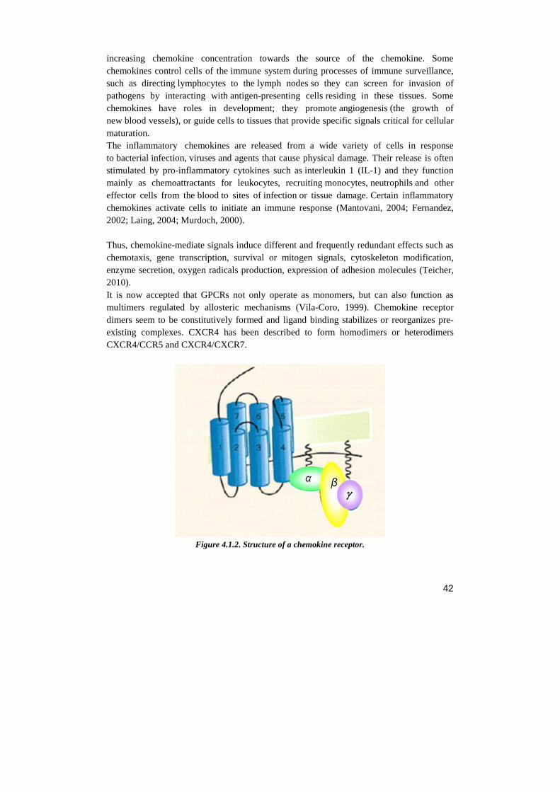
42
increasing chemokine concentration towards the source of the chemokine. Some chemokines control cells of the immune system during processes of immune surveillance, such as directing lymphocytes to the lymph nodes so they can screen for invasion of pathogens by interacting with antigen-presenting cells residing in these tissues. Some chemokines have roles in development; they promote angiogenesis (the growth of new blood vessels), or guide cells to tissues that provide specific signals critical for cellular maturation. The inflammatory chemokines are released from a wide variety of cells in response to bacterial infection, viruses and agents that cause physical damage. Their release is often stimulated by pro-inflammatory cytokines such as interleukin 1 (IL-1) and they function mainly as chemoattractants for leukocytes, recruiting monocytes, neutrophils and other effector cells from the blood to sites of infection or tissue damage. Certain inflammatory chemokines activate cells to initiate an immune response (Mantovani, 2004; Fernandez, 2002; Laing, 2004; Murdoch, 2000). Thus, chemokine-mediate signals induce different and frequently redundant effects such as chemotaxis, gene transcription, survival or mitogen signals, cytoskeleton modification, enzyme secretion, oxygen radicals production, expression of adhesion molecules (Teicher, 2010). It is now accepted that GPCRs not only operate as monomers, but can also function as multimers regulated by allosteric mechanisms (Vila-Coro, 1999). Chemokine receptor dimers seem to be constitutively formed and ligand binding stabilizes or reorganizes pre-existing complexes. CXCR4 has been described to form homodimers or heterodimers CXCR4/CCR5 and CXCR4/CXCR7.
Figure 4.1.2. Structure of a chemokine receptor.
43
Fig.4.1.3 Chemokine receptor signalling in migration and survival/proliferation (O'Hayre, 2008) One of the first events of cell migration involves cell polarization in response to a chemoattractant, whereby some receptors and signalling molecules localize toward the source of the chemoattractant, termed the leading edge, while other molecules distribute away at the trailing edge. This process occurs via chemokine:receptor signalling through the class IB PI3Kγ, which activates Rac and subsequently PAK (p21-activated kinase). Protrusion of the leading edge to move in the direction of the chemoattractant is mediated by actin polymerization and focal adhesions activated as chemokines bind to their receptors. Gi-dependent signalling through PI3K and various protein tyrosine kinases induces the activation of Akt, Rac and Cdc42, which lead to downstream F-actin polymerization. At the trailing edge, activation of ROCK (Rho-associated kinase) downstream of Rho is responsible for actomyosin contraction at the rear so the cell can progress forward. Calcium release and PKC activation downstream of PLC can also play important roles in mediating adhesion events. Activation of FAK, pyk2 (proline-rich tyrosine kinase 2 or FAK-related tyrosine kinase), and other tyrosine kinases are also important in this process. FAK activation is important in establishing focal adhesions and activating other molecules involved in cell movement, such as p130cas, crk and paxillin. Integrin receptors that interact with the ECM to mediate cell adhesion, and secreted proteases such as MMPs that can aid in migration by degrading the ECM, can also be activated downstream of chemokine signalling. As described in more detail in the section on signalling, some chemokines, in normal function or in the context of cancer, also activate a variety of survival and proliferation pathways. Anti-apoptotic/survival signalling, transcription of growth and proliferation-related genes, and transcription of MMPs involved in migration and remodelling the microenvironment are all transduced downstream from Akt, ERK, PKC and tyrosine kinase (e.g. Src) activation. GRK phosphorylation of the C-terminus of chemokine receptors allows β-arrestin to bind, leading to receptor desensitization and internalization. However, β-arrestin binding also leads to the activation of several proteins including Src, MAPK (ERK, p38, JNK) and PI3K. Clearly, there is a large degree of overlap between the upstream signalling molecules underlying these various processes, as these pathways are able to elicit a broad spectrum of effects. Note that continuous lines
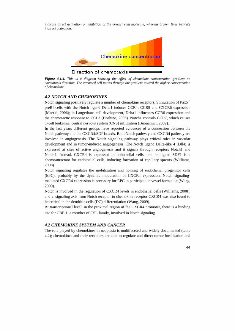
44
indicate direct activation or inhibition of the downstream molecule, whereas broken lines indicate indirect activation.
Figure 4.1.4. This is a diagram showing the effect of chemokine concentration gradient on chemotaxis direction. The attracted cell moves through the gradient toward the higher concentration of chemokine.
4.2 NOTCH AND CHEMOKINES Notch signaling positively regulate a number of chemokine receptors. Stimulation of Pax5-/-
preBI cells with the Notch ligand Delta1 induces CCR4, CCR8 and CXCR6 expression (Maerki, 2006); in Langerhans cell development, Delta1 influences CCR6 expression and the chemotactic response to CCL3 (Hoshino, 2005). Notch1 controls CCR7, which causes T-cell leukemia central nervous system (CNS) infiltration (Buonamici, 2009). In the last years different groups have reported evidences of a connection between the Notch pathway and the CXCR4/SDF1α axis. Both Notch pathway and CXCR4 pathway are involved in angiogenesis. The Notch signaling pathway plays critical roles in vascular development and in tumor-induced angiogenesis. The Notch ligand Delta-like 4 (Dll4) is expressed at sites of active angiogenesis and it signals through receptors Notch1 and Notch4. Instead, CXCR4 is expressed in endothelial cells, and its ligand SDF1 is a chemoattractant for endothelial cells, inducing formation of capillary sprouts (Williams, 2008). Notch signaling regulates the mobilization and homing of endothelial progenitor cells (EPC), probably by the dynamic modulation of CXCR4 expression. Notch signaling-mediated CXCR4 expression is necessary for EPC to participate in vessel formation (Wang, 2009). Notch is involved in the regulation of CXCR4 levels in endothelial cells (Williams, 2008), and a signaling axis from Notch receptor to chemokine receptor CXCR4 was also found to be critical in the dendritic cells (DC) differentiation (Wang, 2009). At transcriptional level, in the proximal region of the CXCR4 promoter, there is a binding
site for CBF-1, a member of CSL family, involved in Notch signaling. 4.2 CHEMOKINE SYSTEM AND CANCER The role played by chemokines in neoplasia is multifaceted and widely documented (table 4.2); chemokines and their receptors are able to regulate and direct tumor localization and
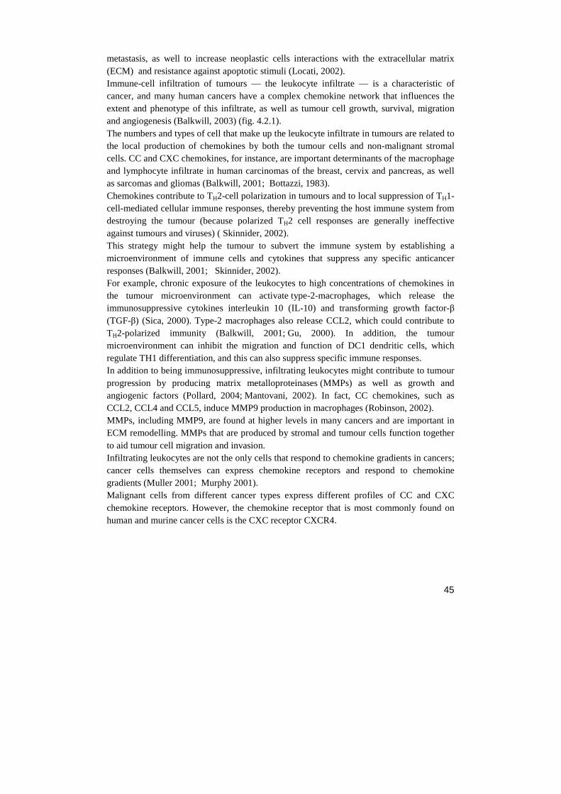
45
metastasis, as well to increase neoplastic cells interactions with the extracellular matrix (ECM) and resistance against apoptotic stimuli (Locati, 2002). Immune-cell infiltration of tumours — the leukocyte infiltrate — is a characteristic of cancer, and many human cancers have a complex chemokine network that influences the extent and phenotype of this infiltrate, as well as tumour cell growth, survival, migration and angiogenesis (Balkwill, 2003) (fig. 4.2.1). The numbers and types of cell that make up the leukocyte infiltrate in tumours are related to the local production of chemokines by both the tumour cells and non-malignant stromal cells. CC and CXC chemokines, for instance, are important determinants of the macrophage and lymphocyte infiltrate in human carcinomas of the breast, cervix and pancreas, as well as sarcomas and gliomas (Balkwill, 2001; Bottazzi, 1983). Chemokines contribute to TH2-cell polarization in tumours and to local suppression of TH1-cell-mediated cellular immune responses, thereby preventing the host immune system from destroying the tumour (because polarized TH2 cell responses are generally ineffective against tumours and viruses) ( Skinnider, 2002). This strategy might help the tumour to subvert the immune system by establishing a microenvironment of immune cells and cytokines that suppress any specific anticancer responses (Balkwill, 2001; Skinnider, 2002). For example, chronic exposure of the leukocytes to high concentrations of chemokines in the tumour microenvironment can activate type-2-macrophages, which release the immunosuppressive cytokines interleukin 10 (IL-10) and transforming growth factor-β (TGF-β) (Sica, 2000). Type-2 macrophages also release CCL2, which could contribute to TH2-polarized immunity (Balkwill, 2001; Gu, 2000). In addition, the tumour microenvironment can inhibit the migration and function of DC1 dendritic cells, which regulate TH1 differentiation, and this can also suppress specific immune responses. In addition to being immunosuppressive, infiltrating leukocytes might contribute to tumour progression by producing matrix metalloproteinases (MMPs) as well as growth and angiogenic factors (Pollard, 2004; Mantovani, 2002). In fact, CC chemokines, such as CCL2, CCL4 and CCL5, induce MMP9 production in macrophages (Robinson, 2002). MMPs, including MMP9, are found at higher levels in many cancers and are important in ECM remodelling. MMPs that are produced by stromal and tumour cells function together to aid tumour cell migration and invasion. Infiltrating leukocytes are not the only cells that respond to chemokine gradients in cancers; cancer cells themselves can express chemokine receptors and respond to chemokine gradients (Muller 2001; Murphy 2001). Malignant cells from different cancer types express different profiles of CC and CXC chemokine receptors. However, the chemokine receptor that is most commonly found on human and murine cancer cells is the CXC receptor CXCR4.
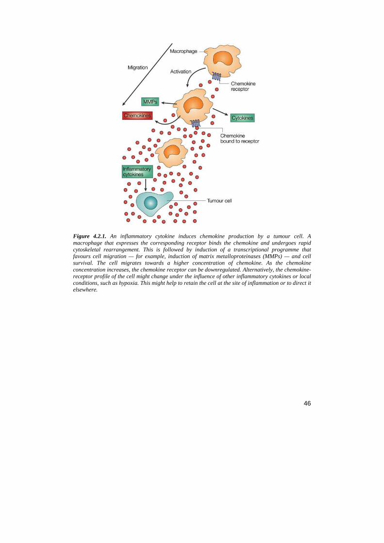
46
Figure 4.2.1. An inflammatory cytokine induces chemokine production by a tumour cell. A macrophage that expresses the corresponding receptor binds the chemokine and undergoes rapid cytoskeletal rearrangement. This is followed by induction of a transcriptional programme that favours cell migration — for example, induction of matrix metalloproteinases (MMPs) — and cell survival. The cell migrates towards a higher concentration of chemokine. As the chemokine concentration increases, the chemokine receptor can be downregulated. Alternatively, the chemokine-receptor profile of the cell might change under the influence of other inflammatory cytokines or local conditions, such as hypoxia. This might help to retain the cell at the site of inflammation or to direct it elsewhere.
47
Table 4.2. Some of the chemokine receptors that are expressed on cancer cells.
4.3 THE CXCR4/SDF-1 AXIS Stromal cell-derived factor 1 (SDF-1 or CXCL-12) is a chemokine originally isolated from a bone marrow stromal cell line (Doranz, 1999). SDF-1 is a 68-amino acid small (8 kDa) cytokine that belongs to the CXC chemokine family. SDF-1 is expressed in two isoforms, SDF-1α and SDF-1β, as two splice variants encoded by single gene mapped in chromosome 10. The two encoded proteins are almost identical, except for the last four amino acids of SDF-1β, which are absent in SDF-1α isoform. Biological and functional differences between the SDF-1α isoforms have not been described. It was long thought that CXCL12 bound exclusively to CXCR4 and that CXCR4 was its sole receptor, however recently CXCR7 was identified as another receptor for CXCL12 (Kryczek, 2007). SDF-1α binds to the receptors trough its RFFESH motif (amino acids 12 to 17). The gene CXCR4 maps on chromosome 2, at 2q21, and produces a 352-amino-acid protein; as the other chemokine receptor, CXCR4 is a 7-α-helix transmembrane metabolotropic receptors. Four cysteine residues situated on the extracellular side of CXCR4 generate two disulfide bonds pin respectively the base of the N-terminal segment to the tip of helix VII and the beginning to the end of the extracellular loop 2; these bonds are necessary to modulate ECL2 and the N-terminal segment (residues 27 to 34) shape to allow the binding of the ligand (Berson, 1996; Beili, 2010); The binding of SDF-1 to CXCR4 occurs on the N-terminus of CXCR4 and the first extracellular loop. CXCR4 is expressed at high levels by various immune cells including monocytes, B cells, and naive T cells in peripheral blood (Aiuti, 1999). SDF-1, unlike most chemokines, is constitutively expressed in a broad range of tissues and therefore may have a role in immune surveillance rather than in inflammation (Bleul, 1996). The most important sources of SDF-1 are bone marrow-, lymph node-, muscle- and
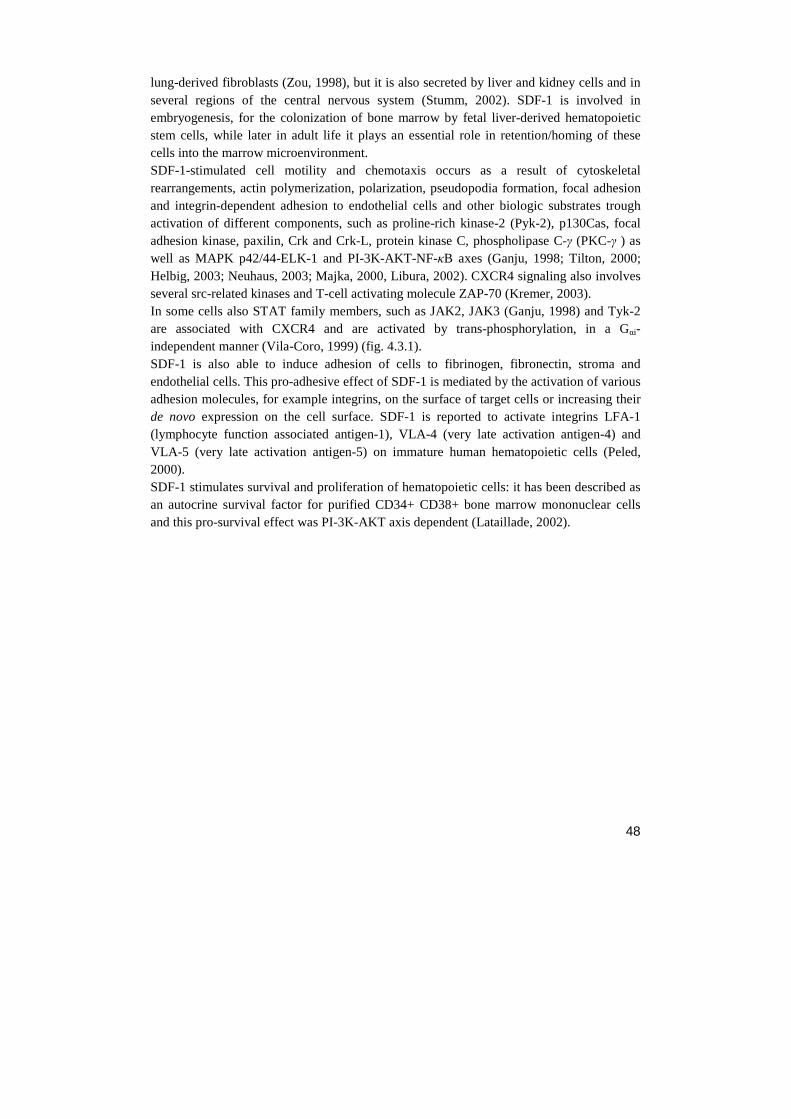
48
lung-derived fibroblasts (Zou, 1998), but it is also secreted by liver and kidney cells and in several regions of the central nervous system (Stumm, 2002). SDF-1 is involved in embryogenesis, for the colonization of bone marrow by fetal liver-derived hematopoietic stem cells, while later in adult life it plays an essential role in retention/homing of these cells into the marrow microenvironment. SDF-1-stimulated cell motility and chemotaxis occurs as a result of cytoskeletal rearrangements, actin polymerization, polarization, pseudopodia formation, focal adhesion and integrin-dependent adhesion to endothelial cells and other biologic substrates trough activation of different components, such as proline-rich kinase-2 (Pyk-2), p130Cas, focal adhesion kinase, paxilin, Crk and Crk-L, protein kinase C, phospholipase C-γ (PKC-γ ) as well as MAPK p42/44-ELK-1 and PI-3K-AKT-NF-κB axes (Ganju, 1998; Tilton, 2000; Helbig, 2003; Neuhaus, 2003; Majka, 2000, Libura, 2002). CXCR4 signaling also involves several src-related kinases and T-cell activating molecule ZAP-70 (Kremer, 2003). In some cells also STAT family members, such as JAK2, JAK3 (Ganju, 1998) and Tyk-2 are associated with CXCR4 and are activated by trans-phosphorylation, in a Gαi-independent manner (Vila-Coro, 1999) (fig. 4.3.1). SDF-1 is also able to induce adhesion of cells to fibrinogen, fibronectin, stroma and endothelial cells. This pro-adhesive effect of SDF-1 is mediated by the activation of various adhesion molecules, for example integrins, on the surface of target cells or increasing their de novo expression on the cell surface. SDF-1 is reported to activate integrins LFA-1 (lymphocyte function associated antigen-1), VLA-4 (very late activation antigen-4) and VLA-5 (very late activation antigen-5) on immature human hematopoietic cells (Peled, 2000). SDF-1 stimulates survival and proliferation of hematopoietic cells: it has been described as an autocrine survival factor for purified CD34+ CD38+ bone marrow mononuclear cells and this pro-survival effect was PI-3K-AKT axis dependent (Lataillade, 2002).
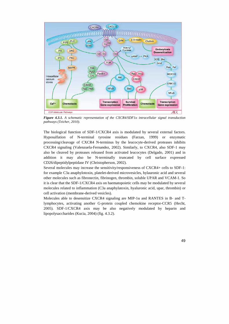
49
Figure 4.3.1. A schematic representation of the CXCR4/SDF1α intracellular signal transduction pathways (Teicher, 2010).
The biological function of SDF-1/CXCR4 axis is modulated by several external factors. Hyposulfation of N-terminal tyrosine residues (Farzan, 1999) or enzymatic processing/cleavage of CXCR4 N-terminus by the leucocyte-derived proteases inhibits CXCR4 signaling (Valenzuela-Fernandez, 2002). Similarly, to CXCR4, also SDF-1 may also be cleaved by proteases released from activated leucocytes (Delgado, 2001) and in addition it may also be N-terminally truncated by cell surface expressed CD26/dipeptidylpeptidase IV (Christopherson, 2002). Several molecules may increase the sensitivity/responsiveness of CXCR4+ cells to SDF-1: for example C3a anaphylotoxin, platelet-derived microvesicles, hylauronic acid and several other molecules such as fibronectin, fibrinogen, thrombin, soluble UPAR and VCAM-1. So it is clear that the SDF-1/CXCR4 axis on haematopoietic cells may be modulated by several molecules related to inflammation (C3a anaphylatoxin, hyaluronic acid, upar, thrombin) or cell activation (membrane-derived vesicles). Molecules able to desensitize CXCR4 signaling are MIP-1α and RANTES in B- and T-lymphocytes, activating another G-protein coupled chemokine receptor-CCR5 (Hecht, 2005). SDF-1/CXCR4 axis may be also negatively modulated by heparin and lipopolysaccharides (Kucia, 2004) (fig. 4.3.2).
50
Figure 4.3.2. A schematic representation of the CXCR4-SDF- 1 pathway regulation (Kucia, 2004).
In the BM, CXCL-12 is produced by stromal cells and it can be regulated by several factors. For example, oncostatin M, a regulator of HSCs homeostasis, retains HSCs in bone marrow through upregulation of CXCL-12 expression. Conversely, fibroblast growth factor 2 (FGF-2) downregulate expression of CXCL-12 in BM stromal cells (Broxmeyer, 2008).
4.3.1 The CXCR4/SDF-1 pathway in cancer Tumour cells from at least 23 different types of human cancers of epithelial, mesenchymal and haematopoietic origin express CXCR4 (Balkwill, 2004) and blockade of CXCR4–CXCL12 interactions has been extensively investigated as a potential cancer therapeutic. Not all cancerous cells in the primary tumour are CXCR4 positive. In ovarian and non-small-cell lung cancer, for instance, only a sub population of cells expresses this receptor (Scotton, 2001; Kijima, 2002).When it has been possible to study freshly isolated tumour cells the CXCR4 receptor is functional and various signaling pathways are activated. CXCL12 is the only known ligand for CXCR4. It is found in primary tumour sites in lymphoma and glioma, and ovarian and pancreatic cancer (Corcione, 2000; Zhou, 2002; Koshiba, 2000; Scotton, 2002) and at sites of metastasis in breast and thyroid cancer, neuroblastoma and haematological malignancies (Muller, 2001, Hwang, 2003; Geminder, 2001). Increased expression of CXCR4, is associated with a poor prognosis and are associated with advanced and metastatic disease (Sun, 2003; Kato, 2003). The ability of tumour cells to use CXCR4–CXCL12 during the process of metastasis might depend on chemokine gradients in the primary tumour, as well as common sites of spread, levels of functional receptor, and the presence of other cytokines and proteases that can cleave ligand and receptor.
51
4.3.1.1 Role of CXCR4/SDF-1 pathway in MM Given its normal functions, it is easy to understand that the SDF-1/CXCR4 axis is involved in tumor progression. The BM microenvironment facilitates the survival, differentiation and proliferation of malignant cells through the secretion of factor such as IL-6 and SDF-1, while integrins-mediated adhesion sequesters tumor cells to this niche. Indeed, the SDF-1/CXCR4 pathway is responsible for retention in the BM of acute lymphoid leukemia (ALL), acute myeloid leukemia (AML) and multiple myeloma (MM) cells (Juarez, 2007; Nervi, 2009; Azab, 2009). The SDF1α/CXCR4 axis is a key-regulator of MM cell homing, adhesion and motility (Katz, 2010). The CXCR4/SDF1α axis, regulates the mobilization and extravasation of tumor cells in bone (Alsayed, 2007). In MM, SDF-1 is expressed by stromal cells and led to a rapid activation of pERK1/2 and pAKT downstream of PI3K and this leads to cells migration; while CXCR4 is expressed by myeloma cells and endothelium, but not by marrow stromal cells (Baggiolini, 1998; Durig, 2001). Migration of myeloma cells across the endothelium lining the bone marrow sinuses is a critical step in the pathogenesis of multiple myeloma, which leads to homing and localization of these cells. As mentioned above, several studies have suggested that SDF-1 is a chemoattractant for human CD34+ hemopoietic progenitor cells. Similarly, in multiple myeloma, there is a positive correlation between SDF-1 protein levels and of chemotactic activity: SDF-1/CXCR4 promotes transendothelial migration of myeloma cells by transient upregulation of VLA-4 (α4β1)/VCAM-1, inducing cell adhesion to the endothelium, and contributing to the trafficking of myeloma cells in the bone marrow microenvironment (Parmo-Cabanas, 2004; Wright, 2003). Once myeloma cells are within the bone marrow, they localize in close proximity to stromal cells, forming tumor niche. In addition, SDF-1 secretion by marrow stromal cells is upregulated by adhesion of MM cells to stromal cells, thus promoting greater expression of integrins which enhance homing (Hideshima, 2002). CXCR4/SCDF-1 also plays a key role in chemotherapy-based mobilization of hematopoietic stem cells (HSC) and progenitor cells from BM to peripheral blood. In vivo studies have shown decreased serum levels of SDF-1 in mobilized myeloma cells (Gazitt 2004) potentially to confine the cells to the marrow and to prevent further trafficking of the cells (Alsayed, 2007). Interestingly, SDF-1 can also be protective against dexamethasone-induced apoptosis through activation of the mitogen-activated protein (MAP) Akt pathway, suggesting a role in drug resistance. SDF-1/ CXCR4 induces NF-κB activation in MM cells, which is consistent with previous reports of SDF-1α-induced NF-κB activation in primary osteocytes (Hideshima, 2002; Han, 2001). NF-κB has both growth-inducing and anti-apoptotic roles in normal cells as well as myeloma cells. SDF-1/CXCR4 also plays an indirect role in promoting growth, survival, and migration of MM cells by increased IL-6 and VEGF secretion in marrow stromal cells (Hideshima, 2002).
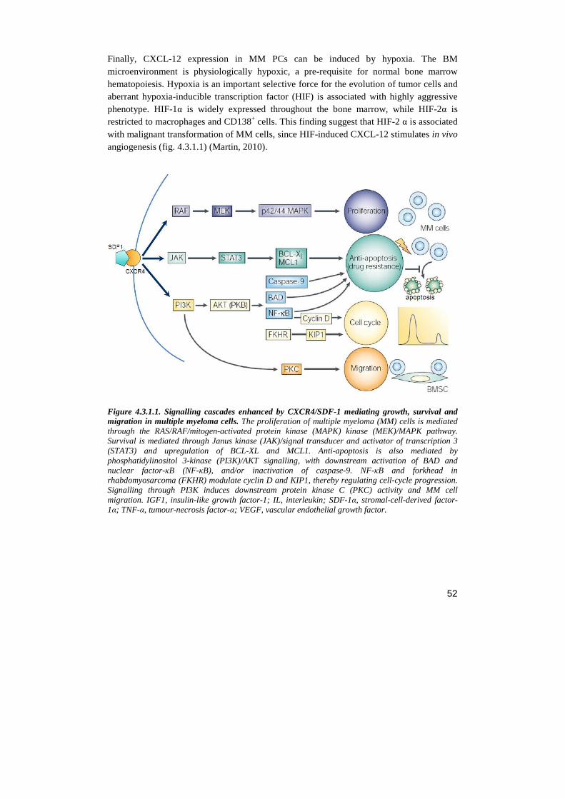
52
Finally, CXCL-12 expression in MM PCs can be induced by hypoxia. The BM microenvironment is physiologically hypoxic, a pre-requisite for normal bone marrow hematopoiesis. Hypoxia is an important selective force for the evolution of tumor cells and aberrant hypoxia-inducible transcription factor (HIF) is associated with highly aggressive phenotype. HIF-1α is widely expressed throughout the bone marrow, while HIF-2α is restricted to macrophages and CD138+ cells. This finding suggest that HIF-2 α is associated with malignant transformation of MM cells, since HIF-induced CXCL-12 stimulates in vivo angiogenesis (fig. 4.3.1.1) (Martin, 2010).
Figure 4.3.1.1. Signalling cascades enhanced by CXCR4/SDF-1 mediating growth, survival and migration in multiple myeloma cells. The proliferation of multiple myeloma (MM) cells is mediated through the RAS/RAF/mitogen-activated protein kinase (MAPK) kinase (MEK)/MAPK pathway. Survival is mediated through Janus kinase (JAK)/signal transducer and activator of transcription 3 (STAT3) and upregulation of BCL-XL and MCL1. Anti-apoptosis is also mediated by phosphatidylinositol 3-kinase (PI3K)/AKT signalling, with downstream activation of BAD and nuclear factor-κB (NF-κB), and/or inactivation of caspase-9. NF-κB and forkhead in rhabdomyosarcoma (FKHR) modulate cyclin D and KIP1, thereby regulating cell-cycle progression. Signalling through PI3K induces downstream protein kinase C (PKC) activity and MM cell migration. IGF1, insulin-like growth factor-1; IL, interleukin; SDF-1α, stromal-cell-derived factor-1α; TNF-α, tumour-necrosis factor-α; VEGF, vascular endothelial growth factor.
53
MATERIALS AND METHODS
1. CELL CULTURES 1.1 Single cultures The Multiple Myeloma (MM) cell lines used were: KMS-12: cell line established from the bone marrow of a 64-year-old woman with a multiple myeloma. Cells negative for CD3, CD80, CD19 e CD20 and positive for CD138. They grow slightly adherent. RPMI-8226: cell line established from the peripheral blood of a 61-year-old man with multiple myeloma (IgG lambda-type) at diagnosis in 1966; described to produce and secrete only lambda light chains (but not heavy chains). Cells negative for CD19 and CD20 and positive for CD28 , CD138 and CD49. They grow slightly adherent. OPM-2: cell line established from the peripheral blood of a 56-year-old woman with multiple myeloma (IgG lambda) in leukemic phase (relapse, terminal) in 1982. Cells negative for CD3 , CD10, CD80, CD19 and CD20 and positive for CD138. They grow in suspension. All MM cell lines were maintained in 75 cm2 flask in RPMI 1640 medium (Sigma-Aldrich Co., St Louis, MO), supplemented with 10% (v/v) FBS (Gibco, Rockville, MD), 2 mM L-glutamine (Invitrogen Corporation Carlsbad, CA, USA), 100 U/ml penicillin and 100 µg/ml streptomycin. The serum was de-complemented for 30’ at 56°C before use. Cells were cultured in 5% CO2 at 37°C, maintaining the optimum concentration at 3x10⁵cells/ml with complete change of medium every two days. The Bone marrow stromal cell (BMSC) line used was: NIH-3T3 : cell line of mouse embryonic fibroblasts isolated in 1962 at the New York University School of Medicine Department of Pathology. 3T3 refers to the sub-cultivation protocol and it means "3-day transfer, inoculum 3 x 10^5 cells" (Todaro G.J., 1963). They grow adherent. Cell line was maintained in 10 cm2 plate dishes , in DMEM medium (Sigma-Aldrich Co., St Louis, MO, USA), supplemented with 10% (v/v) FBS (Gibco, Rockville, MD, USA), 2 mM L-glutamine (Invitrogen Corporation Carlsbad, CA, USA), 100 U/ml penicillin and 100 µg/ml streptomycin. The serum was de-complemented for 30’ at 56°C before use. Cells were cultured in 5% CO2 at 37°C. Cells have a doubling time of 18-20h and were divided three times/ week. 1.2 Co-culture of MM/BMSC lines NIH-3T3 cells were plated in 24 multi-well plates at the concentration of 150000/ml. After 24h NIH-3T3 medium was discarded and OPM-2 MM cells were plated on top of NIH-3T3
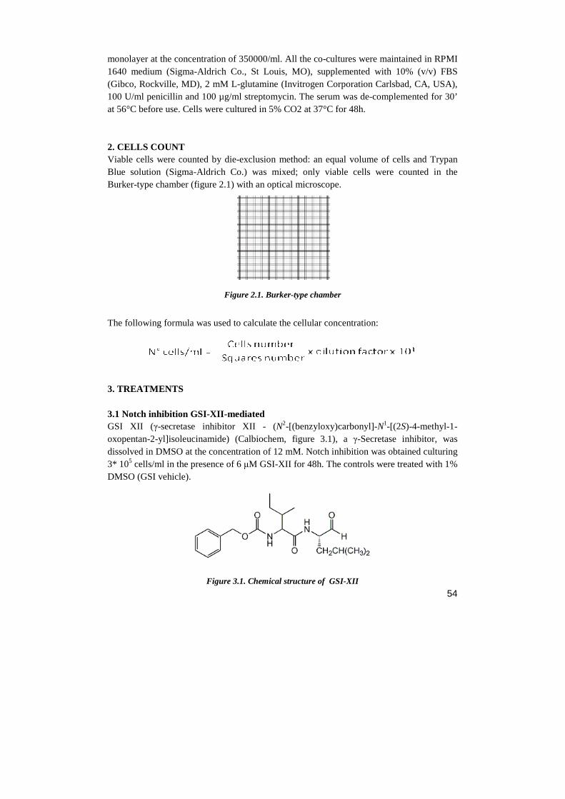
54
monolayer at the concentration of 350000/ml. All the co-cultures were maintained in RPMI 1640 medium (Sigma-Aldrich Co., St Louis, MO), supplemented with 10% (v/v) FBS (Gibco, Rockville, MD), 2 mM L-glutamine (Invitrogen Corporation Carlsbad, CA, USA), 100 U/ml penicillin and 100 µg/ml streptomycin. The serum was de-complemented for 30’ at 56°C before use. Cells were cultured in 5% CO2 at 37°C for 48h. 2. CELLS COUNT Viable cells were counted by die-exclusion method: an equal volume of cells and Trypan Blue solution (Sigma-Aldrich Co.) was mixed; only viable cells were counted in the Burker-type chamber (figure 2.1) with an optical microscope.
Figure 2.1. Burker-type chamber
The following formula was used to calculate the cellular concentration:
3. TREATMENTS 3.1 Notch inhibition GSI-XII-mediated GSI XII (γ-secretase inhibitor XII - (N2-[(benzyloxy)carbonyl]-N1-[(2S)-4-methyl-1-oxopentan-2-yl]isoleucinamide) (Calbiochem, figure 3.1), a γ-Secretase inhibitor, was dissolved in DMSO at the concentration of 12 mM. Notch inhibition was obtained culturing 3* 105 cells/ml in the presence of 6 µM GSI-XII for 48h. The controls were treated with 1% DMSO (GSI vehicle).
Figure 3.1. Chemical structure of GSI-XII
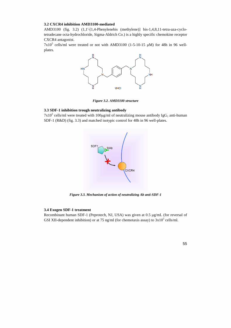
55
3.2 CXCR4 inhibition AMD3100-mediated AMD3100 (fig. 3.2) (1,1′-[1,4-Phenylenebis (methylene)] bis-1,4,8,11-tetra-aza-cyclo-tetradecane octa-hydrochloride, Sigma-Aldrich Co.) is a highly specific chemokine receptor CXCR4 antagonist. 7x10⁵ cells/ml were treated or not with AMD3100 (1-5-10-15 µM) for 48h in 96 well-plates.
Figure 3.2. AMD3100 structure 3.3 SDF-1 inhibition trough neutralizing antibody 7x10⁵ cells/ml were treated with 100µg/ml of neutralizing mouse antibody IgG1 anti-human SDF-1 (R&D) (fig. 3.3) and matched isotypic control for 48h in 96 well-plates.
Figure 3.3. Mechanism of action of neutralizing Ab anti-SDF-1
3.4 Exogen SDF-1 treatment Recombinant human SDF-1 (Peprotech, NJ, USA) was given at 0.5 µg/mL (for reversal of GSI XII-dependent inhibition) or at 75 ng/ml (for chemotaxis assay) to 3x10⁵ cells/ml.
56
4. GENE EXPRESSION ANALYSIS 4.1 RNA isolation The total RNA isolation was based on acid guanidinium thiocyanate-phenol-chloroform extraction (Chomczynski P., 1987).The protocol is optimized for 106 cells:
• Cells were washed two times with cold 1X PBS.
• After centrifugation at room temperature, pellet was resuspended in 150 µl of D-solution.
• Sequentially it was added: - 15 µl Sodium Acetate 2M pH 4 - 150 µl water-saturated Phenol - 30 µl Chloroform-Isoamyl alcohol (98% Chloroforme, 2% Isoamyl Alcohol)
• Sample was mixed by vortex and incubated at 4°C for 10’.
• After centrifugation for 10’ at 14.000 rpm at 4°C, the aqueous phase was collected in a new tube.
• One volume of Phenol-Chloroform was added to the aqueous phase.
• Sample was mixed by vortex and centrifuged for 10’ at 14.000 rpm at 4°C.
• The aqueous phase was collected in a new tube. • One volume of cold isopropanol was added to the aqueous phase.
• Sample was incubated for 30’ at –20°C.
• After centrifugation for 30’ at 14.000 rpm at 4°C, the supernatant was discarded.
• Pellet was resuspended in 50 µl of D-Solution and precipitated with 1 volume of cold isopropanol.
• Sample was incubated for 30’ at –20°C.
• After centrifugation for 30’ at 14.000 rpm at 4°C, the supernatant was discarded.
• Pellet was washed with cold ethanol 70% .
• After centrifugation for 10’ at 14.000 rpm at 4°C, the ethanol was discarded.
• Pellet was dried with vacuum system and resuspended in 30 µl of H2O DEPC.
• To obtain an homogenous solution, RNA was heated at 65°C for 5’. PBS 1X pH 7,4 - 4,3 mM Na2HPO4 - 1,47 mM KH2PO4 - 137 mM NaCl - 2,7mM KCl
D-Solution pH 7: - 4 M guanidinium isothiocyanate - 25 mM sodium citrate tribasic dehydrate - 18.4 mM sodium lauroyl sarcosinate - 100 mM β-mercaptoethanol
57
4.2 RNA quantification RNA was quantified by spectrophotometric measure, using 2 µl of RNA in 700 µl of H2O Milli-Q in quartz cuvettes at two different wavelengths: 260nm (A1) and 280nm (A2). Since: 1 OD260nm = 40 µg/ml The concentration in µg/ml was calculated as:
A260 x 40 ng/µl x dilution factor
High quality RNA was used (A1/A2 ratio closed to 2).
4.3 Reverse transcription The cDNA was obtained by reverse transcription with M-MLV RT KIT (Sigma-Aldrich Co.).
• A reaction mix of 20 µl was prepared with:
- 2 µl of Random primers (250ng/µl)
- 4 µl of 10 mM dNTPs (2.5mM each)
- 2 µg RNA
- H2O DEPC up to 10 µl
• The sample was heated at 65°C for 5’.
• After centrifugation it was added to the mix:
- 2µl of 10x M-MLV RT Buffer
- 1µl of M-MLV Reverse Transcriptase (200 U/µl)
- 7µl of H2O DEPC
• Sample was incubated 10’ at room temperature, then at 37°C for 50 minutes and finally stored at –20°C.
4.4 PCR (Polymerase Chain Reaction) The cDNA obtained by reverse transcription was used as template for PCR reaction. A final reaction volume of 20 µl was prepared with: - 4 µl 5X Buffer Green (Promega) - 1.6 µl Mg 2+ 25mM - 1.6 µl of 2.5mM dNTPs - 4 µl Primer mix (5 µM each) - 0.1 µl Taq Polimerase (5U/µl, Promega) - 2 µg cDNA - H2O MilliQ to 20 µl
cDNA levels were evalueted by densitometric analysis through agarose gel electrophoresis, and normalized with GAPDH. Primer sequences used for cDNA amplification are displayed in Table 4.4.1:
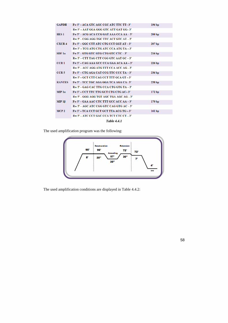
58
Table 4.4.1
The used amplification program was the following:
The used amplification conditions are displayed in Table 4.4.2:
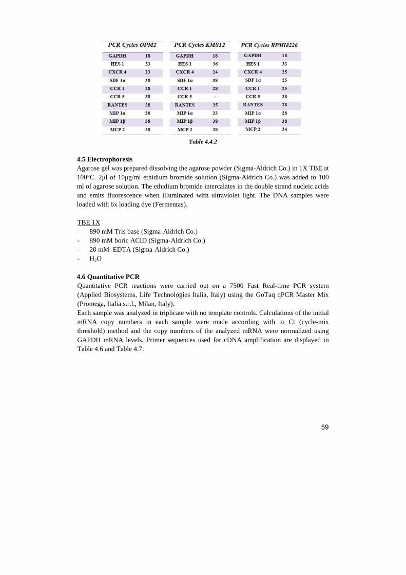
59
Table 4.4.2
4.5 Electrophoresis Agarose gel was prepared dissolving the agarose powder (Sigma-Aldrich Co.) in 1X TBE at 100°C. 2µl of 10µg/ml ethidium bromide solution (Sigma-Aldrich Co.) was added to 100 ml of agarose solution. The ethidium bromide intercalates in the double strand nucleic acids and emits fluorescence when illuminated with ultraviolet light. The DNA samples were loaded with 6x loading dye (Fermentas). TBE 1X - 890 mM Tris base (Sigma-Aldrich Co.) - 890 mM boric ACID (Sigma-Aldrich Co.) - 20 mM EDTA (Sigma-Aldrich Co.) - H2O 4.6 Quantitative PCR Quantitative PCR reactions were carried out on a 7500 Fast Real-time PCR system (Applied Biosystems, Life Technologies Italia, Italy) using the GoTaq qPCR Master Mix (Promega, Italia s.r.l., Milan, Italy). Each sample was analyzed in triplicate with no template controls. Calculations of the initial mRNA copy numbers in each sample were made according with to Ct (cycle-mix threshold) method and the copy numbers of the analyzed mRNA were normalized using GAPDH mRNA levels. Primer sequences used for cDNA amplification are displayed in Table 4.6 and Table 4.7:
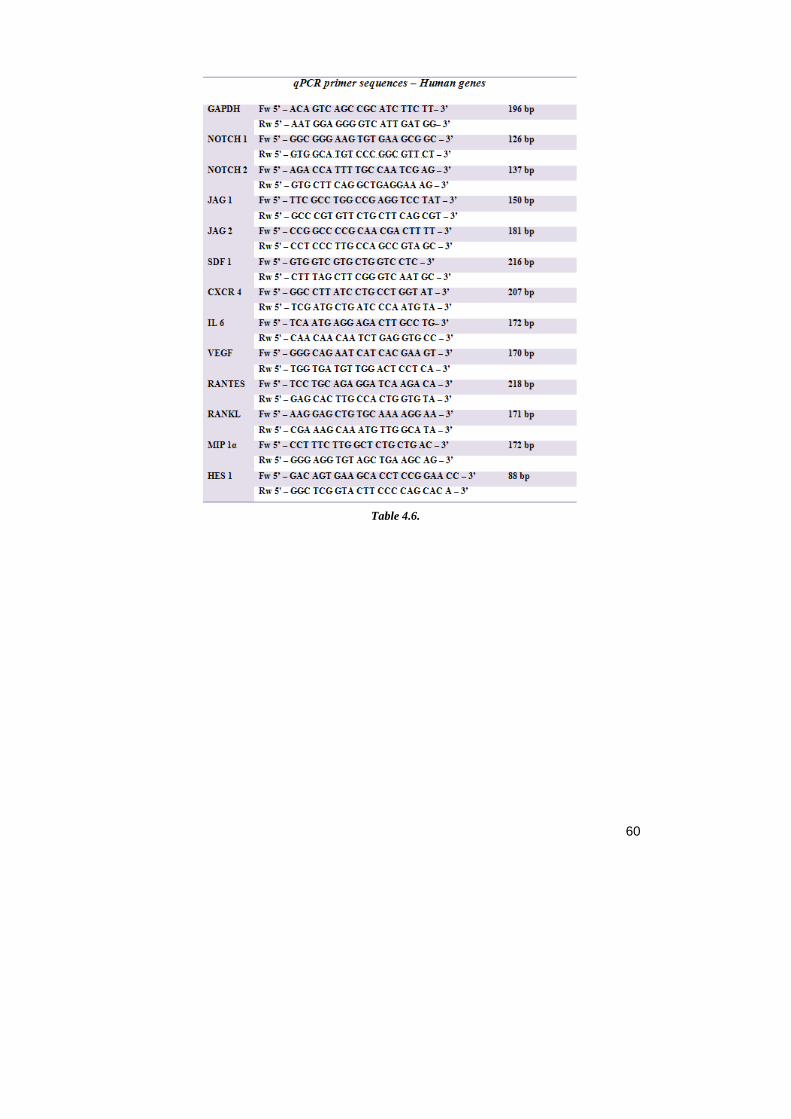
60
Table 4.6.
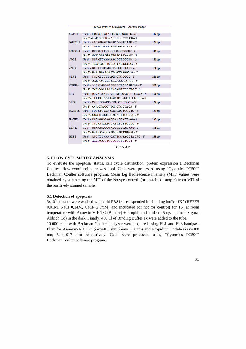
61
Table 4.7.
5. FLOW CYTOMETRY ANALYSIS To evaluate the apoptosis status, cell cycle distribution, protein expression a Beckman Coulter flow cytofluorimeter was used. Cells were processed using “Cytomics FC500” Beckman Coulter software program. Mean log fluorescence intensity (MFI) values were obtained by subtracting the MFI of the isotype control (or unstained sample) from MFI of the positively stained sample. 5.1 Detection of apoptosis 3x10⁵ cells/ml were washed with cold PBS1x, resuspended in “binding buffer 1X” (HEPES 0,01M, NaCl 0,14M, CaCl2 2,5mM) and incubated (or not for control) for 15’ at room temperature with Annexin-V FITC (Bender) + Propidium Iodide (2,5 ug/ml final, Sigma-Aldrich Co) in the dark. Finally, 400 µl of Binding Buffer 1x were added to the tube. 10.000 cells with Beckman Coulter analyzer were acquired using FL1 and FL3 bandpass filter for Annexin-V FITC (λex=488 nm; λem=520 nm) and Propidium Iodide (λex=488 nm; λem=617 nm) respectively. Cells were processed using “Cytomics FC500” BeckmanCoulter software program.
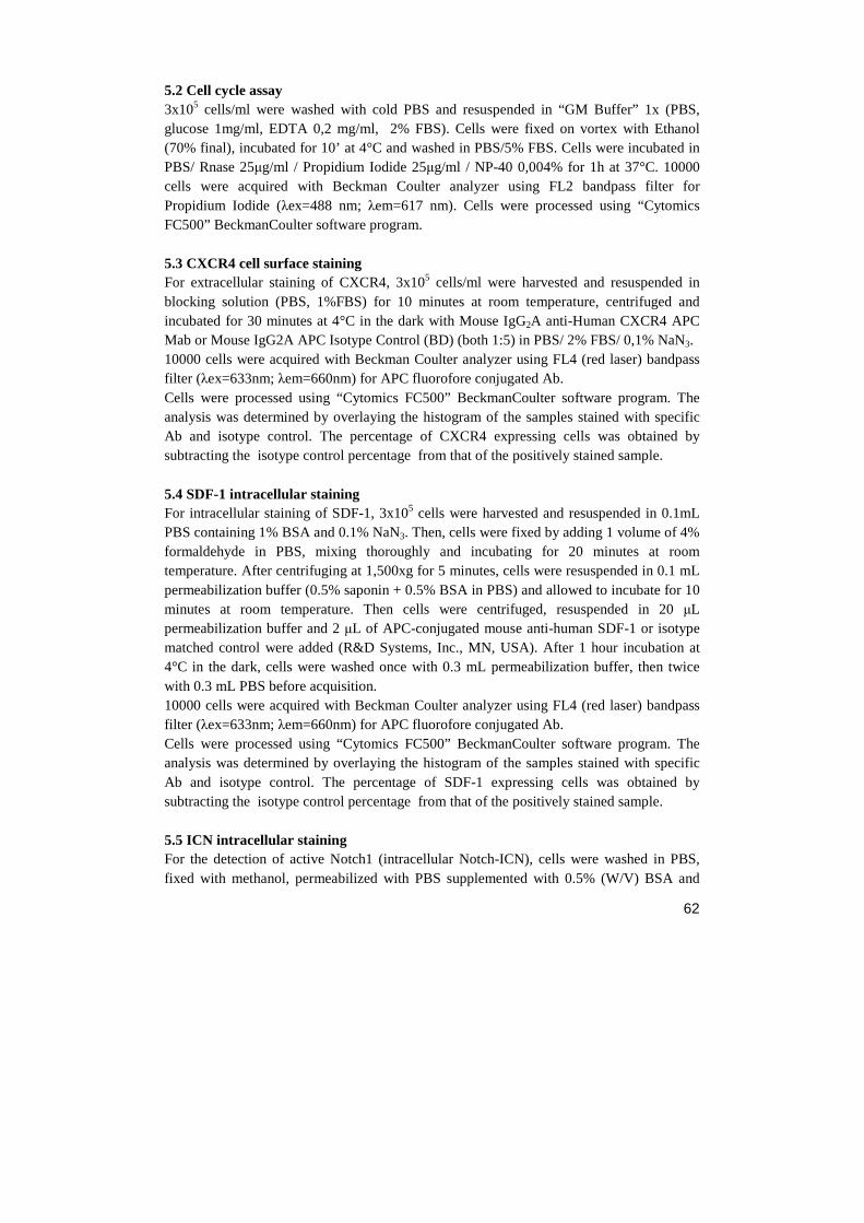
62
5.2 Cell cycle assay 3x10⁵ cells/ml were washed with cold PBS and resuspended in “GM Buffer” 1x (PBS, glucose 1mg/ml, EDTA 0,2 mg/ml, 2% FBS). Cells were fixed on vortex with Ethanol (70% final), incubated for 10’ at 4°C and washed in PBS/5% FBS. Cells were incubated in PBS/ Rnase 25µg/ml / Propidium Iodide 25µg/ml / NP-40 0,004% for 1h at 37°C. 10000 cells were acquired with Beckman Coulter analyzer using FL2 bandpass filter for Propidium Iodide (λex=488 nm; λem=617 nm). Cells were processed using “Cytomics FC500” BeckmanCoulter software program. 5.3 CXCR4 cell surface staining For extracellular staining of CXCR4, 3x10⁵ cells/ml were harvested and resuspended in blocking solution (PBS, 1%FBS) for 10 minutes at room temperature, centrifuged and incubated for 30 minutes at 4°C in the dark with Mouse IgG2A anti-Human CXCR4 APC Mab or Mouse IgG2A APC Isotype Control (BD) (both 1:5) in PBS/ 2% FBS/ 0,1% NaN3. 10000 cells were acquired with Beckman Coulter analyzer using FL4 (red laser) bandpass filter (λex=633nm; λem=660nm) for APC fluorofore conjugated Ab. Cells were processed using “Cytomics FC500” BeckmanCoulter software program. The analysis was determined by overlaying the histogram of the samples stained with specific Ab and isotype control. The percentage of CXCR4 expressing cells was obtained by subtracting the isotype control percentage from that of the positively stained sample. 5.4 SDF-1 intracellular staining For intracellular staining of SDF-1, 3x10⁵ cells were harvested and resuspended in 0.1mL PBS containing 1% BSA and 0.1% NaN3. Then, cells were fixed by adding 1 volume of 4% formaldehyde in PBS, mixing thoroughly and incubating for 20 minutes at room temperature. After centrifuging at 1,500xg for 5 minutes, cells were resuspended in 0.1 mL permeabilization buffer (0.5% saponin + 0.5% BSA in PBS) and allowed to incubate for 10 minutes at room temperature. Then cells were centrifuged, resuspended in 20 µL permeabilization buffer and 2 µL of APC-conjugated mouse anti-human SDF-1 or isotype matched control were added (R&D Systems, Inc., MN, USA). After 1 hour incubation at 4°C in the dark, cells were washed once with 0.3 mL permeabilization buffer, then twice with 0.3 mL PBS before acquisition. 10000 cells were acquired with Beckman Coulter analyzer using FL4 (red laser) bandpass filter (λex=633nm; λem=660nm) for APC fluorofore conjugated Ab. Cells were processed using “Cytomics FC500” BeckmanCoulter software program. The analysis was determined by overlaying the histogram of the samples stained with specific Ab and isotype control. The percentage of SDF-1 expressing cells was obtained by subtracting the isotype control percentage from that of the positively stained sample. 5.5 ICN intracellular staining For the detection of active Notch1 (intracellular Notch-ICN), cells were washed in PBS, fixed with methanol, permeabilized with PBS supplemented with 0.5% (W/V) BSA and
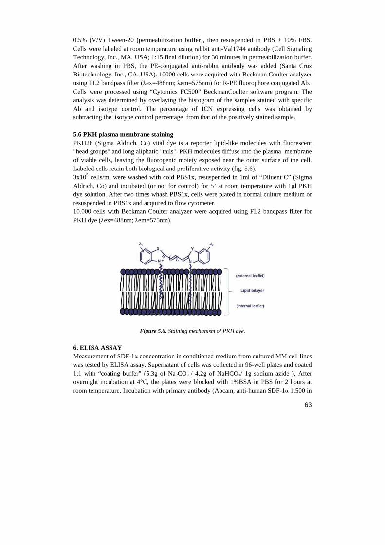
63
0.5% (V/V) Tween-20 (permeabilization buffer), then resuspended in PBS + 10% FBS. Cells were labeled at room temperature using rabbit anti-Val1744 antibody (Cell Signaling Technology, Inc., MA, USA; 1:15 final dilution) for 30 minutes in permeabilization buffer. After washing in PBS, the PE-conjugated anti-rabbit antibody was added (Santa Cruz Biotechnology, Inc., CA, USA). 10000 cells were acquired with Beckman Coulter analyzer using FL2 bandpass filter (λex=488nm; λem=575nm) for R-PE fluorophore conjugated Ab. Cells were processed using “Cytomics FC500” BeckmanCoulter software program. The analysis was determined by overlaying the histogram of the samples stained with specific Ab and isotype control. The percentage of ICN expressing cells was obtained by subtracting the isotype control percentage from that of the positively stained sample. 5.6 PKH plasma membrane staining PKH26 (Sigma Aldrich, Co) vital dye is a reporter lipid-like molecules with fluorescent "head groups" and long aliphatic "tails". PKH molecules diffuse into the plasma membrane of viable cells, leaving the fluorogenic moiety exposed near the outer surface of the cell. Labeled cells retain both biological and proliferative activity (fig. 5.6). 3x10⁵ cells/ml were washed with cold PBS1x, resuspended in 1ml of “Diluent C” (Sigma Aldrich, Co) and incubated (or not for control) for 5’ at room temperature with 1µl PKH dye solution. After two times whash PBS1x, cells were plated in normal culture medium or resuspended in PBS1x and acquired to flow cytometer. 10.000 cells with Beckman Coulter analyzer were acquired using FL2 bandpass filter for PKH dye (λex=488nm; λem=575nm).
Figure 5.6. Staining mechanism of PKH dye.
6. ELISA ASSAY Measurement of SDF-1α concentration in conditioned medium from cultured MM cell lines was tested by ELISA assay. Supernatant of cells was collected in 96-well plates and coated 1:1 with “coating buffer” (5.3g of Na2CO3 / 4.2g of NaHCO3/ 1g sodium azide ). After overnight incubation at 4°C, the plates were blocked with 1%BSA in PBS for 2 hours at room temperature. Incubation with primary antibody (Abcam, anti-human SDF-1α 1:500 in
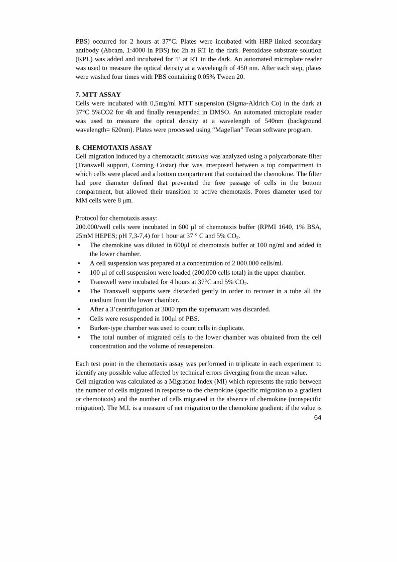
64
PBS) occurred for 2 hours at 37°C. Plates were incubated with HRP-linked secondary antibody (Abcam, 1:4000 in PBS) for 2h at RT in the dark. Peroxidase substrate solution (KPL) was added and incubated for 5’ at RT in the dark. An automated microplate reader was used to measure the optical density at a wavelength of 450 nm. After each step, plates were washed four times with PBS containing 0.05% Tween 20. 7. MTT ASSAY Cells were incubated with 0,5mg/ml MTT suspension (Sigma-Aldrich Co) in the dark at 37°C 5%CO2 for 4h and finally resuspended in DMSO. An automated microplate reader was used to measure the optical density at a wavelength of 540nm (background wavelength= 620nm). Plates were processed using “Magellan” Tecan software program. 8. CHEMOTAXIS ASSAY Cell migration induced by a chemotactic stimulus was analyzed using a polycarbonate filter (Transwell support, Corning Costar) that was interposed between a top compartment in which cells were placed and a bottom compartment that contained the chemokine. The filter had pore diameter defined that prevented the free passage of cells in the bottom compartment, but allowed their transition to active chemotaxis. Pores diameter used for MM cells were 8 µm. Protocol for chemotaxis assay: 200.000/well cells were incubated in 600 µl of chemotaxis buffer (RPMI 1640, 1% BSA, 25mM HEPES; pH 7,3-7,4) for 1 hour at 37 ° C and 5% CO2.
• The chemokine was diluted in 600µl of chemotaxis buffer at 100 ng/ml and added in the lower chamber.
• A cell suspension was prepared at a concentration of 2.000.000 cells/ml.
• 100 µl of cell suspension were loaded (200,000 cells total) in the upper chamber.
• Transwell were incubated for 4 hours at 37°C and 5% CO2.
• The Transwell supports were discarded gently in order to recover in a tube all the medium from the lower chamber.
• After a 3’centrifugation at 3000 rpm the supernatant was discarded.
• Cells were resuspended in 100µl of PBS.
• Burker-type chamber was used to count cells in duplicate.
• The total number of migrated cells to the lower chamber was obtained from the cell concentration and the volume of resuspension.
Each test point in the chemotaxis assay was performed in triplicate in each experiment to identify any possible value affected by technical errors diverging from the mean value. Cell migration was calculated as a Migration Index (MI) which represents the ratio between the number of cells migrated in response to the chemokine (specific migration to a gradient or chemotaxis) and the number of cells migrated in the absence of chemokine (nonspecific migration). The M.I. is a measure of net migration to the chemokine gradient: if the value is
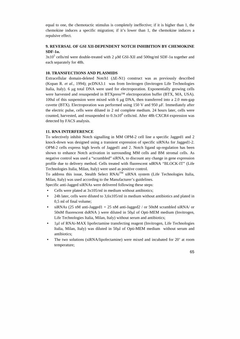
65
equal to one, the chemotactic stimulus is completely ineffective; if it is higher than 1, the chemokine induces a specific migration; if it’s lower than 1, the chemokine induces a repulsive effect. 9. REVERSAL OF GSI XII-DEPENDENT NOTCH INHIBITION B Y CHEMOKINE SDF-1α. 3x10⁵ cells/ml were double-treated with 2 µM GSI-XII and 500ng/ml SDF-1α together and each separately for 48h. 10. TRANSFECTIONS AND PLASMIDS Extracellular domain-deleted Notch1 (∆E-N1) construct was as previously described (Kopan R. et al., 1994); pcDNA3.1 was from Invitrogen (Invitrogen Life Technologies Italia, Italy). 6 µg total DNA were used for electroporation. Exponentially growing cells were harvested and resuspended in BTXpress™ electroporation buffer (BTX, MA, USA). 100ul of this suspension were mixed with 6 µg DNA, then transferred into a 2.0 mm-gap cuvette (BTX). Electroporation was performed using 150 V and 950 µF. Immediately after the electric pulse, cells were diluted in 2 ml complete medium. 24 hours later, cells were counted, harvested, and resuspended to 0.3x10⁶ cells/ml. After 48h CXCR4 expression was detected by FACS analysis. 11. RNA INTERFERENCE To selectively inhibit Notch signalling in MM OPM-2 cell line a specific Jagged1 and 2 knock-down was designed using a transient expression of specific siRNAs for Jagged1-2. OPM-2 cells express high levels of Jagged1 and 2. Notch ligand up-regulation has been shown to enhance Notch activation in surrounding MM cells and BM stromal cells. As negative control was used a “scrambled” siRNA, to discount any change in gene expression profile due to delivery method. Cells treated with fluorescent sdRNA “BLOCK-IT” (Life Technologies Italia, Milan, Italy) were used as positive control. To address this issue, Stealth Select RNAiTM siRNA system (Life Technologies Italia, Milan, Italy) was used according to the Manufacturer’s guidelines. Specific anti-Jagged siRNAs were delivered following these steps:
• Cells were plated at 3x10⁵/ml in medium without antibiotics;
• 24h later, cells were diluted to 3,6x10⁵/ml in medium without antibiotics and plated in 0,5 ml of final volume;
• siRNAs (25 nM anti-Jagged1 + 25 nM anti-Jagged2 / or 50nM scrambled siRNA/ or 50nM fluorescent dsRNA ) were diluted in 50µl of Opti-MEM medium (Invitrogen, Life Technologies Italia, Milan, Italy) without serum and antibiotics;
• 1µl of RNAi-MAX lipofectamine transfecting reagent (Invitrogen, Life Technologies Italia, Milan, Italy) was diluted in 50µl of Opti-MEM medium without serum and antibiotics;
• The two solutions (siRNA/lipofectamine) were mixed and incubated for 20’ at room temperature;
66
• 100µl of lipofectamine/siRNA mix was added to the cells (final cells concentration 3x10⁵/ml);
• Every 48h cells were diluted 1:1 with medium antibiotics-free and treated again with JAG1-2 siRNA up to 8 days
Cells were maintained in RPMI 1640 medium (Sigma-Aldrich Co., St Louis, MO) supplemented with 10% (v/v) FBS (Gibco, Rockville, MD) and 2 mM L-glutamine (Invitrogen Corporation Carlsbad, CA, USA) without antibiotics and incubated in 5% CO2 at 37°C. To confirm that the transfection occurred successfully, the percentage of BLOCK-IT positive cells were checked trough flow cytometry analysis at each time point. 10000 cells were acquired with Beckman Coulter analyzer using FL2 bandpass filter (λex=488nm; λem=575nm) for BLOCK-IT fluorophore conjugated sdRNA. Jagged1 and 2 effective silencing induced by specific siRNAs was assessed by quantitative PCR compared to scrambled siRNA-receiving cells.
12. JAGGED1-2 KNOCK-DOWN AND DETECTION OF APOPTOSIS IN CO-CULTURED CELLS.
• OPM-2 cells were seeded at 300.000 cells/ml in medium without antibiotics and after 24h the JAG1-2 siRNA treatment was added following the protocol previously described.
• After 24h NIH-3T3 cells were labeled (or not for negative control) with PKH vital dye (Sigma-Aldrich, Co) and plated in 24 multi-well plates at the concentration of 150.000/ml
• 24h later, OPM-2 cells were diluted and treated once again with silencing treatment
• After 8h NIH-3T3 medium was discarded while OPM-2 cells were washed with PBS1x and plated on the monolayer of NIH-3T3 cells at final 1:1 ratio.
Co-cultures were manteined for 48h, obtaining in this way a condition in which MM cells were silenced from 4 days. Cells treated with scrambled siRNA were used as negative control. All the cultures for this experiment were maintained in RPMI 1640 medium (Sigma-Aldrich Co., St Louis, MO) supplemented with 10% (v/v) FBS (Gibco, Rockville, MD) and 2 mM L-glutamine (Invitrogen Corporation Carlsbad, CA, USA) without antibiotics and incubated in 5% CO2 at 37°C. 48h co-cultured cell lines were collected and tested for the detection of apoptosis using the following method: cells were washed with cold PBS, re-suspended in “binding buffer 1X” (HEPES 0,01M, NaCl 0,14M, CaCl2 2,5mM) and incubated (or not for control) for 15’ at room temperature with Annexin-V FITC (Bender) in the dark. Finally, 400 µl of Binding Buffer 1x were added to the tube.
67
10.000 cells with Beckman Coulter analyzer were acquired using FL1 and FL2 bandpass filter for Annexin-V FITC (λex=488 nm; λem=520 nm) and PKH fluorescent dye (λex=488nm; λem=575nm) respectively. Cells were processed using “Cytomics FC500” BeckmanCoulter software program. Mean log fluorescence intensity (MFI) values were obtained by subtracting the MFI of the unstained sample from MFI of the positively stained sample. Using this staining method it was obtained a mixed population in which is possible to distinguish four regions (fig. 12.1):
1. PKH+/Ann- population indicating NIH-3T3 living cells 2. PKH+/Ann+ population indicating NIH-3T3 apoptotic cells 3. PKH-/Ann- population indicating OPM-2 living cells 4. PKH-/Ann+ population indicating OPM-2 apoptotic cells
Figure 12.1. Schematic representation of four Dot-Plot regions resulted from PKH/Annexin cellular staining.
13. STATISTICAL ANALYSIS All experiments were performed in triplicate and repeated 3 times. Statistical analysis has been performed by t-test and ANOVA test. Significative differences have been reported: * = p<0,05; ** = p<0,01. Non-significative differences have been reported: # = p> 0,05.
FL1 Annexin FITC
FL2PKH 1 2
3 4
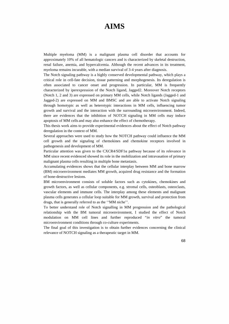
68
AIMS
Multiple myeloma (MM) is a malignant plasma cell disorder that accounts for approximately 10% of all hematologic cancers and is characterized by skeletal destruction, renal failure, anemia, and hypercalcemia. Although the recent advances in its treatment, myeloma remains incurable, with a median survival of 3-4 years after diagnosis. The Notch signaling pathway is a highly conserved developmental pathway, which plays a critical role in cell-fate decision, tissue patterning and morphogenesis. Its deregulation is often associated to cancer onset and progression. In particular, MM is frequently characterized by iperexpression of the Notch ligand, Jagged2. Moreover Notch receptors (Notch 1, 2 and 3) are expressed on primary MM cells, while Notch ligands (Jagged-1 and Jagged-2) are expressed on MM and BMSC and are able to activate Notch signaling through homotypic as well as heterotypic interactions in MM cells, influencing tumor growth and survival and the interaction with the surrounding microenvironment. Indeed, there are evidences that the inhibition of NOTCH signaling in MM cells may induce apoptosis of MM cells and may also enhance the effect of chemotherapy. This thesis work aims to provide experimental evidences about the effect of Notch pathway deregulation in the context of MM. Several approaches were used to study how the NOTCH pathway could influence the MM cell growth and the signaling of chemokines and chemokine receptors involved in pathogenesis and development of MM. Particular attention was given to the CXCR4/SDF1α pathway because of its relevance in MM since recent evidenced showed its role in the mobilization and intravasation of primary malignant plasma cells resulting in multiple bone metastasis. Accumulating evidences shows that the cellular interplay between MM and bone marrow (BM) microenvironment mediates MM growth, acquired drug resistance and the formation of bone-destructive lesions. BM microenvironment consists of soluble factors such as cytokines, chemokines and growth factors, as well as cellular components, e.g. stromal cells, osteoblasts, osteoclasts, vascular elements and immune cells. The interplay among these elements and malignant plasma cells generates a cellular loop suitable for MM growth, survival and protection from drugs, that is generally referred to as the ‘‘MM niche’’. To better understand role of Notch signalling in MM progression and the pathological relationship with the BM tumoral microenvironment, I studied the effect of Notch modulation on MM cell lines and further reproduced “ in vitro” the tumoral microenvironment conditions through co-culture experiments. The final goal of this investigation is to obtain further evidences concerning the clinical relevance of NOTCH signaling as a therapeutic target in MM.
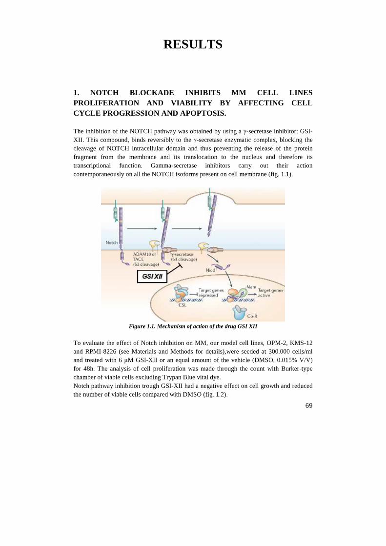
69
RESULTS 1. NOTCH BLOCKADE INHIBITS MM CELL LINES PROLIFERATION AND VIABILITY BY AFFECTING CELL CYCLE PROGRESSION AND APOPTOSIS. The inhibition of the NOTCH pathway was obtained by using a γ-secretase inhibitor: GSI-XII. This compound, binds reversibly to the γ-secretase enzymatic complex, blocking the cleavage of NOTCH intracellular domain and thus preventing the release of the protein fragment from the membrane and its translocation to the nucleus and therefore its transcriptional function. Gamma-secretase inhibitors carry out their action contemporaneously on all the NOTCH isoforms present on cell membrane (fig. 1.1).
Figure 1.1. Mechanism of action of the drug GSI XII
To evaluate the effect of Notch inhibition on MM, our model cell lines, OPM-2, KMS-12 and RPMI-8226 (see Materials and Methods for details),were seeded at 300.000 cells/ml and treated with 6 µM GSI-XII or an equal amount of the vehicle (DMSO, 0.015% V/V) for 48h. The analysis of cell proliferation was made through the count with Burker-type chamber of viable cells excluding Trypan Blue vital dye. Notch pathway inhibition trough GSI-XII had a negative effect on cell growth and reduced the number of viable cells compared with DMSO (fig. 1.2).
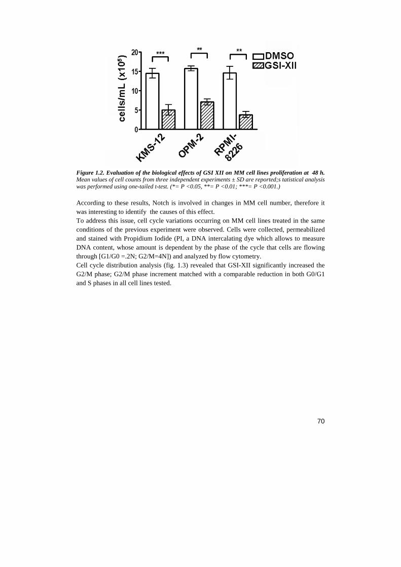
70
Figure 1.2. Evaluation of the biological effects of GSI XII on MM cell lines proliferation at 48 h. Mean values of cell counts from three independent experiments ± SD are reported;s tatistical analysis was performed using one-tailed t-test. (*= P <0.05, **= P <0.01; ***= P <0.001.) According to these results, Notch is involved in changes in MM cell number, therefore it was interesting to identify the causes of this effect. To address this issue, cell cycle variations occurring on MM cell lines treated in the same conditions of the previous experiment were observed. Cells were collected, permeabilized and stained with Propidium Iodide (PI, a DNA intercalating dye which allows to measure DNA content, whose amount is dependent by the phase of the cycle that cells are flowing through [G1/G0 =.2N; G2/M=4N]) and analyzed by flow cytometry. Cell cycle distribution analysis (fig. 1.3) revealed that GSI-XII significantly increased the G2/M phase; G2/M phase increment matched with a comparable reduction in both G0/G1 and S phases in all cell lines tested.

71
Figure 1.3. Cell cycle analysis of MM cell lines with and without GSI-XII. DNA content was measured after 48h of GSI-XII treatment, The values indicate the percentage of cells in each phase of cell cycle and are representative of three independent experiments. This result indicates that the reduced cell growth occurring after GSI-XII treatment is, at least in part, due to G2/M phase cell cycle arrest. GSI-XII-triggered cell growth inhibition effect on MM cell lines apoptosis rate was investigated. Apoptosis assay was performed on MM cell lines treated or not with GSI-XII trough the Annexin-IV/PI double staining method and analyzing positive cells by flow-cytometry (see Material and Methods). GSI-XII treatment resulted in ~ 40-50% increased frequency of Annexin-V+ cells compared with controls (fig. 1.4).
72
Figure 1.4. Apoptosis assay of MM cell lines treated (or not) with GSI-XII for 24h and 48h. Dot-plots deriving by flow cytometric analysis are reported. Values represent the percentage of cells in early (Ann+/PI-) or late (Ann+/PI+)apoptosis and are representative of three independent experiments.
2. NOTCH REGULATES SEVERAL CHEMOKINE RECEPTORS AND THEIR LIGANDS IN MM CELL LINES Several chemokine receptors are overexpressed in MM and are involved in osteolysis occurring at tumor BM localization sites. This consideration prompted us to investigate the effect of NOTCH on the expression of chemokine receptors and their ligands. The same MM cell lines were plated at the concentration of 300,000 cells/ml and treated with GSI XII 6µM or the vehicle for 48h. The effect of NOTCH inhibition on chemokine receptors and their ligands was analyzed through RT-PCR: this first screening was performed on CXCR4, CCR1, CCR5 and their ligands, which were chosen on the basis of their relevance in MM system, as reported in Introduction. The expression level of Hes1
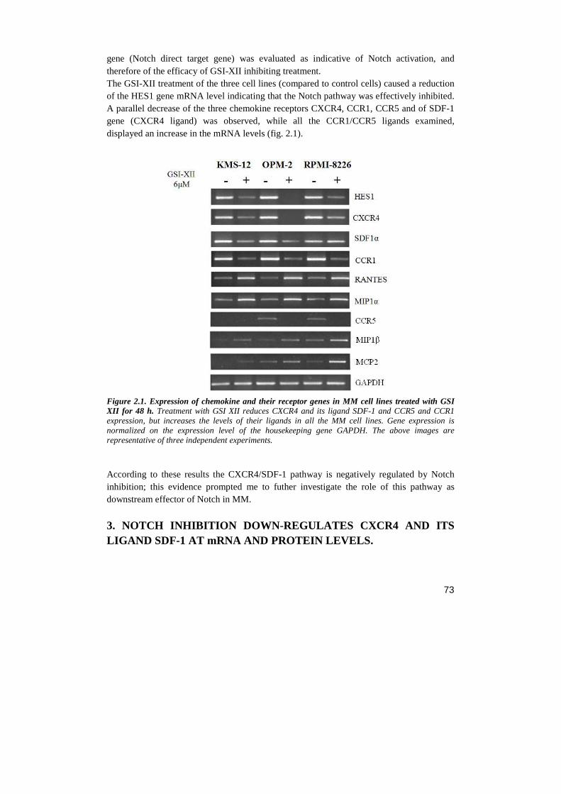
73
gene (Notch direct target gene) was evaluated as indicative of Notch activation, and therefore of the efficacy of GSI-XII inhibiting treatment. The GSI-XII treatment of the three cell lines (compared to control cells) caused a reduction of the HES1 gene mRNA level indicating that the Notch pathway was effectively inhibited. A parallel decrease of the three chemokine receptors CXCR4, CCR1, CCR5 and of SDF-1 gene (CXCR4 ligand) was observed, while all the CCR1/CCR5 ligands examined, displayed an increase in the mRNA levels (fig. 2.1).
Figure 2.1. Expression of chemokine and their receptor genes in MM cell lines treated with GSI XII for 48 h. Treatment with GSI XII reduces CXCR4 and its ligand SDF-1 and CCR5 and CCR1 expression, but increases the levels of their ligands in all the MM cell lines. Gene expression is normalized on the expression level of the housekeeping gene GAPDH. The above images are representative of three independent experiments.
According to these results the CXCR4/SDF-1 pathway is negatively regulated by Notch inhibition; this evidence prompted me to futher investigate the role of this pathway as downstream effector of Notch in MM.
3. NOTCH INHIBITION DOWN-REGULATES CXCR4 AND ITS LIGAND SDF-1 AT mRNA AND PROTEIN LEVELS.
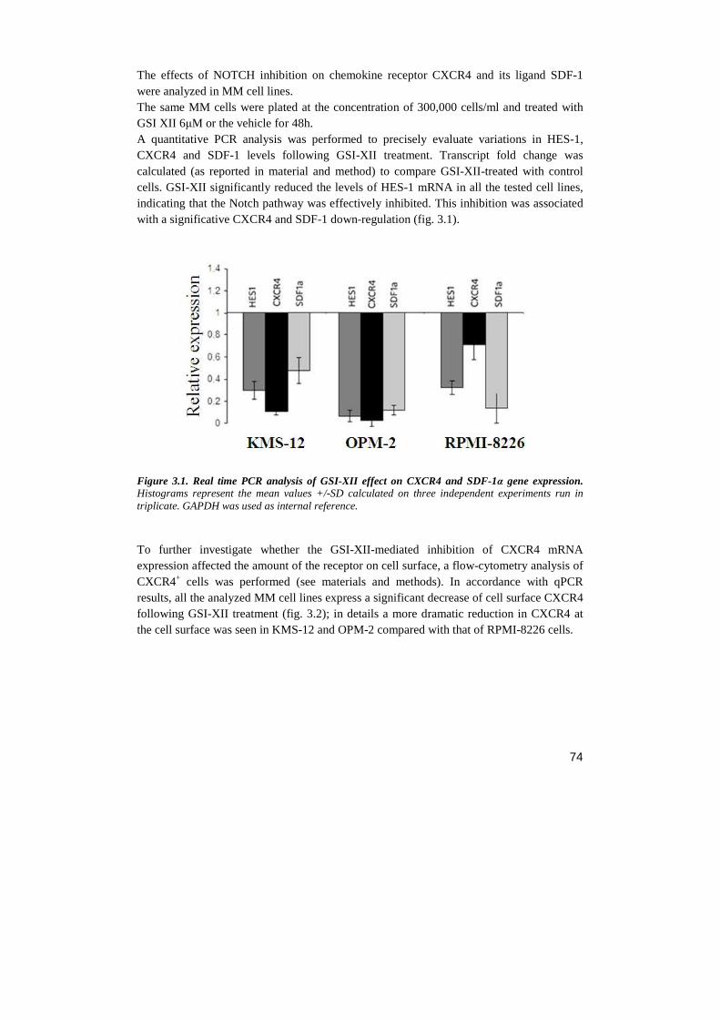
74
The effects of NOTCH inhibition on chemokine receptor CXCR4 and its ligand SDF-1 were analyzed in MM cell lines. The same MM cells were plated at the concentration of 300,000 cells/ml and treated with GSI XII 6µM or the vehicle for 48h. A quantitative PCR analysis was performed to precisely evaluate variations in HES-1, CXCR4 and SDF-1 levels following GSI-XII treatment. Transcript fold change was calculated (as reported in material and method) to compare GSI-XII-treated with control cells. GSI-XII significantly reduced the levels of HES-1 mRNA in all the tested cell lines, indicating that the Notch pathway was effectively inhibited. This inhibition was associated with a significative CXCR4 and SDF-1 down-regulation (fig. 3.1).
Figure 3.1. Real time PCR analysis of GSI-XII effect on CXCR4 and SDF-1α gene expression. Histograms represent the mean values +/-SD calculated on three independent experiments run in triplicate. GAPDH was used as internal reference. To further investigate whether the GSI-XII-mediated inhibition of CXCR4 mRNA expression affected the amount of the receptor on cell surface, a flow-cytometry analysis of CXCR4+ cells was performed (see materials and methods). In accordance with qPCR results, all the analyzed MM cell lines express a significant decrease of cell surface CXCR4 following GSI-XII treatment (fig. 3.2); in details a more dramatic reduction in CXCR4 at the cell surface was seen in KMS-12 and OPM-2 compared with that of RPMI-8226 cells.
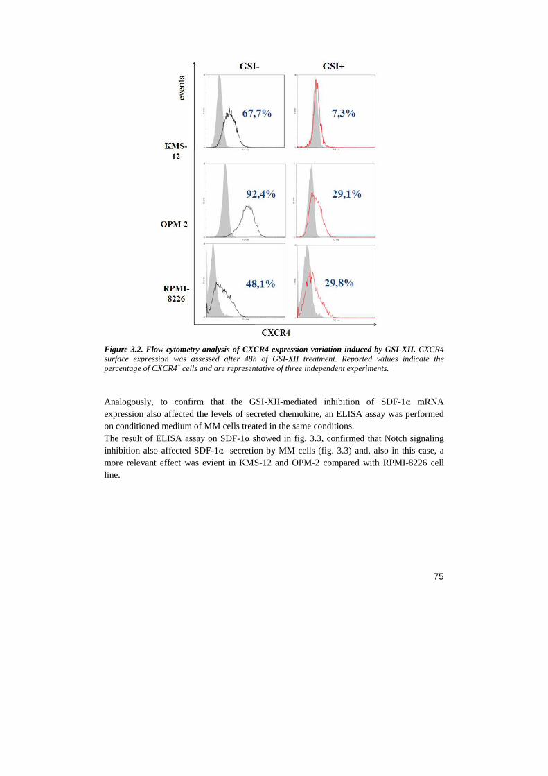
75
Figure 3.2. Flow cytometry analysis of CXCR4 expression variation induced by GSI-XII. CXCR4 surface expression was assessed after 48h of GSI-XII treatment. Reported values indicate the percentage of CXCR4+ cells and are representative of three independent experiments. Analogously, to confirm that the GSI-XII-mediated inhibition of SDF-1α mRNA expression also affected the levels of secreted chemokine, an ELISA assay was performed on conditioned medium of MM cells treated in the same conditions. The result of ELISA assay on SDF-1α showed in fig. 3.3, confirmed that Notch signaling inhibition also affected SDF-1α secretion by MM cells (fig. 3.3) and, also in this case, a more relevant effect was evient in KMS-12 and OPM-2 compared with RPMI-8226 cell line.
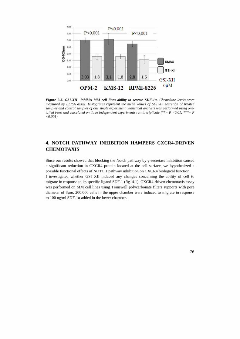
76
Figure 3.3. GSI-XII inhibits MM cell lines ability to secrete SDF-1α. Chemokine levels were measured by ELISA assay. Histograms represent the mean values of SDF-1α secretion of treated samples and control samples of one single experiment. Statistical analysis was performed using one-tailed t-test and calculated on three independent experiments run in triplicate (**= P <0.01; ***= P <0.001).
4. NOTCH PATHWAY INHIBITION HAMPERS CXCR4-DRIVEN CHEMOTAXIS Since our results showed that blocking the Notch pathway by γ-secretase inhibition caused a significant reduction in CXCR4 protein located at the cell surface, we hypothesized a possible functional effects of NOTCH pathway inhibition on CXCR4 biological function. I investigated whether GSI XII induced any changes concerning the ability of cell to migrate in response to its specific ligand SDF-1 (fig. 4.1). CXCR4-driven chemotaxis assay was performed on MM cell lines using Transwell polycarbonate filters supports with pore diameter of 8µm. 200.000 cells in the upper chamber were induced to migrate in response to 100 ng/ml SDF-1α added in the lower chamber.
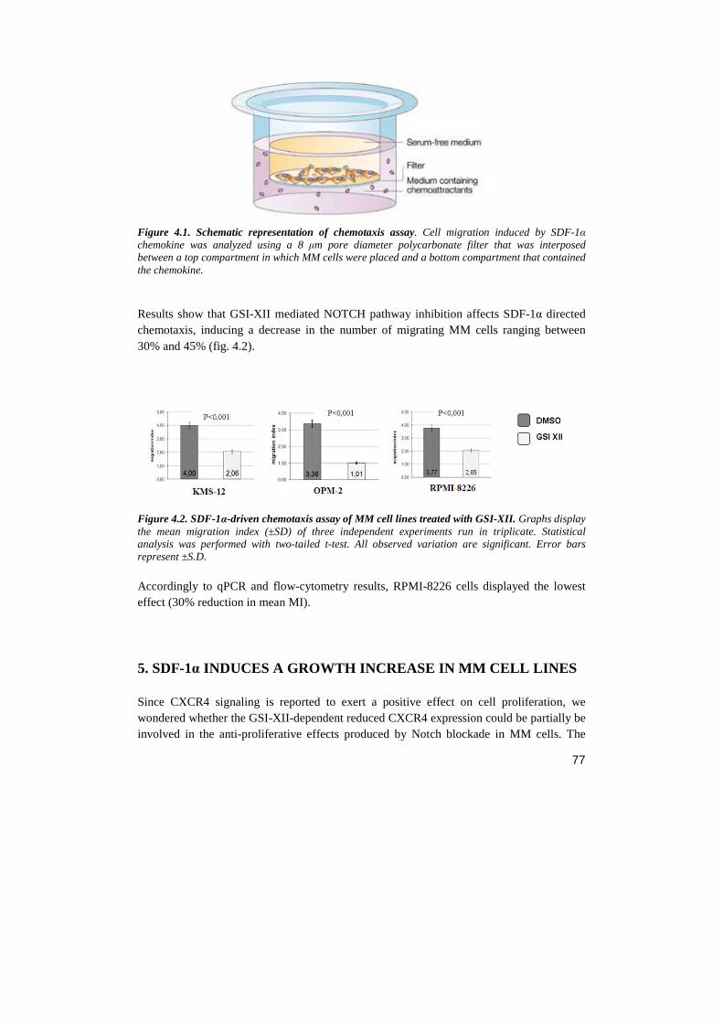
77
Figure 4.1. Schematic representation of chemotaxis assay. Cell migration induced by SDF-1α chemokine was analyzed using a 8 µm pore diameter polycarbonate filter that was interposed between a top compartment in which MM cells were placed and a bottom compartment that contained the chemokine. Results show that GSI-XII mediated NOTCH pathway inhibition affects SDF-1α directed chemotaxis, inducing a decrease in the number of migrating MM cells ranging between 30% and 45% (fig. 4.2).
Figure 4.2. SDF-1α-driven chemotaxis assay of MM cell lines treated with GSI-XII. Graphs display the mean migration index (±SD) of three independent experiments run in triplicate. Statistical analysis was performed with two-tailed t-test. All observed variation are significant. Error bars represent ±S.D. Accordingly to qPCR and flow-cytometry results, RPMI-8226 cells displayed the lowest effect (30% reduction in mean MI).
5. SDF-1α INDUCES A GROWTH INCREASE IN MM CELL LINES Since CXCR4 signaling is reported to exert a positive effect on cell proliferation, we wondered whether the GSI-XII-dependent reduced CXCR4 expression could be partially be involved in the anti-proliferative effects produced by Notch blockade in MM cells. The
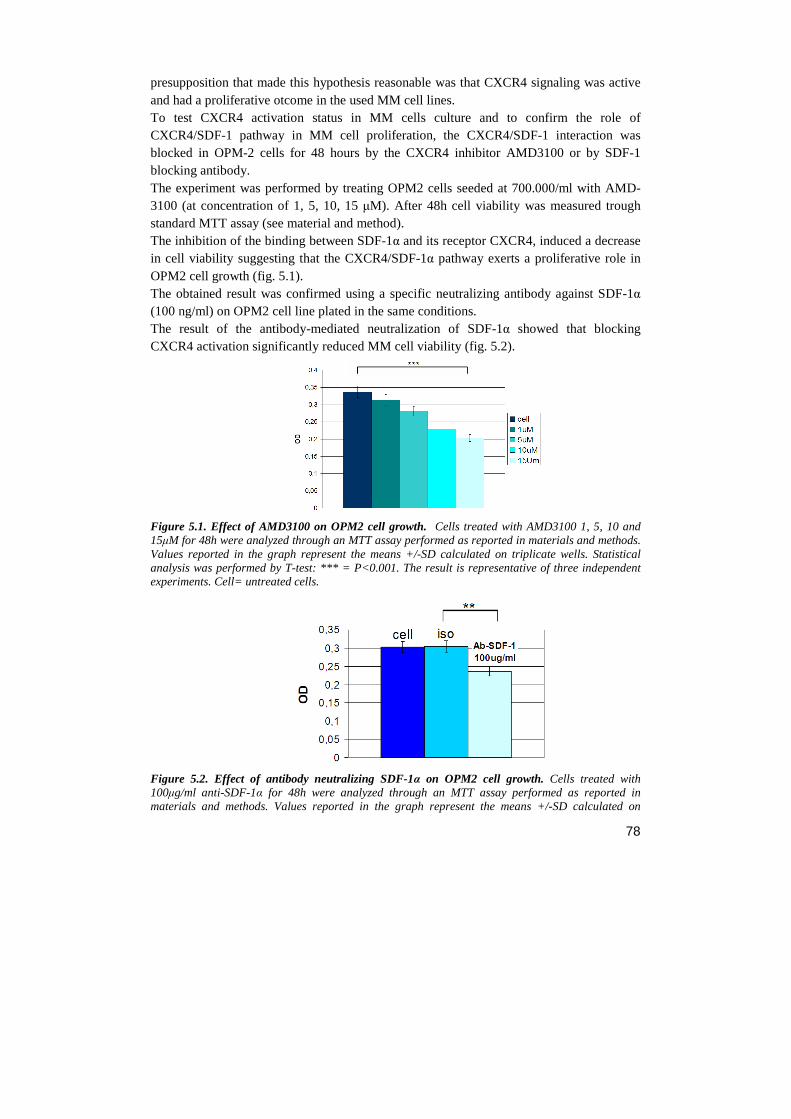
78
presupposition that made this hypothesis reasonable was that CXCR4 signaling was active and had a proliferative otcome in the used MM cell lines. To test CXCR4 activation status in MM cells culture and to confirm the role of CXCR4/SDF-1 pathway in MM cell proliferation, the CXCR4/SDF-1 interaction was blocked in OPM-2 cells for 48 hours by the CXCR4 inhibitor AMD3100 or by SDF-1 blocking antibody. The experiment was performed by treating OPM2 cells seeded at 700.000/ml with AMD-3100 (at concentration of 1, 5, 10, 15 µM). After 48h cell viability was measured trough standard MTT assay (see material and method). The inhibition of the binding between SDF-1α and its receptor CXCR4, induced a decrease in cell viability suggesting that the CXCR4/SDF-1α pathway exerts a proliferative role in OPM2 cell growth (fig. 5.1). The obtained result was confirmed using a specific neutralizing antibody against SDF-1α (100 ng/ml) on OPM2 cell line plated in the same conditions. The result of the antibody-mediated neutralization of SDF-1α showed that blocking CXCR4 activation significantly reduced MM cell viability (fig. 5.2).
Figure 5.1. Effect of AMD3100 on OPM2 cell growth. Cells treated with AMD3100 1, 5, 10 and 15µM for 48h were analyzed through an MTT assay performed as reported in materials and methods. Values reported in the graph represent the means +/-SD calculated on triplicate wells. Statistical analysis was performed by T-test: *** = P<0.001. The result is representative of three independent experiments. Cell= untreated cells.
Figure 5.2. Effect of antibody neutralizing SDF-1α on OPM2 cell growth. Cells treated with 100µg/ml anti-SDF-1α for 48h were analyzed through an MTT assay performed as reported in materials and methods. Values reported in the graph represent the means +/-SD calculated on
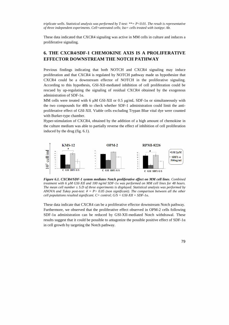
79
triplicate wells. Statistical analysis was performed by T-test: **= P<0.01. The result is representative of three independent experiments. Cell=untreated cells; Iso= cells treated with isotipyc Ab. These data indicated that CXCR4 signaling was active in MM cells in culture and induces a proliferative signaling.
6. THE CXCR4/SDF-1 CHEMOKINE AXIS IS A PROLIFERATIV E EFFECTOR DOWNSTREAM THE NOTCH PATHWAY Previous findings indicating that both NOTCH and CXCR4 signaling may induce proliferation and that CXCR4 is regulated by NOTCH pathway made us hypothesize that CXCR4 could be a downstream effector of NOTCH in the proliferative signaling. According to this hypothesis, GSI-XII-mediated inhibition of cell proliferation could be rescued by up-regulating the signaling of residual CXCR4 obtained by the exogenous administration of SDF-1α. MM cells were treated with 6 µM GSI-XII or 0.5 µg/mL SDF-1α or simultaneously with the two compounds for 48h to check whether SDF-1 administration could limit the anti-proliferative effect of GSI-XII. Viable cells excluding Trypan Blue vital dye were counted with Burker-type chamber. Hyper-stimulation of CXCR4, obtained by the addition of a high amount of chemokine in the culture medium was able to partially reverse the effect of inhibition of cell proliferation induced by the drug (fig. 6.1).
Figure 6.1. CXCR4/SDF-1 system mediates Notch proliferative effect on MM cell lines. Combined treatment with 6 µM GSI-XII and 100 ng/ml SDF-1α was performed on MM cell lines for 48 hours. The mean cell number ± S.D of three experiments is displayed. Statistical analysis was performed by ANOVA and Tukey post-test: # = P> 0.05 (non significant). The comparison between all the other cell populations resulted significant. C= control; G/S = GSI-XII + SDF-1α. These data indicate that CXCR4 can be a proliferative effector downstream Notch pathway. Furthermore, we observed that the proliferative effect observed in OPM-2 cells following SDF-1α administration can be reduced by GSI-XII-mediated Notch withdrawal. These results suggest that it could be possible to antagonize the possible positive effect of SDF-1α in cell growth by targeting the Notch pathway.
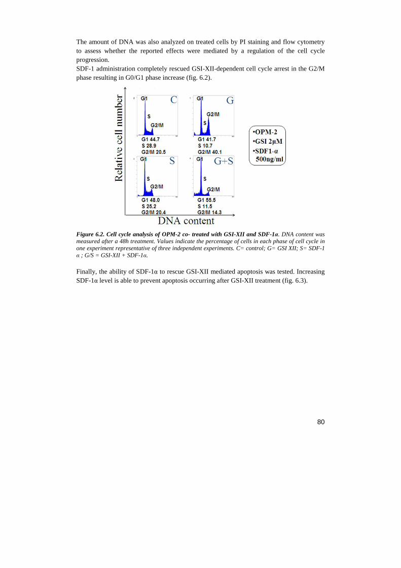
80
The amount of DNA was also analyzed on treated cells by PI staining and flow cytometry to assess whether the reported effects were mediated by a regulation of the cell cycle progression. SDF-1 administration completely rescued GSI-XII-dependent cell cycle arrest in the G2/M phase resulting in G0/G1 phase increase (fig. 6.2).
Figure 6.2. Cell cycle analysis of OPM-2 co- treated with GSI-XII and SDF-1α. DNA content was measured after a 48h treatment. Values indicate the percentage of cells in each phase of cell cycle in one experiment representative of three independent experiments. C= control; G= GSI XII; S= SDF-1 α ; G/S = GSI-XII + SDF-1α. Finally, the ability of SDF-1α to rescue GSI-XII mediated apoptosis was tested. Increasing SDF-1α level is able to prevent apoptosis occurring after GSI-XII treatment (fig. 6.3).
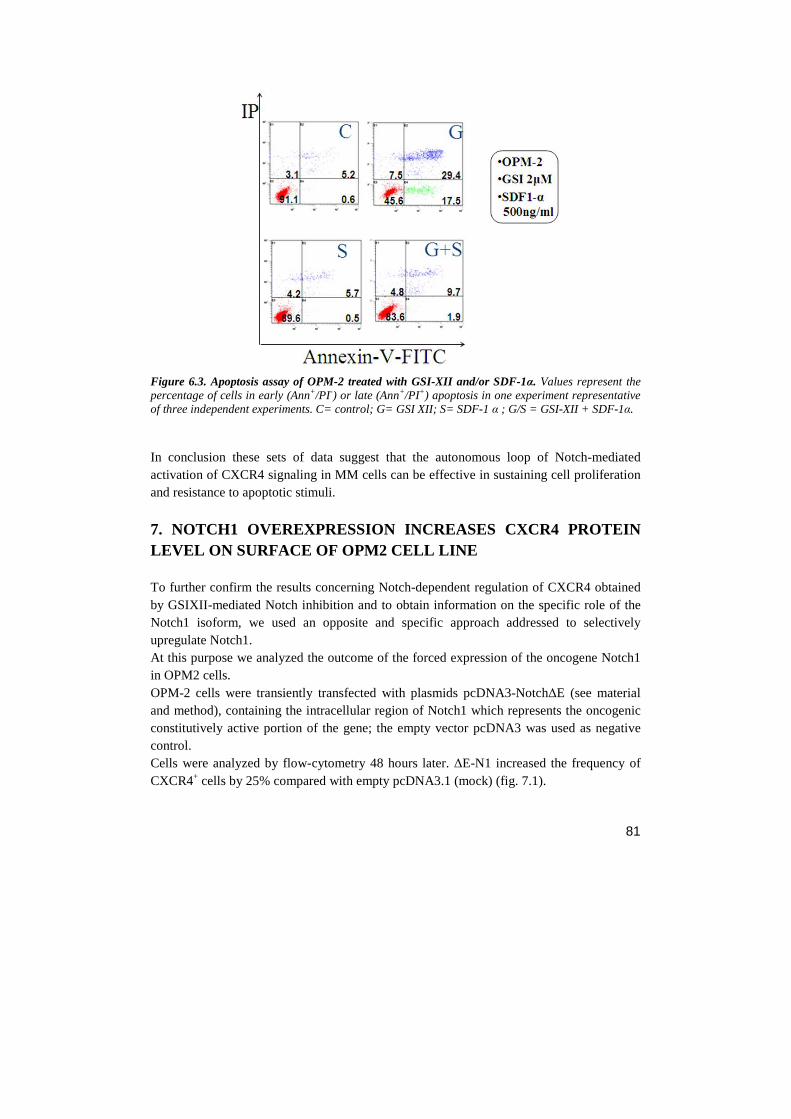
81
Figure 6.3. Apoptosis assay of OPM-2 treated with GSI-XII and/or SDF-1α. Values represent the percentage of cells in early (Ann+/PI-) or late (Ann+/PI+) apoptosis in one experiment representative of three independent experiments. C= control; G= GSI XII; S= SDF-1 α ; G/S = GSI-XII + SDF-1α. In conclusion these sets of data suggest that the autonomous loop of Notch-mediated activation of CXCR4 signaling in MM cells can be effective in sustaining cell proliferation and resistance to apoptotic stimuli.
7. NOTCH1 OVEREXPRESSION INCREASES CXCR4 PROTEIN LEVEL ON SURFACE OF OPM2 CELL LINE To further confirm the results concerning Notch-dependent regulation of CXCR4 obtained by GSIXII-mediated Notch inhibition and to obtain information on the specific role of the Notch1 isoform, we used an opposite and specific approach addressed to selectively upregulate Notch1. At this purpose we analyzed the outcome of the forced expression of the oncogene Notch1 in OPM2 cells. OPM-2 cells were transiently transfected with plasmids pcDNA3-Notch∆E (see material and method), containing the intracellular region of Notch1 which represents the oncogenic constitutively active portion of the gene; the empty vector pcDNA3 was used as negative control. Cells were analyzed by flow-cytometry 48 hours later. ∆E-N1 increased the frequency of CXCR4+ cells by 25% compared with empty pcDNA3.1 (mock) (fig. 7.1).
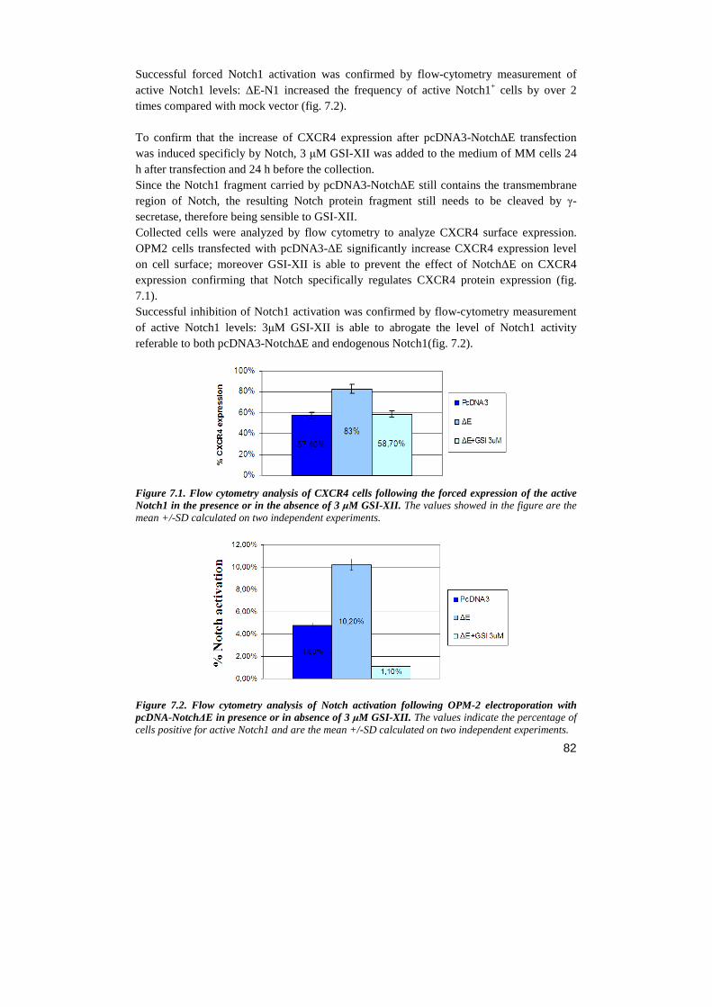
82
Successful forced Notch1 activation was confirmed by flow-cytometry measurement of active Notch1 levels: ∆E-N1 increased the frequency of active Notch1+ cells by over 2 times compared with mock vector (fig. 7.2). To confirm that the increase of CXCR4 expression after pcDNA3-Notch∆E transfection was induced specificly by Notch, 3 µM GSI-XII was added to the medium of MM cells 24 h after transfection and 24 h before the collection. Since the Notch1 fragment carried by pcDNA3-Notch∆E still contains the transmembrane region of Notch, the resulting Notch protein fragment still needs to be cleaved by γ-secretase, therefore being sensible to GSI-XII. Collected cells were analyzed by flow cytometry to analyze CXCR4 surface expression. OPM2 cells transfected with pcDNA3-∆E significantly increase CXCR4 expression level on cell surface; moreover GSI-XII is able to prevent the effect of Notch∆E on CXCR4 expression confirming that Notch specifically regulates CXCR4 protein expression (fig. 7.1). Successful inhibition of Notch1 activation was confirmed by flow-cytometry measurement of active Notch1 levels: 3µM GSI-XII is able to abrogate the level of Notch1 activity referable to both pcDNA3-Notch∆E and endogenous Notch1(fig. 7.2).
Figure 7.1. Flow cytometry analysis of CXCR4 cells following the forced expression of the active Notch1 in the presence or in the absence of 3 µM GSI-XII. The values showed in the figure are the mean +/-SD calculated on two independent experiments.
Figure 7.2. Flow cytometry analysis of Notch activation following OPM-2 electroporation with pcDNA-Notch∆E in presence or in absence of 3 µM GSI-XII. The values indicate the percentage of
cells positive for active Notch1 and are the mean +/-SD calculated on two independent experiments.
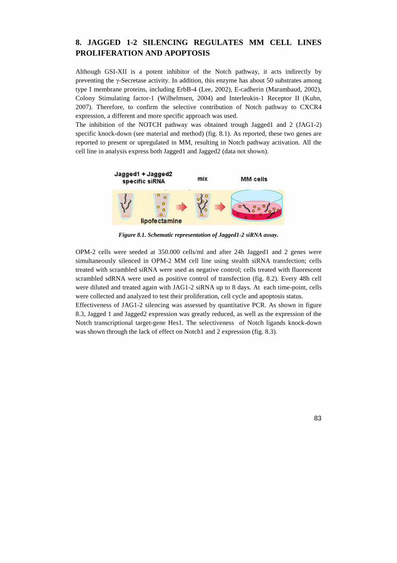
83
8. JAGGED 1-2 SILENCING REGULATES MM CELL LINES PROLIFERATION AND APOPTOSIS Although GSI-XII is a potent inhibitor of the Notch pathway, it acts indirectly by preventing the γ-Secretase activity. In addition, this enzyme has about 50 substrates among type I membrane proteins, including ErbB-4 (Lee, 2002), E-cadherin (Marambaud, 2002), Colony Stimulating factor-1 (Wilhelmsen, 2004) and Interleukin-1 Receptor II (Kuhn, 2007). Therefore, to confirm the selective contribution of Notch pathway to CXCR4 expression, a different and more specific approach was used. The inhibition of the NOTCH pathway was obtained trough Jagged1 and 2 (JAG1-2) specific knock-down (see material and method) (fig. 8.1). As reported, these two genes are reported to present or upregulated in MM, resulting in Notch pathway activation. All the cell line in analysis express both Jagged1 and Jagged2 (data not shown).
Figure 8.1. Schematic representation of Jagged1-2 siRNA assay.
OPM-2 cells were seeded at 350.000 cells/ml and after 24h Jagged1 and 2 genes were simultaneously silenced in OPM-2 MM cell line using stealth siRNA transfection; cells treated with scrambled siRNA were used as negative control; cells treated with fluorescent scrambled sdRNA were used as positive control of transfection (fig. 8.2). Every 48h cell were diluted and treated again with JAG1-2 siRNA up to 8 days. At each time-point, cells were collected and analyzed to test their proliferation, cell cycle and apoptosis status. Effectiveness of JAG1-2 silencing was assessed by quantitative PCR. As shown in figure 8.3, Jagged 1 and Jagged2 expression was greatly reduced, as well as the expression of the Notch transcriptional target-gene Hes1. The selectiveness of Notch ligands knock-down was shown through the lack of effect on Notch1 and 2 expression (fig. 8.3).
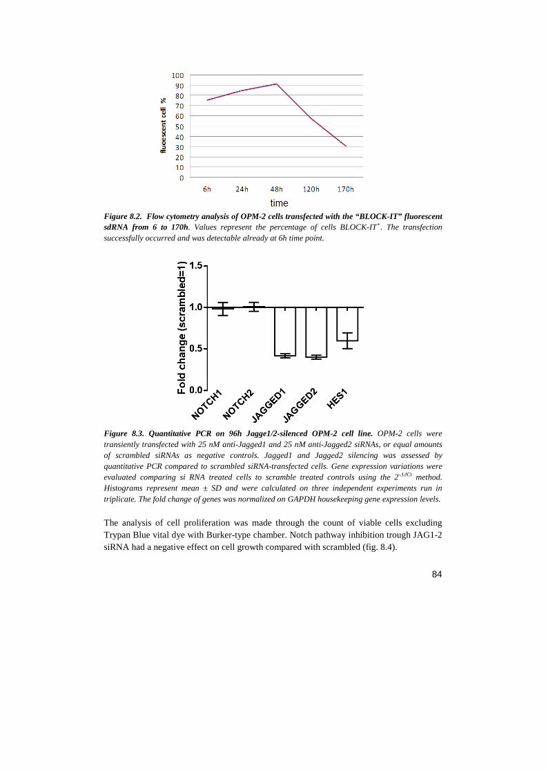
84
Figure 8.2. Flow cytometry analysis of OPM-2 cells transfected with the “BLOCK-IT” fluorescent sdRNA from 6 to 170h. Values represent the percentage of cells BLOCK-IT+. The transfection successfully occurred and was detectable already at 6h time point.
Figure 8.3. Quantitative PCR on 96h Jagge1/2-silenced OPM-2 cell line. OPM-2 cells were transiently transfected with 25 nM anti-Jagged1 and 25 nM anti-Jagged2 siRNAs, or equal amounts of scrambled siRNAs as negative controls. Jagged1 and Jagged2 silencing was assessed by quantitative PCR compared to scrambled siRNA-transfected cells. Gene expression variations were evaluated comparing si RNA treated cells to scramble treated controls using the 2-∆∆Ct method. Histograms represent mean ± SD and were calculated on three independent experiments run in triplicate. The fold change of genes was normalized on GAPDH housekeeping gene expression levels.
The analysis of cell proliferation was made through the count of viable cells excluding Trypan Blue vital dye with Burker-type chamber. Notch pathway inhibition trough JAG1-2 siRNA had a negative effect on cell growth compared with scrambled (fig. 8.4).
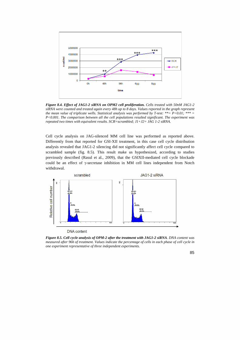
85
Figure 8.4. Effect of JAG1-2 siRNA on OPM2 cell proliferation. Cells treated with 50nM JAG1-2 siRNA were counted and treated again every 48h up to 8 days. Values reported in the graph represent the mean value of triplicate wells. Statistical analysis was performed by T-test: **= P<0.01; *** = P<0.001. The comparison between all the cell populations resulted significant. The experiment was repeated two times with equivalent results. SCR=scrambled; J1+J2= JAG 1-2 siRNA. Cell cycle analysis on JAG-silenced MM cell line was performed as reported above. Differently from that reported for GSI-XII treatment, in this case cell cycle distribution analysis revealed that JAG1-2 silencing did not significantly affect cell cycle compared to scrambled sample (fig. 8.5). This result make us hypothesized, according to studies previously described (Rasul et al., 2009), that the GSIXII-mediated cell cycle blockade could be an effect of γ-secretase inhibition in MM cell lines independent from Notch withdrawal.
Figure 8.5. Cell cycle analysis of OPM-2 after the treatment with JAG1-2 siRNA. DNA content was measured after 96h of treatment. Values indicate the percentage of cells in each phase of cell cycle in one experiment representative of three independent experiments.
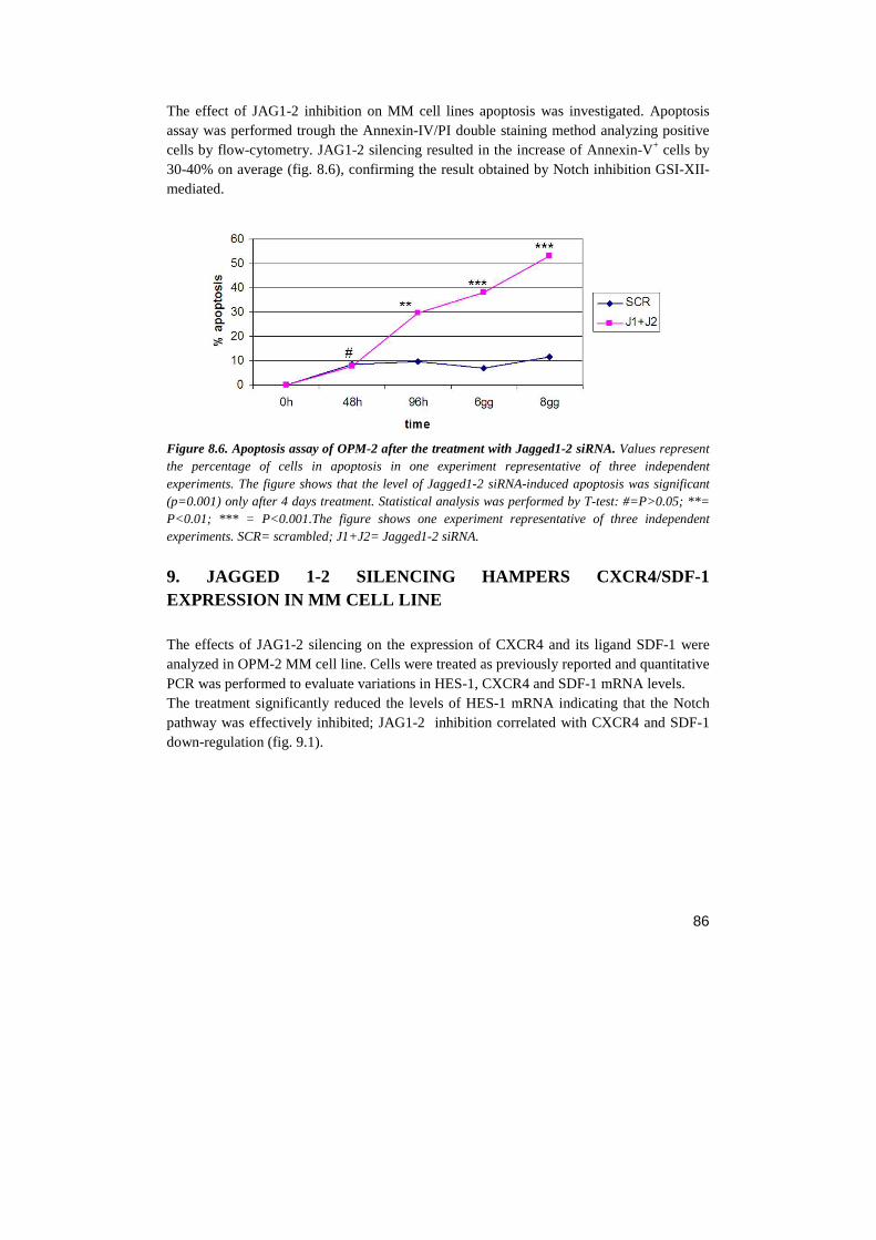
86
The effect of JAG1-2 inhibition on MM cell lines apoptosis was investigated. Apoptosis assay was performed trough the Annexin-IV/PI double staining method analyzing positive cells by flow-cytometry. JAG1-2 silencing resulted in the increase of Annexin-V+ cells by 30-40% on average (fig. 8.6), confirming the result obtained by Notch inhibition GSI-XII-mediated.
Figure 8.6. Apoptosis assay of OPM-2 after the treatment with Jagged1-2 siRNA. Values represent the percentage of cells in apoptosis in one experiment representative of three independent experiments. The figure shows that the level of Jagged1-2 siRNA-induced apoptosis was significant (p=0.001) only after 4 days treatment. Statistical analysis was performed by T-test: #=P>0.05; **= P<0.01; *** = P<0.001.The figure shows one experiment representative of three independent experiments. SCR= scrambled; J1+J2= Jagged1-2 siRNA.
9. JAGGED 1-2 SILENCING HAMPERS CXCR4/SDF-1 EXPRESSION IN MM CELL LINE The effects of JAG1-2 silencing on the expression of CXCR4 and its ligand SDF-1 were analyzed in OPM-2 MM cell line. Cells were treated as previously reported and quantitative PCR was performed to evaluate variations in HES-1, CXCR4 and SDF-1 mRNA levels. The treatment significantly reduced the levels of HES-1 mRNA indicating that the Notch pathway was effectively inhibited; JAG1-2 inhibition correlated with CXCR4 and SDF-1 down-regulation (fig. 9.1).
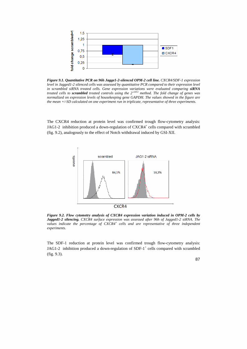
87
Figure 9.1. Quantitative PCR on 96h Jagge1-2-silenced OPM-2 cell line. CXCR4/SDF-1 expression level in Jagged1-2 silenced cells was assessed by quantitative PCR compared to their expression level in scrambled siRNA treated cells. Gene expression variations were evaluated comparing siRNA treated cells to scrambled treated controls using the 2-∆∆Ct method. The fold change of genes was normalized on expression levels of housekeeping gene GAPDH. The values showed in the figure are the mean +/-SD calculated on one experiment run in triplicate, representative of three experiments. The CXCR4 reduction at protein level was confirmed trough flow-cytometry analysis: JAG1-2 inhibition produced a down-regulation of CXCR4+ cells compared with scrambled (fig. 9.2), analogously to the effect of Notch withdrawal induced by GSI-XII.
Figure 9.2. Flow cytometry analysis of CXCR4 expression variation induced in OPM-2 cells by Jagged1-2 silencing. CXCR4 surface expression was assessed after 96h of Jagged1-2 siRNA. The values indicate the percentage of CXCR4+ cells and are representative of three independent experiments. The SDF-1 reduction at protein level was confirmed trough flow-cytometry analysis: JAG1-2 inhibition produced a down-regulation of SDF-1+ cells compared with scrambled (fig. 9.3).
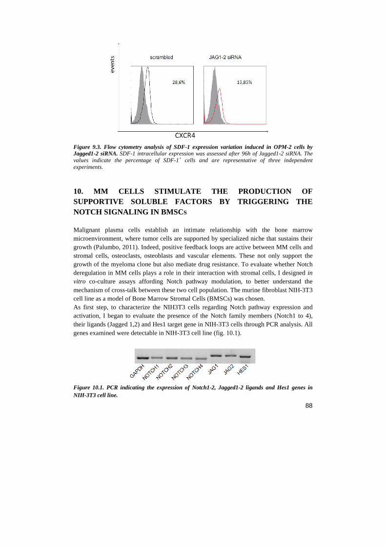
88
Figure 9.3. Flow cytometry analysis of SDF-1 expression variation induced in OPM-2 cells by Jagged1-2 siRNA. SDF-1 intracellular expression was assessed after 96h of Jagged1-2 siRNA. The values indicate the percentage of SDF-1+ cells and are representative of three independent experiments.
10. MM CELLS STIMULATE THE PRODUCTION OF SUPPORTIVE SOLUBLE FACTORS BY TRIGGERING THE NOTCH SIGNALING IN BMSC S Malignant plasma cells establish an intimate relationship with the bone marrow microenvironment, where tumor cells are supported by specialized niche that sustains their growth (Palumbo, 2011). Indeed, positive feedback loops are active between MM cells and stromal cells, osteoclasts, osteoblasts and vascular elements. These not only support the growth of the myeloma clone but also mediate drug resistance. To evaluate whether Notch deregulation in MM cells plays a role in their interaction with stromal cells, I designed in vitro co-culture assays affording Notch pathway modulation, to better understand the mechanism of cross-talk between these two cell population. The murine fibroblast NIH-3T3 cell line as a model of Bone Marrow Stromal Cells (BMSCs) was chosen. As first step, to characterize the NIH3T3 cells regarding Notch pathway expression and activation, I began to evaluate the presence of the Notch family members (Notch1 to 4), their ligands (Jagged 1,2) and Hes1 target gene in NIH-3T3 cells through PCR analysis. All genes examined were detectable in NIH-3T3 cell line (fig. 10.1).
Figure 10.1. PCR indicating the expression of Notch1-2, Jagged1-2 ligands and Hes1 genes in NIH-3T3 cell line.
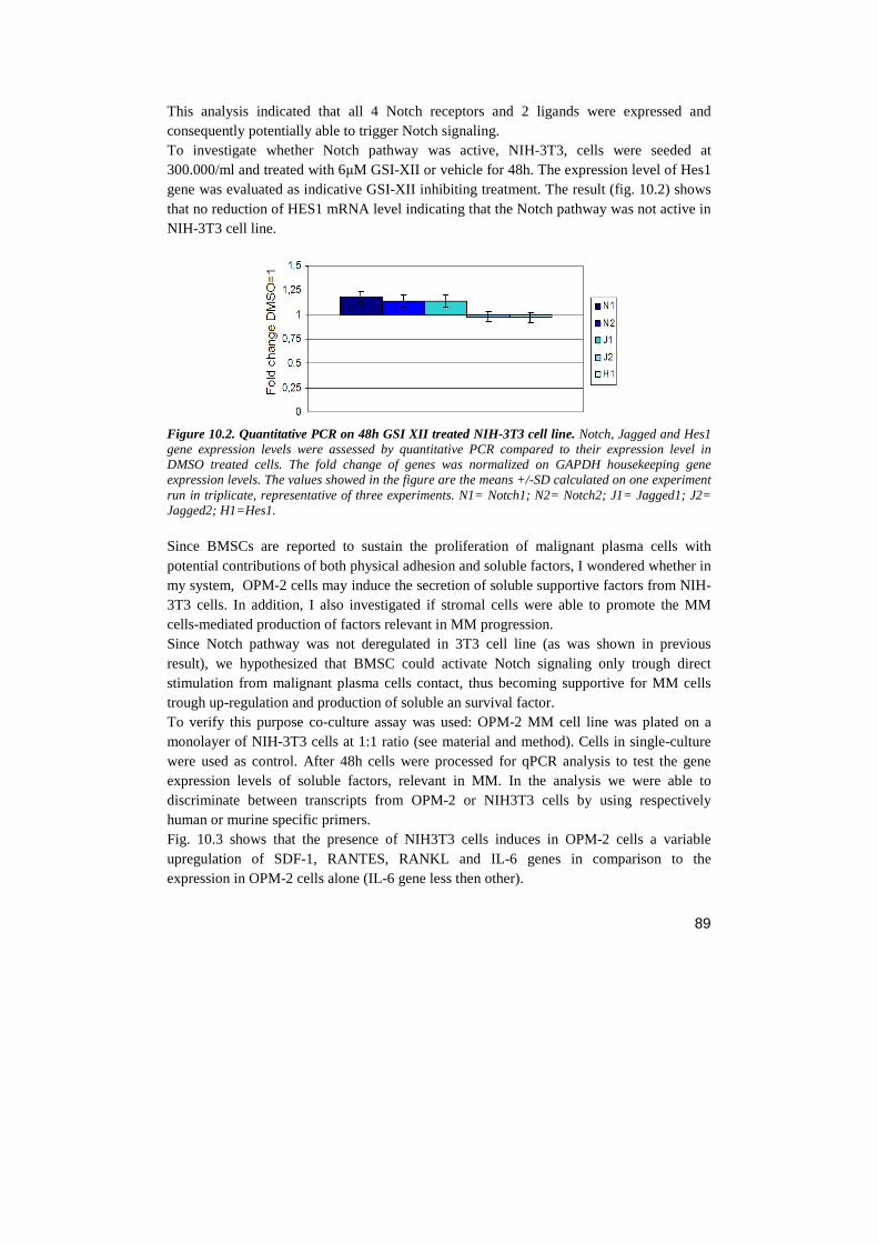
89
This analysis indicated that all 4 Notch receptors and 2 ligands were expressed and consequently potentially able to trigger Notch signaling. To investigate whether Notch pathway was active, NIH-3T3, cells were seeded at 300.000/ml and treated with 6µM GSI-XII or vehicle for 48h. The expression level of Hes1 gene was evaluated as indicative GSI-XII inhibiting treatment. The result (fig. 10.2) shows that no reduction of HES1 mRNA level indicating that the Notch pathway was not active in NIH-3T3 cell line.
Figure 10.2. Quantitative PCR on 48h GSI XII treated NIH-3T3 cell line. Notch, Jagged and Hes1 gene expression levels were assessed by quantitative PCR compared to their expression level in DMSO treated cells. The fold change of genes was normalized on GAPDH housekeeping gene expression levels. The values showed in the figure are the means +/-SD calculated on one experiment run in triplicate, representative of three experiments. N1= Notch1; N2= Notch2; J1= Jagged1; J2= Jagged2; H1=Hes1. Since BMSCs are reported to sustain the proliferation of malignant plasma cells with potential contributions of both physical adhesion and soluble factors, I wondered whether in my system, OPM-2 cells may induce the secretion of soluble supportive factors from NIH-3T3 cells. In addition, I also investigated if stromal cells were able to promote the MM cells-mediated production of factors relevant in MM progression. Since Notch pathway was not deregulated in 3T3 cell line (as was shown in previous result), we hypothesized that BMSC could activate Notch signaling only trough direct stimulation from malignant plasma cells contact, thus becoming supportive for MM cells trough up-regulation and production of soluble an survival factor. To verify this purpose co-culture assay was used: OPM-2 MM cell line was plated on a monolayer of NIH-3T3 cells at 1:1 ratio (see material and method). Cells in single-culture were used as control. After 48h cells were processed for qPCR analysis to test the gene expression levels of soluble factors, relevant in MM. In the analysis we were able to discriminate between transcripts from OPM-2 or NIH3T3 cells by using respectively human or murine specific primers. Fig. 10.3 shows that the presence of NIH3T3 cells induces in OPM-2 cells a variable upregulation of SDF-1, RANTES, RANKL and IL-6 genes in comparison to the expression in OPM-2 cells alone (IL-6 gene less then other).
90
The expression of MIP-1α gene was down-regulated compared to its expression in single culture. CXCR4 and VEGF genes were not affected. Hes1 in OPM-2 cells was not affected by the contact with NIH3T3 cells, indicating that Notch pathway was not up-regulated by BMSC contact.
Figure 10.3. Quantitative PCR on the expression of soluble factors and CXCR4 genes in 48h co-cultured OPM-2/NIH-3T3 cells. Gene expression variations were evaluated comparing OPM-2 co-cultured cells to OPM-2 in single culture using the 2-∆∆Ct method. The fold change of genes was normalized on the expression levels of the housekeeping gene, GAPDH. The values showed in the figure are the means +/-SD calculated on one experiment run in triplicate, representative of three experiments. On the other side, fig. 10.4 displays that NIH-3T3 cells are induced by the presence of OPM-2 to upregulate the expression of SDF-1, CXCR4, IL-6, VEGF, RANTES, MIP-1α genes. The expression of RANKL gene had no variations compared to its expression in single culture. Hes1 expression also was up-regulated in co-culture compared with its expression in single culture, indicating that Notch pathway was activated in NIH-3T3 cells by contact with MM cells.
Figure 10.4. Quantitative PCR on the expression of soluble factors and CXCR4 genes in 48h co-cultured OPM-2/NIH-3T3 cells. Gene expression variations were evaluated comparing NIH-3T3 co-cultured cells to NIH-3T3 in single culture using the 2-∆∆Ct method. The fold change of genes was
91
normalized on the expression levels of the housekeeping gene, GAPDH. The values showed in the figure are the means +/-SD calculated on one experiment run in triplicate, representative of three experiments. All these results indicate that MM cells are able to trigger Notch signaling in NIH-3T3 cells and the release of soluble factors which sustain MM cell progression. By contrast these results do not support the possibility that BMSCs activate the Notch pathway in MM cells, supporting the possibility that Notch signalling activation is thoroughly due to the expression of the two Notch ligands Jagged1 and 2. From this evidence stems the consideration that the observed upregulation in the expression of SDF1, RANKL and RANTES is OPM-2 cell is autonomous and not due to the presence of NIH-3T3 cells. Finally, to clarify which is the Notch contribute in this cross-talk and to investigate whether the Jagged ligands expressed by MM cells are able to trigger the Notch signaling in BMSCs, I designed a co-culture assay in which NIH-3T3 cells were in contact with OPM-2 cells depleted or not of Jagge1 and 2. To selectively inhibit MM-jagged, it was used siRNA approach. OPM-2 cells were seeded at 300.000 cells/ml and after 24h the JAG1-2 siRNA treatment was added. After 48h cells were diluted and treated once again with silencing treatment. After 8h OPM-2 cells were washed and finally plated on a monolayer of NIH-3T3 cells at 1:1 ratio (see material and method). Co-cultures were manteined for 48h, obtaining in this way a condition in wich MM cells were silenced from 4 days (since a 48h silencing was not sufficient to induce a great response in MM cells). Cells treated with scrambled siRNA were used as negative control. The results in fig. 10.6 show that when NIH-3T3 cell line are placed in co-culture with OPM-2 treated with JAG1-2 siRNA, the expression of all tested genes was down-regulated if compared to the same expression in NIH-3T3 cells co-cultured with control OPM-2 cells (treatment with scrambled siRNA). The parallel strong down regulation of Hes1 expression, indicated that Jagged-expressing MM cells trigger the Notch pathway in NIH-3T3, which in turn produce soluble factors. In fact, when the Jagged ligands are turned-off in MM cells, NIH-3T3 cells are no more able to produce soluble factors fundamental for MM progression.
92
Figure 10.5. Quantitative PCR on the expression of soluble factors and CXCR4 genes in 48h NIH-3T3 co-cultured with Jagged1-2 silenced OPM-2. Gene expression levels in NIH-3T3 co-cultured with silenced OPM-2 were assessed by quantitative PCR compared to their expression levels in NIH-3T3 co-cultured with non-silenced OPM-2. The fold change of genes was normalized on GAPDH housekeeping gene expression levels. The figure shows the “reduction index” which indicate the reduction percentage of gene expression compared with the genhe expression leves showed in figure 10.4. The values showed in the figure are representative of three independent experiments. Overall these results shows that the production of soluble factors by BMSC occurs only when Notch pathway in MM is active, in particular when Jagged1-2 expressed on MM cell bind the Notch receptor on BMSC surface. In conclusion, these results are consistent with the possibility that upregulated Jagged ligands in MM cells are responsible of deregulated Notch signaling in BMSC, resulting in the secretion of soluble factors promoting MM progression.
11. JAGGED 1 AND 2 INDUCE IN MM CELLS INTRINSIC SURVIVAL MECHANISM WHICH IS NOT DEPENDENT BY BMSC The above reported results represent a rationale for blocking of Jagged ligands as a therapeutic approach to disrupt the pathological interaction of MM cells with BMSCs. In literature is largely known that stroma in BM of MM patiens is able to support MM cells, inducing survival from apoptosis and drug resistance (Hideshima, 2002a). Indeed if BMSCs were able to save MM cells from apoptosis induced by Jagged blockade (previously reported in fig. 8.5), any Jagged tailored therapeutic approach would be ineffective. To address this issue, I investigated whether BMSCs would influence the apoptotic response of myeloma cells to the inhibition of Jagged1 and 2. OPM-2 cells were treated with JAG1-2 siRNA and co-cultured as previously reported (10.2). The result in figure 11.1 shows that the stromal layer of NIH-3T3 is not able to revert the apoptosis induced by Jagged withdrawal, confirming the hypothesis that the surviving loop occurring between MM and BMSC cells is induced by upstream MM-jagged signaling and
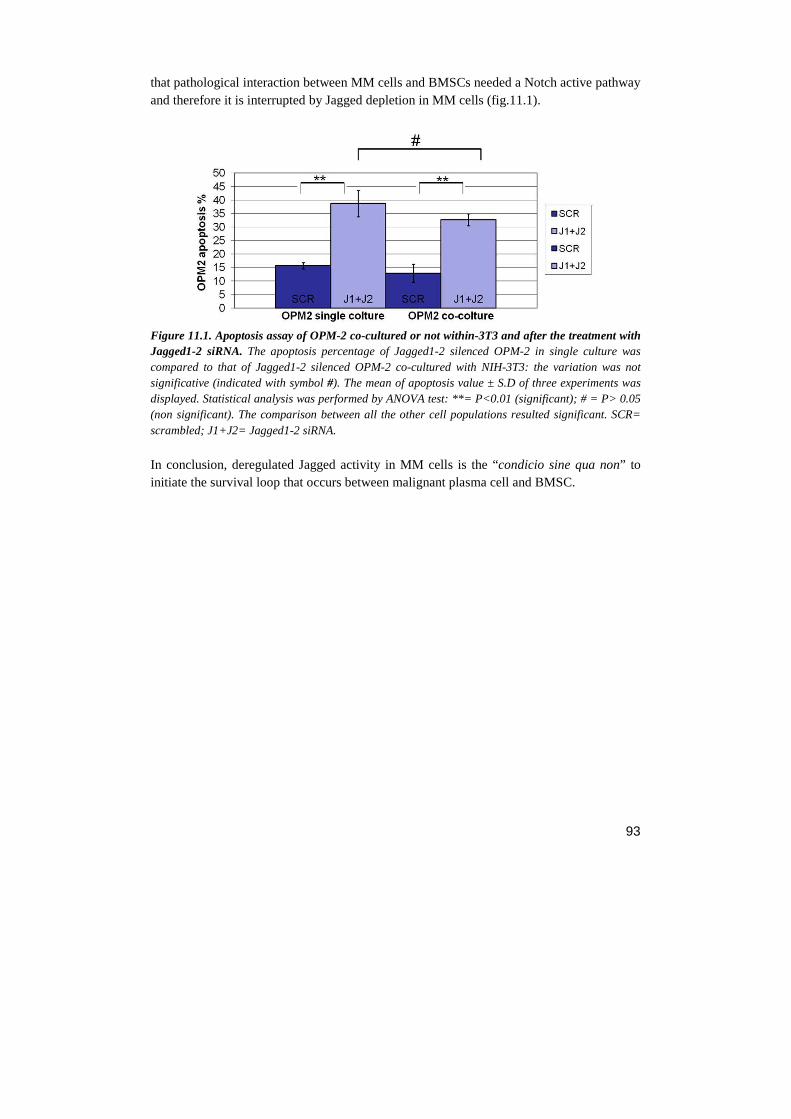
93
that pathological interaction between MM cells and BMSCs needed a Notch active pathway and therefore it is interrupted by Jagged depletion in MM cells (fig.11.1).
Figure 11.1. Apoptosis assay of OPM-2 co-cultured or not within-3T3 and after the treatment with Jagged1-2 siRNA. The apoptosis percentage of Jagged1-2 silenced OPM-2 in single culture was compared to that of Jagged1-2 silenced OPM-2 co-cultured with NIH-3T3: the variation was not significative (indicated with symbol #). The mean of apoptosis value ± S.D of three experiments was displayed. Statistical analysis was performed by ANOVA test: **= P<0.01 (significant); # = P> 0.05 (non significant). The comparison between all the other cell populations resulted significant. SCR= scrambled; J1+J2= Jagged1-2 siRNA.
In conclusion, deregulated Jagged activity in MM cells is the “condicio sine qua non” to initiate the survival loop that occurs between malignant plasma cell and BMSC.
94
DISCUSSION
Multiple myeloma (MM) is a neoplastic plasma cell disorder that results in end-organ damage: beyond hypercalcemia, renal insufficiency and anemia, the most important injury associated to negative prognosis is represented by skeletal lesions (Kyle, 2009). Bone destruction is due to MM cells localization at the bone marrow due to chemokine system deregulation. Malignant plasma cells migrated at the bone marrow interact with bone marrow stromal cells (BMSC) through direct cell-cell contact and secretion of soluble mediators, activating several pathways which bring to osteoclasts (OCL) activation and bone resorption. MM therapy is rarely curative, and a median survival is about 3-4 years after diagnosis which may increase to 5–7 years or more with advanced treatments (Kyle, 2004). The treatment of MM is complex and it exploits different approaches, including:
• Chemotherapy;
• Bone marrow stem cell transplantation (autologus or allogenic);
• Immune modulating treatments such as thalidomide (Thalomid®), lenalidomide (Revlimid®), and bortezomib (Velcade®);
• Corticosteroids (such as prednisone or dexamethasone) treatment. Initial chemotherapy for patients with symptomatic MM depends on the possibility to perform autologous stem cell transplantation (ASCT), which depends from age, performance status, and the presence of comorbid conditions in the patients. Patients who are ineligible for ASCT are submitted to initial chemotherapies. Currently, the preferred therapy is Melphalan/Prednisone/Thalidomide for the treatment of standard-risk myeloma patients, whereas Bortezomib/Melphalan/Prednisone is recommended for patients with high-risk disease (Kyle, 2009). Although associated to side effects, treatments with thalidomide, lenalidomide, and bortezomib represent a therapeutic advance in MM therapy, nevertheless, almost all patients systematically relapse, and therefore MM remains an incurable disease (Kumar, 2009). Therefore, the need of novel drug targets and therapeutic strategies to selectively target molecules relevant in MM cell progression and pharmacological resistance is evident. In the last years several studies were address to find rational molecular targets for cancer treatment, in order to develop target-selective “smart” drugs, on the basis of characterized mechanisms of action (Miele, 2006). Recently, Podar et al. (2009) described the possibility to develop an innovative therapy aiming at the inhibition of pathways activated in MM (i.e. Notch/JAG, PI3K/Akt, JAK/Stat, Raf/MEK/MAPK, NFκB and Wnt) which regulate cell proliferation, survival, migration and drug resistance.
95
The inhibition of the Notch pathway represents a new target-based therapy for those tumors characterized by Notch activation. From 1990s to present, Notch signaling aberrations have been shown to be linked with several hematological malignancies, such as T-cell acute lymphoblastic leukemia (T-ALL), acute myeloid leukemia (AML), lymphoma and MM (Mirandola, 2011a). Deregulated Notch signaling has been reported not only to affect MM cell proliferation and apoptosis, but also to influence MM cell interaction with the BM milieu, resulting in increased resistance to chemotherapeutics and osteolysis (Nefedova, 2004; Schwarzer, 2008). In MM, no mutations directly affecting Notch receptors or ligands are known, nonetheless Notch pathway deregulation seems to be a critical step in disease evolution. The expression of Notch1 and Jagged1 is deregulated upon disease progression from MGUS to MM (Skrtic, 2010). Furthermore, Jagged2 overexpression in MGUS and MM patients correlates with staging and it may induce IL-6, VEGF and IGF-1 production in a paracrine fashion (Houde, 2004). Jagged2 up-regulation may result from promoter demethylation (Houde, 2004), from constitutive core promoter acetylation associated to reduced levels of the SMRT co-repressor (Ghosal, 2009) or at post-transcriptional level from aberrant expression of Jagged2 ubiquitin ligase skeletrophin (Takeuchi, 2005). The localization of MM cells in the bone marrow gives origin to several interactions with BMSCs. The outcome of Jagged2 upregulation in MM cells is the hyper-activation of Notch pathway in neighbouring healthy and tumor cells. These interactions activate several pathways which generate a vicious cycle, from which tumor cells take source for survival and proliferation. There is also evidence that Notch1 activation induces CCR6 expression in osteoclasts, promoting OCL recruitment to osteolysis sites and OCL activation; moreover, MM cell-BMSC interaction, induces abnormal expression of RANKL by osteoblasts, which induces the maturation of OCL progenitors and subsequent bone resorption (Roodman, 2006). The above reported evidences on the role of Notch signaling in MM, suggests that Notch may be a rational specific target in MM therapy. The most diffused method to hamper Notch signaling is based on the inhibition of γ-secretase, a membrane-integral protease complex, essential for Notch receptor activation. Although GSI’s efficacy in vitro has been demonstrated, in patients GSI displayed significant gastrointestinal toxicity and no clinical response in T-ALL. Gastrointestinal side effects have been studies in animal models, where chronic administration of GSIs boosted the formation of secretory globet cells in the intestine (Searfoss, 2003). Side effect in intestine is associated to Notch2 inhibition which is the consequence of the inability of GSI to distinguish between Notch proteins. At the moment, several phase I clinical trials were evaluated for the use of GSIs in clinical practice, such as breast cancer, T-ALL and lymphoma, metasttic melanoma, colorectal and pancreatic adenocarcinoma; despite GSIs side effects, encouraging results suggest that Notch targeting trough γ-secretase inhibitors (GSIs), are potential novel cancer therapeutic agents.
96
This work aims to confirm that Notch signaling is a rational therapeutic target in MM and to identify a strategy of inhibition that avoid the above reported side effect. To confirm that the rational exists for a Notch tailored therapy, I proceeded by steps: 1) confirming the effects of Notch deregulation in MM cell lines. 2) identifying other possible effects of Notch in MM progression different from those already described and involving the biology of tumor cells but also their interaction with stromal cells. 3) investigating the underlying mechanisms involved. Notch inhibition negatively regulates MM cell proliferation Concerning the role of Notch in MM, it is generally acknowledged that Notch exerts an anti-apoptotic role in MM (Nefedova, 2008), by contrast, both proliferative (Nefedova, 2008; Schwarzer, 2008; Jundt, 2004) and anti-proliferative (Nefedova 2004) roles of Notch have been reported. To confirm the effect of Notch deregulation in MM cell lines, I used several in vitro strategies to assess in vitro the molecular and biological effects of Notch inhibition. As first step, Notch pathway was inhibited by using GSI-XII in three different MM cell lines (OPM2, KMS-12, RPMI-8226). Treatment with GSI-XII displayed a negative effect on tumor cell growth, due to cell cycle arrest in G2/M phase and contemporaneous decrease of cells in the S phase and to an apoptosis rate increase; these results are consistent with previous works indicating that NOTCH signaling deregulation promotes MM cell growth and inhibits apoptosis. The CXCR4/SDF-1 chemokine system is a down-stream Notch effector which regulates cell growth, proliferation and motility In order to identify other possible effects of Notch in MM progression different from those already described in the literature, I further investigated other putative Notch downstream effectors. I focused my investigation on the possible cross-talk between Notch and chemokines system relevant for the pathogenesis of MM. Although my preliminary results indicated that Notch was able to regulate the transcription of CXCR4, CCR1 and CCR5 and all their ligands; a particular attention was given to the CXCR4/SDF-1 axis whose relevance in MM was shown by the evidence that it promotes MM cell localization at the BM and osteolytic lesions (Aggarwal, 2006; Diamond, 2009; Zannettino, 2005) and is associated with poor prognosis and disease progression in MM patients (Van de Broek, 2006). In this study I found that Notch controlled the expression (both at transcriptional and protein levels) and function of both CXCR4 and SDF-1 in MM cells. The possible Notch-mediated regulation of CXCR4/SDF-1 was investigated not only by inhibiting Notch signaling (through GSI-XII treatment), but also by forcing the expression of the constitutively active Notch Intracellular Domain (NICD). The forced expression of Notch1 displayed an increased level of CXCR4 expression on cell surface and moreover in a specific Notch-mediated manner. This result confirmed that Notch pathway may control
97
CXCR4 expression levels and that, in particular, the Notch1 isoform plays this role. This does not exclude any possible role of the other Notch receptors. The relevance of these findings is evident when considering the role played by CXCR4/SDF-1 in MM progression. CXCR4/SDF-1 axis activates an important pathway involved in several features of tumor progression, including angiogenesis, metastasis and survival. In particular, in MM cells, SDF-1α triggers MAPK, PI3K/Akt and NF-κB, and promotes proliferation, migration, and protects against Dexamethasone-induced apoptosis (Hideshima, 2002). Within the bone marrow microenvironment, SDF-1 upregulates the secretion of IL-6 and VEGF in BMSCs, thereby promoting tumor cell growth (Hideshima, 2002). Also, Zannettino et al. (2005) demonstrated that the plasma level of SDF-1 correlated with the presence of lytic bone disease in MM patients, suggesting a potential role for SDF-1 in osteoclast precursor maturation and activation. Therefore Notch ability to control CXCR4 and SDF1 expression is a first indication that it could affect further biological features of MM cells besides those already investigated. Indeed my subsequent work demonstrated that Notch was able to regulate biological effect mediated by CXCR4 signaling on MM cell lines. The negative effect of GSI-XII on MM cell viability could be rescued by stimulating the residual CXCR4 receptor with exogenous administration of SDF-1α. I observed both a reversion of the G2/M blockage and apoptosis induced by GSI-XII treatment. On the whole, these results suggest that Notch favours the activation of an autonomous loop of stimulation mediated by the contemporary expression of CXCR4 and SDF-1 in MM cells. The activity of this chemokine axis is effective in sustaining cell growth by increasing cell proliferation and resistance to apoptotic stimuli, and GSI-XII-mediated Notch inhibition can uncouple it. My results also indicated that GSI-XII treatment antagonizes the proliferative effect associated to SDF-1 α mediated CXCR4 engagement. This observation futher confirms that Notch signalling can be a relevant therapeutic target in MM. Indeed the high amount of SDF-1 present in the BM are associated to high levels of CXCR4 signaling in MM cells, which therefore contribute to create a confortable niche further stimulating MM cells proliferation. The inhibition of Notch signalling could reduce the beneficial effect of SDF1 produced by BMSCs. Since chemotaxis is the better known function promoted by chemokines and SDF-1α is able to stimulate MM cell migration by engaging CXCR4, I also verified if GSI-XII treatment was able to affect SDF-1α driven chemotaxis. My results indicating that Notch inhibition reduced MM cells migration suggest that Notch pathway is able to control SDF-1α-driven MM cell migration. In consideration that the axis CXCR4/SDF-1α is the main responsible of MM localization at the bone marrow, these results strongly suggest that Notch pathway upregulation can positively regulate MM cells migration to the bone. This is a critical step in MM development and progression, necessary for pre-mailgnant plasma cells to accumulate mutations essential for their malignant transformation and for beginning the pathological interaction with BM stroma resulting in drug resistance and bone disease (Hideshima, 2002).
98
Jagged1-2 silencing regulates MM cell lines proliferation, apoptosis and CXCR4 expression Since GSIs inhibit γ-secretase, whose activity is not exclusively involved in Notch receptors activation but also in the processing of different surface proteins (Kopan, 2004), a possibility exists that the observed biological outcomes of the treatment with GSI-XII are not exclusively related to the reduction of Notch activity. Therefore, data obtained with GSI-XII were also confirmed through a more specific approach. Due to the frequently observed deregulation of Jagged ligand reported in MM patients and to the high level of expression of the two ligands in the used cells lines, I induced Notch signaling inhibition by performing Jagged1 and 2 (JAG1-2) specific knock-down trough siRNA. This different inhibitory approach confirmed the previously obtained results with the exception of changes in the cell cycle. The possibility to inhibit Notch signaling in MM by targeting the Notch ligands open important opportunities in the therapeutic approaches for MM. Indeed, as reported conventional approaches targeting Notch signaling based on non-selective inhibition of all Notch isoforms by GSIs result in severe goblet cell metaplasia and cause severe gastrointestinal toxicity. This problem could be circumvented by silencing the deregulated JAG ligands. In particular the early upregulation and the reported role of JAG2 in MM, make it the most promising therapeutic target. Jagged1-2 expressed in MM cells are the elements upstream the MM/BMSC cross-talk A characteristic feature of myeloma cells is the requirement for an intimate relationship with the BM microenvironment, where specialized niches support plasma cells survival. Direct interactions between MM cells and BM cells are directly responsible for the failure of available therapies. The cross talk between MM cells and BM milieu activates signaling pathways that mediate growth, survival and migration of MM cells as well as osteoclastogenesis and cell-adhesion-mediated drug resistance (CAM-DR) (Damiano, 1999). To better understand the mechanism of cross-talk between MM cells and BMSCs and to evaluate the role of Notch in this system, I investigated whether Notch activity in MM cells could influence the outcome of their interaction with mouse NIH-3T3, fibroblasts used as surrogate of BMSCs. JAG1-2 siRNA were used, to specifically inhibit Jagged1-2 translation in MM cells co-cultured on a layer of NIH-3T3 cells. The results indicate that The contact of the two cell lines induces up-regulation in MM cells of SDF-1, RANTES, RANKL and IL-6 soluble factors and that MM cells activate Notch signaling in NIH-3T3 cells by activity of Jagged ligands expressed in MM cells: Notch signaling activation in NIH-3T3 stromal cells is associated to the production from stromal cells of SDF-1, CXCR4, IL-6, VEGF, RANTES, MIP-1α soluble factors which in turn sustain MM cell proliferation, migration and drug resistance. The inhibition of Jagged1-2 expressed on MM cells bind is associated to a decreased production of supportive soluble
99
factors by NIH-3T3 fibroblasts supporting the potential efficacy of a Jagged-tailored therapeutic approach in MM. The potential effectiveness of this approach is further confirmed by the evidence that the stromal layer is not able to rescue from the apoptosis MM cells induced by Jagged withdrawal, confirming the hypothesis that the Notch/Jagged activity in MM cells is the upstream initiator of the surviving loop occurring between MM and BMSC cells.
100
CONCLUSIONS
This thesis work shows that the Notch pathway has a key role in MM system since it is able to modulate MM cell proliferation, apoptosis and migration by deregulating the CXCR4/SDF-1 axis activity; in addition Notch seems to be essential for the pathologic cross-talk between MM cells and BMSCs critical for MM progression. The future perspective of this work could be to further extend information on the role of Notch in the pathological relationship between MM cells and the BM tumoral microenvironment, including stromal cells osteoclasts, osteoblasts and endotheial cells. Concluding, taken together, the results of this thesis if confirmed by clinical study on MM patients, could provide a rational for a therapeutic approach since both Notch and chemokine receptors are targeted by emerging drugs. Therefore, a Notch- and CXCR4- focused therapy could be exploited to significantly improve outcome and extend survival, by complementing conventional front-line treatments
101
REFERENCES
Aiuti A., Tavian M., Cipponi A., Ficara F., Zappone E., Hoxie J., et al., (1999) Expression of CXCR4, the receptor for stromal cell-derived factor-1 on fetal and adult human lympho-hematopoietic progenitors. European Journal of Immunology 29:1823–31
Allenspach E.J., Maillard I., Aster J.C., Pear W.S., (2002) Notch signaling in cancer. Cancer Biology & Therapy 466-476.
Allman D., Punt J.A., Izon D.J., Aster J.C., Pear W.S., (2002) An invitation to T and more: notch signaling in lymphopoiesis. Cell 109: S1–S11.
Alsayed Y. et al., (2007) Mechanisms of regulation of CXCR4/SDF-1 (CXCL12)-dependent migration and homing in multiple myeloma. Blood 109, 2708–17.
American Cancer Society. Multiple myeloma 2012.
Apelqvist A., Li H., Sommer L., Beatus P., Anderson D.J., Honjo T., Hrabe de Angelis M., Lendahl U., Edlund H., (1999) Notch signaling controls pancreatic cell differentiation. Nature 400:877–881
Artavanis-Tsakonas S., Rand M.D., Lake R.J., (1999) Notch signaling: cell fate control and signal integration in development. Science 284: 770–776
Ascano J.M., Beverly L.J. and Capobianco A.J. (2003) The C-terminal PDZ-ligand of JAGGED1 is essential for cellular transformation. The Journal of Biological Chemistry 278, 8771–8779.
Austin J., and Kimble J., (1987) glp-1 is required in the germ line for regulation of the decision between mitosis and meiosis in C. elegans. Cell 51, 589–599.
Azab A.K. et al., (2009) CXCR4 inhibitor AMD3100 disrupts the interaction of multiple myeloma cells with the bone marrow microenvironment and enhances their sensitivity to therapy. Blood 113, 4341–51.
Baggiolini M., (1998) Chemokines and leukocyte traffic. Nature. 392:565–568.
Bakkus M.H. et al., (1992) Evidence that multiple myeloma Ig heavy chain VDJ genes contain somatic mutations but show no intraclonal variation. Blood 80, 2326–35.
Balkwill, F. & Mantovani, A., (2001) Inflammation and cancer: back to Virchow. Lancet 357, 539–545.
Balkwill, F., (2003) Chemokine biology in cancer. Semin. Immunol. 15, 49–55.
102
Balkwill, F., (2004) The significance of cancer cell expression of CXCR4. Semin. Cancer Biol. 14, 171–179.
Barillé S. et al., (1997) Metalloproteinases in multiple myeloma: production of matrix metalloproteinase-9 (MMP-9), activation of proMMP-2, and induction of MMP-1 by myeloma cells. Blood 90, 1649–55.
Beili Wu, et al., (2010) Structures of the CXCR4 Chemokine GPCR with Small-Molecule and Cyclic Peptide Antagonists. Science 230: 1066-1071.
Bennett L.D., Fox J.M. and Signoret N., (2011) Mechanisms regulating chemokine receptor activity. Immunology 134, 246–56.
Bergsagel P.L. et al., (2005) Cyclin D dysregulation: an early and unifying pathogenic event in multiple myeloma. Blood 106, 296–303.
Bergsagel P.L. et al., (1996) Promiscuous translocations into immunoglobulin heavy chain switch regions in multiple myeloma. Proceedings of the National Academy of Sciences of the United States of America 93, 13931–6.
Berson J.F., Long D., Doranz B.J. et al., (1996) A seven-transmembrane domain receptor involved in fusion and entry of T-cell-tropic human immunodeficiency virus type 1 strains. J. Virol. 70(9): 6288-95.
Blank U., Karlsson G., and Karlsson S., (2008) “Signaling pathways governing stem-cell fate,” Blood, vol. 111, no. 2, pp. 492–503.
Blaumueller C., Qi H., Zagouras P., Artavanis-Tsakonas S., (1997) Intracellular cleavage of Notch leads to a heterodimeric receptor on the plasma membrane. Cell 90, 281–291.
Bleul, Fuhlbrigge et al., (1996) A highly efficacious lymphocyte chemoattractant, stromal cell-derived factor 1 (SDF-1). J. Exp. Med. 184: 1101-1109
Bolo´s V., et al., (2007) Notch Signaling in Development and Cancer. Endocrine Reviews, 28(3):339–363
Borggrefe T., and Oswald F., (2009). The Notch signaling pathway: transcriptional regulation at Notch target genes. Cell Mol Life Sci 66, 1631:1646
Borset M. et al., (1996) Concomitant expression of hepatocyte growth factor/scatter factor and the receptor c-MET in human myeloma cell lines. The Journal of biological chemistry 271, 24655–61.
Bottazzi B., et al., (1983) Regulation of the macrophage content of neoplasms by chemoattractants. Science 220, 210–212.
Bray S.J., (2006) Notch signalling: a simple pathway becomes complex. Nat. Rev. Mol. Cell Biol. 7, 678–689.
103
Broderick P., et al., (2012) Common variation at 3p22.1 and 7p15.3 influences multiple myeloma risk. Nature genetics 44, 58–61.
Brou C., Logeat F., Gupta N., Bessia C., LeBail O., Doedens J., Cumano A., Roux P., Black R., Israël A., (2000) A novel proteolytic cleavage involved in Notch signaling: the role of the disintegrin-metalloprotease TACE. Mol. Cell 5, 207–216.
Brown M.,Ye J., Rawson R., Goldstein J., (2000) Regulated intramembrane proteolysis: a control mechanism conserved from bacteria to humans. Cell 100, 391–398.
Broxmeyer H.E., (2008) Chemokines in hematopoiesis. Current Opinion in Hematology 15, 49–58.
Buonamici S., Trimarchi T., Rocco M.G., et al., (2009) CCR7 signaling as an essential regulator of CNS infiltration in T-cell leukaemia. Nature 459: 1000-1004
Carlesso N., Aster J.C., Sklar J., Scadden D.T., (1999) Notch1- induced delay of human hematopoietic progenitor cell differentiation is associated with altered cell cycle kinetics. Blood 93: 838–848.
Chapman M.A. et al., (2011) Initial genome sequencing and analysis of multiple myeloma. Nature 471, 467–72.
Chauhan D. et al., (1996) Multiple myeloma cell adhesion-induced interleukin-6 expression in bone marrow stromal cells involves activation of NF-kappa B. Blood 87, 1104–12.
Chiaramonte R., et al., (2005) A wide role for NOTCH1 signaling in acute leukemia. Cancer Letters 219, 113–120.
Chigaev A. et al., (2009) Real-time analysis of conformation-sensitive antibody binding provides new insights into integrin conformational regulation. The Journal of biological chemistry 284, 14337–46.
Chillakuri C.R., Sheppard D., Lea S.M., Handford P.A. (2012) Notch receptor-ligand binding and activation: Insights from molecular studies. Semin. Cell Dev. Biol 23, 421– 428
Cho O.H., Shin H.M., Miele L., et al., (2009) “Notch regulates cytolytic effector function in CD8+ T cells,” Journal of Immunology, vol. 182, no. 6, pp. 3380–3389.
Choi S.J. et al., (2001) Antisense inhibition of macrophage inflammatory protein 1-α blocks bone destruction in a model of myeloma bone disease. The Journal of Clinical Investigation 108, 1833–1841.
Christopherson K.W., Cooper S., Broxmeyer H.E., (2003) Cell surface peptidase CD26/DPPIV mediates G-CSF mobilization of mouse progenitor cells. Blood 101: 4680–4686
104
Conlon R.A., Reaume A.G., Rossant J., (1995). "Notch1 is required for the coordinate segmentation of somites". Development 121 (5): 1533–45.
Corcione, A. et al., (2000) Stromal cell-derived factor-1 as a chemoattractant for follicular center lymphoma B cells. J. Natl Cancer Inst. 92, 628–635.
Cordle J., Johnson S., Tay J.Z., Roversi P., Wilkin M.B., de Madrid B.H., Shimizu H., Jensen S., Whiteman P., Jin B., Redfield C., Baron M., Lea S.M., Handford P.A., (2008) A conserved face of the Jagged/Serrate DSL domain is involved in Notch trans activation and cis-inhibition. Nat Struct Mol Biol. 15(8):849-57
Corre J. et al., (2007) Bone marrow mesenchymal stem cells are abnormal in multiple myeloma. Leukemia 5, 1079–1088.
Costa R.M., Honjo T., and Silva A.J., (2003) Learning and memory deficits in Notch mutant mice. Curr. Biol. 13, 1348 1354
Crosnier C., Vargesson N., Gschmeissner S., Ariza-McNaughton L., Morrison A., Lewis J., (2005) Delta-Notch signalling controls commitment to a secretory fate in the zebrafish intestine. Development 132:1093–1104
D’Souza B., Miyamoto A., Weinmaster G., (2008) The many facets of Notch ligands. Oncogene 27, 5148-5167
Damiano J.S. et al., (1999) Cell adhesion mediated drug resistance (CAM-DR): role of integrins and resistance to apoptosis in human myeloma cell lines. Blood 93, 1658–67.
Dankbar B. et al., (2000) Vascular endothelial growth factor and interleukin-6 in paracrine tumor-stromal cell interactions in multiple myeloma. Blood 95, 2630–2636.
De Strooper B., Annaert W., Cupers P., Saftig P., Craessaerts K., Mumm J.S., Schroeter E.H., Schrijvers V., Wolfe M.S., Ray W.J., Goate A., Kopan R., (1999) A presenilin-1-dependent gamma-secretase-like protease mediates release of Notch intracellular domain. Nature. 398: 518-522.
Deftos M.L., He Y.W., Ojala E.W., Bevan M.J., (1998) Correlating notch signaling with thymocyte maturation. Immunity 9:777–786.
Deftos M.L., Huang E., Ojala E.W., Forbush K.A., Bevan M.J., (2000) Notch1 signaling promotes the maturation of CD4 and CD8 SP thymocytes. Immunity 13:73–84.
Delgado M.B., Clark-Lewis I., Loetscher P., Langen H., Thelen M., Baggiolini M., Wolf M., (2001) Rapid inactivation of stromal cellderived factor-1 by cathepsin G associated with lymphocytes. Eur J Immunol 31: 699–707
Derksen P.W.B. et al., (2002) Cell surface proteoglycan syndecan-1 mediates hepatocyte growth factor binding and promotes Met signaling in multiple myeloma. Blood 99, 1405–1410.
105
Desgrosellier J.S. and Cheresh D.A., (2010) Integrins in cancer: biological implications and therapeutic opportunities. Nature reviews. Cancer 10, 9–22.
Dexter J.S., (1914) The analysis of a case of continuous variation in Drosophila by a study of its linkage relations. Am. Nat. 48, 712–758.
Dispenzieri A., et al., (2007) Treatment of newly diagnosed multiple myeloma based on Mayo Stratification of Myeloma and Risk-adapted Therapy (mSMART): consensus statement. Mayo Clinic Proceedings 82, 323-341.
Doranz BJ, Orsini MJ, Turner JD, Hoffman TL, Berson JF, et al. Identification of CXCR4 domains that support coreceptor and chemokine receptor functions. J. Virol. (1999); 73: 2752–61
Durie B.G.M. and Salmon S.E., (1975) A clinical staging system for multiple myeloma. Correlation of measured myeloma cell mass with presenting clinical features, response to treatment and survival. Cancer 36, 842-854.
Durig J., Schmucker U., Duhrsen U., (2001) Differential expression of chemokine receptors in B cell malignancies. Leukemia. 15:752–756.
Espinosa L., Ingles-Esteve J., Robert-Moreno A., Bigas A., (2003) IkBa and p65 regulate the cytoplasmic shuttling of nuclear corepressors: cross-talk between Notch and NFkB pathways. Mol. Biol. Cell 14, 491–502.
Farzan M, Mirzabekov T, Kolchinsky P et al., (1999) Tyrosine sulfation of the amino terminus of CCR5 facilitates HIV-1 entry. Cell 96: 667–676
Ferlin M. et al., (2000) Insulin-like growth factor induces the survival and proliferation of myeloma cells through an interleukin-6-independent transduction pathway. British journal of haematology 111, 626–34.
Fernandez E., Lolis E., (2002) "Structure, function, and inhibition of chemokines". Annu Rev Pharmacol Toxicol 42: 469–499.
Fleming R.J., (1998) Structural conservation of Notch receptors and ligands. Semin Cell Dev Biol. 9(6):599-607.
Fowlkes B.J., Robey E.A., (2002) A reassessment of the effect of activated Notch1 on CD4 and CD8 T cell development. J Immunol 169: 1817–1821.
Francis R., McGrath G., Zhang J., Ruddy D.A., Sym M., Apfeld J., Nicoll M., Maxwell M., Hai B., Ellis M.C., Parks A.L., Xu W., Li J., Gurney M., Myers R.L., Himes C.S., Hiebsch R., Ruble C., Nye J.S., Curtis D., (2002) aph-1 and pen-2 are required for Notch pathway signaling, gamma-secretase cleavage of betaAPP, and presenilin protein accumulation. Dev. Cell. 3: 85-97.

106
Fryer C.J., White J.B. and Jones K.A., (2004) Mastermind recruits CycC:CDK8 to phosphorylate the Notch ICD and coordinate activation with turnover. Molecular cell 16, 509–20.
Fukushima H. et al., (2008) The association of Notch2 and NF-kappaB accelerates RANKL-induced osteoclastogenesis. Molecular and cellular biology 28, 6402–12.
Furukawa T., Mukherjee S., Bao Z.Z., Morrow E.M., Cepko C.L., (2000) Rax, Hes1, and Notch1 promote the formation of Muller glia by postnatal retinal progenitor cells. Neuron 26:383–394
Ganju RK, Brubaker SA, Meyer J et al., (1998) The α-chemokine stromal cell-derived factor-1α binds to the transmembrane G-protein-coupled CXCR-4 receptor and activates multiple signal transduction pathways. J Biol Chem 273: 23169–23175
Gazitt Y, Akay C., (2004) Mobilization of myeloma cells involves SDF-1/CXCR4 signaling and downregulation of VLA-4. Stem Cells. 22:65–73.
Geminder, H. et al., (2001) A possible role for CXCR4 and its ligand, the CXC chemokine stromal cell-derived factor-1, in the development of bone marrow metastases and neuroblastoma. J. Immunol. 167, 4747–4757.
Ghoshal P. et al., (2009) Loss of the SMRT/NCoR2 Corepressor Correlates with JAG2 Overexpression in Multiple Myeloma. Cancer Reasearch 69, 4380-4387.
Giuliani N., (2001) Myeloma cells induce imbalance in the osteoprotegerin/osteoprotegerin ligand system in the human bone marrow environment. Blood 98, 3527–3533.
González D. et al., (2007) Immunoglobulin gene rearrangements and the pathogenesis of multiple myeloma. Blood 110, 3112–21.
Greenwald I., (1998) LIN-12/Notch signaling lessons from worms and flies. Genes Dev 12, 1751-1762
Greipp P.R. et al., (2005) International staging system for multiple myeloma. Journal of clinical oncology 23, 3412–20.
Gu, L. et al., (2000) Control of TH2 polarization by the chemokine monocyte chemoattractant protein-1. Nature 404, 407–411.
Guidos C.J., (2002) Notch signaling in lymphocyte development. Semin Immunol 14: 395–404.
Haines N., and Irvine K.D., (2003) Glycosylation regulates Notch signalling Nature Reviews Molecular Cell Biology 4, 786-797.
107
Han Y., He T., Huang D.R., Pardo C.A., Ransohoff R.M.., (2001) TNF-alpha mediates SDF-1 alpha-induced NF-kappa B activation and cytotoxic effects in primary astrocytes. J Clin Invest. 108:425– 435.
Harousseau J.L., Shaughnessy J. and Richardson P., (2004) Multiple myeloma. Hematology 237–56.
Hayward P., Brennan K., Sanders P., Balayo T., Das Gupta R., Perrimon N., Martinez Arias A., (2005) Notch modulates Wnt signalling by associating with Armadillo/ßcatenin and regulating its transcriptional activity Development. 132(8): 1819–1830.
Hazlehurst L.A and Dalton W.S., (2001) Mechanisms associated with cell adhesion mediated drug resistance (CAM-DR) in hematopoietic malignancies. Cancer metastasis reviews 20, 43–50.
Hecht I., Cahalon L., Hershkoviz R., Lahat A., Franitza S., Lider O., (2003) Heterologous desensitization of T cell functions by CCR5 and CXCR4 ligands: Inhibition of cellular signaling, adhesion and chemotaxis. Int Immunol; 15: 29–38
Helbig G., Christopherson K.W., Bhat-Nakshatri P., et al., (2003) NF-κB promotes breast cancer cell migration and metastasis by inducing the expression of the chemokine receptor CXCR4. J Biol Chem 278: 21631–21638
Hellstrom M., et. al., (2007) Dll4 signalling through Notch1 regulates formation of tip cells during angiogenesis. Nature 445:776-80.
Hideshima T., Chauhan D., Hayashi T., et al., (2002) The biological sequelae of stromal cell-derived factor-1alpha in multiple myeloma. Mol Cancer Ther. 1:539–544.
Hideshima T., Mitsiades C., Tonon G., Richardson P.G., Anderson K.C., (2007) Understanding multiple myeloma pathogenesis in the bone marrow to identify new therapeutic targets Nat Rev Cancer. 7(8):585-98
Hideshima T. and Anderson, K.C., (2002a) Molecular mechanisms of novel therapeutic approaches for multiple myeloma. Nature reviews. Cancer 2, 927–37.
Hideshima T. et al., (2002b) The Biological Sequelae of Stromal Cell-derived Factor-1 α in Multiple Myeloma. Molecular Cancer Therapeutics 1, 539–544.
Hideshima T. et al., (2001) The role of tumor necrosis factor alpha in the pathophysiology of human multiple myeloma: therapeutic applications. Oncogene 20, 4519–27.
Hodkinson P.S. et al., (2007) Mammalian NOTCH-1 Activates beta1 Integrins via the Small GTPase R-Ras. Journal of Biological Chemistry 282, 28991–29001.
Hoshino N, Katayama N, Shibasaki T, et el., (2005) A novel role for Notch ligand Delta-1 as a regulator of human Langerhans cell development from blood monocytes. J Leukoc Biol 78: 921–9
108
Houde C. et al., (2004) Overexpression of the NOTCH ligand JAG2 in malignant plasma cells from multiple myeloma patients and cell lines. Blood 3697–3704.
Huang E.Y., Gallegos A.M., Richards S.M., Lehar S.M., Bevan M.J., (2003) Surface expression of Notch1 on thymocytes: correlation with the double-negative to double-positive transition. J Immunol 171: 2296–2304.
Hwang, J. H. et al., (2003) CXC chemokine receptor 4 expression and function in human anaplastic thyroid cancer cells. J. Clin. Endocrinol. Metab. 88, 408–416.
Izon D.J., Aster J.C., He Y., et al., (2002) Deltex1 redirects lymphoid progenitors to the B cell lineage by antagonizing notch1. Immunity 16: 231–243.
Izon D.J., Punt J.A., Xu L., Karnell F.G., Allman D., et al., (2001) Notch1 regulates maturation of CD4þ and CD8þ thymocytes by modulating TCR signal strength. Immunity 14:253–264.
Janeway C.A. et al., (2005) Immunobiology: the immune system in health and disease. 6th edition, Piccinin Nuova Libraria S.p.A.
Jensen J., Pedersen E.E., Galante P., Hald J., Heller R.S., Ishibashi M., Kageyama R., Guillemot F., Serup P., Madsen O.D., (2000) Control of endodermal endocrine development by Hes-1. Nat Genet 24:36–44
Juarez J. et al., (2007) CXCR4 antagonists mobilize childhood acute lymphoblastic leukemia cells into the peripheral blood and inhibit engraftment. Leukemia 21, 1249–57.
Jundt F., Schultze Probsting K., Anagnostopoulos I., et al., (2004) Jagged1-induced Notch signaling drives proliferation of multiple myeloma cells. Blood 103: 3511-3515
Jurynczyk M., Jurewicz A., Raine C. S, and Selmaj K., (2008) “Notch3 inhibition in myelin-reactive T cells down-regulates protein kinase Cθ and attenuates experimental autoimmune encephalomyelitis,” Journal of Immunology, vol. 180, no. 4, pp. 2634–2640.
Karanu F.N., Murdoch B., Gallacher L., et al., (2000) The notch ligand jagged-1 represents a novel growth factor of human hematopoietic stem cells. J Exp Med 192: 1365–1372.
Kato, M., Kitayama, J., Kazama, S. & Nagawa, H., (2003) Expression pattern of CXC chemokine receptor-4 iscorrelated with lymph node metastasis in human invasive ductal carcinoma. Breast Cancer Res. 5, 144–150.
Katz B.Z., (2010) Adhesion molecules--The lifelines of multiple myeloma cells. Semin Cancer Biol. 20(3):186-95.
Kijima T. et al., (2002) Regulation of cellular proliferation, cytoskeletal function, and signal transduction through CXCR4 and c-Kit in small cell lung cancer cells. Cancer Res. 62, 6304–6311.
109
Koch U., Lacombe T.A., Holland D., et al., (2001) Subversion of the T/B lineage decision in the thymus by Lunatic Fringe-mediated inhibition of Notch-1. Immunity 15: 225–236.
Koch U., Radtke F., (2007) Notch and cancer: a double-edged sword. Cell. Mol. Life Sci. 64: 2746–2762.
Kokubo H., Tomita-Miyagawa S., Hamada Y., Saga Y., (2007). "Hesr1 and Hesr2 regulate atrioventricular boundary formation in the developing heart through the repression of Tbx2". Development 134 (4): 747–55.
Kondo M., Scherer D.C., King A.G., Manz M., Weissman I.L., (2001) Lymphocyte development from hematopoietic stem cells. Curr Opin Genet Dev 11: 520–526
Kopan R. and Ilagan M.X.G., (2009) The canonical Notch signaling pathway: unfolding the activation mechanism. Cell 137, 216–233.
Kopan R. and Turner D.L., (1996) The Notch pathway: democracy and aristocracy in the selection of cell fate. Curr Opin Neurobiol 6, 594-601
Koshiba, T. et al., (2000) Expression of stromal cell-derived factor 1 and CXCR4 ligand receptor system in pancreatic cancer: a possible role for tumor progression. Clin. Cancer Res. 6, 3530–3535.
Krebs L., Xue Y., Norton C., Sundberg J., Beatus P., et al., (2003) Characterization of Notch3-deficient mice: Normal embryonic development. Genesis 3:139 143.
Krebs L.T., Xue Y., Norton C.R., et al., (2000) Notch signaling is essential for vascular morphogenesis in mice. Genes Dev 14: 1343–1352.
Kremer K.N., Humphreys T.D., Kumar A., Qian N.X., Hedin K.E., (2003) Distinct role of ZAP-70 and Src homology 2 domain-containing leukocyte protein of 76 kDa in the prolonged activation of extracellular signal-regulated protein kinase by the stromal cell-derived factor-1alpha/CXCL12 chemokine. J Immunol 171: 360–367
Kryczek I., Wei S., Keller E., Liu R., Zou W., (2007) Stroma-derived factor (SDF-1/CXCL12) and human tumor pathogenesis. Am J Physiol Cell Physiol. 292(3): C987-95.
Kucia M., Jankowski K., et al., (2004) CXCR4–SDF-1 signalling, locomotion, chemotaxis and adhesion. J. of Molecular Histology 35: 233-245
Kuhn P.H., Marjaux E., Imhof A., De Strooper B., Haass C., Lichtenthaler S.F., (2007) Regulated intramembrane proteolysis of the interleukin-1 receptor II by alpha-, beta-, and gamma-secretase. J Biol Chem 282(16): 11982-11995
Kumano K., Chiba S., Kunisato A. et al., (2003) Notch1 but not Notch2 is essential for generating hematopoietic stem cells from endothelial cells. Immunity 18: 699–711.
110
Kumar S.K., et al., (2008) Improved survival in multiple myeloma and the impact of novel therapies. Blood 111, 2516–20.
Kume T., (2012). "Ligand-dependent Notch signaling in vascular formation". Adv. Exp. Med. Biol. 727: 210–22.
Kurooka H., Kuroda K., and Honjo T., (1998) Roles of the ankyrin repeats and C-terminal region of the mouse notch1 intracellular region Acids Res. 1; 26(23): 5448–5455.
Kyle R.A., et al., (2004a) Incidence of multiple myeloma in Olmsted County, Minnesota: Trend over 6 decades. Cancer 101, 2667–74.
Kyle R.A., Vincent R.S., (2004b) Multiple Myeloma N Engl J Med 2004;351:1860-73.
Kyle R.A., et al., (2007) Clinical course and prognosis of smoldering (asymptomatic) multiple myeloma. The New England journal of medicine 356, 2582–90.
Lai E.C., Bodner R., Posakony J.W. (2000) The Enhancer of split Complex of Drosophila includes four Notchregulated members of the Bearded gene family. Development 127:3441–3455.
Laing K., Secombes C., (2004). "Chemokines". Dev Comp Immunol 28 (5): 443–460
Lammert E., (2000) Brown J, Melton DA. Notch gene expression during pancreatic organogenesis. Mech Dev 94:199 203
Landgren O., and Weiss B.M., (2009) Patterns of monoclonal gammopathy of undetermined significance and multiple myeloma in various ethnic/racial groups: support for genetic factors in pathogenesis. Leukemia 23, 1691–7.
Lataillade J.J., Clay D., Bourin P., Herodin F., et al., (2002) Stromal cell-derived factor 1 regulates primitive hematopoiesis by suppressing apoptosis and by promoting G0/G1 transition in CD34+ cells: Evidence for an autocrine/paracrine mechanism. Blood 99: 1117–1129
LaVoie, Selkoe D.J., (2003) The Notch ligands, Jagged and Delta are sequentially processed by alpha-secretase and presenilin/gamma-secretase and release signaling fragments. J Biol Chem 278:34427–37.
Le Borgne R., (2006) Regulation of Notch signalling by endocytosis and endosomal sorting. Current opinion in cell biology 18, 213–22.
Lee H.J., Jung K.M., Huang Y.Z., Bennett L.B., Lee J.S., Mei L., et al., (2002) Presenilin-dependent gamma-secretase-like intramembrane cleavage of ErbB4. J Biol Chem 277(8): 6318-6323
Lee S.H., Jeong E.G. and Yoo N.J., (2007) Mutational analysis of NOTCH1, 2, 3 and 4 genes in common solid cancers and acute leukemias, APMIS 115, 1357–1363.
111
Levin M., (2005) Left-right asymmetry in embryonic development:a comprehensive review. Mech Dev 122(1):3–25
Libura J., Drukala J., Majka M., Tomescu O., Navenot J.M., et al., (2002) CXCR4–SDF-1 signaling is active in rhabdomyosarcoma cells and regulates locomotion, chemotaxis, and adhesion. Blood 100: 2597–2606
Lieber T., Kidd S., Young M.W., (2002) kuzbanian-mediated cleavage of Drosophila Notch. Genes Dev. 16: 209-221.
Lobov I.B., et. al., (2007) Delta-like ligand 4 (Dll4) is induced by VEGF as a negative regulator of angiogenic sprouting. Proc Natl Acad Sci USA 104:3219-24.
Locati, M. et al., (2002) Analysis of the gene expression profile activated by the CC chemokine ligand 5/RANTES and by lipopolysaccharide in human monocytes. J. Immunol. 168, 3557–3562.
Lodé L. et al., (2010) Mutations in TP53 are exclusively associated with del(17p) in multiple myeloma. Haematologica 95, 1973–6.
Logeat F., Bessia C., Brou C., LeBail O., Jarriault S., Seidah N., Israel A., (1998) The Notch1 receptor is cleaved constitutively by a furin-like convertase. Proc. Natl. Acad. Sci. U.S.A. 95, 8108–8112.
Lopes-Carvalho T., Kearney J.F., (2004) Development and selection of marginal zone B cells. Immunol Rev 197:192–205.
Luty W.H., Rodeberg D., Parness J., Vyas Y.M., (2007) Antiparallel segregation of Notch components in the immunological synapse directs reciprocal signaling in allogeneic Th:DC conjugates. J Immunol 179: 819–829.
Maerki S., Ceredig R., Rolink A., (2006) Induction of chemokine receptor expression during early stages of T cell development. Immunol Lett 104: 110-117
Majka M., Ratajczak J., Kowalska M.A., Ratajczak M.Z., (2000) Binding of stromal derived factor-1alpha (SDF-1alpha) to CXCR4 chemokine receptor in normal human megakaryoblasts but not in platelets induces phosphorylation of mitogen-activated protein kinase p42/44 (MAPK), ELK-1 transcription factor and serine/threonine kinase AKT. Eur J Haematol 64: 164–172
Malecki M.J., Sanchez-Irizarry C., Mitchell J.L., Histen G., Xu M.L., Aster J.C., Blacklow S.C., (2006) Leukemia associated mutations within the NOTCH1 heterodimerization domain fall into at least two distinct mechanistic classes. Mol Cell Biol 26:4642–4651.
Maliekal T.T., Bajaj J., Giri .R, et al. (2008) The role of Notch signaling in human cervical cancer: implications for solid tumors. Oncogene 27, 5110-5114
112
Mantovani A., Sica A., Sozzani S., Allavena P., Vecchi A., Locati M., (2004) The chemokine system in diverse forms of macrophage activation and polarization. Trends in Immunology 12: 677-686.
Mantovani A., Sozzani S., Locati M., Allavena P. & Sica A., (2002) Macrophage polarization: tumor-associated macrophages as a paradigm for polarized M2 mononuclear phagocytes. Trends Immunol. 23, 549–555.
Marambaud P., Shioi J., Serban G., Georgakopoulos A., Sarner S., Nagy V., et al., (2002) A presenilin-1/gamma-secretase cleavage releases the E-cadherin intracellular domain and regulates disassembly of adherens junctions. Embo J 21(8): 1948-1956
Martin S.K., et al., (2010) Hypoxia-inducible factor-2 is a novel regulator of aberrant CXCL12 expression in multiple myeloma plasma cells. Haematologica 95, 776–84.
Meurette O., Stylianou S., Rock R., Collu G.M., Gilmore A.P., Brennan K., (2009) Notch activation induces Akt signaling via an autocrine loop to prevent apoptosis in breast epithelial cells. Cancer Res. 15;69(12):5015-22.
Mirandola L., et al., (2011b) Notch1 produces a deregulation of CXCR4/SDF-1 chemokine signaling in multiple myeloma cells. The Journal of Immunology 186, 149.12.
Mirandola L., et al., (2011a) Notch-ing from T-cell to B-cell lymphoid malignancies. Cancer letters 308, 1–13.
Mitsiades C.S., et al., (2004) Inhibition of the insulin-like growth factor receptor-1 tyrosine kinase activity as a therapeutic strategy for multiple myeloma, other hematologic malignancies, and solid tumors. Cancer cell 5, 221–30.
Morgan G.J., Walker B.A. and Davies F.E. (2012) The genetic architecture of multiple myeloma. Nature Reviews Cancer 12, 335–348.
Morgan M.R., Humphries M.J. and Bass M.D. (2007) Synergistic control of cell adhesion by integrins and syndecans. Nature Reviews Molecular and Cell Biology 8, 957–969.
Morgan T., (1917) The theory of the gene. Am. Nat. 51, 513–544.
Muller, A. et al., (2001) Involvement of chemokine receptors in breast cancer metastasis. Nature 410, 50–56.
Mumm J., Kopan R., (2000) Notch signaling: from the outside in. Dev. Biol. 228, 151–165.
Mumm, J., Schroeter, E., Saxena, M., Griesemer, A., Tian, X., Pan, D., Ray, W., Kopan, R., (2000) A ligand-induced extracellular cleavage regulates gamma-secretase-like proteolytic activation of Notch1. Mol. Cell 5, 197–206.
Murdoch C. and Finn A., (2000) "Chemokine receptors and the role in inflammation and infectious disease". Journal of the American Society of Hematology 95 (10): 3032–3043.
113
Murphy P.M., (2001) Chemokines and molecular basis of cancer metastasis. N. Engl. J. Med. 354, 833–835.
Nefedova Y. and Gabrilovich D., (2008a) Mechanisms and clinical prospects of Notch inhibitors in the therapy of hematological malignancies. Drug Resistance Updates 11, 210–218.
Nefedova Y., et al., (2008b) Inhibition of Notch signaling induces apoptosis of myeloma cells and enhances sensitivity to chemotherapy. Blood 111, 2220–9.
Nefedova Y., et al., (2004) Involvement of Notch-1 signaling in bone marrow stroma-mediated de novo drug resistance of myeloma and other malignant lymphoid cell lines. Blood 103, 3503-3510.
Nemir M., Croquelois A., et al., (2006) Induction of cardiogenesis in embryonic stem cells via downregulation of Notch1 signaling. Circ Res 98:1471–1478
Nervi B., et al., (2009) Chemosensitization of acute myeloid leukemia ( AML ) following mobilization by the CXCR4 antagonist AMD3100. Blood 113, 6206–6214.
Neuhaus T., Stier S., Totzke G., Gruenewald E., et al., (2003) Stromal cells-derived factor 1 alpha (SDF-1 alpha) induces gene-expression of early growth response-1 (Egr-1) and VEGF in human arterial endothelial cells and enhances VEGF induced cell proliferation. Cell Prolif; 36: 75–86
Nicolas M., Wolfer A., Raj K., et al., (2003) Notch 1 function as tumor suppressor in mouse skin. Nat Genet 33, 416-421.
Niessen K., and Karsan A., (2008) Notch Signaling in Cardiac Development. Circulation Research 2008, 102:1169-1181
Noborio-Hatano K., et al., (2009) Bortezomib overcomes cell-adhesion-mediated drug resistance through downregulation of VLA-4 expression in multiple myeloma. Oncogene 28, 231–42.
Nobta M., Tsukazaki T., Shibata Y., Xin C., Moriishi T., Sakano S., Shindo H., Yamaguchi A., (2005) Critical regulation of bone morphogenetic protein-induced osteoblastic differentiation by Delta1/Jagged1-activated Notch1 signaling. J Biol Chem 280:15842–15848
Nobuyoshi M. et al., (1991) Increased expression of the c-myc gene may be related to the aggressive transformation of human myeloma cells. British Journal of Haematology 77, 523-8.
Oakley F., et al., (2003) Basal expression of IkBa is controlled by the mammalian transcriptional repressor RBP-J (CBF1) and its activator Notch1. J. Biol. Chem. 278, 24359–24370.
114
O'Hayre M., Salanga C.L.et al., (2008) Chemokines and cancer: migration, intracellular signalling and intercellular communication in the microenvironment. Biochemical Journal 409, 635-649
Ohishi K., Katayama N., Shiku H., Varnum-Finney B., Bernstein I.D., (2003) Notch signalling in hematopoiesis. Semin Cell Dev Biol 14: 143–150.
Okajima T., Xu A., Irvine K.D., (2003) Modulation of notch–ligand binding by protein O-fucosyltransferase 1 and fringe. J Biol Chem 278:42340–5.
Okajima T., Xu A., Lei L., Irvine K.D., (2005) Chaperone activity of O-fucosyltransferase 1 promotes NOTCH receptor folding. Science 307: 1599-1630.
Osborne B.A. and Minter L.M., (2007) Notch signalling during peripheral T-cell activation and differentiation. Nature reviews. Immunology 7, 64–75.
Palomero T., et al., (2006) CUTLL1, a novel human T-cell lymphoma cell line with t(7;9) rearrangement, aberrant NOTCH1 activation and high sensitivity to g-secretase inhibitors. Leukemia 20, 1279–1287.
Pals S.T., de Gorter D.J.J. and Spaargaren M., (2007) Lymphoma dissemination: the other face of lymphocyte homing. Blood 110, 3102–11.
Palumbo A. and Anderson K., (2011) Multiple myeloma. The New England journal of medicine 364, 1046–60.
Parmo-Cabanas M., Bartolome R.A., Wright N., Hidalgo A., Drager A.M., Teixido J., (2004) Integrin alpha4betal involvement in stromal cell derived factor-1 alpha-promoted myeloma cell transendothelial migration and adhesion: role of cAMP and the actin cytoskeleton in adhesion. Exp Cell Res. 294:571–580.
Pear W.S., Radtke F., (2003) Notch signaling in lymphopoiesis. Semin Immunol 15: 69–79.
Peled A., Kollet O., Ponomaryov T., Petit I., et al., (2000) The chemokine SDF-1 activates the integrins LFA-1, VLA-4, and VLA-5 on immature human CD34+ cells: Role in transendothelial/stromal migration and engraftment of NOD.SCID mice. Blood 95: 3289–3296
Pellat-Deceunynck C., et al., (1995) Adhesion Molecules on Human Myeloma Cells : Significant Changes in Expression Related to Malignancy , Tumor Spreading , and Immortalization. Cancer Research 55, 3647–3653.
Perumalsamy L.R., Nagala M., Banerjee P., Sarin A., (2009) A hierarchical cascade activated by non-canonical Notch signaling and the mTOR-Rictor complex regulates neglect-induced death in mammalian cells. Cell Death Differ. 16(6):879-89.
Pillai S., Cariappa A., Moran S.T., (2005) Marginal zone B cells. Annu Rev Immunol 23:161–196.
115
Podar K. et al., (2001) Vascular endothelial growth factor triggers signaling cascades mediating multiple myeloma cell growth and migration. Blood 98, 428–35.
Pollard, J. W., (2004) Tumour-educated macrophages promote tumour progression and metastasis. Nature Rev. Cancer 4, 71–78.
Pui J.C., Allman D., Xu L., et al., (1999) Notch1 expression in early lymphopoiesis influences B versus T lineage determination. Immunity 11: 299–308.
Qiang Y.W., et al., (2008) Myeloma-derived Dickkopf-1 disrupts Wnt-regulated osteoprotegerin and RANKL production by osteoblasts: a potential mechanism underlying osteolytic bone lesions in multiple myeloma. Blood 112, 196–207.
Radtke F., Wilson A., Ernst B., MacDonald H.R., (2002) The role of Notch signaling during hematopoietic lineage commitment. Immunol Rev 187: 65–74.
Radtke F., Wilson A., Mancini S.J., MacDonald H.R., (2004) Notch regulation of lymphocyte development and function. Nat Immunol 5:247–253.
Radtke F., Fasnacht N., and MacDonald H. R., (2010) “Notch signaling in the immune system,” Immunity, vol. 32, no. 1, pp. 14–27.
Rajkumar S.V., (2012) Multiple myeloma: 2012 update on diagnosis, risk-stratification, and management. American journal of hematology 87, 78–88.
Rand M.D., Grimm L.M., Artavanis-Tsakonas S., Patriub V., Blacklow S.C., Sklar J., and Asterr J.C., (2000) Calcium Depletion Dissociates and Activates Heterodimeric Notch Receptors. Mol Cell Biol. 20(5): 1825–1835.
Rasul S., Balasubramanian R., Filipovic´ A., Slade M.J., Yagu E. and Coombes R.C., (2009) Inhibition of γ-secretase induces G2/M arrest and triggers apoptosis in breast cancer cells. British Journal of Cancer 100, 1879 – 1888
Rebay I., Flemming R.J., Fehon R.G., Cherbas L., Cherbas P., Artavanis-Tsakonas S., (1991) Specific EGF repeats of Notch mediate interactions with Delta and Serrate: implications for Notch as a multifunctional receptor. Cell 67:687–99.
Redmond L., et. al., (2000) Nuclear Notch1 signaling and the regulation of dendritic development. Nat. Neurosci. 3, 30–40.
Reya T., Duncan A. W., Ailles L. et al., (2003) “A role for Wnt signalling in self-renewal of haematopoietic stem cells,” Nature, vol. 423, no. 6938, pp. 409–414.
Ribatti D., Nico B. and Vacca A., (2006) Importance of the bone marrow microenvironment in inducing the angiogenic response in multiple myeloma. Oncogene 25, 4257–66.
116
Robey E., Chang D., Itano A., Cado D., Alexander H., et al., (1996) An activated form of Notch influences the choice between CD4 and CD8 T cell lineages. Cell 87:483–492.
Robinson, S. C., Scott, K. A. & Balkwill, F. R., (2002) Chemokine stimulation of monocyte matrix metalloproteinase-9 requires endogenous TNF-α. Eur. J. Immunol. 32, 404–412.
Roodman G.D., (2006) New potential targets for treating myeloma bone disease. Clinical cancer research: an official journal of the American Association for Cancer Research 12, 6270s–6273s.
Saito T., Chiba S., Ichikawa M., Kunisato A., Asai T., et al., (2003) Notch2 is preferentially expressed in mature B cells and indispensable for marginal zone B lineage development. Immunity 18:675– 685.
Sanalkumar R., Dhanesh S.B., and James J., (2010) “Non-canonical activation of Notch signaling/target genes in vertebrates,” Cellular and Molecular Life Sciences, vol. 67, no. 17, pp. 2957– 2968.
Sanchez-Irizarry C., Carpenter A.C., Weng A.P., Pear W.S., Aster J.C., Blacklow S.C. (2004) Notch subunit heterodimerization and prevention of ligand-independent proteolytic activation depend, respectively on a novel domain and the LNR repeats. Mol Cell Biol 24:9265–73.
Sanderson R.D. and Børset M., (2002) Syndecan-1 in B lymphoid malignancies. Annals of hematology 81, 125–35.
Scheer N., Groth A., Hans S., Campos-Ortega J.A., (2001). "An instructive function for Notch in promoting gliogenesis in the zebrafish retina". Development 128 (7): 1099–107.
Schroeter E.H., Kisslinger J.A., Kopan R., (1998) Notch-1 signalling requires ligand-induced proteolytic release of intracellular domain. Nature. 393: 382-386.
Schwartz R.N., (2008) Current and Emerging Treatments for Multiple Myeloma J Manag Care Pharm. 14(7)(suppl S):S12-S18
Schwarzer R. et al., (2008) Notch inhibition blocks multiple myeloma cell-induced osteoclast activation. Leukemia 22, 2273-77.
Scotton, C.J., et al., (2002) Multiple actions of the chemokine CXCL12 on epithelial tumor cells in human ovarian cancer. Cancer Res. 62, 5930–5938.
Scotton, C. J., Wilson, J. L., Milliken, D., Stamp, G. & Balkwill, F. R., (2001) Epithelial cancer cell migration: a role for chemokine receptors? Cancer Res. 61, 4961–4965.
Searfoss G.H. et al., (2003) Adipsin, a Biomarker of Gastrointestinal Toxicity Mediated by a Functional γ-Secretase Inhibitor. Journ. Biol. Chem.. 278, 46107–46116.
117
Sezer O., (2009) Myeloma bone disease: recent advances in biology, diagnosis, and treatment. The oncologist 14, 276–83.
Shin H.M., et al., (2006) Notch1 augments NF-kB activity by facilitating its nuclear retention. EMBO J. 25, 129–138.
Sica, A. et al., (2000) Autocrine production of IL-10 mediates defective IL-12 production and NF-κB activation in tumorassociated macrophages. J. Immunol. 164, 762–767.
Siekmann A.F., Lawson N.D., (2007) Notch signalling limits angiogenic cell behaviour in developing zebrafish arteries. Nature 445:781-4.
Silvestris F., et al., (2009) Bone-resorbing cells in multiple myeloma: osteoclasts, myeloma cell polykaryons, or both? The oncologist 14, 264–75.
Skinnider B.F. & Mak T.W., (2002) The role of cytokines in classical Hodgkin lymphoma. Blood 99, 4283–4297.
Souabni A., Cobaleda C., Schebesta M., Busslinger M., (2002) Pax5 promotes B lymphopoiesis and blocks T cell development by repressing Notch1. Immunity 17:781-793.
Sprinzak D., Lakhanpal A., Le Bon L., Santat L.A., Fontes M.E., Anderson G.A., Garcia-Ojalvo J., Elowitz M.B., (2010) Cis-interactions between Notch and Delta generate mutually exclusive signaling states. Nature 465: 86–90.
Staal F.J.T. and Luis T.C., (2010) “Wnt signaling in hematopoiesis: crucial factors for self-renewal, proliferation, and cell fate decisions,” Journal of Cellular Biochemistry, vol. 109, no. 5, pp. 844–849.
Stier S., Cheng T., Dombkowski D., Carlesso N., Scadden D.T., (2002) Notch1 activation increases hematopoietic stem cell self-renewal in vivo and favors lymphoid over myeloid lineage outcome. Blood 99: 2369–2378.
Stumm R.K., Rummel J., et al., (2002) A dual role for the SDF-1/CXCR4 chemokine receptor system in adult brain: isoform-selective regulation of SDF-1 expression modulates CXCR4-dependent neuronal plasticity and cerebral leukocyte recruitment after focal ischemia. Journal of Neuroscience 22(14): 5865-78
Sulis M.L. et al., (2008) NOTCH1 extracellular juxtamembrane expansion mutations in T-ALL. Blood 112, 733–740.
Sun Y.X., et al., (2003) Expression of CXCR4 and CXCL12 (SDF-1) in human prostate cancers (PCa) in vivo. J. Cell Biochem. 89, 462–473.
Tagami S., Okochi M., Yanagida K., Ikuta A., Fukumori A., Matsumoto N., Ishizuka-Katsura Y., Nakayama T., Itoh N., Jiang J., et al., (2008) Regulation of Notch signaling by dynamic changes in the precision in S3 cleavage of Notch-1. Mol Cell Biol 28: 165–176.
118
Takeuchi T., et al., (2005) Skeletrophin, a Novel Ubiquitin Ligase to the Intracellular Region of Jagged-2, Is Aberrantly Expressed in Multiple Myeloma. The American Journal of Pathology 166, 1817-1826.
Talora C., Campese A.F., Bellavia D. et al., (2008) “Notch signaling and diseases: an evolutionary journey from a simple beginning to complex outcomes,” Biochimica et Biophysica Acta, vol. 1782, no. 9, pp. 489–497.
Tamura K., Taniguchi Y., Minoguchi S., Sakai T., Tun T., Furukawa T., et al. (1995) Physical interaction between a novel domain of the receptor Notch and the transcription factor RBO-J kappa/Su(H). Curr Biol 5:1416–23.
Teicher B. A and Fricker S.P., (2010) CXCL12 (SDF-1)/CXCR4 pathway in cancer. Clinical Cancer Research 16, 2927–31.
Tilton B., Ho L., Oberlin E., Loetscher P. et al., (2000) Signal transduction by CXC chemokine receptor 4: Stromal cell-derived factor 1 stimulates prolonged protein kinase B and extracellular signal-regulated kinase 2 activation in T lymphocytes. J Exp Med 192: 313–324
Timmerman L.A., et. al., (2004) Notch promotes epithelial-mesenchymal transition during cardiac development and oncogenic transformation. Genes Dev. 18: 99–115
Trowbridge J.J., Xenocostas A., Moon R. T., and Bhatia M., (2006) “Glycogen synthase kinase-3 is an in vivo regulator of hematopoietic stem cell repopulation,” Nature Medicine, vol. 12, no. 1, pp. 89–98.
Uchiyama H., et al., (1992) Characterization of adhesion molecules on human myeloma cell lines. Blood 80, 2306–14.
Vacca A., et al., (2006) Notch3 and pre-TCR interaction unveils distinct NF-kB pathways in T-cell development and leukemia. EMBO J. 25, 1000–1008.
Valenzuela-Fernandez A., Planchenault T., Baleux F., Staropoli I., et al., (2002) Leukocyte elastase negatively regulates stromal cell-derived factor-1 (SDF-1)/CXCR4 binding and functional by amino-terminal processing of SDF-1 and CXCR4. J Biol Chem 277: 15677–15689
Van de Broek I., Leleu X., Schots R., et al. (2006) Clinical significance of chemokine receptor (CCR1, CCR2 and CXCR4) expression in human myeloma cells: the association with disease activity and survival. Haematologica 91(2):200-206.
Van Tetering G., van Diest P., Verlaan I., van der Wall E., Kopan R., Vooijs M., (2009) Metalloprotease ADAM10 is required for Notch1 site 2 cleavage. J. Biol. Chem. 284: 31018-31027.
119
Varnum-Finney B., Brashem-Stein C., Bernstein I.D., (2003) Combined effects of Notch signaling and cytokines induce a multiple log increase in precursors with lymphoid and myeloid reconstituting ability. Blood 101: 1784–1789.
Varnum-Finney B., Xu L., Brashem-Stein C., et al., (2000) Pluripotent, cytokine-dependent, hematopoietic stem cells are immortalized by constitutive notch1 signaling. Nat Med 6: 1278–1281.
Vila-Coro A.J., et al., (1999) The chemokine SDF-1 α triggers CXCR4 receptor dimerization and activates the JAK / STAT pathway. The Journal of the Federation of American Societies for Experimental Biology 13, 1699–1710.
Vilimas T., Mascarenhas J., Palomero T., Mandal M., Buonamici S., Meng F., Thompson B., Spaulding C., Macaroun S., Alegre M.L., Kee B.L., Ferrando A., Miele L., Aifantis I., (2007) Targeting the NF-κB signaling pathway in Notch1-induced T-cell leukemia Nature Medicine 13, 70 – 77.
Wang Y.C., Hu X.B., He F., Feng F., Wang L., Li W., Zhang P., et al., (2009) Lipopolysaccharide-induced maturation of bone marrow-derived dendritic cells is regulated by NOTCH signaling through the up-regulation of CXCR4. J Biol Chem. 284(23): 15993-6003
Wang J., et al., (2001) Human Notch-1 inhibits NF-kB activity in the nucleus through a direct interaction involving a novel domain. J. Immunol. 167, 289–295.
Watanabe N., et al., (2003) Suppression of differentiation and proliferation of early chondrogenic cells by Notch. J Bone Miner Metab 21:344–352
Weiss B.M. et al., (2009) A monoclonal gammopathy precedes multiple myeloma in most patients. Blood 113, 5418–22.
Weng A.P., Ferrando A.A., Lee W., et al., (2004) Activating mutations of Notch1 in human T cell acute lymphoblastic leukemia. Science 306 (n° 5694): 269-271.
Wharton K.A., Johansen K.M., Xu T., Artavanis-Tsakonas S., (1985) Nucleotide sequence from the neurogenic locus notch implies a gene product that shares homology with proteins containing EGF-like repeats. Cell 43:567-581
Wilhelmsen K, van der Geer P., (2004) Phorbol 12-myristate 13-acetate-induced release of the colony-stimulating factor 1 receptor cytoplasmic domain into the cytosol involves two separate cleavage events. Mol Cell Biol 24(1): 454-464
Williams C.K., Segarra M., De La Luz Sierra M., et al., (2008) Regulation of CXCR4 by the Notch Ligand Delta-like 4 in endothelial cells. Cancer Research 68: 1889-1895
Wilson A., MacDonald H.R., Radtke F., (2001) Notch 1-deficient common lymphoid precursors adopt a B cell fate in the thymus. J Exp Med 194: 1003–1012.
120
Wolfer A., Bakker T., Wilson A., et al., (2001) Inactivation of Notch 1 in immature thymocytes does not perturb CD4 or CD8T cell development. Nat Immunol 2: 235–241.
Wolfer A., Wilson A., Nemir M., MacDonald H.R., Radtke F., (2002) Inactivation of Notch1 impairs VDJbeta rearrangement and allows pre TCR-independent survival of early alpha beta Lineage Thymocytes. Immunity 16: 869–879.
Wright L.M. et al., (2005) Stromal cell-derived factor-1 binding to its chemokine receptor CXCR4 on precursor cells promotes the chemotactic recruitment, development and survival of human osteoclasts. Bone 36, 840–53.
Wright N., de Lera T.L., Garcia-Moruja C., et al., (2003) Transforming growth factor-betal down-regulates expression of chemokine stromal cell-derived factor-1: functional consequences in cell migration and adhesion. Blood. 102:1978–1984.
Wu L. and Griffin J., (2004) Modulation of Notch signaling by mastermind-like (MAML) transcriptional co-activators and their involvement in tumorigenesis. Semin. Cancer Biol. 14, 348–356.
Xu S., et al., (2012) Impaired osteogenic differentiation of mesenchymal stem cells derived from multiple myeloma patients is associated with a blockade in the deactivation of the Notch signaling pathway. Leukemia 1–4.
Yamada T., Yamazaki H., Yamane T., Yoshino M., Okuyama H., Tsuneto M., Kurino T., Hayashi S., Sakano S., (2003) Regulation of osteoclast development by Notch signaling directed to osteoclast precursors and through stromal cells. Blood 101:2227–2234
Yamane T., Kunisada T., Tsukamoto H. et al., (2001) “Wnt signaling regulates hemopoiesis through stromal cells,” Journal of Immunology, vol. 167, no. 2, pp. 765–772.
Yeh T.S., Lin Y.M., Hsieh R.H., Tseng M.J. (2003) Association of transcription factor YY1 with the high molecular weight Notch complex suppresses the transactivation activity of Notch. The Journal of Biol. Chem. 278 (43): 41963 41969.
Yin L., (2005) Chondroitin synthase 1 is a key molecule in myeloma cell-osteoclast interactions. J. Biol. Chem. 280, 15666–15672.
Zannettino A.C.W., et al., (2005) Elevated serum levels of stromal-derived factor-1alpha are associated with increased osteoclast activity and osteolytic bone disease in multiple myeloma patients. Cancer research 65, 1700–9.
Zanotti S., et al. (2008) Notch inhibits osteoblast differentiation and causes osteopenia. Endocrinology 149, 3890–9.
Zhou S.F. and Hayward S.D., (2001) Nuclear localization of CBF1 is regulated by interactions with the SMRT corepressor complex. Mol. Cell. Biol. 21, 6222-6232.
121
Zhou, Y., Larsen, P.H., Hao, C. & Yong, V.W., (2002) CXCR4 is a major chemokine receptor on glioma cells and mediates their survival. J. Biol. Chem. 277, 49481–49487.
Zou Y.R., Kottmann A.H., et al., (1998) Function of the chemokine receptor CXCR4 in haematopoiesis and in cerebellar development. Nature; 393: 595–599
Zweidler-McKay P.A., et al., (2005) Notch signaling is a potent inducer of growth arrest and apoptosis in a wide range of B-cell malignancies. Blood 106, 3898–906.