Tissue inhibitor of metalloproteinase-1 (TIMP-1) microRNA
8
Tissue inhibitor of metalloproteinase-1 (TIMP-1) regulates mesenchymal stem cells through let-7f microRNA and Wnt/β-catenin signaling Virginia Egea a , Stefan Zahler b , Nicole Rieth c , Peter Neth a , Tanja Popp a , Kai Kehe d , Marianne Jochum a , and Christian Ries a,1 a Division of Clinical Chemistry and Clinical Biochemistry, Surgical Department, and c Medical Policlinic, Ludwig-Maximilians-University, 80336 Munich, Germany; b Department of Pharmacy, Center for Drug Research, Ludwig-Maximilians-University, 81377 Munich, Germany; and d Bundeswehr Institute of Pharmacology and Toxicology, 80937 Munich, Germany Edited* by Zena Werb, University of California, San Francisco, CA, and approved December 12, 2011 (received for review September 21, 2011) Tissue inhibitor of metalloproteinases 1 (TIMP-1) is a matrix metal- loproteinase (MMP)-independent regulator of growth and apopto- sis in various cell types. The receptors and signaling pathways that are involved in the growth factor activities of TIMP-1, however, remain controversial. RNA interference of TIMP-1 has revealed that endogenous TIMP-1 suppresses the proliferation, metabolic activ- ity, and osteogenic differentiation capacity of human mesenchymal stem cells (hMSCs). The knockdown of TIMP-1 in hMSCs activated the Wnt/β-catenin signaling pathway as indicated by the increased stability and nuclear localization of β-catenin in TIMP-1–deficient hMSCs. Moreover, TIMP-1 knockdown cells exhibited enhanced β-catenin transcriptional activity, determined by Wnt/β-catenin tar- get gene expression analysis and a luciferase-based β-catenin– activated reporter assay. An analysis of a mutant form of TIMP-1 that cannot inhibit MMP indicated that the effect of TIMP-1 on β-catenin signaling is MMP independent. Furthermore, the binding of CD63 to TIMP-1 on the surface of hMSCs is essential for the TIMP-1–mediated effects on Wnt/β-catenin signaling. An array analysis of microRNAs (miRNAs) and transfection studies with spe- cific miRNA inhibitors and mimics showed that let-7f miRNA is cru- cial for the regulation of β-catenin activity and osteogenic differ- entiation by TIMP-1. Let-7f was up-regulated in TIMP-1–depleted hMSCs and demonstrably reduced axin 2, an antagonist of β-cat- enin stability. Our results demonstrate that TIMP-1 is a direct reg- ulator of hMSC functions and reveal a regulatory network in which let-7f modulates Wnt/β-catenin activity. plasticity | osteogenesis | canonical Wnt signaling T issue inhibitor of metalloproteinases 1 (TIMP-1) is a glyco- protein with a relative molecular weight of 28,500 that is ubiquitously expressed in numerous human cells and tissues. TIMP-1 belongs to a group of four endogenous inhibitors (TIMP-1–TIMP-4) that control the activity of matrix metal- loproteinases (MMP) and other metalloproteinases (1). In this respect, TIMP-1 is an important regulator of extracellular matrix turnover. TIMP-1 was discovered as a humoral protein that pro- moted the proliferation of human erythroid progenitor cells (2). Since then, TIMP-1 has been shown to exhibit multiple activities in the regulation of various biological processes such as cell growth, apoptosis, and differentiation that are independent of its metallo- proteinase inhibitory activity (3). In this context, CD63, a member of the tetraspanin family, has been identified as a binding protein of TIMP-1 on the cell surface (4). However, little is known about the downstream mechanisms of TIMP-1–mediated cell signaling. Previously, we reported the constitutively high expression of TIMP-1 in human mesenchymal stem cells (hMSCs) (5). This observation prompted us to hypothesize that autocrine TIMP-1 may influence basic stem cell functions in these cells. Bone marrow hMSCs are multipotent progenitor cells that are capable of differentiating into osteocytes, chondrocytes, adipocytes, and various nonmesodermal cell types, including neural cells (6–8). These features make hMSCs valuable candidates for cell-based therapies to regenerate tissues under various pathological con- ditions, such as skeletal trauma, bone loss, cardiac disorders, and neurodegenerative diseases (9–11). More recent findings provide evidence that hMSCs may also be used therapeutically to mod- ulate immune response in patients with tissue injury, trans- plantation, and autoimmune disease because of their ability to release diverse cytokines and chemokines (12). The canonical Wnt/β-catenin pathway appears to play impor- tant roles in multiple physiological and pathological processes in hMSCs (13). In the absence of Wnt ligands, cytoplasmatic β-catenin is constantly degraded through the activity of the axin complex (14). Activation of the Wnt/β-catenin pathway is char- acterized by the binding of Wnt ligands to specific receptors, which enables the stabilization of cytoplasmatic β-catenin. In the absence of the axin degradation complex, β-catenin is translocated into the nucleus where it interacts with lymphoid-enhancer–binding fac- tor-1/T-cell factor-1 (LEF/TCF) transcription factors, resulting in the transcription of target genes such as cyclin D1 and membrane- type 1 matrix metalloproteinase (MT1-MMP) (14–16). In recent years, noncoding RNA molecules known as micro- RNAs (miRNAs) have increasingly gained attention because of their important roles as posttranscriptional gene regulators in numerous biological processes such as development, stem cell regulation, and human disease (17, 18). miRNAs are mostly transcribed from intragenic or intergenic regions and undergo further processing by Drosha and other downstream ribonu- cleases before being converted into mature 18- to 25-nt miRNA transcripts that are capable of gene silencing. Several studies have demonstrated that specific miRNAs or miRNA expression profiles are associated with the intrinsic stem cell properties of self-renewal and pluripotency (19, 20). Less work, however, has focused on the roles of miRNAs in known regulatory networks that control cell fate and differentiation in response to endoge- nous and exogenous stimuli. In the present study, we provide evidence that endogenous TIMP-1 acts as a suppressor of hMSC growth and osteogenic dif- ferentiation through negative modulation of Wnt/β-catenin sig- naling activity. This effect requires the interaction of TIMP-1 and CD63 on the cell surface and is independent of MMP-inhibitory Author contributions: V.E. and C.R. designed research; V.E., S.Z., and N.R. performed research; S.Z., N.R., and P.N. contributed new reagents/analytic tools; V.E., P.N., T.P., K.K., M.J., and C.R. analyzed data; and C.R. wrote the paper. The authors declare no conflict of interest. *This Direct Submission article had a prearranged editor. 1 To whom correspondence should be addressed. E-mail: [email protected] muenchen.de. See Author Summary on page 1826. This article contains supporting information online at www.pnas.org/lookup/suppl/doi:10. 1073/pnas.1115083109/-/DCSupplemental. www.pnas.org/cgi/doi/10.1073/pnas.1115083109 PNAS | February 7, 2012 | vol. 109 | no. 6 | E309–E316 CELL BIOLOGY PNAS PLUS
Transcript of Tissue inhibitor of metalloproteinase-1 (TIMP-1) microRNA
Tissue inhibitor of metalloproteinase-1 (TIMP-1)regulates mesenchymal stem cells through let-7fmicroRNA and Wnt/β-catenin signalingVirginia Egeaa, Stefan Zahlerb, Nicole Riethc, Peter Netha, Tanja Poppa, Kai Kehed, Marianne Jochuma,and Christian Riesa,1
aDivision of Clinical Chemistry and Clinical Biochemistry, Surgical Department, and cMedical Policlinic, Ludwig-Maximilians-University, 80336 Munich,Germany; bDepartment of Pharmacy, Center for Drug Research, Ludwig-Maximilians-University, 81377 Munich, Germany; and dBundeswehr Instituteof Pharmacology and Toxicology, 80937 Munich, Germany
Edited* by Zena Werb, University of California, San Francisco, CA, and approved December 12, 2011 (received for review September 21, 2011)
Tissue inhibitor of metalloproteinases 1 (TIMP-1) is a matrix metal-loproteinase (MMP)-independent regulator of growth and apopto-sis in various cell types. The receptors and signaling pathways thatare involved in the growth factor activities of TIMP-1, however,remain controversial. RNA interference of TIMP-1 has revealed thatendogenous TIMP-1 suppresses the proliferation, metabolic activ-ity, and osteogenic differentiation capacity of humanmesenchymalstem cells (hMSCs). The knockdown of TIMP-1 in hMSCs activatedthe Wnt/β-catenin signaling pathway as indicated by the increasedstability and nuclear localization of β-catenin in TIMP-1–deficienthMSCs. Moreover, TIMP-1 knockdown cells exhibited enhancedβ-catenin transcriptional activity, determined by Wnt/β-catenin tar-get gene expression analysis and a luciferase-based β-catenin–activated reporter assay. An analysis of a mutant form of TIMP-1that cannot inhibit MMP indicated that the effect of TIMP-1 onβ-catenin signaling is MMP independent. Furthermore, the bindingof CD63 to TIMP-1 on the surface of hMSCs is essential for theTIMP-1–mediated effects on Wnt/β-catenin signaling. An arrayanalysis of microRNAs (miRNAs) and transfection studies with spe-cific miRNA inhibitors and mimics showed that let-7f miRNA is cru-cial for the regulation of β-catenin activity and osteogenic differ-entiation by TIMP-1. Let-7f was up-regulated in TIMP-1–depletedhMSCs and demonstrably reduced axin 2, an antagonist of β-cat-enin stability. Our results demonstrate that TIMP-1 is a direct reg-ulator of hMSC functions and reveal a regulatory network in whichlet-7f modulates Wnt/β-catenin activity.
plasticity | osteogenesis | canonical Wnt signaling
Tissue inhibitor of metalloproteinases 1 (TIMP-1) is a glyco-protein with a relative molecular weight of 28,500 that is
ubiquitously expressed in numerous human cells and tissues.TIMP-1 belongs to a group of four endogenous inhibitors(TIMP-1–TIMP-4) that control the activity of matrix metal-loproteinases (MMP) and other metalloproteinases (1). In thisrespect, TIMP-1 is an important regulator of extracellular matrixturnover. TIMP-1 was discovered as a humoral protein that pro-moted the proliferation of human erythroid progenitor cells (2).Since then, TIMP-1 has been shown to exhibit multiple activities inthe regulation of various biological processes such as cell growth,apoptosis, and differentiation that are independent of its metallo-proteinase inhibitory activity (3). In this context, CD63, a memberof the tetraspanin family, has been identified as a binding proteinof TIMP-1 on the cell surface (4). However, little is known aboutthe downstream mechanisms of TIMP-1–mediated cell signaling.Previously, we reported the constitutively high expression of
TIMP-1 in human mesenchymal stem cells (hMSCs) (5). Thisobservation prompted us to hypothesize that autocrine TIMP-1may influence basic stem cell functions in these cells. Bonemarrow hMSCs are multipotent progenitor cells that are capableof differentiating into osteocytes, chondrocytes, adipocytes, andvarious nonmesodermal cell types, including neural cells (6–8).
These features make hMSCs valuable candidates for cell-basedtherapies to regenerate tissues under various pathological con-ditions, such as skeletal trauma, bone loss, cardiac disorders, andneurodegenerative diseases (9–11). More recent findings provideevidence that hMSCs may also be used therapeutically to mod-ulate immune response in patients with tissue injury, trans-plantation, and autoimmune disease because of their ability torelease diverse cytokines and chemokines (12).The canonical Wnt/β-catenin pathway appears to play impor-
tant roles in multiple physiological and pathological processesin hMSCs (13). In the absence of Wnt ligands, cytoplasmaticβ-catenin is constantly degraded through the activity of the axincomplex (14). Activation of the Wnt/β-catenin pathway is char-acterized by the binding ofWnt ligands to specific receptors, whichenables the stabilization of cytoplasmatic β-catenin. In the absenceof the axin degradation complex, β-catenin is translocated into thenucleus where it interacts with lymphoid-enhancer–binding fac-tor-1/T-cell factor-1 (LEF/TCF) transcription factors, resulting inthe transcription of target genes such as cyclin D1 andmembrane-type 1 matrix metalloproteinase (MT1-MMP) (14–16).In recent years, noncoding RNA molecules known as micro-
RNAs (miRNAs) have increasingly gained attention because oftheir important roles as posttranscriptional gene regulators innumerous biological processes such as development, stem cellregulation, and human disease (17, 18). miRNAs are mostlytranscribed from intragenic or intergenic regions and undergofurther processing by Drosha and other downstream ribonu-cleases before being converted into mature 18- to 25-nt miRNAtranscripts that are capable of gene silencing. Several studieshave demonstrated that specific miRNAs or miRNA expressionprofiles are associated with the intrinsic stem cell properties ofself-renewal and pluripotency (19, 20). Less work, however, hasfocused on the roles of miRNAs in known regulatory networksthat control cell fate and differentiation in response to endoge-nous and exogenous stimuli.In the present study, we provide evidence that endogenous
TIMP-1 acts as a suppressor of hMSC growth and osteogenic dif-ferentiation through negative modulation of Wnt/β-catenin sig-naling activity. This effect requires the interaction of TIMP-1 andCD63 on the cell surface and is independent of MMP-inhibitory
Author contributions: V.E. and C.R. designed research; V.E., S.Z., and N.R. performedresearch; S.Z., N.R., and P.N. contributed new reagents/analytic tools; V.E., P.N., T.P.,K.K., M.J., and C.R. analyzed data; and C.R. wrote the paper.
The authors declare no conflict of interest.
*This Direct Submission article had a prearranged editor.1To whom correspondence should be addressed. E-mail: [email protected].
See Author Summary on page 1826.
This article contains supporting information online at www.pnas.org/lookup/suppl/doi:10.1073/pnas.1115083109/-/DCSupplemental.
www.pnas.org/cgi/doi/10.1073/pnas.1115083109 PNAS | February 7, 2012 | vol. 109 | no. 6 | E309–E316
CELL
BIOLO
GY
PNASPL
US
activity. Furthermore, we found that the underlying regulatorynetwork involved the miRNA let-7f as a key factor targeting thetranslation of axin 2, which promotes β-catenin degradation.
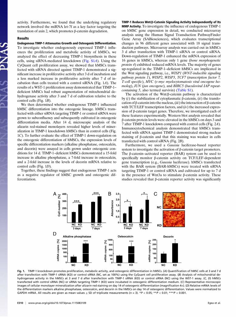
ResultsEndogenous TIMP-1 Attenuates Growth and Osteogenic Differentiation.To investigate whether endogenously expressed TIMP-1 influ-ences the proliferation and metabolic activity of hMSCs, weanalyzed the effect of decreasing TIMP-1 biosynthesis in thesecells, using siRNA-mediated knockdown (Fig. S1A). Using theCyQuant cell proliferation assay, we showed that hMSCs trans-fected with siRNA directed against TIMP-1 demonstrated a sig-nificant increase in proliferative activity after 3 d of incubation anda less marked increase in proliferative activity after 7 d of in-cubation than cells treated with a control siRNA (Fig. 1A). Theresults of aWST-1 proliferation assay demonstrated that TIMP-1–deficient hMSCs had robust augmentation of mitochondrial de-hydrogenase activity after 3 and 7 d of cultivation relative to thecontrol cells (Fig. 1B).We then determined whether endogenous TIMP-1 influenced
hMSC differentiation into the osteogenic lineage. hMSCs trans-fected with either siRNA targeting TIMP-1 or control siRNA weregrown to subconfluency and subsequently cultivated in osteogenicdifferentiation media. After 14 d, microscopic analysis of thealizarin red-stained monolayers revealed higher levels of miner-alization in TIMP-1 knockdown hMSCs than in control cells (Fig.1C). To further evaluate the effect of TIMP-1 down-regulation onthe osteogenic differentiation of hMSCs, the expression levels ofspecific differentiation markers (alkaline phosphatase, osteocalcin,and decorin) were assayed in cells grown under osteogenic con-ditions for 14 d. TIMP-1–deficient hMSCs demonstrated a 15-foldincrease in alkaline phosphatase, a 7-fold increase in osteocalcin,and a 2-fold increase in the levels of decorin mRNA relative tocontrol cells (Fig. 1D).Together, these findings suggest that endogenous TIMP-1 acts
as a negative regulator of hMSC growth and osteogenic dif-ferentiation.
TIMP-1 Reduces Wnt/β-Catenin Signaling Activity Independently of ItsMMP Activity. To investigate the influence of endogenous TIMP-1on hMSC gene expression in detail, we conducted microarrayanalysis using the Human Signal Transduction PathwayFinderGene Array (SABiosciences), which evaluates transcriptionalchanges in 96 different genes associated with 18 signal trans-duction pathways. Microarray analysis was carried out in hMSCs3 d after transfection with TIMP-1 siRNA or control siRNA.Down-regulation of TIMP-1 enhanced the mRNA expression of16 genes in hMSCs, whereas only 1 gene (bone morphogeneticprotein 4) exhibited reduced mRNA levels. The majority of genesup-regulated in the TIMP-1–deficient hMSCs are implicated inthe Wnt signaling pathway, i.e., WISP1 (WNT-inducible signalingpathway protein 1), WISP2, WISP3, TCF7 (transcription factor 7,T-cell specific), MYC (v-myc myelocytomatosis viral oncogene ho-molog), JUN (jun oncogene), and BIRC5 (baculoviral IAP repeat-containing 5, also termed survivin) (Table S1).The activation of the Wnt/β-catenin pathway is characterized
by (i) the stabilization of cytoplasmatic β-catenin, (ii) the translo-cationofβ-catenin into thenucleus, (iii) the interactionof β-cateninwith TCT/LEF transcription factors, and (iv) the increased expres-sion of β-catenin target genes. Therefore, we investigated each ofthese features experimentally. Western blot analysis revealed thatβ-catenin protein levels were elevated in the hMSCs on days 3 and7 after TIMP-1 knockdown compared with control cells (Fig. 2A).Immunocytochemical analysis demonstrated that hMSCs trans-fected with siRNA against TIMP-1 demonstrated strong nuclearstaining of β-catenin and that this staining was weaker in cellstransfected with control siRNA (Fig. 2B).Furthermore, we used a Gaussia luciferase-based reporter
system to investigate the activation of β-catenin target promoters.The β-catenin–activated reporter (BAR) system can be used tospecifically monitor β-catenin activity on TCF/LEF-dependentgene transcription (e.g., Gaussia luciferase). hMSCs transfectedwith the BAR system (BAR-hMSCs) were treated with siRNAtargeting TIMP-1 or control siRNA and cultivated for up to 7 din the presence of Wnt3a to stimulate β-catenin activity. Thesestudies revealed that β-catenin reporter activity was significantly
Fig. 1. TIMP-1 knockdown promotes proliferation, metabolic activity, and osteogenic differentiation in hMSCs. (A) Quantification of hMSC cells at 3 and 7 dafter transfection with TIMP-1 siRNA (KD) or control siRNA (NC, set as 100%) using the CyQuant cell proliferation assay. (B) Analysis of mitochondrial de-hydrogenase activity in the hMSCs at 3 and 7 d after transfection with TIMP-1 siRNA (KD) or control siRNA (NC) using the WST-1 assay. (C, D) hMSCstransfected with control siRNA (NC) or siRNA targeting TIMP-1 (KD) were incubated in osteogenic differentiation medium. (C) Representative microscopicimages of cellular monolayer mineralization after alizarin red staining on day 14 of osteogenic differentiation (magnification 4×). (D) Relative mRNA levels ofthe differentiation markers alkaline phosphatase, osteocalcin, and decorin in the hMSCs on day 14 of osteogenic differentiation. Values were normalized toGAPDH mRNA. All results are given as mean values ± SD of triplicate measurements (n = 3). *P < 0.05; **P < 0.01; ***P < 0.001.
E310 | www.pnas.org/cgi/doi/10.1073/pnas.1115083109 Egea et al.
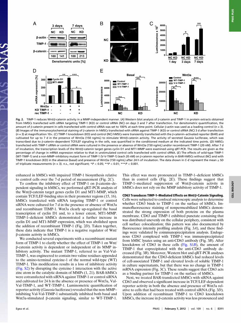
enhanced in hMSCs with impaired TIMP-1 biosynthesis relativeto control cells over the 7-d period of measurement (Fig. 2C).To confirm the inhibitory effect of TIMP-1 on β-catenin–de-
pendent signaling in hMSCs, we performed qRT-PCR analysis ofthe Wnt/β-catenin target genes cyclin D1 and MT1-MMP, whichcontain TCF/LEF-binding sites in their promoter regions (15, 16).hMSCs transfected with siRNA targeting TIMP-1 or controlsiRNA were cultured for 7 d in the presence or absence of Wnt3aand recombinant TIMP-1. Wnt3a clearly up-regulated the basaltranscription of cyclin D1 and, to a lesser extent, MT1-MMP.TIMP-1–deficient hMSCs demonstrated a further increase incyclin D1 and MT1-MMP expression, which was abolished uponthe addition of recombinant TIMP-1 (Fig. 2D). Taken together,these data indicate that TIMP-1 is a negative regulator of Wnt/β-catenin activity in hMSCs.We conducted several experiments with a recombinant mutant
form of TIMP-1 to clarify whether the effect of TIMP-1 on Wnt/β-catenin activity is dependent or independent of its MMP in-hibitory activity. The mutant TIMP-1, referred to as Val-Val-TIMP-1, was engineered to contain two valine residues appendedto the amino-terminal cysteine-1 of the normal wild-type (WT)TIMP-1. This modification results in a loss of inhibitory activity(Fig. S2) by disrupting the cysteine-1 interaction with the activezinc atom in the catalytic domain of MMPs (1, 21). BAR-hMSCswere cotransfected with siRNA against TIMP-1 or control siRNAand cultivated for 24 h in the absence or presence of Wnt3a, Val-Val-TIMP-1, and WT-TIMP-1. Luminometric quantification ofreporter activity (Gaussia luciferase) revealed that the non-MMP–inhibiting Val-Val-TIMP-1 substantially inhibited both basal andWnt3a-stimulated β-catenin signaling, similar to WT-TIMP-1.
This effect was more pronounced in TIMP-1–deficient hMSCsthan in control cells (Fig. 2E). These findings suggest thatTIMP-1–mediated suppression of Wnt/β-catenin activity inhMSCs does not rely on the MMP inhibitory activity of TIMP-1.
CD63 Transduces TIMP-1–Mediated Effects on Wnt/β-Catenin Signaling.Cells were subjected to confocal microscopic analysis to determinewhether CD63 binds to TIMP-1 on the surface of hMSCs. Im-munofluorescence staining of nonpermeabilized hMSCs demon-strated the strong expression of both proteins on the plasmamembrane. CD63 and TIMP-1 exhibited punctate costaining thatwas distributed unevenly on the cellular periphery, consistent withcell surface colocalization; this pattern was also documented byfluorescence intensity profiling analysis (Fig. 3A), and these find-ings were validated by coimmunoprecipitation analysis. Endoge-nous CD63 complexed with TIMP-1 was immunoprecipitatedfrom hMSC lysates using an anti-CD63 antibody (Fig. 3B). Afterknockdown of CD63 in these cells (Fig. S1B), the amount ofTIMP-1 that coprecipitated with the anti-CD63 antibody de-creased (Fig. 3B). Moreover, Western blot and qRT-PCR analysesdemonstrated that the CD63-deficient hMSCs had reduced levelsof cell-associated TIMP-1 and elevated levels of soluble TIMP-1in culture supernatants, but that there was no change in TIMP-1mRNA expression (Fig. 3C). These results suggest that CD63 actsas a binding partner for TIMP-1 on the surface of hMSCs.Next, we treated BAR-transfected hMSCs with siRNA against
CD63 and observed a significant increase in TCF/LEF-dependentreporter activity in both the absence and presence of Wnt3a rel-ative to cells that had been treated with control siRNA (Fig. 3D).Upon addition of recombinant TIMP-1 to CD63 knockdownhMSCs, the increase in β-catenin activity was less pronounced and
Fig. 2. TIMP-1 reduces Wnt/β-catenin activity in a MMP-independent manner. (A) Western blot analysis of β-catenin and TIMP-1 in protein extracts obtainedfrom hMSCs transfected with siRNA targeting TIMP-1 (KD) or control siRNA (NC) on days 3 and 7 after transfection. For densitometric quantification, theamount of β-catenin present in cells transfected with control siRNA was set to 100% at each time point. Cellular β-actin was used as a loading control (n = 3).(B) Images of the immunocytochemical staining of β-catenin in hMSCs transfected with siRNA against TIMP-1 (KD) or control siRNA (NC) 3 d after transfection(n = 3) at magnification 10×. (C) TIMP-1 knockdown (KD) and control (NC) hMSCs were transiently transfected with the β-catenin–activated reporter (BAR) andcultivated for up to 7 d in the presence of Wnt3a (150 ng/mL) to stimulate Wnt/β-catenin activity. The activity of secreted Gaussia luciferase, which wastranscribed due to β-catenin–dependent TCF/LEF signaling in the cells, was quantified in the conditioned medium at the indicated time points. (D) hMSCstransfected with TIMP-1 siRNA or control siRNA were cultured in the presence or absence of Wnt3a (150 ng/mL) and/or recombinant TIMP-1 (20 nM). After 7 dof incubation, the transcription levels of the Wnt/β-catenin target genes cyclin D1 and MT1-MMP were examined using qRT-PCR. The results are given as thepercentage of change in mRNA expression relative to that in unstimulated control cells transfected with control siRNA. (E) The effects of wild-type TIMP-1(WT-TIMP-1) and a non-MMP–inhibitory mutant form of TIMP-1 (V-V-TIMP-1) (each 20 nM) on β-catenin reporter activity in BAR-hMSCs without (NC) and withTIMP-1 knockdown (KD) in the absence (basal) and presence of Wnt3a (150 ng/mL) after 24 h of incubation. The data shown in C–E represent the mean ± SDof triplicate measurements (n = 3). n.s., not significant; *P < 0.05; **P < 0.01; ***P < 0.001.
Egea et al. PNAS | February 7, 2012 | vol. 109 | no. 6 | E311
CELL
BIOLO
GY
PNASPL
US
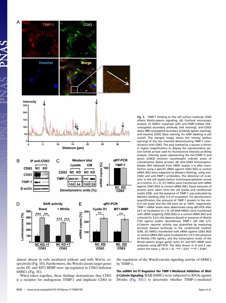
almost absent in cells incubated without and with Wnt3a, re-spectively (Fig. 3D). Furthermore, theWnt/β-catenin target genescyclin D1 and MT1-MMP were up-regulated in CD63-deficienthMSCs (Fig. 3E).When taken together, these findings demonstrate that CD63
is a receptor for endogenous TIMP-1 and implicate CD63 in
the regulation of the Wnt/β-catenin signaling activity of hMSCsby TIMP-1.
The miRNA let-7f Regulates the TIMP-1-Mediated Inhibition of Wnt/β-Catenin Signaling. BAR-hMSCs were subjected to RNAi againstDrosha (Fig. S1C) to determine whether TIMP-1–mediated
Fig. 3. TIMP-1 binding to the cell surface molecule CD63affects Wnt/β-catenin signaling. (A) Confocal microscopicanalysis of hMSCs costained with anti-TIMP-1/Alexa 543–conjugated secondary antibody (red staining), anti-CD63/Alexa 488–conjugated secondary antibody (green staining),and Hoechst 33342 (blue staining, for DNA labeling in cellnuclei). The merged image shows the overlay (yellowstaining) of the two channels demonstrating TIMP-1 coloc-alization with CD63. The area marked by a square is shownin higher magnification to display the representative sec-tion (white arrow) used for fluorescence intensity profilinganalysis. Intensity peaks representing the red (TIMP-1) andgreen (CD63) emission wavelengths indicate areas ofcolocalization (black arrows). (B) Anti-CD63 immunopreci-pitates (IPs) obtained from hMSC lysates 3 d after trans-fection using a specific siRNA against CD63 (KD) or controlsiRNA (NC) were subjected to Western blotting, using anti-CD63 and anti-TIMP-1 antibodies. The detection of β-cat-enin in the cell lysates before immunoprecipitation servedas a control. (n = 3). (C) hMSCs were transfected with siRNAagainst CD63 (KD) or control siRNA (NC). Equal amounts ofprotein were taken from the cell lysates and conditionedmedia (CM), and the presence of TIMP-1 was evaluated byWestern blotting after 3 d of incubation. For densitometricquantification, the amounts of TIMP-1 present in the con-trol cell lysate and the CM were set as 100%, respectively.TIMP-1 mRNA levels were determined using qRT-PCR after24 h of incubation (n = 3). (D) BAR-hMSCs were transfectedwith siRNA targeting CD63 (KD) or a control siRNA (NC) andcultured for 3 d in the absence (basal) or presence of Wnt3a(150 ng/mL) and/or recombinant TIMP-1 (20 nM) (+T).β-Catenin reporter activity was quantified by measuringsecreted Gaussia luciferase in the conditioned medium(CM). (E) hMSCs transfected with siRNA against CD63 (KD)or control siRNA (NC) were incubated for 3 d in the presenceof Wnt3a (150 ng/mL), and the transcription levels of theWnt/β-catenin target genes cyclin D1 and MT1-MMP wereanalyzed using qRT-PCR. The data shown in D and E rep-resent the mean ± SD (n = 3). **P < 0.01; ***P < 0.001.
E312 | www.pnas.org/cgi/doi/10.1073/pnas.1115083109 Egea et al.

modulation of the Wnt/β-catenin activity in hMSCs is associatedwith changes in miRNA expression. Drosha is a key regulatoryenzyme in miRNA biogenesis. At 3 d after transfection, Drosha-deficient hMSCs demonstrated a significant decrease in sus-ceptibility to the TIMP-1 knockdown-mediated up-regulation ofβ-catenin activity (Fig. 4A).To identify specific miRNAs involved in the regulation of
hMSCs by TIMP-1, we silenced TIMP-1 expression and analyzedthe subsequent transcriptional changes in 88 abundantly expressedand well-characterized miRNA sequences, using the HumanmiFinder RT2 miRNA PCR Array (SABiosciences). Microarrayanalysis was carried out in hMSCs after 3 d of incubation followingtransfection with an siRNA targeting TIMP-1 or a control siRNA.
The down-regulation of TIMP-1 in hMSCs modulated the tran-scription of numerous miRNAs, with the greatest increase ob-served in let-7f (Fig. 4B and Table S2). Quantification by qRT-PCRdemonstrated an 18-fold augmentation of let-7f in TIMP-1–de-pleted hMSCs (Fig. 4C). Moreover, transcription of let-7f was in-creased 2- to 3-fold upon knockdown of CD63 in the cells (Fig. 4C).Next, we studied the role of let-7f in more detail by transfecting
hMSCs with let-7f miRNA (chemically synthesized, double-stranded RNA that mimics mature endogenous let-7f) and a let-7f miRNA inhibitor (chemically modified, single-stranded RNAdesigned to specifically bind and inhibit endogenous let-7f). Theaddition of the let-7f mimic to hMSCs slightly augmented β-cat-enin activity, whereas the inhibition of let-7f efficiently blocked
Fig. 4. The miRNA let-7f is implicated in the TIMP-1–mediated inhibition of β-catenin activity and osteogenic differentiation. (A) BAR-hMSCs were trans-fected with control siRNA (NC), siRNA targeting TIMP-1 (KD), and/or siRNA targeting Drosha (KD). After 3 d of culture, β-catenin reporter activity was de-termined by luminometric analysis of Gaussia luciferase in the culture supernatants. (B) hMSCs were transfected with control siRNA (NC) or siRNA targetingTIMP-1 (KD) and incubated for 3 d. RNA was collected and subjected to miRNA analysis, using PCR array technology. The results are shown in a diagram withthe crossing point (Ct) values in TIMP-1 knockdown cells plotted against the respective Cts in control cells and fitted well to a straight line, revealing that let-7fwas the most up-regulated miRNA tested. (C) Quantitation of let-7f transcription in control cells (NC, set as 1) and hMSCs deficient (KD) in TIMP-1 and CD63using qRT-PCR analysis. The values shown were normalized to GAPDH mRNA. (D) BAR-hMSCs were transfected with control siRNA (NC), siRNA against TIMP-1(KD), a let-7f mimic (MI), and/or a let-7f inhibitor (IN). After 24 h of incubation, β-catenin reporter activity was quantified by luminometric measurement ofsecreted Gaussia luciferase in the conditioned medium (the control was set as 100%). (E) hMSCs were cotransfected with a Renilla luciferase reporter constructbearing the axin 2 3′-UTR and a let-7f mimic (10–40 nM) or control miRNA (40 nM). After 24 h, axin 2 reporter activity was quantified by luminometricmeasurement of Renilla luciferase in the cell extracts. (F) hMSCs were transfected with control siRNA (NC), siRNA against TIMP-1 (KD), a let-7f mimic (MI), and/or a let-7f inhibitor (IN) and cultivated in the presence of Wnt3a (150 ng/mL) to stimulate Wnt/β-catenin signaling. After 24 h, axin 2 protein expression wasanalyzed using Western blotting, and axin 2 mRNA expression was analyzed using qRT-PCR. β-Actin served as a loading control. For densitometric quanti-fication, the amount of axin 2 protein present in the control cells (NC) was set as 100%. The qRT-PCR values shown were normalized to the GAPDH mRNAlevels. (G–I) hMSCs transfected with control siRNA (NC), siRNA targeting TIMP-1 (KD), a let-7f mimic (MI), and/or a let-7f inhibitor (IN) were incubated for 14 din osteogenic differentiation medium. (G) Representative images of cellular monolayer mineralization with alizarin red staining. (H) The relative absorbanceof the alizarin red stain extracts (the NC was set as 100%). (I) Relative mRNA levels of the osteogenic markers alkaline phosphatase and osteocalcin weredetermined using qRT-PCR (the NC was set as 1). The values are normalized to the GAPDH mRNA levels. Data shown in A, C–F, H, and I are given as meanvalues ± SD of triplicate measurements (n = 3). n.s., not significant; *P < 0.05; **P < 0.01; ***P < 0.001.
Egea et al. PNAS | February 7, 2012 | vol. 109 | no. 6 | E313
CELL
BIOLO
GY
PNASPL
US
the up-regulation of β-catenin signaling in TIMP-1–deficienthMSCs (Fig. 4D). These data indicate that let-7f participates inthe TIMP-1–mediated regulation of β-catenin activity in hMSCs.We then searched for potential let-7f targets, i.e., negative
regulators of the Wnt/β-catenin signaling pathway that are com-plementary to (and therefore may be repressed by) let-7f. Usingan in silico prediction algorithm, we discovered that let-7f maytarget axin 2 (axis inhibition protein 2), a cytoplasmatic proteinthat plays a key role in β-catenin degradation (22). The targetingwas verified experimentally using a luciferase reporter assay andthe 3′-untranslated region (UTR) of axin 2. When hMSCs werecotransfected with the reporter construct and increasing amountsof the let-7f mimic, the axin 2-specific luciferase activity was re-duced in both unstimulated and Wnt3a-stimulated cells in a dose-dependent manner (Fig. 4E). Moreover, Western blot analysisdemonstrated a clear decrease in the biosynthesis of axin 2 pro-tein in the presence of the let-7f mimic and a rescue of axin 2protein levels in TIMP-1 knockdown hMSCs upon inhibition oflet-7f (Fig. 4F). Axin 2 mRNA expression was not affected byeither a let-7f mimic or a let-7f inhibitor (Fig. 4F), indicating thatlet-7f regulates axin 2 at the posttranscriptional level. Taken to-gether, these results provide evidence that let-7f promotesβ-catenin activity in hMSCs by repressing the translation of axin 2.Finally, we examined whether let-7f is involved in the regulation
of osteogenic differentiation. Transfection with a let-7f mimicfurther increased cellular mineralization and osteogenic markerexpression in hMSCs under osteogenic conditions. In contrast, theinhibition of let-7f activity by the transfection of a specific inhibitorabolished the increase of mineralization (Fig. 4 G and H) andosteogenic marker expression (Fig. 4I), as observed in TIMP-1–deficient cells grown in osteogenic media. These data indicate thatlet-7f expression in hMSCs promotes osteogenic differentiation.
DiscussionOur current study describes a mechanism by which TIMP-1 reg-ulates essential stem cell functions. The inhibition of TIMP-1expression in hMSCs significantly promoted cell growth and dif-ferentiation into adult cells of the osteogenic lineage, indicatingthat TIMP-1 is an endogenous attenuator of these processes.Previous reports have shown that TIMP-1 inhibits epithelial cellproliferation and osteoblastic differentiation (23, 24) but stim-ulates differentiation in hematopoietic progenitor cells, B cells,and Burkitt lymphoma cells (25–27). However, none of thesestudies has provided information regarding the underlying mo-lecular mechanisms. Because TIMP-1 is secreted from hMSCs atrelatively high levels in the absence of its major target proteaseMMP-9 (5), we hypothesized that TIMP-1 may also mediatesignaling that affects gene expression and inhibits the growth anddifferentiation of these cells.Our studies revealed that knockdown of TIMP-1 production in
hMSCs up-regulates the stability, nuclear translocation, andpromoter activity of β-catenin, demonstrating that TIMP-1 is aninhibitor of β-catenin–dependent signaling. The Wnt/β-cateninsignaling pathway is closely associated with the growth and de-velopment of stem cells (13). In hMSCs, increased stability and/or activity of β-catenin has been shown to promote osteogenicdifferentiation, which may also occur independently of stimula-tion with Wnt (28, 29). In the present study, both endogenousTIMP-1 and exogenously added recombinant TIMP-1 acted asnegative regulators of β-catenin activity in hMSCs, thereby pre-venting up-regulation of the β-catenin target genes cyclin D1 andMT1-MMP. Consistent with our data, TIMP-1 down-regulatescyclin D1 in human breast epithelial (MCF10A) cells (30).We showed that TIMP-1 colocalizes with CD63 on the surface
of hMSCs, which appears to be one extracellular mechanismby which TIMP-1 reassociates with the hMSC plasma membrane.In addition to CD63, other molecular structures appear to act asdocking proteins for TIMP-1 on the plasmamembrane of hMSCs;
these structures will need to be identified by further studies.Nevertheless, CD63 plays an important role in the TIMP-1–me-diated interference of the Wnt/β-catenin pathway. Functionaldepletion of CD63 in hMSCs significantly up-regulated Wnt/β-catenin activity even though TIMP-1 was still expressed in thesecells. This result is consistent with previous reports demonstratingthat CD63 can regulate the activity of the phosphatidylinositol 3-kinase, focal adhesion kinase, Src, and Akt signaling pathways, allof which have been implicated in the anti-apoptotic activity ofTIMP-1 (31–33). Addition of an excess of TIMP-1 to CD63-de-pleted hMSCs attenuated the increase ofWnt/β-catenin activity inthese cells, again indicating that CD63 is not the exclusive struc-ture that interacts with TIMP-1 and thereby affects gene expres-sion in these cells.To further elucidate the intracellular molecular mechanism by
which TIMP-1 suppresses the stability and activity of β-catenin inhMSCs, we focused on miRNAs, which have very recently beenshown to play important roles in the self-renewal and differenti-ation of hMSCs (20). Functional disruption of the miRNA pro-cessing machinery significantly inhibited the increase in β-cateninactivity in TIMP-1–depleted hMSCs, suggesting that precise levelsof mature miRNAs are critical for the TIMP-1–mediated modu-lation of β-catenin. This finding is consistent with other recentdata demonstrating that various miRNAs can act as positive ornegative regulators of β-catenin transcriptional activity (34).Among the miRNAs that were up-regulated after knockdown ofTIMP-1 biosynthesis in our hMSC experiments, let-7f exhibitedthe most pronounced increase. This is especially interesting be-cause the let-7 family of miRNAs is thought to represent “pro-differentiation” factors with “antistemness” properties (35). Forexample, mature let-7 miRNAs are undetectable in embryonicstem cells but accumulate strongly after the onset of differentia-tion in these cells (36). Moreover, other studies in hMSCs havedemonstrated that specific let-7 miRNAs are up-regulated duringosteogenic differentiation and presumably down-regulate factorsthat inhibit osteogenesis (37, 38).miRNAs can modulate β-catenin–dependent transcription in
several ways, including the direct inhibition of β-catenin mRNA orthe indirect inhibition of negative and positive regulators ofβ-catenin activity (34). Axin 2 is a negative regulator of the Wnt/β-catenin signaling pathway and was identified in silico and in vitroas a let-7f target in hMSCs. Similar to its homolog axin 1, axin 2 isthought to act as a scaffolding protein that organizes a multi-protein complex that can degrade β-catenin (39). Indeed, axin 2translation is abrogated in TIMP-1–depleted hMSCs, concomitantwith the increased β-catenin activity observed in these cells. Fur-thermore, axin 2 protein levels were rescued upon let-7f inhibition,confirming that axin 2 is a target of let-7f. Axin 2 down-regulationby let-7f may explain the direct increase in β-catenin activity by let-7f in hMSCs. Although additional miRNAs are clearly involved inthe control of osteogenic differentiation in hMSCs, our findingsindicate that let-7f is a key miRNA in this process.In conclusion, we have demonstrated that TIMP-1 is a negative
regulator of the growth and osteogenic differentiation of hMSCsand thus promotes a quiescent state in these cells. TIMP-1 bindingto CD63 on the surface of hMSCs results in the repression oflet-7f miRNA expression, thereby stabilizing the levels of axin 2and subsequently promoting the degradation of β-catenin. Impor-tantly, our results show that the functional depletion of TIMP-1or an increase in let-7f accelerates osteogenic differentiation inhMSCs. A deeper knowledge of this regulatory network mayimprove the development of novel therapeutic approaches toenhance bone formation during pathological bone loss.
Experimental ProceduresCultivation, Osteogenic Differentiation, and Alizarin Red Staining of hMSCs.hMSCs from bone marrow aspirates were purchased from Lonza as frozenvials of passage-one cells. Each hMSC lot was tested by Lonza for purity using
E314 | www.pnas.org/cgi/doi/10.1073/pnas.1115083109 Egea et al.
flow cytometry and for their ability to differentiate into osteogenic, chon-drogenic, and adipogenic lineages. The cells were positive for CD105, CD166,CD29, and CD44 and negative for CD14, CD34, and CD45.
hMSCswere cultured as described previously, using themesenchymal stem-cell growth medium (MSCGM) BulletKit (Lonza) (8). For experiments underserum-free conditions, hMSCs were washed with serum-free medium and in-cubated in DMEM supplemented with 1% Nutridoma SP (Roche Applied Sci-ence) in the presence or absence of 20 nM human recombinant TIMP-1(Peprotech), thenull-inhibitionmutantVal-Val-TIMP-1, or its activeanalogWTTIMP-1, both kindly provided by P. Nelson, Ludwig-Maximilians-University.
Osteogenic differentiation and alizarin red staining of hMSCs were per-formed as previously described (40). Alizarin red stain incorporation wasquantified by extracting stained monolayers with a solution containing 50%ethanol, 10% methanol, and 2% SDS. Aliquots of the extracted dye werethen transferred to 96-well plates and the absorbance at 405 nm wasmeasured using a plate reader (Tecan). All studies were carried out withhMSCs from the fifth or sixth passage.
CyQuant Cell Proliferation Assay and WST-1 Assay for Cell Metabolic Activity.Cell proliferationwas quantified using the CyQuant cell proliferation assay kit(Invitrogen) as previously described (40). Briefly, nucleic acids present in celllysates were stained with fluorescent CyQuant GR dye before measurementat 480 nm (excitation) and 530 nm (emission). For each experiment, a stan-dard calibration curve was generated by plotting the measured fluorescencevalues of the samples vs. the corresponding number of cells, which had beendetermined before staining using a hematocytometer.
TheWST-1 assay (Roche Applied Science), which is based on the cleavage ofthe tetrazolium salt WST-1 by mitochondrial dehydrogenases in viable cells,was conducted according to the manufacturer’s protocol.
qRT-PCR Analysis of mRNA and miRNA Expression. RNA isolation, cDNA syn-thesis, and PCR were performed as described in ref. 5. qRT-PCR was carriedout on a LightCycler (Roche Applied Science), using the LightCycler-FastStartDNA Master SYBR Green I Kit (Roche Applied Science). For amplification ofspecific human transcripts, the LightCycler Primer Sets for alkaline phos-phate, osteocalcin, decorin, cyclin D1, MT1-MMP, axin 2, and the house-keeping gene standard GAPDH were used following the manufacturer’sinstructions (Search LC).
The expression of select miRNAs was determined using the miScript PCRSystem (Qiagen), which converts miRNA into cDNA, which was detected usingSYBR Green-based RT-PCR. The let-7f primer sequence is given in Table S3.
Transfection of hMSCs with siRNA, miRNA Mimics, and miRNA Inhibitors. RNAinterference (RNAi) technology was used to generate specific knockdowns ofTIMP-1, CD63, and Drosha expression in hMSCs. siRNA sense and antisenseoligonucleotides targeting human TIMP-1 mRNA were designed in our lab-oratory according to the protocol of Reynolds et al. (41) and synthesized byQiagen. siRNA against CD63, Drosha, and a nonspecific siRNA with no targetin the human transcriptome (used as a negative control) were purchasedfrom Qiagen. For studies of miRNA function and gene regulation, miScriptmiRNA mimics and miScript miRNA inhibitors of let-7f were used (Qiagen).The sequences of all siRNAs, miRNA mimics, and miRNA inhibitors used arelisted in Table S3. hMSCs were transfected with miRNA mimics (20 nM) andmiRNA inhibitors (20 nM) according to a previously described method forhMSC transfection with siRNA (5).
miRNA Target Prediction and Experimental Validation of Target Genes. Weused the computational target prediction algorithm miRanda to identifymature gene transcripts that might be targeted by let-7f at their 3′-UTRs (42).Axin 2 was then experimentally verified as a let-7f target gene in hMSCsaccording to the protocol suggested by Huang et al. (43).
Reporter assays used a LightSwitch GoClone 3′-UTR Reporter Construct(BioCat) based on the 3′-UTR sequence of axin 2, which was cloned down-stream of the Renilla luciferase gene. hMSCs grown in microtiter plates werecotransfected with the axin 2 reporter plasmid (100 ng) and either the let-7fmimic (10–40 nM) or the control siRNA (40 nM) (Qiagen). After 24 h of in-cubation, 100 μL of LightSwitch reagent (BioCat) was added to each well,and luminescence was measured using a plate-reading luminometer (Tecan).
The effect of let-7f on axin 2was examined byWestern blot analysis of axin2 protein levels in hMSC extracts 24 h after treatment with the let-7f mimic orinhibitor. qRT-PCR analysis was performed to quantitate the level of axin 2transcript in the hMSCs 1 d after incubation with the let-7f mimic or inhibitor.
Transcription-Based Reporter Assay of Wnt/β-Catenin Signaling. The activity ofthe Wnt/β-catenin signaling pathway, which culminates in the TCF/LEF-
dependent regulation of target gene transcription, was monitored in thehMSCs, using a reporter assay. This assay was based on the BAR, whichcontains multimerized TCF/LEF DNA-binding sites, and the control reporter,found-unresponsive BAR (fuBAR) (44). We modified the BAR/fuBAR bycloning Gaussia luciferase as the reporter gene to allow the detection of thereporter in the supernatant; after its transcription is activated by β-catenin,Gaussia luciferase is secreted from the cells, so the cells do not have to bedestroyed to assay its transcription.
hMSCs were transfected with the BAR (or fuBAR, which harbors mutantTCF-binding sites) Gaussia luciferase reporter plasmid and cultivated in thepresence of geneticin (100 μg/mL) to eliminate nontransfected cells. Then,the BAR/fuBAR-hMSCs were used for the stimulation and/or knockdownexperiments. Conditioned medium was collected after the indicated timeintervals and analyzed for the presence of reporter protein, using theGaussia luciferase assay kit (New England Biolabs) according to the manu-facturer’s protocol. The light intensity was determined using a plate-readingluminometer (Tecan). No Gaussia luciferase activity was detected in condi-tioned media from hMSCs transfected with the fuBAR system in any ex-periment performed in this study.
Western Blot and Coimmunoprecipitation Analyses. Protein extraction fromthe hMSCs, SDS/polyacrylamide gel electrophoresis, and Western blottingwere performed as described in ref. 5. The blotted membranes were in-cubated overnight with monoclonal antibodies against β-actin (1:2,000)and TIMP-1 (1:500) (both from Abcam), β-catenin (1:500) (Santa CruzBiotechnology), CD63 (1:1,000) (Millipore), or axin 2 (1:1,000) (Cell Sig-naling Technology) diluted in Tris-buffered saline containing 0.1%Tween-20 (TBS-Tween buffer) containing 5% BSA. The membranes werewashed in TBS-Tween buffer and then incubated with peroxidase-conju-gated anti-rabbit IgG (Cell Signaling Technology) at a dilution of 1:8,000in TBS-Tween for 30 min. Bound antibodies were detected using theenhanced chemiluminescence system (GE Healthcare Life Sciences).Recombinant protein standards (Invitrogen) were used for molecularmass determination.
Coimmunoprecipitation was performed to investigate protein inter-actions. Total hMSC lysates were prepared in lysis buffer as described in ref. 5.Aliquots (10 μg of protein) were incubated overnight at 4 °C with anti-CD63(2 μg) (Millipore) or control IgG in TBS-Tween. The immunocomplexes werecollected on protein A agarose beads (Invitrogen), following the manu-facturer’s instructions. Bound proteins/complexes were then eluted andsubjected to Western blotting.
Immunocytochemistry and Confocal Microscopic Analysis. The subcellular lo-calization of β-catenin protein in the hMSCs was examined by immunocy-tochemistry as previously described (8). Culture slides were incubated withmouse anti–β-catenin antibodies (Santa Cruz Biotechnology) at a dilutionof 1:500 in a solution containing 0.5% Triton X-100 and 10% normal goatserum (NGS) for 2 h at 37 °C. After several washes in PBS, the culture slideswere incubated with rhodamine-conjugated anti-rabbit IgG-R (Santa CruzBiotechnology) at a dilution of 1:500 for 45 min at room temperature andvisualized using an Olympus IX70 microscope equipped with an Olympus20× objective lens (numerical aperture 0.55) at 20 °C.
For confocal microscopic analysis, the cells were seeded on Ibidi eight-wellslides and incubated overnight with conditioned hMSC medium. The mediumcontained secreted TIMP-1 to saturate potential TIMP-1 surface docking re-ceptors on the hMSCs. The cells were fixed with methanol-free 4% formalinand thenwashed and stainedwithmonoclonal anti-TIMP-1 (Chemicon) (1:500)and anti-CD63 (Santa Cruz) (1:125) antibodies. Nuclei were fluorescently la-beled with Hoechst 33342 (Thermo Scientific). All secondary antibodies werepurchased from Invitrogen. Confocal images were obtained using a Zeiss LSM510 microscope equipped with a 40× oil immersion objective. Pinhole diam-eters were adjusted to visualize 1.5-μm optical slices. To detect spots ofcolocalization of both proteins, fluorescence intensity profiles were obtainedfrom representative images. Intensity peaks that were detected at bothemission wavelengths indicated areas of colocalization.
Statistical Analysis. Statistical significance was assessed by comparing mean(±SD) values, using Student’s t tests for independent groups. One-wayanalysis of variance followed by Dunnett’s test was used to compare mul-tiple groups. Significance was assumed for P < 0.05. Statistical analysis wasconducted using Origin 8.0 software (OriginLab).
ACKNOWLEDGMENTS. The β-catenin–activated reporter (BAR) and the con-trol reporter (found-unresponsive BAR, fuBAR) were established and kindlyprovided by Randall T. Moon (University of Washington). Active MMP-3 was
Egea et al. PNAS | February 7, 2012 | vol. 109 | no. 6 | E315
CELL
BIOLO
GY
PNASPL
US
a kind gift from Hideaki Nagase (Imperial College). We thank Thomas Pitschfor his technical assistance. This work was supported by a contract from
the German Federal Ministry of Defense research project, M/SABX/8A002(to V.E., M.J., and C.R.).
1. Brew K, Nagase H (2010) The tissue inhibitors of metalloproteinases (TIMPs): Anancient family with structural and functional diversity. Biochim Biophys Acta 1803:55–71.
2. Avalos BR, et al. (1988) K562 cells produce and respond to human erythroid-potentiating activity. Blood 71:1720–1725.
3. Stetler-Stevenson WG (2008) Tissue inhibitors of metalloproteinases in cell signaling:Metalloproteinase-independent biological activities. Sci Signal 1:re6.
4. Jung KK, Liu XW, Chirco R, Fridman R, Kim HR (2006) Identification of CD63 as a tissueinhibitor of metalloproteinase-1 interacting cell surface protein. EMBO J 25:3934–3942.
5. Ries C, et al. (2007) MMP-2, MT1-MMP, and TIMP-2 are essential for the invasivecapacity of human mesenchymal stem cells: Differential regulation by inflammatorycytokines. Blood 109:4055–4063.
6. Pittenger MF, et al. (1999) Multilineage potential of adult human mesenchymal stemcells. Science 284:143–147.
7. Jiang Y, et al. (2002) Pluripotency of mesenchymal stem cells derived from adultmarrow. Nature 418:41–49.
8. Egea V, et al. (2011) TNF-α respecifies human mesenchymal stem cells to a neural fateand promotes migration toward experimental glioma. Cell Death Differ 18:853–863.
9. Prockop DJ, Kota DJ, Bazhanov N, Reger RL (2010) Evolving paradigms for repair oftissues by adult stem/progenitor cells (MSCs). J Cell Mol Med 14:2190–2199.
10. Martinez EC, Kofidis T (2011) Adult stem cells for cardiac tissue engineering. J Mol CellCardiol 50:312–319.
11. Joyce N, et al. (2010) Mesenchymal stem cells for the treatment of neurodegenerativedisease. Regen Med 5:933–946.
12. Singer NG, Caplan AI (2011) Mesenchymal stem cells: Mechanisms of inflammation.Annu Rev Pathol 6:457–478.
13. Clevers H (2006) Wnt/beta-catenin signaling in development and disease. Cell 127:469–480.
14. MacDonald BT, Tamai K, He X (2009) Wnt/beta-catenin signaling: Components,mechanisms, and diseases. Dev Cell 17:9–26.
15. Shtutman M, et al. (1999) The cyclin D1 gene is a target of the beta-catenin/LEF-1pathway. Proc Natl Acad Sci USA 96:5522–5527.
16. Takahashi M, Tsunoda T, Seiki M, Nakamura Y, Furukawa Y (2002) Identification ofmembrane-type matrix metalloproteinase-1 as a target of the beta-catenin/Tcf4complex in human colorectal cancers. Oncogene 21:5861–5867.
17. Bartel DP (2009) MicroRNAs: Target recognition and regulatory functions. Cell 136:215–233.
18. Yi R, Fuchs E (2011) MicroRNAs and their roles in mammalian stem cells. J Cell Sci 124:1775–1783.
19. Ivey KN, Srivastava D (2010) MicroRNAs as regulators of differentiation and cell fatedecisions. Cell Stem Cell 7:36–41.
20. Guo L, Zhao RC, Wu Y (2011) The role of microRNAs in self-renewal and differ-entiation of mesenchymal stem cells. Exp Hematol 39:608–616.
21. Wingfield PT, et al. (1999) Biophysical and functional characterization of full-length,recombinant human tissue inhibitor of metalloproteinases-2 (TIMP-2) produced inEscherichia coli. Comparison of wild type and amino-terminal alanine appendedvariant with implications for the mechanism of TIMP functions. J Biol Chem 274:21362–21368.
22. Behrens J, et al. (1998) Functional interaction of an axin homolog, conductin, withbeta-catenin, APC, and GSK3beta. Science 280:596–599.
23. Fata JE, Leco KJ, Moorehead RA, Martin DC, Khokha R (1999) Timp-1 is important forepithelial proliferation and branching morphogenesis during mouse mammarydevelopment. Dev Biol 211:238–254.
24. Schiltz C, Marty C, de Vernejoul MC, Geoffroy V (2008) Inhibition of osteoblasticmetalloproteinases in mice prevents bone loss induced by oestrogen deficiency. J CellBiochem 104:1803–1817.
25. Guedez L, et al. (2001) Tissue inhibitor of metalloproteinases 1 regulation ofinterleukin-10 in B-cell differentiation and lymphomagenesis. Blood 97:1796–1802.
26. Guedez L, et al. (2005) Tissue inhibitor of metalloproteinase 1 (TIMP-1) promotesplasmablastic differentiation of a Burkitt lymphoma cell line: Implications in thepathogenesis of plasmacytic/plasmablastic tumors. Blood 105:1660–1668.
27. Dassé E, et al. (2007) Tissue inhibitor of metalloproteinase-1 promotes hematopoieticdifferentiation via caspase-3 upstream theMEKK1/MEK6/p38alpha pathway. Leukemia21:595–603.
28. Day TF, Guo X, Garrett-Beal L, Yang Y (2005) Wnt/beta-catenin signaling inmesenchymal progenitors controls osteoblast and chondrocyte differentiation duringvertebrate skeletogenesis. Dev Cell 8:739–750.
29. Qiang YW, et al. (2009) Bortezomib induces osteoblast differentiation via Wnt-independent activation of beta-catenin/TCF signaling. Blood 113:4319–4330.
30. Taube ME, Liu XW, Fridman R, Kim HR (2006) TIMP-1 regulation of cell cycle in humanbreast epithelial cells via stabilization of p27(KIP1) protein. Oncogene 25:3041–3048.
31. Liu XW, BernardoMM, Fridman R, Kim HR (2003) Tissue inhibitor of metalloproteinase-1 protects human breast epithelial cells against intrinsic apoptotic cell death via thefocal adhesion kinase/phosphatidylinositol 3-kinase and MAPK signaling pathway.J Biol Chem 278:40364–40372.
32. Liu XW, et al. (2005) Tissue inhibitor of metalloproteinase-1 protects human breastepithelial cells from extrinsic cell death: A potential oncogenic activity of tissueinhibitor of metalloproteinase-1. Cancer Res 65:898–906.
33. Chirco R, Liu XW, Jung KK, Kim HR (2006) Novel functions of TIMPs in cell signaling.Cancer Metastasis Rev 25:99–113.
34. Huang K, et al. (2010) MicroRNA roles in beta-catenin pathway. Mol Cancer 9:252.35. Peter ME (2009) Let-7 and miR-200 microRNAs: Guardians against pluripotency and
cancer progression. Cell Cycle 8:843–852.36. Viswanathan SR, Daley GQ, Gregory RI (2008) Selective blockade of microRNA
processing by Lin28. Science 320:97–100.37. Oskowitz AZ, et al. (2008) Human multipotent stromal cells from bone marrow and
microRNA: Regulation of differentiation and leukemia inhibitory factor expression.Proc Natl Acad Sci USA 105:18372–18377.
38. Goff LA, et al. (2008) Differentiating human multipotent mesenchymal stromal cellsregulate microRNAs: Prediction of microRNA regulation by PDGF duringosteogenesis. Exp Hematol 36:1354–1369.
39. Jho EH, et al. (2002) Wnt/beta-catenin/Tcf signaling induces the transcription ofAxin2, a negative regulator of the signaling pathway. Mol Cell Biol 22:1172–1183.
40. Neth P, et al. (2006) Wnt signaling regulates the invasion capacity of humanmesenchymal stem cells. Stem Cells 24:1892–1903.
41. Reynolds A, et al. (2004) Rational siRNA design for RNA interference. Nat Biotechnol22:326–330.
42. John B, et al. (2004) Human MicroRNA targets. PLoS Biol 2:e363.43. Huang Y, et al. (2010) A study of miRNAs targets prediction and experimental
validation. Protein Cell 1:979–986.44. Biechele TL, Adams AM, Moon RT (2009) Transcription-based reporters of Wnt/beta-
catenin signaling. Cold Spring Harb Protoc 2009(6): pdb.prot5233.
E316 | www.pnas.org/cgi/doi/10.1073/pnas.1115083109 Egea et al.


![Original Article EMMPRIN, SP1 and microRNA-27a mediate ... · pathways and mechanisms, including loss of function of the tumor suppressor p53 [10], upregulated vascular endothelial](https://static.fdocument.org/doc/165x107/5e22380d3a89c23c53196456/original-article-emmprin-sp1-and-microrna-27a-mediate-pathways-and-mechanisms.jpg)