The role of the cortical cytoskeleton: F-actin...
13
INTRODUCTION The mechanical properties of the cytoplasm are important determiners of cell shape and cell motility and can therefore greatly influence morphogenesis and cell division. Actin filaments and proteins that crosslink the filaments and control their size are major contributors to the mechanical properties of the cytoplasm (Luby-Phelps, 1994). Cells contain a variety of actin filament crosslinkers that bind actin with a range of affinities, have different subcellular localizations, and are dif- ferently regulated. This combination allows cells to change the cytoplasm from a rigid to a dynamic network (Wachsstock et al., 1994). In Dictyostelium discoideum α-actinin and the 120 kDa gelation factor (ABP120) are major crosslinking proteins. They belong to a family of crosslinking proteins based on their homology in the actin-binding site and their similar overall structure (Matsudaira, 1991). Both proteins are found through- out the cytoplasm, for the gelation factor an enrichment in pseudopods and an association with the Triton insoluble cytoskeleton during pseudopod formation have been reported (Condeelis et al., 1988). Using a rheological approach with a torsion pendulum it could be shown that the viscoelastic prop- erties of F-actin gels crosslinked by either α-actinin or the 120 kDa gelation factor are different. α-actinin/F-actin networks responded to deformation with a strongly damped oscillation, whereas the gelation factor/F-actin networks reacted in a more elastic, weakly damped way (Janssen et al., 1996). This could explain on a functional basis the presence of many different F- actin crosslinking proteins at the same time and region in a cell. In addition, F-actin crosslinking proteins can serve as substi- tutes for large amounts of actin. It required only traces of Dic- tyostelium gelation factor in relatively weak filamentous 2679 Journal of Cell Science 109, 2679-2691 (1996) Printed in Great Britain © The Company of Biologists Limited 1996 JCS4248 We generated Dictyostelium double mutants lacking the two F-actin crosslinking proteins α-actinin and gelation factor by inactivating the corresponding genes via homol- ogous recombination. Here we investigated the conse- quences of these deficiencies both at the single cell level and at the multicellular stage. We found that loss of both proteins severely affected growth of the mutant cells in shaking suspension, and led to a reduction of cell size from 12 μm in wild-type cells to 9 μm in mutant cells. Moreover the cells did not exhibit the typical polarized morphology of aggregating Dictyostelium cells but had a more rounded cell shape, and also exhibited an increased sensitivity towards osmotic shock and a reduced rate of phagocytosis. Development was heavily impaired and never resulted in the formation of fruiting bodies. Expression of develop- mentally regulated genes and the final developmental stages that were reached varied, however, with the substrata on which the cells were deposited. On phosphate buffered agar plates the cells were able to form tight aggre- gates and mounds and to express prespore and prestalk cell specific genes. Under these conditions the cells could perform chemotactic signalling and cell behavior was normal at the onset of multicellular development as revealed by time-lapse video microscopy. Double mutant cells were motile but speed was reduced by approximately 30% as compared to wild type. These changes were reversed by expressing the gelation factor in the mutant cells. We conclude that the actin assemblies that are formed and/or stabilized by both F-actin crosslinking proteins have a protective function during osmotic stress and are essential for proper cell shape and motility. Key words: α-actinin, 120 kDa gelation factor, Development, Video microscopy, Dictyostelium SUMMARY The role of the cortical cytoskeleton: F-actin crosslinking proteins protect against osmotic stress, ensure cell size, cell shape and motility, and contribute to phagocytosis and development Francisco Rivero 1, *, Bernd Köppel 1, * ,† , Barbara Peracino 2 , Salvatore Bozzaro 2 , Florian Siegert 3 , Cornelis J. Weijer 3,‡ , Michael Schleicher 4 , Richard Albrecht 1 and Angelika A. Noegel 1,§ 1 Max-Planck-Institut für Biochemie, Am Klopferspitz 18a, D-82152 Martinsried, Germany 2 Dipartimento di Scienze Cliniche e Biologiche, Ospedale S. Luigi Gonzaga, Regione Gonzole 10, 10043 Orbassano-Torino, Italy 3 Zoologisches Institut, Ludwig-Maximilians-Universität, Luisenstrasse 14, 80333 München, Germany 4 Institut für Zellbiologie, Ludwig-Maximilians-Universität, Schillerstrasse 42, 80336 München, Germany *These two authors have equally contributed to this work † Present address: MRC Laboratory of Molecular Biology, Hills Road, Cambridge CB2 2QH, UK ‡ Present address: Department of Anatomy and Physiology, Old Medical School, Dundee DD1 4AN, UK § Author for correspondence
-
Upload
duongduong -
Category
Documents
-
view
220 -
download
2
Transcript of The role of the cortical cytoskeleton: F-actin...
2679Journal of Cell Science 109, 2679-2691 (1996)Printed in Great Britain © The Company of Biologists Limited 1996JCS4248
The role of the cortical cytoskeleton: F-actin crosslinking proteins protect
against osmotic stress, ensure cell size, cell shape and motility, and
contribute to phagocytosis and development
Francisco Rivero1,*, Bernd Köppel1,*,†, Barbara Peracino2, Salvatore Bozzaro2, Florian Siegert3,Cornelis J. Weijer3,‡, Michael Schleicher4, Richard Albrecht1 and Angelika A. Noegel1,§
1Max-Planck-Institut für Biochemie, Am Klopferspitz 18a, D-82152 Martinsried, Germany2Dipartimento di Scienze Cliniche e Biologiche, Ospedale S. Luigi Gonzaga, Regione Gonzole 10, 10043 Orbassano-Torino, Italy3Zoologisches Institut, Ludwig-Maximilians-Universität, Luisenstrasse 14, 80333 München, Germany4Institut für Zellbiologie, Ludwig-Maximilians-Universität, Schillerstrasse 42, 80336 München, Germany
*These two authors have equally contributed to this work†Present address: MRC Laboratory of Molecular Biology, Hills Road, Cambridge CB2 2QH, UK‡Present address: Department of Anatomy and Physiology, Old Medical School, Dundee DD1 4AN, UK§Author for correspondence
We generated Dictyostelium double mutants lacking thetwo F-actin crosslinking proteins α-actinin and gelationfactor by inactivating the corresponding genes via homol-ogous recombination. Here we investigated the conse-quences of these deficiencies both at the single cell level andat the multicellular stage. We found that loss of bothproteins severely affected growth of the mutant cells inshaking suspension, and led to a reduction of cell size from12 µm in wild-type cells to 9 µm in mutant cells. Moreoverthe cells did not exhibit the typical polarized morphologyof aggregating Dictyostelium cells but had a more roundedcell shape, and also exhibited an increased sensitivitytowards osmotic shock and a reduced rate of phagocytosis.Development was heavily impaired and never resulted inthe formation of fruiting bodies. Expression of develop-mentally regulated genes and the final developmentalstages that were reached varied, however, with the
substrata on which the cells were deposited. On phosphatebuffered agar plates the cells were able to form tight aggre-gates and mounds and to express prespore and prestalk cellspecific genes. Under these conditions the cells couldperform chemotactic signalling and cell behavior wasnormal at the onset of multicellular development asrevealed by time-lapse video microscopy. Double mutantcells were motile but speed was reduced by approximately30% as compared to wild type. These changes werereversed by expressing the gelation factor in the mutantcells. We conclude that the actin assemblies that are formedand/or stabilized by both F-actin crosslinking proteins havea protective function during osmotic stress and areessential for proper cell shape and motility.
Key words: α-actinin, 120 kDa gelation factor, Development, Videomicroscopy, Dictyostelium
SUMMARY
INTRODUCTION
The mechanical properties of the cytoplasm are importantdeterminers of cell shape and cell motility and can thereforegreatly influence morphogenesis and cell division. Actinfilaments and proteins that crosslink the filaments and controltheir size are major contributors to the mechanical propertiesof the cytoplasm (Luby-Phelps, 1994). Cells contain a varietyof actin filament crosslinkers that bind actin with a range ofaffinities, have different subcellular localizations, and are dif-ferently regulated. This combination allows cells to change thecytoplasm from a rigid to a dynamic network (Wachsstock etal., 1994). In Dictyostelium discoideum α-actinin and the 120kDa gelation factor (ABP120) are major crosslinking proteins.They belong to a family of crosslinking proteins based on theirhomology in the actin-binding site and their similar overall
structure (Matsudaira, 1991). Both proteins are found through-out the cytoplasm, for the gelation factor an enrichment inpseudopods and an association with the Triton insolublecytoskeleton during pseudopod formation have been reported(Condeelis et al., 1988). Using a rheological approach with atorsion pendulum it could be shown that the viscoelastic prop-erties of F-actin gels crosslinked by either α-actinin or the 120kDa gelation factor are different. α-actinin/F-actin networksresponded to deformation with a strongly damped oscillation,whereas the gelation factor/F-actin networks reacted in a moreelastic, weakly damped way (Janssen et al., 1996). This couldexplain on a functional basis the presence of many different F-actin crosslinking proteins at the same time and region in a cell.In addition, F-actin crosslinking proteins can serve as substi-tutes for large amounts of actin. It required only traces of Dic-tyostelium gelation factor in relatively weak filamentous
2680 F. Rivero and others
networks to reach the same viscoelastic strength as was foundwith high F-actin concentrations alone (Janssen et al., 1996).The individual input of different F-actin crosslinking proteinscould be assayed even on whole Dictyostelium cells by usingrheological methods, and it became obvious that lack of α-actinin caused a significant reduction of the viscoelasticresponse after deformation at high frequencies. These datasuggest that in Dictyostelium amoebae α-actinin is responsiblefor organizing the cytoskeleton against fast and strong impactsfrom outside, whereas the 120 kDa gelation factor helps tobuild a three-dimensional meshwork of filaments as it isrequired in the cell cortex and during pseudopod formation(Eichinger et al., 1996).
One of the major advantages of the Dictyostelium system forstudying these topics is the possibility to generate cytoskeletalmutants (Schleicher et al., 1995). For investigation of the 120kDa gelation factor two mutants have been described, one wasisolated after nitrosoguanidine-mutagenesis (Brink et al.,1990), in the second one the gene was inactivated using homol-ogous recombination (Cox et al., 1992, 1996). The chemicallyinduced mutant exhibited normal motile behavior, mutant cellsisolated by gene disruption showed reduced pseudopodformation and a slower rate of translocation. Double mutantslacking α-actinin and gelation factor failed to complete thedevelopmental cycle and the ability to form fruiting bodies wasstrongly reduced (Witke et al., 1992). The parent strains ofthese double mutants had been generated by chemical muta-genesis and were deficient in either α-actinin or gelation factor.
Since it is a concern that during nitrosoguanidine treatmentof cells several genes might be affected, we have now usedhomologous recombination to specifically inactivate bothgenes by targeted disruption. Double and single mutants wereanalyzed at the single cell level and during multicellular devel-opment. Special emphasis was put on aspects that are con-trolled by the cortical actin cytoskeleton. In the double mutantswe observed a cell size reduction, the generation of nucleusfree particles, reduced resistance against osmotic shock,reduced rate of phagocytosis and altered cell shape and cellmotility. A comparison with mutants lacking either α-actininor 120 kDa gelation factor indicated that the proteins have adifferent impact on these properties and that they rather act inconcert. Their combined loss leads to a general weakening ofthe cortical cytoskeleton with severe consequences for devel-opment.
MATERIALS AND METHODS
Dictyostelium strains and growth conditionsDictyostelium discoideum strain AX2-214, an axenically growingderivative of wild strain NC-4, is the parent strain of all mutants. Agelation factor negative transformant (GHR) was generated by dis-rupting the gelation factor gene via homologous recombination. Thetransformation vector used was pDgel1.5 (Witke et al., 1992) carryinga 1.5 kb N-terminal EcoRI fragment of the gelation factor cDNA(Noegel et al., 1989). The isolation of mutants lacking α-actinin andgelation factor is described by Eichinger et al. (1996).
D. discoideum strain AX2 and mutant strains were either grown at21°C in liquid nutrient medium with shaking at 160 rpm (Claviez etal., 1982) or on SM agar plates with Klebsiella aerogenes (Williamsand Newell, 1976) or on nutrient agar plates with Escherichia coli B/2(Noegel et al., 1985).
Construction of the gelation factor expression vector andisolation of mutantsTo express the gelation factor in the G418- and phleomycin-resistantstrain AGHR2, a vector was constructed which allowed expressionunder the control of the actin 15 promoter and actin 8 terminator(Knecht et al., 1986) using hygromycin as selection marker. Thehygromycin-resistance gene from pDE109 (Egelhoff et al., 1989) wascloned into the SmaI and SalI sites of pUC19 to generate pHY. Thisplasmid was linearized with HindIII, blunt-ended with S1-nucleaseand ligated to a 0.8 kb fragment encoding the actin 15 promoter andactin 8 terminator derived from pDEX H (Faix et al., 1992). Theresulting Dictyostelium expression vector pHEX contains a uniqueHindIII site between the actin 15 promoter and the actin 8 terminatorwhich can be used for cloning. For expression of gelation factor, a 2.7kb fragment encoding the full-length cDNA sequence was obtainedfrom cDNA clone p1-10 (Noegel et al., 1989) using PCR and clonedinto the HindIII site of pHEX to generate plasmid pHGE.
Transformation of D. discoideum was performed as described(Witke et al., 1987). For selection of hygromycin resistant transfor-mants, cells were grown in HL-5 medium, pH 7.5 (Egelhoff et al.,1989). After transformation cells were allowed to recover overnightand selection was started with 3 µg/ml hygromycin B (Calbiochem,La Jolla, CA). Concentrations of the antibiotics were increasedstepwise to 5, 7.5 and 10 µg/ml until control plates were cleared.Transformants were identified by colony blotting (Wallraff et al.,1986) using gelation factor specific monoclonal antibody 82-471-17(Brink et al., 1990).
Western, Southern and northern blottingSDS-polyacrylamide gel electrophoresis was done as described byLaemmli (1970). Western blotting was performed using 125I-labelledantibodies or the ECL detection system (Amersham, Braunschweig,Germany). DNA and RNA were isolated as described (Noegel et al.,1985). After transfer to nylon membranes (Amersham), hybridizationwas performed at 37°C for 12-16 hours in hybridization buffer con-taining 50% formamide and 2× SSC. The blots were washed for 2×5 minutes in 2× SSC containing 0.1% SDS at room temperature and1× 60 minutes in wash buffer containing 50% formamide and 2× SSCat 37°C. 32P-labelled probes were generated using a random primelabelling kit (Stratagene, La Jolla, CA).
Development of DictyosteliumFor analysis of development cells were allowed to develop on nitro-cellulose filters (Millipore type HA, Millipore, Molsheim, France) at21°C as described (Newell et al., 1969) or on phosphate-buffered agarplates (17 mM Soerensen phosphate buffer, pH 6.0). For developmentin shaking suspension cells were washed in Soerensen phosphatebuffer, resuspended at a density of 1×10⁷ cells/ml and shaken at 21°Cand 150 rpm. Morphogenesis was studied by allowing cells to developon 1.2% (w/v) water agar plates or phosphate-buffered agar plates.Development was also examined with cells growing on SM agarplates on a lawn of K. aerogenes (Williams and Newell, 1976) or onnutrient agar plates on a lawn of E. coli B/2 (Noegel et al., 1985).
Cell size determination D. discoideum strain AX2 and mutant strains were grown in liquidmedium until a density of 2×10⁶ cells/ml was reached. Cells werewashed with cold Soerensen phosphate buffer and left on ice for 20minutes after vortexing. This procedure led to single essentially sphericalcells. Cells were photographed and diameters determined from theprints. Furthermore, cell size distributions were also determined using aCoulter Counter ZM (Coulter Electronics, Luton, England).
Osmotic shockCells were grown to a density of 3×10⁶ cells/ml, washed twice inSoerensen phosphate buffer and resuspended to a density of 3×10⁷
cells/ml. The cells were kept shaking at 21°C and 160 rpm for 1 hour,
2681Role of the cortical actin cytoskeleton
then sorbitol was added to a final concentration of 0.4 M and shakingwas continued for 2 hours. At various time points samples were taken,diluted into Soerensen phosphate buffer containing K. aerogenes andplated onto SM agar plates to assay for viability. Cell lysis uponosmotic shock was also estimated by determining the protein contentin the cellular pellet.
Phagocytosis microassayPhagocytosis was measured by a modification of a previously describedassay (Bozzaro et al., 1987). E. coli B/r at a final concentration of1×1010/ml were labelled with 0.1 mg/ml FITC (Sigma) as described byVogel (1987), except that the incubation time and temperature were,respectively, 90 minutes and 23°C. Dictyostelium cells starved for 30minutes in Soerensen phosphate buffer were mixed with cold FITC-labelled bacteria at a final concentration of 4×10⁶ and 5×10⁹/ml, respec-tively, in a final volume of 0.1 ml in 5 ml polystyrene tubes. The tubeswere shaken at 200 vibrations per minute. At various time points thecells were washed free of unbound bacteria by diluting with 5 ml cold50 mM Na-phosphate buffer, pH 9.2, and centrifuging at 110 g for 3minutes. After an additional washing, the cell pellet was resuspendedin 1 ml Na-phosphate buffer and lysed for 30 minutes with 0.2% TritonX-100. Fluorescence in cell lysates was measured in a spectroflu-orimeter SFM 25 (Kontron) at an excitation wave length of 470 nm andemission wave length of 520 nm. To determine the number of bacteria,a calibration curve was made by serial dilutions of FITC-labeledbacteria lysed with 1% SDS for 2 minutes at 90°C (Vogel, 1987).
ChemotaxisFor qualitative evaluation of chemotaxis, cells were starved for 6hours in Soerensen phosphate buffer in shaking suspension and trans-ferred onto a glass surface. Cells were stimulated with a micropipettefilled with 10−4 M cAMP and the ability of the cells to move towardsthe micropipette was examined (Gerisch and Keller, 1981; André etal., 1989). Quantitative analysis of cell motility and chemotaxis wasperformed using an image-processing system (Segall et al., 1987) anda chemotaxis chamber (Fisher et al., 1989) with a maximum cAMPconcentration of 5×10⁻⁸ M (Brink et al., 1990).
Immunofluorescence studiesCells were allowed to settle on a coverslip and fixed in cold methanol(−20°C). Profilin I and contact site A were detected with monoclonalantibodies 153-246-10 (Haugwitz et al., 1991) and 33-249-17(Berthold et al., 1985), respectively, followed by incubation with Cy3labelled anti-mouse IgG antibody according to the method of Weineret al. (1993). Actin distribution was determined by incubation with anactin specific monoclonal antibody (Simpson et al., 1984). Nucleiwere stained with 4′,6-diamidino-2-phenylindole (DAPI).
Synergy experimentsFor synergy experiments AX2 cells expressing the green fluorescentprotein under the control of the actin 15 promotor (AX2-gfp)(Rietdorf et al., 1996) or the bacterial β-galactosidase gene under thecontrol of the actin 6 promoter (AX2-gal) (Dingermann et al., 1989)were mixed with AGHR2 cells at the vegetative stage and allowed todevelop through aggregation to the mound stage. In some experimentsAGHR2 cells labelled with rhodamine-dextran (Sigma), were mixedwith 0.5% AX2-gfp cells before plating. For rhodamine-dextranlabelling 1×10⁷ AGHR2 cells were incubated with 1 ml rhodamine-dextran solution (10 mg/ml in KK2, 20 mM potassium phosphatebuffer, pH 6.8). This cell suspension was then pressed once througha Nythal filter (Seidengazefabrik AG Thal, Switzerland) with 7 µmpore size. Thereby about 1-3% of the cells were labelled and retainedthe label for more than 24 hours (Siegert and Weijer, 1992). In thesynergy experiments with AX2-gal cells the mounds were fixed andstained (Bichler and Weijer, 1994).
Videomicroscopy and digital image processingPlates for videomicroscopy were prepared as previously described(Siegert and Weijer, 1989) and incubated at 21°C until mounds hadformed. Video films of mounds were made by placing the agar plateson a Zeiss IM 35 inverted microscope equipped with ×6.3 or ×10objectives and an image intensified CCD camera (SANYO VC 2512).Video images were captured at 5 or 10 second intervals and stored ona time-lapse video recorder (Sony EVT 801CE). In several experi-ments background noise was reduced by averaging 8-16 video imagesin real time (25 frames/second) before storing the result as a singleimage on the video recorder. Fluorescence measurements wereconducted with a ×16 Neofluar objective and a high sensitvity SITcamera (Hamamatsu C2400-8). To reduce damage of the cells theexcitation light was reduced 64 times by a grey filter and the camerawas adjusted to maximum sensitivity. Between successive time pointsof the time lapse measurements (every 10 seconds) the excitation lightwas shut off by a computer controlled shutter.
Digital image processing was performed with an IBM compatible486 66 Mhz computer equipped with an Imaging Technology board(AFG, resolution 768×512 pixel). Analysis of dark field waves wascarried out essentially as previously described (Siegert and Weijer,1989, 1995). Briefly, the gray values inside a narrow window placedacross a mound were averaged along the short axis to create a one-dimensional representation of optical density values. These opticaldensity slices were taken every 5 or 10 seconds and stored on a harddisc. For analysis successive measurements were placed below eachother to obtain a time-space plot which was analyzed by otherprograms. The movement of single cells was measured by manuallytracking fluorescent cells (Siegert and Weijer, 1992).
RESULTS
Disruption of the α-actinin and gelation factor genesFor disruption of the α-actinin and gelation factor genes we con-structed vectors p∆AA and p∆GF which contain a marker genebetween the 5′ and the 3′ coding sequences of the respectivegene (Eichinger et al., 1996). AX2 cells were transformed withlinearized plasmid p∆AA; G418 resistant transformants werecloned and tested with mAb 47-62-17 (Schleicher et al., 1988)to identify α-actinin negative cells. Restriction enzyme analysisshowed that in eleven out of twelve independent α-actininnegative transformants a gene disruption had occurred, in onetransformant (AHR) a gene replacement event was identifiedsince an internal 0.7 kb EcoRI fragment of the gene was lacking.Mutant AHR cells were transformed with p∆GF for disruptionof the gelation factor gene and transformants screened with mAb82-471-14 (Brink et al., 1990). Two independent clones,AGHR1 and 2, lacking gelation factor were isolated. In bothmutants inactivation of the gene was due to a gene disruption.
The gene replacement and disruption events were stablymaintained, since after growth of AGHR1 and AGHR2 cellsfor more than 100 generations without selection pressure inliquid medium no revertants were observed when singlecolonies or whole cell extracts were analyzed by westernblotting with specific antibodies.
In all subsequent analyses the following strains wereassayed: strain AX2 as wild-type parent strain, strain AHR asα-actinin deficient transformant, strain GHR as gelation factorminus mutant in which the gene was disrupted by a single copyinsertion of vector pDgel1.5 (Witke et al., 1992) and strainsAGHR1 and 2 as double mutants (Fig. 1). Since both double

2682 F. Rivero and others
Fig. 1. Western blot analysis of wild-type and mutant strains. Totalcellular protein from 2×10⁵ cells of AX2 wild type, single mutantsAHR and GHR and double mutant AGHR2 was separated by SDS-PAGE (10% acrylamide), blotted onto nitrocellulose and probed withα-actinin specific mAb 47-62-17 or gelation factor specific mAb 82-471-14. The blot was subsequently probed with cap34 specific mAb409 (Hartmann et al., 1989) to show comparable loading.
mutant strains have similar characteristics, in most cases onlythe data for AGHR2 are shown.
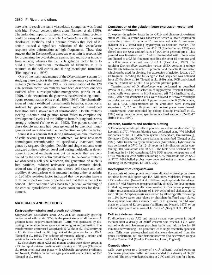
Growth of the double mutant strainsWhen grown under axenic conditions AX2 wild-type cells aswell as mutant cells of strains AHR and GHR reached final celldensities of 1×10⁷ cells/ml and higher. In contrast, AGHR2 cellsnever reached similar densities and consistently stopped growthmuch earlier. The doubling time was also prolonged from 10hours for AX2 to 19 hours for AGHR2 (Fig. 2). For AGHR2cells we noted a substantial shedding of cellular material duringgrowth in shaken suspension. These particles often haddiameters of up to 3 µm. They contained cytosolic proteins asvisualized by staining for profilin but were devoid of nuclei. Thisproperty could be traced back to a gelation factor deficiencysince GHR cells exhibited a similar phenotype (Fig. 3).
Time (hours)
0 20 40 60 80 100 120
Cel
l den
sity
(×1
06 /ml)
0
2
4
6
8
10
12
14AX2
AHR
GHR
AGHR2
Fig. 2. Growth of wild-type and mutant cells in shaking culture.Growth of wild-type AX2, AHR and GHR in shaking suspensionwas comparable, whereas AGHR2 had a reduced growth rate and didnot reach similar cell densities.
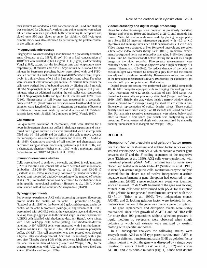
Cell size of AGHR mutantsAGHR2 cultures contained both smaller as well as significantlylarger cells than wild-type cultures. For further analysis the sizedistribution of AGHR2 cells was determined and compared tothe one of AX2, AHR and GHR cells (Fig. 4). Both AHR andGHR cells were almost indistinguishable in size from AX2cells, with an average diameter of 12 µm whereas most AGHR2cells had a diameter of 9 µm. In addition, there was a secondpopulation of enlarged AGHR2 cells with diameters of approx-imately 25 µm. This second population accounted for about 5%of the total cell number. Immunofluorescence studies using theDNA binding dye DAPI revealed that the large-sized AGHR2cells were multinucleated. Determination of cell sizes in anautomated counter supported the finding that the majority ofAGHR2 cells was smaller than AX2 cells.
Response to osmotic shock Exposure of cells to high osmolarities evokes a variety ofresponses. The accumulation of compatible osmolytes raises theinternal osmotic potential and the synthesis of stress proteinsprotects cellular components facilitating repair and recovery(Kwon and Handler, 1995). Whereas these are slow processesthat occur in pro- and eukaryotes, eukaryotic cells in additioncan quickly respond to changes in cell volume by dis- andreassembly of their cytoskeletons. When wild-type and mutantcells were treated for up to two hours with 0.4 M sorbitol andthen diluted into a solution of low osmolarity, survival of wild-type cells was much better than that of AGHR2 cells. Theosmotic shock led to a marked reduction in the proportion ofviable AGHR2 cells, which was 66% after one hour and 14%after two hours of treatment with sorbitol (Fig. 5). Comparableresults were obtained when cell lysis was estimated by deter-mining the decrease in protein content in the cellular pellet. Nosignificant lysis was observed for AX2 cells and AHR and GHRwere comparable to AX2 in their resistance to osmotic shock.
Phagocytosis of mutant strainsImmunofluorescence studies suggest that both crosslinkers areinvolved in different steps of the phagocytic process, gelationfactor being located at the phagocytic cup (Cox et al., 1996)and α-actinin in phagosomes (Furukawa and Fechheimer,1994). This has prompted us to test single and double mutantsfor their ability to phagocytose bacteria. After 30 minutes ofincubation with fluorescently labelled E. coli, AGHR2 cellsshowed a significant reduction (40-60%) in the rate of bacterialuptake, compared to the single mutants and the wild-type strainAX2 (Fig. 6). As internal control we used HSB50, a severelyimpaired phagocytosis mutant, which is also defective in cell-substrate adhesion and is blocked at the mound stage of devel-opment (Ceccarelli and Bozzaro, 1992). In contrast to HSB50,the rate of phagocytosis in AGHR2 seems to be normal for thefirst 5 minutes, suggesting that the double mutation probablyaffects later steps in the phagocytic process more strongly.
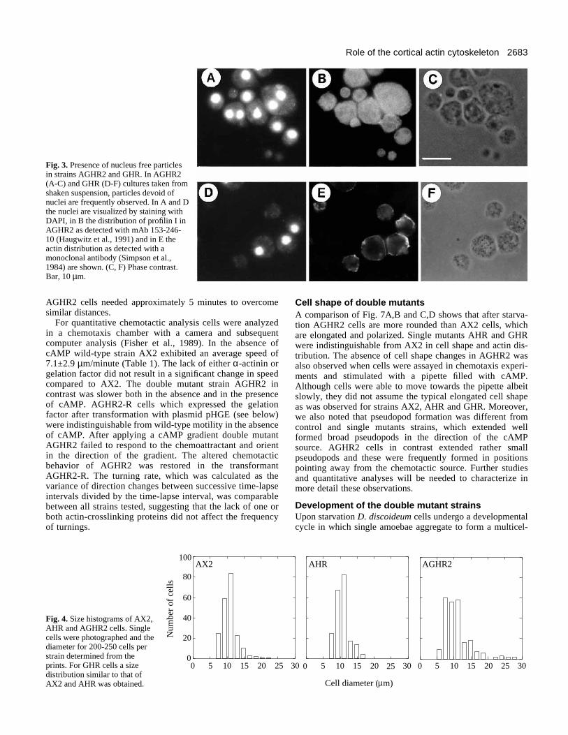
Motility and chemotactic behavior of mutant strainsChemotaxis of starving cells towards cAMP was analyzed inthe capillary assay where cells are stimulated with amicropipette filled with cAMP (10−4 M). AGHR2 cells wereable to move towards the pipette, however, they respondedmuch more slowly than wild-type cells. In a typical experimentAX2 cells arrived at the pipette within a minute, whereas
2683Role of the cortical actin cytoskeleton
Fig. 3. Presence of nucleus free particlesin strains AGHR2 and GHR. In AGHR2(A-C) and GHR (D-F) cultures taken fromshaken suspension, particles devoid ofnuclei are frequently observed. In A and Dthe nuclei are visualized by staining withDAPI, in B the distribution of profilin I inAGHR2 as detected with mAb 153-246-10 (Haugwitz et al., 1991) and in E theactin distribution as detected with amonoclonal antibody (Simpson et al.,1984) are shown. (C, F) Phase contrast.Bar, 10 µm.
AGHR2 cells needed approximately 5 minutes to overcomesimilar distances.
For quantitative chemotactic analysis cells were analyzedin a chemotaxis chamber with a camera and subsequentcomputer analysis (Fisher et al., 1989). In the absence ofcAMP wild-type strain AX2 exhibited an average speed of7.1±2.9 µm/minute (Table 1). The lack of either α-actinin orgelation factor did not result in a significant change in speedcompared to AX2. The double mutant strain AGHR2 incontrast was slower both in the absence and in the presenceof cAMP. AGHR2-R cells which expressed the gelationfactor after transformation with plasmid pHGE (see below)were indistinguishable from wild-type motility in the absenceof cAMP. After applying a cAMP gradient double mutantAGHR2 failed to respond to the chemoattractant and orientin the direction of the gradient. The altered chemotacticbehavior of AGHR2 was restored in the transformantAGHR2-R. The turning rate, which was calculated as thevariance of direction changes between successive time-lapseintervals divided by the time-lapse interval, was comparablebetween all strains tested, suggesting that the lack of one orboth actin-crosslinking proteins did not affect the frequencyof turnings.
80
60
40
20
0
100AX2
Num
ber
of c
ells
0 5 10 15 20 25
Fig. 4. Size histograms of AX2,AHR and AGHR2 cells. Singlecells were photographed and thediameter for 200-250 cells perstrain determined from theprints. For GHR cells a sizedistribution similar to that ofAX2 and AHR was obtained.
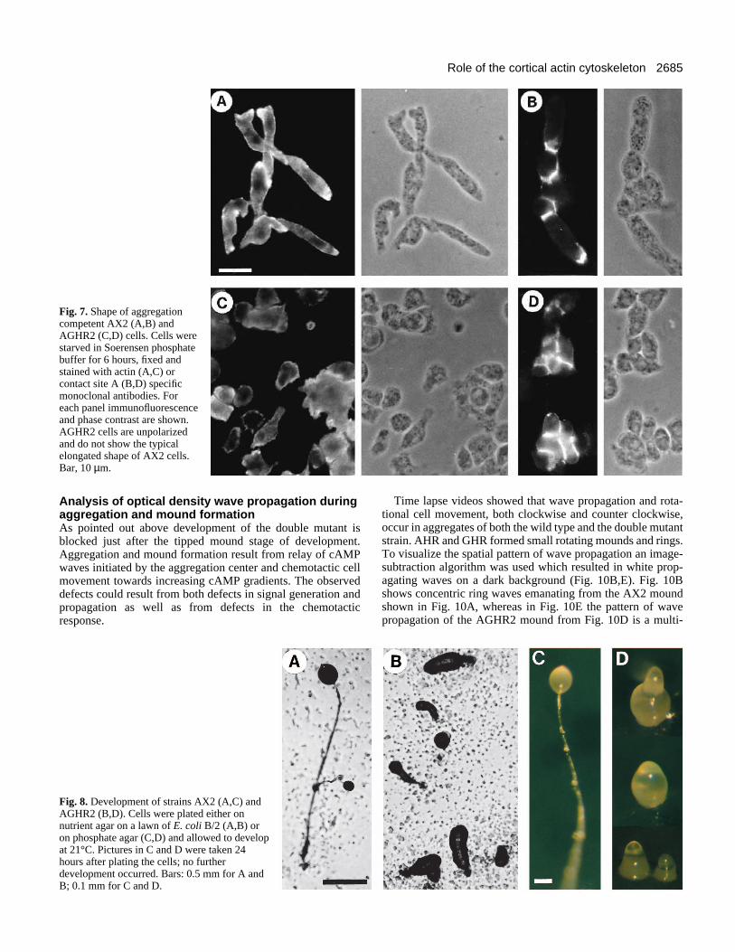
Cell shape of double mutantsA comparison of Fig. 7A,B and C,D shows that after starva-tion AGHR2 cells are more rounded than AX2 cells, whichare elongated and polarized. Single mutants AHR and GHRwere indistinguishable from AX2 in cell shape and actin dis-tribution. The absence of cell shape changes in AGHR2 wasalso observed when cells were assayed in chemotaxis experi-ments and stimulated with a pipette filled with cAMP.Although cells were able to move towards the pipette albeitslowly, they did not assume the typical elongated cell shapeas was observed for strains AX2, AHR and GHR. Moreover,we also noted that pseudopod formation was different fromcontrol and single mutants strains, which extended wellformed broad pseudopods in the direction of the cAMPsource. AGHR2 cells in contrast extended rather smallpseudopods and these were frequently formed in positionspointing away from the chemotactic source. Further studiesand quantitative analyses will be needed to characterize inmore detail these observations.
Development of the double mutant strainsUpon starvation D. discoideum cells undergo a developmentalcycle in which single amoebae aggregate to form a multicel-
5 10 15 20 25 30
AHR AGHR2
Cell diameter ( m)
0201510530
µ
30250
2684 F. Rivero and others
Table 1. Motility of wild-type and mutant strains Speed Orientation Turning rate
[µm/min] [cosθ] [rad2/min]
Strain N exp. Buffer Gradient Buffer Gradient Buffer Gradient
AX2 6 7.1±2.9 10.7±2.1 0.02±0.11 0.23±0.08 2.0±0.5 1.7±0.9GHR 3 5.6±2.3 8.3±0.5 0.09±0.12 0.14±0.03 1.2±0.3 1.5±0.1AHR 3 10.2±5.9 11.6±3.1 0.08±0.10 0.11±0.06 1.3±0.3 1.6±0.4AGHR2 4 3.8±1.8 7.3±1.6* 0.01±0.03 0.06±0.05* 2.2±0.6 1.3±0.2AGHR2R 2 7.5±0.1 8.3±0.4 0.04±0.03 0.14±0.01 1.8±0.4 1.6±0.3
Cell tracks were recorded over four 30 minutes periods. In each period 41 images were taken with a time lapse of 45 seconds between subsequent images.During the first two half hours movement in buffer was analyzed, during the second two half hour periods a linear cAMP gradient with a steepness of 2.5×10⁻⁸ McAMP/mm was applied. The values shown in the table were recorded in the first and third half hour period. In each period 50-80 individual cells were averaged.To avoid cells not only responding to the applied gradient but also to cAMP secreted by other cells, cell densities were used where the average distances betweenadjacent cells were at least 50 µm. Furthermore, care was taken that no cell aggregates were present. The orientation is expressed as cosθ, i.e. the higher thevalues the better the orientation in the gradient. Values are given as means ± s.d. *P<0.05 relative to AX2 (ANOVA).
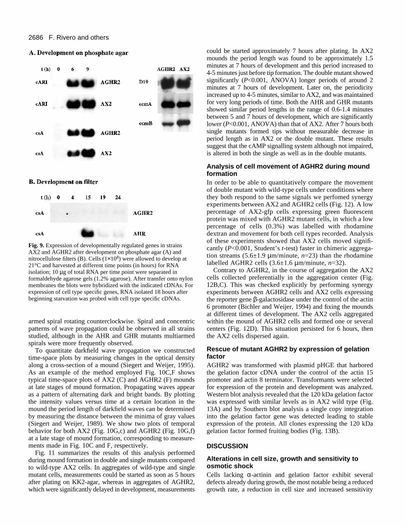
lular fruiting body. This cycle involves differentiation intospore and stalk cells and requires the sequential expression ofdevelopmentally regulated genes. In strain AGHR2 develop-ment was heavily impaired. On phosphate agar plates AGHR2cells were able to form long aggregation streams similar to thatof AX2 wild-type cells (see Figs 10A,D and 12A). Later duringaggregation many AGHR2 streams broke up, leading to theformation of small aggregates. Under these conditions tip andfinger formation and occasionally short thick slugs could beobserved, but no further development occurred (Fig. 8).Overall the developmental pattern of the double mutant cellswas significantly delayed as compared to wild-type cells. Thesingle mutants AHR and GHR usually formed smaller aggre-gation territories which also resulted in smaller mounds ascompared to AX2 cells. Surprisingly both single mutantsneeded less time to form tipped aggregates. Furthermore theywere able to complete development resulting in the formationof normal looking fruiting bodies.
Further investigations of AGHR2 showed that the stage atwhich development was blocked varied with the substrateused. When deposited on nitrocellulose filters AGHR2 cells
Time (hours)
0 1 2
% v
iabl
e ce
lls
0
20
40
60
80
100
120
140
AX2 + bufferAX2 + sorbitolAGHR2 + bufferAGHR2 + sorbitol
Fig. 5. Resistance to osmotic shock of strains AX2 and AGHR2.Cells were shaken in phosphate buffer in the presence or absence of0.4 M sorbitol for the times indicated, diluted into phosphate bufferand plated onto SM agar plates with K. aerogenes. Cell viability after1 or 2 hours of treatment was determined as the percentage ofcolonies in relation to 0 hours, which was assigned 100%. Each pointis the average of colony counts from 5 plates. The results of onerepresentative experiment are shown.
did not develop into aggregates. In parallel, mRNAs specificfor the aggregation stage like contact site A message (Noegelet al., 1986) were not expressed or were present at reducedlevels, as the mRNA corresponding to cAMP receptor cAR1(Klein et al., 1988). The messages of genes later expressed indevelopment such as the D19 (Early et al., 1988), ecmA andecmB genes (Jermyn et al., 1987) were not detected in strainAGHR2, suggesting that a differentiation into prespore andprestalk cells did not take place. During development onphosphate agar strain AGHR2 aggregated. cARI- as well ascsA-specific RNA could be detected at 6 hours after beginningstarvation, and at 18 hours the cell type specific mRNAscoding for PsA and EcmA and EcmB proteins were expressed.Whereas PsA and EcmA expression is comparable in AX2 andAGHR2, slightly lower levels of EcmB mRNA are apparent(Fig. 9). The expression of these markers did not substantiallydiffer between strains AX2, AHR and GHR.
Starvation in suspension culture also allowed developmentof AGHR2 as determined by the presence of the aggregationspecific csA protein, which is enriched at sites of cell to cellcontact (Fig. 7B,D).
Time (min)0 10 20 30
Num
ber
of b
acte
ria/
cell
0
20
40
60AX2 AHR GHR AGHR2 HSB50
Fig. 6. Phagocytosis of bacteria by wild-type and mutant cells. Cellswere incubated with FITC-labelled E.coli B/r under shakingconditions for the times indicated, and the number of cell-associatedbacteria determined as described in Materials and Methods. Eachpoint is the average of determinations made in triplicate. The resultsof one representative experiment are shown. Similar results wereobtained in three independent experiments.
2685Role of the cortical actin cytoskeleton
Fig. 7. Shape of aggregationcompetent AX2 (A,B) andAGHR2 (C,D) cells. Cells werestarved in Soerensen phosphatebuffer for 6 hours, fixed andstained with actin (A,C) orcontact site A (B,D) specificmonoclonal antibodies. Foreach panel immunofluorescenceand phase contrast are shown.AGHR2 cells are unpolarizedand do not show the typicalelongated shape of AX2 cells.Bar, 10 µm.
Analysis of optical density wave propagation duringaggregation and mound formationAs pointed out above development of the double mutant isblocked just after the tipped mound stage of development.Aggregation and mound formation result from relay of cAMPwaves initiated by the aggregation center and chemotactic cellmovement towards increasing cAMP gradients. The observeddefects could result from both defects in signal generation andpropagation as well as from defects in the chemotacticresponse.
Fig. 8. Development of strains AX2 (A,C) andAGHR2 (B,D). Cells were plated either onnutrient agar on a lawn of E. coli B/2 (A,B) oron phosphate agar (C,D) and allowed to developat 21°C. Pictures in C and D were taken 24hours after plating the cells; no furtherdevelopment occurred. Bars: 0.5 mm for A andB; 0.1 mm for C and D.
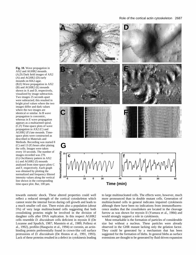
Time lapse videos showed that wave propagation and rota-tional cell movement, both clockwise and counter clockwise,occur in aggregates of both the wild type and the double mutantstrain. AHR and GHR formed small rotating mounds and rings.To visualize the spatial pattern of wave propagation an image-subtraction algorithm was used which resulted in white prop-agating waves on a dark background (Fig. 10B,E). Fig. 10Bshows concentric ring waves emanating from the AX2 moundshown in Fig. 10A, whereas in Fig. 10E the pattern of wavepropagation of the AGHR2 mound from Fig. 10D is a multi-
2686 F. Rivero and others
Fig. 9. Expression of developmentally regulated genes in strainsAX2 and AGHR2 after development on phosphate agar (A) andnitrocellulose filters (B). Cells (1×10⁸) were allowed to develop at21°C and harvested at different time points (in hours) for RNAisolation; 10 µg of total RNA per time point were separated informaldehyde agarose gels (1.2% agarose). After transfer onto nylonmembranes the blots were hybridized with the indicated cDNAs. Forexpression of cell type specific genes, RNA isolated 18 hours afterbeginning starvation was probed with cell type specific cDNAs.
armed spiral rotating counterclockwise. Spiral and concentricpatterns of wave propagation could be observed in all strainsstudied, although in the AHR and GHR mutants multiarmedspirals were more frequently observed.
To quantitate darkfield wave propagation we constructedtime-space plots by measuring changes in the optical densityalong a cross-section of a mound (Siegert and Weijer, 1995).As an example of the method employed Fig. 10C,F showstypical time-space plots of AX2 (C) and AGHR2 (F) moundsat late stages of mound formation. Propagating waves appearas a pattern of alternating dark and bright bands. By plottingthe intensity values versus time at a certain location in themound the period length of darkfield waves can be determinedby measuring the distance between the minima of gray values(Siegert and Weijer, 1989). We show two plots of temporalbehavior for both AX2 (Fig. 10G,c) and AGHR2 (Fig. 10G,f)at a late stage of mound formation, corresponding to measure-ments made in Fig. 10C and F, respectively.
Fig. 11 summarizes the results of this analysis performedduring mound formation in double and single mutants comparedto wild-type AX2 cells. In aggregates of wild-type and singlemutant cells, measurements could be started as soon as 5 hoursafter plating on KK2-agar, whereas in aggregates of AGHR2,which were significantly delayed in development, measurements
could be started approximately 7 hours after plating. In AX2mounds the period length was found to be approximately 1.5minutes at 7 hours of development and this period increased to4-5 minutes just before tip formation. The double mutant showedsignificantly (P<0.001, ANOVA) longer periods of around 2minutes at 7 hours of development. Later on, the periodicityincreased up to 4-5 minutes, similar to AX2, and was maintainedfor very long periods of time. Both the AHR and GHR mutantsshowed similar period lengths in the range of 0.6-1.4 minutesbetween 5 and 7 hours of development, which are significantlylower (P<0.001, ANOVA) than that of AX2. After 7 hours bothsingle mutants formed tips without measurable decrease inperiod length as in AX2 or the double mutant. These resultssuggest that the cAMP signalling system although not impaired,is altered in both the single as well as in the double mutants.
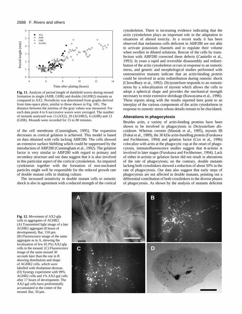
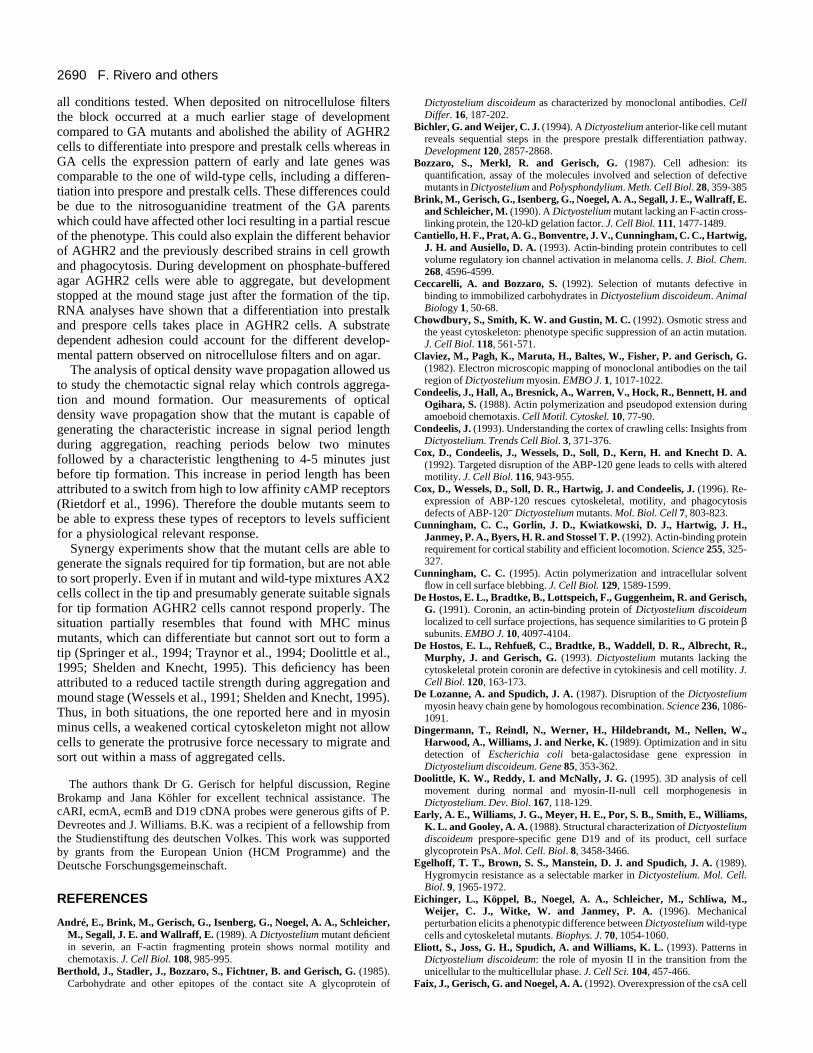
Analysis of cell movement of AGHR2 during moundformationIn order to be able to quantitatively compare the movementof double mutant with wild-type cells under conditions wherethey both respond to the same signals we perfomed synergyexperiments between AX2 and AGHR2 cells (Fig. 12). A lowpercentage of AX2-gfp cells expressing green fluorescentprotein was mixed with AGHR2 mutant cells, in which a lowpercentage of cells (0.3%) was labelled with rhodaminedextran and movement for both cell types recorded. Analysisof these experiments showed that AX2 cells moved signifi-cantly (P<0.001, Student’s t-test) faster in chimeric aggrega-tion streams (5.6±1.9 µm/minute, n=23) than the rhodaminelabelled AGHR2 cells (3.6±1.6 µm/minute, n=32).
Contrary to AGHR2, in the course of aggregation the AX2cells collected preferentially in the aggregation center (Fig.12B,C). This was checked explicitly by performing synergyexperiments between AGHR2 cells and AX2 cells expressingthe reporter gene β-galactosidase under the control of the actin6 promoter (Bichler and Weijer, 1994) and fixing the moundsat different times of development. The AX2 cells aggregatedwithin the mound of AGHR2 cells and formed one or severalcenters (Fig. 12D). This situation persisted for 6 hours, thenthe AX2 cells dispersed again.
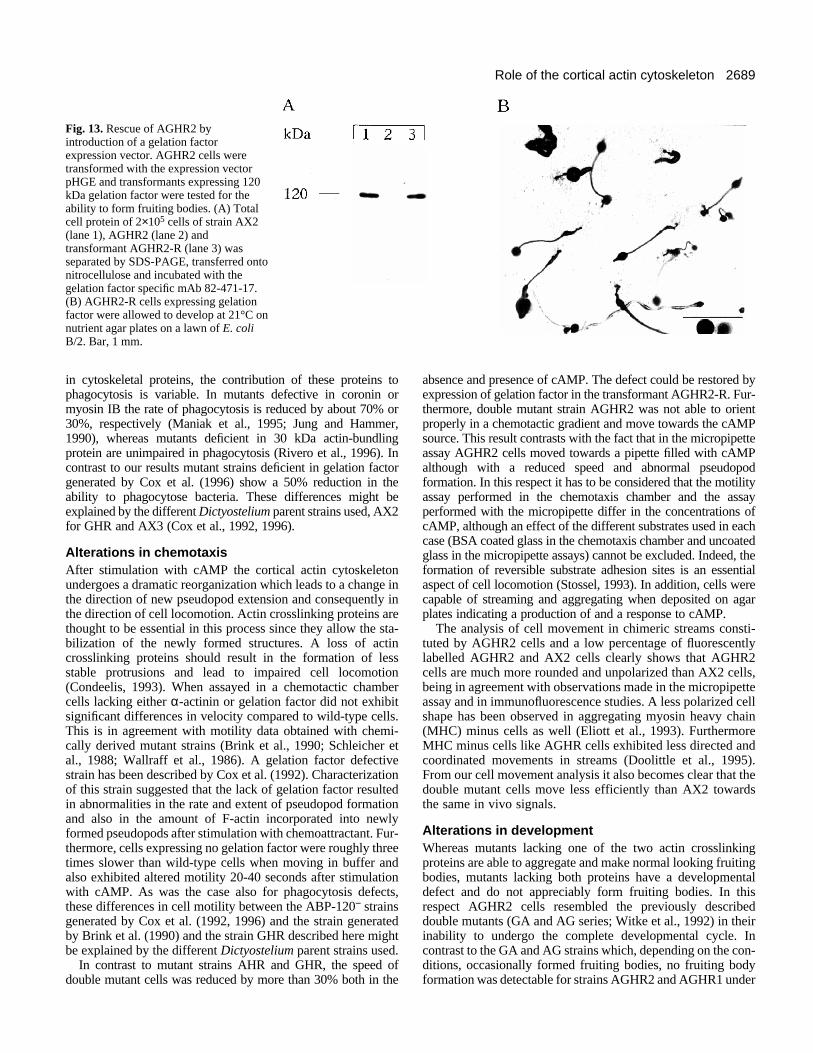
Rescue of mutant AGHR2 by expression of gelationfactorAGHR2 was transformed with plasmid pHGE that harboredthe gelation factor cDNA under the control of the actin 15promoter and actin 8 terminator. Transformants were selectedfor expression of the protein and development was analyzed.Western blot analysis revealed that the 120 kDa gelation factorwas expressed with similar levels as in AX2 wild type (Fig.13A) and by Southern blot analysis a single copy integrationinto the gelation factor gene was detected leading to stableexpression of the protein. All clones expressing the 120 kDagelation factor formed fruiting bodies (Fig. 13B).
DISCUSSION
Alterations in cell size, growth and sensitivity toosmotic shockCells lacking α-actinin and gelation factor exhibit severaldefects already during growth, the most notable being a reducedgrowth rate, a reduction in cell size and increased sensitivity
2687Role of the cortical actin cytoskeleton
A
B
C
D
F
EN
orm
aliz
ed in
ten
sity
Time (min)
15
-1515 30 45 15 30 450 0
Gc f
Fig. 10. Wave propagation inAX2 and AGHR2 mounds.(A,D) Dark field images of AX2(A) and AGHR2 (D) earlymounds on KK2-agar.(B,E) Wave propagation in AX2(B) and AGHR2 (E) moundsshown in A and D, respectively,visualized by image subtraction.Two images 25 seconds apartwere subtracted; this resulted inbright pixel values where the twoimages differ and dark valueswhere the two images areidentical or similar. In B wavepropagation is concentric,whereas in E wave propagationappears as a multiarmed spiral.(C,F) Time-space plots of wavepropagation in AX2 (C) andAGHR2 (F) late mounds. Time-space plots were constructed asdescribed in Materials andMethods. Recording was started 8(C) and 13 (F) hours after platingthe cells. Images were takenevery 10 seconds. The number ofimages recorded was 270.(G) Oscillatory pattern in AX2(c) and AGHR2 (f) moundsanalyzed from time-space plots Cand F, respectively. Each graphwas obtained by plotting thenormalized and frequency filteredintensity values along the verticalline shown in the correspondingtime-space plot. Bar, 100 µm.
towards osmotic shock. These altered properties could wellreflect a reduced strength of the cortical cytoskeleton whichcannot resist the internal forces during cell growth and leads toa much smaller cell size. There exists also a population (about5%) of very large multinucleated cells suggesting that bothcrosslinking proteins might be involved in the division ofdaughter cells after DNA replication. In this respect AGHR2cells resemble D. discoideum cells deficient in myosin II (DeLozanne and Spudich, 1987; Manstein et al., 1989; Pollenz etal., 1992), profilin (Haugwitz et al., 1994) or coronin, an actin-binding protein preferentially found in crown-like cell surfaceprotrusions of D. discoideum (De Hostos et al., 1991, 1993).Lack of these proteins resulted in a defect in cytokinesis leading
to large multinucleated cells. The effects were, however, muchmore pronounced than in double mutant cells. Generation ofmultinucleated cells in general indicates impaired cytokinesisalthough there have been no indications from immunofluores-cence studies that the crosslinkers are located in the cleavagefurrow as was shown for myosin II (Yumura et al., 1984) andwould strongly support a role in cytokinesis.
Most remarkable is the formation of particles of considerablesize but without a nucleus. These particles were alreadyobserved in the GHR mutant lacking only the gelation factor.They could be generated by a mechanism that has beensuggested for the formation of blebs. In general blebs as surfaceextensions are thought to be generated by fluid driven expansion
2688 F. Rivero and others
Time after plating (hours)
4 6 8 10 12 14
0
1
2
3
4
5
6
AX2 AHR GHR AGHR2
Peri
od le
ngth
(m
in)
Fig. 11. Analysis of period length of darkfield waves during moundformation in single (AHR, GHR) and double (AGHR2) mutants ascompared to AX2. Periodicity was determined from graphs derivedfrom time-space plots, similar to those shown in Fig. 10G. Thedistance between the minima of the gray values was measured. Foreach data point 4 to 6 successive waves were averaged. The numberof mounds analyzed was 13 (AX2), 20 (AGHR2), 6 (AHR) and 13(GHR). Mounds were recorded for 15 to 80 minutes.
of the cell membrane (Cunningham, 1995). The expansiondecreases as cortical gelation is achieved. This model is basedon data obtained with cells lacking ABP280. The cells showedan extensive surface blebbing which could be suppressed by theintroduction of ABP280 (Cunningham et al., 1992). The gelationfactor is very similar to ABP280 with regard to primary andsecondary structure and our data suggest that it is also involvedin this particular aspect of the cortical cytoskeleton. An impairedcytokinesis together with the formation of non-nucleatedparticles might well be responsible for the reduced growth rateof double mutant cells in shaking culture.
The increased sensitivity in double mutant cells to osmoticshock is also in agreement with a reduced strength of the cortical
A
C
Fig. 12. Movement of AX2-gfpcells in aggregates of AGHR2.(A) Transmitted light image of a lateAGHR2 aggregate (8 hours ofdevelopment). Bar, 150 µm.(B) Fluorescence image of the sameaggregate as in A, showing thelocalization of few (0.3%) AX2-gfpcells in the mound. (C) Fluorescenceimage of the same mound 30seconds later than the one in Bshowing distribution and shapeof AGHR2 cells, which werelabelled with rhodamine-dextran.(D) Synergy experiment with 99%AGHR2 cells and 1% AX2-gal cellsafter 17 hours of development. TheAX2-gal cells have preferentiallyaccumulated in the center of themound. Bar, 50 µm.
cytoskeleton. There is increasing evidence indicating that theactin cytoskeleton plays an important role in the adaptation tosituations of altered tonicity. In a recent study it has beenobserved that melanoma cells deficient in ABP280 are not ableto activate potassium channels and to regulate their volumewhen swollen in diluted solutions. Rescue of the cells by trans-fection with ABP280 corrected these defects (Cantiello et al.,1993). In yeast a rapid and reversible disassembly and redistri-bution of the actin cytoskeleton occurs in response to an osmoticstress, and genetic and morphological studies performed withosmosensitive mutants indicate that an actin-binding proteincould be involved in actin redistribution during osmotic shock(Chowdbury et al., 1992). Dictyostelium responds to an osmoticstress by a relocalization of myosin which allows the cells toadopt a spherical shape and provides the mechanical strengthnecessary to resist extensive shrinkage (Kuwayama et al., 1996).These reports along with the results reported here point to aninterplay of the various components of the actin cytoskeleton inresponse to osmotic stress whose details remain to be elucidated.
Alterations in phagocytosisBesides actin, a variety of actin-binding proteins have beenshown to be involved in phagocytosis in Dictyostelium dis-coideum. Whereas coronin (Maniak et al., 1995), myosin IB(Fukui et al., 1989), the 30 kDa actin-bundling protein (Furukawaand Fechheimer, 1994) and gelation factor (Cox et al., 1996)colocalize with actin at the phagocytic cup at the onset of phago-cytosis, immunofluorescence studies suggest that α-actinin isinvolved in later stages (Furukawa and Fechheimer, 1994). Lackof either α-actinin or gelation factor did not result in alterationsof the rate of phagocytosis; on the contrary, double mutantslacking both crosslinkers showed a reduction of about 50% in therate of phagocytosis. Our data also suggest that early steps ofphagocytosis are not affected in double mutants, pointing out adifferential contribution of both crosslinkers to the diverse phasesof phagocytosis. As shown by the analysis of mutants deficient
B
D
2689Role of the cortical actin cytoskeleton
Fig. 13. Rescue of AGHR2 byintroduction of a gelation factorexpression vector. AGHR2 cells weretransformed with the expression vectorpHGE and transformants expressing 120kDa gelation factor were tested for theability to form fruiting bodies. (A) Totalcell protein of 2×10⁵ cells of strain AX2(lane 1), AGHR2 (lane 2) andtransformant AGHR2-R (lane 3) wasseparated by SDS-PAGE, transferred ontonitrocellulose and incubated with thegelation factor specific mAb 82-471-17.(B) AGHR2-R cells expressing gelationfactor were allowed to develop at 21°C onnutrient agar plates on a lawn of E. coliB/2. Bar, 1 mm.
in cytoskeletal proteins, the contribution of these proteins tophagocytosis is variable. In mutants defective in coronin ormyosin IB the rate of phagocytosis is reduced by about 70% or30%, respectively (Maniak et al., 1995; Jung and Hammer,1990), whereas mutants deficient in 30 kDa actin-bundlingprotein are unimpaired in phagocytosis (Rivero et al., 1996). Incontrast to our results mutant strains deficient in gelation factorgenerated by Cox et al. (1996) show a 50% reduction in theability to phagocytose bacteria. These differences might beexplained by the different Dictyostelium parent strains used, AX2for GHR and AX3 (Cox et al., 1992, 1996).
Alterations in chemotaxisAfter stimulation with cAMP the cortical actin cytoskeletonundergoes a dramatic reorganization which leads to a change inthe direction of new pseudopod extension and consequently inthe direction of cell locomotion. Actin crosslinking proteins arethought to be essential in this process since they allow the sta-bilization of the newly formed structures. A loss of actincrosslinking proteins should result in the formation of lessstable protrusions and lead to impaired cell locomotion(Condeelis, 1993). When assayed in a chemotactic chambercells lacking either α-actinin or gelation factor did not exhibitsignificant differences in velocity compared to wild-type cells.This is in agreement with motility data obtained with chemi-cally derived mutant strains (Brink et al., 1990; Schleicher etal., 1988; Wallraff et al., 1986). A gelation factor defectivestrain has been described by Cox et al. (1992). Characterizationof this strain suggested that the lack of gelation factor resultedin abnormalities in the rate and extent of pseudopod formationand also in the amount of F-actin incorporated into newlyformed pseudopods after stimulation with chemoattractant. Fur-thermore, cells expressing no gelation factor were roughly threetimes slower than wild-type cells when moving in buffer andalso exhibited altered motility 20-40 seconds after stimulationwith cAMP. As was the case also for phagocytosis defects,these differences in cell motility between the ABP-120− strainsgenerated by Cox et al. (1992, 1996) and the strain generatedby Brink et al. (1990) and the strain GHR described here mightbe explained by the different Dictyostelium parent strains used.
In contrast to mutant strains AHR and GHR, the speed ofdouble mutant cells was reduced by more than 30% both in the
absence and presence of cAMP. The defect could be restored byexpression of gelation factor in the transformant AGHR2-R. Fur-thermore, double mutant strain AGHR2 was not able to orientproperly in a chemotactic gradient and move towards the cAMPsource. This result contrasts with the fact that in the micropipetteassay AGHR2 cells moved towards a pipette filled with cAMPalthough with a reduced speed and abnormal pseudopodformation. In this respect it has to be considered that the motilityassay performed in the chemotaxis chamber and the assayperformed with the micropipette differ in the concentrations ofcAMP, although an effect of the different substrates used in eachcase (BSA coated glass in the chemotaxis chamber and uncoatedglass in the micropipette assays) cannot be excluded. Indeed, theformation of reversible substrate adhesion sites is an essentialaspect of cell locomotion (Stossel, 1993). In addition, cells werecapable of streaming and aggregating when deposited on agarplates indicating a production of and a response to cAMP.
The analysis of cell movement in chimeric streams consti-tuted by AGHR2 cells and a low percentage of fluorescentlylabelled AGHR2 and AX2 cells clearly shows that AGHR2cells are much more rounded and unpolarized than AX2 cells,being in agreement with observations made in the micropipetteassay and in immunofluorescence studies. A less polarized cellshape has been observed in aggregating myosin heavy chain(MHC) minus cells as well (Eliott et al., 1993). FurthermoreMHC minus cells like AGHR cells exhibited less directed andcoordinated movements in streams (Doolittle et al., 1995).From our cell movement analysis it also becomes clear that thedouble mutant cells move less efficiently than AX2 towardsthe same in vivo signals.
Alterations in developmentWhereas mutants lacking one of the two actin crosslinkingproteins are able to aggregate and make normal looking fruitingbodies, mutants lacking both proteins have a developmentaldefect and do not appreciably form fruiting bodies. In thisrespect AGHR2 cells resembled the previously describeddouble mutants (GA and AG series; Witke et al., 1992) in theirinability to undergo the complete developmental cycle. Incontrast to the GA and AG strains which, depending on the con-ditions, occasionally formed fruiting bodies, no fruiting bodyformation was detectable for strains AGHR2 and AGHR1 under
2690 F. Rivero and others
all conditions tested. When deposited on nitrocellulose filtersthe block occurred at a much earlier stage of developmentcompared to GA mutants and abolished the ability of AGHR2cells to differentiate into prespore and prestalk cells whereas inGA cells the expression pattern of early and late genes wascomparable to the one of wild-type cells, including a differen-tiation into prespore and prestalk cells. These differences couldbe due to the nitrosoguanidine treatment of the GA parentswhich could have affected other loci resulting in a partial rescueof the phenotype. This could also explain the different behaviorof AGHR2 and the previously described strains in cell growthand phagocytosis. During development on phosphate-bufferedagar AGHR2 cells were able to aggregate, but developmentstopped at the mound stage just after the formation of the tip.RNA analyses have shown that a differentiation into prestalkand prespore cells takes place in AGHR2 cells. A substratedependent adhesion could account for the different develop-mental pattern observed on nitrocellulose filters and on agar.
The analysis of optical density wave propagation allowed usto study the chemotactic signal relay which controls aggrega-tion and mound formation. Our measurements of opticaldensity wave propagation show that the mutant is capable ofgenerating the characteristic increase in signal period lengthduring aggregation, reaching periods below two minutesfollowed by a characteristic lengthening to 4-5 minutes justbefore tip formation. This increase in period length has beenattributed to a switch from high to low affinity cAMP receptors(Rietdorf et al., 1996). Therefore the double mutants seem tobe able to express these types of receptors to levels sufficientfor a physiological relevant response.
Synergy experiments show that the mutant cells are able togenerate the signals required for tip formation, but are not ableto sort properly. Even if in mutant and wild-type mixtures AX2cells collect in the tip and presumably generate suitable signalsfor tip formation AGHR2 cells cannot respond properly. Thesituation partially resembles that found with MHC minusmutants, which can differentiate but cannot sort out to form atip (Springer et al., 1994; Traynor et al., 1994; Doolittle et al.,1995; Shelden and Knecht, 1995). This deficiency has beenattributed to a reduced tactile strength during aggregation andmound stage (Wessels et al., 1991; Shelden and Knecht, 1995).Thus, in both situations, the one reported here and in myosinminus cells, a weakened cortical cytoskeleton might not allowcells to generate the protrusive force necessary to migrate andsort out within a mass of aggregated cells.
The authors thank Dr G. Gerisch for helpful discussion, RegineBrokamp and Jana Köhler for excellent technical assistance. ThecARI, ecmA, ecmB and D19 cDNA probes were generous gifts of P.Devreotes and J. Williams. B.K. was a recipient of a fellowship fromthe Studienstiftung des deutschen Volkes. This work was supportedby grants from the European Union (HCM Programme) and theDeutsche Forschungsgemeinschaft.
REFERENCES
André, E., Brink, M., Gerisch, G., Isenberg, G., Noegel, A. A., Schleicher,M., Segall, J. E. and Wallraff, E. (1989). A Dictyostelium mutant deficientin severin, an F-actin fragmenting protein shows normal motility andchemotaxis. J. Cell Biol. 108, 985-995.
Berthold, J., Stadler, J., Bozzaro, S., Fichtner, B. and Gerisch, G. (1985).Carbohydrate and other epitopes of the contact site A glycoprotein of
Dictyostelium discoideum as characterized by monoclonal antibodies. CellDiffer. 16, 187-202.
Bichler, G. and Weijer, C. J. (1994). A Dictyostelium anterior-like cell mutantreveals sequential steps in the prespore prestalk differentiation pathway.Development 120, 2857-2868.
Bozzaro, S., Merkl, R. and Gerisch, G. (1987). Cell adhesion: itsquantification, assay of the molecules involved and selection of defectivemutants in Dictyostelium and Polysphondylium. Meth. Cell Biol. 28, 359-385
Brink, M., Gerisch, G., Isenberg, G., Noegel, A. A., Segall, J. E., Wallraff, E.and Schleicher, M. (1990). A Dictyostelium mutant lacking an F-actin cross-linking protein, the 120-kD gelation factor. J. Cell Biol. 111, 1477-1489.
Cantiello, H. F., Prat, A. G., Bonventre, J. V., Cunningham, C. C., Hartwig,J. H. and Ausiello, D. A. (1993). Actin-binding protein contributes to cellvolume regulatory ion channel activation in melanoma cells. J. Biol. Chem.268, 4596-4599.
Ceccarelli, A. and Bozzaro, S. (1992). Selection of mutants defective inbinding to immobilized carbohydrates in Dictyostelium discoideum. AnimalBiology 1, 50-68.
Chowdbury, S., Smith, K. W. and Gustin, M. C. (1992). Osmotic stress andthe yeast cytoskeleton: phenotype specific suppression of an actin mutation.J. Cell Biol. 118, 561-571.
Claviez, M., Pagh, K., Maruta, H., Baltes, W., Fisher, P. and Gerisch, G.(1982). Electron microscopic mapping of monoclonal antibodies on the tailregion of Dictyostelium myosin. EMBO J. 1, 1017-1022.
Condeelis, J., Hall, A., Bresnick, A., Warren, V., Hock, R., Bennett, H. andOgihara, S. (1988). Actin polymerization and pseudopod extension duringamoeboid chemotaxis. Cell Motil. Cytoskel. 10, 77-90.
Condeelis, J. (1993). Understanding the cortex of crawling cells: Insights fromDictyostelium. Trends Cell Biol. 3, 371-376.
Cox, D., Condeelis, J., Wessels, D., Soll, D., Kern, H. and Knecht D. A.(1992). Targeted disruption of the ABP-120 gene leads to cells with alteredmotility. J. Cell Biol. 116, 943-955.
Cox, D., Wessels, D., Soll, D. R., Hartwig, J. and Condeelis, J. (1996). Re-expression of ABP-120 rescues cytoskeletal, motility, and phagocytosisdefects of ABP-120− Dictyostelium mutants. Mol. Biol. Cell 7, 803-823.
Cunningham, C. C., Gorlin, J. D., Kwiatkowski, D. J., Hartwig, J. H.,Janmey, P. A., Byers, H. R. and Stossel T. P. (1992). Actin-binding proteinrequirement for cortical stability and efficient locomotion. Science 255, 325-327.
Cunningham, C. C. (1995). Actin polymerization and intracellular solventflow in cell surface blebbing. J. Cell Biol. 129, 1589-1599.
De Hostos, E. L., Bradtke, B., Lottspeich, F., Guggenheim, R. and Gerisch,G. (1991). Coronin, an actin-binding protein of Dictyostelium discoideumlocalized to cell surface projections, has sequence similarities to G protein βsubunits. EMBO J. 10, 4097-4104.
De Hostos, E. L., Rehfueß, C., Bradtke, B., Waddell, D. R., Albrecht, R.,Murphy, J. and Gerisch, G. (1993). Dictyostelium mutants lacking thecytoskeletal protein coronin are defective in cytokinesis and cell motility. J.Cell Biol. 120, 163-173.
De Lozanne, A. and Spudich, J. A. (1987). Disruption of the Dictyosteliummyosin heavy chain gene by homologous recombination. Science 236, 1086-1091.
Dingermann, T., Reindl, N., Werner, H., Hildebrandt, M., Nellen, W.,Harwood, A., Williams, J. and Nerke, K. (1989). Optimization and in situdetection of Escherichia coli beta-galactosidase gene expression inDictyostelium discoideum. Gene 85, 353-362.
Doolittle, K. W., Reddy, I. and McNally, J. G. (1995). 3D analysis of cellmovement during normal and myosin-II-null cell morphogenesis inDictyostelium. Dev. Biol. 167, 118-129.
Early, A. E., Williams, J. G., Meyer, H. E., Por, S. B., Smith, E., Williams,K. L. and Gooley, A. A. (1988). Structural characterization of Dictyosteliumdiscoideum prespore-specific gene D19 and of its product, cell surfaceglycoprotein PsA. Mol. Cell. Biol. 8, 3458-3466.
Egelhoff, T. T., Brown, S. S., Manstein, D. J. and Spudich, J. A. (1989).Hygromycin resistance as a selectable marker in Dictyostelium. Mol. Cell.Biol. 9, 1965-1972.
Eichinger, L., Köppel, B., Noegel, A. A., Schleicher, M., Schliwa, M.,Weijer, C. J., Witke, W. and Janmey, P. A. (1996). Mechanicalperturbation elicits a phenotypic difference between Dictyostelium wild-typecells and cytoskeletal mutants. Biophys. J. 70, 1054-1060.
Eliott, S., Joss, G. H., Spudich, A. and Williams, K. L. (1993). Patterns inDictyostelium discoideum: the role of myosin II in the transition from theunicellular to the multicellular phase. J. Cell Sci. 104, 457-466.
Faix, J., Gerisch, G. and Noegel, A. A. (1992). Overexpression of the csA cell
2691Role of the cortical actin cytoskeleton
adhesion molecule under its own cAMP-regulated promoter impairsmorphogenesis in Dictyostelium discoideum. J. Cell Sci. 102, 203-214.
Fisher, P. R., Merkl, R. and Gerisch, G. (1989). Quantitative analysis of cellmotility and chemotaxis in Dictyostelium discoideum by using an imageprocessing system and a novel chemotaxis chamber providing stationarychemical gradients. J. Cell Biol. 108, 973-984.
Fukui, Y., Lynch, T. J., Brzeska, H. and Korn, E. D. (1989). Myosin I islocated at the leading edges of locomoting Dictyostelium amoebae. Nature341, 328-331.
Furukawa, R. and Fechheimer, M. (1994). Differential localization of α-actinin and the 30 kD actin-bundling protein in the cleavage furrrow,phagocytic cup, and contractile vacuole of Dictyostelium discoideum. CellMotil. Cytoskel. 29, 46-56.
Gerisch, G. and Keller, H. U. (1981). Chemotactic reorientation ofgranulocytes stimulated with micropipettes containing fMet-Leu-Phe. J. CellSci. 52, 1-10.
Hartmann, H., Noegel, A. A., Eckerskorn, C., Rapp, S. and Schleicher, M.(1989). Ca2+ independent F-actin capping proteins: Cap32/34, a cappingprotein from Dictyostelium discoideum, does not share sequence homologieswith known capping proteins. J. Biol. Chem. 264, 12639-12647.
Haugwitz, M., Noegel, A. A., Rieger, D., Lottspeich, F. and Schleicher, M.(1991). Dictyostelium discoideum contains two profilin isoforms that differin structure and function. J. Cell Sci. 100, 481-489.
Haugwitz, M., Noegel, A. A., Karakesisoglou, J. and Schleicher, M. (1994).Dictyostelium amoebae that lack G-actin-sequestering profilins show defectsin F-actin content, cytokinesis, and development. Cell 79, 303-314.
Janssen, K.-P., Eichinger, L., Janmey, P., Noegel, A. A., Schliwa, M.,Witke, W. and Schleicher, M. (1996). Viscoelastic properties of F-actinsolutions in the presence of normal and mutated actin-binding proteins. Arch.Biochem. Biophys. 325, 183-189.
Jermyn, K. A., Berks, M., Kay, R. R. and Williams, J. E. (1987). Twodistinct classes of prestalk-enriched mRNA sequences in Dictyosteliumdiscoideum. Development 100, 745-755.
Jung, G. and Hammer, J. A. (1990). Generation and characterization ofDictyostelium cells deficient in a myosin I heavy chain isoform. J. Cell Biol.110, 1955-1964.
Klein, P. S., Sun, T. J., Saxe III, C. L., Kimmel, A. R., Johnson, R. L. andDevreotes, P. N. (1988). A chemoattractant receptor controls developmentin Dictyostelium discoideum. Science 241, 1467-1472.
Knecht, D. A., Cohen, S. M., Loomis, W. F. and Lodish, H. F. (1986).Developmental regulation of Dictyostelium discoideum actin gene fusionscarried on low-copy and high-copy transformation vectors. Mol. Cell. Biol. 6,3973-3983.
Kuwayama, H., Ecke, M., Gerisch, G. and Van Haastert, P. (1996).Protection against osmotic stress by cGMP mediated myosinphosphorylation. Science 271, 207-209.
Kwon, H. M. and Handler, J. S. (1995). Cell volume regulated transporters ofcompatible osmolytes. Curr. Opin. Cell Biol. 7, 465-471.
Laemmli, U. K. (1970). Cleavage of structural proteins during the assembly ofthe head of bacteriophage T4. Nature 227, 680-685.
Luby-Phelps, K. (1994). Physical properties of cytoplasm. Curr. Opin. CellBiol. 6, 3-9.
Maniak, M., Rauchenberger, R., Albrecht, R., Murphy, J. and Gerisch, G.(1995). Coronin involved in phagocytosis: dynamics of particle-inducedrelocalization visualized by a green fluorescent protein tag. Cell 83, 915-924.
Manstein, D. J., Titus, M. A., De Lozanne, A. and Spudich, J. A. (1989).Gene replacement in Dictyostelium: generation of myosin null mutants.EMBO J. 8, 923-932.
Matsudaira, P. (1991). Modular organization of actin crosslinking proteins.Trends Biochem. Sci. 16, 87-92.
Newell, P. C., Telser, A. and Sussman, M. (1969). Alternative developmentalpathways determined by environmental conditions in the cellular slimemould Dictyostelium discoideum. J. Bacteriol. 100, 763-768.
Noegel, A. A., Harloff, C., Hirth, P., Merkl, R., Modersitzki, M., Stadler, J.,Weinhart, U., Westphal, M. and Gerisch, G. (1985). Probing an adhesionmutant of Dictyostelium discoideum with cDNA clones and monoclonalantibodies indicates a specific defect in the contact site A glycoprotein.EMBO J. 4, 3805-3810.
Noegel, A. A., Gerisch, G., Stadler, J. and Westphal, M. (1986). Completesequence and transcript regulation of a cell adhesion protein fromaggregating Dictyostelium cells. EMBO J. 5, 1473-1476.
Noegel, A. A., Rapp, S., Lottspeich, F., Schleicher, M. and Stewart, M. (1989).The Dictyostelium gelation factor shares a putative actin binding site with α-
actinins and dystrophin and also has a rod domain containing six 100-residuemotifs that appear to have a cross-beta conformation. J. Cell Biol. 109, 607-618.
Pollenz, R. S., Chen, T.-L. L., Trivinos-Lagos, L. and Chisholm, R. L.(1992). The Dictyostelium essential light chain is required for myosinfunction. Cell 69, 951-962.
Rietdorf, J., Siegert, F. and Weijer, C. J. (1996). Analysis of optical densitywave propagation and cell movement during mound formation inDictyostelium discoideum. Dev. Biol. 177, 427-438.
Rivero, F., Furukawa, R., Noegel, A. A. and Fechheimer, M. (1996).Dictyostelium discoideum cells lacking the 34,000 Dalton actin bindingprotein can grow, locomote and develop, but exhibit defects in regulation ofcell structure and movement: A case of partial redundancy. J. Cell Biol. (inpress).
Schleicher, M., Noegel, A. A., Schwarz, T., Wallraff, E., Brink, M., Faix, J.,Gerisch, G. and Isenberg, G. (1988). A Dictyostelium mutant with severedefects in α-actinin: Its characterization by cDNA probes and monoclonalantibodies. J. Cell Sci. 90, 59-71.
Schleicher, M., André, B., Andréoli, C., Eichinger, L., Haugwitz, M.,Hofmann, A., Karakesisoglou, J., Stöckelhuber, M. and Noegel, A. A.(1995). Structure/function studies on cytoskeletal proteins in Dictyosteliumamoebae as a paradigm. FEBS Lett. 369, 38-42.
Segall, J. E., Fisher, P. R. and Gerisch, G. (1987). Selection of chemotaxismutants of Dictyostelium discoideum. J. Cell Biol. 10, 151-161.
Shelden, E. and Knecht, D. A. (1995). Mutants lacking myosin II cannot resistforces generated during multicellular morphogenesis. J. Cell Sci. 108, 1105-1115.
Siegert, F. and Weijer, C. (1989). Digital image processing of optical densitywave propagation in Dictyostelium discoideum and analysis of the effects ofcaffeine and ammonia. J. Cell Sci. 93, 325-335.
Siegert, F. and Weijer, C. J. (1992). Three-dimensional scroll waves organizeDictyostelium slugs. Proc. Nat. Acad. Sci. USA 89, 6433-6437.
Siegert, F. and Weijer, C. (1995). Spiral and concentric waves organizemulticellular Dictyostelium mounds. Curr. Biol. 5, 937-943.
Simpson, P. A., Spudich, J. A. and Parham, P. (1984). Monoclonalantibodies prepared against Dictyostelium actin: characterization andinteraction with actin. J. Cell Biol. 99, 287-295.
Springer, M. L., Patterson, B. and Spudich, J. A. (1994). Stage specificrequirement for myosin II during Dictyostelium development. Development120, 2651-2660.
Stossel, T. P. (1993). On the crawling of animal cells. Science 260, 1086-1094.Traynor, D., Tasaka, M., Takeuchi, I. and Williams, J. (1994). Aberrant
pattern formation in myosin heavy chain mutants of Dictyostelium.Development 120, 591-601.
Vogel, G. (1987). Endocytosis and recognition mechanisms in Dictyosteliumdiscoideum. Meth. Cell Biol. 28, 129-138.
Wachsstock, D. H., Schwarz, W. H. and Pollard, T. D. (1994). Cross-linkerdynamics determine the mechanical properties of actin gels. Biophys. J. 66,801-809.
Wallraff, E., Schleicher, M., Modersitzki, M., Rieger, D., Isenberg, G. andGerisch, G. (1986). Selection of Dictyostelium mutants defective incytoskeletal proteins: Use of an antibody that binds to the ends of α-actininrods. EMBO J. 5, 61-67.
Weiner, O. H., Murphy, J., Griffiths, G., Schleicher, M. and Noegel, A. A.(1993). The actin-binding protein comitin (p24) is a component of the Golgiapparatus. J. Cell Biol. 123, 23-34.
Wessels, D., Soll, D. R., Knecht, D. A., Loomis, W. F., De Lozanne, A. andSpudich, J. A. (1988). Cell motility and chemotaxis in Dictyosteliumamebae lacking myosin heavy chain. Dev. Biol. 128, 164-177.
Williams, K. L. and Newell, P. C. (1976). A genetic study of aggregation inthe cellular slime mould Dictyostelium discoideum using complementationanalysis. Genetics 82, 287-307.
Witke, W., Nellen, W. and Noegel, A. A. (1987). Homologous recombinationin the Dictyostelium α-actinin gene leads to an altered mRNA and lack of theprotein. EMBO J. 6, 4143-4148.
Witke, W., Schleicher, M. and Noegel, A. A. (1992). Redundancy in themicrofilament system: Abnormal development of Dictyostelium cells lackingtwo F-actin crosslinking proteins. Cell 68, 53-62.
Yumura, S., Mori, H. and Fukui, Y. (1984). Localization of actin and myosinfor the study of amoeboid movement in Dictyostelium using improvedimmunofluorescence. J. Cell Biol. 99, 894-899.
(Received 23 April 1996 – Accepted 6 August 1996)
![Stability of the human polymerase δ holoenzyme …thesis (TLS)] so that pol δ may resume synthesis (9–12). How-ever, studies on the human pol δ holoenzyme are lacking, and hence,](https://static.fdocument.org/doc/165x107/5ecf5ac71e33ba350c72b907/stability-of-the-human-polymerase-holoenzyme-thesis-tls-so-that-pol-may.jpg)