A morphogen gradient of Wnt/β-catenin signalling...
13
INTRODUCTION The vertebrate central nervous system (CNS) is divided anteroposteriorly into forebrain, midbrain, hindbrain and spinal cord during early embryogenesis. Neural inducers and modifiers establish a crude anteroposterior (AP) pattern before and during gastrulation that becomes refined during later stages (Saha and Grainger, 1992; Lumsden and Krumlauf, 1996; Sasai and De Robertis, 1997; Chang and Hemmati-Brivanlou, 1998a; Beddington and Robertson, 1999; Gamse and Sive, 2000; Stern, 2001). In amphibia, classical experiments have established a central role for Spemann’s organizer in this process. This organizer region does not only induce ectoderm to acquire neural fate, it also emits regionally specific inducers, with anterior mesendoderm inducing forebrain and posterior chordamesoderm inducing spinal cord (Gilbert and Saxén, 1993; Ruiz i Altaba, 1993; Gould and Grainger, 1997; Harland and Gerhart, 1997; Nieuwkoop, 1997). Similarly, classical experiments by Nieuwkoop, Saxén, Toivonen and colleagues have suggested a two-step model where a gradient of a posteriorizing factor (‘transformer’) confers progressive posterior identity to tissue which has initially been induced as anterior neuroectoderm by early involuting mesendoderm (Gilbert and Saxén, 1993; Doniach and Musci, 1995; Nieuwkoop, 1997; Sasai and De Robertis, 1997). The molecular identity of the transforming gradient has remained elusive, although a number of candidate posteriorizing signals have been proposed, notably fibroblast growth factors (FGFs), retinoic acid (RA) and Wnts (Durston et al., 1989; Ruiz i Altaba, 1993; Slack, 1994; Doniach, 1995; McGrew et al., 1995; Pownall et al., 1996; Kolm and Sive, 1997; McGrew et al., 1997; Moon et al., 1997; Isaacs et al., 1998; Pownall et al., 1998; Wodarz and Nusse, 1998; Holowacz and Sokol, 1999; Gavalas and Krumlauf, 2000; Ribisi et al., 2000; Gamse and Sive, 2001). Yet, for none of them has it been shown that (1) it is required for AP patterning, (2) it acts directly on neuroectoderm and (3) it functions endogenously in a graded fashion. Among the candidate transformers, Wnts have recently been implicated as endogenous antagonists of the head organizer (Niehrs, 1999). Consistent with the distinct inducing activities of anterior mesendoderm and chordamesoderm noted by Spemann and Mangold, inhibitors of transforming growth factors β (TGFβs) and Wnts, which are thought to mediate inductions by the organizer, are differentially expressed along the AP axis of the organizer and its derivatives (Harland and Gerhart, 1997; De Robertis et al., 2000). Notably, Wnt inhibitors are expressed predominantly in the anterior mesendoderm (Niehrs, 1999; De Robertis et al., 2000; Kiecker and Niehrs, 2001). In line with these inhibitors regulating AP neural patterning, co-injection of Wnt and bone morphogenetic protein (BMP) inhibitors on the ventral side of Xenopus embryos induces ectopic heads including forebrain structures while BMP inhibitors only induce ectopic trunk including spinal cord (Glinka et al., 1997). Thus, a distinguishing feature of the head organizer is the repression of Wnt signalling (Niehrs, 1999). Other data support the idea that Wnts antagonize the head 4189 Development 128, 4189-4201 (2001) Printed in Great Britain © The Company of Biologists Limited 2001 DEV2746 Anteroposterior (AP) patterning of the vertebrate neural plate is initiated during gastrulation and is regulated by Spemann’s organizer and its derivatives. The prevailing model for AP patterning predicts a caudally increasing gradient of a ‘transformer’ which posteriorizes anteriorly specified neural cells. However, the molecular identity of the transforming gradient has remained elusive. We show that in Xenopus embryos (1) dose-dependent Wnt signalling is both necessary and sufficient for AP patterning of the neuraxis, (2) Wnt/β-catenin signalling occurs in a direct and long-range fashion within the ectoderm, and (3) that there is an endogenous AP gradient of Wnt/β-catenin signalling in the presumptive neural plate of the Xenopus gastrula. Our results indicate that an activity gradient of Wnt/β-catenin signalling acts as transforming morphogen to pattern the Xenopus central nervous system. Key words: Anteroposterior neuraxis, AP, β-catenin, Gastrulation, Head organizer, Long-range signalling, Morphogen gradient, Neural patterning, Neuroectoderm, Nieuwkoop, Spemann, Transformer, Wnt protein, Xenopus SUMMARY A morphogen gradient of Wnt/β-catenin signalling regulates anteroposterior neural patterning in Xenopus Clemens Kiecker and Christof Niehrs* Division of Molecular Embryology, Deutsches Krebsforschungszentrum, Im Neuenheimer Feld 280, 69120 Heidelberg, Germany *Author for correspondence (e-mail: [email protected]) Accepted 2 August 2001
Transcript of A morphogen gradient of Wnt/β-catenin signalling...

INTRODUCTION
The vertebrate central nervous system (CNS) is dividedanteroposteriorly into forebrain, midbrain, hindbrain and spinalcord during early embryogenesis. Neural inducers andmodifiers establish a crude anteroposterior (AP) pattern beforeand during gastrulation that becomes refined during later stages(Saha and Grainger, 1992; Lumsden and Krumlauf, 1996;Sasai and De Robertis, 1997; Chang and Hemmati-Brivanlou,1998a; Beddington and Robertson, 1999; Gamse and Sive,2000; Stern, 2001). In amphibia, classical experiments haveestablished a central role for Spemann’s organizer in thisprocess. This organizer region does not only induce ectodermto acquire neural fate, it also emits regionally specific inducers,with anterior mesendoderm inducing forebrain and posteriorchordamesoderm inducing spinal cord (Gilbert and Saxén,1993; Ruiz i Altaba, 1993; Gould and Grainger, 1997; Harlandand Gerhart, 1997; Nieuwkoop, 1997). Similarly, classicalexperiments by Nieuwkoop, Saxén, Toivonen and colleagueshave suggested a two-step model where a gradient of aposteriorizing factor (‘transformer’) confers progressiveposterior identity to tissue which has initially been induced asanterior neuroectoderm by early involuting mesendoderm(Gilbert and Saxén, 1993; Doniach and Musci, 1995;Nieuwkoop, 1997; Sasai and De Robertis, 1997). Themolecular identity of the transforming gradient has remainedelusive, although a number of candidate posteriorizing signalshave been proposed, notably fibroblast growth factors (FGFs),retinoic acid (RA) and Wnts (Durston et al., 1989; Ruiz i
Altaba, 1993; Slack, 1994; Doniach, 1995; McGrew et al.,1995; Pownall et al., 1996; Kolm and Sive, 1997; McGrew etal., 1997; Moon et al., 1997; Isaacs et al., 1998; Pownall et al.,1998; Wodarz and Nusse, 1998; Holowacz and Sokol, 1999;Gavalas and Krumlauf, 2000; Ribisi et al., 2000; Gamse andSive, 2001). Yet, for none of them has it been shown that (1)it is required for AP patterning, (2) it acts directly onneuroectoderm and (3) it functions endogenously in a gradedfashion.
Among the candidate transformers, Wnts have recently beenimplicated as endogenous antagonists of the head organizer(Niehrs, 1999). Consistent with the distinct inducing activitiesof anterior mesendoderm and chordamesoderm noted bySpemann and Mangold, inhibitors of transforming growthfactors β (TGFβs) and Wnts, which are thought to mediateinductions by the organizer, are differentially expressed alongthe AP axis of the organizer and its derivatives (Harland andGerhart, 1997; De Robertis et al., 2000). Notably, Wntinhibitors are expressed predominantly in the anteriormesendoderm (Niehrs, 1999; De Robertis et al., 2000; Kieckerand Niehrs, 2001). In line with these inhibitors regulating APneural patterning, co-injection of Wnt and bone morphogeneticprotein (BMP) inhibitors on the ventral side of Xenopusembryos induces ectopic heads including forebrain structureswhile BMP inhibitors only induce ectopic trunk includingspinal cord (Glinka et al., 1997). Thus, a distinguishing featureof the head organizer is the repression of Wnt signalling(Niehrs, 1999).
Other data support the idea that Wnts antagonize the head
4189Development 128, 4189-4201 (2001)Printed in Great Britain © The Company of Biologists Limited 2001DEV2746
Anteroposterior (AP) patterning of the vertebrate neuralplate is initiated during gastrulation and is regulated bySpemann’s organizer and its derivatives. The prevailingmodel for AP patterning predicts a caudally increasinggradient of a ‘transformer’ which posteriorizes anteriorlyspecified neural cells. However, the molecular identity ofthe transforming gradient has remained elusive. We showthat in Xenopus embryos (1) dose-dependent Wntsignalling is both necessary and sufficient for AP patterningof the neuraxis, (2) Wnt/β-catenin signalling occurs in adirect and long-range fashion within the ectoderm, and (3)
that there is an endogenous AP gradient of Wnt/β-cateninsignalling in the presumptive neural plate of the Xenopusgastrula. Our results indicate that an activity gradient ofWnt/β-catenin signalling acts as transforming morphogento pattern the Xenopuscentral nervous system.
Key words: Anteroposterior neuraxis, AP, β-catenin, Gastrulation,Head organizer, Long-range signalling, Morphogen gradient, Neuralpatterning, Neuroectoderm, Nieuwkoop, Spemann, Transformer, Wntprotein, Xenopus
SUMMARY
A morphogen gradient of Wnt/ β-catenin signalling regulates anteroposterior
neural patterning in Xenopus
Clemens Kiecker and Christof Niehrs*
Division of Molecular Embryology, Deutsches Krebsforschungszentrum, Im Neuenheimer Feld 280, 69120 Heidelberg, Germany*Author for correspondence (e-mail: [email protected])
Accepted 2 August 2001
4190
organizer and have posteriorizing properties. Overexpressionof various Wnts (Christian and Moon, 1993; McGrew et al.,1995; Fredieu et al., 1997; McGrew et al., 1997; Saint-Jeannet et al., 1997; Chang and Hemmati-Brivanlou, 1998b;McGrew et al., 1999; Gamse and Sive, 2001), the Wntpathway components β-catenin and XTcf3 (McGrew et al.,1995; Darken and Wilson, 2001; Hamilton et al., 2001) ortreatment with the artificial activator of the Wnt pathwaylithium chloride (Fredieu et al., 1997) all lead to repressionof anterior and concomittant induction of posterior neuralmarkers in Xenopus. Conversely, overexpression of secretedWnt antagonists anteriorizes Xenopusand zebrafish embryos(Itoh et al., 1995; Hoppler et al., 1996; Leyns et al., 1997;Deardorff et al., 1998; Glinka et al., 1998; Hsieh et al., 1999a;Fekany-Lee et al., 2000; Hashimoto et al., 2000; Shinya etal., 2000). A posteriorizing role for Wnts has also beensuggested from studies on transgenic mice expressing Wnt8(Pöpperl et al., 1997). Furthermore, mice mutant for Wnt3A(Takada et al., 1994), the nuclear transducers of Wntsignalling Lef1 and Tcf1 (Galceran et al., 1999) and the Wntco-receptor LRP6 (Pinson et al., 2000) show posteriortruncations, indicating a requirement for Wnt signalling inposterior structures. Finally, inhibition of Wnt signalling isrequired for formation of anterior neural structures asevidenced by interference with inhibitors of Wnt signalling:both inhibition of the secreted Wnt antagonist Dkk1 inXenopus(Glinka et al., 1998; Kazanskaya et al., 2000), aswell as inactivation of the Wnt repressors tcf3and axin1inthe zebrafish headlessand masterblindmutants, respectively,lead to microcephalic embryos (Kim et al., 2000; Heisenberget al., 2001).
These properties make Wnts likely candidates forNieuwkoop’s posteriorizing factor. Thus, we investigatedwhether an endogenous Wnt signalling gradient could act astransformer in AP neural patterning during gastrulation, i.e.before the well-established function of Wnts during thepattern-refinement phase of neural development (Patapoutianand Reichardt, 2000; Wilson and Rubenstein, 2000). We showthat dose-dependent Wnt signalling is both necessary andsufficient for AP patterning of the neuraxis, and that it occursin a direct and long-range fashion. Furthermore, we reveal anAP activity gradient of Wnt/β-catenin signalling across thepresumptive neural plate. Our results implicate a morphogengradient of Wnt/β-catenin signalling as a component ofNieuwkoop’s transformer.
MATERIALS AND METHODS
Recombinant proteinsXWnt8 and Fc-tagged MFz8-CRD proteins were produced asdescribed (Hsieh et al., 1999b). XWnt8 protein concentration ofconditioned media was determined by comparing its signal intensityproduced in Western blot with signal intensities of known amounts of35S-labelled, Myc-tagged XWnt8 protein synthesized by in vitrotranslation.
Embryos and explantsMicroinjections, conjugation and grafting of animal caps wereperformed in agarose-coated dishes containing 1× modified Barth’ssolution (MBS). Microsurgical operations were performed usingeyebrow knives. For ectodermal grafts (Figs 5, 6), a small piece of
ectoderm was cut from the pigmented donor embryo, and a hole ofthe same shape and size was cut into the presumptive neural plate ofthe albino host. The graft was positioned in the recipient’s hole withgentle pressure and transferred to culture dishes after ~20 minutes ofhealing. Explants and operated embryos were cultured at 15°C inplastic dishes coated with HEMA (poly(2-hydroxyethylmethacrylate))in 0.5× MBS. Explantation of early (stage 13) neuroectoderm forluciferase assays (Fig. 7C) was performed in 1×Ca2+- and Mg2+-freemodified Barth’s solution (CMFB). The presumptive neural plate wasexcised and cut into four AP slices together with the underlying tissue.After few minutes, the ectoderm disadhered and could easily beseparated from the underlying material. Ectodermal explants werecollected in 1×MBS and processed for luciferase assay.
Treatment of dissociated ectodermal cells with proteinConcentrated XWnt8-conditioned and control media were dialyzedovernight at 4°C against 0.5×CMFB. Protein dilutions were preparedon ice using control medium, adjusted to 0.1 mg/ml heparin and keptat 15°C. For cell dissociation, 15 to 20 animal caps were collected persample and transferred to a HEMA-coated tube containing 0.5 ml 1×CMFB + 2mM EDTA and incubated at room temperature for 5minutes. Subsequently, the CMFB + EDTA was replaced by 0.2 mlof protein sample and the caps were resuspended after furtherincubation for 20 minutes by gentle pipetting using a HEMA-treatedplastic tip. The tubes were incubated for 3 hours at room temperaturein a horizontal position to prevent aggregation of the dissociated cells.Following treatment, the tubes were briefly centrifuged and thesupernatant was replaced by 0.5 ml 0.5× MBS. The tubes wereincubated in an upright position overnight at 18°C to allowreaggregation of the animal cap cells. When control siblings reachedthe desired stage, RNA was extracted from aggregates using Trizol(Gibco BRL).
RT-PCR and luciferase assayRT-PCR analysis was carried out as described previously (Gawantkaet al., 1995) using gene-specific primer pairs for Bf1 (Bourguignon etal., 1998), En2 (Hemmati-Brivanlou et al., 1991), H4, muscle actin,Otx2, sia, Xbra (Glinka et al., 1998), Krox20, NCAM2(Hemmati-Brivanlou et al., 1994) and Xnr3 (Smith et al., 1995). Reporter geneassays on embryonic explants were performed using the Dual-Luciferase Reporter Assay System (Promega).
ConstructsThe expression construct for the activated form of Xenopus β-cateninlacking the N-terminal 90 amino acids was cloned by oligonucleotide-mediated ‘loop-out’ mutagenesis (Sambrook et al., 1989), usingpCS2+Xβcat-GFP (Miller and Moon, 1997) to yield the plasmidpCS2+Xβcat*-GFP.
In situ hybridization and β-catenin immunostainingIn situ hybridization was carried out using standard procedures(Gawantka et al., 1995; Hollemann et al., 1998b). All embryos andexplants were photographed following immersion in 80% glycerol/phosphate-buffered saline (PBS). The embryos in Fig. 5D werebleached by illumination in 30% H2O2/methanol/water (1:1:1). β-catenin staining on mid-gastrula embryo halves was performed asreported previously (Schneider et al., 1996) using a rabbit β-cateninantibody (generous gift from H. Steinbeisser). Stained halves weretreated with Murray’s clear, mounted on glass slides and pictureswere taken using a Zeiss Axioplan microscope, a Sony Power HAD3CCD color video camera and the Adobe Photoshop software. Eightconsecutive pictures were recorded along the AP axis within thepresumptive neural plate, each window typically containing 25 to 40cells within the analysed ectodermal layer. Grey values weredetermined for all nuclei using the NIH Image software, the greyvalue of the cytoplasmic background was subtracted and theresulting densities were averaged for individual pictures.
C. Kiecker and C. Niehrs

4191AP neural patterning by a Wnt gradient
RESULTS
Wnts posteriorize anterior neuroectoderm in aconcentration-dependent mannerAn important experimental approach used to reveal thefunction of TGFβs as morphogens during dorsoventral (DV)mesoderm induction was the use of dissociated Xenopusectodermal (animal cap) cells which were treated with differentdoses of Activin protein (Green and Smith, 1990). Previously,similar experiments using Wnts were difficult because theycould not be obtained in soluble form. Recently though,various sources of soluble, bioactive vertebrate Wnts havebeen described (Shibamoto et al., 1998; Hsieh et al., 1999b;Piccolo et al., 1999). We used a soluble XWnt8 produced asconditioned medium from DrosophilaS2 cells (Hsieh et al.,1999b) to test its dose-dependent effects on neuralized animalcap cells. Animal caps were explanted at late blastula stage anddissociated in Ca2+- and Mg2+-free medium. This treatmentcharacteristically results in anterior neural inductionpresumably owing to dilution of anti-neuralizing BMPs (Changand Hemmati-Brivanlou, 1998a). Cells were treated withXWnt8-conditioned medium until control embryos developedto late gastrula stage. After reaggregation, the cells werefurther incubated without XWnt8 and harvested at neural tubestage for analysis of marker gene expression by RT-PCR (Fig.1A).
Dissociation of animal cap cells lead to induction of the pan-neural marker NCAM2(Kintner and Melton, 1987), theforebrain marker Bf1(Bourguignon et al., 1998) and Otx2, amarker of fore- and midbrain (Blitz and Cho, 1995; Pannese etal., 1995), indicative of anterior neural induction (Fig. 1B).Treatment with increasing doses of XWnt8 lead to aprogressive posteriorization of the neural character of thesecells. At 2 nM XWnt8, Bf1expression was repressed and Otx2was maintained, while the mid-hindbrain boundary marker En2(Hemmati-Brivanlou and Harland, 1989) becameinduced. At 20 nM XWnt8, these anterior neuralmarkers were repressed and the hindbrain markerKrox20 (Bradley et al., 1993) became induced.
To confirm that this posteriorization was due toXWnt8 in the conditioned medium, we alsoperformed XWnt8 treatment in the presence ofMFz8-CRD, a secreted form of the ligand-bindingdomain of mouse Frizzled8, which binds to XWnt8and acts as Wnt antagonist (Deardorff et al., 1998;Hsieh et al., 1999b). MFz8-CRD abolished the ability
of XWnt8 to induce Krox20, indicating that posteriorizationwas due to Wnt signalling (Fig. 1C). Furthermore,posteriorization by XWnt8 was not accompanied by mesoderminduction, as indicated by the absence of muscle actinexpression, and hence occurred in a direct fashion (Fig. 1B,C).
These results show that Wnt protein can posteriorize anteriorneuroectoderm in a dose-dependent manner, specifying withincreasing dose forebrain (Bf1), midbrain (Otx2, not Bf1), mid-hindbrain (En2) and hindbrain (Krox20), respectively. Themodulation of marker gene expression occurs over a 10-foldconcentration range.
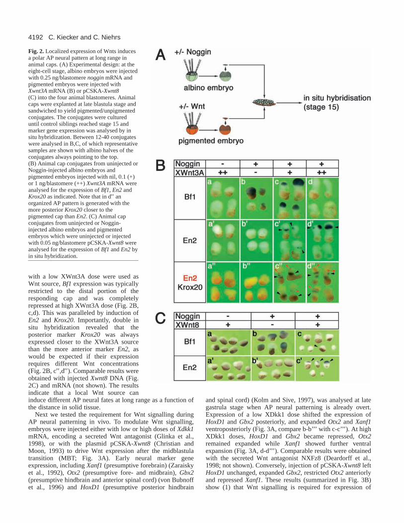
We tested if Wnts also elicit dose-dependent responses insolid tissue and whether the responses occur as a function ofthe distance from a local Wnt source. We performed animalcap sandwich experiments similar to those used by Gurdon andcolleagues (Gurdon et al., 1994). Animal caps were taken fromalbino embryos injected with noggin mRNA (Smith andHarland, 1992) and were used as anterior neuralizedresponding tissue. These caps were conjugated with animalcaps from pigmented embryos injected with Xwnt3AmRNA(Christian et al., 1991) or Xwnt8 DNA as sources ofposteriorizing Wnt protein (Fig. 2A). The conjugates wereallowed to develop until control embryos reached neural platestage and expression of neural markers was analysed by in situhybridization. The use of albino and pigmented animal capsallowed easy identification of the Wnt-responding tissue. Cellmixing of pigmented with albino cells was negligible underthese conditions, as shown previously (Gurdon et al., 1994),and this was also confirmed by lineage tracing with colloidalgold labelled caps (not shown).
Without Noggin, no neural induction occurred, even whenXwnt3A was expressed (Fig. 2B, a-a′′). Noggin injectionresulted in induction of Bf1 throughout the responding albinocap but no expression of the posterior markers En2or Krox20was detectable (Fig. 2B, b-b′′ ). When pigmented caps injected
Fig. 1. Recombinant XWnt8 protein posteriorizesneuralized animal cap cells in a dose-dependent manner.(A) Experimental design. RT-PCR analyses were carriedout when control embryos reached neural tube stage (stage20). (B) RT-PCR analysis of whole embryos (we), intactcontrol animal caps (co) and caps that were dissociatedand reaggregated (diss.) as depicted in A and treatedduring dissociation with 0, 2 nM or 20 nM recombinantXWnt8. (C) RT-PCR analysis of whole embryos (we),intact animal caps (co) and caps that have been dissociatedand reaggregated as depicted in A, and treated duringdissociation with 25 nM XWnt8 (+) in the absence (–) orpresence (+) of recombinant MFz8-CRD. H4, histone4fornormalization; –RT, negative control without reversetranscriptase.
4192
with a low XWnt3A dose were used asWnt source, Bf1expression was typicallyrestricted to the distal portion of theresponding cap and was completelyrepressed at high XWnt3A dose (Fig. 2B,c,d). This was paralleled by induction ofEn2 and Krox20. Importantly, double insitu hybridization revealed that theposterior marker Krox20was alwaysexpressed closer to the XWnt3A sourcethan the more anterior marker En2, aswould be expected if their expressionrequires different Wnt concentrations(Fig. 2B, c′′,d′′). Comparable results wereobtained with injected Xwnt8DNA (Fig.2C) and mRNA (not shown). The resultsindicate that a local Wnt source caninduce different AP neural fates at long range as a function ofthe distance in solid tissue.
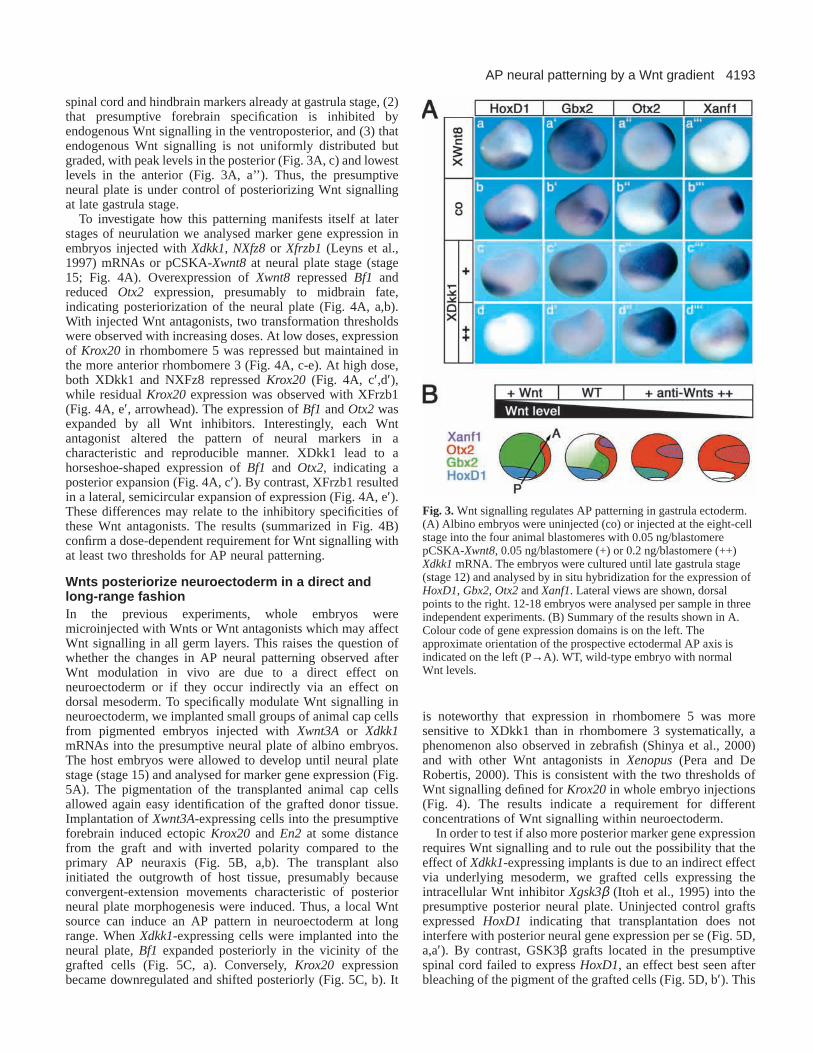
Next we tested the requirement for Wnt signalling duringAP neural patterning in vivo. To modulate Wnt signalling,embryos were injected either with low or high doses of Xdkk1mRNA, encoding a secreted Wnt antagonist (Glinka et al.,1998), or with the plasmid pCSKA-Xwnt8 (Christian andMoon, 1993) to drive Wnt expression after the midblastulatransition (MBT; Fig. 3A). Early neural marker geneexpression, including Xanf1(presumptive forebrain) (Zaraiskyet al., 1992), Otx2 (presumptive fore- and midbrain), Gbx2(presumptive hindbrain and anterior spinal cord) (von Bubnoffet al., 1996) and HoxD1 (presumptive posterior hindbrain
and spinal cord) (Kolm and Sive, 1997), was analysed at lategastrula stage when AP neural patterning is already overt.Expression of a low XDkk1 dose shifted the expression ofHoxD1 and Gbx2 posteriorly, and expanded Otx2 and Xanf1ventroposteriorly (Fig. 3A, compare b-b’’’ with c-c’’’). At highXDkk1 doses, HoxD1and Gbx2became repressed, Otx2remained expanded while Xanf1showed further ventralexpansion (Fig. 3A, d-d’’’). Comparable results were obtainedwith the secreted Wnt antagonist NXFz8 (Deardorff et al.,1998; not shown). Conversely, injection of pCSKA-Xwnt8leftHoxD1unchanged, expanded Gbx2, restricted Otx2anteriorlyand repressed Xanf1. These results (summarized in Fig. 3B)show (1) that Wnt signalling is required for expression of
C. Kiecker and C. Niehrs
Fig. 2.Localized expression of Wnts inducesa polar AP neural pattern at long range inanimal caps. (A) Experimental design: at theeight-cell stage, albino embryos were injectedwith 0.25 ng/blastomere nogginmRNA andpigmented embryos were injected withXwnt3AmRNA (B) or pCSKA-Xwnt8(C) into the four animal blastomeres. Animalcaps were explanted at late blastula stage andsandwiched to yield pigmented/unpigmentedconjugates. The conjugates were cultureduntil control siblings reached stage 15 andmarker gene expression was analysed by insitu hybridization. Between 12-40 conjugateswere analysed in B,C, of which representativesamples are shown with albino halves of theconjugates always pointing to the top.(B) Animal cap conjugates from uninjected orNoggin-injected albino embryos andpigmented embryos injected with nil, 0.1 (+)or 1 ng/blastomere (++) Xwnt3AmRNA wereanalysed for the expression of Bf1, En2andKrox20as indicated. Note that in d′′ anorganized AP pattern is generated with themore posterior Krox20closer to thepigmented cap than En2. (C) Animal capconjugates from uninjected or Noggin-injected albino embryos and pigmentedembryos which were uninjected or injectedwith 0.05 ng/blastomere pCSKA-Xwnt8wereanalysed for the expression of Bf1and En2byin situ hybridization.
4193AP neural patterning by a Wnt gradient
spinal cord and hindbrain markers already at gastrula stage, (2)that presumptive forebrain specification is inhibited byendogenous Wnt signalling in the ventroposterior, and (3) thatendogenous Wnt signalling is not uniformly distributed butgraded, with peak levels in the posterior (Fig. 3A, c) and lowestlevels in the anterior (Fig. 3A, a’’). Thus, the presumptiveneural plate is under control of posteriorizing Wnt signallingat late gastrula stage.
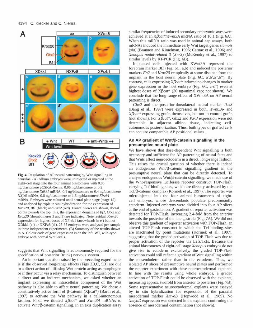
To investigate how this patterning manifests itself at laterstages of neurulation we analysed marker gene expression inembryos injected with Xdkk1, NXfz8or Xfrzb1(Leyns et al.,1997) mRNAs or pCSKA-Xwnt8 at neural plate stage (stage15; Fig. 4A). Overexpression of Xwnt8 repressed Bf1andreduced Otx2 expression, presumably to midbrain fate,indicating posteriorization of the neural plate (Fig. 4A, a,b).With injected Wnt antagonists, two transformation thresholdswere observed with increasing doses. At low doses, expressionof Krox20 in rhombomere 5 was repressed but maintained inthe more anterior rhombomere 3 (Fig. 4A, c-e). At high dose,both XDkk1 and NXFz8 repressed Krox20 (Fig. 4A, c′,d′),while residual Krox20expression was observed with XFrzb1(Fig. 4A, e′, arrowhead). The expression of Bf1 and Otx2wasexpanded by all Wnt inhibitors. Interestingly, each Wntantagonist altered the pattern of neural markers in acharacteristic and reproducible manner. XDkk1 lead to ahorseshoe-shaped expression of Bf1 and Otx2, indicating aposterior expansion (Fig. 4A, c′). By contrast, XFrzb1 resultedin a lateral, semicircular expansion of expression (Fig. 4A, e′).These differences may relate to the inhibitory specificities ofthese Wnt antagonists. The results (summarized in Fig. 4B)confirm a dose-dependent requirement for Wnt signalling withat least two thresholds for AP neural patterning.
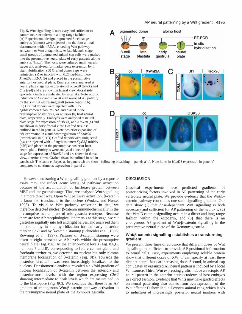
Wnts posteriorize neuroectoderm in a direct andlong-range fashionIn the previous experiments, whole embryos weremicroinjected with Wnts or Wnt antagonists which may affectWnt signalling in all germ layers. This raises the question ofwhether the changes in AP neural patterning observed afterWnt modulation in vivo are due to a direct effect onneuroectoderm or if they occur indirectly via an effect ondorsal mesoderm. To specifically modulate Wnt signalling inneuroectoderm, we implanted small groups of animal cap cellsfrom pigmented embryos injected with Xwnt3A or Xdkk1mRNAs into the presumptive neural plate of albino embryos.The host embryos were allowed to develop until neural platestage (stage 15) and analysed for marker gene expression (Fig.5A). The pigmentation of the transplanted animal cap cellsallowed again easy identification of the grafted donor tissue.Implantation of Xwnt3A-expressing cells into the presumptiveforebrain induced ectopic Krox20and En2at some distancefrom the graft and with inverted polarity compared to theprimary AP neuraxis (Fig. 5B, a,b). The transplant alsoinitiated the outgrowth of host tissue, presumably becauseconvergent-extension movements characteristic of posteriorneural plate morphogenesis were induced. Thus, a local Wntsource can induce an AP pattern in neuroectoderm at longrange. When Xdkk1-expressing cells were implanted into theneural plate, Bf1expanded posteriorly in the vicinity of thegrafted cells (Fig. 5C, a). Conversely, Krox20 expressionbecame downregulated and shifted posteriorly (Fig. 5C, b). It
is noteworthy that expression in rhombomere 5 was moresensitive to XDkk1 than in rhombomere 3 systematically, aphenomenon also observed in zebrafish (Shinya et al., 2000)and with other Wnt antagonists in Xenopus(Pera and DeRobertis, 2000). This is consistent with the two thresholds ofWnt signalling defined for Krox20 in whole embryo injections(Fig. 4). The results indicate a requirement for differentconcentrations of Wnt signalling within neuroectoderm.
In order to test if also more posterior marker gene expressionrequires Wnt signalling and to rule out the possibility that theeffect of Xdkk1-expressing implants is due to an indirect effectvia underlying mesoderm, we grafted cells expressing theintracellular Wnt inhibitor Xgsk3β (Itoh et al., 1995) into thepresumptive posterior neural plate. Uninjected control graftsexpressed HoxD1 indicating that transplantation does notinterfere with posterior neural gene expression per se (Fig. 5D,a,a′). By contrast, GSK3βgrafts located in the presumptivespinal cord failed to express HoxD1, an effect best seen afterbleaching of the pigment of the grafted cells (Fig. 5D, b′). This
Fig. 3.Wnt signalling regulates AP patterning in gastrula ectoderm.(A) Albino embryos were uninjected (co) or injected at the eight-cellstage into the four animal blastomeres with 0.05 ng/blastomerepCSKA-Xwnt8, 0.05 ng/blastomere (+) or 0.2 ng/blastomere (++)Xdkk1mRNA. The embryos were cultured until late gastrula stage(stage 12) and analysed by in situ hybridization for the expression ofHoxD1, Gbx2, Otx2and Xanf1. Lateral views are shown, dorsalpoints to the right. 12-18 embryos were analysed per sample in threeindependent experiments. (B) Summary of the results shown in A.Colour code of gene expression domains is on the left. Theapproximate orientation of the prospective ectodermal AP axis isindicated on the left (P→A). WT, wild-type embryo with normalWnt levels.
4194
suggests that Wnt signalling is autonomously required for thespecification of posterior (trunk) nervous system.
An important question raised by the preceding experimentsis if the observed long-range effects (Figs 2B,C, 5B) are dueto a direct action of diffusing Wnt protein acting as morphogenor if they occur via a relay mechanism. To distinguish betweena direct and an indirect mechanism, we asked whether animplant expressing an intracellular component of the Wntpathway is also able to affect neural patterning. We chose aconstitutively active form of β-catenin (Xβcat*) (Barth et al.,1997) to activate the Wnt pathway in a cell-autonomousfashion. First, we titrated Xβcat* and Xwnt3A mRNAs toactivate Wnt/β-catenin signalling. In an axis duplication assay
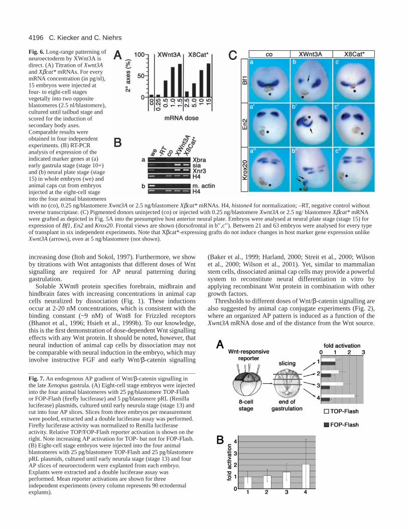
similar frequencies of induced secondary embryonic axes wereachieved at an Xβcat*/Xwnt3AmRNA ratio of 10:1 (Fig. 6A).When this mRNA ratio was used in animal cap assays, bothmRNAs induced the immediate early Wnt target genes siamois(sia) (Brannon and Kimelman, 1996; Carnac et al., 1996) andXenopus nodal-related 3(Xnr3) (McKendry et al., 1997) tosimilar levels by RT-PCR (Fig. 6B).
Implanted cells injected with XWnt3A repressed theforebrain marker Bf1(Fig. 6C, a,b) and induced the posteriormarkers En2and Krox20ectopically at some distance from theimplant in the host neural plate (Fig. 6C, a′,b′,a′′,b′′). Bycontrast, cells expressing Xβcat* induced no changes in markergene expression in the host embryo (Fig. 6C, c-c′′ ) even athighest doses of Xβcat* (20 ng/animal cap; not shown). Weconclude that the long-range effect of XWnt3A on AP neuralpatterning is direct.
Gbx2 and the posterior-dorsolateral neural marker Pax3(Bang et al., 1997) were expressed in both, Xwnt3A- andXβcat*-expressing grafts themselves, but not in control grafts(not shown). For Xβcat*, Gbx2and Pax3expression were notdetectable in adjacent albino tissue, indicating cell-autonomous posteriorization. Thus, both types of grafted cellscan acquire comparable AP positional values.
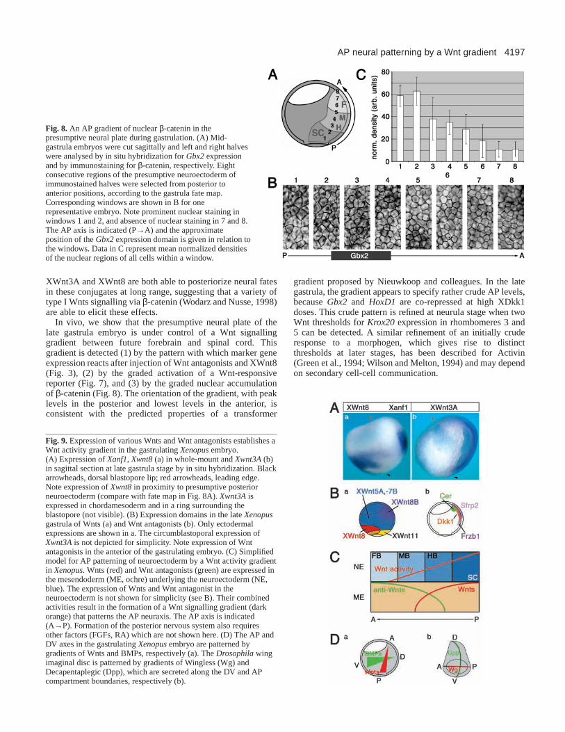
An AP gradient of Wnt/ β-catenin signalling in thepresumptive neural plateWe have shown that dose-dependent Wnt signalling is bothnecessary and sufficient for AP patterning of neural fates andthat Wnts affect neuroectoderm in a direct, long-range fashion.This raises the crucial question of whether there is indeedan endogenous Wnt/β-catenin signalling gradient in thepresumptive neural plate that can be directly detected. Toanalyse endogenous Wnt/β-catenin signalling, we made use ofthe Wnt-responsive luciferase reporter construct TOP-Flash,carrying Tcf-binding sites, which are directly activated by theTcf/β-catenin complex (Korinek et al., 1997). The reporter wasmicroinjected into the four animal blastomeres of eight-cell embryos, whose descendants populate predominantlyectoderm. Injected embryos were divided into four AP slicesat the end of gastrulation. A gradient of reporter activation wasdetected for TOP-Flash, increasing 2.4-fold from the anteriortowards the posterior of the late gastrula (Fig. 7A). We did notobserve this gradient of reporter activation with FOP-Flash, analtered TOP-Flash construct in which the Tcf-binding sitesare inactivated by point mutations (Korinek et al., 1997),suggesting that the graded activation of TOP-Flash was due toproper activation of the reporter via Lefs/Tcfs. Because theanimal blastomeres of eight-cell stage Xenopusembryos do notgive rise to ectoderm exclusively, the graded TOP-Flashactivation could still reflect a gradient of Wnt signalling withinthe mesendoderm rather than in the ectoderm. Thus, weisolated AP slices of presumptive neural plates and performedthe reporter experiment with these neuroectodermal explants.In line with the results using whole embryos, a gradedactivation of TOP-Flash could be observed with the explants,increasing approx. twofold from anterior to posterior (Fig. 7B).Some representative neuroectodermal explants were assayedby in situ hybridization for expression of the paraxialmesodermal marker XmyoD(Hopwood et al., 1989). NoXmyoDexpression was detected in the explants confirming theabsence of mesodermal contamination (not shown).
C. Kiecker and C. Niehrs
Fig. 4. Regulation of AP neural patterning by Wnt signalling inneurulae. (A) Albino embryos were uninjected or injected at theeight-cell stage into the four animal blastomeres with 0.05ng/blastomere pCSKA-Xwnt8, 0.05 ng/blastomere or 0.2ng/blastomere Xdkk1mRNA, 0.1 ng/blastomere or 0.4 ng/blastomereNXfz8mRNA, 0.8 ng/blastomere or 1.6 ng/blastomere Xfrzb1mRNA. Embryos were cultured until neural plate stage (stage 15)and analysed by triple in situ hybridization for the expression ofKrox20, Bf1(black) and Otx2(red). Frontal views are shown, dorsalpoints towards the top. In a, the expression domains of Bf1, Otx2andKrox20(rhombomeres 3 and 5) are indicated. Note residual Krox20expression for highest doses of XFrzb1 (arrowheads in e′) but notXDkk1 (c′) or NXFz8 (d′). 25-35 embryos were analysed per samplein three independent experiments. (B) Summary of the results shownin A. Colour code of gene expression is on the left. WT, wild-typeembryo with normal Wnt levels.
4195AP neural patterning by a Wnt gradient
However, measuring a Wnt signalling gradient by a reporterassay may not reflect acute levels of pathway activationbecause of the accumulation of luciferase protein betweenMBT and late gastrula stage. Thus, we analysed Wnt signallingin a more direct way. Upon Wnt pathway activation, β-cateninis known to translocate to the nucleus (Wodarz and Nusse,1998). To visualize Wnt pathway activation in situ, wetherefore detected nuclear β-catenin immunochemically in thepresumptive neural plate of mid-gastrula embryos. Becausethere are few AP morphological landmarks at this stage, we cutgastrulae sagittally into left and right halves, and analysed themin parallel by in situ hybridization for the early posteriormarker Gbx2and by β-catenin staining (Schneider et al., 1996;Rowning et al., 1997). Pictures of β-catenin staining weretaken at eight consecutive AP levels within the presumptiveneural plate (Fig. 8A). At the anterior-most levels (Fig. 8A,B,numbers 7 and 8), corresponding to future cement gland andforebrain territories, we detected no nuclear but only plasmamembrane localization of β-catenin (Fig. 8B). Towards theposterior, β-catenin was seen increasingly localized to thenucleus. Densitometric analysis revealed a sixfold gradient ofnuclear localization of β-catenin between the anterior- andposterior-most levels, with the region expressing Gbx2showing intermediate to peak levels which are maintained upto the blastopore (Fig. 8C). We conclude that there is an APgradient of endogenous Wnt/β-catenin pathway activation inthe presumptive neural plate of the Xenopusgastrula.
DISCUSSION
Classical experiments have predicted gradients ofposteriorizing factors involved in AP patterning of the earlyvertebrate neural plate. We provide evidence that the Wnt/β-catenin pathway constitutes one such signalling gradient. Ourdata show (1) that dose-dependent Wnt signalling is bothnecessary and sufficient for AP patterning of the neuraxis, (2)that Wnt/β-catenin signalling occurs in a direct and long-rangefashion within the ectoderm, and (3) that there is anendogenous AP gradient of Wnt/β-catenin signalling in thepresumptive neural plate of the Xenopusgastrula.
Wnt/β-catenin signalling establishes a transforminggradientWe present three lines of evidence that different doses of Wntsignalling are sufficient to provide AP positional informationto neural cells. First, experiments employing soluble proteinshow that different doses of XWnt8 can specify at least threedistinct neural fates at increasing dose. Second, in animal capconjugates an organized AP neural pattern is induced by a localWnt source. Third, Wnt expressing grafts induce an ectopic APneural pattern in the anterior neuroectoderm of host embryosin a direct fashion. Evidence that Wnts may have graded effectson neural patterning also comes from overexpression of theWnt effector Dishevelled in Xenopusanimal caps, which leadsto induction of increasingly posterior neural markers with
Fig. 5. Wnt signalling is necessary and sufficient topattern neuroectoderm in a long-range fashion.(A) Experimental design: pigmented 8-cell stageembryos (donors) were injected into the four animalblastomeres with mRNAs encoding Wnt pathwayactivators or Wnt antagonists. At late blastula stage,small groups of pigmented animal cap cells were graftedinto the presumptive neural plate of early gastrula albinoembryos (hosts). The hosts were cultured until neurulastages and analysed for marker gene expression by insitu hybridization. (B) Grafted donor caps wereuninjected (a) or injected with 0.25 ng/blastomereXwnt3AmRNA (b) and placed in the presumptiveanterior host neural plate. Embryos were analysed atneural plate stage for expression of Krox20(black) andEn2(red) and are shown in lateral view, dorsal sideupwards. Grafts are indicated by asterisks. Note ectopicinduction of En2and Krox20with reversed AP polarityby the Xwnt3Aexpressing graft (arrowheads in b).(C) Grafted donors were injected with 0.25ng/blastomereXdkk1mRNA and placed in thepresumptive posterior (a) or anterior (b) host neuralplate, respectively. Embryos were analysed at neuralplate stage for expression of Bf1(a) and Krox20(b) andare shown in dorsofrontal view. Grafted tissue isoutlined in red in panel a. Note posterior expansion ofBf1expression in a and downregulation of Krox20(arrowheads in b). (D) Grafted donors were uninjected(a,a′) or injected with 1.5 ng/blastomereXgsk3β mRNA(b,b′) and placed in the presumptive posterior hostneural plate. Embryos were analysed at neural platestage for expression of HoxD1and are shown in dorsalview, anterior down. Grafted tissue is outlined in red inpanels a,b. The same embryos as in panels a,b are shown following bleaching in panels a′,b′. Note holes in HoxD1expression in panel b′compared to continuous expression in panel a′.
4196
increasing dose (Itoh and Sokol, 1997). Furthermore, we showby titrations with Wnt antagonists that different doses of Wntsignalling are required for AP neural patterning duringgastrulation.
Soluble XWnt8 protein specifies forebrain, midbrain andhindbrain fates with increasing concentrations in animal capcells neuralized by dissociation (Fig. 1). These inductionsoccur at 2-20 nM concentrations, which is consistent with thebinding constant (~9 nM) of Wnt8 for Frizzled receptors(Bhanot et al., 1996; Hsieh et al., 1999b). To our knowledge,this is the first demonstration of dose-dependent Wnt signallingeffects with any Wnt protein. It should be noted, however, thatneural induction of animal cap cells by dissociation may notbe comparable with neural induction in the embryo, which mayinvolve instructive FGF and early Wnt/β-catenin signalling
(Baker et al., 1999; Harland, 2000; Streit et al., 2000; Wilsonet al., 2000; Wilson et al., 2001). Yet, similar to mammalianstem cells, dissociated animal cap cells may provide a powerfulsystem to reconstitute neural differentiation in vitro byapplying recombinant Wnt protein in combination with othergrowth factors.
Thresholds to different doses of Wnt/β-catenin signalling arealso suggested by animal cap conjugate experiments (Fig. 2),where an organized AP pattern is induced as a function of theXwnt3AmRNA dose and of the distance from the Wnt source.
C. Kiecker and C. Niehrs
Fig. 6.Long-range patterning ofneuroectoderm by XWnt3A isdirect. (A) Titration of Xwnt3Aand Xβcat* mRNAs. For everymRNA concentration (in pg/nl),15 embryos were injected atfour- to eight-cell stagesvegetally into two oppositeblastomeres (2.5 nl/blastomere),cultured until tailbud stage andscored for the induction ofsecondary body axes.Comparable results wereobtained in four independentexperiments. (B) RT-PCRanalysis of expression of theindicated marker genes at (a)early gastrula stage (stage 10+)and (b) neural plate stage (stage15) in whole embryos (we) andanimal caps cut from embryosinjected at the eight-cell stageinto the four animal blastomereswith no (co), 0.25 ng/blastomere Xwnt3Aor 2.5 ng/blastomere Xβcat* mRNAs. H4, histone4 for normalization; –RT, negative control withoutreverse transcriptase. (C) Pigmented donors uninjected (co) or injected with 0.25 ng/blastomere Xwnt3Aor 2.5 ng/ blastomere Xβcat* mRNAwere grafted as depicted in Fig. 5A into the presumptive host anterior neural plate. Embryos were analysed at neural plate stage (stage 15) forexpression of Bf1, En2and Krox20. Frontal views are shown (dorsofrontal in b′′,c′′). Between 21 and 63 embryos were analysed for every typeof transplant in six independent experiments. Note that Xβcat*-expressing grafts do not induce changes in host marker gene expression unlikeXwnt3A(arrows), even at 5 ng/blastomere (not shown).
Fig. 7. An endogenous AP gradient of Wnt/β-catenin signalling inthe late Xenopusgastrula. (A) Eight-cell stage embryos were injectedinto the four animal blastomeres with 25 pg/blastomere TOP-Flashor FOP-Flash (firefly luciferase) and 5 pg/blastomere pRL (Renillaluciferase) plasmids, cultured until early neurula stage (stage 13) andcut into four AP slices. Slices from three embryos per measurementwere pooled, extracted and a double luciferase assay was performed.Firefly luciferase activity was normalized to Renilla luciferaseactivity. Relative TOP/FOP-Flash reporter activation is shown on theright. Note increasing AP activation for TOP- but not for FOP-Flash.(B) Eight-cell stage embryos were injected into the four animalblastomeres with 25 pg/blastomere TOP-Flash and 25 pg/blastomerepRL plasmids, cultured until early neurula stage (stage 13) and fourAP slices of neuroectoderm were explanted from each embryo.Explants were extracted and a double luciferase assay wasperformed. Mean reporter activations are shown for threeindependent experiments (every column represents 90 ectodermalexplants).
4197AP neural patterning by a Wnt gradient
XWnt3A and XWnt8 are both able to posteriorize neural fatesin these conjugates at long range, suggesting that a variety oftype I Wnts signalling via β-catenin (Wodarz and Nusse, 1998)are able to elicit these effects.
In vivo, we show that the presumptive neural plate of thelate gastrula embryo is under control of a Wnt signallinggradient between future forebrain and spinal cord. Thisgradient is detected (1) by the pattern with which marker geneexpression reacts after injection of Wnt antagonists and XWnt8(Fig. 3), (2) by the graded activation of a Wnt-responsivereporter (Fig. 7), and (3) by the graded nuclear accumulationof β-catenin (Fig. 8). The orientation of the gradient, with peaklevels in the posterior and lowest levels in the anterior, isconsistent with the predicted properties of a transformer
gradient proposed by Nieuwkoop and colleagues. In the lategastrula, the gradient appears to specify rather crude AP levels,because Gbx2 and HoxD1 are co-repressed at high XDkk1doses. This crude pattern is refined at neurula stage when twoWnt thresholds for Krox20expression in rhombomeres 3 and5 can be detected. A similar refinement of an initially cruderesponse to a morphogen, which gives rise to distinctthresholds at later stages, has been described for Activin(Green et al., 1994; Wilson and Melton, 1994) and may dependon secondary cell-cell communication.
Fig. 8.An AP gradient of nuclear β-catenin in thepresumptive neural plate during gastrulation. (A) Mid-gastrula embryos were cut sagittally and left and right halveswere analysed by in situ hybridization for Gbx2expressionand by immunostaining for β-catenin, respectively. Eightconsecutive regions of the presumptive neuroectoderm ofimmunostained halves were selected from posterior toanterior positions, according to the gastrula fate map.Corresponding windows are shown in B for onerepresentative embryo. Note prominent nuclear staining inwindows 1 and 2, and absence of nuclear staining in 7 and 8.The AP axis is indicated (P→A) and the approximateposition of the Gbx2expression domain is given in relation tothe windows. Data in C represent mean normalized densitiesof the nuclear regions of all cells within a window.
Fig. 9. Expression of various Wnts and Wnt antagonists establishes aWnt activity gradient in the gastrulating Xenopusembryo.(A) Expression of Xanf1, Xwnt8(a) in whole-mount and Xwnt3A(b)in sagittal section at late gastrula stage by in situ hybridization. Blackarrowheads, dorsal blastopore lip; red arrowheads, leading edge.Note expression of Xwnt8in proximity to presumptive posteriorneuroectoderm (compare with fate map in Fig. 8A). Xwnt3Aisexpressed in chordamesoderm and in a ring surrounding theblastopore (not visible). (B) Expression domains in the late Xenopusgastrula of Wnts (a) and Wnt antagonists (b). Only ectodermalexpressions are shown in a. The circumblastoporal expression ofXwnt3Ais not depicted for simplicity. Note expression of Wntantagonists in the anterior of the gastrulating embryo. (C) Simplifiedmodel for AP patterning of neuroectoderm by a Wnt activity gradientin Xenopus. Wnts (red) and Wnt antagonists (green) are expressed inthe mesendoderm (ME, ochre) underlying the neuroectoderm (NE,blue). The expression of Wnts and Wnt antagonist in theneuroectoderm is not shown for simplicity (see B). Their combinedactivities result in the formation of a Wnt signalling gradient (darkorange) that patterns the AP neuraxis. The AP axis is indicated(A→P). Formation of the posterior nervous system also requiresother factors (FGFs, RA) which are not shown here. (D) The AP andDV axes in the gastrulating Xenopusembryo are patterned bygradients of Wnts and BMPs, respectively (a). The Drosophilawingimaginal disc is patterned by gradients of Wingless (Wg) andDecapentaplegic (Dpp), which are secreted along the DV and APcompartment boundaries, respectively (b).
4198
Competence factors restrict AP patterning by WntsignallingAlthough the titrations with Wnt antagonists (Figs 4, 5) reveala dose-dependent requirement for Wnt signalling, the gradualexpansion of more anterior markers such as Bf1 and Otx2is incontrast to the thresholds observed in dissociated animal capcells responding to different doses of XWnt8 protein (Fig. 1).For example, in the in vitro experiments, populations of cellsexpressing only Bf1 and Otx2or only Krox20are observed atdifferent Wnt doses. Extrapolating from the in vitro results onemight expect that in vivo the entire neural plate expressesKrox20 when Wnts, or Bf1when Wnt antagonists areoverexpressed. However, such a complete respecification of theneural plate is never observed in vivo, rather the changes inmarker gene expression respect specific boundaries. This ismost obvious for Bf1in Fig. 4, whose anterior stripe ofexpression only becomes broader but not significantly thickerwith increasing doses of Wnt inhibitors. Similarly, afterimplantation of Xdkk1-expressing cells as in Fig. 5C (panel a),ectopic Bf1 expression is never seen to extend into the midlinebut straddles the dorsal neural plate posteriorly. This suggeststhat factors in addition to Wnts restrict the competence of cellsto express anterior markers. Obvious candidates for suchmodifiers are RA, Sonic Hedgehog, Nodals, BMPs and FGFs,which have been implicated in neural patterning (Lumsden andKrumlauf, 1996; Pownall et al., 1996; Sasai and De Robertis,1997; Chang and Hemmati-Brivanlou, 1998a; Isaacs et al.,1998; Pownall et al., 1998; Gamse and Sive, 2000; Ribisi etal., 2000; Wilson and Rubenstein, 2000). To test if theinhibition of these factors abolishes this restriction ofcompetence we implanted cells co-expressing Xdkk1and eithermouseHip1, cerberus-short, noggin or FGF receptor-1andFGF receptor-4aectodomains into the neural plate, whichencode inhibitors of Hedgehog, Nodal, BMP and FGFsignalling, respectively (Zimmerman et al., 1996; Ye et al.,1998; Chuang and McMahon, 1999; Piccolo et al., 1999; Streitet al., 2000). None of these growth factor antagonists inconjunction with XDkk1 was able to expand Bf1 expressioninto the entire neural plate (not shown). Similarly, inhibition ofRA signalling by injecting the host embryos with XCYP26mRNA, encoding a RA degrading enzyme (Hollemann et al.,1998a), did not allow Xdkk1-expressing grafts to expand Bf1expression further, despite a posterior shift of hindbrainmarkers induced by XCYP26(not shown). The restriction ofcompetence for anterior markers to become expressedposteriorly may occur via factors other than those tested.Alternatively, this/these factor(s) may be active before earlygastrula, the stage of grafting. An ectodermal prepatternregulating the expression of Otx2 has also been identified inthe zebrafish central nervous system. This initial AP prepatterndepends on a differential competence of the epiblast and is notimposed by organizer-derived signals (Koshida et al., 1998).We observed the same phenomenon in our experiments, whereOtx2 expression never expanded into the ventroposteriorectoderm even at high XDkk1 dose (Fig. 3A, d’’).
Clearly then, a morphogen gradient of Wnt/β-cateninsignalling is not sufficient to account for all AP patterning ofthe neural plate and indeed other signalling molecules, such asRA (Ruiz i Altaba, 1993; Kolm and Sive, 1997) and FGFs(Slack and Isaacs, 1994; Cox and Hemmati-Brivanlou, 1995;Doniach, 1995; Kengaku and Okamoto, 1995; Lamb and
Harland, 1995; Holowacz and Sokol, 1999; Ribisi et al., 2000)have been implicated in posteriorization. FGFs are potentposteriorizing agents and FGF8 mediates effects of the isthmicorganizer (Pownall et al., 1996; Isaacs et al., 1998; Pownall etal., 1998; Rhinn and Brand, 2001). However, the posteriorizingeffects of FGFs can be reversed with Wnt antagonists,suggesting that they may be mediated through Wnts at leastpartially (McGrew et al., 1997; Kazanskaya et al., 2000). Adirect requirement for RA signalling during early neuralpatterning has been demonstrated in the hindbrain but not inanterior head structures (Blumberg et al., 1997; Kolm et al.,1997; Hollemann et al., 1998a; Gavalas and Krumlauf, 2000;Niederreither et al., 2000; Dupé and Lumsden, 2001). Thus,hindbrain and spinal cord patterning are regulated by FGF, RAand Wnt signalling.
An endogenous AP gradient of Wnt/ β-cateninsignalling in the presumptive neural plateA major finding of our study is the discovery of a Wnt/β-catenin signalling gradient across the presumptive gastrulaneural plate. How is this gradient established? Several Wntsare expressed in the Xenopusgastrula, including Xwnt3A,Xwnt5A, Xwnt7B, Xwnt8, Xwnt8Band Xwnt11(Fig. 9B, a).Xwnt3A and Xwnt8 are expressed in chordamesoderm andlateroventral mesoderm, respectively (Fig. 9A) (Christian andMoon, 1993; McGrew et al., 1997; Bang et al., 1999), bothof which harbour posteriorizing activity in Xenopus, zebrafishand chick (Hemmati-Brivanlou et al., 1990; Doniach andMusci, 1995; Bang et al., 1997; Muhr et al., 1997; Woo andFraser, 1997). As we show that Wnts are able to signal at longrange, XWnt3A and XWnt8 may diffuse from these sourcesto set up a concentration gradient. Xwnt5Aand Xwnt11,although expressed in the gastrula, are unlikely to beposteriorizing because they do not appear to signal via β-catenin (Ku and Melton, 1993; Moon et al., 1993; Sokol,2000). Xwnt7B and Xwnt8B are both expressed maternallyand may contribute to posteriorization (Cui et al., 1995;Chang and Hemmati-Brivanlou, 1998b). Thus, the signallinggradient probably involves multiple Wnts. A Wnt signalling-free zone is maintained by Wnt antagonists secreted withinthe anterior neuroectoderm and from adjacent anteriormesendoderm, including Cerberus, Dkk1, Frzb1,Crescent/Frzb2 and Sfrp2 (Bouwmeester et al., 1996; Leynset al., 1997; Glinka et al., 1998; Piccolo et al., 1999; Bradleyet al., 2000; Pera and De Robertis, 2000; Fig. 9B, b). As thesefactors are potentially diffusible, they may also contribute tothe establishment of a Wnt activity gradient. In conclusion,we suggest that the XenopusAP neuraxis is patterned by aWnt activity gradient formed in the presumptiveneuroectoderm of the Xenopusgastrula by the interplaybetween posteriorizing Wnts and Wnt antagonists expressedin the anterior (Fig. 9C).
That multiple Wnts are involved in setting up the gradient isalso suggested by the response of marker genes to differentWnt antagonists. Unlike XDkk1 and NXFz8, XFrzb1 failed tocompletely repress Krox20expression. In addition, theexpansion of Bf1expression elicited by XDkk1 and XFrzb1was reproducibly of different shapes, horseshoe versussemicircular, respectively (Fig. 4A). These differencesprobably reflect different specificities of the Wnt antagonists.For example, XFrzb1 is unable to inhibit signalling by
C. Kiecker and C. Niehrs
4199AP neural patterning by a Wnt gradient
XWnt3A, unlike XDkk1 (Wang et al., 1997; Krupnik et al.,1999; Kazanskaya et al., 2000).
The formation of the Wnt/β-catenin signalling gradientresembles formation of the BMP morphogen gradient involvedin DV patterning. BMP2, BMP4, BMP7and BMP7Rareexpressed in overlapping domains in the gastrula and anactivity gradient is formed by the diffusion of multiple BMPantagonists secreted from Spemann’s organizer (Harland andGerhart, 1997; Barth et al., 1999; Dale and Wardle, 1999).Thus, the BMP and Wnt morphogen gradients regulatepatterning in the Xenopusgastrula, providing DV and APpositional information, respectively (Fig. 9D, a). Interestingly,a system of orthogonal Decapentaplegic and Winglessmorphogen gradients regulate AP and DV patterning of theDrosophila wing imaginal disc, respectively (Fig. 9D, b)(Strigini and Cohen, 1999). This raises the possibility thatorthogonal BMP-Wnt gradients represent an evolutionarilyconserved module that specifies bilateral symmetry.
We are indebted to Drs J. Nathans and H. Steinbeisser for generousgifts of XWnt8-expressing insect cells and β-catenin antibody,respectively, and for advice. Drs H. Clevers, M. Eilers, J. Gurdon, R.Harland, D. Kimelmann, P. Klein, J. Nathans and R. Moon kindlyprovided reagents. We thank C. Rumig and P. Steinmetz for supportwith in situ hybridization and RT-PCR, and C. Walter for skillfulassistance in insect cell culture. We are grateful to Drs G. Davidson,O. Kazanskaya and J. Wittbrodt for critical reading of the manuscript.C. N. was supported by the DFG.
REFERENCES
Baker, J. C., Beddington, R. S. and Harland, R. M.(1999). Wnt signalingin Xenopus embryos inhibits bmp4expression and activates neuraldevelopment. Genes Dev.13, 3149-3159.
Bang, A. G., Papalopulu, N., Kintner, C. and Goulding, M. D.(1997).Expression of Pax-3is initiated in the early neural plate by posteriorizingsignals produced by the organizer and by posterior non-axial mesoderm.Development124, 2075-2085.
Bang, A. G., Papalopulu, N., Goulding, M. D. and Kintner, C.(1999).Expression of Pax-3in the lateral neural plate is dependent on a Wnt-mediated signal from posterior nonaxial mesoderm. Dev. Biol.212, 366-380.
Barth, A. I., Pollack, A. L., Altschuler, Y., Mostov, K. E. and Nelson, W.J. (1997). NH2-terminal deletion of β-catenin results in stable colocalizationof mutant β-catenin with adenomatous polyposis coli protein and alteredMDCK cell adhesion. J. Cell Biol.136, 693-706.
Barth, K. A., Kishimoto, Y., Rohr, K. B., Seydler, C., Schulte-Merker, S.and Wilson, S. W.(1999). Bmp activity establishes a gradient of positionalinformation throughout the entire neural plate. Development126, 4977-4987.
Beddington, R. S. P. and Robertson, E. J.(1999). Axis development andearly asymmetry in mammals. Cell96, 195-209.
Bhanot, P., Brink, M., Samos, C. H., Hsieh, J.-C., Wang, Y., Macke, J. P.,Andrew, D., Nathans, J. and Nusse, R.(1996). A new member of thefrizzled family from Drosophilafunctions as a Wingless receptor. Nature382, 225-230.
Blitz, I. L. and Cho, K. W. Y. (1995). Anterior neuroectoderm is progressivelyinduced during gastrulation: the role of the Xenopushomeobox geneorthodenticle. Development121, 993-1004.
Blumberg, B., Bolado, J. J., Moreno, T. A., Kintner, C., Evans, R. M. andPapalopulu, N. (1997). An essential role for retinoid signaling inanteroposterior neural patterning. Development124, 373-379.
Bourguignon, C., Li, J. and Papalopulu, N.(1998). XBF-1, a winged helixtranscription factor with dual activity, has a role in positioning neurogenesisin Xenopuscompetent ectoderm. Development125, 4889-4900.
Bouwmeester, T., Kim, S.-H., Sasai, Y., Lu, B. and De Robertis, E. M.(1996). Cerberus is a head-inducing secreted factor expressed in the anteriorendoderm of Spemann’s organizer. Nature382, 595-601.
Bradley, L. C., Snape, A., Bhatt, S. and Wilkinson, D. G.(1993). Thestructure and expression of the Xenopus Krox-20gene: conserved anddivergent patterns of expression in rhombomeres and neural crest. Mech.Dev.40, 73-84.
Bradley, L., Sun, B., Collins-Racie, L., LaVallie, E., McCoy, J. and Sive,H. (2000). Different activities of the frizzled-related proteins frzb2 andsizzled2 during Xenopusanteroposterior patterning. Dev. Biol.227, 118-132.
Brannon, M. and Kimelman, D. (1996). Activation of Siamois by the Wntpathway. Dev. Biol.180, 344-347.
Carnac, G., Kodjachbachian, L., Gurdon, J. B. and Lemaire, P.(1996).The homeobox gene siamoisis a target of the wnt dorsalization pathwayand triggers organiser activity in the absence of mesoderm. Development122, 3055-3065.
Chang, C. and Hemmati-Brivanlou, A. (1998a). Cell fate determination inembryonic ectoderm. J. Neurobiol.36, 128-151.
Chang, C. and Hemmati-Brivanlou, A. (1998b). Neural crest induction byXwnt7B in Xenopus. Dev. Biol.194, 129-134.
Christian, J. L. and Moon, R. T. (1993). Interactions between Xwnt-8 andSpemann organizer signaling pathways generate dorsoventral pattern in theembryonic mesoderm of Xenopus. Genes Dev.7, 13-28.
Christian, J. L., Gavin, B. J., McMahon, A. P. and Moon, R. T.(1991).Isolation of cDNAs partially encoding four XenopusWnt-1/int-1-relatedproteins and characterization of their transient expression during embryonicdevelopment. Dev. Biol.143, 230-234.
Chuang, P. T. and McMahon, A. P.(1999). Vertebrate Hedgehog signallingmodulated by induction of a Hedgehog-binding protein. Nature397, 617-621.
Cox, W. G. and Hemmati-Brivanlou, B. A.(1995). Caudalization of neuralfate by tissue recombination and bFGF. Development121, 4349-4358.
Cui, Y., Brown, J. D., Moon, R. T. and Christian, J. L.(1995). Xwnt-8b: amaternally expressed Xenopus Wntgene with a potential role in establishingthe dorsoventral axis. Development121, 2177-2186.
Dale, L. and Wardle, F. C. (1999). A gradient of BMP activity specifiesdorsal-ventral fates in early Xenopusembryos. Semin. Cell Dev. Biol.10,319-326.
Darken, R. S. and Wilson, P. A.(2001). Axis induction by Wnt signaling:Target promoter responsiveness regulates competence. Dev. Biol.234, 42-54.
De Robertis, E. M., Larrain, J., Oelgeschläger, M. and Wessely, O.(2000).The establishment of Spemann’s organizer and patterning of the vertebrateembryo. Nat. Rev. Genet.1, 171-181.
Deardorff, M. A., Tan, C., Conrad, L. J. and Klein, P. S.(1998). Frizzled-8 is expressed in the Spemann organizer and plays a role in earlymorphogenesis. Development125, 2687-2700.
Doniach, T.(1995). Basic FGF as an inducer of anteroposterior neural pattern.Cell 83, 1067-1070.
Doniach, T. and Musci, T. J. (1995). Induction of anteroposterior neuralpattern in Xenopus: evidence for a quantitative mechanism. Mech. Dev.53,403-413.
Dupé, V. and Lumsden, A.(2001). Hindbrain patterning involves gradedresponses to retinoic acid signalling. Development128, 2199-2208.
Durston, A. J., Timmermans, J. P., Hage, W. J., Hendriks, H. F., de Vries,N. J., Heideveld, M. and Nieuwkoop, P. D.(1989). Retinoic acid causesan anteroposterior transformation in the developing central nervous system.Nature340, 140-144.
Fekany-Lee, K., Gonzalez, E., Miller-Bertoglio, V. and Solnica-Krezel, L.(2000). The homeobox gene bozozokpromotes anterior neuroectodermformation in zebrafish through negative regulation of BMP2/4 and Wntpathways. Development127, 2333-2345.
Fredieu, J. R., Cui, Y., Maier, D., Danilchik, M. V. and Christian, J. L.(1997). Xwnt-8 and lithium can act upon either dorsal mesodermal orneuroectodermal cells to cause a loss of forebrain in Xenopusembryos. Dev.Biol. 186, 100-114.
Galceran, J., Farinas, I., Depew, M. J., Clevers, H. and Grosschedl, R.(1999). Wnt3a–/–like phenotype and limb deficiency in Lef1−/−Tcf1−/− mice.Genes Dev.13, 709-717.
Gamse, J. and Sive, H.(2000). Vertebrate anteroposterior patterning: theXenopusneurectoderm as a paradigm. BioEssays22, 976-986.
Gamse, J. T. and Sive, H.(2001). Early anteroposterior division of thepresumptive neurectoderm in Xenopus. Mech. Dev.104, 21-36.
Gavalas, A. and Krumlauf, R. (2000). Retinoid signalling and hindbrainpatterning. Curr. Opin. Genet. Dev.10, 380-386.
Gawantka, V., Delius, H., Hirschfeld, K., Blumenstock, C. and Niehrs, C.

4200
(1995). Antagonizing the Spemann organizer: role of the homeobox geneXvent-1. EMBO J.14, 6268-6279.
Gilbert, S. F. and Saxén, L.(1993). Spemann’s organizer: models andmolecules. Mech. Dev.41, 73-89.
Glinka, A., Wu, W., Onichtchouk, D., Blumenstock, C. and Niehrs, C.(1997). Head induction by simultaneous repression of Bmp and Wntsignalling in Xenopus. Nature389, 517-519.
Glinka, A., Wu, W., Delius, H., Monaghan, P. A., Blumenstock, C. andNiehrs, C. (1998). Dickkopf-1 is a member of a new family of secretedproteins and functions in head induction. Nature391, 357-362.
Gould, S. E. and Grainger, R. M. (1997). Neural induction and antero-posterior patterning in the amphibian embryo: past, present and future. Cell.Mol. Life Sci.53, 319-338.
Green, J. B. and Smith, J. C.(1990). Graded changes in dose of a Xenopusactivin A homologue elicit stepwise transitions in embryonic cell fate.Nature347, 391-394.
Green, J. B. A., Smith, J. C. and Gerhard, J. C.(1994). Slow emergence ofa multithreshold response to activin requires cell-contact-dependentsharpening but not prepattern. Development120, 2271-2278.
Gurdon, J. B., Harger, P., Mitchell, A. and Lemaire, P.(1994). Activinsignalling and response to a morphogen gradient. Nature371, 487-492.
Hamilton, F. S., Wheeler, G. N. and Hoppler, S. (2001). Difference in XTcf-3 dependency accounts for change in response to β-catenin-mediated Wntsignalling in Xenopusblastula. Development128, 2063-2073.
Harland, R. (2000). Neural induction. Curr. Opin. Genet. Dev.10, 357-362.Harland, R. M. and Gerhart, J. (1997). Formation and function of
Spemann’s organizer. Annu. Rev. Dev. Biol.13, 611-667.Hashimoto, H., Itoh, M., Yamanaka, Y., Yamashita, S., Shimizu, T.,
Solnica-Krezel, L., Hibi, M. and Hirano, T. (2000). Zebrafish Dkk1Functions in Forebrain Specification and Axial Mesendoderm Formation.Dev. Biol.217, 138-152.
Heisenberg, C. P., Houart, C., Take-Uchi, M., Rauch, G. J., Young, N.,Coutinho, P., Masai, I., Caneparo, L., Concha, M. L., Geisler, R., Dale,T. C., Wilson, S. W. and Stemple, D. L.(2001). A mutation in the Gsk3-binding domain of zebrafish Masterblind/Axin1leads to a fate transformationof telencephalon and eyes to diencephalon. Genes Dev.15, 1427-1434.
Hemmati-Brivanlou, A. and Harland, R. M. (1989). Expression of anengrailed-related protein is induced in the anterior neural ectoderm of earlyXenopusembryos. Development106, 611-617.
Hemmati-Brivanlou, A., Stewart, R. M. and Harland, R. M. (1990).Region-specific neural induction of an engrailed protein by anteriornotochord in Xenopus. Science250, 800-802.
Hemmati-Brivanlou, A., de la Torre, J. R., Holt, C. and Harland, R. M.(1991). Cephalic expression and molecular characterization of Xenopus En-2. Development111, 715-724.
Hemmati-Brivanlou, A., Kelly, O. G. and Melton, D. A. (1994). Follistatin,an antagonist of activin, is expressed in the Spemann organizer and displaysdirect neuralizing activity. Cell77, 283-295.
Hollemann, T., Chen, Y., Grunz, H. and Pieler, T.(1998a). Regionalizedmetabolic activity establishes boundaries of retinoic acid signalling. EMBOJ. 17, 7361-7372.
Hollemann, T., Panitz, F. and Pieler, T. (1998b). In situ hybridizationtechniques with Xenopusembryos. In Comparative Methods Approach tothe Study of Oocytes and Embryos(ed. J. D. Richter), pp. 279-290. Oxford:Oxford University Press.
Holowacz, T. and Sokol, S.(1999). FGF is required for posterior neuralpatterning but not for neural induction. Dev. Biol.205, 296-308.
Hoppler, S., Brown, J. D. and Moon, R. T.(1996). Expression of a dominant-negative wnt blocks induction of MyoD in Xenopusembryos. Genes Dev.10, 2805-2817.
Hopwood, N. D., Pluck, A. and Gurdon, J. B.(1989). MyoDexpression inthe forming somites is an early response to mesoderm induction in Xenopusembryos. EMBO J.8, 3409-3417.
Hsieh, J. C., Kodjabachian, L., Rebbert, M. L., Rattner, A., Smallwood,P. M., Samos, C. H., Nusse, R., Dawid, I. B. and Nathans, J.(1999a). Anew secreted protein that binds to Wnt proteins and inhibits their activities.Nature398, 431-436.
Hsieh, J. C., Rattner, A., Smallwood, P. M. and Nathans, J.(1999b).Biochemical characterization of Wnt-frizzled interactions using a soluble,biologically active vertebrate Wnt protein. Proc. Natl. Acad. Sci. USA96,3546-3551.
Isaacs, H. V., Pownall, M. E. and Slack, J. M.(1998). Regulation of Hoxgene expression and posterior development by the Xenopus caudalhomologue Xcad3. EMBO J.17, 3413-3427.
Itoh, K. and Sokol, S. Y. (1997). Graded amounts of Xenopusdishevelledspecify discrete anteroposterior cell fates in prospective ectoderm. Mech.Dev.61, 113-125.
Itoh, K., Tang, T. L., Neel, B. G. and Sokol, S. Y.(1995). Specific modulationof ectodermal cell fates in Xenopusembryos by glycogen synthase kinase.Development121, 3979-3988.
Kazanskaya, O., Glinka, A. and Niehrs, C.(2000). The role of Xenopusdickkopf1 in prechordal plate specification and neural patterning.Development127, 4981-4992.
Kengaku, M. and Okamoto, H.(1995). bFGF as a possible morphogen forthe anteroposterior axis of the central nervous system in Xenopus.Development121, 3121-3130.
Kiecker, C. and Niehrs, C.(2001). The role of prechordal mesendoderm inneural patterning. Curr. Opin. Neurobiol.11, 27-33.
Kim, C.-H., Oda, T., Itoh, M., Jiang, D. I., Artinger, K.,Chandrasekharappa, S. C., Driever, W. and Chitnis, A. B.(2000).Repressor activity of Headless/Tcf3 is essential for vertebrate headformation. Nature407, 916-920.
Kintner, C. R. and Melton, D. A. (1987). Expression of Xenopus N-CAMRNA in ectoderm is an early response to neural induction. Development99,311-325.
Kolm, P. J. and Sive, H. L.(1997). Retinoids and posterior neural induction:a reevaluation of Nieuwkoop’s two-step hypothesis. Cold Spring Harb.Symp. Quant. Biol.62, 511-521.
Kolm, P. J., Apekin, V. and Sive, H.(1997). Xenopushindbrain patterningrequires retinoid signaling. Dev. Biol.192, 1-16.
Korinek, V., Barker, N., Morin, P. J., van Wichen, D., de Weger, R.,Kinzler, K. W. and Vogelstein, B. (1997). Constitutive transcriptionalactivation by a β-catenin-Tcf complexes in APC/colon carcinoma. Science275, 1784-1787.
Koshida, S., Shinya, M., Mizuno, T., Kuroiwa, A. and Takeda, H.(1998).Initial anteroposterior pattern of the zebrafish central nervous system isdetermined by differential competence of the epiblast. Development125,1957-1966.
Krupnik, V. E., Sharp, J. D., Jiang, C., Robison, K., Chickering, T. W.,Amaravadi, L., Brown, D. E., Guyot, D., Mays, G., Leiby, K. et al.(1999). Functional and structural diversity of the human Dickkopf genefamily. Gene238, 301-313.
Ku, M. and Melton, D. A. (1993). Xwnt-11: a maternally expressed Xenopuswnt gene. Development119, 1161-1173.
Lamb, T. M. and Harland, R. M. (1995). Fibroblast growth factor is a directneural inducer, which combined with noggin generates anterior-posteriorneural pattern. Development121, 3627-3636.
Leyns, L., Bouwmeester, T., Kim, S.-H., Piccolo, S. and De Robertis, E. M.(1997). Frzb-1 is a secreted antagonist of wnt-signals expressed in theSpemann organizer. Cell88, 747-756.
Lumsden, A. and Krumlauf, R. (1996). Patterning the vertebrate neuraxis.Science274, 1109-1115.
McGrew, L. L., Lai, C. J. and Moon, R. T. (1995). Specification of theanteroposterior neural axis through synergistic interaction of the Wntsignaling cascade with noggin and follistatin. Dev. Biol.172, 337-342.
McGrew, L. L., Hoppler, S. and Moon, R. T.(1997). Wnt and FGF pathwayscooperatively pattern anteroposterior neural ectoderm in Xenopus. Mech.Dev.69, 105-114.
McGrew, L. L., Takemaru, K., Bates, R. and Moon, R. T.(1999). Directregulation of the Xenopus engrailed-2promoter by the Wnt signalingpathway, and a molecular screen for Wnt-responsive genes, confirm a rolefor Wnt signaling during neural patterning in Xenopus. Mech. Dev.87, 21-32.
McKendry, R., Hsu, S. C., Harland, R. M. and Grosschedl, R.(1997). LEF-1/TCF proteins mediate wnt-inducible transcription from the Xenopusnodal-related 3promoter. Dev. Biol.192, 420-431.
Miller, J. R. and Moon, R. T. (1997). Analysis of the signaling activities oflocalization mutants of β-catenin during axis specification in Xenopus. J.Cell Biol. 139, 229-243.
Moon, R. T., Campbell, R. M., Christian, J. L., McGrew, L. L., Shih, J.and Fraser, S.(1993). Xwnt-5A: a maternal Wnt that affects morphogeneticmovements after overexpression in embryos of Xenopus laevis.Development119, 97-111.
Moon, R. T., Brown, J. D. and Torres, M.(1997). Wnts modulate cell fateand behavior during vertebrate development. Trends Genet.13, 157-162.
Muhr, J., Jessell, T. M. and Edlund, T.(1997). Assignment of early caudalidentity to neural plate cells by a signal from caudal paraxial mesoderm.Neuron19, 487-502.
C. Kiecker and C. Niehrs
4201AP neural patterning by a Wnt gradient
Niederreither, K., Vermot, J., Schuhbaur, B., Chambon, P. and Dollé, P.(2000). Retinoic acid synthesis and hindbrain patterning in the mouseembryo. Development127, 75-85.
Niehrs, C. (1999). Head in the Wnt – the molecular nature of Spemann’s headorganizer. Trends Genet.15, 314-319.
Nieuwkoop, P. D.(1997). Short historical survey of pattern formation in theendo-mesoderm and the neural anlage in the vertebrates: the role of verticaland planar inductive actions. Cell. Mol. Life Sci.53, 305-318.
Pannese, M., Polo, C., Andreazzoli, M., Vignali, R., Kablar, B., Barsacchi,G. and Boncinelli, E.(1995). The Xenopushomologue of Otx2is a maternalhomeobox gene that demarcates and specifies anterior body regions.Development121, 707-720.
Patapoutian, A. and Reichardt, L. F.(2000). Roles of Wnt proteins in neuraldevelopment and maintenance. Curr. Opin. Neurobiol.10, 392-399.
Pera, E. M. and De Robertis, E. M.(2000). A direct screen for secretedproteins in Xenopusembryos identifies distinct activities for the Wntantagonists Crescent and Frzb-1. Mech. Dev.96, 183-195.
Piccolo, S., Agius, E., Leyns, L., Bhattacharyya, S., Grunz, H.,Bouwmeester, T. and De Robertis, E. M.(1999). The head inducerCerberus is a multifunctional antagonist of Nodal, BMP and Wnt signals.Nature397, 707-710.
Pinson, K. I., Brennan, J., Monkley, S., Avery, B. J. and Skarnes, W. C.(2000). An LDL-receptor-related protein mediates Wnt signalling in mice.Nature407, 535-538.
Pöpperl, H., Schmidt, C., Wilson, V., Hume, C. R., Dodd, J., Krumlauf, R.and Beddington, R. S. P.(1997). Misexpression of Cwnt8cin the mouseinduces an ectopic embryonic axis and causes a truncation of the anteriorneuroectoderm. Development124, 2997-3005.
Pownall, M. E., Tucker, A. S., Slack, J. M. and Isaacs, H. V. (1996). eFGF,Xcad3 and Hox genes form a molecular pathway that establishes theanteroposterior axis in Xenopus. Development122, 3881-3892.
Pownall, M. E., Isaacs, H. V. and Slack, J. M.(1998). Two phases of Hoxgene regulation during early Xenopusdevelopment. Curr. Biol.8, 673-676.
Rhinn, M. and Brand, M. (2001). The midbrain-hindbrain boundaryorganizer. Curr. Opin. Neurobiol.11, 34-42.
Ribisi, S., Mariani, F. V., Aamar, E., Lamb, T. M., Frank, D. and Harland,R. M. (2000). Ras-mediated FGF signaling is required for the formation ofposterior but not anterior neural tissue in Xenopus laevis. Dev. Biol.227,183-196.
Rowning, B. A., Wells, J., Wu, M., Gerhart, J. C., Moon, R. T. andLarabell, C. A. (1997). Microtubule-mediated transport of organelles andlocalization of β-catenin to the future dorsal side of Xenopuseggs. Proc.Natl. Acad. Sci. USA94, 1224-1229.
Ruiz i Altaba, A. (1993). Induction and axial patterning of the neural plate:planar and vertical signals. J. Neurobiol.24, 1276-1304.
Saha, M. S. and Grainger, R. M.(1992). A labile period in the determinationof the anterior-posterior axis during early neural development in Xenopus.Neuron8, 1003-1014.
Saint-Jeannet, J.-P., He, X., Varmus, H. and Dawid, I. B.(1997). Regulationof dorsal fate in the neuraxis by Wnt-1 and Wnt-3a. Proc. Natl. Acad. Sci.USA94, 13713-13718.
Sambrook, J., Fritsch, E. F. and Maniatis, T.(1989). Molecular Cloning. ALaboratory Manual. Cold Spring Harbor: Cold Spring Harbor LaboratoryPress.
Sasai, Y. and De Robertis, E. M.(1997). Ectodermal patterning in vertebrateembryos. Dev. Biol.182, 5-20.
Schneider, S., Steinbeisser, H., Warga, R. M. and Hausen, P.(1996). β-catenin translocation into nuclei demracates the dorsalizing centers in frogand fish embryos. Mech. Dev.57, 191-198.
Shibamoto, S., Higano, K., Takada, R., Ito, F., Takeichi, M. and Takada,S. (1998). Cytoskeletal reorganization by soluble Wnt-3a protein signalling.Genes Cells3, 659-670.
Shinya, M., Eschbach, C., Clark, M., Lehrach, H. and Furutani-Seiki, M.(2000). Zebrafish Dkk1, induced by the pre-MBT Wnt signaling, is secretedfrom the prechordal plate and patterns the anterior neural plate. Mech. Dev.98, 3-17.
Slack, J. M. (1994). Inducing factors in Xenopusearly embryos. Curr. Biol.4, 116-126.
Slack, J. M. and Isaacs, H. V.(1994). The role of fibroblast growth factorsin early Xenopusdevelopment. Biochem. Soc. Trans.22, 585-589.
Smith, W. C. and Harland, R. M. (1992). Expression cloning of noggin, anew dorsalizing factor localized to the Spemann organizer in Xenopusembryos. Cell 70, 829-840.
Smith, W. C., McKendry, R., Ribisi, S. J. and Harland, R. M.(1995). Anodal-relatedgene defines a physical and functional domain within theSpemann organizer. Cell82, 37-46.
Sokol, S.(2000). A role for Wnts in morpho-genesis and tissue polarity. Nat.Cell Biol. 2, E124-E125.
Stern, C. D. (2001). Initial patterning of the central nervous system: Howmany organizers? Nat. Rev. Neurosci.2, 92-98.
Streit, A., Berliner, A. J., Papanayotou, C., Sirulnik, A. and Stern, C. D.(2000). Initiation of neural induction by FGF signalling before gastrulation.Nature406, 74-78.
Strigini, M. and Cohen, S. M.(1999). Formation of morphogen gradients inthe Drosophila wing. Semin. Cell Dev. Biol.10, 335-344.
Takada, S., Stark, K. L., Shea, M. J., Vassileva, G., McMahon, J. A. andMcMahon, A. P. (1994). Wnt-3aregulates somite and tailbud formation inthe mouse embryo. Genes Dev.8, 174-189.
von Bubnoff, A., Schmidt, J. E. and Kimelman, D.(1996). The Xenopuslaevis homeobox gene Xgbx-2 is an early marker of anteroposteriorpatterning in the ectoderm. Mech. Dev.54, 149-160.
Wang, S., Krinks, M. and Moos, M. J.(1997). Frzb-1, an antagonist of Wnt-1 and Wnt-8, does not block signaling by Wnts3A, -5A, or -11. Biochem.Biophys. Res. Commun.236, 502-504.
Wilson, P. A. and Melton, D. A.(1994). Mesodermal patterning by an inducergradient depends on secondary cell-cell communication. Curr. Biol. 4, 676-686.
Wilson, S. I., Graziano, E., Harland, R., Jessell, T. M. and Edlund, T.(2000). An early requirement for FGF signalling in the acquisition of neuralcell fate in the chick embryo. Curr. Biol. 10, 421-429.
Wilson, S. W. and Rubenstein, J. L.(2000). Induction and dorsoventralpatterning of the telencephalon. Neuron28, 641-651.
Wilson, S., Rydstrom, A., Trimborn, T., Willert, K., Nusse, R., Jessell, T.M. and Edlund, T. (2001). The status of Wnt signalling regulates neuraland epidermal fates in the chick embryo. Nature411, 325-330.
Wodarz, A. and Nusse, R.(1998). Mechanisms of Wnt signaling indevelopment. Annu. Rev. Cell Dev. Biol.14, 59-88.
Woo, K. and Fraser, S. E.(1997). Specification of the zebrafish nervoussystem by nonaxial signals. Science277, 254-257.
Ye, W., Shimamura, K., Rubenstein, J. L., Hynes, M. A. and Rosenthal,A. (1998). FGF and Shh signals control dopaminergic and serotonergic cellfate in the anterior neural plate. Cell93, 755-766.
Zaraisky, A. G., Lukyanov, S. A., Vasiliev, O. L., Smirnov, Y. V., Belyavsky,A. V. and Kazanskaya, O. V.(1992). A novel homeobox gene expressedin the anterior neural plate of the Xenopusembryo. Dev. Biol.152, 373-382.
Zimmerman, L. B., De Jesús-Escobar, J.-E. and Harland, R. M.(1996).The Spemann organizer signal noggin binds and inactivates bonemorphogenetic protein-4. Cell86, 599-606.


![Stability of the human polymerase δ holoenzyme …thesis (TLS)] so that pol δ may resume synthesis (9–12). How-ever, studies on the human pol δ holoenzyme are lacking, and hence,](https://static.fdocument.org/doc/165x107/5ecf5ac71e33ba350c72b907/stability-of-the-human-polymerase-holoenzyme-thesis-tls-so-that-pol-may.jpg)