1 integrins are required for normal CNS myelination and...
8
2717 RESEARCH ARTICLE INTRODUCTION In the vertebrate nervous system, myelin sheaths insulate axons and limit membrane depolarization to the nodes of Ranvier, where the machinery propagating action potentials is concentrated (Sherman and Brophy, 2005). Integrins, a family of heterodimeric transmembrane receptors consisting of α and β subunits, are thought to be important for myelination. Integrins containing the β1 subunit are expressed in Schwann cells, the myelinating glial cells of the peripheral nervous system, where they mediate interactions with laminin that are crucial for myelination (Berti et al., 2006). Integrins are also expressed in oligodendrocytes (Milner and ffrench-Constant, 1994; Milner et al., 1997), the myelinating glial cells of the CNS, but less is known about their function in these cells. In vitro studies show that the integrin αvβ1 promotes migration of oligodendrocyte progenitors, whereas α6β1 regulates oligodendrocyte survival and myelin membrane formation in response to laminin 2 (Ln2; Lama2 – Mouse Genome Informatics) (Buttery and ffrench-Constant, 1999; Corley et al., 2001; Frost et al., 1999; Milner et al., 1996; Relvas et al., 2001; Tiwari-Woodruff et al., 2001). Consistent with the in vitro studies, Ln2 is expressed in premyelinating axonal tracts in the CNS, and oligodendrocyte death is increased in the brainstem of mice lacking integrin α6 (Chun et al., 2003; Colognato et al., 2002; Farina et al., 1998; Jones et al., 2001). However, since α6- deficient mice die at birth, it has remained unclear whether the defects in oligodendrocyte survival lead to persistent changes in myelination. In fact, genetic studies addressing the function of β1 integrins in oligodendrocytes have led to contradictory results. Whereas oligodendrocyte-specific expression of a dominant- negative (DN) β1 integrin in a transgenic mouse model was associated with CNS hypomyelination (Camara et al., 2009; Lee et al., 2006), conditional ablation of a floxed allele of the β1 gene (Itgb1) in oligodendrocytes using a CNPase-Cre (Cnp-Cre – Mouse Genome Informatics) driver caused no myelination defects (Benninger et al., 2006). The interpretation of these experiments is complex, as DN β1 integrins can also ectopically activate integrin signaling (Lukashev et al., 1994), and CNPase-Cre induces recombination in only a subset of oligodendrocytes by P0 (Benninger et al., 2006). To circumvent the complications associated with the previous studies, we have analyzed mice in which β1 integrins were inactivated in the ventricular neuroepithelium, including in the oligodendrocyte precursors (referred to here as Itgb1CNS-ko mice). We show that in the absence β1 integrins, the thickness of myelin sheets was reduced in axonal tracts in the spinal cord, optic nerves and cerebellum. Defects in myelination were also observed in the spinal cord from mice in which β1 was inactivated solely in NG2 (CSPG4 – Mouse Genome Informatics)-positive oligodendrocyte precursors. Myelination defects were not caused by defects in oligodendrocyte differentiation or survival. Instead, studies with cultured oligodendrocytes show that β1 integrins regulate the outgrowth of myelin sheets. Interestingly, it has recently been shown that overexpression of a constitutively active form of the serine/threonine kinase AKT (AKT1 – Mouse Genome Informatics) in transgenic mice leads to enhanced myelin sheath formation in the CNS without affecting oligodendrocyte proliferation or survival (Flores et al., 2008). We show here that β1 integrins regulate AKT activity in oligodendrocytes, and demonstrate that constitutively active AKT rescues the defect in myelin sheet outgrowth in cultured β1 integrin-deficient oligodendrocytes. Our findings define the role of β1 integrins in CNS myelination and establish a link between β1 integrins and AKT in the control of CNS axonal ensheathment. β1 integrins are required for normal CNS myelination and promote AKT-dependent myelin outgrowth Claudia S. Barros 1, *, Tom Nguyen 2, *, Kathryn S. R. Spencer 1 , Akiko Nishiyama 3 , Holly Colognato 2,† and Ulrich Müller 1,† Oligodendrocytes in the central nervous system (CNS) produce myelin sheaths that insulate axons to ensure fast propagation of action potentials. β1 integrins regulate the myelination of peripheral nerves, but their function during the myelination of axonal tracts in the CNS is unclear. Here we show that genetically modified mice lacking β1 integrins in the CNS present a deficit in myelination but no defects in the development of the oligodendroglial lineage. Instead, in vitro data show that β1 integrins regulate the outgrowth of myelin sheaths. Oligodendrocytes derived from mutant mice are unable to efficiently extend myelin sheets and fail to activate AKT (also known as AKT1), a kinase that is crucial for axonal ensheathment. The inhibition of PTEN, a negative regulator of AKT, or the expression of a constitutively active form of AKT restores myelin outgrowth in cultured β1- deficient oligodendrocytes. Our data suggest that β1 integrins play an instructive role in CNS myelination by promoting myelin wrapping in a process that depends on AKT. KEY WORDS: β1 integrins, Myelination, Oligodendrocytes, Mouse Development 136, 2717-2724 (2009) doi:10.1242/dev.038679 1 The Scripps Research Institute, Department of Cell Biology, Institute of Childhood and Neglected Disease, 10550 N. Torrey Pines Road, La Jolla, CA 92037, USA. 2 Department of Pharmacology, State University of New York, Stony Brook, NY 11794, USA. 3 Department of Physiology and Neurobiology, University of Connecticut, Storrs, CT 06269, USA. *These authors contributed equally to this work † Authors for correspondence (e-mails: [email protected]; [email protected]) Accepted 11 June 2009 DEVELOPMENT DEVELOPMENT
Transcript of 1 integrins are required for normal CNS myelination and...
2717RESEARCH ARTICLE
INTRODUCTIONIn the vertebrate nervous system, myelin sheaths insulate axonsand limit membrane depolarization to the nodes of Ranvier, wherethe machinery propagating action potentials is concentrated(Sherman and Brophy, 2005). Integrins, a family of heterodimerictransmembrane receptors consisting of α and β subunits, arethought to be important for myelination. Integrins containing theβ1 subunit are expressed in Schwann cells, the myelinating glialcells of the peripheral nervous system, where they mediateinteractions with laminin that are crucial for myelination (Berti etal., 2006). Integrins are also expressed in oligodendrocytes(Milner and ffrench-Constant, 1994; Milner et al., 1997), themyelinating glial cells of the CNS, but less is known about theirfunction in these cells. In vitro studies show that the integrin αvβ1promotes migration of oligodendrocyte progenitors, whereasα6β1 regulates oligodendrocyte survival and myelin membraneformation in response to laminin 2 (Ln2; Lama2 – MouseGenome Informatics) (Buttery and ffrench-Constant, 1999;Corley et al., 2001; Frost et al., 1999; Milner et al., 1996; Relvaset al., 2001; Tiwari-Woodruff et al., 2001). Consistent with the invitro studies, Ln2 is expressed in premyelinating axonal tracts inthe CNS, and oligodendrocyte death is increased in the brainstemof mice lacking integrin α6 (Chun et al., 2003; Colognato et al.,2002; Farina et al., 1998; Jones et al., 2001). However, since α6-deficient mice die at birth, it has remained unclear whether thedefects in oligodendrocyte survival lead to persistent changes in
myelination. In fact, genetic studies addressing the function of β1integrins in oligodendrocytes have led to contradictory results.Whereas oligodendrocyte-specific expression of a dominant-negative (DN) β1 integrin in a transgenic mouse model wasassociated with CNS hypomyelination (Camara et al., 2009; Leeet al., 2006), conditional ablation of a floxed allele of the β1 gene(Itgb1) in oligodendrocytes using a CNPase-Cre (Cnp-Cre –Mouse Genome Informatics) driver caused no myelination defects(Benninger et al., 2006). The interpretation of these experimentsis complex, as DN β1 integrins can also ectopically activateintegrin signaling (Lukashev et al., 1994), and CNPase-Creinduces recombination in only a subset of oligodendrocytes by P0(Benninger et al., 2006).
To circumvent the complications associated with theprevious studies, we have analyzed mice in which β1 integrinswere inactivated in the ventricular neuroepithelium, includingin the oligodendrocyte precursors (referred to here asItgb1CNS-ko mice). We show that in the absence β1 integrins,the thickness of myelin sheets was reduced in axonal tracts inthe spinal cord, optic nerves and cerebellum. Defects inmyelination were also observed in the spinal cord from mice inwhich β1 was inactivated solely in NG2 (CSPG4 – MouseGenome Informatics)-positive oligodendrocyte precursors.Myelination defects were not caused by defects inoligodendrocyte differentiation or survival. Instead, studieswith cultured oligodendrocytes show that β1 integrins regulatethe outgrowth of myelin sheets. Interestingly, it has recentlybeen shown that overexpression of a constitutively active formof the serine/threonine kinase AKT (AKT1 – Mouse GenomeInformatics) in transgenic mice leads to enhanced myelinsheath formation in the CNS without affecting oligodendrocyteproliferation or survival (Flores et al., 2008). We show here thatβ1 integrins regulate AKT activity in oligodendrocytes, anddemonstrate that constitutively active AKT rescues the defectin myelin sheet outgrowth in cultured β1 integrin-deficientoligodendrocytes. Our findings define the role of β1 integrinsin CNS myelination and establish a link between β1 integrinsand AKT in the control of CNS axonal ensheathment.
β1 integrins are required for normal CNS myelination andpromote AKT-dependent myelin outgrowthClaudia S. Barros1,*, Tom Nguyen2,*, Kathryn S. R. Spencer1, Akiko Nishiyama3, Holly Colognato2,† andUlrich Müller1,†
Oligodendrocytes in the central nervous system (CNS) produce myelin sheaths that insulate axons to ensure fast propagation ofaction potentials. β1 integrins regulate the myelination of peripheral nerves, but their function during the myelination of axonaltracts in the CNS is unclear. Here we show that genetically modified mice lacking β1 integrins in the CNS present a deficit inmyelination but no defects in the development of the oligodendroglial lineage. Instead, in vitro data show that β1 integrinsregulate the outgrowth of myelin sheaths. Oligodendrocytes derived from mutant mice are unable to efficiently extend myelinsheets and fail to activate AKT (also known as AKT1), a kinase that is crucial for axonal ensheathment. The inhibition of PTEN, anegative regulator of AKT, or the expression of a constitutively active form of AKT restores myelin outgrowth in cultured β1-deficient oligodendrocytes. Our data suggest that β1 integrins play an instructive role in CNS myelination by promoting myelinwrapping in a process that depends on AKT.
KEY WORDS: β1 integrins, Myelination, Oligodendrocytes, Mouse
Development 136, 2717-2724 (2009) doi:10.1242/dev.038679
1The Scripps Research Institute, Department of Cell Biology, Institute of Childhoodand Neglected Disease, 10550 N. Torrey Pines Road, La Jolla, CA 92037, USA.2Department of Pharmacology, State University of New York, Stony Brook,NY 11794, USA. 3Department of Physiology and Neurobiology, University ofConnecticut, Storrs, CT 06269, USA.
*These authors contributed equally to this work†Authors for correspondence (e-mails: [email protected];[email protected])
Accepted 11 June 2009 DEVELO
PMENT
DEVELO
PMENT
2718
MATERIALS AND METHODSMouse lines and western blotsAll mouse lines have been previously described (Graus-Porta et al., 2001;Stephens et al., 1995; Zhu et al., 2008). Western blots were carried out asdescribed (Colognato et al., 2002; Graus-Porta et al., 2001). Densitometryand quantification of relative levels of protein expression were performedon scanned images of western blots using MetaMorph software (UniversalImaging). Antibodies used were β1 integrin (Graus-Porta et al., 2001), MBP(Boehringer), PLP (Serotec), actin (Sigma), AKT phospho-S473 and AKT(Cell Signaling).
Oligodendrocyte culturesOligodendrocyte progenitors were isolated and cultured following publishedprocedures (Colognato et al., 2007; Colognato et al., 2004), with the additionof a panning step to remove microglia (Cahoy et al., 2008). Briefly,oligodendrocyte progenitor cells (OPCs) were isolated from 10-days-in-vitro (DIV) mixed glial cultures using a 20-hour mechanical dissociationfollowed by a 30-minute differential adhesion step with uncoated petridishes. Unattached cells were subjected to two rounds of panning on lectin-coated dishes to remove remaining microglia. Lectin-coated dishes wereprepared by incubating petri dishes with 2.3 μg/ml BS-lectin-1 inDulbecco’s PBS (D-PBS) for 4 hours at room temperature, followed by fourwashes with D-PBS immediately prior to use (Cahoy et al., 2008). PurifiedOPCs were added to poly-D-lysine (PDL)- or laminin-coated Permanoxchamber slides in a modified SATO’s medium (SATO with 0.5% FCS fordifferentiation experiments). Human placental laminin (laminin 211, Sigma)was used to coat the surfaces of slides and dishes at 10 μg/ml in PBS for 4hours at 37°C. Surface coating with PDL (Sigma) was performed similarlybut was instead diluted in dH2O to obtain 10 μg/ml. Mixed glial culturesfrom neonatal spinal cords were grown on PDL-coated chamber slides andswitched to SATO with 0.5% FCS to differentiate at 10 DIV. Recombinantprotein comprising the EGF-like domain of neuregulin 1 was used at 100ng/ml (PeproTech). The PTEN inhibitor bpV(pic) was used at 31 nM, theIC50 for PTEN inhibition (Calbiochem) (Schmid et al., 2004). Wild-type andβ1-CNS-mutant mixed glial cultures were transfected using FuGENE(Roche) as described (Colognato et al., 2004), with either MYR-AKT-EGFPor control EGFP plasmids (a kind gift from Dr Bing-Hua Jiang, WestVirginia University, Morgantown, WV, USA). Transfected oligodendrocyteswere differentiated within the mixed glial cultures and identified 6 days post-transfection using combined GFP and/or MBP immunocytochemistry (seebelow). A minimum of 25 fields per transfection were evaluated bymorphometric analysis.
Electron microscopy and immunochemistryElectron microscopy (EM) samples were prepared as described (Graus-Portaet al., 2001). EM images were acquired using a Philips CM100 microscope(FEI, Hillsbrough, OR, USA). Morphometric analysis of nerve fibers wasperformed using MetaMorph software. Immunohistochemistry followed ourpublished procedures (Belvindrah et al., 2007; Blaess et al., 2004). Primaryantibodies used were PDGFαR (a kind gift from W. Stallcup, BurnhamInstitute, La Jolla, CA, USA), CC1 (Calbiotech), OLIG2 (Chemicon) andcleaved caspase 3 (Cell Signaling). Secondary antibodies were Alexa Fluor488 and 568-conjugated (Molecular Probes). Nuclei were stained withTO-PRO-3 (Molecular Probes). TUNEL assays were carried using theApopTag Red In Situ Apoptosis Detection Kit (Chemicon). Forimmunocytochemistry, cells were fixed in 4% PFA (15 minutes) or 100%methanol (5 minutes, –20°C). Blocking and primary antibody incubationswere in PBS with 10% donkey serum (with 0.1% Triton X-100 for PFA-fixed cells). Cells undergoing GFP/MBP immunocytochemistry wereblocked in PBS with 10% donkey serum and 0.05% Triton X-100. ForGALC staining, live cells were labeled with antibody for 30 minutes inDMEM, 1% FCS, followed by washes and PFA fixation. Primary antibodieswere NG2 (Chemicon), β1 integrin (Chemicon, MAB1997), GALC(Sigma), CNP (Sigma), MBP (Serotec), GFP (Molecular Probes) and OLIG2(IBL). Secondary antibodies were FITC- or Texas Red-conjugated(Jackson). Nuclei were stained with DAPI. Images were collected on anOlympus Fluoview 500 confocal microscope, an Olympus AX70microscope, and a Zeiss Axioplan microscope. MetaMorph software
(Universal Imaging) was used for morphometric measures. The AxioVisionInteractive Measurement Module was used for myelin sheet and processlength cell measures. To ensure consistent analysis across differentexperiments, MBP-positive cells and MBP-positive sheet-bearing cells weredetermined using intensity thresholding relative to background intensity(AxioVision). Morphometric analysis to determine the area of coverage ofmyelin sheets was performed by tracing the outer perimeter of thresholdedMBP-positive sheet-bearing oligodendrocytes (AxioVision, Zeiss). Processlength measurements were performed on corresponding phase micrographsby tracing from the cell body/process border to the tip of the longest process(AxioVision). Examples of morphometric tracing can be observed in Fig.7F,G.
Statistical analysisStatistical analysis was performed using GraphPad Prism. Data show mean± standard error (mean±s.e.m.). Statistical significance for g-ratio analysiswas performed using the Mann-Whitney (rank sum) test. The number ofanimals analyzed (n) was three unless otherwise indicated. Student’s t-testwas used for all other assays described. Statistical significance was set atP<0.05 (*, significant), P<0.01 (**, very significant) and P<0.001 (***,highly significant).
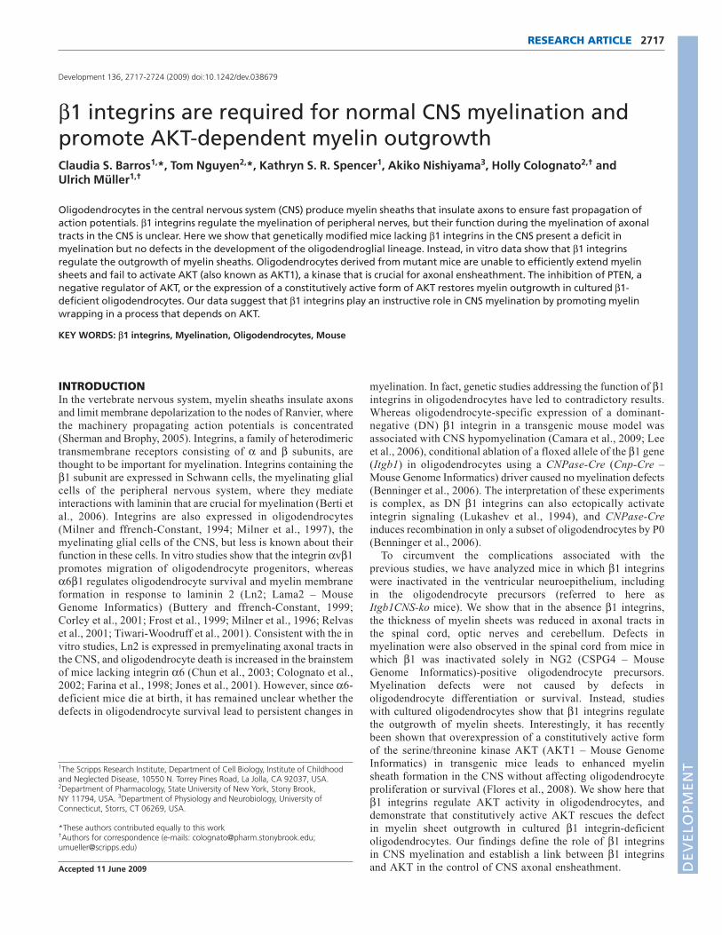
RESULTSCNS myelination defects in Itgb1-CNSko andItgb1-OL-ko miceTo define the functions of β1 integrins in CNS myelination, weinactivated β1 expression by crossing mice carrying a floxedintegrin β1 gene (Itgb1-flox) with nestin-Cre mice. In this nestin-Creline, CRE-mediated recombination occurs in neural precursors in theventricular zone as early as embryonic day (E) 10.5, leading to theinactivation of floxed alleles in developing neurons and glia,including cells at all stages of oligodendrocyte development (Graus-Porta et al., 2001). The mutant mice are viable and fertile and showno obvious behavioral abnormalities (Graus-Porta et al., 2001), andwill be referred to as Itgb1-CNSko mice (Fig. 1A). We confirmedrecombination of the Itgb1-flox allele by PCR and the absence of β1protein by immunoblotting in the spinal cords and brains of mutantmice (Fig. 1B,C; C.S.B., unpublished) (Graus-Porta et al., 2001). Inall assays, Itgb1-CNSko mice were compared with littermates thatdid not express CRE but were homozygous for the Itgb1-flox alleleor carried one Itgb1-flox and one Itgb1-null allele. Control mice arereferred to as wild type (WT) because previous studies revealed thatthe loxP sites or heterozygosity for Itgb1-null do not affect β1function (Graus-Porta et al., 2001; Stephens et al., 1995).Histological analysis showed that spinal cords from Itgb1-CNSkoadult mice were not obviously different in overall morphology fromWT (Fig. 1D).
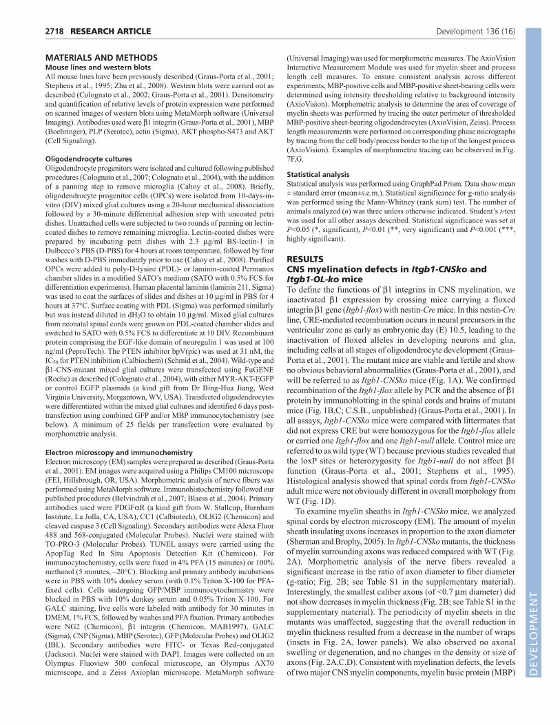
To examine myelin sheaths in Itgb1-CNSko mice, we analyzedspinal cords by electron microscopy (EM). The amount of myelinsheath insulating axons increases in proportion to the axon diameter(Sherman and Brophy, 2005). In Itgb1-CNSko mutants, the thicknessof myelin surrounding axons was reduced compared with WT (Fig.2A). Morphometric analysis of the nerve fibers revealed asignificant increase in the ratio of axon diameter to fiber diameter(g-ratio; Fig. 2B; see Table S1 in the supplementary material).Interestingly, the smallest caliber axons (of <0.7 μm diameter) didnot show decreases in myelin thickness (Fig. 2B; see Table S1 in thesupplementary material). The periodicity of myelin sheets in themutants was unaffected, suggesting that the overall reduction inmyelin thickness resulted from a decrease in the number of wraps(insets in Fig. 2A, lower panels). We also observed no axonalswelling or degeneration, and no changes in the density or size ofaxons (Fig. 2A,C,D). Consistent with myelination defects, the levelsof two major CNS myelin components, myelin basic protein (MBP)
RESEARCH ARTICLE Development 136 (16)
DEVELO
PMENT
DEVELO
PMENT
and proteolipid protein (PLP, PLP1 – Mouse Genome Informatics)(Simons and Trajkovic, 2006), were modestly but significantlyreduced in Itgb1-CNSko mice (Fig. 2E).
To investigate whether other axonal tracts in the CNS weresimilarly affected, we analyzed the optic nerves and the cerebellum.As in the spinal cord, myelin sheaths enwrapping the larger caliberaxons were reduced in thickness (Fig. 3A,B; see Table S1 in thesupplementary material). We also attempted to analyze myelinationin the corpus callosum, but its organization was significantlydisrupted in Itgb1-CNSko mice due to the perturbations in corticalsize and structure (Graus-Porta et al., 2001), preventing ameaningful analysis.
To determine the extent to which β1 integrins in oligodendrogliaare required for myelination, we inactivated their expression usingNg2-Cre mice. In this mouse line, CRE specifically induces therecombination of floxed alleles in NG2-positive oligodendrocyteprecursors (Zhu et al., 2008). The analysis of axonal tracts in thespinal cord of these mutants (named Itgb1-OL-ko mice) revealed areduction in myelin thickness (Fig. 4A,B). As in Itgb1-CNSko mice,myelin thickness was affected in axons with diameters >0.7 μm,although the phenotype was less severe, and some of the largestdiameter axons appeared unaffected (Fig. 4C). The less severe defectin myelination could be a consequence of less effective inactivation
of β1 integrins by Ng2-Cre compared with nestin-Cre. To test thishypothesis, we purified oligodendrocytes from mutant mice andanalyzed integrin expression by immunohistochemistry and westernblotting. Staining for NG2 and OLIG2 confirmed thatoligodendrocytes were purified to near homogeneity (see Fig. S1A-C in the supplementary material). Whereas β1 integrins were almostcompletely absent in oligodendrocytes from Itgb1-CNSko mice (Fig.6A,B,I), a low amount of β1 integrin expression could still bedetected in oligodendrocytes from Itgb1-OL-ko mice (see Fig.S1D,E in the supplementary material), suggesting that the lesssevere myelination defect in Itgb1-OL-ko mice was a consequenceof less efficient CRE-mediated recombination. However, it cannotbe excluded that β1 integrins in axons, which were inactivated bynestin-Cre but not by Ng2-Cre, also contributed to myelination.Taken together, our findings demonstrate that β1 integrins are
2719RESEARCH ARTICLEβ1 integrins in CNS myelination
Fig. 1. CRE-mediated gene inactivation leads to loss of β1integrin protein. (A) Diagram of nestin-Cre-mediated geneinactivation in Itgb1-CNSko mice mutants. The Itgb1 (encodes β1integrin) floxed allele before and after recombination (rec) and theItgb1-null allele are shown. LoxP sites are indicated as black triangles,exons (blue boxes) are numbered. neo indicates neomycin cassette.(B) DNA from the P19 spinal cord of WT and Itgb1-CNSko mice (β1mt)was analyzed by PCR. With DNA from Itgb1-CNSko mice, bandscorresponding to the nestin-Cre transgene, the Itgb1-null allele and therecombined Itgb1-flox allele were detected. (C) Immunoblotting with β1integrin antibody using spinal cord extracts from mice at P7, P19 andP30 showed loss of β1 protein in the mutants. Tubulin served as aloading control. (D) Spinal cord sections from WT and Itgb1-CNSkomice at P30 stained with Toluidine Blue. No obvious differences inoverall morphology were detected. Scale bar: 500μm.
Fig. 2. Myelination defects in the spinal cords of Itgb1-CNSkomutants. (A) Electron microscopy (EM) analysis of myelinated fibers inspinal cord sections from P30 Itgb1-CNSko and WT mice. Asterisksindicate axons of similar size. Insets in lower panels show highermagnifications of myelin wraps from indicated axons. Scale bars: 5μm;100 nm for insets in lower panels. (B) G-ratio (axon diameter/fiberdiameter) of fibers grouped by axon diameter. Values are shown asmean±s.e.m. In Itgb1-CNSko mutants, the g-ratio was significantlyincreased, with the exception of axons with a diameter less than0.7μm. See Table S1 in the supplementary material for statistical values.(C,D) No significant change was detected in either the density (C) orsize (diameter, D) of axons in Itgb1-CNSko mutants [density: β1mt0.101±0.01/μm2 (103.52±1.14% with respect to WT), WT 0.097±0.01/μm2, P>0.05, n=1660 axons from three WT, n=2009 axons fromthree Itgb1-CNSko mutants; size (diameter): β1mt 1.61±0.03μm, WT1.59±0.03μm, P>0.05, n=1660 axons from three WT, n=2009 axonsfrom three Itgb1-CNSko mutants]. Values are shown as mean±s.e.m.(E) Immunobloting for MBP and PLP in spinal cord extracts from P7 andP30 Itgb1-CNSko mutant and WT mice. Tubulin served as a control forloading and subsequent densitometry analysis. In mutant spinal cordsat P7, the relative levels of MBP and PLP proteins were reduced by23.1±2.5% (n=3, ***P<0.001) and 24.9±6.8% (n=3, *P<0.05),respectively. At P30, MBP and PLP levels were decreased in mutantspinal cords by 36.5±12.1% (n=3, *P<0.05) and 13.5±3% (n=2,*P<0.05), respectively. n.s., not significant.
DEVELO
PMENT
DEVELO
PMENT

2720
required for the myelination of axonal tracts in the spinal cord, opticnerve and cerebellum, and that they act, at least in part, inoligodendrocytes to carry out their function.
Oligodendrocyte lineage development in Itgb1-CNSko mutantsThe hypomyelination of axonal tracts in the CNS of Itgb1-CNSkomutants could arise from defects in oligodendrocyte development.We therefore analyzed their development using stage-specificmolecular markers. The number of PDGFαR-positiveoligodendrocyte progenitors at postnatal day (P) 0, prior to axonalensheathment, was not reduced in the mutants (Fig. 5A,B,G; β1mt17.88±1.09/μm2, WT 17.95±1.21/μm2, P>0.05, n=3). Similarly,there was no reduction in the numbers of CC1-positiveoligodendrocytes at P19, when myelination is in progress (Fig.5C,D,H; β1mt 32.71±0.89/μm2, WT 33.5±1.36/μm2, P>0.05, n=3),or at P60, after myelin sheaths have formed (Fig. 5E,F,H; β1mt34.17±1.05/μm2, WT 34.89±0.78/μm2, P>0.05, n=3). Detection ofOLIG2, which labels oligodendroglia throughout their development,also revealed no defects in mutant animals (see Fig. S2 in thesupplementary material). Finally, we analyzed cell death in the
mutants and observed no differences in either the developing oradult spinal cords (see Fig. S3 in the supplementary material).Analysis of the developing cerebellum also revealed no significantincrease in the numbers of dying cells (see Fig. S4 in thesupplementary material). We conclude that the reduction in myelinensheathment in Itb1-CNSko mice is probably not caused by the lossof oligodendrocytes.
To further define the cell-autonomous function of β1 integrins inoligodendrocytes, we purified their progenitors from WT and Itgb1-CNSko mice to near homogeneity (see Fig. S1A-C in thesupplementary material) and followed their differentiation in vitro(Fig. 6). Immunostaining and western blots confirmed the efficientinactivation of β1 integrins in oligodendrocytes from Itgb1-CNSkomice (Fig. 6A,B,I). The percentage of NG2-positiveoligodendrocyte precursors that were present 1 day after plating intodifferentiation medium was not significantly different between WTand β1-deficient cells (Fig. 6C,D,J; β1mt 46.83±3.02%, WT50.90±5.32%, n=4, P>0.05). No difference was observed in thepercentage of GALC-positive oligodendrocytes at days 2 or 4 (Fig.6E,F,K; β1mt 67.05±2.1%, WT 66.89±3.72%, n=4, P>0.05 at day2; β1mt 81.6±2.82%, WT 78.87±4.29%, n=4, P>0.05 at day 4), orMBP-positive mature oligodendrocytes at 4 or 6 days postplating indifferentiation medium (Fig. 6G,H,L; β1mt 33.26±4.37%, WT37.39±4.54%, n=4, P>0.05 at day 4; β1mt 23.28±2.95%, WT32.97±4.22%, n=4, P>0.05 at day 6). These data are in agreementwith our in vivo analysis (Fig. 5; see Fig. S2 in the supplementarymaterial) and confirm that β1 integrins are not essential for theformation of mature oligodendrocytes.
RESEARCH ARTICLE Development 136 (16)
Fig. 3. Myelination defects in the optic nerves and cerebellum ofItgb1-CNSko mutants. (A,B) EM analysis of myelinated fibers in opticnerve (A) and cerebellum (B) cross-sections from P30 Itgb1-CNSko andWT mice. Asterisks indicate axons of similar size. The g-ratios (diameteraxon/diameter fiber) of fibers grouped by axon diameter are also shown(mean±s.e.m.). In Itgb1-CNSko mutant optic nerves and cerebellumthere was an increase in the g-ratio of fibers with larger axonal caliberscompared with WT. See Table S1 in the supplementary material forstatistical data. n.s., not significant. Scale bars: 1μm.
Fig. 4. Myelination defects in the spinal cords of Itgb1-OL-komutants derived using Ng2-Cre. (A) EM analysis of myelinated fibersin spinal cord sections from P30 Itgb1-OL-ko and WT mice generatedusing the Ng2-Cre driver. (B) Higher magnification of fibers boxed in A.(C) G-ratio of fibers grouped by axon diameter (mean±s.e.m.). In Itgb1-OL-ko mutants, the g-ratio was significantly increased in axons with adiameter between 0.7 and 2.5μm. n.s., not significant. See Table S1 inthe supplementary material for statistical values. Scale bars: 5μm in A;2.5μm in B.
DEVELO
PMENT
DEVELO
PMENT
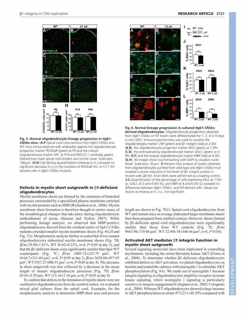
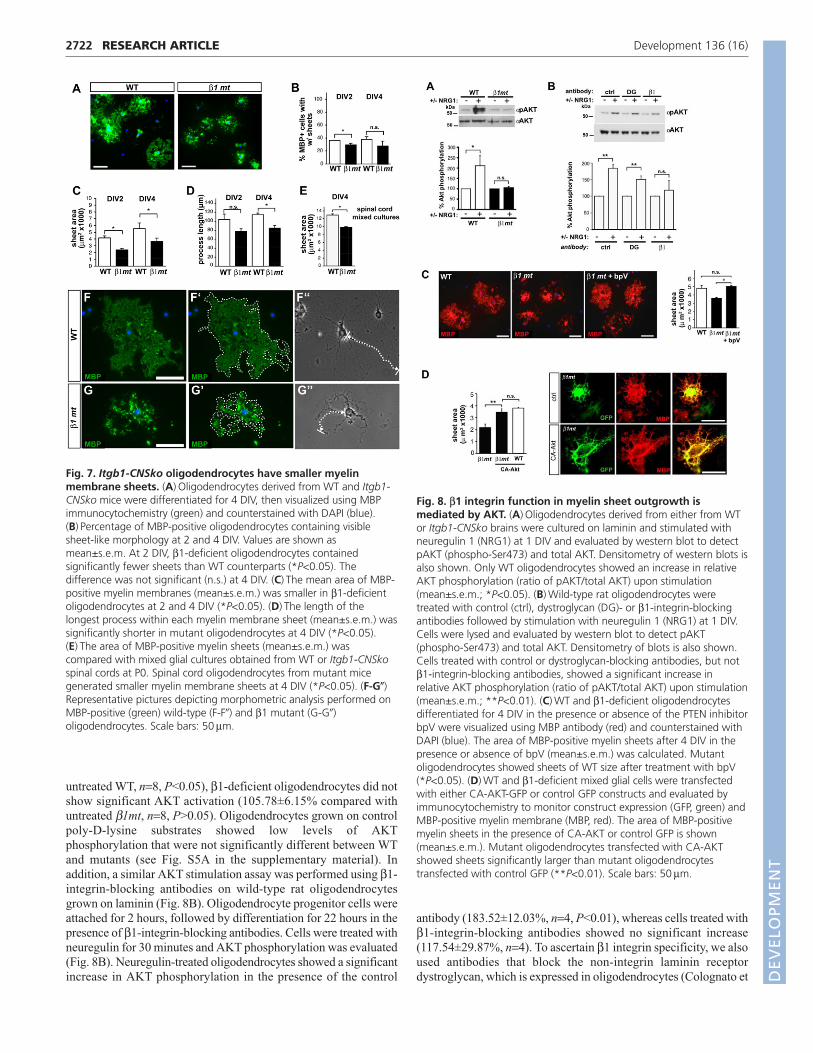
Defects in myelin sheet outgrowth in β1-deficientoligodendrocytesMyelin membrane sheets are formed by the extension of branchedprocesses surrounded by a specialized plasma membrane enrichedwith myelin proteins such as MBP (Richardson et al., 2006). Myelinmembrane sheet formation is therefore thought to mimic many ofthe morphological changes that take place during oligodendrocyteensheathment of axons (Simons and Trotter, 2007). Whileperforming lineage analysis, we observed that MBP-positiveoligodendrocytes derived from the cerebral cortex of Itgb1-CNSkomutants extended smaller myelin membrane sheets (Fig. 6G,H andFig. 7A). Morphometric analysis further revealed that fewer mutantoligodendrocytes elaborated myelin membrane sheets (Fig. 7B;β1mt 29.98±1.81%, WT 36.62±0.23%, n=4, P<0.05 at day 2), andthat the β1-deficient sheets were significantly smaller than their WTcounterparts (Fig. 7C; β1mt 2409.12±225.79 μm2, WT4164.7±311.60 μm2, n=4, P<0.05 at day 2; β1mt 3658.04±457.05μm2, WT 5557.27±806.51 μm2, n=4, P<0.05 at day 4). The decreasein sheet outgrowth was also reflected by a decrease in the meanlength of mutant oligodendrocyte processes (Fig. 7D; β1mt85.01±5.59 μm, WT 115.16±3.16 μm, n=4, P<0.05 at day 4).
To confirm that defects in the formation of myelin sheets were notconfined to oligodendrocytes from the cerebral cortex, we evaluatedmixed glial cultures from the spinal cord. Examples for themorphometric analysis to determine MBP sheet area and process
length are shown in Fig. 7F,G. Spinal cord oligodendrocytes fromWT and mutant mice on average elaborated larger membrane sheetsthan those prepared from cerebral cortices. However, sheets formedby β1-deficient spinal cord oligodendrocytes were significantlysmaller than those from WT controls (Fig. 7E; β1mt9863.98±719.86 μm2, WT 12,944.54±346.64 μm2, n=4, P<0.05).
Activated AKT mediates β1 integrin function inmyelin sheet outgrowthSeveral signaling molecules have been implicated in controllingmyelination, including the serine/threonine kinase AKT (Flores etal., 2008). To determine whether β1-deficient oligodendrocytesexhibited defects in AKT activation, we plated oligodendrocytes onlaminin and treated the cultures with neuregulin 1 to stimulate AKTphosphorylation (Fig. 8A). We made use of neuregulin 1 becauseintegrin signaling in oligodendrocytes amplifies receptor tyrosinekinase signaling, where neuregulin 1 signaling is particularlysensitive to integrin engagement (Colognato et al., 2002; Colognatoet al., 2004). Whereas WT oligodendrocytes showed a large increasein AKT phosphorylation at serine 473 (211±48.29% compared with
2721RESEARCH ARTICLEβ1 integrins in CNS myelination
Fig. 5. Normal oligodendrocyte lineage progression in Itgb1-CNSko mice. (A-F) Spinal cord cross-sections from Itgb1-CNSko andWT mice immunostained with antibodies against the oligodendrocyteprogenitor marker PDGFαR (green) at P0 and the matureoligodendrocyte marker APC at P19 and P60 (CC1 antibody, green).Dashed lines mark spinal cord borders and central canal. Scale bars:100μm. (G,H) Cell density quantifications (mean±s.e.m.) showed nosignificant decrease (n.s.) in the numbers of PDGFαR (G)- or CC1 (H)-positive cells in Itgb1-CNSko mutants.
Fig. 6. Normal lineage progression in cultured Itgb1-CNSko-derived oligodendrocytes. Oligodendrocyte progenitors obtainedfrom Itgb1-CNSko or WT brains were differentiated for 1, 2, 4 or 6 daysin vitro (DIV). Immunocytochemistry was used to visualize theoligodendroglia marker CNP (green) and β1 integrin (red) at 2 DIV(A,B), the oligodendrocyte progenitor marker NG2 (green) at 1 DIV(C,D), the premyelinating oligodendrocyte marker GALC (green) at 4DIV (E,F) and the mature oligodendrocyte marker MBP (red) at 4 DIV(G,H). All images show counterstaining with DAPI to visualize nuclei(blue). Scale bars: 50μm. (I) Western blot analysis of lysates obtainedfrom oligodendrocytes purified from wild-type and Itgb1-CNSko micerevealed a severe reduction in the levels of β1 integrin protein inmutant cells (β1mt). Actin blots were performed as a loading control.(J-L) Quantification of the percentage of cells expressing NG2 at 1 DIV(J); GALC at 2 and 4 DIV (K); and MBP at 4 and 6 DIV (L) revealed nodifferences between Itgb1-CNSko- and WT-derived cells. Values areshown as mean±s.e.m. n.s., not significant.
DEVELO
PMENT
DEVELO
PMENT
2722
untreated WT, n=8, P<0.05), β1-deficient oligodendrocytes did notshow significant AKT activation (105.78±6.15% compared withuntreated β1mt, n=8, P>0.05). Oligodendrocytes grown on controlpoly-D-lysine substrates showed low levels of AKTphosphorylation that were not significantly different between WTand mutants (see Fig. S5A in the supplementary material). Inaddition, a similar AKT stimulation assay was performed using β1-integrin-blocking antibodies on wild-type rat oligodendrocytesgrown on laminin (Fig. 8B). Oligodendrocyte progenitor cells wereattached for 2 hours, followed by differentiation for 22 hours in thepresence of β1-integrin-blocking antibodies. Cells were treated withneuregulin for 30 minutes and AKT phosphorylation was evaluated(Fig. 8B). Neuregulin-treated oligodendrocytes showed a significantincrease in AKT phosphorylation in the presence of the control
antibody (183.52±12.03%, n=4, P<0.01), whereas cells treated withβ1-integrin-blocking antibodies showed no significant increase(117.54±29.87%, n=4). To ascertain β1 integrin specificity, we alsoused antibodies that block the non-integrin laminin receptordystroglycan, which is expressed in oligodendrocytes (Colognato et
RESEARCH ARTICLE Development 136 (16)
Fig. 7. Itgb1-CNSko oligodendrocytes have smaller myelinmembrane sheets. (A) Oligodendrocytes derived from WT and Itgb1-CNSko mice were differentiated for 4 DIV, then visualized using MBPimmunocytochemistry (green) and counterstained with DAPI (blue).(B) Percentage of MBP-positive oligodendrocytes containing visiblesheet-like morphology at 2 and 4 DIV. Values are shown asmean±s.e.m. At 2 DIV, β1-deficient oligodendrocytes containedsignificantly fewer sheets than WT counterparts (*P<0.05). Thedifference was not significant (n.s.) at 4 DIV. (C) The mean area of MBP-positive myelin membranes (mean±s.e.m.) was smaller in β1-deficientoligodendrocytes at 2 and 4 DIV (*P<0.05). (D) The length of thelongest process within each myelin membrane sheet (mean±s.e.m.) wassignificantly shorter in mutant oligodendrocytes at 4 DIV (*P<0.05).(E) The area of MBP-positive myelin sheets (mean±s.e.m.) wascompared with mixed glial cultures obtained from WT or Itgb1-CNSkospinal cords at P0. Spinal cord oligodendrocytes from mutant micegenerated smaller myelin membrane sheets at 4 DIV (*P<0.05). (F-G�)Representative pictures depicting morphometric analysis performed onMBP-positive (green) wild-type (F-F�) and β1 mutant (G-G�)oligodendrocytes. Scale bars: 50μm.
Fig. 8. β1 integrin function in myelin sheet outgrowth ismediated by AKT. (A) Oligodendrocytes derived from either from WTor Itgb1-CNSko brains were cultured on laminin and stimulated withneuregulin 1 (NRG1) at 1 DIV and evaluated by western blot to detectpAKT (phospho-Ser473) and total AKT. Densitometry of western blots isalso shown. Only WT oligodendrocytes showed an increase in relativeAKT phosphorylation (ratio of pAKT/total AKT) upon stimulation(mean±s.e.m.; *P<0.05). (B) Wild-type rat oligodendrocytes weretreated with control (ctrl), dystroglycan (DG)- or β1-integrin-blockingantibodies followed by stimulation with neuregulin 1 (NRG1) at 1 DIV.Cells were lysed and evaluated by western blot to detect pAKT(phospho-Ser473) and total AKT. Densitometry of blots is also shown.Cells treated with control or dystroglycan-blocking antibodies, but notβ1-integrin-blocking antibodies, showed a significant increase inrelative AKT phosphorylation (ratio of pAKT/total AKT) upon stimulation(mean±s.e.m.; **P<0.01). (C) WT and β1-deficient oligodendrocytesdifferentiated for 4 DIV in the presence or absence of the PTEN inhibitorbpV were visualized using MBP antibody (red) and counterstained withDAPI (blue). The area of MBP-positive myelin sheets after 4 DIV in thepresence or absence of bpV (mean±s.e.m.) was calculated. Mutantoligodendrocytes showed sheets of WT size after treatment with bpV(*P<0.05). (D) WT and β1-deficient mixed glial cells were transfectedwith either CA-AKT-GFP or control GFP constructs and evaluated byimmunocytochemistry to monitor construct expression (GFP, green) andMBP-positive myelin membrane (MBP, red). The area of MBP-positivemyelin sheets in the presence of CA-AKT or control GFP is shown(mean±s.e.m.). Mutant oligodendrocytes transfected with CA-AKTshowed sheets significantly larger than mutant oligodendrocytestransfected with control GFP (**P<0.01). Scale bars: 50μm.
DEVELO
PMENT
DEVELO
PMENT
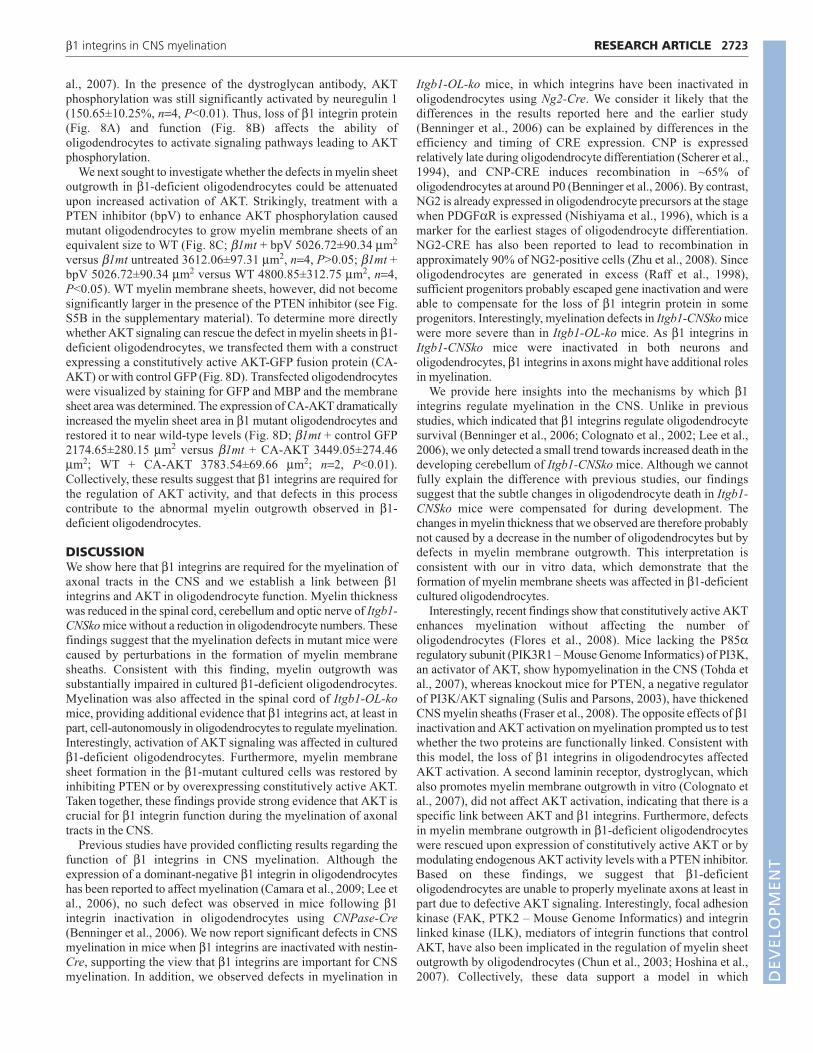
al., 2007). In the presence of the dystroglycan antibody, AKTphosphorylation was still significantly activated by neuregulin 1(150.65±10.25%, n=4, P<0.01). Thus, loss of β1 integrin protein(Fig. 8A) and function (Fig. 8B) affects the ability ofoligodendrocytes to activate signaling pathways leading to AKTphosphorylation.
We next sought to investigate whether the defects in myelin sheetoutgrowth in β1-deficient oligodendrocytes could be attenuatedupon increased activation of AKT. Strikingly, treatment with aPTEN inhibitor (bpV) to enhance AKT phosphorylation causedmutant oligodendrocytes to grow myelin membrane sheets of anequivalent size to WT (Fig. 8C; β1mt + bpV 5026.72±90.34 μm2
versus β1mt untreated 3612.06±97.31 μm2, n=4, P>0.05; β1mt +bpV 5026.72±90.34 μm2 versus WT 4800.85±312.75 μm2, n=4,P<0.05). WT myelin membrane sheets, however, did not becomesignificantly larger in the presence of the PTEN inhibitor (see Fig.S5B in the supplementary material). To determine more directlywhether AKT signaling can rescue the defect in myelin sheets in β1-deficient oligodendrocytes, we transfected them with a constructexpressing a constitutively active AKT-GFP fusion protein (CA-AKT) or with control GFP (Fig. 8D). Transfected oligodendrocyteswere visualized by staining for GFP and MBP and the membranesheet area was determined. The expression of CA-AKT dramaticallyincreased the myelin sheet area in β1 mutant oligodendrocytes andrestored it to near wild-type levels (Fig. 8D; β1mt + control GFP2174.65±280.15 μm2 versus β1mt + CA-AKT 3449.05±274.46μm2; WT + CA-AKT 3783.54±69.66 μm2; n=2, P<0.01).Collectively, these results suggest that β1 integrins are required forthe regulation of AKT activity, and that defects in this processcontribute to the abnormal myelin outgrowth observed in β1-deficient oligodendrocytes.
DISCUSSIONWe show here that β1 integrins are required for the myelination ofaxonal tracts in the CNS and we establish a link between β1integrins and AKT in oligodendrocyte function. Myelin thicknesswas reduced in the spinal cord, cerebellum and optic nerve of Itgb1-CNSko mice without a reduction in oligodendrocyte numbers. Thesefindings suggest that the myelination defects in mutant mice werecaused by perturbations in the formation of myelin membranesheaths. Consistent with this finding, myelin outgrowth wassubstantially impaired in cultured β1-deficient oligodendrocytes.Myelination was also affected in the spinal cord of Itgb1-OL-komice, providing additional evidence that β1 integrins act, at least inpart, cell-autonomously in oligodendrocytes to regulate myelination.Interestingly, activation of AKT signaling was affected in culturedβ1-deficient oligodendrocytes. Furthermore, myelin membranesheet formation in the β1-mutant cultured cells was restored byinhibiting PTEN or by overexpressing constitutively active AKT.Taken together, these findings provide strong evidence that AKT iscrucial for β1 integrin function during the myelination of axonaltracts in the CNS.
Previous studies have provided conflicting results regarding thefunction of β1 integrins in CNS myelination. Although theexpression of a dominant-negative β1 integrin in oligodendrocyteshas been reported to affect myelination (Camara et al., 2009; Lee etal., 2006), no such defect was observed in mice following β1integrin inactivation in oligodendrocytes using CNPase-Cre(Benninger et al., 2006). We now report significant defects in CNSmyelination in mice when β1 integrins are inactivated with nestin-Cre, supporting the view that β1 integrins are important for CNSmyelination. In addition, we observed defects in myelination in
Itgb1-OL-ko mice, in which integrins have been inactivated inoligodendrocytes using Ng2-Cre. We consider it likely that thedifferences in the results reported here and the earlier study(Benninger et al., 2006) can be explained by differences in theefficiency and timing of CRE expression. CNP is expressedrelatively late during oligodendrocyte differentiation (Scherer et al.,1994), and CNP-CRE induces recombination in ~65% ofoligodendrocytes at around P0 (Benninger et al., 2006). By contrast,NG2 is already expressed in oligodendrocyte precursors at the stagewhen PDGFαR is expressed (Nishiyama et al., 1996), which is amarker for the earliest stages of oligodendrocyte differentiation.NG2-CRE has also been reported to lead to recombination inapproximately 90% of NG2-positive cells (Zhu et al., 2008). Sinceoligodendrocytes are generated in excess (Raff et al., 1998),sufficient progenitors probably escaped gene inactivation and wereable to compensate for the loss of β1 integrin protein in someprogenitors. Interestingly, myelination defects in Itgb1-CNSko micewere more severe than in Itgb1-OL-ko mice. As β1 integrins inItgb1-CNSko mice were inactivated in both neurons andoligodendrocytes, β1 integrins in axons might have additional rolesin myelination.
We provide here insights into the mechanisms by which β1integrins regulate myelination in the CNS. Unlike in previousstudies, which indicated that β1 integrins regulate oligodendrocytesurvival (Benninger et al., 2006; Colognato et al., 2002; Lee et al.,2006), we only detected a small trend towards increased death in thedeveloping cerebellum of Itgb1-CNSko mice. Although we cannotfully explain the difference with previous studies, our findingssuggest that the subtle changes in oligodendrocyte death in Itgb1-CNSko mice were compensated for during development. Thechanges in myelin thickness that we observed are therefore probablynot caused by a decrease in the number of oligodendrocytes but bydefects in myelin membrane outgrowth. This interpretation isconsistent with our in vitro data, which demonstrate that theformation of myelin membrane sheets was affected in β1-deficientcultured oligodendrocytes.
Interestingly, recent findings show that constitutively active AKTenhances myelination without affecting the number ofoligodendrocytes (Flores et al., 2008). Mice lacking the P85αregulatory subunit (PIK3R1 – Mouse Genome Informatics) of PI3K,an activator of AKT, show hypomyelination in the CNS (Tohda etal., 2007), whereas knockout mice for PTEN, a negative regulatorof PI3K/AKT signaling (Sulis and Parsons, 2003), have thickenedCNS myelin sheaths (Fraser et al., 2008). The opposite effects of β1inactivation and AKT activation on myelination prompted us to testwhether the two proteins are functionally linked. Consistent withthis model, the loss of β1 integrins in oligodendrocytes affectedAKT activation. A second laminin receptor, dystroglycan, whichalso promotes myelin membrane outgrowth in vitro (Colognato etal., 2007), did not affect AKT activation, indicating that there is aspecific link between AKT and β1 integrins. Furthermore, defectsin myelin membrane outgrowth in β1-deficient oligodendrocyteswere rescued upon expression of constitutively active AKT or bymodulating endogenous AKT activity levels with a PTEN inhibitor.Based on these findings, we suggest that β1-deficientoligodendrocytes are unable to properly myelinate axons at least inpart due to defective AKT signaling. Interestingly, focal adhesionkinase (FAK, PTK2 – Mouse Genome Informatics) and integrinlinked kinase (ILK), mediators of integrin functions that controlAKT, have also been implicated in the regulation of myelin sheetoutgrowth by oligodendrocytes (Chun et al., 2003; Hoshina et al.,2007). Collectively, these data support a model in which
2723RESEARCH ARTICLEβ1 integrins in CNS myelination
DEVELO
PMENT
DEVELO
PMENT
2724
extracellular ligands that activate β1 integrins induce AKT activityto control myelin outgrowth and axonal wrapping in the CNS. In thefuture, it will be important to define the downstream effectors ofAKT and the extent to which they integrate biosynthetic andcytoskeletal pathways to control the outgrowth of myelinmembranes that enwrap axonal processes in the CNS.
AcknowledgementsWe thank T. Bossing for critical reading of the manuscript; M. Wood for EMtechnical support; D. Park for technical support; and Dr Bian-Hua Jiang for thegenerous gift of MYR-AKT-GFP. This work was supported by funding from theNational Institutes of Health (U.M., NS046456, MH078833; H.C., NS054042),a Christopher Reeve Foundation fellowship (C.S.B.), a National MultipleSclerosis Society Career Transition Fellowship (H.C.) and a NSF/IGERT 3MTfellowship (T.N., 0549370). Deposited in PMC for release after 12 months.
Supplementary materialSupplementary material for this article is available athttp://dev.biologists.org/cgi/content/full/136/16/2717/DC1
ReferencesBelvindrah, R., Graus-Porta, D., Goebbels, S., Nave, K. A. and Muller, U.
(2007). Beta1 integrins in radial glia but not in migrating neurons are essentialfor the formation of cell layers in the cerebral cortex. J. Neurosci. 27, 13854-13865.
Benninger, Y., Colognato, H., Thurnherr, T., Franklin, R. J., Leone, D. P.,Atanasoski, S., Nave, K. A., ffrench-Constant, C., Suter, U. and Relvas, J.B. (2006). Beta1-integrin signaling mediates premyelinating oligodendrocytesurvival but is not required for CNS myelination and remyelination. J. Neurosci.26, 7665-7673.
Berti, C., Nodari, A., Wrabetz, L. and Feltri, M. L. (2006). Role of integrins inperipheral nerves and hereditary neuropathies. Neuromolecular Med. 8, 191-204.
Blaess, S., Graus-Porta, D., Belvindrah, R., Radakovits, R., Pons, S.,Littlewood-Evans, A., Senften, M., Guo, H., Li, Y., Miner, J. H. et al. (2004).Beta1-integrins are critical for cerebellar granule cell precursor proliferation. J.Neurosci. 24, 3402-3412.
Buttery, P. C. and ffrench-Constant, C. (1999). Laminin-2/integrin interactionsenhance myelin membrane formation by oligodendrocytes. Mol. Cell. Neurosci.14, 199-212.
Cahoy, J. D., Emery, B., Kaushal, A., Foo, L. C., Zamanian, J. L.,Christopherson, K. S., Xing, Y., Lubischer, J. L., Krieg, P. A., Krupenko, S.A. et al. (2008). A transcriptome database for astrocytes, neurons, andoligodendrocytes: a new resource for understanding brain development andfunction. J. Neurosci. 28, 264-278.
Camara, J., Wang, Z., Nunes-Fonseca, C., Friedman, H. C., Grove, M.,Sherman, D. L., Komiyama, N. H., Grant, S. G., Brophy, P. J., Peterson, A.et al. (2009). Integrin-mediated axoglial interactions initiate myelination in thecentral nervous system. J. Cell Biol. 185, 699-712.
Chun, S. J., Rasband, M. N., Sidman, R. L., Habib, A. A. and Vartanian, T.(2003). Integrin-linked kinase is required for laminin-2-induced oligodendrocytecell spreading and CNS myelination. J. Cell Biol. 163, 397-408.
Colognato, H., Baron, W., Avellana-Adalid, V., Relvas, J. B., Baron-VanEvercooren, A., Georges-Labouesse, E. and ffrench-Constant, C. (2002).CNS integrins switch growth factor signalling to promote target-dependentsurvival. Nat. Cell Biol. 4, 833-841.
Colognato, H., Ramachandrappa, S., Olsen, I. M. and ffrench-Constant, C.(2004). Integrins direct Src family kinases to regulate distinct phases ofoligodendrocyte development. J. Cell Biol. 167, 365-375.
Colognato, H., Galvin, J., Wang, Z., Relucio, J., Nguyen, T., Harrison, D.,Yurchenco, P. D. and ffrench-Constant, C. (2007). Identification ofdystroglycan as a second laminin receptor in oligodendrocytes, with a role inmyelination. Development 134, 1723-1736.
Corley, S. M., Ladiwala, U., Besson, A. and Yong, V. W. (2001). Astrocytesattenuate oligodendrocyte death in vitro through an alpha(6) integrin-laminin-dependent mechanism. Glia 36, 281-294.
Farina, L., Morandi, L., Milanesi, I., Ciceri, E., Mora, M., Moroni, I.,Pantelioni, C. and Savoiardo, M. (1998). Congenital muscular dystrophywith merosin deficiency: MRI findings in five patients. Neuroradiology 40, 807-811.
Flores, A. I., Narayanan, S. P., Morse, E. N., Shick, H. E., Yin, X., Kidd, G.,Avila, R. L., Kirschner, D. A. and Macklin, W. B. (2008). Constitutively activeAkt induces enhanced myelination in the CNS. J. Neurosci. 28, 7174-7183.
Fraser, M. M., Bayazitov, I. T., Zakharenko, S. S. and Baker, S. J. (2008).Phosphatase and tensin homolog, deleted on chromosome 10 deficiency inbrain causes defects in synaptic structure, transmission and plasticity, andmyelination abnormalities. Neuroscience 151, 476-488.
Frost, E. E., Buttery, P. C., Milner, R. and ffrench-Constant, C. (1999). Integrinsmediate a neuronal survival signal for oligodendrocytes. Curr. Biol. 9, 1251-1254.
Graus-Porta, D., Blaess, S., Senften, M., Littlewood-Evans, A., Damsky, C.,Huang, Z., Orban, P., Klein, R., Schittny, J. C. and Muller, U. (2001). Beta1-class integrins regulate the development of laminae and folia in the cerebral andcerebellar cortex. Neuron 31, 367-379.
Hoshina, N., Tezuka, T., Yokoyama, K., Kozuka-Hata, H., Oyama, M. andYamamoto, T. (2007). Focal adhesion kinase regulates laminin-inducedoligodendroglial process outgrowth. Genes Cells 12, 1245-1254.
Jones, K. G., Morgan, G., Johnston, H., Tobias, V., Ouvrier, R. A., Wilkinson,I. and North, K. N. (2001). The expanding phenotype of laminin alpha2 chain(merosin) abnormalities: case series and review. J. Med. Genet. 38, 649-657.
Lee, K. K., de Repentigny, Y., Saulnier, R., Rippstein, P., Macklin, W. B. andKothary, R. (2006). Dominant-negative beta1 integrin mice have region-specificmyelin defects accompanied by alterations in MAPK activity. Glia 53, 836-844.
Lukashev, M. E., Sheppard, D. and Pytela, R. (1994). Disruption of integrinfunction and induction of tyrosine phosphorylation by the autonomouslyexpressed beta 1 integrin cytoplasmic domain. J. Biol. Chem. 269, 18311-18314.
Milner, R. and ffrench-Constant, C. (1994). A developmental analysis ofoligodendroglial integrins in primary cells: changes in alpha v-associated betasubunits during differentiation. Development 120, 3497-3506.
Milner, R., Edwards, G., Streuli, C. and ffrench-Constant, C. (1996). A role inmigration for the alpha V beta 1 integrin expressed on oligodendrocyteprecursors. J. Neurosci. 16, 7240-7252.
Milner, R., Frost, E., Nishimura, S., Delcommenne, M., Streuli, C., Pytela, R.and ffrench-Constant, C. (1997). Expression of alpha vbeta3 and alpha vbeta8integrins during oligodendrocyte precursor differentiation in the presence andabsence of axons. Glia 21, 350-360.
Nishiyama, A., Lin, X. H., Giese, N., Heldin, C. H. and Stallcup, W. B. (1996).Co-localization of NG2 proteoglycan and PDGF alpha-receptor on O2Aprogenitor cells in the developing rat brain. J. Neurosci. Res. 43, 299-314.
Raff, M. C., Durand, B. and Gao, F. B. (1998). Cell number control and timing inanimal development: the oligodendrocyte cell lineage. Int. J. Dev. Biol. 42, 263-267.
Relvas, J. B., Setzu, A., Baron, W., Buttery, P. C., LaFlamme, S. E., Franklin, R.J. and ffrench-Constant, C. (2001). Expression of dominant-negative andchimeric subunits reveals an essential role for beta1 integrin during myelination.Curr. Biol. 11, 1039-1043.
Richardson, W. D., Kessaris, N. and Pringle, N. (2006). Oligodendrocyte wars.Nat. Rev. Neurosci. 7, 11-18.
Scherer, S. S., Braun, P. E., Grinspan, J., Collarini, E., Wang, D. Y. andKamholz, J. (1994). Differential regulation of the 2’,3�-cyclic nucleotide 3�-phosphodiesterase gene during oligodendrocyte development. Neuron 12,1363-1375.
Schmid, A. C., Byrne, R. D., Vilar, R. and Woscholski, R. (2004).Bisperoxovanadium compounds are potent PTEN inhibitors. FEBS Lett. 566, 35-38.
Sherman, D. L. and Brophy, P. J. (2005). Mechanisms of axon ensheathment andmyelin growth. Nat. Rev. Neurosci. 6, 683-690.
Simons, M. and Trajkovic, K. (2006). Neuron-glia communication in the controlof oligodendrocyte function and myelin biogenesis. J. Cell Sci. 119, 4381-4389.
Simons, M. and Trotter, J. (2007). Wrapping it up: the cell biology ofmyelination. Curr. Opin. Neurobiol. 17, 533-540.
Stephens, L. E., Sutherland, A. E., Klimanskaya, I. V., Andrieux, A., Meneses,J., Pedersen, R. A. and Damsky, C. H. (1995). Deletion of beta 1 integrins inmice results in inner cell mass failure and peri-implantation lethality. Genes Dev.9, 1883-1895.
Sulis, M. L. and Parsons, R. (2003). PTEN: from pathology to biology. Trends CellBiol. 13, 478-483.
Tiwari-Woodruff, S. K., Buznikov, A. G., Vu, T. Q., Micevych, P. E., Chen, K.,Kornblum, H. I. and Bronstein, J. M. (2001). OSP/claudin11 forms a complexwith a novel member of the tetraspanin super family and beta1 integrin andregulates proliferation and migration of oligodendrocytes. J. Cell Biol. 153, 295-305.
Tohda, C., Nakanishi, R. and Kadowaki, M. (2007). Learning deficits andagenesis of synapses and myelinated axons in phosphoinositide-3 kinase-deficient mice. Neurosignals 15, 293-306.
Zhu, X., Bergles, D. E. and Nishiyama, A. (2008). NG2 cells generate botholigodendrocytes and gray matter astrocytes. Development 135, 145-157.
RESEARCH ARTICLE Development 136 (16)
DEVELO
PMENT
DEVELO
PMENT