The interaction of tiotropium with long lasting β2 ...edoc.unibas.ch/34104/1/merged_Luigi...
96
1 The interaction of tiotropium with long lasting β2- agonists on lung cell function Inauguraldissertation Zur Erlangung der Würde eines Doktors der Philosophie Vorgelegt der Philosophisch-Naturwissenschaftlichen Fakultät Der Universität Basel Von Luigi Costa Aus Ischia, Italien Basel, 2014
Transcript of The interaction of tiotropium with long lasting β2 ...edoc.unibas.ch/34104/1/merged_Luigi...
1
The interaction of tiotropium with long lasting β2-agonists on lung cell function
Inauguraldissertation
Zur Erlangung der Würde eines Doktors der Philosophie
Vorgelegt der Philosophisch-Naturwissenschaftlichen Fakultät
Der Universität Basel
Von Luigi Costa
Aus Ischia, Italien
Basel, 2014

2
© Luigi Costa, 2013 Genehmigt von der Philosophisch-Naturwissenschaftlichen Fakultät Auf Antrag von Prof. Michael Roth Prof. Markus A. Ruegg Prof. Luigi Terracciano Basel, den 10.12.2013 Prof. Dr. J. Schibler Dekan der Philosophisch-Naturwissenschaftlichen Fakultät
3
Acknowledgments
I would like to thank Prof. Michael Tamm and Prof. Michael Roth for giving me the
opportunity to do my thesis in their laboratory. Things were not always easy over the past four
years but in the end, I had the chance to develop my independence in research and to learn
from my own mistakes. Thanks for giving me chances to present my data at international
scientific conferences, which I enjoyed and where I found the motivation to continue with my
work.
Thanks to my previous and new colleagues for sharing the ups and down of a Ph.D. thesis
with me; and for giving me advice when I was getting frustrated with my experiments.
Luigi
4
Table of content
Acknowledgments 2
List of abbreviations 5
Summary and Implications of the thesis 6
Chapter 1- Asthma 11
1.1. Definition and pathogenesis of asthma 11
1.2. New aspects of asthma pathologies and therapeutic targets 12
1.3. Pathological mechanisms in asthma 15
1.4. The muscarinic receptor on lung fibroblasts: function and role in asthma 17
1.5. β2-adrenergic receptor in lung fibroblasts: function and role in asthma 22
1.6. Tissue remodeling in asthma 25
1.7. The role of airway smooth muscle cells in asthma associated airway remodeling 27
1.8. The role of airway fibroblast in asthma associated airway remodeling 27
1.9. The epithelial-mesenchymal trophic unit and its role in airway remodeling 32
1.10. Enzymes that deregulate the EMTU 35
1.11. Mast cells and other immune activated cell types 37
1.12. Asthma relevant cytokines that are produced by airway fibroblasts 39
I.12.a Platelet-derived growth factor-BB (PDGF-BB) 39
I.12.b Tumor necrosis factor (TNF)-α 41
I.12.c Interleukin-1β 42
Chapter 2- Materials and Methods 45
2.1. Primary human lung fibroblasts… 45
2.2. Primary human lung fibroblasts characterization 45
2.3. Drug treatment 45
2.4. Cytokine secretion 46
2.5. Protein expression 46
5
2.6. CREB EMSA 47
2.7. cAMP detection 47
2.8. Statistics 48
Chapter 3- Effect of muscarinic receptors and b2-adregernic receptors on
Fibroblast proliferation 49
3.1. Control of fibroblast proliferation in asthma and COPD by muscarinic receptors
and β2 receptors 49
3.2. On going studies and preliminary results 51
References 54
Chapter 4- Results and publication. 74 4.1. Publication # I
Role of cyclic AMP in the interaction of muscarinic receptors and β2-adrenergic receptors. 74
4.1.1 Introduction 76
4.1.2 Methods 77
4.1.2.1 Primary human lung fibroblasts 77
4.1.2.2 Fibroblast treatment 78
4.1.2.3 Cytokine secretion 79
4.1.2.4 Protein expression 79
4.1.2.5 cAMP detection 79
4.1.2.6 Primary human lung fibroblasts characterization 80
4.1.3 Results 81
4.1.3.1 Fibroblasts characterization 81
4.1.3.2 Dose-dependent induction of IL-6 and IL-8 by PDGF, TNF-α and IL-1β 82
4.1.3.3 Tiotropium counteracts the IL-1β and carbachol-induced release of IL-6
and IL-8 83
4.1.3.4 The combination of olodaterol plus tiotropium reduces IL-1β-induced
IL-6 and IL-8 secretion 84
4.1.3.5 Tiotropium restores olodaterol-induced cAMP formation 85
4.1.4 Discussion 87
4.1.5 References 89
6
List of abbreviations
WHO World Health Organization Th-1, 2 T-cell helper β2 Beta two ASM Airway smooth muscle COPD Chronic Obstructive Pulmonary Disease IL-1β Interleukin one beta TNF-α Tumor necrosis factor-alpha VEGF Vascular endothelial growth factor EMT Epithelial-mesenchimal transition GAG Glycosaminoglycans HBEC Human bronchial epithelial cells TGF-β1 Transforming growth factor- beta one LABA Long acting beta agonist ECM Extracellular matrix AHR Airway hyper-responsiveness PGE 2 Prostaglandin E two MMPs Matrix metalloproteinase’s FCS Fetal calf serum EMTU Epithelial mesenchymal trophic unit PDGF-ββ Platlet derived growth factor –beta beta EGF Epidermal growt factor BALF Bronchioalveolar fluid SNPs Single nucleotide polymorfisms uPAR Urokinase plasminogen activator receptor uPAI-1 Plasminogen activator inhibitor-one mRNA Messenger ribonucleic acid p38MAPK p38 mitogen-activated protein kinases
ERK1, 2 Extracellular-signal-regulated kinases one, two PI3 Phosphatidylinositide three Akt Protein Kinase B FGF10 Fibroblast growth factor Wnt Wnt signaling pathways I-CAM Intercellular Adhesion Molecule 1 CCR3,1 Chemokine (C-C motif) receptor 3, 1 cAMP Cyclic adenosine monophosphate CREB Cyclic AMP response element binding protein CCL2 Chemokine (C-C motif) ligand 2 M1,2,3 Muscarinic receptor 1,2,3 LAMA Long acting muscarinic antagonist ºC Celsius CO2 Carbon dioxide mM Millimolar min Minute ml Milliliter M Molar ng Nanogram HCL Hydrochloric acid PBS Phosphate-Buffered Saline µg Microgram IBMX Iso-butyl-methylxantine ELISA Enzyme-linked immune sorbent assay SEM Standard error of mean GCs Glucocorticoids PDE4D Phosphodiesterase-4D GRK2 G-protein-coupled kinase 2 CHO Chinese hamster ovary GM-CSF Granulocyte/macrophage-colony stimulating factor
7
Summary and Implications of the thesis
The major question addressed in this thesis was to find the mechanism(s) by which
muscarinic receptors interact with β2-adrenergic receptors in human airway fibroblasts.
This question is of importance to understand the molecular biological basis of the
clinical observation that blocking the muscarinic receptots, while activating the β2-
adernergic receptor allows better symptom control in COPD and asthma then increasing
the concentration of a single drug. This knowledge will also help to improve and
optimize the action of the two drugs when combined.
The question what is the molecular biological basis of the improved beneficial clinical
effects observed in COPD patients treated by a combination of muscarinic receptor
inhibitors and long acting β2-aganists became of sepcial interest for asthma therapy after
Grainge et al (2011) described that airway remodelling when induced by allergens or
cholinergic stimuli was prevented when the patients had inhaled a short acting β2-
agonist. This study not only indicated a novel unknown interactive mechanism between
the muscarinic receptor and the β2-adrenergic receptor, in addition, it provided for the
first time clear in vivo experimental evidence in humans, that asthma associated airway
wall remodelling is independent of preceding inflammation and thast it occurs within
days and does not need months as indicated by animal models.
In my thesis I provide first evidence, that not only airway smmoth muscle cells, but also
human primary lung fibroblasts, isolated from lungs of asthma patients, do secrete more
pro-inflammatory cytokines than cells isolated from non-asthmatic patients (including
COPD). This disease specific pro-inflammatory response, however, was not occuring
under all conditions, but was depnednent on the type of stimulus used. Comparing the
effect of three different asthma relevant stimuli, PDGF-BB, IL-1β and TNF-α, it was
obvious that TNF-α had a stronger indcutive effect on IL-6 secretion in fibroblasts of
asthma patients compared to non-asthmatic cells, while it had a stimulating but not
disease specific effect on IL-8 secretion. PDGF-BB had a similar inductive effect on IL-
6 secretionin both asthmatic and non-asthmatic firboblasts, while it had a signifcant
stronger inducing effect on IL-8 secretion by asthmatic fibroblasts compared to control
cells. In contrast, stimulation with IL-1β significantly stronger up-regulated the
secretion of IL-6 and IL-8 by control fibroblasts compared to cells of asthma patients.
Carbachole, a muscarinic receptor activator, had no stimulative effect on either cytokine,
neither in asthmatic nor in control fibroblasts. However, when combined with especially
8
IL-1β it further increased the cytokine secretion.Therefore, it can be concluded that sub-
epithelial fibroblasts in the airway wall represent an additional source of pro-
inflammatory cytokines. These initial findings were the reason why the combination of
IL-1β with carbachole was used in all sub-seqeunt experiments to investigate the effects
of the long acting β2 agonist olodaterol and the muscarinic receptor inhibitor tiotropium
on cytokine secretion by fibroblasts.
Both classes of drugs, olodaterol and tiotropium, alone significantly reduced the IL-1β
induced secretion of IL-6 and IL-8. When combined their inhibitory effects were only
additive. Thus, the therapeutic combination of both classes of drugs may be beneficial,
but has to be proven for other compounds.
Our group has provided earlier data that showed the expression of the β2-adrenergic
receptor on the cell surface of human lung fibroblasts, but there was no data for the type
of muscarinic receptor (MR1-5) was expressed by the cells. Using RT-PCR we showed
that the majority of muscarinic receptors expressed by human lung fibroblasts were if
type-3 and only little of type-1.
We further investigated the sigballing pathway underlying the anti-inflammatory effect
of the β2-agonist and the muscarinic receptor inhibitor. The data showed thatin part the
inhibitory mwechanism invloves the increase of intracellular cAMP levels, which is
known to mediate the muscle relaxing effect of this class of drugs. However, it was
surprising that carbachol overruled the anti-inflammatory effect of the β2-agonist which
was not in line with the in vivo results presented by Grainge et al (2011), but would fit
with other studies showing only a limited ant-inflammatory effect of β2-agonsists.
However, when the cells were pre-incubated with the muscarinic receptor inhibitor
tiotropium for 30 minutes prior to the addition of carbachol the β2-agonist dependent
increase of the intracellular cAMP levekl was rescued, followed by activation of the
cyclic AMP response element (CREB). These findings may explain why the anti-
inflammatory effect of the combined drugs was additive rather than synergistic.
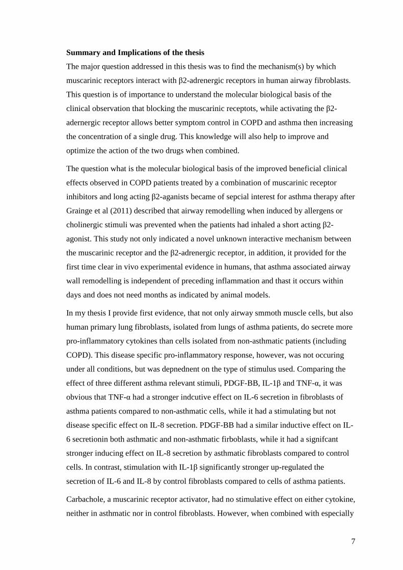
Initially it was assumed that the clinically observed beneficial effect of the combined
drugs may result from the interaction of specific G-proteins to which both receptor types
are linked (see figure below). However, the obtained data did not indicate any role of G-
proteins in the anti-inflammatory effect of neither the β2 adreneregic receptor nor of the
muscarinic receptor.
9
Proposed separation of the anti-
inflammatory and anti-
proliferative signalling
interaction of muscarinic and β2-
adrenergic receptors in human
lung fibroblasts.
Beside inflammation, airway wall remodelling is characterized by an extensive increase
of sub-epithelial fibroblast numbers and extracellular matrix deposition. According to
the literature there is no drug in asthma therapy which has a significant reducing effect
on airway wall remodelling. In earlier studies our research group had shown that the
lack of anti-proliferative effect of at least glucocorticoids is due to the lack of a
differentiation/cell cycle control factor, CCAAT enhancer binding protein-α (C/EBP-α),
which disease sepcifically missing in airway smooth muscle cells of asthma patients.
Unpublished data suggested that at least the β2-agonists, formoterol and salmeterol,
have an anti-proliferative effect of 30% reduction in airway smooth muscle cells. The
literature reported simliar small effects of anti-proliferative action of β2-agonists and
even reported pro-proliferative actions. In contrast to glucocortisoids which depend on
C/EBP-α and sub-seqeunt activity of p21(Waf), the anti-proliferative action of β2-
agonists invloved p27(Kip). In this thesis, we tested the inhibitory potebtial of olodaterol
on fibroblast proliferation induced by PDGF-BB.
At the time of this thesis, there was no data published showing an inhibitory effect of
muscarinic receptor inhibitors in regard of fibroblast proliferation.
Olodaterol confirmed an inhibitory effect of β2-agonists on lung fibroblast proliferation,
when the cells were stimulated with PDGF-BB. The anti-proliferative effect of
olodaterol was dose-dependent and was paralleled by the increase of intracellular
cAMP. Based on our earlier data with formoterol and salmeterol we concluded that this
beneficial effect applies to all β2-agonists.
10
Combining olodaterol with the muscarinic receptor inhibitor, however, did not improve
tha anti-proliferative effect of the β2-agonist. In contrast the muscarinic receptor
inhibitor counteracted the anti-proliferative effect of olodaterol to a certain extend.
Surprizingly, neither the anti-proliferative effect of olodaterol nor the counteractive
effect of tiotropium could be linked to the rescue of β2-agonist cAMP level increase or
to G-protein activity. Importantly, similar results have been recently published by others
and suggest a novel anti-proliferative acting signalling pathway for β2-adrenergic
receptors through so called β-arrestins, which are involved in muscarinic receptor
activity, however, with controversial results. Therefore, no conclusion on the role of β-
arrestins as an anti-proliferative proetin in the signalling of combined β2 agonists and
muscarinic receptor inhibitors can be made. It would be interesting to screen our
samples for the expression of β-arrestins.
The impact of this thesis on the undertsanding of the interaction of β2-adrenergic
receptors and muscarinic receptor signalling is as follows:
(i) The intracellular signalling cascades that get activated by the muscarinic receptor-3
or by the β2-adrenergic receptor are not involving G-proteins;
(ii) That combined β2-agonsists and muscarinic receptor inhibitors indeed have
beneficial additive anti-inflammatory action which may be due to a rescuing effect of
muscrainic receptor inhibition on β2-adrenergic receptor dependent intracellular cAMP
activation.
(iii) The combination of the two classes of drugs is not beneficial for airwaywall
remodelling based on increased fibrotic lesions, and thus is in line with clinical data.
However, proliferation is only one part of remodelling which also includes increased
deposition of extracellular matrix.
(iv) Preliminary experiments on the deposition of collagens and fibronectin show that
β2-agonsits reduce TGF-β and endothelin induced depsoition of extracelllular matrix
components but do not have a general inhibitory effect. Some of the inhibitory effects of
β2-agonists on cllagens can be explained by the increase of cAMP, while the inhibitory
effect on fibronectin is independent of cAMP. The available data indicates that long
actinng β2-agonists (fornmoterol, salmeterol) reduce the deposition of specifically
collagen type-I and of fibronectin through a cAMP dependent and a cAMP independent
pathway.
11
In summary, the data obtained in this thesis answered some aspects of the interaction of
the two receptor types, but also raised further questions. In addition, it became clear that
some of the beneficial anti-inflammatory actions of combined β2-adrenergic receptor
agonsists with inhibitors of muscarinic receptors invloves the action of cAMP while
others do not. The beneficail anti-inflammatory action of combined β2-adrenergic
receptor agonsists with inhibitors of muscarinic receptors is clear, but the net-effect of
the two drugs on tissue remodelling has to be further investigated.
Finally, it became clear, that we need to better understand how β2-adrenergic receptors
and muscarinic receptors mediate their signals to the cells, it seems that we do not yet
know all the details and novel mechanisms will be described soon.
12
I Asthma:
I.1 Definition and pathogenesis of asthma
The World Health Organization (WHO) estimates that asthma affects more than 250
million people worldwide (http://www.who.int/topics/asthma/en/). The guidelines of the
World Health Organization (WHO) define asthma as an inflammatory disease of the
airways:”It is a disease characterized by recurrent attacks of breathlessness and
wheezing, which vary in severity and frequency from person to person. In an individual,
they may occur from hour to hour and day to day. This condition is due to inflammation
of the air passages in the lungs and affects the sensitivity of the nerve endings in the
airways so they become easily irritated. In an attack, the lining of the passages swell
causing the airways to narrow and reducing the flow of air in and out of the lungs”
(http://www.who.int/respiratory/asthma/definition/en/).
The WHO also states that children represent more than 50% of all asthma patients and
that asthma is not curable, only the disease symptoms can be controlled by inhaled
drugs. It had been claimed that many children "grow out" off asthma; however, recent
studies suggest that asthma re-occurs at older age. Asthma shows a strange gender-
related association as it occurs more frequent in boys at young age and more often in
women at older age, thus suggesting the influence of hormones (Dijk et al 2013;
Moreno-Macías et al 2013). Several studies aimed to link asthma with susceptibility
genes and thus with genetic pre-dispositions, but so far there is no clear evidence for any
inheritable factors that pre-condition for asthma (Anderson et al 2013; Berenguer et al
2013; Boudier et al 20913; Li et al 2013; Lloyd et al 2013; Macintyre et al 2013). More
recently so-called epigenetic mechanisms have been linked to asthma including age
related DNA methylation patterns (Harris et al 2013). Nutrition and living conditions
also affect asthma. Beside a beneficial effect of Mediterranean food and increased intake
of oxygen radical scavengers such as vitamin C and D, it was claimed that there exists a
North-South gradient with lower asthma prevalence in the equatorial countries (Lang et
al 2013; Malinovschi et al 2013; Tsai et al 2013; Wegienka et al 2013). This hypothesis
has to be rejected and it is more likely that living conditions in rural areas is protective
to asthma, while life in cities furthers asthma (Anderson et al 2013; Macintyre et al
2013). However, beside the epidemiologic evidence, there is no biological mechanisms
known that could explain how the different triggers, allergic and non-allergic, can lead
to one disease phenotype "asthma".
13
I.2 New aspects of asthma pathologies and therapeutic targets:
New studies imply that the pre-condition to develop asthma is set during pregnancy or in
the first years of life, and therefore, many asthma patients suffer from birth throughout
their life (Covaciu et al 2013; Dijk et al 2013; Malmström et al 2013; van Schayck et al
2013; Wright et al 2013). In the past decades asthma was regarded as a chronic
inflammatory disease of the lung caused by a deregulated organ specific immune
response which is triggered mainly by inhaled substances including allergens,
chemicals, or dust (Hams & Fallon 2012; Holtzman 2012; Ozdemir et al 2011). The
hypothesis which sees an over-reactive immune response as the cause of asthma ignores
that 40% of asthma patients have no known allergies and their asthma is caused by
physical or psychological stress, such as exercise, sports (winter- and water sports),
stress or anxiety; furthermore sudden changes of the environment such as temperature,
humidity and air pressure can trigger asthma attacks. All those triggers cannot be
explained by an over-reactive immune response.
Thus the hypothesis of the over-reactive immune system has been challenged recently
and today the role of tissue forming cells in the pathology of asthma is re-investigated
(Black et al 2012; Leonardi et al 2012; Pongdee et al 2013; Thompson et al 2012). There
is increasing evidence for the role of mechano-compressive forces within the asthmatic
airway contributing to airway structural changes. An often asked question is if the re-
occurring strong constriction during an asthma attack could lead to changes of the
airway wall structure.
In contrast to chronic obstructive pulmonary disease (COPD), the airway constriction
which causes the shortness of breath, is reversible in asthma as soon as the muscle
bundles relax, this is the rational to inhale muscle relaxing drugs such short or long
acting β2-agonists, which are investigated in this thesis for their actions on cytokine
release, proliferation, and their interaction with muscarinic receptor signaling. Muscle
relaxing drugs seem not to affect airway wall remodeling, mucus secretion or cytokine
release. In order to control the latter factors many asthma patients use a combination of
β2-agonists with steroids or other anti-inflammatory agents such as steroids, cytokine
inhibitors; or IgE-antibodies for allergic asthma (Kandeel et al; Marandi et al 2013;
Manuyakorn et al 2013; Miller et al 2013; Robinson et al. 2013). However, none of the
available drugs is able to cure asthma. Off course the interaction of the different lung
14
forming cell types with immune cells in the airways has to be taken into consideration
(Ramakrishna et al. 2012). Last but not least inhibition or control of airway remodeling
is getting into the focus of new therapeutic asthma targets (Manuyakorn et al 2013)
Regarding the role of tissue forming lung cells it is interesting to note that the
hypertrophy and hyperplasia of airway wall smooth muscle (ASM) bundles was the first
disease specific pathology described in 22 patients with fatal asthma by Huber &
Koessler in 1922. This pathology was for some time accepted as the explanation of
airway hyper-constriction and obstruction. However, its development could not be
explained well and from the late 1960-ies onwards immunological pathologies were
described in asthma patients and the new dogma was that asthma results from an
overactive immune system and first indications of the role of immunoglobulins to the
pathogenesis of asthma (Hanissian et al 1969; Hilman et al 1969; Koltay et al 1967;
Stenius et al 1969).
In the late 1980-ies to 90-ies animal models were made and confirmed the importance of
the immune system in the pathogenesis of asthma. These animal models suggested that a
shift from Th-1 to Th-2 is important in allergic asthma (Corry et al 1996; Coyle et al
1996; O'Brien et al 1996; Schwarze et al 1997). However, this Th1 – Th2 shift was
never fully confirmed in humans (Holgate 2012; Shalaby & Martin 2010; Warrington
2010).
In the past two decades, the role of the mesenchymal cell types, especially of airway
wall residing fibroblasts and smooth muscle cells, in asthma was re-assessed and
increasing evidence suggest that airway wall remodeling is a central pathology that most
probably causes asthma (Manuyakorn et al 2013). The picture that evolves today
suggests that asthma results from a disrupted interaction of epithelial cell with immune
cell and mesenchymal cell. Furthermore, there is evidence that the condition to develop
asthma is set during the late phase of pregnancy and early childhood (Dotterud et al
2013; Källén et al 2013; Olsson et al 2013; Tedner et al 2012; Yang et al 2012; Wu et al
2012). In several studies it was reported that the increase of fibroblasts and airway
smooth muscle cell hyperplasia and hypertrophy in the airway wall occurs early in life
and in some studies these pathologies, together with wheezing, were recorded even
before asthma was diagnosed by standard clinical parameters and methods (Chawes et al
2011; Donohue et al 2013; Gold et al 2013; Jenkins et al 2003; Konradsen et al 2013¸
Lopez-Guisaet al 2012). Furthermore, airway wall remodeling preceded other symptoms
15
including inflammation in childhood asthma (Jenkins et al 2003; Malmström et al 2013).
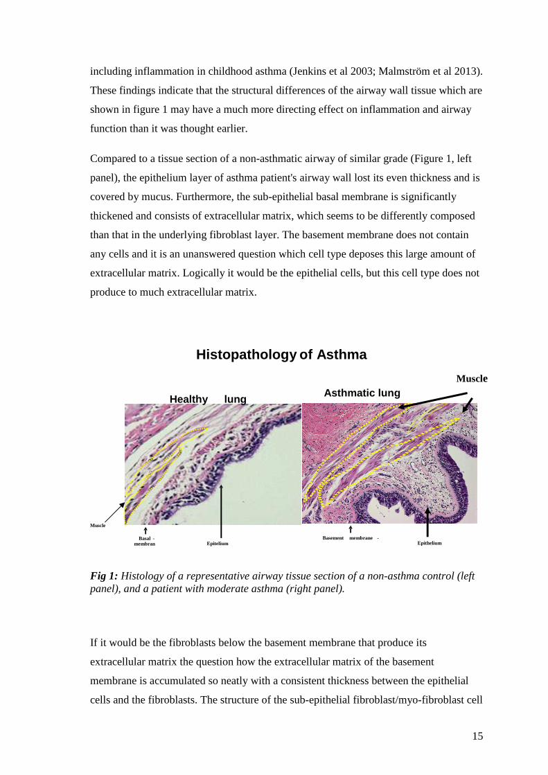
These findings indicate that the structural differences of the airway wall tissue which are
shown in figure 1 may have a much more directing effect on inflammation and airway
function than it was thought earlier.
Compared to a tissue section of a non-asthmatic airway of similar grade (Figure 1, left
panel), the epithelium layer of asthma patient's airway wall lost its even thickness and is
covered by mucus. Furthermore, the sub-epithelial basal membrane is significantly
thickened and consists of extracellular matrix, which seems to be differently composed
than that in the underlying fibroblast layer. The basement membrane does not contain
any cells and it is an unanswered question which cell type deposes this large amount of
extracellular matrix. Logically it would be the epithelial cells, but this cell type does not
produce to much extracellular matrix.
Fig 1: Histology of a representative airway tissue section of a non-asthma control (left panel), and a patient with moderate asthma (right panel).
If it would be the fibroblasts below the basement membrane that produce its
extracellular matrix the question how the extracellular matrix of the basement
membrane is accumulated so neatly with a consistent thickness between the epithelial
cells and the fibroblasts. The structure of the sub-epithelial fibroblast/myo-fibroblast cell
Healthy lung
Epitelium Basal -membran
Histopathology of Asthma
Asthmatic lung
Epithelium Basement membrane -
Muscle
Muscle
16
layer has produced a large amount of extracellular matrix which contains large areas
which seem to be cell free (no nuclei). This fibroblast layer is followed by an even more
increased layer of smooth muscle cells forming clear contractile bundles (Figure 1, right
panel).
I.3 Pathological mechanisms in asthma
Beside the fact that in the past 20 years 92’337 scientific articles were published on
human asthma the cause of asthma remains unclear. As mentioned above, it was
assumed for a long time that the major reason to develop chronic inflammation was a
deregulated immune response (Corry et al 1996; Coyle et al 1996; O'Brien et al 1996;
Schwarze et al 1997), which, however, was never fully confirmed in humans (Holgate
2012; Shalaby & Martin 2010; Warrington 2010). Therefore, other explanations must be
found, such as airway wall remodeling, which can lead to inflammation and immune
cell activation (Berair et al 2013; Siddiqui et al 2013; Xiao et al 2013). In order to
understand the interaction between the different cell types it is important to study the
crosstalk of inflammatory factors and their corresponding receptors, especially of the
muscarinic receptor with others (McGraw et al 2007; Oenema et al 2013; Quizon et al
2012; Verhein et al 2009).
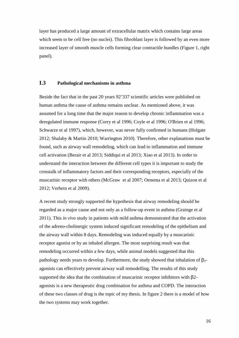
A recent study strongly supported the hypothesis that airway remodeling should be
regarded as a major cause and not only as a follow-up event in asthma (Grainge et al
2011). This in vivo study in patients with mild asthma demonstrated that the activation
of the adreno-cholinergic system induced significant remodeling of the epithelium and
the airway wall within 8 days. Remodeling was induced equally by a muscarinic
receptor agonist or by an inhaled allergen. The most surprising result was that
remodeling occurred within a few days, while animal models suggested that this
pathology needs years to develop. Furthermore, the study showed that inhalation of β2-
agonists can effectively prevent airway wall remodelling. The results of this study
supported the idea that the combination of muscarinic receptor inhibitors with β2–
agonists is a new therapeutic drug combination for asthma and COPD. The interaction
of these two classes of drug is the topic of my thesis. In figure 2 there is a model of how
the two systems may work together.
17
The signaling pathway
Presentation March 28 Pulmonary Cell Research, University Basel
Fig.2: Interaction of Muscarinic and β2 adrenergic receptors in asthma.
Most studies suggested that inflammation and remodeling are linked, however, there is
increasing evidence that remodelling occurs independent of inflammation and does not
need years to develop, as it was suggested by earlier studies in humans and animal
models (Blackquiere et al 2010; Evans et al 2010; Nihlberg et al 2010 ; Van Hove et al
2009).
Airway wall remodeling consists of two major parts: (i) the increase of mesenchymal
cells in numbers and (ii), the increase of extracellular matrix deposition. Both events are
independent of each other, but they affect each other. Airway wall remodeling includes
structural changes of the tissue such as modified epithelial cell characteristics, increased
mass of airway smooth muscle cells and increased numbers of active fibroblasts turning
into myo-fibroblasts; fibrosis and increased vascularization. Cytokines released by
inflammatory cells activate the epithelium and stimulate a network of extracellular
signals that determine the tissue structural changes in asthma. The structural defect of
the epithelium, include reduced cell-cell contact and allows allergens to migrate into the
sub-epithelial cell layer where they come into direct contact with the mesenchymal cell
types (fibroblasts, smooth muscle cells). Interestingly, airway mesenchymal cells
express antigen recognizing receptors and immunoglobulin receptors and directly
respond to allergens by releasing pro-inflammatory (Gounni et al 2005; Grunstein et al
18
2002; Redhu et al 2013; Redhu et al 2009; Xia et al 2011). It is currently investigated if
blocking immunolglobulin receptors may prevent or even reverse asthma associated
airway wall remodeling (Rabe et al 2011). In monkey fibroblast cells anti-IgE antibodies
blocked their activation (Takai et al 2011) and similarly in human fibroblast like cells
(Smith et al 1995). Furthermore, IgG can directly interact with fibrocyte cells and
activate them (Pilling et al 2006). In summary, immunoglobulin exposure of fibroblast
like cells activated the production of pro-inflammatory mediators, induced cell
constriction and altered enzyme secretion (Lee et al 2003). However, the pathogenesis
of asthma cannot depend on a single cell type and the interactions between de-regulated
tissue forming mesenchymal cell types in the airway wall with infiltrating immune cells
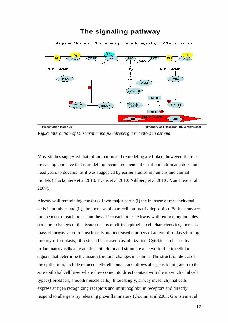
has to be studied in more details. An overview of possible interactions of mesenchymal
cells, immuno-globulins and immune cells in asthma is provided in figure 3.
Fig. 3: Summary of airway wall remodelling through tissue forming mesenchymal cell interaction with mast cells and immuno-globulins.
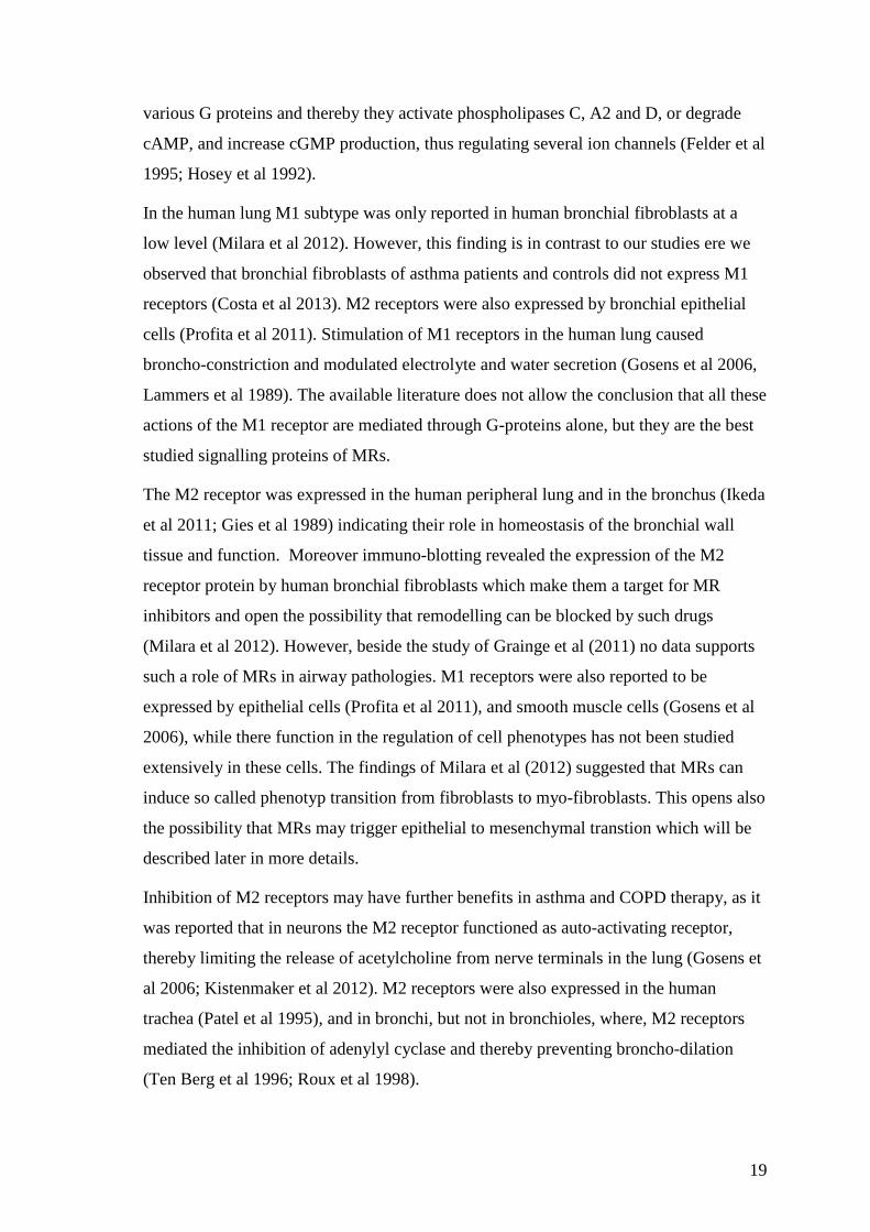
I.4 The muscarinic receptors on lung fibroblasts: function and role in asthma
The interaction of muscarinic receptors (MR) have been since long a target for therapy
in asthma and other chronic inflammatory lung idseaes. Together with other membrane
bound receptor, especially with the asthma relevant β2-adrenoceptor is largely unkown.
MRs modulates different intracellular signal transduction pathways by coupling to
19
various G proteins and thereby they activate phospholipases C, A2 and D, or degrade
cAMP, and increase cGMP production, thus regulating several ion channels (Felder et al
1995; Hosey et al 1992).
In the human lung M1 subtype was only reported in human bronchial fibroblasts at a
low level (Milara et al 2012). However, this finding is in contrast to our studies ere we
observed that bronchial fibroblasts of asthma patients and controls did not express M1
receptors (Costa et al 2013). M2 receptors were also expressed by bronchial epithelial
cells (Profita et al 2011). Stimulation of M1 receptors in the human lung caused
broncho-constriction and modulated electrolyte and water secretion (Gosens et al 2006,
Lammers et al 1989). The available literature does not allow the conclusion that all these
actions of the M1 receptor are mediated through G-proteins alone, but they are the best
studied signalling proteins of MRs.
The M2 receptor was expressed in the human peripheral lung and in the bronchus (Ikeda
et al 2011; Gies et al 1989) indicating their role in homeostasis of the bronchial wall
tissue and function. Moreover immuno-blotting revealed the expression of the M2
receptor protein by human bronchial fibroblasts which make them a target for MR
inhibitors and open the possibility that remodelling can be blocked by such drugs
(Milara et al 2012). However, beside the study of Grainge et al (2011) no data supports
such a role of MRs in airway pathologies. M1 receptors were also reported to be
expressed by epithelial cells (Profita et al 2011), and smooth muscle cells (Gosens et al
2006), while there function in the regulation of cell phenotypes has not been studied
extensively in these cells. The findings of Milara et al (2012) suggested that MRs can
induce so called phenotyp transition from fibroblasts to myo-fibroblasts. This opens also
the possibility that MRs may trigger epithelial to mesenchymal transtion which will be
described later in more details.
Inhibition of M2 receptors may have further benefits in asthma and COPD therapy, as it
was reported that in neurons the M2 receptor functioned as auto-activating receptor,
thereby limiting the release of acetylcholine from nerve terminals in the lung (Gosens et
al 2006; Kistenmaker et al 2012). M2 receptors were also expressed in the human
trachea (Patel et al 1995), and in bronchi, but not in bronchioles, where, M2 receptors
mediated the inhibition of adenylyl cyclase and thereby preventing broncho-dilation
(Ten Berg et al 1996; Roux et al 1998).
20
In regard to airway wall remodelling the stimulation of the M2 receptors induced
fibroblasts proliferation (Matthiessen et al 2006; Haag et al 2006) and acetylcholine
enhanced cell proliferation in cells isolated from COPD patients, as compared to healthy
non-smokers, through a process involving ERK1/2 MAPK and NFκB phosphorylation
(Profita et al 2009). Airway smooth muscle thickening is a characteristic pathology of
asthma, and to a lesser extent of COPD. Accumulating evidence suggests that
stimulation of MRs is involved in the proliferation and maturation of airway smooth
muscle cells (Kistenmaker et al 2012). Furthermore, MR activation enhanced the
mitogenic effect of PDGF-BB and of EGF in airway smooth muscle cells (Kong et al
2006; Gosens et al 2007). However, the molecular interaction of the receptor specific
signalling cascades is not clear. Moreover, the expression of myosin light-chain kinase
was augmented by carbachol in human airway smooth muscle cells which had been
stretched by cyclical mechanical forces (Fairbank et al 2008).
Furthermore, and relevant to airway wall remodeling stimulation of MRs supported
TGF-β1-induced expression of contractile proteins by smooth muscle cells and thus
would increase the constrictive forces in an asthma attack (Oenema et al 2012). In
animal models of asthma and COPD, the M1, M2 and m3 receptor inhibitor tiotropium
significantly inhibited airway smooth muscle remodelin and contractile protein
expression in guinea pigs (Gosens et al 2005; Bos et al 2007). The drug also prevented
smooth muscle thickening and the expression of TGF-β1 in the bronchi in an
ovalbumine mouse model (Ohta et al 2010). In a murine asthma model the selective M3
receptor antagonist bencycloquidium bromide had similar beneficial effects as it
inhibited ovalbumin-induced mRNA expression of IL-5, IL-4, and MMP-9, as well as
lung tissue eosinophil infiltration, airway mucus production, and collagen deposition in
lung tissues (Cao et al 2011).
Further, in regard to remodeling in asthma Hypoxia and PDGF-BB induced synthesis of
soluble collagen type I via ERK1/2 and p38 MAP kinase in human lung fibroblasts and
pulmonary vascular smooth muscle cells (Karakiulakis et al 2007). In human lung
fibroblasts stimulation of M2 receptors induced cell proliferation and collagen synthesis
(Matthiessen et al 2006; Haag et al 2008). In a clinical trial, inhalation of methacholine
induced airway remodeling in asthma patients, through the expression of TGF-β and
collagen type-I as shown in bronchial biopsies (Grainge et al 2011). Treatment with
tiotropium inhibited the increased peri-bronchial collagen deposition ina guinea pig
COPD model (Pera et al 2011).
21
The M3 receptor is the primary MR subtype that mediates the contraction of bronchial
and tracheal smooth muscle However, compared to M2 levels the M3 receptor is
expressed at 25% (Roffel et al 1988). In addition, the M3 receptor is expressed by
airway smooth muscle cells (Eglen et al 1996), by human bronchial fibroblasts (Milara
et al 2012), and by human bronchial epithelial cells (Profita et al 2011), and by human
peripheral lung cells (Gies et al 1989). The M3 receptor is predominantly expressed in
the bronchus and its density decreases from the segmental to subsegmental bronchus
tissue, while it has not been reported in lung parenchyma (Ikeda et al 2012). Stimulation
of the M3 receptor in the human lung, and in isolated human bronchus caused broncho-
constriction and mucus secretion from submucosal glands (Gosens et al 2006; Roux et al
1998; Eglen et al 1996; Roffel et al 1990). A summary of the expression and function of
the different MR in the airway wall and its three major tissue forming cell types is
shown in figure 4.
.
Figure 4: Overview of cell type specific muscarinic receptor distribution and their function in the human airway wall. ACH: acetylcholine, EGF: epithelial growth factor, MMP: matrix metalloproteinase, PDGF: platelet-derived growth factor, M1: muscarinic receptor type.
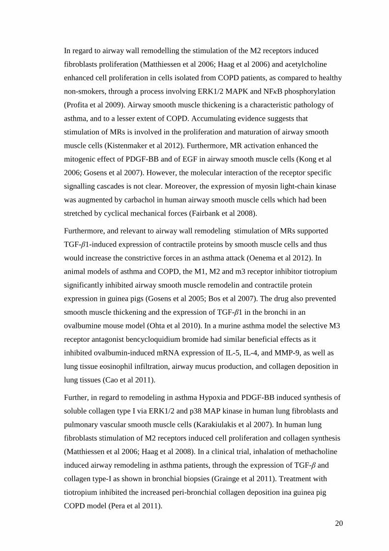
There are a lot of different signalling options for MR, and the interaction with other
signalling pathways is even more complicated, as each single MR subtype can activate
more than one G protein type in a specific cell type. It can therefore be assumed that the
condition of the cell and its phenotype dictate to which G-protein the MR is coupled
under a defined condition, which in consequence alters the signalling pathway and the
cells response (Felder 1995; Hosey 1992; Eglen & Nahorski 2000). MRs can be divided
22
into two primary G-proteins couplings: 1) M2 and M4, which are coupled to pertusiss-
toxin sensitive Gi/o type proteins, while 2) the M1, M3, and M5 receptors are coupled to
Gq/11-type proteins (Felder 1995; Caulfield & Birdsall 1998). However, MRs can also
couple to a wider range of G-proteins as well as to other signaling proteins and thus, it is
difficult to predict their effects (Nathanson 2000; van Koppen 2003). An overview of
known muscarinic receptor signaling is provided in Figure 5.
Figure 5: Overview of known muscarinic receptor coupled G-proteins and subsequent signalling.
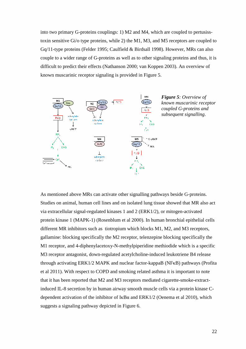
As mentioned above MRs can activate other signalling pathways beside G-proteins.
Studies on animal, human cell lines and on isolated lung tissue showed that MR also act
via extracellular signal-regulated kinases 1 and 2 (ERK1/2), or mitogen-activated
protein kinase 1 (MAPK-1) (Rosenblum et al 2000). In human bronchial epithelial cells
different MR inhibitors such as tiotropium which blocks M1, M2, and M3 receptors,
gallamine: blocking specifically the M2 receptor, telenzepine blocking specifically the
M1 receptor, and 4-diphenylacetoxy-N-methylpiperidine methiodide which is a specific
M3 receptor antagonist, down-regulated acetylcholine-induced leukotriene B4 release
through activating ERK1/2 MAPK and nuclear factor-kappaB (NFκB) pathways (Profita
et al 2011). With respect to COPD and smoking related asthma it is important to note
that it has been reported that M2 and M3 receptors mediated cigarette-smoke-extract-
induced IL-8 secretion by in human airway smooth muscle cells via a protein kinase C-
dependent activation of the inhibitor of IκBα and ERK1/2 (Oenema et al 2010), which
suggests a signaling pathway depicted in Figure 6.
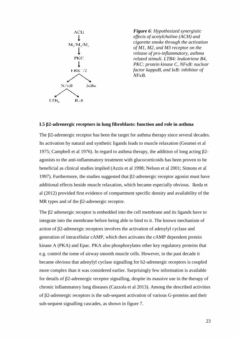
23
Figure 6: Hypothesized synergistic effects of acetylcholine (ACH) and cigarette smoke through the activation of M1, M2, and M3 receptor on the release of pro-inflammatory, asthma related stimuli. LTB4: leukotriene B4, PKC: protein kinase C, NFκB: nuclear factor kappaB, and IκB: inhibitor of NFκB.
I.5 β2-adrenergic receptors in lung fibroblasts: function and role in asthma
The β2-adrenergic receptor has been the target for asthma therapy since several decades.
Its activation by natural and synthetic ligands leads to muscle relaxation (Geumei et al
1975; Campbell et al 1976). In regard to asthma therapy, the addition of long acting β2-
agonists to the anti-inflammatory treatment with glucocorticoids has been proven to be
beneficial as clinical studies implied (Azzis et al 1998; Nelson et al 2001; Simons et al
1997). Furthermore, the studies suggested that β2-adrenergic receptor agonist must have
additional effects beside muscle relaxation, which became especially obvious. Ikeda et
al (2012) provided first evidence of compartment specific density and availability of the
MR types and of the β2-adrenergic receptor.
The β2 adrenergic receptor is embedded into the cell membrane and its ligands have to
integrate into the membrane before being able to bind to it. The known mechanism of
action of β2-adrenergic receptors involves the activation of adenylyl cyclase and
generation of intracellular cAMP, which then activates the cAMP dependent protein
kinase A (PKA) and Epac. PKA also phosphorylates other key regulatory proteins that
e.g. control the tome of airway smooth muscle cells. However, in the past decade it
became obvious that adenylyl cyclase signalling for b2-adrenergic receptors is coupled
more complex than it was considered earlier. Surprisingly few information is available
for details of β2-adrenergic receptor signalling, despite its massive use in the therapy of
chronic inflammatory lung diseases (Cazzola et al 2013). Among the described activities
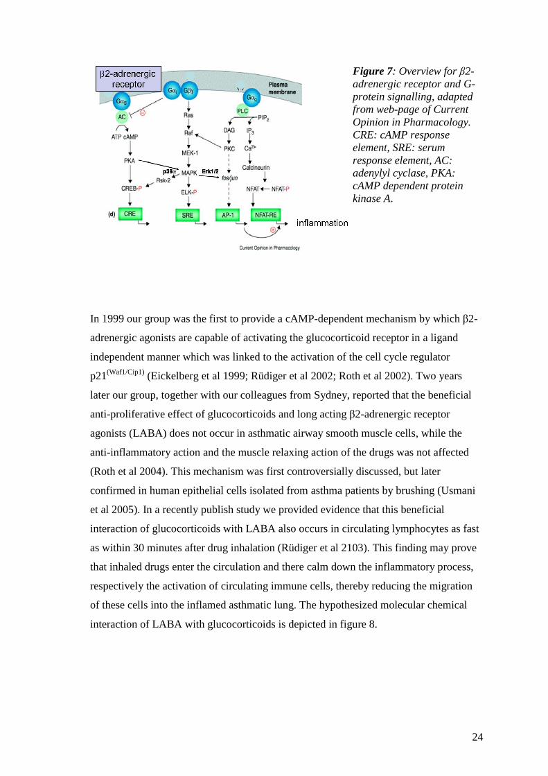
of β2-adrenergic receptors is the sub-sequent activation of various G-proteins and their
sub-sequent signalling cascades, as shown in figure 7.
24
Figure 7: Overview for β2-adrenergic receptor and G-protein signalling, adapted from web-page of Current Opinion in Pharmacology. CRE: cAMP response element, SRE: serum response element, AC: adenylyl cyclase, PKA: cAMP dependent protein kinase A.
In 1999 our group was the first to provide a cAMP-dependent mechanism by which β2-
adrenergic agonists are capable of activating the glucocorticoid receptor in a ligand
independent manner which was linked to the activation of the cell cycle regulator
p21(Waf1/Cip1) (Eickelberg et al 1999; Rüdiger et al 2002; Roth et al 2002). Two years
later our group, together with our colleagues from Sydney, reported that the beneficial
anti-proliferative effect of glucocorticoids and long acting β2-adrenergic receptor
agonists (LABA) does not occur in asthmatic airway smooth muscle cells, while the
anti-inflammatory action and the muscle relaxing action of the drugs was not affected
(Roth et al 2004). This mechanism was first controversially discussed, but later
confirmed in human epithelial cells isolated from asthma patients by brushing (Usmani
et al 2005). In a recently publish study we provided evidence that this beneficial
interaction of glucocorticoids with LABA also occurs in circulating lymphocytes as fast
as within 30 minutes after drug inhalation (Rüdiger et al 2103). This finding may prove
that inhaled drugs enter the circulation and there calm down the inflammatory process,
respectively the activation of circulating immune cells, thereby reducing the migration
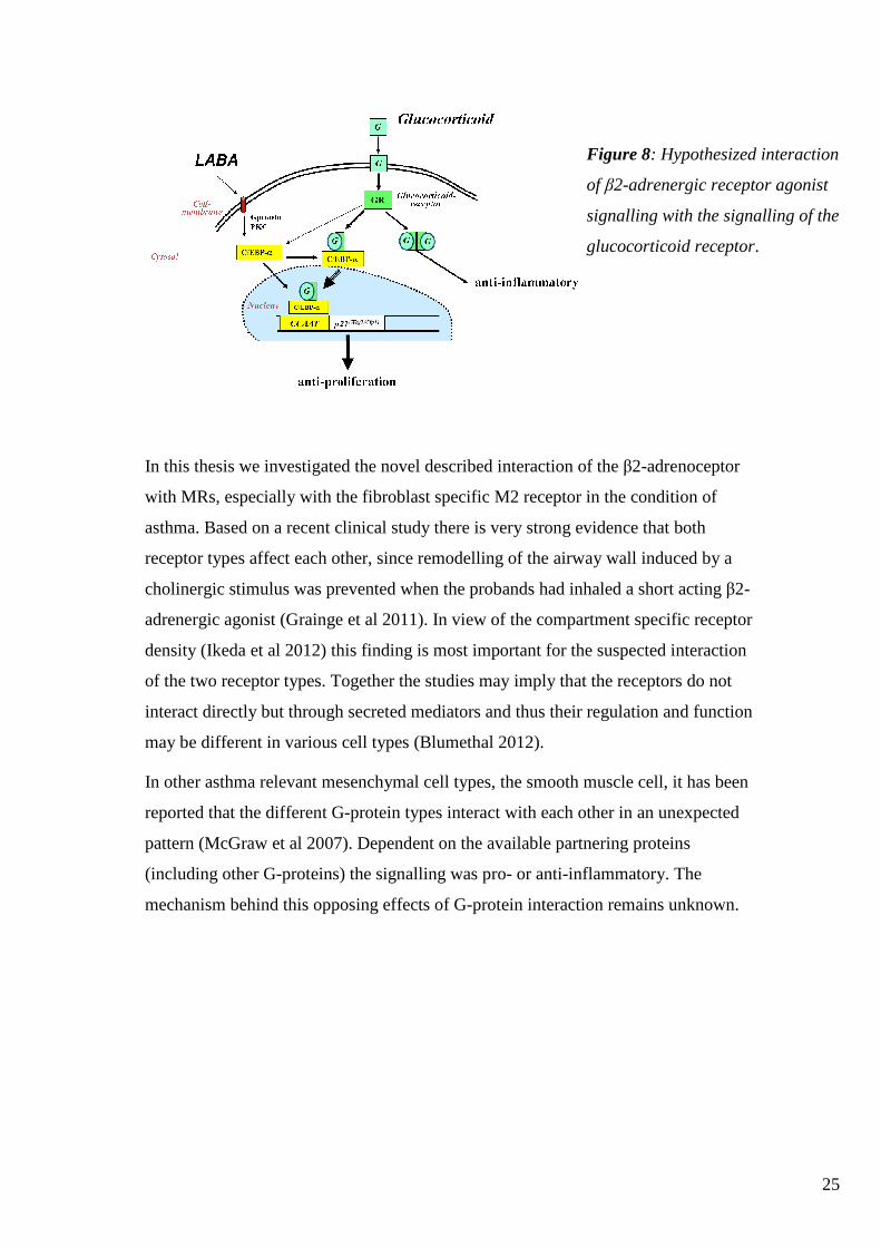
of these cells into the inflamed asthmatic lung. The hypothesized molecular chemical
interaction of LABA with glucocorticoids is depicted in figure 8.
25
Figure 8: Hypothesized interaction
of β2-adrenergic receptor agonist
signalling with the signalling of the
glucocorticoid receptor.
In this thesis we investigated the novel described interaction of the β2-adrenoceptor
with MRs, especially with the fibroblast specific M2 receptor in the condition of
asthma. Based on a recent clinical study there is very strong evidence that both
receptor types affect each other, since remodelling of the airway wall induced by a
cholinergic stimulus was prevented when the probands had inhaled a short acting β2-
adrenergic agonist (Grainge et al 2011). In view of the compartment specific receptor
density (Ikeda et al 2012) this finding is most important for the suspected interaction
of the two receptor types. Together the studies may imply that the receptors do not
interact directly but through secreted mediators and thus their regulation and function
may be different in various cell types (Blumethal 2012).
In other asthma relevant mesenchymal cell types, the smooth muscle cell, it has been
reported that the different G-protein types interact with each other in an unexpected
pattern (McGraw et al 2007). Dependent on the available partnering proteins
(including other G-proteins) the signalling was pro- or anti-inflammatory. The
mechanism behind this opposing effects of G-protein interaction remains unknown.
26
I.6 Tissue remodeling in asthma:
Tissue remodeling can occur in all organs and is often preceded by inflammation, especially
in lung diseases such as fibrosis, asthma, and chronic obstructive pulmonary disease (COPD).
Asthma and COPD share several pathologies and it is difficult to distinguish between both
diseases. In patients with COPD airway remodeling occurs mainly in the small airways. In
asthma patients the changes are significant in the upper and medium size airways (Caramori
et al 2011; Contoli et al 2010; Paredi et al 2009; Plopper et al 2008). Summarizing the large
number of reports on increased pro-inflammatory cytokines that were linked to airway
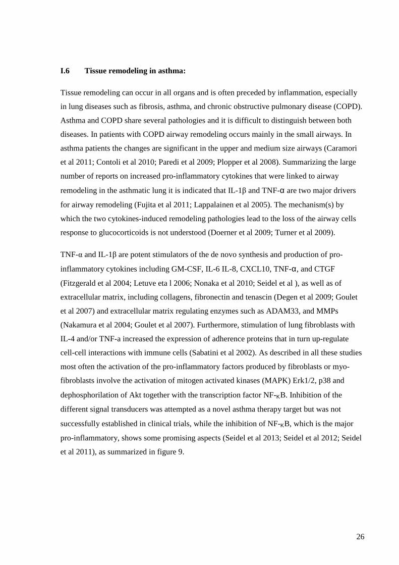
remodeling in the asthmatic lung it is indicated that IL-1β and TNF-α are two major drivers
for airway remodeling (Fujita et al 2011; Lappalainen et al 2005). The mechanism(s) by
which the two cytokines-induced remodeling pathologies lead to the loss of the airway cells
response to glucocorticoids is not understood (Doerner et al 2009; Turner et al 2009).
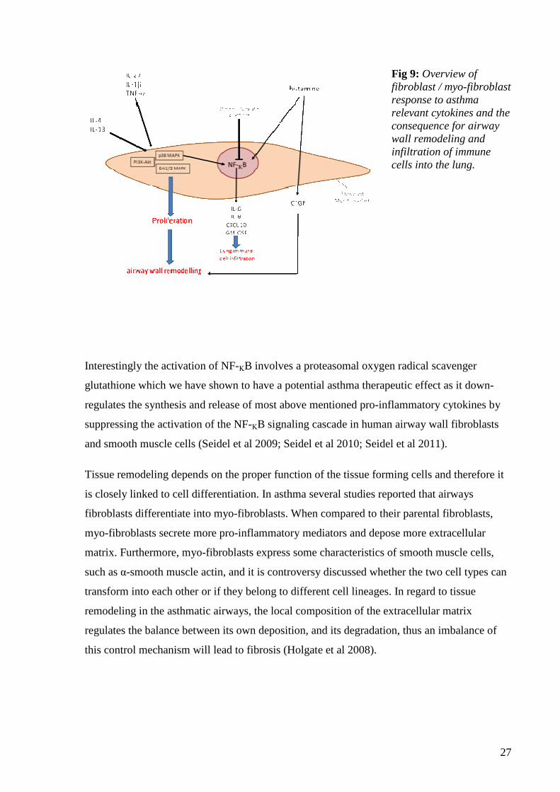
TNF-α and IL-1β are potent stimulators of the de novo synthesis and production of pro-
inflammatory cytokines including GM-CSF, IL-6 IL-8, CXCL10, TNF-α, and CTGF
(Fitzgerald et al 2004; Letuve eta l 2006; Nonaka et al 2010; Seidel et al ), as well as of
extracellular matrix, including collagens, fibronectin and tenascin (Degen et al 2009; Goulet
et al 2007) and extracellular matrix regulating enzymes such as ADAM33, and MMPs
(Nakamura et al 2004; Goulet et al 2007). Furthermore, stimulation of lung fibroblasts with
IL-4 and/or TNF-a increased the expression of adherence proteins that in turn up-regulate
cell-cell interactions with immune cells (Sabatini et al 2002). As described in all these studies
most often the activation of the pro-inflammatory factors produced by fibroblasts or myo-
fibroblasts involve the activation of mitogen activated kinases (MAPK) Erk1/2, p38 and
dephosphorilation of Akt together with the transcription factor NF-ΚB. Inhibition of the
different signal transducers was attempted as a novel asthma therapy target but was not
successfully established in clinical trials, while the inhibition of NF-ΚB, which is the major
pro-inflammatory, shows some promising aspects (Seidel et al 2013; Seidel et al 2012; Seidel
et al 2011), as summarized in figure 9.
27
Fig 9: Overview of fibroblast / myo-fibroblast response to asthma relevant cytokines and the consequence for airway wall remodeling and infiltration of immune cells into the lung.
Interestingly the activation of NF-ΚB involves a proteasomal oxygen radical scavenger
glutathione which we have shown to have a potential asthma therapeutic effect as it down-
regulates the synthesis and release of most above mentioned pro-inflammatory cytokines by
suppressing the activation of the NF-ΚB signaling cascade in human airway wall fibroblasts
and smooth muscle cells (Seidel et al 2009; Seidel et al 2010; Seidel et al 2011).
Tissue remodeling depends on the proper function of the tissue forming cells and therefore it
is closely linked to cell differentiation. In asthma several studies reported that airways
fibroblasts differentiate into myo-fibroblasts. When compared to their parental fibroblasts,
myo-fibroblasts secrete more pro-inflammatory mediators and depose more extracellular
matrix. Furthermore, myo-fibroblasts express some characteristics of smooth muscle cells,
such as α-smooth muscle actin, and it is controversy discussed whether the two cell types can
transform into each other or if they belong to different cell lineages. In regard to tissue
remodeling in the asthmatic airways, the local composition of the extracellular matrix
regulates the balance between its own deposition, and its degradation, thus an imbalance of
this control mechanism will lead to fibrosis (Holgate et al 2008).

28
I.7 The role of airway smooth muscle cells in asthma associated airway remodeling
Airway smooth muscle cells also contribute to airway wall remodeling in that they increase in
number and size. This pathology correlate with the development of a so-called “secretory
phenotype” of the airway smooth muscle cells (Dekkers et al 2009). The phenotype change is
also characterized by an increased expression of cell adhesion receptors, and receptors for
cytokines (Joubert et al 2005). In addition there is increased angiogenesis in the asthmatic
airway wall which is assumed to be based on the overproduction of vascular endothelial
growth factor (VEGF), which stimulates the formation of new blood vessels in the sub-
epithelial cell layers and in addition causes edema formation by loosening the cell-cell contact
between endothelial cells. Edema in turn allows the infiltration of more pro-inflammatory
cells which secrete remodeling enhancing cytokines in the airway wall (Makinde et al 2006).
The increase of the airway smooth muscle cell mass, angiogenesis and fibrosis, contribute to
airway wall thickness which restricts airflow (Pepper et al 2005; Benayoun et al 2003).
I.8 The role of airway fibroblast in asthma associated airway remodeling
The involvement of fibroblasts in the airway wall remodeling process in asthma is assumed to
be initiated through their activation by various cytokines. Once activated the fibroblasts
themselves become a source of more inflammatory cytokines and of more deposed
extracellular matrix. The activated fibroblast is often called “myo-fibroblasts” and shares
some properties with airway smooth muscle cells (Descalzi et al 2007; Michalik et al 2011;
Singh et al 2008).
However, it is difficult to define the border between myo-fibroblasts and smooth muscle cells,
and the only reliable differentiation between both cell types is the fibrilar expression of α-
smooth muscle cell actin (α-SMA) together with increased the myosin heavy chain
(SmMHC), SM22α, and calponin (Descalzi et al 2007; Michalik et al 2011; Shi et al 2013;
Wu et al 2008). The most potent stimulus of myo-fibroblast differentiation and epithelial to
mesenchymal transition is TGF-β, which also is the most potent stimulus for the production
extracellular matrix, and which is furthermore significantly up-regulated in asthma patients
samples (Makinde et al 2007; Shi et al 2103; Qin et al 2012; Singh et al 2008). The action of
TGF-β in myo-fibroblast differentiation involves MAPKs and may be related to the pro-
inflammatory activation cascade shown in figures 3 and 4 (Sabatini et al 2013; Singh et al
29
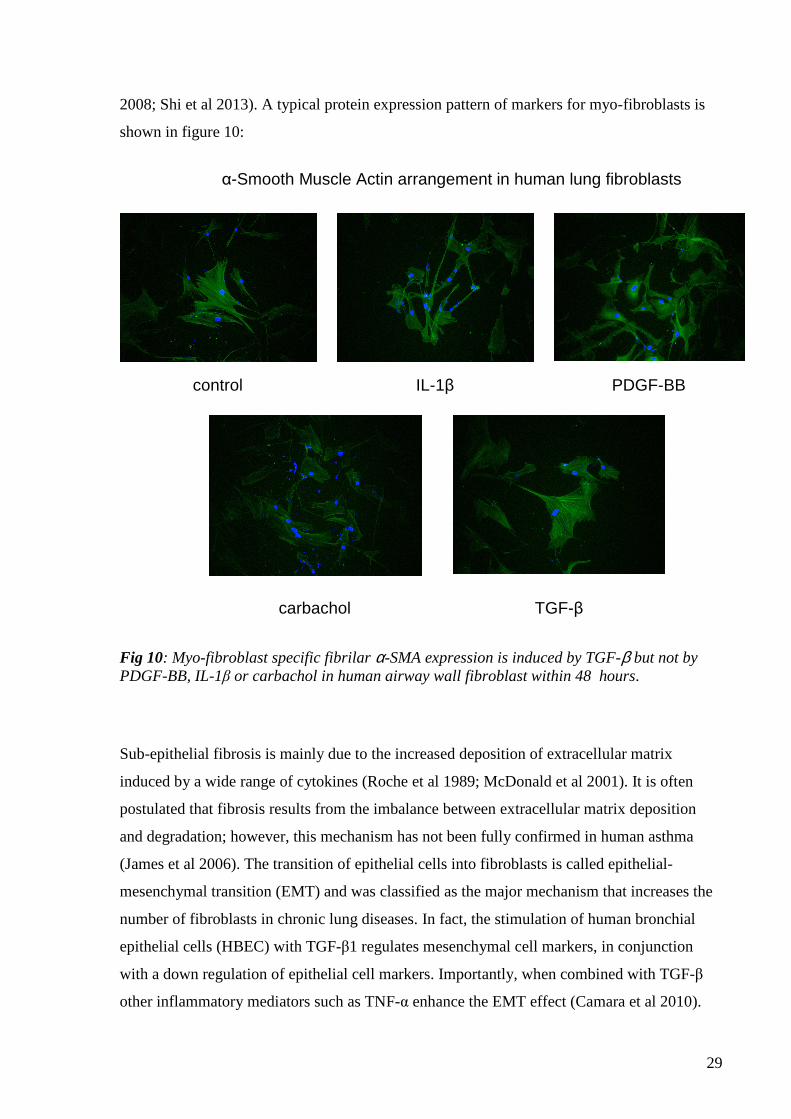
2008; Shi et al 2013). A typical protein expression pattern of markers for myo-fibroblasts is
shown in figure 10:
Fig 10: Myo-fibroblast specific fibrilar α-SMA expression is induced by TGF-β but not by PDGF-BB, IL-1β or carbachol in human airway wall fibroblast within 48 hours.
Sub-epithelial fibrosis is mainly due to the increased deposition of extracellular matrix
induced by a wide range of cytokines (Roche et al 1989; McDonald et al 2001). It is often
postulated that fibrosis results from the imbalance between extracellular matrix deposition
and degradation; however, this mechanism has not been fully confirmed in human asthma
(James et al 2006). The transition of epithelial cells into fibroblasts is called epithelial-
mesenchymal transition (EMT) and was classified as the major mechanism that increases the
number of fibroblasts in chronic lung diseases. In fact, the stimulation of human bronchial
epithelial cells (HBEC) with TGF-β1 regulates mesenchymal cell markers, in conjunction
with a down regulation of epithelial cell markers. Importantly, when combined with TGF-β
other inflammatory mediators such as TNF-α enhance the EMT effect (Camara et al 2010).
TGF-β carbachol
control IL-1β PDGF-BB
α-Smooth Muscle Actin arrangement in human lung fibroblasts
30
Regarding therapies, a large number of studies showed that the combination of corticosteroids
and LABA improves lung function, better controls asthma symptoms and improves quality of
life. Most of these beneficial therapeutic effects are due to a decrease in inflammation
(Greening et al 1994; Pauwels et al 1997; O'Byrne et al 2005).
Inhalation of allergens results increases eosinophils in the lung, which are associated with
increased inflammation of the airways. Today it is assumed that eosinophils causes airway
wall remodeling in asthma (Brannan et al 2012). Treatment with LABA reduced the
inflammatory response and reduced the number of lung infiltrating eosinophils (Kelly et al
2010). Asthmatic airway wall cells incubated with TGF-β 1, and 2 expressed increased
markers of myo-fibrobasts (Michalik et al 2009). Furthermore, inflammatory symptoms in
asthma were resolved within one week of treatment; the increase of airway hyper-
responsiveness (AHR) and markers of extracellular matrix deposition persisted indicating that
the pathology of AHR and remodeling are independent pathological events in asthma. It was
demonstrated that airway inflammation, airway remodeling, can occur independent of
inflammation and importantly remodeling persists after the inflammation has been resolved
(Kariyawasam et al 2007).
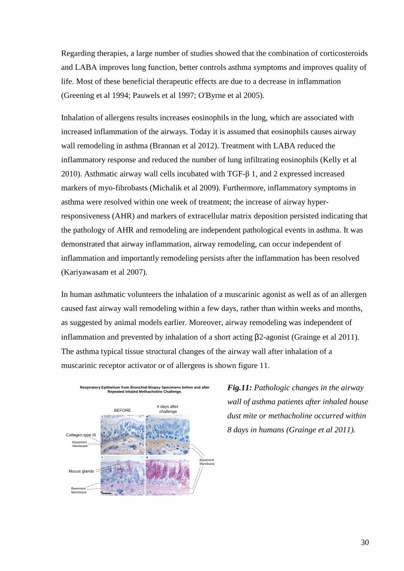
In human asthmatic volunteers the inhalation of a muscarinic agonist as well as of an allergen
caused fast airway wall remodeling within a few days, rather than within weeks and months,
as suggested by animal models earlier. Moreover, airway remodeling was independent of
inflammation and prevented by inhalation of a short acting β2-agonist (Grainge et al 2011).
The asthma typical tissue structural changes of the airway wall after inhalation of a
muscarinic receptor activator or of allergens is shown figure 11.
Respiratory Epithelium from Bronchial-Biopsy Specimens before and after Repeated Inhaled Methacholine Challenge.
BEFORE4 days after
challenge
Collagen type III
Mucus glands
BasementMembrane
BasementMembrane
BasementMembrane
Fig.11: Pathologic changes in the airway
wall of asthma patients after inhaled house
dust mite or methacholine occurred within
8 days in humans (Grainge et al 2011).
31
The process of fibrosis may be caused by the inadequate production of inflammatory factors
by infiltrated immune cells, or by the inadequate response of fibroblasts to these factors
(Portnoy et al 2006). Moore BB et al discovered that PGE 2 (prostaglandin E2) is a potent
inhibitor of fibroblast proliferation (Moore et al 2000). In lung fibroblasts, the production of
PGE2 is regulated by TGF-β and affects cell proliferation, apoptosis, and differentiation
(McAnulty et al 1997; Annu et al 1998).
However, TGF-β is also associated with fibrosis and tissue remodeling in vivo and in vitro
(Sagara et al 2002). Primary human epithelial cells regulated fibroblast proliferation through
the release of TGF-β which stimulated fibroblasts to produce and secrete PGE 2. This in turn
had an antiproliferative feedback mechanism on fibroblasts (Hostettler et al 2008). Using
conditioned medium obtained from airway epithelial cells it was demonstrated that
cyclosporine provoked fibroblast proliferation only when exposed to epithelial cells before.
The fibrogenic potential of cyclosporine was attributed to many different cellular
mechanisms, such as, decreased extracellular matrix degradation through increased
expression of tissue proteinases and matrix metal-proteinase (Duymelinck et al 1998;
Hostettler et al 2004). In a healthy lung, the daily turnover of the extracellular matrix is
estimated at 10 - 15 %( Davidson 1990; Stetler -Stevenson et al 1996), and it is controlled by
three different mechanisms: extracellular matrix de novo synthesis, extracellular matrix
degradation by MMPs and the inhibition of MMP by tissue inhibitor factors (McAnulty &
Laurent 1995; Curran & Murray 1999). The balance of these 3 mechanism guaranties the
function and integrity of all organs and organisms.
The structure of the extracellular matrix, and the vasculature, control the supply of oxygen to
the tissues, which is of specific importance during embryogenesis, fibrosis, and repair of
tissue damage and in tumor progression (Norman et al 2000; Tokuda et al 2000; Chen &
Aplin 2003; Gebb & Jones 2003). One component of the extracellular matrix, collagen type-
IV, is particularly abundant in fibrosis, and it seems specifically increased when there is a low
supply of oxygen, assigned as hypoxia (Steinbrech et al 1999). Fibroblasts are the major
source of collagen type-I and –IV.
It was early demonstrated by our group that there is a link between hypoxia, local nutrition
and secretion of pro-MMP-2. Moreover hypoxia has a distinct effect on different cell types,
and in the lung, hypoxia in combination with collagen type expression, alters the secretion of
32
pro-MMP-2, thereby reducing the chance to properly repair the damaged tissue (Leufgen et al
2005). Late, Goulet et al. (2007) investigated the effect of corticosteroids and LABA on the
deposition of extracellular matrix, on collagen gene expression, on cell proliferation, and on
IL-6, IL-8, and secretion by primary lung fibroblasts (Goulet et al 2007). Fetal calf serum
(FCS, 5%) increased total extracellular matrix synthesis, collagen deposition, cell
proliferation as well, IL-6 IL-8 secretion, and TGF-β1 levels. In starving condition (0.3%
albumin) corticosteroids reduced the deposition of collagens and of total extracellular matrix,
while in 5% FCS, the corticosteroid increased deposition of extracellular matrix (Goulet et al
2007).
In contrast, LABA reduced the deposition of extracellular matrix and collagens in all
conditions. In combination, the drugs had an additive effect in thus largely decreasing the
deposition of extracellular matrix. The study implied that as soon the inflammation of the
airways has been resolved by glucocorticoids the addition of LABA may reduce tissue
remodeling in the asthmatic airways (Goulet et al 2007). In asthmatic fibroblasts the most
produced extracellular matrix components are: fibronectin and tenascin -C (Chiquet-
Ehrismann et al 2003). Both glycoproteins are over expressed in asthma and COPD and
contribute to the progression and pathology.
M. Degen et al. (2009) demonstrated in human lung fibroblasts that treatment with
corticosteroids reduced the expression of tenascin-C, but increased fibronectin. In contrast
LABA did not show a significant effect on either, tenascin-C or fibronectin (Degen et al
2009). Another characteristic of the fibrotic processes in the lung is the modified composition
and production of glycosaminoglycans (GAG) by lung fibroblasts (Kneussl et al 1996;
Moseley et al 1986). When lung fibroblasts were stimulated by platelet-derived growth factor
(PDGF)-BB in normal condition, the cells increased the production of GAGs in a dose-
dependent manner. Under hypoxia the GAG production increased through up-regulated
synthesis of PDGF-BB. Depending on the composition and the length of the sugar chains
different GAG and their degradation products can increase the pro-fibrotic processes in the
damaged lung (Papakonstantinou et al 2000).
33
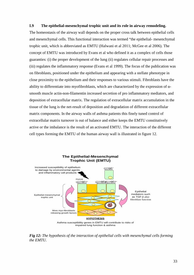
I.9 The epithelial-mesenchymal trophic unit and its role in airway remodeling.
The homeostasis of the airway wall depends on the proper cross talk between epithelial cells
and mesenchymal cells. This functional interaction was termed “the epithelial- mesenchymal
trophic unit, which is abbreviated as EMTU (Halwani et al 2011; McGee et al 2006). The
concept of EMTU was introduced by Evans et al who defined it as a complex of cells those
guaranties: (i) the proper development of the lung (ii) regulates cellular repair processes and
(iii) regulates the inflammatory response (Evans et al 1999). The focus of the publication was
on fibroblasts, positioned under the epithelium and appearing with a stellate phenotype in
close proximity to the epithelium and their responses to various stimuli. Fibroblasts have the
ability to differentiate into myofibroblasts, which are characterized by the expression of α-
smooth muscle actin-non-filamentin increased secretion of pro inflammatory mediators, and
deposition of extracellular matrix. The regulation of extracellular matrix accumulation in the
tissue of the lung is the net-result of deposition and degradation of different extracellular
matrix components. In the airway walls of asthma patients this finely tuned control of
extracellular matrix turnover is out of balance and either keeps the EMTU constitutively
active or the imbalance is the result of an activated EMTU. The interaction of the different
cell types forming the EMTU of the human airway wall is illustrated in figure 12.
The Epithelial-MesenchymalTrophic Unit (EMTU)
Epithelialmediators such as TGF-β alter
fibroblast function
HYPOTHESIS
Asthma susceptibility genes in EMTU will contribute to risks of impaired lung function & asthma
Increased susceptibility of epitheliumto damage by environmental agents
and inflammatory cell products
mucus
Epithelial-mesenchymaltrophic unit
More myo-fibroblastsreleasing growth factors
Fig 12: The hypothesis of the interaction of epithelial cells with mesenchymal cells forming the EMTU.
34
Today it is not clear what is the initiating event that deregulates the balance of the EMTU in
asthma, however, it leads to hyper-responsiveness, and thickening of the airway wall due to
increased extracellular matrix deposition and increased mesenchymal cell number. Together
these factors reduce the airway flexibility and the airway lumen which gives the patient the
feeling of not being able to breathe (Holgate et al 2000). The healthy epithelium acts as a
barrier of the airway wall to the inhaled air which carries a multitude of external factors
including allergens, dust etc, and while in sensitive patients cause an asthma exacerbation.
There is experimental evidence that the epithelium regulates the homeostasis of the sub-
epithelial tissue, and when its integrity or function is compromise, the EMTU is activated and
tries to protect and repair the damaged tissue (Hostettler et al 2008). Damage of the
epithelium initiates a repair process including epithelial to mesenchymal transition (EMT) and
cell migration. Two important cytokines that regulate the EMT are EGF and TGF-β, which
both stimulate myofibroblasts to increase the deposition of extracellular matrix in asthmatics
patients (Brewster et al 1990). If the tissue repair process is normal, the fibroblasts-
myofibroblasts go in apoptosis or return into the sub mucosa, but this doesn’t occur in asthma
and other chronic inflammatory lung diseases (Harold et al 2003). Studies of undifferentiated
epithelial cells and fibroblasts, confirmed that a change of the EMTU stimulates the release of
TGF-β from both cell types (Thompson et al 2006). TGF-β, not only activated fibroblasts to
depose more extracellular matrix, but studying its action led to identify the first genetic pre-
condition implicated in asthma which was linked to the development of airway hyper-
responsiveness: ADAM33 (Van Eerdewegh et al 2002).
ADAM33 expression was associated with a rapid decline in lung function not only in asthma
patients (Jongepier et al 2004), in asthmatics and in subjects with COPD (Gosman et al 2007)
, and in the general population (van Diemen et al 2005). ADAM 33 is a gene consisting of 22
exons that encode a full-length molecule of 813 amino acids containing a metalloprotease
domain, a disintegrin domain, an EGF domain and a cytoplasmic domain. ADAM33 belongs
to the family of multifunctional membrane glycoprotein-anchores that mediates cell-cell
interactions as well as cell-extracellular matrix interaction (Black et al 1998). The discovery
of soluble ADAM33 that contain the metalloprotease domain in the broncho-alveolar fluid
(BALF) of subjects with asthma, but not in BALF of healthy subjects indicated that levels of
sADAM33 are inversely correlated with lung function (Lee et al 2006). The soluble
ADAM33 also promoted new blood vessel formation in the airway wall which is pathology of
severe asthma (Puxeddu et al 2008).
35
The function of the healthy epithelium is that of a barrier which protects the airway tissue
from inhaled substances such as chemicals, pollen, allergens, and dust. Studies examining
bronchial biopsies of subjects with moderate and severe asthma showed that there is an injury
of the epithelium colonnade, with the presence of several markers of cellular stress. It was
therefore assumed that the stress condition causes incorrect tissue remodeling of the damaged
epithelium, resulting in an epithelium cell layer that remains not fully closed at many places.
These gaps are permitting exogenous substances to penetrate the airway wall, as is show in
the figure 13 below, resulting in a chronic airway wall inflammatory response (Truong-Tran
et al 2002). In fact there is evidence that epithelial cells from subjects with chronic airway
inflammation form an incompletely epithelial cell layer (Knight et al 2002; Knight et al,
2003). This condition of a damaged epithelium which is improperly repaired continues to
secrete cytokines and other growth factors. This chronic inflammatory condition causes the
infiltration of a large number of eosinophils, which are the source of TGF-β, and are
responsible for the differentiation of fibroblasts into myo-fibroblasts, which then depose more
extracellular matrix. Eosinophils may also interact with the mucus secreted largely by goblet
cells, thereby increasing the viscosity of the mucus present in individuals with this asthma
(Rose et al 2006).
In summary, the cross talk between the epithelium and the underlying mesenchyme appears to
be a central guide for the homeostasis of the healthy lung and is disrupted in asthma. The
expression of ADAM33 and its involvement in EMTU function helped to understand airway
remodeling in asthma (Jongepier et al 2004). Today we know more than 200 genes which
have been associated with different pathologies of asthma, but unfortunately none of them can
be used as a marker for asthma diagnosis, or pathogenesis, or as a new therapeutic target. The
airway wall can be considered as a complex called EMTU, epithelial mesenchymal trophic
unit, which is fully responsible for the cellular homeostasis of the healthy lung and that an
imbalance of this system leads to remodeling, sometimes even in the absence of any
detectable inflammation (Bousquet et al 2000).
36
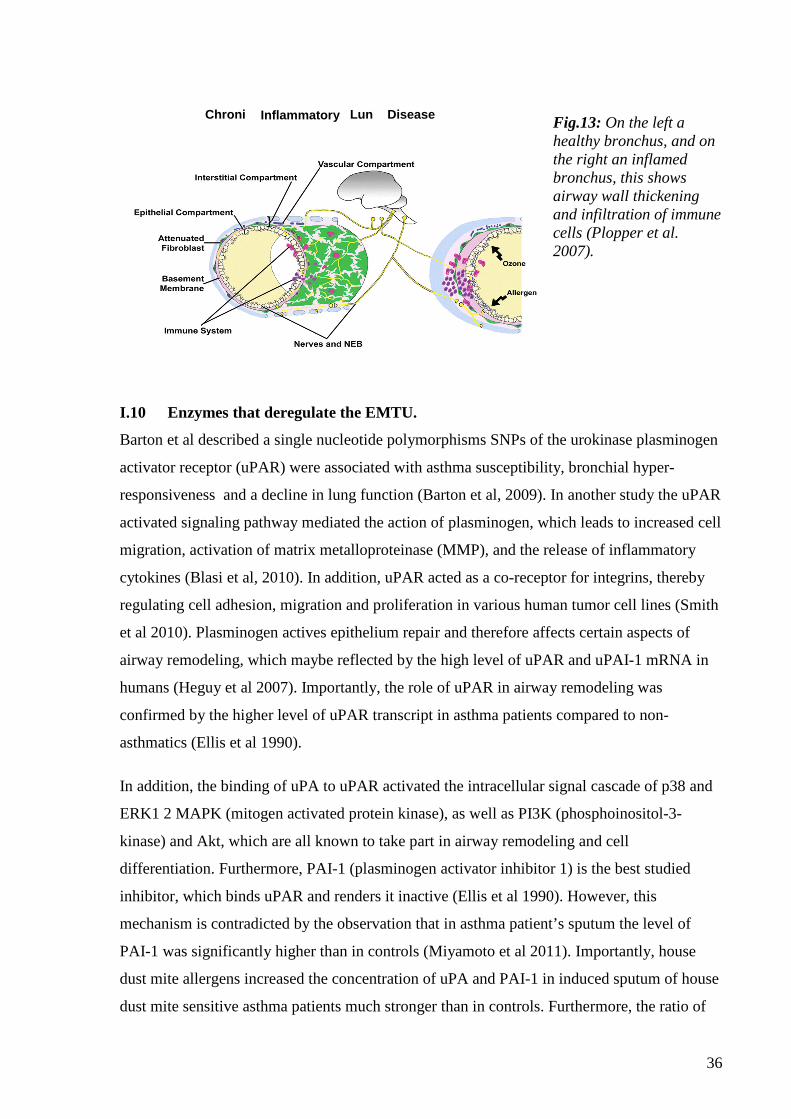
Fig.13: On the left a healthy bronchus, and on the right an inflamed bronchus, this shows airway wall thickening and infiltration of immune cells (Plopper et al. 2007).
I.10 Enzymes that deregulate the EMTU.
Barton et al described a single nucleotide polymorphisms SNPs of the urokinase plasminogen
activator receptor (uPAR) were associated with asthma susceptibility, bronchial hyper-
responsiveness and a decline in lung function (Barton et al, 2009). In another study the uPAR
activated signaling pathway mediated the action of plasminogen, which leads to increased cell
migration, activation of matrix metalloproteinase (MMP), and the release of inflammatory
cytokines (Blasi et al, 2010). In addition, uPAR acted as a co-receptor for integrins, thereby
regulating cell adhesion, migration and proliferation in various human tumor cell lines (Smith
et al 2010). Plasminogen actives epithelium repair and therefore affects certain aspects of
airway remodeling, which maybe reflected by the high level of uPAR and uPAI-1 mRNA in
humans (Heguy et al 2007). Importantly, the role of uPAR in airway remodeling was
confirmed by the higher level of uPAR transcript in asthma patients compared to non-
asthmatics (Ellis et al 1990).
In addition, the binding of uPA to uPAR activated the intracellular signal cascade of p38 and
ERK1 2 MAPK (mitogen activated protein kinase), as well as PI3K (phosphoinositol-3-
kinase) and Akt, which are all known to take part in airway remodeling and cell
differentiation. Furthermore, PAI-1 (plasminogen activator inhibitor 1) is the best studied
inhibitor, which binds uPAR and renders it inactive (Ellis et al 1990). However, this
mechanism is contradicted by the observation that in asthma patient’s sputum the level of
PAI-1 was significantly higher than in controls (Miyamoto et al 2011). Importantly, house
dust mite allergens increased the concentration of uPA and PAI-1 in induced sputum of house
dust mite sensitive asthma patients much stronger than in controls. Furthermore, the ratio of
Chronic
Inflammatory Lung
Diseases
37
uPA / PAI-1 was lower in asthma patients before and after inhalation of house dust mite
allergens (Kowal et al 2010). The results suggest that the reduced ratio of uPA / PAI-1 may
promote airway remodeling and play an important role in the development of bronchial
hyper-reactivity.
In mice which were genetically deficient for PAI-1 exogenous plasminogen reduced sub-
epithelial bronchial wall thickening, as well as collagen deposition, and α-smooth muscle
actin expression (Kuramoto et al 2009). During the embryonic development of the lung,
fibroblast growth factor 10 (FGF10) is expressed by distal epithelial progenitor cells and
prevents the cell’s differentiation, while promoting their proliferation (Bellusci et al 1997).
FGF10 is also secreted by mesenchymal progenitor cells when stimulated with β-catenin,
suggesting that both cell types regulate lung development and differentiations in a controlled
system (De Langhe et al 2008).
In animal experiments naphthalene activated epithelial progenitor cells through Wnt/FGF10, a
process which is usually restricted to embryogenesis. This makes it likely that there are cell
types (stem cells) which are able to be rejuvenate the lung structure by specific stimuli and
thus control airway remodeling in the adult lung (Volckaert et al 2011).
Most exacerbations in asthma are due to infections, bacterial or viral, which first destroy the
epithelium and then penetrate the airway wall into the mesenchymal cell layers. To effectively
combat infection, the epithelium requires the assistance of neutrophils recruited from the
peripheral circulation. Activated neutrophils migrate through the epithelium towards the
tissue lesion. To achieve this, the neutrophils have to produce and release proteolytic enzymes
and after reaching the tissue lesion they produce oxygen radicals in order to destroy the
pathogens. Unfortunately, radical oxygen’s, if produced into large quantities attenuate chronic
inflammation and recruitment more neutrophils into the respiratory tract which further
increases the inflammation in asthma (Tam et al 2011). An overview of EMTU regulating
enzymes and their relevance of asthma and airway wall remodeling is provided in figure 14.
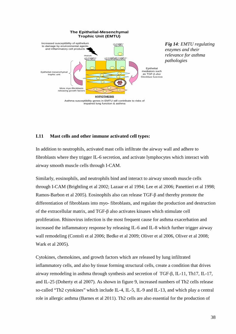
38
The Epithelial-MesenchymalTrophic Unit (EMTU)
Epithelialmediators such as TGF-β alter
fibroblast function
HYPOTHESIS
Asthma susceptibility genes in EMTU will contribute to risks of impaired lung function & asthma
Increased susceptibility of epitheliumto damage by environmental agents
and inflammatory cell products
mucus
Epithelial-mesenchymaltrophic unit
More myo-fibroblastsreleasing growth factors
Fig 14: EMTU regulating enzymes and their relevance for asthma pathologies
I.11 Mast cells and other immune activated cell types:
In addition to neutrophils, activated mast cells infiltrate the airway wall and adhere to
fibroblasts where they trigger IL-6 secretion, and activate lymphocytes which interact with
airway smooth muscle cells through I-CAM.
Similarly, eosinophils, and neutrophils bind and interact to airway smooth muscle cells
through I-CAM (Brightling et al 2002; Lazaar et al 1994; Lee et al 2006; Panettieri et al 1998;
Ramos-Barbon et al 2005). Eosinophils also can release TGF-β and thereby promote the
differentiation of fibroblasts into myo- fibroblasts, and regulate the production and destruction
of the extracellular matrix, and TGF-β also activates kinases which stimulate cell
proliferation. Rhinovirus infection is the most frequent cause for asthma exacerbation and
increased the inflammatory response by releasing IL-6 and IL-8 which further trigger airway
wall remodeling (Contoli et al 2006; Bedke et al 2009; Oliver et al 2006, Oliver et al 2008;
Wark et al 2005).
Cytokines, chemokines, and growth factors which are released by lung infiltrated
inflammatory cells, and also by tissue forming structural cells, create a condition that drives
airway remodeling in asthma through synthesis and secretion of TGF-β, IL-11, Th17, IL-17,
and IL-25 (Doherty et al 2007). As shown in figure 9, increased numbers of Th2 cells release
so-called “Th2 cytokines” which include IL-4, IL-5, IL-9 and IL-13, and which play a central
role in allergic asthma (Barnes et al 2011). Th2 cells are also essential for the production of
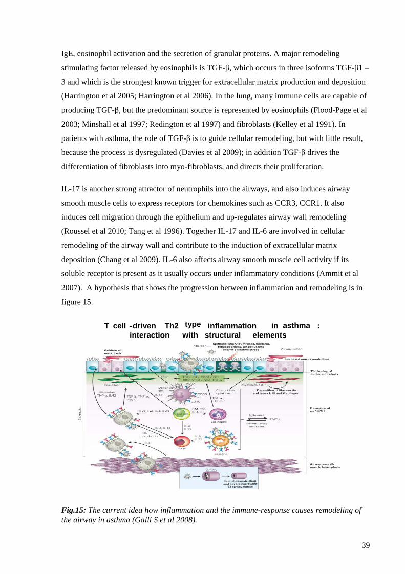
39
IgE, eosinophil activation and the secretion of granular proteins. A major remodeling
stimulating factor released by eosinophils is TGF-β, which occurs in three isoforms TGF-β1 –
3 and which is the strongest known trigger for extracellular matrix production and deposition
(Harrington et al 2005; Harrington et al 2006). In the lung, many immune cells are capable of
producing TGF-β, but the predominant source is represented by eosinophils (Flood-Page et al
2003; Minshall et al 1997; Redington et al 1997) and fibroblasts (Kelley et al 1991). In
patients with asthma, the role of TGF-β is to guide cellular remodeling, but with little result,
because the process is dysregulated (Davies et al 2009); in addition TGF-β drives the
differentiation of fibroblasts into myo-fibroblasts, and directs their proliferation.
IL-17 is another strong attractor of neutrophils into the airways, and also induces airway
smooth muscle cells to express receptors for chemokines such as CCR3, CCR1. It also
induces cell migration through the epithelium and up-regulates airway wall remodeling
(Roussel et al 2010; Tang et al 1996). Together IL-17 and IL-6 are involved in cellular
remodeling of the airway wall and contribute to the induction of extracellular matrix
deposition (Chang et al 2009). IL-6 also affects airway smooth muscle cell activity if its
soluble receptor is present as it usually occurs under inflammatory conditions (Ammit et al
2007). A hypothesis that shows the progression between inflammation and remodeling is in
figure 15.
Fig.15: The current idea how inflammation and the immune-response causes remodeling of the airway in asthma (Galli S et al 2008).
T cell -driven Th2 type inflammation in asthmaa
: interaction with structural elements
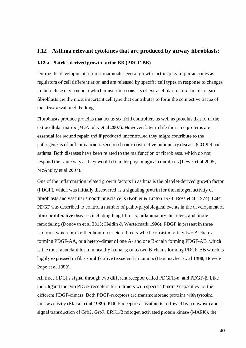
40
I.12 Asthma relevant cytokines that are produced by airway fibroblasts: I.12.a Platelet-derived growth factor-BB (PDGF-BB)
During the development of most mammals several growth factors play important roles as
regulators of cell differentiation and are released by specific cell types in response to changes
in their close environment which most often consists of extracellular matrix. In this regard
fibroblasts are the most important cell type that contributes to form the connective tissue of
the airway wall and the lung.
Fibroblasts produce proteins that act as scaffold controllers as well as proteins that form the
extracellular matrix (McAnulty et al 2007). However, later in life the same proteins are
essential for wound repair and if produced uncontrolled they might contribute to the
pathogenesis of inflammation as seen in chronic obstructive pulmonary disease (COPD) and
asthma. Both diseases have been related to the malfunction of fibroblasts, which do not
respond the same way as they would do under physiological conditions (Lewis et al 2005;
McAnulty et al 2007).
One of the inflammation related growth factors in asthma is the platelet-derived growth factor
(PDGF), which was initially discovered as a signaling protein for the mitogen activity of
fibroblasts and vascular smooth muscle cells (Kohler & Lipton 1974; Ross et al. 1974). Later
PDGF was described to control a number of patho-physiological events in the development of
fibro-proliferative diseases including lung fibrosis, inflammatory disorders, and tissue
remodeling (Donovan et al 2013; Heldin & Westermark 1996). PDGF is present in three
isoforms which form either homo- or heterodimers which consist of either two A-chains
forming PDGF-AA, or a hetero-dimer of one A- and one B-chain forming PDGF-AB, which
is the most abundant form in healthy humans; or as two B-chains forming PDGF-BB which is
highly expressed in fibro-proliferative tissue and in tumors (Hammacher et. al 1988; Bowen-
Pope et al 1989).
All three PDGFs signal through two different receptor called PDGFR-α, and PDGF-β. Like
their ligand the two PDGF receptors form dimers with specific binding capacities for the
different PDGF-dimers. Both PDGF-receptors are transmembrane proteins with tyrosine
kinase activity (Matsui et al 1989). PDGF receptor activation is followed by a downstream
signal transduction of Grb2, Grb7, ERK1/2 mitogen activated protein kinase (MAPK), the
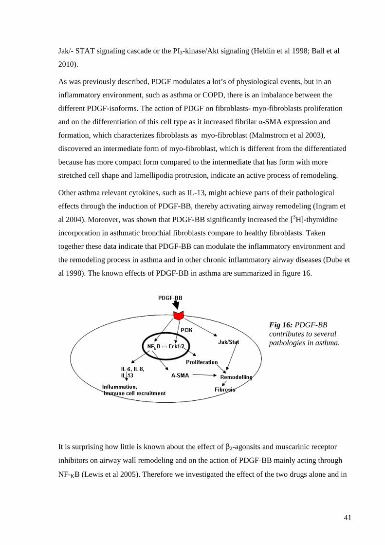
41
Jak/- STAT signaling cascade or the PI3-kinase/Akt signaling (Heldin et al 1998; Ball et al
2010).
As was previously described, PDGF modulates a lot’s of physiological events, but in an
inflammatory environment, such as asthma or COPD, there is an imbalance between the
different PDGF-isoforms. The action of PDGF on fibroblasts- myo-fibroblasts proliferation
and on the differentiation of this cell type as it increased fibrilar α-SMA expression and
formation, which characterizes fibroblasts as myo-fibroblast (Malmstrom et al 2003),
discovered an intermediate form of myo-fibroblast, which is different from the differentiated
because has more compact form compared to the intermediate that has form with more
stretched cell shape and lamellipodia protrusion, indicate an active process of remodeling.
Other asthma relevant cytokines, such as IL-13, might achieve parts of their pathological
effects through the induction of PDGF-BB, thereby activating airway remodeling (Ingram et
al 2004). Moreover, was shown that PDGF-BB significantly increased the [3H]-thymidine
incorporation in asthmatic bronchial fibroblasts compare to healthy fibroblasts. Taken
together these data indicate that PDGF-BB can modulate the inflammatory environment and
the remodeling process in asthma and in other chronic inflammatory airway diseases (Dube et
al 1998). The known effects of PDGF-BB in asthma are summarized in figure 16.
Fig 16: PDGF-BB contributes to several pathologies in asthma.
It is surprising how little is known about the effect of β2-agonsits and muscarinic receptor
inhibitors on airway wall remodeling and on the action of PDGF-BB mainly acting through
NF-ΚB (Lewis et al 2005). Therefore we investigated the effect of the two drugs alone and in
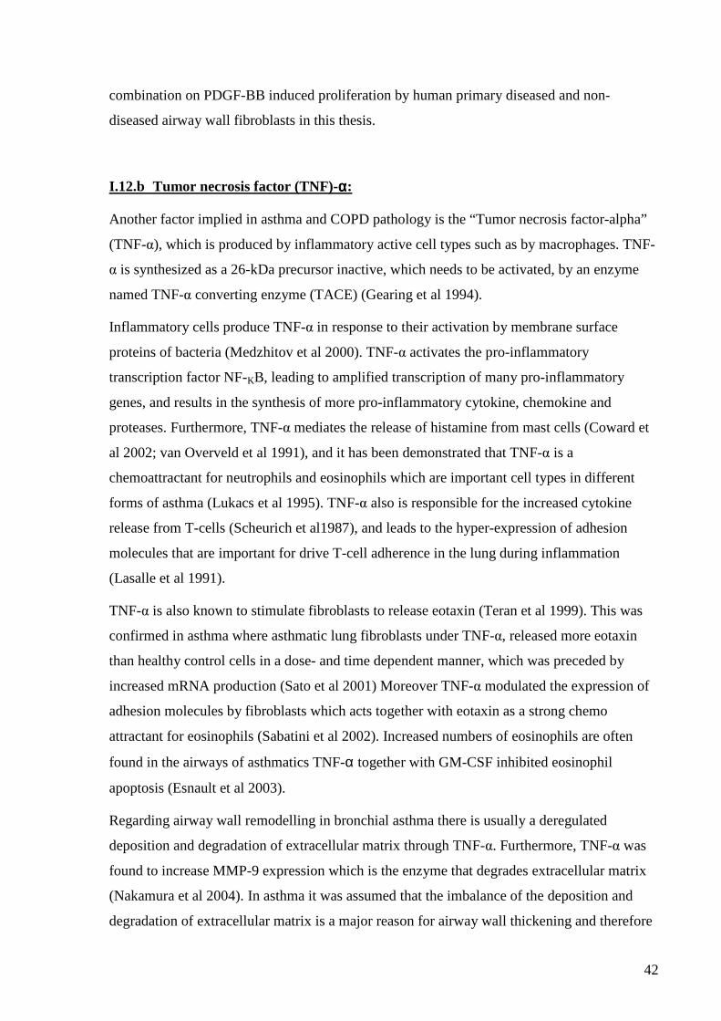
42
combination on PDGF-BB induced proliferation by human primary diseased and non-
diseased airway wall fibroblasts in this thesis.
I.12.b Tumor necrosis factor (TNF)-αααα:
Another factor implied in asthma and COPD pathology is the “Tumor necrosis factor-alpha”
(TNF-α), which is produced by inflammatory active cell types such as by macrophages. TNF-
α is synthesized as a 26-kDa precursor inactive, which needs to be activated, by an enzyme
named TNF-α converting enzyme (TACE) (Gearing et al 1994).
Inflammatory cells produce TNF-α in response to their activation by membrane surface
proteins of bacteria (Medzhitov et al 2000). TNF-α activates the pro-inflammatory
transcription factor NF-KB, leading to amplified transcription of many pro-inflammatory
genes, and results in the synthesis of more pro-inflammatory cytokine, chemokine and
proteases. Furthermore, TNF-α mediates the release of histamine from mast cells (Coward et
al 2002; van Overveld et al 1991), and it has been demonstrated that TNF-α is a
chemoattractant for neutrophils and eosinophils which are important cell types in different
forms of asthma (Lukacs et al 1995). TNF-α also is responsible for the increased cytokine
release from T-cells (Scheurich et al1987), and leads to the hyper-expression of adhesion
molecules that are important for drive T-cell adherence in the lung during inflammation
(Lasalle et al 1991).
TNF-α is also known to stimulate fibroblasts to release eotaxin (Teran et al 1999). This was
confirmed in asthma where asthmatic lung fibroblasts under TNF-α, released more eotaxin
than healthy control cells in a dose- and time dependent manner, which was preceded by
increased mRNA production (Sato et al 2001) Moreover TNF-α modulated the expression of
adhesion molecules by fibroblasts which acts together with eotaxin as a strong chemo
attractant for eosinophils (Sabatini et al 2002). Increased numbers of eosinophils are often
found in the airways of asthmatics TNF-α together with GM-CSF inhibited eosinophil
apoptosis (Esnault et al 2003).
Regarding airway wall remodelling in bronchial asthma there is usually a deregulated
deposition and degradation of extracellular matrix through TNF-α. Furthermore, TNF-α was
found to increase MMP-9 expression which is the enzyme that degrades extracellular matrix
(Nakamura et al 2004). In asthma it was assumed that the imbalance of the deposition and
degradation of extracellular matrix is a major reason for airway wall thickening and therefore
43
of airway lumen reduction. The known effects of PDGF-BB in asthma are summarized in
figure 17.
Fig 17: The known effects of TNF−alpha in asthma are summarized in figure 11.
Surprisingly not much is known about the effect of β2-agonsits and muscarinic receptor
inhibtors on airway wall remodelling and on the action of TNF-α. Therefore we investigated
the effect of the two drugs alone and in combination on TNF-α release by human primary
diseased and non-diseased airway wall fibroblasts in this thesis.
I.12.c Interleukin-1ββββ:
IL-1β is one of the most important cytokines that is assumed to initiate lung inflammation in
asthma and COPD and also plays an important part in the maintenance of inflammatory
conditions (Dinarello et al. 1996). IL-1β is synthesized as a precursor, which is activated by
the interleukin-1 converting enzyme (ICE), which limits its biological availability of IL-1β
(Wilson et al. 1994). IL-1β is produced by many different cell types, including fibroblasts,
monocytes, tissue and alveolar macrophages, smooth muscle cell, vessel wall endothelial
cells, neutrophils, and airway epithelial cells (Ackerman et al. 1994; Wagner et al. 1985;
Chung et al. 2001).
In regard to asthma IL-1β stimulates leukocytosis by inducing neutrophils to infiltrate the
lung. IL-1β also stimulates the secretion of other pro-inflammatory cytokines, such as IL-6,
IL-8, GM-CSF and TNF-α from many cell types. IL-1β stimulates specifically granulocytes,
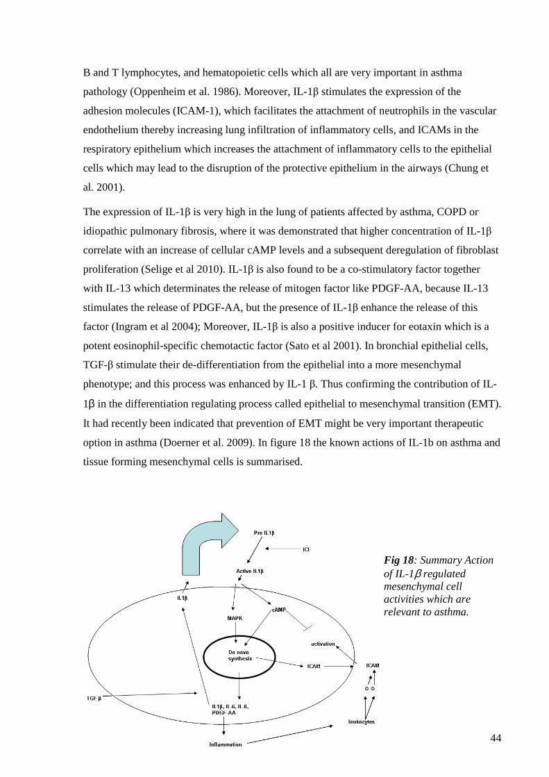
44
B and T lymphocytes, and hematopoietic cells which all are very important in asthma
pathology (Oppenheim et al. 1986). Moreover, IL-1β stimulates the expression of the
adhesion molecules (ICAM-1), which facilitates the attachment of neutrophils in the vascular
endothelium thereby increasing lung infiltration of inflammatory cells, and ICAMs in the
respiratory epithelium which increases the attachment of inflammatory cells to the epithelial
cells which may lead to the disruption of the protective epithelium in the airways (Chung et
al. 2001).
The expression of IL-1β is very high in the lung of patients affected by asthma, COPD or
idiopathic pulmonary fibrosis, where it was demonstrated that higher concentration of IL-1β
correlate with an increase of cellular cAMP levels and a subsequent deregulation of fibroblast
proliferation (Selige et al 2010). IL-1β is also found to be a co-stimulatory factor together
with IL-13 which determinates the release of mitogen factor like PDGF-AA, because IL-13
stimulates the release of PDGF-AA, but the presence of IL-1β enhance the release of this
factor (Ingram et al 2004); Moreover, IL-1β is also a positive inducer for eotaxin which is a
potent eosinophil-specific chemotactic factor (Sato et al 2001). In bronchial epithelial cells,
TGF-β stimulate their de-differentiation from the epithelial into a more mesenchymal
phenotype; and this process was enhanced by IL-1 β. Thus confirming the contribution of IL-
1β in the differentiation regulating process called epithelial to mesenchymal transition (EMT).
It had recently been indicated that prevention of EMT might be very important therapeutic
option in asthma (Doerner et al. 2009). In figure 18 the known actions of IL-1b on asthma and
tissue forming mesenchymal cells is summarised.
Fig 18: Summary Action of IL-1β regulated mesenchymal cell activities which are relevant to asthma.
45
Only little data have been reported for the effect of β2-agonsits and muscarinic receptor
inhibitors on IL-1β stimulated airway wall remodeling suggesting a central role of cAMP
(Selige et al 2010). Therefore we investigated the effect of the two drugs alone and in
combination on IL-1β release by human primary diseased and non-diseased airway wall
fibroblasts in this thesis.
46
II. MATERIALS and METHODS:
II.1 Primary human lung fibroblasts.
Lung tissue specimens were obtained from the Department of Internal Medicine,
Pneumology, University Hospital Basel, Basel, Switzerland, with the approval of the local
Ethnical Committee (EK:05/06) and written consent of each patient.
Primary human lung fibroblasts were isolated from tissue of non-asthma and asthma patient
lung biopsies as previously described and maintained and expanded under standard cell
culture conditions (37 °C, 100% humidity, 5% CO2, 95% air) [Tamm M et al 2001].
Fibroblast growth medium consisted of RPMI 1640 supplemented with 8 mM L-Glutamine,
25mM HEPES, 1x anti-biotic/anti-mycotic, 1x MEM vitamin, and 10% fetal bovine serum
(FBS) (all: Gibco/Invitrogen Corp., Grand Island, NY).
II.2 Primary human lung fibroblasts characterization.
We have characterized primary human lung fibroblasts for the expression of α-smooth muscle
actin (SMA) immuno-staining as shown in figure 5. Cells were grown to 80% confluence and
than fixed with 2 x 5 minutes incubation in 2% formalin (in PBS), followed by blocking
unspecific antibody binding with 1% bovine serum albumin in PBS for 20 minutes. The first
rabbit anti-body, specific to α-smooth muscle actin was diluted 1:1000 and incubated over
night (4oC) before being washed off with 3 x 5 minutes blocking buffer, followed by 1 hour
incubation with a species specific FITC labeled antibody (all Santa Cruz Biotechnology, CA).
Unbound antibodies were washed off by 3 x 5 minutes blocking buffer and 1 x PBS. The
Fluorescence was monitored by microscopy and the arrangement of α-SMA in filaments was
used as indicator for myo-fiborblasts, while diffuse cytosolic staining was the indicator for
fibroblasts (Fig. 5).
II.3 Drug treatment.
Fibroblasts were seeded (104 cells/cm2) into 12 and 24 well plates and grown until 90%
confluence. The cells were deprived of serum for 24 hours before being incubated for 24
hours in the presence of increasing concentration of the LABA olodaterol (10-9 to 10-6 M), or
with the LAMA tiotropium bromide (10-8 to 10-5 M), and/or carbachol (10-8 to 10-5 M) in the
47
following combinations: (i) carbachol + tiotropium followed by stimulation with IL-1β; or (ii)
olodaterol + tiotropium for 30 min followed by stimulation with IL-1β (10 ng/mL).
Recombinant human platelet-derived growth factor (PDGF)-BB (R&D Systems Europe,
Abingdon, United Kingdom) was dissolved in sterile 4 mM HCL to a stock concentration of
100µg/mL; Recombinant human TNF-α (R&D Systems Europe Ltd.) was dissolved to a
concentration of 100µg/mL in sterile PBS (Dulbecco’s PBS 1x, PAA-Laboratories GmbH)
containing 0.1% human or bovine serum albumin; Recombinant Human IL-1β (R&D Systems
Europe Ltd.), was reconstituted at 25 µg/mL in sterile phosphate buffered saline ( PBS 1x,
PAA-Laboratories), containing 0.1% bovine serum albumin. Carbachol and olodaterol (BI
1744 CL) were dissolved in RPMI 1640 medium. Tiotropium bromide (Ba 679 BR) was
dissolved in ethanol. Olodaterol and tiotropium bromide were provided by Boehringer
Ingelheim Pharma GmbH & Co. KG (Biberach, Germany).
II 4 Cytokine secretion.
Sub-confluent fibroblasts (90%) in 12 well plates were serum starved for 24 hours before
treatment. PDGF-BB, TNF-α, or IL-1β, were added to fibroblasts (0.1, 1, 10 ng/mL) in the
presence or absence of the drugs as described in the previous chapter. After 24 hours, cell
culture medium samples were collected. IL-6 and IL-8 levels in the cell culture media were
determined by commercial enzyme-linked immune sorbent assays (ELISA) following the
distributors instruction (Ani Biotech Oy, Orgenium Laboratories, Vantaa, Finland).
II.5 Protein expression.
Cellular proteins were isolated from confluent cells by dissociation in lysis buffer, (62.5 mM
Tris-HCL, pH 6.8; 2% sodium dodecylsulfate, 2% β-mercapto-ethanol, 10% glycerol) and
analyzed by immuno-blotting as described earlier [Roth M et al 2004] for the expression of
cAMP response element binding protein (CREB), phosphorylated CREB (P-CREB) and α-
tubulin (all: Cell Signaling Technology).
48
II.6 CREB EMSA
A non radioactive commercial available CREB EMSA was used to determine CREB DNA
binding following the instruction of producer. Cells were grown to sub-confluence and after
incubation with different drug, were treated for the nuclear protein extraction.
Cell were washed with 10 ml ice-cold PBS7PIB; 10 ml ice-cold PBS/PIB were added and
cells were scraped and transferred into a pre-chilled 15 ml tube and spin at 300 x g for 5
minutes at 4°C.
The pellet was placed in 1 ml ice-cold HB buffer by gentle pipetting and transferring the cells
into a pre-chilled 1.5 ml tube.
Cells were left to swell on ice for 15 minutes; 50 microl 10% Nonidet P-40 (0.5% final) and
vortex the tube vigorously for 10 seconds.
The homogenate was centrifuged for 30 seconds at 4°C; the supernatant (cytoplasmic
fraction) was removed and stored in another tube at -80°C.
The nuclear pellet was re-suspended in 50 microl Complete Lysis Buffer and rock the tube
gently on ice for 30 minutes on a shaking platform.
After centrifuge for 10 minutes at 14000 x g at 4°C we saved the supernatant (nuclear
extract).
Protein concentration was determined by using a Bradford-based assay.
II.7 cAMP detection.
Sub-confluent fibroblasts (90%) grown in 24 well plates were serum deprived for 24 hours.
To block phosphodiesterase activity, cells were pretreated with iso-butyl-methylxantine
(IBMX, Sigma) for 10 minutes, then stimulated as follows: (i) serum-free medium, (ii) IL-1β
(10 ng/ml), (iii) carbachol 10-5 M, (iv) tiotropium 10-6 M, (v) olodaterol 10-6 M, (vi) IL-1β +
olodaterol 10-6 M, (vii) IL-1β + olodaterol 10-6 M + carbachol 10-5 M (added 30 minutes prior
to all other stimuli), (viii) IL-1β + olodaterol 10-6 M + carbachol 10-5 M + tiotropium 10-6 M
(added 30 minutes prior to all other stimuli). After 10 minutes cells were lysed and cAMP
levels were determined by ELISA following the instruction of the producer (R&D Systems
Europe Ltd.).
49
To mimic the activity of tiotropium, cells were 30 minutes pretreated with Pertussis toxin
(ENZO), then stimulated as follows: (i) serum-free medium, (ii) olodaterol 10-6 M, (iii)
olodaterol 10-6 M + IL-1β (5 ng/ml), (iv) carbachol 10-5 M +olodaterol 10-6 M + IL-1β (5
ng/ml), (v) carbachol 10-5 M + olodaterol 10-6 M + IL-1β + Pertussis toxin (added 30 minutes
prior to all other stimuli). After 10 minutes cells were lysed and cyclic AMP levels were
determined by ELISA following the instruction of the producer (R&D Systems Europe Ltd.).
II.8 Statistics.
Cytokine data has been calculated as percent change compared to the release of IL-1β
stimulated cells and the data are presented as mean ± SEM. After checking the data for
normal distribution, paired or unpaired, two-tailed student’s t-test was performed and p-values
< 0.05 were considered significant.
50
III. Effect of muscarinic receptors and b2-adregernic receptors on fibroblast
proliferation.
III.1 Control of fibroblast proliferation in asthma and COPD by muscarinic receptors and β2
receptors
Asthma and COPD are characterised by increased mass of sub-epithelial mesenchymal cells
including fibroblasts, myo-fibroblasts and smooth muscle cells. The origin of these cells is
discussed controversly and several scenarios have been suggested: a) cell migration from
surrounding tissues (Kohan et al 2012), b) cell phenotype change (b.1) from fibroblasts to
myo-fibroblast (Milara et al 2012) or (b.2) epithelial to mesenchymal transition (EMT)
(Crosby & Waters 2010), 3) infiltration of circulating fibrocytes that originate from the bone
marrow (Mehrad & Strieter 2012; Tsai et al 2013), or 4) proliferation of resident tissue cells
(Zhang et al 2012).
Recent studies in volunteering asthma patients have challenged the hypothesis that asthma is
caused by an over reacting immune system (Grainge et al 2011; Bossley et al 2012). Not only
that airway remodelling is one of the first pathologies recognised even in children with
wheezing and asthma (Bossley et al 2012; O’Reilly et al 2012); in addition, it has been shown
that either the stimulation of the muscarinic receptors or the administration of allergens lead
to remodelling of the airway within 8 days (Grainge et al 2011). Remodelling occurred
without any sign of inflammation and was significantly reduced if the patients were pre-
treated with a muscle relaxant. These findings are well in line with studies in non-human
primates (rhesus monkeys), in which it has been impressively shown that the arrangement of
the smooth muscle bundles around the airways is completely different in animals who grew
up in filtered sterile air compared to those growing up with exposure to allergens or ozone
(Plopper et al 2012; 2008; 2007; Evans et al 2010). There is no other animal model in mouse,
rat or sheep which reflects the human disease (asthma). We and others have reported earlier
that human airway cells maintain pathologies which reflect remodelling and inflammation
under cell culture conditions. In some of these studies, it was shown that the regulation of
intracellular signalling and activity is deregulated in asthma.
In 2004, we reported that proliferation control is deregulated in asthmatic smooth muscle cells
and lead to the hyperplasia which is well documented in clinical studies (Roth et al 2004). In
subsequent studies, we have shown that this deregulated proliferation is explained by a faulty
translation of cell differentiation factor specifically in airway smooth muscle cells of asthma
51
patients (Borger et al 2009) and that well known asthma triggers such as house dust mite can
induce the down regulation of this cell differentiation factor, C/EBP-α (Miglino et al 2011).
We have also shown that non allergic asthma and COPD triggers such as cigarette smoke
affect the expression of C/EBP-α and β in fibroblasts of smoker and respective controls,
thereby increasing cell proliferation and secretion of pro-inflammatory cytokine (Miglino et al
2012).
In vitro experiments suggested that proliferation of mesenchymal cell types can be reduced or
blocked by both 2-adrenergic receptor agonists and M2 receptor inhibitors (Milara et al
2012, Tsai et al 2013, Zhang et al 2012). However, there is only little evidence that this effect
of the drugs also occurs in the diseased lung. Investigating the interaction of MRs with β2
adrenergic receptors, we discovered that the release of cAMP (anti-proliferative, anti-
inflammatory) is lower in fibroblast of asthma patients. A similar disease specific low cAMP
level was reported by our collaborator from Sydney in airway smooth muscle cells of asthma
patients (Niimi et al 2012, Trian et al 2011). The lower level of cAMP in asthma fibroblasts
and smooth muscle cells may also explain the increase activity of mitochondria (cells power
supply) which has been described by one of our collaborator from Bordeaux (Trian et al
2007).
Comparing the proliferative and inflammatory effect of different asthma relevant growth
factors, we observed that fibroblast of asthma patients respond with faster proliferation to
PDGF-BB and this effect is further enhanced when the cells are stimulated by a muscarinic
receptor activator in parallel. Importantly, muscarinic receptor activation alone did not
significantly increase proliferation and inflammation of fibroblasts. It would be of great
importance to understand the pathology of asthma as well as to find new diagnostic and
therapeutic targets by characterising the molecular biological difference of the underlying
signalling pathway, especially for the combination of MR activation with other asthma
relevant cytokines, such as PDGF-BB.
Based on the existing literature, we postulated that muscarinic and tyrosine kinase receptor
signalling may merge and support each other through the activation (phosphorylation) of a
protein complex formed by Shc/Grb2/SOS. In other cells, it has been shown that the M2
receptor activated this complex through PI3 kinase, while the same complex was directly
bound to the PDGF-BB receptor (Sharma et al 2005; Lopez-Ilasaca et al 1997). When
activated, this complex stimulated a signalling cascade which was both pro-proliferative and
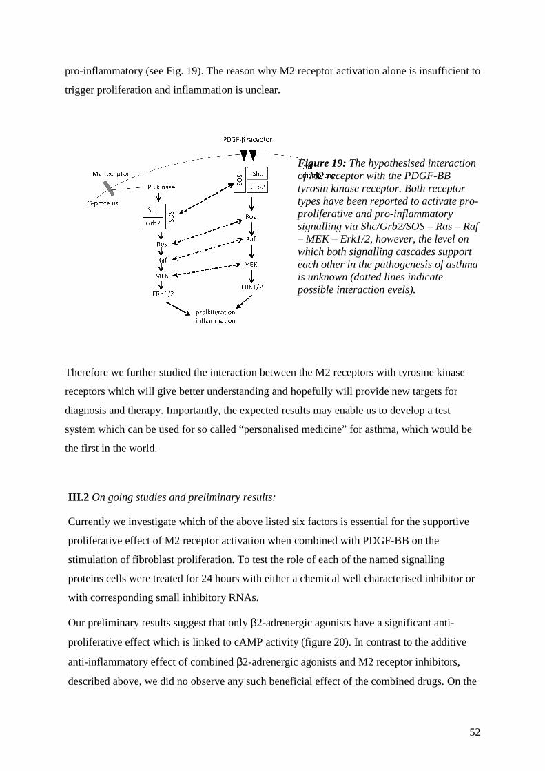
52
pro-inflammatory (see Fig. 19). The reason why M2 receptor activation alone is insufficient to
trigger proliferation and inflammation is unclear.
Figure 19: The hypothesised interaction of M2 receptor with the PDGF-BB tyrosin kinase receptor. Both receptor types have been reported to activate pro-proliferative and pro-inflammatory signalling via Shc/Grb2/SOS – Ras – Raf – MEK – Erk1/2, however, the level on which both signalling cascades support each other in the pathogenesis of asthma is unknown (dotted lines indicate possible interaction evels).
Therefore we further studied the interaction between the M2 receptors with tyrosine kinase
receptors which will give better understanding and hopefully will provide new targets for
diagnosis and therapy. Importantly, the expected results may enable us to develop a test
system which can be used for so called “personalised medicine” for asthma, which would be
the first in the world.
III.2 On going studies and preliminary results:
Currently we investigate which of the above listed six factors is essential for the supportive
proliferative effect of M2 receptor activation when combined with PDGF-BB on the
stimulation of fibroblast proliferation. To test the role of each of the named signalling
proteins cells were treated for 24 hours with either a chemical well characterised inhibitor or
with corresponding small inhibitory RNAs.
Our preliminary results suggest that only β2-adrenergic agonists have a significant anti-
proliferative effect which is linked to cAMP activity (figure 20). In contrast to the additive
anti-inflammatory effect of combined β2-adrenergic agonists and M2 receptor inhibitors,
described above, we did no observe any such beneficial effect of the combined drugs. On the
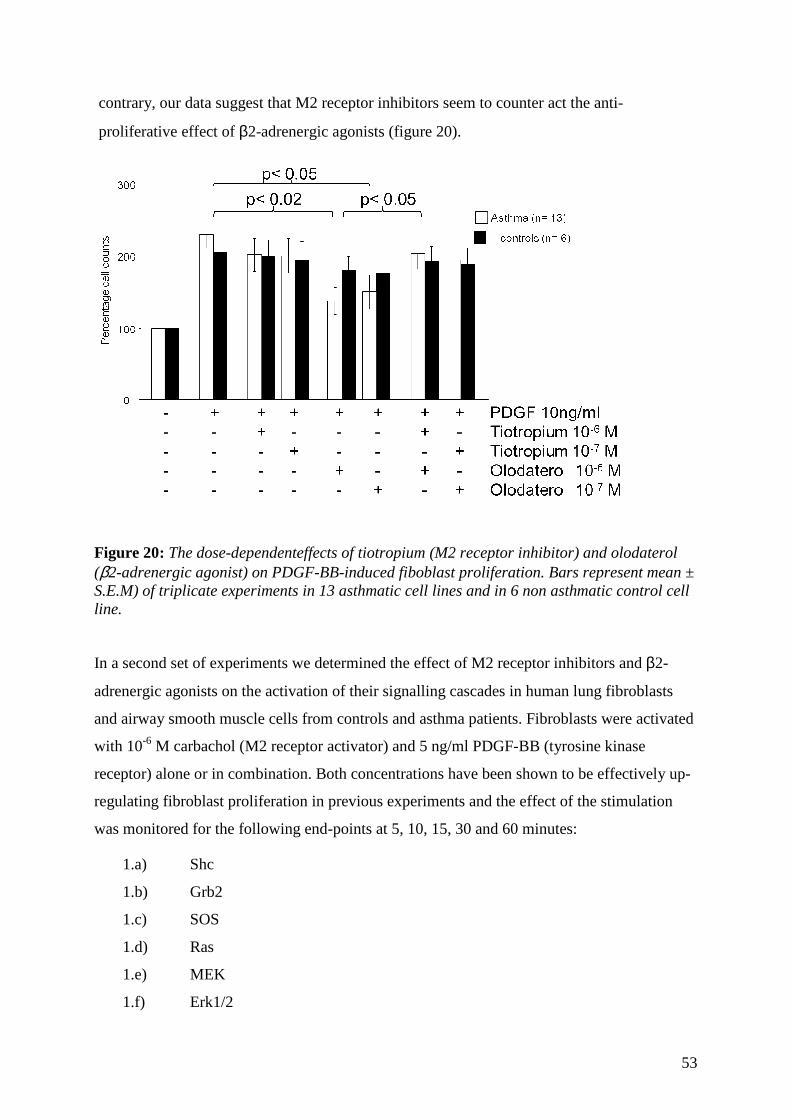
53
contrary, our data suggest that M2 receptor inhibitors seem to counter act the anti-
proliferative effect of β2-adrenergic agonists (figure 20).
Figure 20: The dose-dependenteffects of tiotropium (M2 receptor inhibitor) and olodaterol (β2-adrenergic agonist) on PDGF-BB-induced fiboblast proliferation. Bars represent mean ± S.E.M) of triplicate experiments in 13 asthmatic cell lines and in 6 non asthmatic control cell line.
In a second set of experiments we determined the effect of M2 receptor inhibitors and β2-
adrenergic agonists on the activation of their signalling cascades in human lung fibroblasts
and airway smooth muscle cells from controls and asthma patients. Fibroblasts were activated
with 10-6 M carbachol (M2 receptor activator) and 5 ng/ml PDGF-BB (tyrosine kinase
receptor) alone or in combination. Both concentrations have been shown to be effectively up-
regulating fibroblast proliferation in previous experiments and the effect of the stimulation
was monitored for the following end-points at 5, 10, 15, 30 and 60 minutes:
1.a) Shc
1.b) Grb2
1.c) SOS
1.d) Ras
1.e) MEK
1.f) Erk1/2

54
We hope that the expected results may explain the surprising counteractive effect of combined
M2 receptor inhibitors and β2-adrenergic agonists on proliferation.
55
References
Ackerman,V., M. Marini, E. Vittori, A. Bellini, G. Vassalli, and S. Mattoli. 1994. Detection of cytokines and their cell sources in bronchial biopsy speciments from asthmatic patients. Relationship to atopic status, symptoms, and level of airway hyperresponsiveness. Chest 105:687-696.
Ammit AJ, Moir LM, Oliver BG, Hughes JM, Alkhouri H, Ge Q, Burgess JK, Black JL, Roth M. Effect of IL-6 trans-signaling on the pro-remodeling phenotype of airway smooth muscle. Am J Physiol Lung Cell Mol Physiol. 2007 Jan; 292(1):L199-206.
Anderson WH, Koshy BT, Huang L, Mosteller M, Stinnett SW, Condreay LD, Ortega H. Genetic analysis of asthma exacerbations. Ann Allergy Asthma Immunol. 2013 Jun;110(6):416-422.
Aziz I, McFarlane LC, Lipworth BJ. Concomitant inhaled corticosteroid resensitises cardiac beta2-adrenoceptors in the presence of long-actingbeta2-agonist therapy. Eur J Clin Pharmacol. 1998 Jul;54(5):377-81.
Ball SG, Shuttleworth CA, Kielty CM. Platelet-derived growth factor receptors regulate mesenchymal stem cell fate: implications for neovascularisation. Expert Opinion on Biological Therapy 2010: 10: 57-71.
Barnes PJ. Pathophysiology of allergic inflammation. Immunol Rev. 2011 Jul; 242(1):31-50.
Barton SJ, Koppelman GH, Vonk JM, et al. PLAUR polymorphisms are associated with asthma, PLAUR levels, and lung function decline. J Allergy Clin Immunol 2009;123:1391e400.e17.
Bedke N, Haitchi HM, Xatzipsalti M, Holgate ST, Davies DE. Contribution of bronchial fibroblasts to the antiviral response in asthma. J Immunol 2009; 182: 3660-7.
Bellusci S, Grindley J, Emoto H, Itoh N, Hogan BL. Fibroblast growth factor 10 (FGF10) and branching morphogenesis in the embryonic mouse lung. Development. 1997; 124(23):4867.
Benayoun L, Druilhe A, Dombret MC, Aubier M, Pretolani M. Airway structural alterations selectively associated with severe asthma. Am J Respir Crit Care Med, 2003, pp. 1360-1368.
Berair R, Saunders R, Brightling CE. Origins of increased airway smooth muscle mass in asthma. BMC Med. 2013 Jun 6;11:145. doi: 10.1186/1741-7015-11-145.
Berenguer AG, Rosa A, Brehm A. Asthma-snapshot or motion picture? Front Genet. 2013 Apr 30;4:73. doi: 10.3389/fgene.2013.00073.
Black JL, Panettieri RA Jr, Banerjee A, Berger P. Airway smooth muscle in asthma: just a target for bronchodilation? Clin Chest Med. 2012 Sep;33(3):543-58.
Black RA, White JM. ADAMs: focus on the protease domain. Curr Opin Cell Biol 1998; 10:654–659.
Blackquiere et al Airway inflammation and remodeling in two mouse models of asthma: comparison of males and females. Int Arch Allergy Immunol. 2010;153:173-81.
Blasi F, Sidenius N. The urokinase receptor: focused cell surface proteolysis, cell adhesion and signaling. FEBS Lett 2010;584:1923e30.
56
Blumenthal MN. Role of β2-adrenoceptor, M3 cholinoreceptor genes, andcorticosteroids in asthma and chronic obstructive pulmonary disease. Ann Allergy Asthma Immunol. 2012 Jan;108(1):7-8.
Borger P, et al Impaired translation of CCAAT/enhancer binding protein alpha mRNA in bronchial smooth muscle cells of asthmatic patients. J Allergy Clin Immunol. 2009;123(3):639-45.
Bos I. S. T., R. Gosens, A. B. Zuidhof et al., Inhibitionof allergen-induced airway remodelling by tiotropium andbudesonide: a comparison, Europ Respir J, vol.30, no. 4, pp. 653–661, 2007.
Bossley CJ, et al Pediatric severe asthma is characterized by eosinophilia and remodeling without T(H)2 cytokines. J Allergy Clin Immunol. 2012;129(4):974-82. Boudier A, Curjuric I, Basagaña X, Hazgui H, Anto JM, Bousquet J, Bridevaux PO, Dupuis-Lozeron E, Garcia-Aymerich J, Heinrich J, Janson C, Künzli N, Leynaert B, de Marco R, Rochat T, Schindler C, Varraso R, Pin I, Probst-Hensch N, Sunyer J, Kauffmann F, Siroux V. 10 Year-Follow up of Cluster-Based Asthma Phenotypes in Adults. A Pooled Analysis of Three Cohorts. Am J Respir Crit Care Med. 2013 Jun 18. [Epub ahead of print] PubMed PMID: 23777340.
Bousquet J, Yssel H, Vignola AM. Is allergic asthma associated with delayed fetal maturation or the persistence of conserved fetal genes? Allergy 2000;55:1194-1197.
Bowen-Pope, D. F., C. E. Hart, and R. A. Seifert. 1989. Sera and conditioned media contain different isoforms of platelet-derived growth factor (PDGF) which bind to different classes of PDGF receptors. J. Biol. Chem. 264:2502-2508.
Brannan JD, Lougheed MD. Airway hyperresponsiveness in asthma: mechanisms, clinical significance, and treatment. Front Physiol. 2012;3:460. doi: 10.3389/fphys.2012.00460. Epub 2012 Dec 10.
Brewster CE, Howarth PH, Djukanovic R, Wilson J, Holgate ST, Roche WR. Myofibroblasts and subepithelial fibrosis in bronchial asthma. Am J Respir Cell Mol Biol 1990;3:507–511.
Brightling CE, Bradding P, Symon FA, Holgate ST, Wardlaw AJ, Pavord ID. Mast-cell infiltration of airway smooth muscle in asthma. N Engl J Med 2002; 346:1699-705.
Bumbacea D, Campbell D, Nguyen L, Carr D, Barnes PJ, Robinson D, Chung KF: Parameters associated with persistent airflow obstruction in chronic severe asthma. Eur Respir J 2004, 24:122-128.
Câmara J, Jarai G. Epithelial-mesenchymal transition in primary human bronchial epithelial cells is Smad-dependent and enhanced by fibronectin and TNF-alpha. Fibrogenesis Tissue Repair. 2010 Jan 5;3(1):2.
Campbell AB, Soyka LF. Selective beta2-receptor agonists for the treatment of asthma--therapeutic breakthrough or advertising ploy? A commentary. J Pediatr.<1976 Dec;89(6):1020-6.
Cao R., X. W. Dong, J. X. Jiang et al., M3 muscarinic receptorantagonist bencycloquidium bromide attenuates allergicairway inflammation, hyperresponsiveness and remodeling inmice,” Europ J Pharmacol, vol. 655, no. 1–3, pp.83–90, 2011.
57
Caramori G, COPD pathology in the small airways. Panminerva Med. 2011 Mar;53(1):51-70.
Caulfield M. P. & Birdsall N. J. M. International union of pharmacology. XVII. Classification of muscarinic acetylcholine receptors, Pharmacol Rev, vol. 50, no. 2, pp. 279–290, 1998.
Cazzola M, Page CP, Rogliani P, Matera MG. β2-agonist therapy in lung disease.Am J Respir Crit Care Med. 2013 Apr 1;187(7):690-6.
Chang SH, Dong C: IL-17F: regulation, signaling and function in inflammation. Cytokine 2009, 46:7-11.
Chawes BL. Upper and lower airway pathology in young children with allergic- and non-allergic rhinitis. Dan Med Bull. 2011 May;58(5):B4278.
Chen CP, Aplin JD. 2003. Placental extracellular matrix: Gene expression and deposition by placental fibroblasts and the effect of oxygen. Placenta 24:316–325.
Chiquet-Ehrismann R, Chiquet M. Tenascins: regulation and putative functions during pathological stress. J Pathol 2003.
Chung KF. Cytokines in chronic obstructive pulmonary disease.Eur Respr J Suppl. 2001 Dec;34:50s-59s. Rewiev.
Contoli M, The small airways and distal lung compartment in asthma and COPD: a time for reappraisal. Allergy. 2010 Feb;65(2):141-51.
Contoli M, Message SD, Laza-Stanca V, Edwards MR, Wark PA, Bartlett NW, et al. Role of deficient type III interferon-lambda production in asthma exacerbations.Nat Med 2006;12:1023-6.
Corry DB, Folkesson HG, Warnock ML, Erle DJ, Matthay MA, Wiener-Kronish JP, Locksley RM. Interleukin 4, but not interleukin 5 or eosinophils, is required in a murine model of acute airway hyperreactivity. J Exp Med. 1996 Jan 1;183(1):109-17.
Coward WR, Okayama Y, Sagara H, Wilson SJ, Holgate ST, Church MK. NF-kappa B and TNF-alpha: a positive autocrine loop in human lung mast cells? J Immunol 2002;169(9):5287-93.
Coyle AJ, Wagner K, Bertrand C, Tsuyuki S, Bews J, Heusser C. Central role of immunoglobulin (Ig) E in the induction of lung eosinophil infiltration and T helper 2 cell cytokine production: inhibition by a non-anaphylactogenic anti-IgE antibody. J Exp Med. 1996 Apr 1;183(4):1303-10.
Covaciu C, Bergström A, Lind T, Svartengren M, Kull I. Childhood allergies affect health-related quality of life. J Asthma. 2013 Jun;50(5):522-8.
Crosby LM, Waters CM. Epithelial repair mechanisms in the lung. Am J Physiol Lung Cell Mol Physiol. 2010;298(6):L715-31.
Curran S, Murray GI. 1999. Matrix metalloproteinases in tumour invasion and metastasis. J Pathol 189:300–308.
Davidson JM. 1990. Biochemistry and turnover of lung interstitium. Eur Respir J 3:1048–1063.
Davies DE. The role of the epithelium in airway remodeling in asthma. Proc Am Thorac Soc 2009;6:678–682.
58
Degen M, Goulet S, Ferralli J, Roth M, Tamm M, Chiquet-Ehrisman ;Opposite effect of fluticasone and salmeterol on fibronectin and tenascin-C expression in primary human lung fibroblasts. Clin Exp Allergy. 2009 May.
Dekkers BG, Airway structural components drive airway smooth muscle remodeling in asthma. Proc Am Thorac Soc. 2009 Dec;6(8):683-92.
De Langhe SP, et al. Formation and Differentiation of Multiple Mesenchymal Lineages during Lung Development Is Regulated by beta-catenin Signaling. PLoS One. 2008;3(1):e1516.
Dijk FN, de Jongste JC, Postma DS, Koppelman GH. Genetics of onset of asthma. Curr Opin Allergy Clin Immunol. 2013 Apr;13(2):193-202.
Dinarello CA. Biologic basis for interleukin-1 in disease. Blood 1996;87:2095-2147.
Doerner AM, Zuraw BL. TGF-beta1 induced epithelial to mesenchymal transition (EMT) in human bronchial epithelial cells is enhanced by IL-1beta but not abrogated by corticosteroids. Respir Res. 2009,10.1186/1465-9921-10-100.
Doherty T, Broide D. Cytokines and growth factors in airway remodeling in asthma. Curr Opin Immunol 2007;19:676-80).
Donovan J, Abraham D, Norman J. Platelet-derived growth factor signaling mesenchymal cells. Frontiers in Bioscience 2013; 18:106-19.
Donohue KM, Hoffman EA, Baumhauer H, Guo J, Ahmed FS, Lovasi GS, Jacobs DR Jr, Enright P, Barr RG. Asthma and lung structure on computed tomography: the Multi-Ethnic Study of Atherosclerosis Lung Study. J Allergy Clin Immunol. 2013 Feb;131(2):361-8.e1-11.
Dotterud CK, Storrø O, Simpson MR, Johnsen R, Øien T. The impact of pre- and postnatal exposures on allergy related diseases in childhood: a controlled multicentre intervention study in primary health care. BMC Public Health. 2013 Feb 8;13:123. doi: 10.1186/1471-2458-13-123.
Dubé J, Chakir J, Laviolette M, Saint Martin S, Boutet M, Desrochers C, Auger F, Boulet LP. In vitro procollagen synthesis and proliferative phenotype of bronchial fibroblasts from normal and asthmatic subjects. Lab Invest. 1998 Mar;78(3):297-307
Duymelinck C, Deng JT, Dauwe SE, et al. Inhibition of the matrix metalloproteinase system in a rat model of chronic cyclosporine nephropathy. Kidney Int 1998; 54(3): 80
Eglen R. M., S. S. Hegde, and N. Watson, Muscarinic receptorsubtypes and smooth muscle function, Pharmacol Rev, vol. 48, no. 4, pp. 531–565, 1996.
Eglen R. M. and Nahorski S. R. Themuscarinic M5 receptor: a silent or emerging subtype? Brit J Pharmacol, vol. 130, no. 1, pp. 13–21, 2000.
Eickelberg O, Roth M, Lörx R, Bruce V, Rüdiger J, Johnson M, Block LH. Ligand-independent activation of the glucocorticoid receptor by beta2-adrenergic receptor agonists in primary human lung fibroblasts and vascular smooth muscle cells. J Biol Chem. 1999 Jan 8;274(2):1005-10.
Ellis V, Wun TC, Behrendt N, et al. Inhibition of receptor-bound urokinase by plasminogen-activator inhibitors. J Biol Chem 1990;265:9904e8.
Esnault S, Malter JS. Hyaluronic acid or TNF-alpha plus fibronectin triggers granulocyte macrophage-colony-stimulating factor mRNA stabilization in eosinophils yet engages

59
differential intracellular pathways and mRNA binding proteins. J Immunol. 2003 Dec 15;171(12):6780-7.
Evans et al Reduction of collagen VII anchoring fibrils in the airway basement membrane zone of infant rhesus monkeys exposed to house dust mite. Am J Physiol Lung Cell Mol Physiol. 2010;298:L543-7.
Evans MJ, van Winkle LS, Fanucchi MV, Plopper CG. The attenuated fibroblast sheath of the respiratory tract epithelial-mesenchymal trophic unit. Am J Respir Cell Mol Biol 2000;21:655–657
Evans MJ, et al Postnatal development of the lamina reticularis in primate airways. Anat Rec (Hoboken). 201;293(6):947-54.
Fairbank N. J., S. C. Connolly, J. D. MacKinnon, K. Wehry, L.Deng, and G. N. Maksym, Airway smooth muscle cell toneamplifies contractile function in the presence of chronic cyclicstrain, Am J Physiol vol. 295, no. 3, pp.L479–L488, 2008.
Felder C. C. Muscarinic acetylcholine receptors: signal transduction through multiple effectors, FASEB J, vol. 9, no. 8, pp. 619–625, 1995.
Fitzgerald SM, Chi DS, Lee SA, Hall K, Krishnaswamy G. Inhibition of GM-CSF production in fibroblast-monocyte coculture by prednisone and effects of rhGM-CSFon human lung fibroblasts. Front Biosci. 2004 Jan 1;9:342-8.
Flood-Page P, Menzies-Gow A, Phipps S, Ying S, Wangoo A, Ludwig MS, Barnes N, Robinson D, Kay AB. Anti–IL-5 treatment reduces deposition of ECM proteins in the bronchial subepithelial basement membrane of mild atopic asthmatics. J Clin Invest 2003;112:1029–1036.
Fujita H, Claudin-1 expression in airway smooth muscle exacerbates airway remodeling in asthmatic subjects. J Allergy Clin Immunol. 2011 Jun, 127(6):1612-21.e8. doi: 10.1016/j.jaci.2011.03.039.
Gearing AJ, Beckett P, Christodoulou M, Churchill M, Clements J, Davidson AH, Drummond AH, Galloway WA, Gilbert R, and Gordon JL (1994) Processing of tumour necrosis factor-α precursor by metalloproteinases. Nature (Lond) 370:555-557.
Gebb SA, Jones PL. 2003. Hypoxia and lung branching morphogenesis. Adv Exp Med Biol 543:117–125.
Geumei A, Miller WF, Paez PN, Gast LR. Evaluation of a new oral beta2-adrenoceptor stimulant bronchodilator, terbutaline. Pharmacology.1975;13(3):201-11.
Gies J. P., C. Bertrand, P. Vanderheyden et al., Characterizationof muscarinic receptors in human, guinea pig and rat lung, J Pharmacol ExpTherap,vol. 250, no. 1, pp. 309–315, 1989.
Gold DR. Do rapid infant growth and childhood asthma have common developmental origins? J Allergy Clin Immunol. 2013 Jun;131(6):1537-8.
Gosens R., I. S. T. Bos, J. Zaagsma, and H. Meurs, Protectiveeffects of tiotropium bromide in the progression of airwaysmooth muscle remodeling, Am J Respir CritCare Med, vol. 171, no. 10, pp. 1096–1102,2005.
Gosens R, J. Zaagsma, H. Meurs, and A. J. Halayko, Muscarinic receptor signaling in the pathophysiology of asthma and COPD, Respiry Res, vol. 7, article 73, 2006.
60
Gosens R., G. Dueck, E. Rector et al., Cooperative regulationof GSK-3 by muscarinic and PDGF receptors is associated with airway myocyte proliferation” Europ RespirJ, vol. 293, no. 5, pp. L1348–L1358, 2007.
Gosman MM, Boezen HM, van Diemen CC, Snoeck-Stroband JB, Lapperre TS, Hiemstra PS, Ten Hacken NH, Stolk J, Postma DS A Disintegrin and Metalloproteinase 33 and chronic obstructive pulmonary disease pathophysiology. Thorax 2007;62:242–247.
Goulet S, Bihl MP, Gambazzi F, Tamm M, Roth M. Opposite effect of corticosteroids and long-acting beta(2)-agonists on serumand TGF-beta(1)-induced extracellular matrix deposition by primary human lung fibroblasts. J Cell Physiol 2007
Gounni AS, Wellemans V, Yang J, Bellesort F, Kassiri K, Gangloff S, Guenounou M, Halayko AJ, Hamid Q, Lamkhioued B. Human airway smooth muscle cells express the high affinity receptor for IgE (Fc epsilon RI): a critical role of Fc epsilon RI in human airway smooth muscle cell function. J Immunol. 2005 Aug 15;175(4):2613-21.
Grainge C, Howarth PH. Repeated high-dose inhalation allergen challenge in asthma.Clin Respir J. 2011 Jul;5(3):150-5. doi: 10.1111/j.1752-699X.2010.00212.x. Epub 2010 Jun 7
Grainge CL, et al. Effect of bronchoconstriction on airway remodeling in asthma. N Engl J Med. 2011;364(21):2006-15.
Greening AP, Ind PW, Northfield M, Shaw G. Added salmeterol versus higher-dose corticosteroid in asthma patients with symptoms on existing inhaled corticosteroid.Lancet 1994;344:219-24
Grunstein MM, Hakonarson H, Leiter J, Chen M, Whelan R, Grunstein JS, Chuang S. IL-13-dependent autocrine signaling mediates altered responsiveness of IgE-sensitized airway smooth muscle. Am J Physiol Lung Cell Mol Physiol. 2002 Mar;282(3):L520-8.
Haag S., S. Matthiesen, U. R. Juergens, and K. Rack´e, Muscarinicreceptors mediate stimulation of collagen synthesis inhuman lung fibroblasts, Europ Respir J, vol. 32,no. 3, pp. 555–562, 2008.
Halwani R, Al-Muhsen S, Hamid Q. Airway remodeling in asthma. Curr Opin Pharmacol 2010;10:236-45.
Halwani R, Al-Muhsen S, Al-Jahdali H, Hamid Q. Role of TGF-b in airway remodeling in Asthma. Am J Respir Cell Mol Biol 2011;44:127-33
Hammacher, A., U. Hellman, A. Johnsson, A. Ostman, K. Gunnarsson, B. Westermark, A. Wasteson, and C.-H. Heldin. 1988. A major part of PDGF purified from human platelets is a heterodimer of one A chain and one B chain. J.Biol. Chem. 263:16493-16498.
Hams E, Fallon PG. Innate type 2 cells and asthma. Curr Opin Pharmacol. 2012 Aug;12(4):503-9
Hanissian AS, Crawford LV, Roane J. Antigenic relationship of various house dust antigens. Ann Allergy. 1969 Nov;27(11):571-4.
Harold S. Nelson, Donna E. Davies,James Wicks,Robert M. Powell, Sarah M. Puddicombe, Stephen T. Holgate, Airway remodeling in asthma: New insights. Current Reviews of Allergy and Clinical Immunology, 2003, Pages 215–225
Harrington LE, Hatton RD, Mangan PR, Turner H, Murphy TL, Murphy KM, et al. Interleukin 17-producing CD41 effector T cells develop via a lineage distinct from the T helper type 1 and 2 lineages. Nat Immunol 2005;6: 1123-32.
61
Harrington LE, Mangan PR, Weaver CT. Expanding the effector CD4 T-cell repertoire: the Th17 lineage. Curr Opin Immunol 2006;18:349-56.
Harris RA, Nagy-Szakal D, Kellermayer R. Human metastable epiallele candidates link to common disorders. Epigenetics. 2013 Feb;8(2):157-63.
Heguy A, Harvey BG, Leopold PL, et al. Responses of the human airway epithelium transcriptome to in vivo injury. Physiol Genomics 2007;29:139e48
Heldin, C.-H. and Westermark, B. (1990) Signal transduction by the receptors for platelet-derived growth factor. J. Cell Science 96, 193-196.
Heldin CH, Ostman A, Ronnstr and L. Signal transduction via platelet-derived growth factor receptors. Biochimica et Biophysica Acta 1998: 1378:F79-113.
Hilman BC, Triplett RF, Crawford LV, Kittler FJ, Mattson DH. Intracutaneous immune serum globulin therapy in allergic children. JAMA. 1969 Feb 3;207(5):902-6.
Holgate ST. Pathophysiology of asthma: what has our current understanding taught us about new therapeutic approaches? J Allergy Clin Immunol. 2011 Sep;128(3):495-505
Holgate ST. The airway epithelium is central to the pathogenesis of asthma. Allergol Int 2008;57:1-10.
Holgate ST, Davies DE, Lackie PM, Wilson SJ, Puddicombe SM,Lordan JL. Epithelial-mesenchymal interactions in the pathogenesis of asthma. J Allergy Clin Immunol 2000;105:193–204
Holgate ST. Trials and tribulations in identifying new biologic treatments for asthma. Trends Immunol. 2012 May;33(5):238-46.
Holtzman MJ. Asthma as a chronic disease of the innate and adaptive immune systems responding to viruses and allergens. J Clin Invest. 2012 Aug 1;122(8):2741-8.
Hosey M. M. Diversity of structure, signaling and regulation within the family of muscarinic cholinergic receptors, FASEB J, vol. 6, no. 3, pp. 845–852, 1992.
Hostettler KE, Roth M, Burgess JK, Johnson PR, Glanville AR, Tamm M, Black JL, Borger P. Cyclosporine A mediates fibroproliferation through epithelial cells. Transplantation. 2004 Jun 27;77(12):1886-93.
Hostettler KE, Roth M, Burgess JK, Gencay MM, Gambazzi F, Black JL, Tamm M, Borger P. Airway epithelium-derived transforming growth factor-beta is a regulator of fibroblast proliferation in both fibrotic and normal subjects. Clin Exp Allergy. 2008 Aug. 38(8):1309-17. doi: 10.1111/j.1365-2222.2008.03017
Huber LH, Koessler KK. The pathology of bronchial asthma. Arch Intern Med (Chic). 1922;30(6):689-760.
Hyde DM, Hamid Q, Irvin CG. Anatomy, pathology, and physiology of the tracheobronchial tree: emphasis on the distal airways. J Allergy Clin Immunol 2009; 124(suppl):S72-7.
Ikeda T., A. S. Anisuzzaman, H. Yoshiki et al., Regional quantification of muscarinic acetylcholine receptors and β-adrenoceptors in human airways, Brit J Pharmacol, vol. 166, pp. 1804–1814, 2012.
Ingram JL, Rice AB, Geisenhoffer K, Madtes DK, Bonner JC. IL-13 and IL-1beta promote lung fibroblast growth through coordinated up-regulation of PDGF-AA and PDGF-R alpha. FASEB J. 2004 Jul;18(10):1132-4.
62
James AJ, Penrose JF, Cazaly AM, Holgate ST, Sampson AP: Human bronchial fibroblasts express the 5-lipoxygenase pathway. Respir Res 2006, 7:102.
Jenkins HA, Cool C, Szefler SJ, Covar R, Brugman S, Gelfand EW, Spahn JD.Histopathology of severe childhood asthma: a case series. Chest. 2003 Jul;124(1):32-41.
Jeffery PK. Comparison of the structural and inflammatory features of COPD and Asthma. Giles F. Filley Lecture. Chest 2000;117:251S-260S.
Jongepier H, Boezen HM, Dijkstra A, Howard TD, Vonk JM, Koppelman GH, Zheng SL, Meyers DA, Bleecker ER, Postma DS. Polymorphisms of the ADAM33 gene are associated with accelerated lung function decline in asthma. Clin Exp Allergy 2004;34:757–7.
Joubert P, Lajoie-Kadoch S, Labonte I, Gounni AS, Maghni K, Wellemans V, et al. CCR3 expression and function in asthmatic airway smooth muscle cells. J Immunol 2005;175:2702-8.
Källén B, Finnström O, Nygren KG, Otterblad Olausson P. Maternal drug use during pregnancy and asthma risk among children. Pediatr Allergy Immunol. 2013 Feb;24(1):28-32.
Kaminska M, Foley S, Maghni K, Storness-Bliss C, Coxson H, Ghezzo H, Lemiere C, Olivenstein R, Ernst P, Hamid Q et al.: Airway remodeling in subjects with severe asthma with or without chronic persistent airflow obstruction. J Allergy ClinImmunol 2009, 124:e41-44.
Kandeel M, Balaha M, Inagaki N, Kitade Y. Current and future asthma therapies. Drugs Today (Barc). 2013 May;49(5):325-39.
Karakiulakis G., E. Papakonstantinou, A. J. Aletras, M. Tamm,and M. Roth, Cell type-specific effect of hypoxia and plateletderivedgrowth factor-BB on extracellularmatrix turnover andits consequences for lung remodeling, J Biol Chem, vol. 282, no. 2, pp. 908–915, 2007.
Kariyawasam HH, Aizen M, Barkans J, Robinson DS, Kay AB. Remodeling and airway hyperresponsiveness but not cellular inflammation persist after allergen challenge in asthma. Am. J. Respir. Crit. Care Med. 2007;175:896-904.
Kelley J, Kovacs EJ, Nicholson K, Fabisiak JP. Transforming growth factor–beta production by lung macrophages and fibroblasts. Chest 1991;99 (Suppl 3)85S–86S.
Kelly NM, Effect of budesonide and formoterol on allergen-induced airway response, inflammation, and airway remodelling in asthma .J Allergy Clin Immunol. 2010 Feb)
Kim N, Vu Th, Birth Defects Res C Embryo Today. Mar2006, 78(1):80-9.
Kistemaker L. E., T. A. Oenema, H. Meurs, and R. Gosens, Regulation of airway inflammation and remodeling by muscarinic receptors: perspectives on anticholinergic therapy in asthma and COPD, Life Sci, vol. 91, no. 21-22, pp. 1126–1133, 2012.
Kneussl MP, Land IM, and Brenot FP. Medical management of primary pulmonary hypertension. Eur Respir J 9: 2401–2409, 1996.
Knight D. Increased permeability of asthmatic epithelial cells to pollutants. Does this mean that they are intrinsically abnormal? Clin. Exp. Allergy 2002;32:1263-1265.
Knight DA, Holgate ST. The airway epithelium: structural and functional properties in health and disease. Respirology 2003;8:432-446.
63
Kohan M, Puxeddu I, Reich R, Levi-Schaffer F, Berkman N. Eotaxin-2/CCL24 and eotaxin-3/CCL26 exert differential profibrogenic effects on human lung fibroblasts. Ann Allergy Asthma Immunol. 2010;104(1):66-72.
Kohler, N. and Lipton, A. (1974) Platlet as a source of fibroblast growth-promoting activity. Exp. Cell Res. 87, 297-301.
Koltay M, Osváth P. Severe bronchial asthma in a child with IgA (Beta 2A) deficiency. Allerg Asthma (Leipz). 1967 Aug;13(4):151-6.
Kong K. C., C. K. Billington, U. Gandhi, R. A. Panettieri,and R. B. Penn, Cooperative mitogenic signaling by Gprotein-coupled receptors and growth factors is dependent onG(q/11), The FASEB J, vol. 20, no. 9, pp. 1558–1560,2006.
Konradsen JR, James A, Nordlund B, Reinius LE, Söderhäll C, Melén E, Wheelock A, Lödrup Carlsen KC, Lidegran M, Verhoek M, Boot RG, Dahlén B, Dahlén SE, Hedlin G. The chitinase-like protein YKL-40: A possible biomarker of inflammation and airway remodeling in severe pediatric asthma. J Allergy Clin Immunol. 2013 Apr 26. doi:pii: S0091-6749(13)00422-3. 10.1016/j.jaci.2013.03.003.
Kowal K, Moniuszko M, Zukowski S, Bodzenta-Lukaszyk A. Concentrations of plasminogen activator inhibitor-1 (PAI-1) and urokinase plasminogen activator (uPA) in induced sputum of asthma patients after allergen challenge. Folia Histochem Cytobiol. 2010 Dec;48(4):518-23.
Kuo PL, Hsu YL, Huang MS, Chiang SL, Bronchial epithelium-derived IL-8 and RANTES increased bronchial smooth muscle cell migration and proliferation by Krüppel-like factor 5 in areca nut-mediated airway remodeling. Ko YCToxicol Sci. 2011 May; 121(1):177-90. doi: 10.1093/toxsci/kfr030.
Kuramoto E, Nishiuma T, Kobayashi K, Yamamoto M, Kono Y, Funada Y, Kotani Y, Sisson TH, Simon RH, Nishimura Y Inhalation of urokinase-type plasminogen activator reduces airway remodeling in a murine asthma model. Am J Physiol Lung Cell Mol Physiol. 2009 Mar;296(3):L337-46.
Lammers J. W. J., P. Minette, M. McCusker, and P. J. Barnes, The role of prinzepine-sensitive (M1) muscarinic receptors in vagally mediated bronchoconstriction in humans, Am Rev Respir Dis, vol. 139, no. 2, pp. 446–449, 1989.
Lang JE, Mougey EB, Allayee H, Blake KV, Lockey R, Gong Y, Hossain J, Killen K, Lima JJ; Nemours Network for Asthma Research. Nutrigenetic response to omega-3 fatty acids in obese asthmatics (NOOA): rationale and methods. Contemp Clin Trials. 2013 Mar;34(2):326-35.
Lappalainen U, Interleukin-1beta causes pulmonary inflammation, emphysema, and airway remodeling in the adult murine lung. Am J Respir Cell Mol Biol. 2005 Apr;32
Lasalle P, Delneste Y, Gosset P, Tonnel AB, Capron A. Potential implication of endothelial cells in bronchial asthma. Int Arch Allergy Appl Immunol 1991 ;94(1-4) :233-8.
Lazaar AL, Albelda SM, Pilewski JM, Brennan B, Pure E, Panettieri RA Jr. T lymphocytes adhere to airway smooth muscle cells via integrins and CD44 and induce smooth muscle cell DNA synthesis. J Exp Med 1994;180:807-16.
Lee CW, Lin WN, Lin CC, Luo SF, Wang JS, Pouyssegur J, et al. Transcriptional regulation of VCAM-1 expression by tumor necrosis factor-alpha in human tracheal smooth muscle cells: involvement of MAPKs, NF-kappaB, p300, and histone acetylation. J Cell Physiol 2006;207:174-86.
64
Lee JY, Park SW, Chang HK, Kim HY, Rhim T, Lee JH, Jang AS, Koh ES, Park CS. A disintegrin and metalloproteinase 33 protein in patients with asthma: relevance to airflow limitation. Am J Respir Crit Care Med 2006;173:729–735.
Lee JH, Park HS, Park SW, Jang AS, Uh ST, Rhim T, Park CS, Hong SJ, Holgate ST, Holloway JW, Shin HD. ADAM33 polymorphism: association with bronchial hyper-responsiveness in Korean asthmatics. Clin Exp Allergy. 2004 Jun;34(6):860-5.
Leonardi S, Vitaliti G, Marseglia GL, Caimmi D, Lionetti E, Miraglia Del Giudice M, Salpietro C, Spicuzza L, Ciprandi G, La Rosa M. Function of the airway epithelium in asthma. J Biol Regul Homeost Agents. 2012 Jan-Mar;26(1 Suppl):S41-8.
Létuvé S, Lajoie-Kadoch S, Audusseau S, Rothenberg ME, Fiset PO, Ludwig MS, Hamid Q. IL-17E upregulates the expression of proinflammatory cytokines in lung fibroblasts. J Allergy Clin Immunol. 2006 Mar;117(3):590-6.
Leufgen H, Bihl MP, Rüdiger JJ, Gambazzi J, Perruchoud AP, Tamm M, Roth M. Collagenase expression and activity is modulated by the interaction of collagen types, hypoxia, and nutrition in human lung cells. J Cell Physiol. 2005 Jul;204(1):146-54.
Lewis CC, Chu HW, Westcott JY. Tucker A, Langmack EL, Sutherland ER et al. Airway fibroblasts exhibit a synthetic phenotype in severe asthma. Journal of Allergy and Clinical Immunology 2005; 115:534-40.
Li X, Hawkins GA, Ampleford EJ, Moore WC, Li H, Hastie AT, Howard TD, Boushey HA, Busse WW, Calhoun WJ, Castro M, Erzurum SC, Israel E, Lemanske RF Jr, Szefler SJ, Wasserman SI, Wenzel SE, Peters SP, Meyers DA, Bleecker ER. Genome-wide association study identifies TH1 pathway genes associated with lung function in asthmatic patients. J Allergy Clin Immunol. 2013 Aug;132(2):313-320.
Lloyd CM, Saglani S. T cells in asthma: influences of genetics, environment, and T-cell plasticity. J Allergy Clin Immunol. 2013 May;131(5):1267-74
Lopez-Guisa JM, Powers C, File D, Cochrane E, Jimenez N, Debley JS. Airway epithelial cells from asthmatic children differentially express proremodeling factors. J Allergy Clin Immunol. 2012 Apr;129(4):990-7.
Lopez-Ilasaca M, et al Linkage of G protein-coupled receptors to the MAPK signaling pathway through PI 3-kinase gamma. Science. 1997;275(5298):394-7.
Lukacs NW, Strieter RM, Chensue SW, Widmer M, Kunkel SL. TNF-alpha mediates recruitment of neutrophils and eosiniphils during airway inflammation. J immunol 1995;154(10):5411-7.
Macintyre EA, Carlsten C, Macnutt M, Fuertes E, Melén E, Tiesler CM, Gehring U, Krämer U, Klümper C, Kerkhof M, Chan-Yeung M, Kozyrskyj AL, Berdel D, Bauer CP, Herbarth O, Bauer M, Schaaf B, Koletzko S, Pershagen G, Brunekreef B, Heinrich J, Brauer M. Traffic, asthma and genetics: combining international birth cohort data to examine genetics as a mediator of traffic-related air pollution's impact on childhood asthma. Eur J Epidemiol. 2013 Jul 24. [Epub ahead of print] PubMed PMID: 23880893.
Makinde T, Murphy RF, Agrawal DK. The regulatory role of TGF-beta in airway remodeling in asthma. Immunol Cell Biol. 2007 Jul;85(5):348-56.
Makinde T, Murphy RF, Agrawal DK. Immunomodulatory role of vascular endothelial growth factor and angiopoietin-1 in airway remodeling. Curr Mol Med 2006;6:831-41.
65
Malinovschi A, Fonseca JA, Jacinto T, Alving K, Janson C. Exhaled nitric oxide levels and blood eosinophil counts independently associate with wheeze and asthma events in National Health and Nutrition Examination Survey subjects. J Allergy Clin Immunol. 2013 Jul 25. doi:pii: S0091-6749(13)00919-6.
Malmström J, Tufvesson E, Löfdahl CG, Hansson L, Marko-Varga G, Westergren-Thorsson G. Activation of platelet-derived growth factor pathway in human asthmatic pulmonary-derived mesenchymal cells. Electrophoresis. 2003 Jan;24(1-2):276-85
Malmström K, Pelkonen AS, Mäkelä MJ. Remodeling, inflammation and airway responsiveness in early childhood asthma. Curr Opin Allergy Clin Immunol. 2013 Apr;13(2):203-10.
Malmström J, Tufvesson E, Löfdahl CG, Hansson L, Marko-Varga G, Westergren-Thorsson G. Activation of platelet-derived growth factor pathway in human asthmatic pulmonary-derived mesenchymal cells. Electrophoresis. 2003 Jan;24(1-2):276-85
Manuyakorn W, Howarth PH, Holgate ST. Airway remodelling in asthma and novel therapy. Asian Pac J Allergy Immunol. 2013 Mar;31(1):3-10
Marandi Y, Farahi N, Hashjin GS. Asthma: beyond corticosteroid treatment. Arch Med Sci. 2013 Jun 20;9(3):521-6.
Massague J. TGF-beta signal transduction. Annu Rev Biochem 1998; 67:753–91.
Matsui, T., Heidaran, M., Miki, T., Popescu, N., La Rochelle, W., Kraus, M., Pierce, J., and Aaronson, S. (1989) Isolation of a novel receptor cDNA estabilishes the existence of two PDGF receptor genes. Science 243, 800-804.
Matthiesen S., A. Bahulayan, S. Kempkens et al., Muscarinic receptors mediate stimulation of human lung fibroblast proliferation, Am J Respir Cell MolBiol, vol. 35, no. 6, pp. 621–627, 2006
McAnulty RJ. Fibroblasts and myofibroblasts:their source, function and role in disease. International Journal of Biochemistry and Cell Biology 2007:39:666-71.
McAnulty RJ, Hernandez-Rodriguez NA, Mutsaers SE, Coker RK, Laurent GJ. Indomethacin suppresses the anti-proliferative effects of transforming growth factor-beta isoforms on fibroblast cell cultures. Biochem J 1997; 321 (Part 3):639–43.
McAnulty RJ, Laurent GJ. 1995. Pathogenesis of lung fibrosis and potential new therapeutic strategies. Exp Nephrol 3:96–107.
McDonald DM: Angiogenesis and remodeling of airway vasculature in chronic inflammation. Am J Respir Crit Care Med 2001
McGee HS, Agrawal DK. TH2 cells in the pathogenesis of airway remodeling: regulatory T cells a plausible panacea for asthma. Immunol Res 2006;35.
McGraw DW, Elwing JM, Fogel KM, Wang WC, Glinka CB, Mihlbachler KA, Rothenberg ME, Liggett SB. Crosstalk between Gi and Gq/Gs pathways in airway smooth muscle regulates bronchial contractility and relaxation. J Clin Invest. 2007 May;117(5):1391-8.
Medzhitov R, Janeway C, Jr. Innate immunity. N Engl J Med 2000; 343(5):338-44.
Mehrad B, Strieter RM Fibrocytes and the pathogenesis of diffuse parenchymal lung disease. Fibrogenesis Tissue Repair. 2012;5 Suppl 1:S22.
66
Michalik M, Pierzchalska M, Włodarczyk A, Wójcik KA, Czyż J, Sanak M, Madeja Z. Transition of asthmatic bronchial fibroblasts to myofibroblasts is inhibitedby cell-cell contacts. Respir Med. 2011 Oct;105(10):1467-75.
Michalik M, Pierzchalska M, Legutko A, Ura M, Ostaszewska A, Soja J, Sanak M. Asthmatic bronchial fibroblasts demonstrate enhanced potential to differentiate into myofibroblasts in culture. Med Sci Monit. 2009 Jul;15(7):BR194-201.
Miglino N, et al House dust mite extract downregulates C/EBPα in asthmatic bronchial smooth muscle cells. Eur Respir J. 2011;38(1):50-8.
Miglino N, et al Cigarette smoke inhibits lung fibroblast proliferation by translational mechanisms. Eur Respir J. 2012;39(3):705-11.
Milara J., A. Serrano, T. Peir´o et al., Aclidinium inhibits human lung fibroblast to myofibroblast transition, Thorax, vol. 67, no. 3, pp. 229–237, 2012.
Miller SM, Ortega VE. Pharmacogenetics and the Development of Personalized Approaches for Combination Therapy in Asthma. Curr Allergy Asthma Rep. 2013 Aug 3. [Epub ahead of print] PubMed PMID: 23912588.
Minshall EM, Leung DY, Martin RJ, Song YL, Cameron L, Ernst P, Hamid Q. Eosinophil-associated TGF-beta1 mRNA expression and airways fibrosis in bronchial asthma. Am J Respir Cell Mol Biol 1997;17:326–3
Miyamoto S, Hattori N, Senoo T, Onari Y, Iwamoto H, Kanehara M, Ishikawa N, Fujitaka K, Haruta Y, Murai H, Yokoyama A, Kohno N. Intra-airway administration of small interfering RNA targeting plasminogen activator inhibitor-1 attenuates allergic asthma in mice. Am J Physiol Lung Cell Mol Physiol. 2011 Dec;301(6):L908-16.
Moore BB, Coffey MJ, Christensen P et al. GM-CSF regulates bleomycin-induced pulmonary fibrosis via a prostaglandin dependentmechanism. J Immunol 2000; 165:4032–9.
Moreno-Macías H, Dockery DW, Schwartz J, Gold DR, Laird NM, Sienra-Monge JJ, Del Río-Navarro BE, Ramírez-Aguilar M, Barraza-Villarreal A, Li H, London SJ, Romieu I. Ozone exposure, vitamin C intake, and genetic susceptibility of asthmatic children in Mexico City: a cohort study. Respir Res. 2013 Feb 4;14:14. doi: 10.1186/1465-9921-14-14.
Moseley PL, Hemken C, and Hunninghake GW. Augmentation of fibroblast proliferation by bleomycin. J Clin Invest 78: 1150–1154, 1986..
Nakamura Y, Ghaffar O, Olivenstein R, Taha RA, Soussi-Gounni A, Zhang DH, Ray A, Hamid Q: Gene expression of the GATA-3 transcription factor is increased in atopic asthma. J Allergy Clin Immunol 1999, 103:215-222.
Nakamura Y, Esnault S, Maeda T, Kelly EA, Malter JS, Jarjour NN. Ets-1 regulates TNF-alpha-induced matrix metalloproteinase-9 and tenascin expression in primary bronchial fibroblasts. J Immunol. 2004 Feb 1;172(3):1945-52.
Nathanson N. M. A multiplicity of muscarinic mechanisms: enough signaling pathways to take your breath away, Proc Natl Acad Sci., U S A, vol. 97, no. 12, pp. 6245–6247, 2000.
Nelson HS. Advair: combination treatment with fluticasone propionate/salmeterol in the treatment of asthma. J Allergy Clin Immunol. 2001Feb;107(2):398-416.
Nihlberg et al Altered matrix production in the distal airways of individuals with asthma. Thorax. 2010;65:670-6.
67
Niimi K, et al β2-Agonists upregulate PDE4 mRNA but not protein or activity in human airway smooth muscle cells from asthmatic and nonasthmatic volunteers. Am J Physiol Lung Cell Mol Physiol. 2012;302(3):L334-42.
Nonaka M, Ogihara N, Fukumoto A, Sakanushi A, Kusama K, Pawankar R, Yagi T. Combined stimulation with Poly(I:C), TNF-alpha and Th2 cytokines induces TARC production by human fibroblasts from the nose, bronchioles and lungs. Int Arch Allergy Immunol. 2010;152(4):327-41.
Norman JT, Clarke IM, Garcia PL. 2000. Hypoxia promotes fibrogenesis in human renal fibroblasts. Kidney Int 58:2351–2366.
O'Brien R, Ooi MA, Clarke AH, Thomas WR. Immunologic responses following respiratory sensitization to house dust mite allergens in mice. Immunol Cell Biol. 1996 Apr;74(2):174-9.
O’Byrne PM, Bisgaard H, Godard PP, Pistolesi M, Palmqvist M, Zhu Y, et al. Budesonide/formoterol combination therapy as both maintenance and reliever medicationin asthma. Am J Respir Crit Care Med 2005;171:129-36.
Oenema T. A., S. Kolahian, J. E. Nanninga et al., Pro inflammatory mechanisms of muscarinic receptor stimulation in airway smooth muscle, Respir Res, vol. 11, article 130, 2010.
Oenema T. A., M. Smit, L. Smedinga et al., Muscarinicreceptor stimulation augments TGF-β,1-induced contractileprotein expression by airway smooth muscle cells, Am J Physiol, vol. 303, no. 7, pp. L589–L597, 2012.
Oenema TA, Mensink G, Smedinga L, Halayko AJ, Zaagsma J, Meurs H, Gosens R, Dekkers BG. Cross-Talk between Transforming Growth Factor-β1 and Muscarinic M2 Receptors Augments Airway Smooth Muscle Proliferation. Am J Respir Cell Mol Biol. 2013 Jul;49(1):18-27.
Ohta S., N. Oda, T. Yokoe et al., Effect of tiotropiumbromideon airway inflammation and remodelling in amouse model ofasthma, Clin Exp Allergy, vol. 40, no. 8, pp.1266–1275, 2010.
Oliver BG, Lim S, Wark P, Laza-Stanca V, King N, Black JL, Burgess JK, Roth M, Johnston SL. Rhinovirus exposure impairs immune responses to bacterial products in human alveolar macrophages. Thorax. 2008 Jun;63(6):519-25.
Oliver BG, Johnston SL, Baraket M, Burgess JK, King NJ, Roth M, Lim S, Black JL. Increased proinflammatory responses from asthmatic human airway smooth muscle cells in response to rhinovirus infection. Respir Res. 2006 May 3;7:71.
Olsson D, Mogren I, Forsberg B. Air pollution exposure in early pregnancy and adverse pregnancy outcomes: a register-based cohort study. BMJ Open. 2013 Feb 5;3(2). doi:pii: e001955. 10.1136/bmjopen-2012-001955.
Oppenheim,J.J., E. J. Kovacs, K. Matsushima, and S. K. Durum.1986. There is more than one interleukin 1. Immunol. Today. 7:45-56.
O'Reilly R, et al Increased airway smooth muscle in preschool wheezers who have asthma at school age. J Allergy Clin Immunol. 2012 Oct 12; doi:pii: S0091-6749(12)01459-5.
Ozdemir C, Kucuksezer UC, Akdis M, Akdis CA. Specific immunotherapy and turning off the T cell: how does it work? Ann Allergy Asthma Immunol. 2011 Nov;107(5):381-92.
68
Panettieri RA Jr, Murray RK, Eszterhas AJ, Bilgen G, Martin JG. Repeated allergen inhalations induce DNA synthesis in airway smooth muscle and epithelial cells in vivo. Am J Physiol Lung Cell Mol Physiol 1998;274:L417-24.
Papakonstantinou E, Karakiulakis G, Tamm M, Perruchoud AP, Roth M. Hypoxia modifies the effect of PDGF on glycosaminoglycan synthesis by primary human lung cells.. Am J Physiol Lung Cell Mol Physiol. 2000 Nov
Patel H. J., P. J. Barnes, T. Takahashi, S. Tadjkarimi, M.H. Yacoub, and M. G. Belvisi, Evidence for prejunctionalmuscarinic autoreceptors in human and guinea pig trachea, Am J Respir CritCare Med,vol. 152, no. 3, pp. 872–878, 1995.
Pauwels RA. Buist AS. Calverley PM. Jenkins CR. Hurd SS. Global strategy for the diagnosis, management, and prevention of chronic obstructive pulmonary disease. NHLBI/WHO Global Initiative for Chronic Obstructive Lung Disease (GOLD) Workshop summary. Am J Respir Crit Care Med 2001;163:1256-1276.
Paredi P, Barnes PJ. The airway vasculature: recent advances and clinical implications. Thorax. 2009.
Pauwels RA, Lofdahl CG, Postma DS, Tattersfield AE, O’Byrne P, Barnes PJ, et al. Effect of inhaled formoterol and budesonide on exacerbations of asthma. Formoterol and Corticosteroids Establishing Therapy (FACET) International Study Group. N Engl J Med 1997;337:1405.
Pepper C, Foley S, Shannon J, Lemiere C, Olivenstein R, Ernst P, et al. Differences in airway remodeling between subjects with severe and moderate asthma. J Allergy Clin Immunol 2005.
Pera T., A. Zuidhof, J. Valadas et al., Tiotropium inhibits pulmonary inflammation and remodelling in a guinea pig model of COPD, Europ Respiry J, vol. 38, no. 4,pp. 789–796, 2011.
Pilling D, Tucker NM, Gomer RH. Aggregated IgG inhibits the differentiation of human fibrocytes. J Leukoc Biol. 2006 Jun;79(6):1242-51.
Plopper CG, Smiley-Jewell SM, Miller LA, Fanucchi MV, Evans MJ, Buckpitt AR, Avdalovic M, Gershwin LJ, Joad JP, Kajekar R, Larson S, Pinkerton KE, Van Winkle LS, Schelegle ES, Pieczarka EM, Wu R, Hyde DM. Asthma/allergic airways disease: does postnatal exposure to environmental toxicants promote airway pathobiology? Toxicol Pathol. 2007 Jan;35(1):97-110.
Plopper CG, Hyde DM. The non-human primate as a model for studying COPD and asthma. Pulm Pharmacol Ther. 2008 Oct;21(5):755-66.
Plopper CG, et al Lung effects of inhaled corticosteroids in a rhesus monkey model of childhood asthma. Clin Exp Allergy. 2012;42(7):1104-18.
Pongdee T, Li JT. Exercise-induced bronchoconstriction. Ann Allergy Asthma Immunol. 2013 May;110(5):311-5.
Portnoy J, Pan T, Dinarello CA et al. Alveolar type II cells inhibit fibroblast proliferation: role of IL-1alpha. Am J Physiol Lung Cell Mol Physiol 2006; 290:L307–16.
Profita M., A. Bonanno, L. Siena et al., Smoke, cholineacetyltransferase, muscarinic receptors, and fibroblast proliferationin chronic obstructive pulmonary disease, J Pharmacoly ExpTherap, vol. 329, no. 2,pp. 753–763, 2009.
69
Profita M., A. Bonanno, A. M. Montalbano et al., Cigarettesmoke extract activates human bronchial epithelial cellsaffecting non-neuronal cholinergic system signalling in vitro, Life Sci, vol. 89, no. 1-2, pp. 36–43, 2011.
Puxeddu I, Pang YY, Harvey A, Haitchi HM, Nicholas B, Yoshisue H, Ribatti D, Clough G, Powell RM, Murphy G, et al. The soluble form of a disintegrin and metalloprotease 33 promotes angiogenesis: implications for airway remodeling in asthma. J Allergy Clin Immunol 2008;121:1400–1406
Qin XJ, Zhang GS, Zhang X, Qiu ZW, Wang PL, Li YW, Li W, Xie QM, Ke YH, Lee JJ, Shen HH. Protein tyrosine phosphatase SHP2 regulates TGF-β1 production in airway epithelia and asthmatic airway remodeling in mice. Allergy. 2012 Dec;67(12):1547-56
Quizon A, Colin AA, Pelosi U, Rossi GA. Treatment of disorders characterized by reversible airway obstruction in childhood: are anti-cholinergic agents the answer? Curr Pharm Des. 2012;18(21):3061-85.
Rabe KF, Calhoun WJ, Smith N, Jimenez P. Can anti-IgE therapy prevent airway remodeling in allergic asthma? Allergy. 2011 Sep;66(9):1142-51.
Ramakrishna L, de Vries VC, Curotto de Lafaille MA. Cross-roads in the lung: immune cells and tissue interactions as determinants of allergic asthma. Immunol Res. 2012 Sep;53(1-3):213-28.
Ramos-Barbon D, Presley JF, Hamid QA, Fixman ED, Martin JG. Antigen-specific CD41 T cells drive airway smooth muscle remodeling in experimental asthma. J Clin Invest 2005;115:1580-9.
Redington AE, Madden J, Frew AJ, Djukanovic R, Roche WR, Holgate ST, Howarth PH. Transforming growth factor–beta 1 in asthma: measurement in bronchoalveolar lavage fluid. Am J Respir Crit Care Med 1997;156:642–647
Redhu NS, Saleh A, Shan L, Gerthoffer WT, Kung SK, Halayko AJ, Lamkhioued B, Gounni AS. Proinflammatory and Th2 cytokines regulate the high affinity IgE receptor (FcepsilonRI) and IgE-dependant activation of human airway smooth muscle cells. PLoS One. 2009 Jul 7;4(7):e6153.
Redhu NS, Gounni AS. The high affinity IgE receptor (FcεRI) expression and function in airway smooth muscle. Pulm Pharmacol Ther. 2013 Feb;26(1):86-94.
Robinson PD, Van Asperen P. Newer treatments in the management of pediatric asthma. Paediatr Drugs. 2013 Aug;15(4):291-302.
Roche WR, Beasley R, Williams JH, Holgate ST: Subepithelial fibrosis in the bronchi of asthmatics. Lancet 1989
Roffel A. F., C. R. S. Elzinga, R. G. M. Van Amsterdam, R. A. De Zeeuw, and J. Zaagsma, Muscarinic M2 receptor in bovine tracheal smooth muscle: discrepancies between binding and function, Europ J Pharmacol, vol. 153, no. 1, pp. 73–82, 1988.
Roffel A. F., C. R. Elzinga, and J. Zaagsma, MuscarinicM3 receptors mediate contraction of human central andperipheral airway smooth muscle, Pulm Pharmacol,vol. 3, no. 1, pp. 47–51, 1990.
Rose MC, Voynow JA. Respiratory tract mucin genes and mucin glycoproteins in health and disease. Physiol. Rev. 2006;86:245-278.
70
Rosenblum, K. M. Futter, M. Jones, E. C. Hulme, and T. V. P. Bliss, ERKI/II regulation by the muscarinic acetylcholine receptors in neurons, J Neurosci, vol. 20, no. 3, pp. 977–985, 2000.
Ross, R., Glomset. J., Kariya,B. and Harker, L. (1974) A platelet-dependent serum factor that stimulates the proliferation of arterial smooth muscle cells in vitro. Proc. Natl. Acad. Sci. USA 71, 1207-1210.
Roth M, Johnson PR, Rüdiger JJ, King GG, Ge Q, Burgess JK, Anderson G, Tamm M, Black JL. Interaction between glucocorticoids and beta2 agonists on bronchial airway smooth muscle cells through synchronised cellular signalling. Lancet. 2002 Oct 26;360(9342):1293-9.
Roth M, Johnson PR, Borger P, Bihl MP, Rüdiger JJ, King GG, Ge Q, Hostettler K, Burgess JK, Black JL, Tamm M. Dysfunctional interaction of C/EBP alpha and the glucocorticoid receptor in asthmatic bronchial smooth-muscle cells. N Engl J Med.2004 Aug 5;351(6):560-74.
Roussel L, Houle F, Chan C, Yao Y, Berube J, Olivenstein R, et al. IL-17 promotes p38 MAPK-dependent endothelial activation enhancing neutrophil recruitment to sites of inflammation. J Immunol 2010;184:4531-7.
Roux E., M. Molimard, J. P. Savineau, and R. Marthan, Muscarinic stimulation of airway smooth muscle cells, Gen Pharmacol, vol. 31, no. 3, pp. 349–356, 1998.
Rüdiger JJ, Roth M, Bihl MP, Cornelius BC, Johnson M, Ziesche R, Block LH. Interaction of C/EBP alpha and the glucocorticoid receptor in vivo and in nontransformed human cells. FASEB J. 2002 Feb;16(2):177-84.
Sabatini F, Luppi F, Petecchia L, Stefano AD, Longo AM, Eva A, Vanni C, Hiemstra PS, Sterk PJ, Sorbello V, Fabbri LM, Rossi GA, Ricciardolo FL. Bradykinin-induced asthmatic fibroblast/myofibroblast activities via bradykinin B2 receptor and different MAPK pathways. Eur J Pharmacol. 2013 Jun 15;710(1-3):100-9.
Sabatini F, Silvestri M, Sale R, Scarso L, Defilippi AC, Risso FM, Rossi GA. Fibroblast-eosinophil interaction: modulation of adhesion molecules expression and chemokine release by human fetal lung fibroblasts in response to IL-4 and TNF-alpha. Immunol Lett. 2002 Dec 3;84(3):173-8.
Sato E, Nelson DK, Koyama S, Hoyt JC, Robbins RA. Inflammatory cytokines modulate eotaxin release by human lung fibroblast cell line. Exp Lung Res. 2001 Mar;27(2):173-83.
Sagara H, Okada T, Okumura K et al. Activation of TGF-beta/ Smad2 signaling is associated with airway remodeling in asthma. J Allergy Clin Immunol 2002; 110:249–54.
Scheurich P, Thoma B, Ucer U, Pfizenmaier K. Immunoregolatory activity of recombinant human tumor necrosis factor (TNF)-alpha: induction of TNF receptors on human T cells and TNF-alpha-mediated enhancement of T cell responses. J Immunol 1987;138(6):1786-90.
Schurch W, Seemayer TA, Gabbiani G. The myofibroblast: a quarter century after its discovery. Am J Surg Pathol 1998;22:141-7.
Schwarze J, Hamelmann E, Bradley KL, Takeda K, Gelfand EW. Respiratory syncytial virus infection results in airway hyperresponsiveness and enhanced airway sensitization to allergen. J Clin Invest. 1997 Jul 1;100(1):226-33.
Seidel P, Merfort I, Tamm M, Roth M. Inhibition of NF-κB and AP-1 by dimethylfumarate correlates with down-regulated IL-6 secretion and proliferation in human lung fibroblasts. Swiss Med Wkly. 2010 Dec 7;140:w13132.
71
Seidel P, Hostettler KE, Hughes JM, Tamm M, Roth M. Dimethylfumarate inhibits CXCL10 via haem oxygenase-1 in airway smooth muscle. Eur Respir J. 2013 Jan;41(1):195-202. doi: 10.1183/09031936.00068411. Epub 2012 Jul 12. PubMed PMID: 22790915.
Seidel P, Roth M, Ge Q, Merfort I, S'ng CT, Ammit AJ. IκBα glutathionylation and reduced histone H3 phosphorylation inhibit eotaxin and RANTES. Eur Respir J. 2011 Dec;38(6):1444-52. doi: 10.1183/09031936.00129610.
Seidel P, Goulet S, Hostettler K, Tamm M, Roth M. DMF inhibits PDGF-BB induced airway smooth muscle cell proliferation through induction of heme-oxygenase-1. Respir Res. 2010 Oct 20;11:145. doi: 10.1186/1465-9921-11-145.
Seidel P, Merfort I, Hughes JM, Oliver BG, Tamm M, Roth M. Dimethylfumarate inhibits NF-{kappa}B function at multiple levels to limit airway smooth muscle cell cytokine secretion. Am J Physiol Lung Cell Mol Physiol. 2009Aug;297(2):L326-39.
Selige J, Tenor H, Hatzelmann A, Dunkern T. Cytokine-dependent balance of mitogenic effects in primary human lung fibroblasts related to cyclic AMP signaling and phosphodiesterase 4 inhibition. . J Cell Physiol. 2010 May;223(2):317-26.
Shalaby KH, Martin JG. Overview of asthma; the place of the T cell. Curr Opin Pharmacol. 2010 Jun;10(3):218-25.
Sharma PM, et al. Mechanism of SHIP-mediated inhibition of insulin- and platelet-derived growth factor-stimulated mitogen-activated protein kinase activity in 3T3-L1 adipocytes. Mol Endocrinol. 2005;19(2):421-30.
Shi Y, Dong Y, Duan Y, Jiang X, Chen C, Deng L. Substrate stiffness influences TGF-β1-induced differentiation of bronchial fibroblasts into myofibroblasts in airway remodeling. Mol Med Rep. 2013 Feb;7(2):419-24.
Siddiqui S, Novali M, Tsuchiya K, Hirota N, Geller BJ, McGovern TK, Risse PA, Jo T, Zeroual MA, Martin JG. The modulation of large airway smooth muscle phenotype and effects of epidermal growth factor receptor inhibition in the repeatedly allergen-challenged rat. Am J Physiol Lung Cell Mol Physiol. 2013 Jun;304(12):L853-62.
Simons FE. A comparison of beclomethasone, salmeterol, and placebo in children with asthma. Canadian Beclomethasone Dipropionate-Salmeterol Xinafoate StudyGroup. N Engl J Med. 1997 Dec 4;337(23):1659-65.
Singh SR, Hall IP. Airway myofibroblasts and their relationship with airway myocytes and fibroblasts. Proc Am Thorac Soc. 2008 Jan 1;5(1):127-32.
Smith HW, Marshall CJ. Regulation of cell signalling by uPAR. Nat Rev Mol Cell Biol 2010;11:23e36.
Smith SJ, Galvin A, Hall I, Shakib F. Circulating human IgG autoanti-IgE antibodies in asthma patients block the binding of IgE to its high affinityreceptor. Clin Mol Pathol. 1995 Jun;48(3):M148-52.
Steinbrech DS, Longaker MT, Mehrara BJ, Saadeh PB, Gerrets RP, Chau DC, Rowe NM, Gittes GK. 1999. Fibroblast response to hypoxia: The relationship between angiogenesis and matrix regulation. J Surg Res 84:127–133.
Stenius B, Wide L. Reaginic antibody (IgE), skin, and provocation tests to Dermatophagoides culinae and house dust in respiratory allergy. Lancet. 1969 Aug 30;2(7618):455-8.
Stetler-Stevenson WG. 1996. Dynamics of matrix turnover during pathologic remodeling of the extracellular matrix. Am J Pathol 148:1345–1350.
72
Takai T, Okumura K, Ra C, Yokota T, Okumura Y. Expression of humanized Fab fragments that recognize the IgE-binding domain of human Fc(epsilon)RIalpha in COS and CHO cells. J Biochem. 2001 Jan;129(1):5-12.
Tam A, Wadsworth S, Dorscheid D, Man SF, Sin DD. The airway epithelium: more than just a structural barrier. Ther Adv Respir Dis. 2011 Aug
Tang W, Geba GP, Zheng T, Ray P, Homer RJ, Kuhn C 3rd, et al. Targeted expression of IL-11 in the murine airway causes lymphocytic inflammation, bronchial remodeling, and airways obstruction. J Clin Invest 1996;98:(12):2845-53.
Tamm M, Roth M, Malouf M, Chhajed P, Johnson P, Black J, Glanville A.Primary fibroblast cell cultures from transbronchial biopsies of lung transplant recipients. Transplantation. 2001; 71: 337-339
Tedner SG, Örtqvist AK, Almqvist C. Fetal growth and risk of childhood asthma and allergic disease. Clin Exp Allergy. 2012 Oct;42(10):1430-47.
Ten Berge R. E. J., J. Zaagsma, and A. F. Roffel, Muscarinicinhibitory autoreceptors in different generations of humanairways, Am J Respir Crit Care Med, vol. 154, no. 1, pp. 43–49, 1996.
Teran LM, Mochizuki M, Bartels J, Valencia EL, Nakajima T, Hirai K, Schröder JM. Th1- and Th2-type cytokines regulate the expression and production of eotaxin and RANTES by human lung fibroblasts. Am J Respir Cell Mol Biol. 1999 Apr;20(4):777-86.
Thompson HG, Mih JD, Krasieva TB, Tromberg BJ, George SC. Epithelial-derived TGF-beta2 modulates basal and wound-healing subepithelial matrix homeostasis. Am J Physiol Lung Cell Mol Physiol 2006;291:L1277–L1285.
Thomson NC, Bicknell S, Chaudhuri R. Bronchial thermoplasty for severe asthma. Curr Opin Allergy Clin Immunol. 2012 Jun;12(3):241-8.
Tokuda Y, Crane S, Yamaguchi Y, Zhou L, Falanga V. 2000. The levels and kinetics of oxygen tension detectable at the surface of human dermal fibroblast cultures. J Cell Physiol 182:414–420
Trian T, et al β2-Agonist induced cAMP is decreased in asthmatic airway smooth muscle due to increased PDE4D. PLoS One. 2011;6(5):e20000. doi:10.1371/journal.pone.0020000.
Trian T, et al Bronchial smooth muscle remodeling involves calcium-dependent enhanced mitochondrial biogenesis in asthma. J Exp Med. 2007;204(13):3173-81.
Truong-Tran AQ, Ruffin RE, Foster PS. Altered zinc homeostasis and caspase-3 activity in murine allergic airway inflammation. Am. J. Respir. Cell Mol. Biol. 2002;27:286- 296.
Tsai CL, Delclos GL, Huang JS, Hanania NA, Camargo CA Jr. Age-related differences in asthma outcomes in the United States, 1988-2006. Ann Allergy Asthma Immunol. 2013 Apr;110(4):240-6
Tsai YM, Hsu SC, Zhang J, Zhou YF, Plunkett B, Huang SK, Gao PS. Functional interaction of cockroach allergens and mannose receptor (CD206) in human circulating fibrocytes. PLoS One. 2013;8(5):e64105.
Turner J, Jones CE. Regulation of mucin expression in respiratory diseases. Biochem Soc Trans 2009;37:877-81.
73
Usmani OS, Ito K, Maneechotesuwan K, Ito M, Johnson M, Barnes PJ, Adcock IM.Glucocorticoid receptor nuclear translocation in airway cells after inhaledcombination therapy. Am J Respir Crit Care Med. 2005 Sep 15;172(6):704-12.
Van Diemen CC, Postma DS, Vonk JM, Bruinenberg M, Schouten JP, Boezen HM. A Disintegrin A Metalloprotease 33 polymorphisms and lung function decline in the general population. Am J Respir Crit Care Med 2005;172:329–333.4
Van Eerdewegh P, Little RD, Dupuis J, Del Mastro RG, Falls K, Simon J, Torrey D, Pandit S, McKenny J, Brausnschweiger K, et al. Association of the ADAM-33 gene with asthma and bronchial hyper-responsiveness. Nature 2002;418:426–430.
Van Hove et al Comparison of acute inflammatory and chronic structural asthma-like responses between C57BL/6 and BALB/c mice. Int Arch Allergy Immunol. 2009;149:195-207.
Van Koppen C. J. & Kaiser B. “Regulation of muscarinic acetylcholine receptor signaling,” Pharmacol Therap, vol. 98, no. 2, pp. 197–220, 2003.
Van Overveld FJ, Jorens PG, Rampart M, de Backer W, Vermeire PA. Tumour necrosis factor stimulates human skin mast cells to release histamine and tryptase. Clin. Exp Allergy 1991;21(6):711-4.
Van Schayck OC. Global strategies for reducing the burden from asthma. Prim Care Respir J. 2013 Jun;22(2):239-43.
Verhein KC, Fryer AD, Jacoby DB. Neural control of airway inflammation. Curr Allergy Asthma Rep. 2009 Nov;9(6):484-90.
Volckaert T, Dill E, Campbell A, Tiozzo C, Majka S, Bellusci S, De Langhe SP. Parabronchial smooth muscle constitutes an airway epithelial stem cell niche in the mouse lung after injury. J Clin Invest. 2011 Nov 1;121(11):4409-19.
Wagner, C.R., R. M. Vetto, and D.R. Burger. 1985. Expression of I-region-associated antigen (Ia) and interleukin 1by subcultured human endothelial cells. Cell. Immunol. 93:91-104.
Wark PA, Johnston SL, Bucchieri F, Powell R, Puddicombe S, Laza-Stanca V, et al. Asthmatic bronchial epithelial cells have a deficient innate immune response to infection with rhinovirus. J Exp Med 2005;201:937-47.
Warrington R. Immunotherapy in asthma. Immunotherapy. 2010 Sep;2(5):711-25.
Wegienka G, Johnson CC, Zoratti E, Havstad S. Racial differences in allergic sensitization: recent findings and future directions. Curr Allergy Asthma Rep. 2013 Jun;13(3):255-61.
Wilson, K.P., J.F. Black, J.A Thomson, E.E. Kim, J.P. Griffith, M. A. Navia, M.A. Murcko, S. P. Chambers, R.A. Aldape, S.A. Raybuck, and D.J. Livingstom. 1994. Structure and mechanism of interleukine-1β converting enzyme. Nature 370:270-275.
Wright RJ, Brunst KJ. Programming of respiratory health in childhood: influence of outdoor air pollution. Curr Opin Pediatr. 2013 Apr;25(2):232-9.
Wu Y, Huang Y, Herring BP, Gunst SJ. Integrin-linked kinase regulates smooth muscle differentiation marker gene expression in airway tissue. Am J Physiol LungCell Mol Physiol. 2008 Dec;295(6):L988-97.
Wu P. Maternal smoking during pregnancy and its effect on childhood asthma: understanding the puzzle. Am J Respir Crit Care Med. 2012 Nov 15;186(10):941-2.
74
Xia YC, Schuliga M, Shepherd M, Powell M, Harris T, Langenbach SY, Tan PS, Gerthoffer WT, Hogarth PM, Stewart AG, Mackay GA. Functional expression of IgG-Fc receptors in human airway smooth muscle cells. Am J Respir Cell Mol Biol. 2011 May;44(5):665-72.
Xiao M, Zhu T, Wang T, Wen FQ. Hydrogen-rich saline reduces airway remodeling via inactivation of NF-κB in a murine model of asthma. Eur Rev Med Pharmacol Sci. 2013 Apr;17(8):1033-43.
Yang KD. Perinatal programming of childhood asthma. Clin Dev Immunol. 2012;2012:438572. doi: 10.1155/2012/438572.
Zhang J, Shan L, Koussih L, Redhu NS, Halayko AJ, Chakir J, Gounni AS. Pentraxin 3 (PTX3) expression in allergic asthmatic airways: role in airway smooth muscle migration and chemokine production. PLoS One. 2012;7(4):e34965.
75
IV. RESULTS and PUBLICATIONS
Role of cyclic AMP in the interaction of muscarinic receptors and ββββ2-adrenergic receptors
IV.1 Publication # I
(accepted for Pulmonary Pharmacology & Therapy Nov. 2013)
Introduction: In asthma fibroblasts are indicated as an important source of pro-inflammatory
cytokines which regulate remodeling. Second, fibroblasts are the major source of extracellular
matrix deposited in the airway wall. Together the two events increase the airway wall
thickness and reduce the airway lumen, thereby causing the restricted breathing capacity in an
asthma attack. In the first study we assessed the possible effect of muscarinic receptors by
activating them through carbachol together with other asthma relevant cytokines (PDGF-BB,
TNF-α, IL-1β). We also determined the effect of a muscarinic receptor inhibitor, tiotropium,
alone and in combination with a long acting β2-agonist, olodaterol. Both drugs in
combination are approved by the European Union to be used for the treatment of chronic
obstructive pulmonary disease (COPD) and are in the pre-clinical phase for asthma therapy.
In the Mesenchymal cells (fibroblasts) of the airway wall respond to cholinergic stimulation
by releasing pro-inflammatory and chemotactic cytokines and may thus contribute to chronic
inflammation of the lung. Here, we studied the anti-inflammatory potential of olodaterol, a
long acting β2-adrenergic receptor agonist, and tiotropium, a long-acting muscarinic receptor
antagonist, and whether they interact at the level of the cyclic AMP dependent signaling
pathway. Pulmonary fibroblasts of asthmatic (n=9) and non-asthmatic (n=8) subjects were
stimulated with the muscarinic receptor agonist carbachol and interleukin-1β (IL-1 beta) in
presence or absence of tiotropium or olodaterol alone, or their combination.. We also
measured cAMP levels and phosphorylation of the cAMP response element binding protein
(CREB). As single components, carbachol, olodaterol and tiotropium did not affect IL-6 and
IL-8 release. Carbachol concentration-dependently enhanced the production of IL-1β-induced
IL-6 and IL-8, which was blocked by the simultaneous addition of tiotropium. The
combination of olodaterol plus tiotropium further reduced IL-6 and IL-8 release. Olodaterol
induced cAMP and the phosphorylation of CREB, an effect counteracted by carbachol, but
rescued by tiotropium. We conclude that olodaterol plus tiotropium cooperate to decrease the
inflamamtory response in pulmonary fibroblasts in vitro.
76
A respective manuscript has been accepted for publication in “Pulmonary Pharmacology &
Therapeutics” in 2013.
“Tiotropium sustains the anti-inflammatory action of olodaterol via the cyclic AMP
pathway”.
Luigi Costa1, Michael Roth2, Nicola Miglino1, Laura Keglowich1, Jun Zhong1, Didier
Lardinois3, Michael Tamm2, Pieter Borger1.
1Pulmonary Cell Research, Department of Biomedicine, University Basel,
2Pulmonology, Department of Internal medicine, University Hospital Basel,
3Department of Thoracic Surgery, University Hospital Basel, CH-4031 Basel, Switzerland
Corresponding address:
Dr. P. Borger, Pulmonary Cell Research, Lab 305, Department of Biomedicine, University
Hospital Basel, Hebelstrasse 20, 4031 CH, Basel, Switzerland.
Tel: +41 61 265 3254
Fax: +41 61 265 2350
Email: [email protected]
Supported by the Swiss National Foundation (grant number SNF320030-124905), the Nora
van Meeuven-Häfliger Stiftung and Boehringer Ingelheim (Biberach an der Riss).
Abstract
Mesenchymal cells (fibroblasts) of the airway wall respond to cholinergic stimulation by
releasing pro-inflammatory and chemotactic cytokines and may thus contribute to chronic
inflammation of the lung. Here, we studied the anti-inflammatory potential of olodaterol, a
long acting β2-adrenergic receptor agonist, and tiotropium, a long-acting muscarinic receptor
antagonist, and whether they interact at the level of the cyclic AMP dependent signaling
pathway. Pulmonary fibroblasts of asthmatic (n=9) and non-asthmatic (n=8) subjects were
stimulated with the muscarinic receptor agonist carbachol and interleukin-1β (IL-1 beta) in
presence or absence of tiotropium or olodaterol alone, or their combination.. We also
measured cAMP levels and phosphorylation of the cAMP response element binding protein
77
(CREB). As single components, carbachol, olodaterol and tiotropium did not affect IL-6 and
IL-8 release. Carbachol concentration-dependently enhanced the production of IL-1β-induced
IL-6 and IL-8, which was blocked by the simultaneous addition of tiotropium. The
combination of olodaterol plus tiotropium further reduced IL-6 and IL-8 release. Olodaterol
induced cAMP and the phosphorylation of CREB, an effect counteracted by carbachol, but
rescued by tiotropium. We conclude that olodaterol plus tiotropium cooperate to decrease the
inflamamtory response in pulmonary fibroblasts in vitro.
1. Introduction
Asthma is a heterogenic respiratory disease and represents a public health problem with
increasing prevalence worldwide. It manifests as chronic airway inflammation associated with
bronchial hyper-responsiveness, recurrent episodes of respiratory depression, wheezing, chest
tightness and cough. Asthma exacerbation can be caused by a wide range of environmental
factors, including exposure to airborne or food-contained allergens, temperature changes,
humidity, as well as by physical and psychological stress [1]. Several cell types of the
immunological compartment have been studied to understand and/or explain the
inflammatory conditions of the airways, most prominently T-lymphocytes, macrophages and
eosinophils [2, 3]. Recent studies showed, however, that the observed airway inflammation is
more than an activation and infiltration of immune cells into the airways, but also involves the
activity of tissue forming airway resident cells, such as fibroblasts, smooth muscle cells and
epithelial cells [4].
In an inflammatory environment, such as the asthmatic lung, fibroblasts become activated and
secrete pro-inflammatory mediators and cytokines including platelet derived growth factor-
BB (PDGF-BB), tumor necrosis factor- α (TNF-α) and interleukin-1β (IL/1β) which can
stimulate each other’s synthesis and action [5 - 7]. Pulmonary fibroblasts have been found to
produce IL-6, IL-8, and CCL2 (chemokine c-c motif ligand 2) [8-10], and are an important
source of pro-inflammatory and chemotactic cytokines of the lung [11]. The activity of
pulmonary fibroblasts can be modulated by β2-adrenergic receptor agonists, as well as by
glucocorticoids [12]. In addition, fibroblasts express muscarinic receptors which, upon
triggering, exert pro-inflammatory, pro-proliferative and pro-fibrotic actions in the airways
[13, 14]. Muscarinic receptors (M1, M2 and M3) present on lung tissue regulate airway
smooth muscle contraction and mucus secretion through binding of locally produced
78
acetylcholine – the endogenous ligand of muscarinic receptors. After allergen exposure,
inflammatory, neuronal and epithelial cells release acetylcholine, which triggers
bronchoconstriction through M3 receptors present on airway smooth muscle cells [15]. This
mechanism explains why anti-cholinergic drugs are well known for their beneficial
bronchodilatory effect.
Currently, most common drugs used in asthma treatment are a combination of glucocorticoids
(GC) and long-acting β2-adrenergic receptor agonists (LABAs) [16]. GCs have an overall
immuno-suppressive effect, which is partly mediated via a transcription inhibitory effect of
the glucocorticoids receptor. LABAs bind to β2-adrenergic receptors and induce intracellular
cAMP levels leading to the phosphorylation of the cAMP response element binding protein
(CREB). In addition to GCs and LABAs, the long-acting muscarinic receptor antagonist
(LAMA) tiotropium has been clinically tested in patients with difficult to treat or poorly
controlled asthma [17-19]. Tiotropium, however, is currently not approved for the use in
asthma. Although the aforementioned clinical data clearly demonstrated additional (or
supportive) effects of tiotropium to control asthma symptoms, which may extend beyond
inducing bronchodilation, the underlying mechanism is largely unknown.
In this study we investigated the effect of the LABA olodaterol in combination with the
LAMA tiotropium on the secretion of pro-inflammatory cytokines (IL-6, IL-8) by human
bronchial fibroblasts obtained from asthma and non-asthma patients. In addition, we studied
the effects of these drugs on the cAMP dependent signaling pathway.
2. Methods
2.1 Primary human lung fibroblasts
Lung tissue specimens were obtained from the Department of Internal Medicine,
Pulmonology, University Hospital Basel, Basel, Switzerland, with the approval of the local
Ethnical Committee (EK:05/06) and written consent of each tissue donor (non-asthmatic
n=10, asthmatics n=12). The information of the fibroblast donors (gender, age, FEV1,
therapy) is shown in table I.
Primary human lung fibroblasts were isolated from tissue of non-asthma and asthma patient
lung biopsies as previously described at standard cell culture conditions (37 °C, 100%
humidity, 5% CO2, 95% air). Fibroblast growth medium consisted of RPMI 1640
supplemented with 8 mM L-Glutamine, 25mM HEPES, 1x anti-biotic/anti-mycotic, 1x MEM
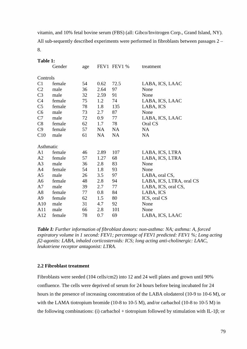
79
vitamin, and 10% fetal bovine serum (FBS) (all: Gibco/Invitrogen Corp., Grand Island, NY).
All sub-sequently described experiments were performed in fibroblasts between passages 2 –
8.
Table 1: Gender age FEV1 FEV1 % treatment Controls C1 female 54 0.62 72.5 LABA, ICS, LAAC C2 male 36 2.64 97 None C3 male 32 2.59 91 None C4 female 75 1.2 74 LABA, ICS, LAAC C5 female 78 1.8 135 LABA, ICS C6 male 73 2.7 87 None C7 male 72 0.9 77 LABA, ICS, LAAC C8 female 62 1.7 78 Oral CS C9 female 57 NA NA NA C10 male 61 NA NA NA Asthmatic A1 female 46 2.89 107 LABA, ICS, LTRA A2 female 57 1.27 68 LABA, ICS, LTRA A3 male 36 2.8 83 None A4 female 54 1.8 93 None A5 male 26 3.5 97 LABA, oral CS, A6 female 48 2.8 94 LABA, ICS, LTRA, oral CS A7 male 39 2.7 77 LABA, ICS, oral CS, A8 female 77 0.8 84 LABA, ICS A9 female 62 1.5 80 ICS, oral CS A10 male 31 4.7 92 None A11 male 66 2.8 101 None A12 female 78 0.7 69 LABA, ICS, LAAC
Table I: Further information of fibroblast donors: non-asthma: NA; asthma: A, forced expiratory volume in 1 second: FEV1; percentage of FEV1 predicted: FEV1 %; Long acting β2-agonits: LABA, inhaled corticosteroids: ICS; long acting anti-cholinergic: LAAC, leukotriene receptor antagonist: LTRA.
2.2 Fibroblast treatment
Fibroblasts were seeded (104 cells/cm2) into 12 and 24 well plates and grown until 90%
confluence. The cells were deprived of serum for 24 hours before being incubated for 24
hours in the presence of increasing concentration of the LABA olodaterol (10-9 to 10-6 M), or
with the LAMA tiotropium bromide (10-8 to 10-5 M), and/or carbachol (10-8 to 10-5 M) in
the following combinations: (i) carbachol + tiotropium followed by stimulation with IL-1β; or
80
(ii) olodaterol + tiotropium for 30 min followed by stimulation with IL-1β (10 ng/mL).
Recombinant human platelet-derived growth factor (PDGF)-BB (R&D Systems Europe,
Abingdon, United Kingdom) was dissolved in sterile 4 mM HCL to a stock concentration of
100µg/mL; Recombinant human TNF-α (R&D Systems Europe Ltd.) was dissolved to a
concentration of 100µg/mL in sterile PBS (Dulbecco’s PBS 1x, PAA-Laboratories GmbH)
containing 0.1% human or bovine serum albumin; Recombinant Human IL-1β (R&D Systems
Europe Ltd.), was reconstituted at 25 µg/mL in sterile phosphate buffered saline (PBS 1x,
PAA-Laboratories), containing 0.1% bovine serum albumin. Carbachol and olodaterol (BI
1744 CL) were dissolved in RPMI 1640 medium. Tiotropium bromide (Ba 679 BR) was
dissolved in ethanol. Olodaterol and tiotropium bromide were provided by Boehringer
Ingelheim Pharma GmbH & Co. KG (Biberach, Germany).
2.3 Cytokine secretion
Sub-confluent fibroblasts (90%) in 12 well plates were serum starved for 24 hours before
treatment. PDGF-BB, TNF-α, or IL-1β, were added to fibroblasts (0.1, 1, 10 ng/mL) and after
24 hours, cell culture medium samples were collected. IL-6 and IL-8 levels in the cell culture
media were determined by commercial enzyme-linked immune sorbent assays (ELISA)
following the distributors instruction (Ani Biotech Oy, Orgenium Laboratories, Vantaa,
Finland).
2.4 Protein expression
Cellular proteins were isolated from confluent cells by dissociation in lysis buffer, (62.5 mM
Tris-HCL, pH 6.8; 2% sodium dodecylsulfate, 2% β-mercapto-ethanol, 10% glycerol) and
analyzed by immuno-blotting for the expression of cAMP response element binding protein
(CREB), phosphorylated CREB (P-CREB) and α-tubulin (all: Cell Signaling Technology) as
described earlier for other proteins [20].
2.5 cAMP detection.
Sub-confluent fibroblasts (90%) grown in 24 well plates were serum deprived for 24 hours.
To block phosphodiesterase activity, cells were pretreated with iso-butyl-methylxantine
(IBMX, Sigma) for 10 minutes, then stimulated as follows: (i) serum-free medium, (ii) IL-1β
(10 ng/ml), (iii) carbachol 10-5 M, (iv) tiotropium 10-6 M, (v) olodaterol 10-6 M, (vi) IL-1β +
olodaterol 10-6 M, (vii) IL-1β + olodaterol 10-6 M + carbachol 10-5 M (added 30 minutes
prior to all other stimuli), (viii) IL-1β + olodaterol 10-6 M + carbachol 10-5 M + tiotropium
10-6 M (added 30 minutes prior to all other stimuli). After 10 minutes cells were lysed and
81
cAMP levels were determined by ELISA following the instruction of the producer (R&D
Systems Europe Ltd.). In order to investigate the role of G-proteins fibroblasts were pre-
incubated with pertussis toxin (10 ng/ml) for 30 minutes prior to all other treatments.
2.6 Primary human lung fibroblasts characterization
We characterized primary human lung fibroblasts for the expression of α-smooth muscle actin
(α-SMA) and fibronectin in immune-chemical staining as described earlier [21] 2.7 Statistics.
Cytokine expression was calculated as percent change of IL-1β stimulated cells and the data
are presented as mean ± SEM. After checking the data for normal distribution, paired or
unpaired, two-tailed student’s t-test was performed and p-values < 0.05 were considered
significant.
82
3. Results
3.1 Fibroblast characterization:
Fibroblasts were characterized by immunostaining for the expression of α-SMA, with fibrillar
α-SMA as indicator for myo-fibroblast phenotype. Non-stimulated fibroblasts showed a
diffuse cytoplasmic expression of α-SMA (Fig. 1A), and treatment with either 5 ng/ml IL-1β
(Fig 1B), or 1 x 10⁻⁵ M carbachol (Fig 1C), or 1 x 10⁻⁶ M tiotropium (Fig. 1D), or 1 x 10⁻⁶ M
olodaterol (Fig. 1E), or 1 x 10⁻⁶ M tiotropium plus 1 x 10⁻⁶ M olodaterol (Fig. 1F). In contrast,
treatment with either 10 ng/ml PDGF-BB (Fig. 1G), or 5 ng/ml TGF-β1 (Fig.1H) stimulated
the fibrillar arrangement of α-SMA typical for myo-fibroblasts, with the effect being most
prominent in TGF-β1 stimulated cells.
Figure 1: Representative immune-chemical characterisation of α-SMA in human lung fibroblasts treated for 24 hours with different stimuli and drugs investigated in this study: (A) non-stimulated serum starved fibroblasts, (B) 5 ng/ml IL-1β, (C) 1 x 10⁻⁵ M carbachol, (D) 1 x 10⁻⁶ M tiotropium, (E) 1 x 10⁻⁶ M olodaterol, (F) 1 x 10⁻⁶ M tiotropium plus 1 x 10⁻⁶ M olodaterol, (G) 10 ng/ml PDGF-BB, or (H) 5 ng/ml TGF-β1. Similar results were obtained in four additional fibroblast lines.
The expression of M1-3 receptor was analyzed by PCR and observed that fibroblasts mainly
expressed M2 receptor, and little of M3 receptor, while none of fibroblast lines expressed M1
receptor (data not shown) which is in line with other reports [14].
83
3.2 Dose-dependent induction of IL-6 and IL-8 by PDGF, TNF-α and IL-1β
Primary fibroblasts released IL-6 and IL-8, while GM-CSF and eotaxin levels were below the
detection level. PDGF-BB significantly increased IL-6 release by fibroblasts of asthma and
non-asthma patients, with no difference between the groups (Fig.2A). TNF-α also increased
IL-6 release, however, the effect was more pronounced in fibroblasts from asthma patients
(Fig.2B). IL-1β induced the release of IL-6 in both asthma and non-asthma fibroblasts, with
no significant difference between the groups (Fig.2C). As shown in figure 2D, PDGF-BB also
significantly increased IL-8 release with the effect being stronger in cells from asthma
patients. TNF-α and IL-1β concentration-dependently increased the release of IL-8 release
from both asthma and non-asthma fibroblasts (Fig 2E, F). Compared to each other and with
regard to asthma fibroblast specific action IL-1β was the most potent cytokine, therefore we
focused on its modification by the drugs. Based on direct cell counts we did not observe any
significant increase of fibroblast numbers within the observation period thus the increase of
cytokines cannot be explained by cell numbers (data not shown).
Figure 2: Release of IL-6 and IL-8 by fibroblasts of asthmatic (n≥5) and non-asthmatic (n≥5) subjects incubated with different concentrations (0.1, 1, 10 ng/ml) of PDGF-BB (A, D), TNF-α (B, E) and IL-1β (C, F). Bars represent the mean ± S.E.M. of independent experiments in lung fibroblasts obtained from asthmatic and non-asthmatic subjects. *p<0.05 asthma or non-asthma compared to un-stimulated cells; †
p<0.05 asthma compared to non-asthma.
84
3.3 Tiotropium counteracts the IL-1β and carbachol-induced release of IL-6 and IL-8
To simulate an inflammatory environment, which is one of the characteristics of the lungs of
asthma patients, and to optimally induce both IL-6 and IL-8, fibroblasts were stimulated IL-
1β (10 ng/ml) with and without carbachol (10-8 to 10-5 M). As shown in figure3A carbachol
alone had no effect, but concentration-dependently enhanced the IL-1β-induced IL-6 release
by fibroblasts of asthma patients, whereas only a trend was observed in fibroblasts of non-
asthma donors (p=0.064).
Figure 3: Effect of 10-5M carbachol (A, D), tiotropium (B, E), and their combination (C, F) on the release of IL-6 and IL-8 by fibroblasts of asthmatic (n≥5) and non-asthmatic (n≥5) subjects. Bars represent the mean ± S.E.M. of independent experiments in lung fibroblasts obtained from asthmatic and non-asthmatic subjects. *p<0.05 compared to IL-1β (10 ng/ml); # p<0.05 carbachol compared to carbachol plus tiotropium. † p<0.05 asthma compared to non-asthma.
85
Tiotropium alone did not affect IL-1β-induced IL-6 secretion (Fig. 3B), but it significantly
counteracted the additive effect of carbachol on IL-1β induced IL-6 secretion (Fig. 3C). Only
high concentrations of carbachol (10-5 and 10-6 M) significantly enhanced IL-1β-induced
release of IL-8, and alone it had no effect (Fig. 3D). Surprisingly, tiotropium (10-6 M)
significantly reduced IL-1β-induced IL-8 secretion by asthma fibroblasts, but not in control
cells, alone the drug had no effect (Fig 3E). In addition, tiotropium (10-8 and 10-6 M)
completely reversed the additive effect of carbachol on IL-1β induced IL-8 secretion in
fibroblasts from asthmatic and non-asthmatic patients (Fig 3F).
3.4 The combination of olodaterol plus tiotropium reduces IL-1β-induced IL-6 and IL-8
secretion
Figure 4: Effects of olodaterol (A, C) and the combination of olodaterol plus tiotropium (B, D) on the release of IL-1β-induced IL-6 and IL-8 release by fibroblasts of asthmatic (n≥5) and non-asthmatic subjects (n≥5). Bars represent the mean ± S.E.M. of independent experiments in lung fibroblasts obtained from asthmatic and non-asthmatic subjects. *p<0.05 compared to IL-1β (10 ng/ml); # p<0.05 olodaterol alone compared to olodaterol plus tiotropium.
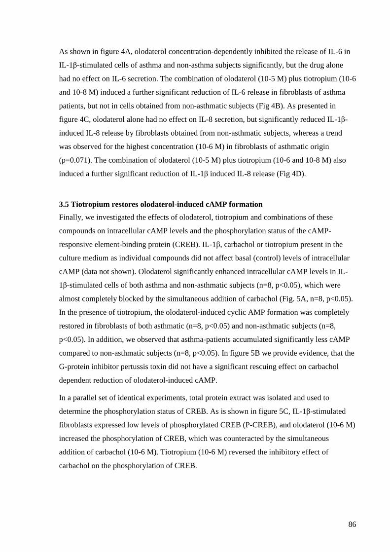
86
As shown in figure 4A, olodaterol concentration-dependently inhibited the release of IL-6 in
IL-1β-stimulated cells of asthma and non-asthma subjects significantly, but the drug alone
had no effect on IL-6 secretion. The combination of olodaterol (10-5 M) plus tiotropium (10-6
and 10-8 M) induced a further significant reduction of IL-6 release in fibroblasts of asthma
patients, but not in cells obtained from non-asthmatic subjects (Fig 4B). As presented in
figure 4C, olodaterol alone had no effect on IL-8 secretion, but significantly reduced IL-1β-
induced IL-8 release by fibroblasts obtained from non-asthmatic subjects, whereas a trend
was observed for the highest concentration (10-6 M) in fibroblasts of asthmatic origin
(p=0.071). The combination of olodaterol (10-5 M) plus tiotropium (10-6 and 10-8 M) also
induced a further significant reduction of IL-1β induced IL-8 release (Fig 4D).
3.5 Tiotropium restores olodaterol-induced cAMP formation
Finally, we investigated the effects of olodaterol, tiotropium and combinations of these
compounds on intracellular cAMP levels and the phosphorylation status of the cAMP-
responsive element-binding protein (CREB). IL-1β, carbachol or tiotropium present in the
culture medium as individual compounds did not affect basal (control) levels of intracellular
cAMP (data not shown). Olodaterol significantly enhanced intracellular cAMP levels in IL-
1β-stimulated cells of both asthma and non-asthmatic subjects (n=8, p<0.05), which were
almost completely blocked by the simultaneous addition of carbachol (Fig. 5A, n=8, p<0.05).
In the presence of tiotropium, the olodaterol-induced cyclic AMP formation was completely
restored in fibroblasts of both asthmatic (n=8, p<0.05) and non-asthmatic subjects (n=8,
p<0.05). In addition, we observed that asthma-patients accumulated significantly less cAMP
compared to non-asthmatic subjects (n=8, p<0.05). In figure 5B we provide evidence, that the
G-protein inhibitor pertussis toxin did not have a significant rescuing effect on carbachol
dependent reduction of olodaterol-induced cAMP.
In a parallel set of identical experiments, total protein extract was isolated and used to
determine the phosphorylation status of CREB. As is shown in figure 5C, IL-1β-stimulated
fibroblasts expressed low levels of phosphorylated CREB (P-CREB), and olodaterol (10-6 M)
increased the phosphorylation of CREB, which was counteracted by the simultaneous
addition of carbachol (10-6 M). Tiotropium (10-6 M) reversed the inhibitory effect of
carbachol on the phosphorylation of CREB.
87
Figure 5: (A) Absolute cAMP (pg/ml) produced by lung fibroblasts of asthma patients (n=8) and controls (n=8). Cells were stimulated for 10 minutes either with IL-1β alone or in the presence of the drugs. Bars represent the mean ± S.E.M. of independent experiments. * and ** p<0.05 effect of olodaterol compared to IL-1β+IBMX-treated cells; # and ## p<0.05 effect of carbachol compared to olodaterol+IL1β +IBMX-treated cells; φ and φφ effect of tiotropium compared to carbachol+olodaterol+IL1β +IBMX-treated cells; @ p<0.05 intra group comparison asthma versus non-asthma. (B) relative changes of olodaterol induced cAMP levels (100%) in the presence or absence of drugs, carbachol or pertussis toxin. (n=3 each group), bars represent the mean ± S.E.M. of independent experiments. * indicate significant difference of the drugs effect compared to IL-1β + olodaterol with p<0.05 (Student’s t-test, paired); n.s.: not significant. (C) The effect of IL-1β (5 ng/ml), olodaterol (10-6 M), carbachol (10-6 M), tiotropium (10-6 M) and combinations of these compounds on the phosphorylation of CREB (p-CREB) at 30 minutes after the addition of the stimuli or drugs. The immune-blots are representative examples of four independent experiments).
88
4. Discussion
There is abundance of evidence that the signaling systems triggered by muscarinic- and β2-
adrenergic receptors are integrated in an intricate interacting network [12]. Although both
systems are triggered by distinctly different receptors, they affect the activity of the same pool
of G proteins [13]. This may explain why tiotropium and olodaterol cooperate, and why they -
- when added together -- have beneficial effects in the treatment of asthma and COPD [17, 18,
22, 23]. In a recent paper, it was shown that add-on therapy with tiotropium provided
sustained bronchodilation and resulted in reduced asthma exacerbations in patients who were
symptomatic and had persistent airflow limitation on top of the use of LABAs and inhaled
GCs [19]. These observations suggest a role of muscarinic receptor antagonists that extends
beyond their properties as bronchodilators. Indeed, in a guinea-pig model of asthma
tiotropium was found to counteract remodeling- and inflammatory responses [24-26]. In
addition, in vitro studies showed that stress-activated airway smooth muscle cells exposed to
carbachol secreted increased levels of pro-inflammatory cyclooxygenases, as well as IL-1β,
IL-6 and IL-8 [27]. In airway smooth muscle cells, muscarinic receptor stimulation induced
the secretion of IL-6 and IL-8 and this effect was significantly greater in the presence of
cigarette smoke and inhibited by tiotropium [28, 29].
In our present study, separately IL-1β and carbachol elicited the release of IL-6 and IL-8 by
fibroblasts of asthmatic and non-asthmatic origin, and their combination further enhanced IL-
6 and IL-8. This shows that in an inflammatory environment, such as in the asthmatic lung,
endogenous acetylcholine may further aggravate the inflammation. Our in vitro data with
primary human lung fibroblasts demonstrate that tiotropium exerted clear anti-inflammatory
effects by reducing carbachol-induced IL-6 and IL-8 protein levels. Likewise, tiotropium may
counteract the increased acetylcholine levels reported in asthma patients [30-32], thus
relieving pulmonary inflammation.
The cAMP-dependent signaling pathway, which is triggered by olodaterol, provides a
negative signal for inflammatory responses, including the production and release of pro-
inflammatory cytokines and chemokines [33, 34]. How the signaling pathway(s) coupled to
the muscarinic receptor system are integrated and/or interact with those initiated by the β2-
adrenergic receptor is still largely unclear. It has been shown, however, that the airways of
mice deficient in a cAMP-specific phosphodiesterase-4D (PDE4D) are no longer responsive
to cholinergic stimulation [35]. In addition, Chinese Hamster ovary cells transfected with the
M3- and ß2-adrenergic receptor, muscarinic (M3) receptor stimulation resulted in β2-
89
adrenergic receptor phosphorylation and subsequent desensitization [36]. The desensitization
of the β2-adrenergic receptor system had been shown earlier in cells of asthma patients,
including peripheral blood cells [37, 38] and airway smooth muscle cells [39]. An intriguing
observation is that lung fibroblasts of asthma patients also produce significantly lower levels
of cAMP in contrast to fibroblasts of non-asthma patients, demonstrating this to be a general
phenomenon of cells of asthma patient. Although G-protein-coupled kinase-2 (GRK2) [37,
38] and PDE4 [39] have been implicated, the mechanism to explain the reduced clevels in
cells of asthma patients is still elusive. Drugs that help to increase intracellular cAMP levels –
either directly or indirectly – would be helpful to treat inflammatory and bronchial obstructive
lung diseases.
Our present study also underpinned the importance of the cAMP-dependent signaling
pathway in M2 - and β2-adrenergic receptor cross talk: tiotropium significantly restored
cAMP levels in fibroblasts of both asthmatic and non-asthmatic subjects and it reversed the
inhibitory effect of carbachol on the phosphorylation of CREB. In the presence of tiotropium,
intracellular cAMP levels were restored beyond levels induced by olodaterol alone. The
muscarinic M3 receptor subtype is predominantly expressed in the bronchus and is absent in
lung parenchyma. On the other hand, the M1 subtype only occurs in the lung parenchyma,
whereas the M2 subtype is distributed ubiquitously in the bronchus and lungs [40]. The
dominating muscarinic receptor present on primary human lung fibroblasts is the M2 receptor
[41]. Therefore, the M2 receptor was most likely mediating the carbachol-induced inhibitory
effects on cAMP accumulation and phosphorylation of CREB. A well-known protein
regulating the activity of β2-adrenergic receptors is the GRK2, which facilitates the binding of
β-arrestins, thus desensitizing the β2-adrenergic receptor by uncoupling it from the Gs protein
[42]. Although β2-adrenergic receptor phosphorylation has been reported after M3 activation
in CHO cells, the phosphorylation was independent of GRK2 [36]. Muscarinic receptor
activation can reduce cAMP levels through mobilization of intracellular calcium
(predominantly via M3), PLC (phospholipase C) and of calmodulin-dependent
phosphodiesterase which is enhanced in asthmatic subjects [43]. Furthermore, the muscarinic
receptor was found to be "negative coupled" to adenylate cyclase by the Gi subunit of the G
protein [44]. However, our data show that the G-protein inhibitor pertussis toxin did not have
the same rescuing effect on olodaterol induced cAMP as observed for tiotropium. Thus, it
might indicate that the rescue effect of tiotropium be largely independent of G-protein activity
through the M2-receptor. Although the precise mechanism remains unclear, the observed
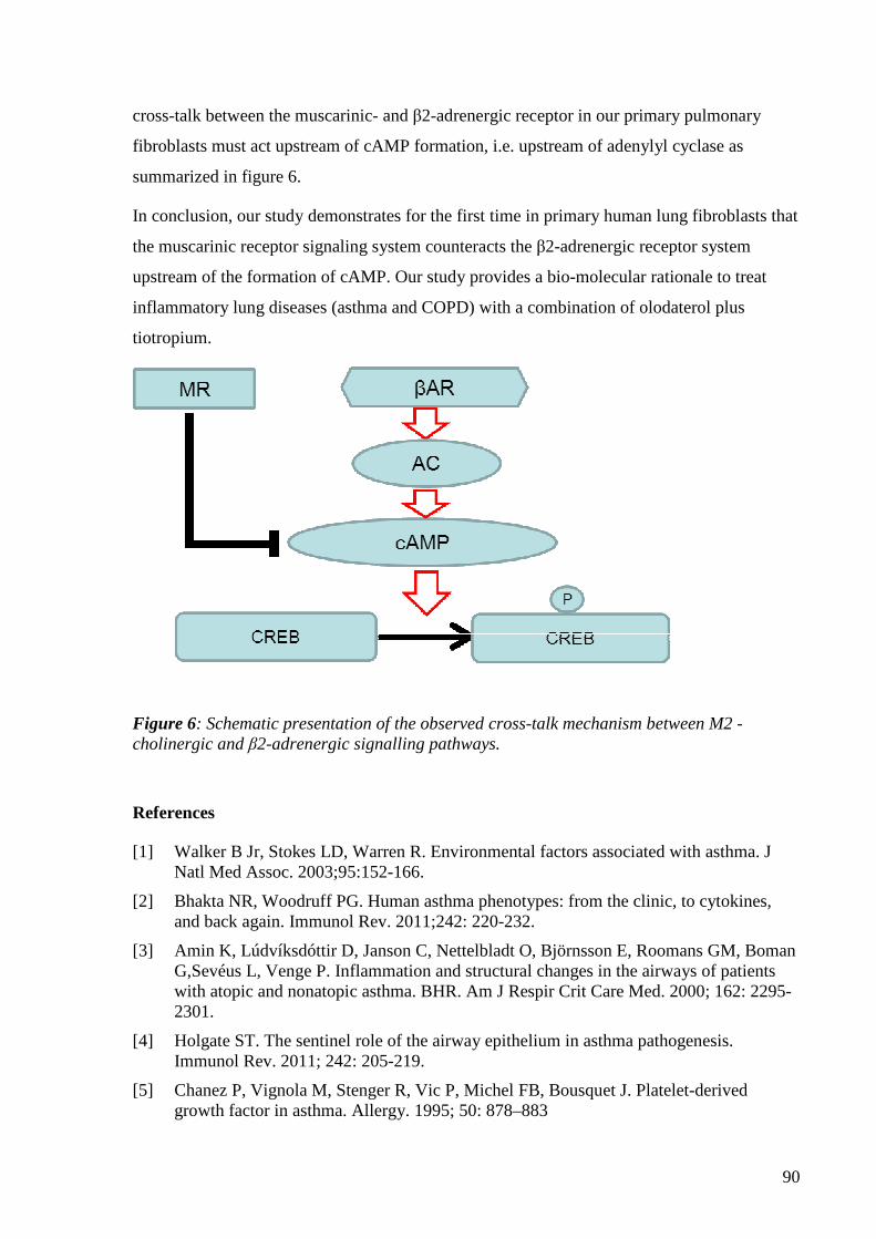
90
cross-talk between the muscarinic- and β2-adrenergic receptor in our primary pulmonary
fibroblasts must act upstream of cAMP formation, i.e. upstream of adenylyl cyclase as
summarized in figure 6.
In conclusion, our study demonstrates for the first time in primary human lung fibroblasts that
the muscarinic receptor signaling system counteracts the β2-adrenergic receptor system
upstream of the formation of cAMP. Our study provides a bio-molecular rationale to treat
inflammatory lung diseases (asthma and COPD) with a combination of olodaterol plus
tiotropium.
Figure 6: Schematic presentation of the observed cross-talk mechanism between M2 -cholinergic and β2-adrenergic signalling pathways.
References
[1] Walker B Jr, Stokes LD, Warren R. Environmental factors associated with asthma. J Natl Med Assoc. 2003;95:152-166.
[2] Bhakta NR, Woodruff PG. Human asthma phenotypes: from the clinic, to cytokines, and back again. Immunol Rev. 2011;242: 220-232.
[3] Amin K, Lúdvíksdóttir D, Janson C, Nettelbladt O, Björnsson E, Roomans GM, Boman G,Sevéus L, Venge P. Inflammation and structural changes in the airways of patients with atopic and nonatopic asthma. BHR. Am J Respir Crit Care Med. 2000; 162: 2295-2301.
[4] Holgate ST. The sentinel role of the airway epithelium in asthma pathogenesis. Immunol Rev. 2011; 242: 205-219.
[5] Chanez P, Vignola M, Stenger R, Vic P, Michel FB, Bousquet J. Platelet-derived growth factor in asthma. Allergy. 1995; 50: 878–883
91
[6] Ingram JL, Rice AB, Geisenhoffer K, Madtes DK, Bonner JC. IL-13 and IL-1beta promote lung fibroblast growth through coordinated up-regulation of PDGF-AA and PDGF-Ralpha. FASEB J. 2004; 18: 1132-4.
[7] Howarth PH, Babu KS, Arshad HS, Lau L, Buckley M, McConnell W, Beckett P, Al Ali M, Chauhan A, Wilson SJ, Reynolds A, Davies DE, Holgate ST. Tumour necrosis factor (TNFalpha) as a novel therapeutic target in symptomatic corticosteroid dependent asthma. Thorax. 2005; 60: 1012-8.
[8] Miglino N, Roth M, Lardinois D, Sadowski C, Tamm M, Borger P. Cigarette smoke inhibits lung fibroblast proliferation by translational mechanisms. Eur Respir J. 2012;39:705-11.
[9] Tager AM, LaCamera P, Shea BS, et al. The lysophosphatidic acid receptor LPA1 links pulmonary fibrosis to lung injury by mediating fibroblast recruitment and vascular leak. Nat Med 2008; 14:45–54.
[10] Chen D, Carpenter A, Abrahams J, et al. Protease-activated receptor 1 activation is necessary for monocyte chemoattractant protein 1-dependent leukocyte recruitment in vivo. J Exp Med 2008; 205:1739–1746.
[11] Orihara K, Dil N, Anaparti V, Moqbel R. What's new in asthma pathophysiology and immunopathology? Expert Rev Respir Med. 2010; 4: 605-29.
[12] Todorova L, Bjermer L, Westergren-Thorsson G, Miller-Larsson A. TGFβ-induced matrix production by bronchial fibroblasts in asthma: budesonide and formoterol effects. Respir Med. 2011; 105: 1296-307.
[13] Gosens R, Zaagsma J, Meurs H, Halayko. Muscarinic receptor signaling in the pathophysiology of asthma and COPD. Respir Res. 2006; 7:73.
[14] Meurs H, Dekkers BG, Maarsingh H, Halayko AJ, Zaagsma J, Gosens R. Muscarinic receptors on airway mesenchymal cells: Novel findings for an ancient target. Pulm Pharmacol Ther. 2013; 26: 145-55
[15] Pan J, Rhode HK, Undem BJ, Myers AC. Neurotransmitters in airway parasympathetic neurons altered by neurotrophin-3 and repeated allergen challenge. Am J Respir Cell Mol Biol. 2010; 43: 452-457.
[16] Lemanske RF Jr, Mauger DT, Sorkness CA, Jackson DJ, Boehmer SJ, Martinez FD, Strunk RC, Szefler SJ, Zeiger RS, Bacharier LB, Covar RA, Guilbert TW, Larsen G, Morgan WJ, Moss MH, Spahn JD, Taussig LM; Childhood Asthma Research and Education (CARE) Network of the National Heart, Lung, and Blood Institute. Step-up therapy for children with uncontrolled asthma receiving inhaled corticosteroids. N Engl J Med. 2010; 362: 975-985.
[17] Peters SP, Kunselman SJ, Icitovic N, Moore WC, Pascual R, Ameredes BT, Boushey HA, Calhoun WJ, Castro M, Cherniack RM, Craig T, Denlinger L, Engle LL, DiMango EA, Fahy JV, Israel E, Jarjour N, Kazani SD, Kraft M, Lazarus SC, Lemanske RF Jr, Lugogo N, Martin RJ, Meyers DA, Ramsdell J, Sorkness CA, Sutherland ER, Szefler SJ, Wasserman SI, Walter MJ, Wechsler ME, Chinchilli VM, Bleecker ER; National Heart, Lung, and Blood Institute Asthma Clinical Research Network. Tiotropium bromide step-up therapy for adults with uncontrolled asthma. N Engl J Med. 2010; 363: 1715-1726.
92
[18] Kanazawa H. Anticholinergic agents in asthma: chronic bronchodilator therapy, relief of acute severe asthma, reduction of chronic viral inflammation and prevention of airway remodeling. Curr Opin Pulm Med. 2006; 12: 60-67.
[19] Kerstjens HA, Engel M, Dahl R, Paggiaro P, Beck E, Vandewalker M, Sigmund R, Seibold W, Moroni-Zentgraf P, Bateman ED. Tiotropium in Asthma Poorly Controlled with Standard Combination Therapy. N Engl J Med. 2012 Sep 2. [Epub ahead of print].
[20] Borger P, Matsumoto H, Boustany S, Gencay MM, Burgess JK, King GG, Black JL, Tamm M, Roth M. Disease-specific expression and regulation of CCAAT/enhancer-binding proteins in asthma and chronic obstructive pulmonary disease. J Allergy Clin Immunol. 2007; 119: 98-105.
[21] Papakonstantinou E, Klagas I, Karakiulakis G, Hostettler K, S'ng CT, Kotoula V, Savic S, Tamm M, Roth M. Steroids and β2-agonists regulate hyaluronan metabolism in asthmatic airway smooth muscle cells. Am J Respir Cell Mol Biol. 2012; 47: 759-67.
[22] Proskocil BJ, Fryer AD. Beta2-agonist and anticholinergic drugs in the treatment of lung disease. Proc Am Thorac Soc. 2005; 2: 305-10.
[23] Powrie DJ, Wilkinson TM, Donaldson GC, Jones P, Scrine K, Viel K, Kesten S, Wedzicha JA. Effect of tiotropium on sputum and serum inflammatory markers and exacerbations in COPD. Eur Respir J. 2007; 30: 472-478.
[24] Buels KS, Jacoby DB, Fryer AD. Non-bronchodilating mechanisms of tiotropium prevent airway hyperreactivity in a guinea-pig model of allergic asthma. Br J Pharmacol. 2012;165: 1501-14.
[25] Ohta S, Oda N, Yokoe T, Tanaka A, Yamamoto Y, Watanabe Y, Minoguchi K, Ohnishi T, Hirose T, Nagase H, Ohta K, Adachi M. Effect of tiotropium bromide on airway inflammation and remodelling in a mouse model of asthma. Clin Exp Allergy. 2010; 40: 1266-75.
[26] Pera T, Zuidhof A, Valadas J, Smit M, Schoemaker RG, Gosens R, Maarsingh H, Zaagsma J, Meurs H. Tiotropium inhibits pulmonary inflammation and remodelling in a guinea pig model of COPD. Eur Respir J. 2011; 38: 789-96.
[27] Kanefsky J, Lenburg M, Hai CM. Cholinergic receptor and cyclic stretch-mediated inflammatory gene expression in intact ASM. Am J Respir Cell Mol Biol. 2006; 34: 417-425.
[28] Gosens R, Rieks D, Meurs H, Ninaber DK, Rabe KF, Nanninga J, Kolahian S, Halayko AJ, Hiemstra PS, Zuyderduyn S. Muscarinic M3 receptor stimulation increases cigarette smoke-induced IL-8 secretion by human airway smooth muscle cells. Eur Respir J. 2009; 34: 1436-1443.
[29] Oenema TA, Kolahian S, Nanninga JE, Rieks D, Hiemstra PS, Zuyderduyn S, Halayko AJ, Meurs H, Gosens R. Pro-inflammatory mechanisms of muscarinic receptor stimulation in airway smooth muscle. Respir Res. 2010 Sep 28;11:130. doi: 10.1186/1465-9921-11-130.
[30] Ayala LE, Ahmed T. Is there loss of protective muscarinic receptor mechanism in asthma? Chest 1989, 96: 1285-1291.
[31] Minette PA, Lammers JW, Dixon CM, McCusker MT, Barnes PJ. A muscarinic agonist inhibits reflex bronchoconstriction in normal but not in asthmatic subjects. J Appl Physiol 1989, 67: 2461-2465.
93
[32] Novelli F, Malagrinò L, Dente FL, Paggiaro P. Efficacy of anticholinergic drugs in asthma. Expert Rev Respir Med. 2012; 6: 309-19.
[33] Borger P, Postma DS, Vellenga E, Kauffman HF. Regulation of asthma-related T-cell cytokines by the cyclic AMP-dependent signalling pathway. Clin Exp Allergy. 2000; 30: 920-6.
[34] Yamagata T, Ichinose M. Agents against cytokine synthesis or receptors. Eur J Pharmacol. 2006; 533: 289-301.
[35] Hansen G, Jin S, Umetsu DT, Conti M. Absence of muscarinic cholinergic airway responses in mice deficient in the cyclic nucleotide phosphodiesterase PDE4D. Proc Natl Acad Sci U S A. 2000; 97: 6751-6.
[36] Budd DC, Challiss RA, Young KW, Tobin AB. Cross talk between m3-muscarinic and beta(2)-adrenergic receptors at the level of receptor phosphorylation and desensitization. Mol Pharmacol. 1999; 56: 813-23.
[37] Meurs H, Koëter GH, de Vries K, Kauffman HF. The beta-adrenergic system and allergic bronchial asthma: changes in lymphocyte beta-adrenergic receptor number and adenylate cyclase activity after an allergen-induced asthmatic attack. J Allergy Clin Immunol. 1982; 70: 272-80.
[38] Borger P, Postma DS, Vellenga E, Kauffman HF. Regulation of asthma-related T-cell cytokines by the cyclic AMP-dependent signalling pathway. Clin Exp Allergy. 2000; 30: 920-6.
[39] Trian T, Burgess JK, Niimi K, Moir LM, Ge Q, Berger P, Liggett SB, Black JL, Oliver BG. β2-Agonist induced cAMP is decreased in asthmatic airway smooth muscle due to increased PDE4D. PLoS One. 2011; 6: e20000.
[40] Ikeda T, Anisuzzaman AS, Yoshiki H, Sasaki M, Koshiji T, Uwada J, Nishimune A, Itoh H, Muramatsu I. Regional quantification of muscarinic acetylcholine receptors and β-adrenoceptors in human airways. Br J Pharmacol. 2012; 166: 1804-14.
[41] Haag S, Matthiesen S, Juergens UR, Racké K. Muscarinic receptors mediate stimulation of collagen synthesis in human lung fibroblasts. Eur Respir J. 2008; 3: 555-62.
[42] Shenoy, S. K., McDonald, P. H., Kohout, T. A., and Lefkowitz, R. J. Regulation of receptor fateby ubiquitination of activated beta 2-adrenergic receptor and beta-arrestin. Science. 2001; 294: 1307-13.
[43] Budd DC, Willars GB, McDonald JE, Tobin AB.Phosphorylation of the Gq/11-coupled m3-muscarinic receptor is involved in receptor activation of the ERK-1/2 mitogen-activated protein kinase pathway. J Biol Chem. 2001; 276: 4581-7.
[44] Nathanson NM. A multiplicity of muscarinic mechanisms: enough signaling pathways to take your breath away. Proc Natl Acad Sci U S A. 2000; 97: 6245-7.
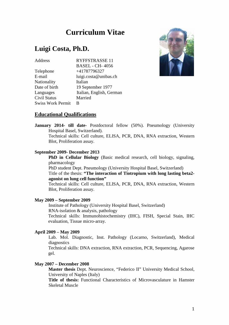
1
Curriculum Vitae Luigi Costa, Ph.D. Address RYFFSTRASSE 11
BASEL - CH- 4056 Telephone +41787796327 E-mail [email protected] Nationality Italian Date of birth 19 September 1977 Languages Italian, English, German Civil Status Married Swiss Work Permit B Educational Qualifications January 2014- till date- Postdoctoral fellow (50%). Pneumology (University
Hospital Basel, Switzerland). Technical skills: Cell culture, ELISA, PCR, DNA, RNA extraction, Western Blot, Proliferation assay.
September 2009- December 2013
PhD in Cellular Biology (Basic medical research, cell biology, signaling, pharmacology PhD student Dept. Pneumology (University Hospital Basel, Switzerland) Title of the thesis: “The interaction of Tiotropium with long lasting beta2-agonist on lung cell function” Technical skills: Cell culture, ELISA, PCR, DNA, RNA extraction, Western Blot, Proliferation assay.
May 2009 – September 2009 Institute of Pathology (University Hospital Basel, Switzerland) RNA-isolation & analysis, pathology
Technical skills: Immunohistochemistry (IHC), FISH, Special Stain, IHC evaluation, Tissue micro-array.
April 2009 – May 2009
Lab. Mol. Diagnostic, Inst. Pathology (Locarno, Switzerland), Medical diagnostics Technical skills: DNA extraction, RNA extraction, PCR, Sequencing, Agarose gel.
May 2007 – December 2008
Master thesis Dept. Neuroscience, “Federico II” University Medical School, University of Naples (Italy) Title of thesis: Functional Characteristics of Microvasculature in Hamster Skeletal Muscle
2
Research topic: Research on micro-vascular network in hamster skin muscle and on rat microcirculation. Technical skills: Western Blot, cell culture.
March 2006 – March 2007
Internship at Dept. Biomorphological and Functional Sciences, “Federico II” University Medical School, University of Naples (Italy) Technical skills: Dissection of small rodents, PCR, microscopy, molecular biology.
October 2002 - March 2007 Bachelor’s degree in Biotechnology Sciences Title of thesis: Molecular Characteristics of Neuroblastoma “Federico II” University Medical School, University of Naples (Italy) Faculty of Biotechnology
Publication:
1. Costa L, Roth M, Miglino N, Keglowich L, Zhong J, Lardinois D, Tamm M, Borger P: Tiotropium supports olodaterol action on cytokine secretion by human lung fibroblasts via cyclic AMP. Pulmonary Pharmacology and Therapeutics 2014:27:29-37. Impact Factor: 2.3
2. Seidel P, Costa L, Keglowich L, Lardinos D, Tamm M, Roth M: The MNK-1/eIF4E pathway as a new therapeutic pathway to target inflammation and remodelling in asthma. American Journal of Physiology (Lung) 2014 – submitted
3. Christopher Lambers1, Ying Qi2, Papakonstantinou E3, Luigi Costa2, Jun Zhong2, Michael Tamm2, Lutz-Henning Block1, Michael Roth2 : Extracellular matrix composition is modified by β2-agonists through cAMP in COPD Biochemical Pharmacology 2014.
4. Seidel P, Costa L, Tamm M, Roth M:The mTOR signalling pathway as a new therapeutic pathway to target inflammation and remodeling in asthma American Journal Physiology (Lung) 2014 – submitted.
5. Ying Qi, Zhong J, Costa L, Lambers C, S’ng CT, Tamm M, Roth M,: Long acting beta2-agonists reduce airway smooth muscle cell proliferation through p27(Kip) in a cAMP independent mechanism. Pulmonary Research 2014 - submitted
Presentations at International congress: 1. ERS Barcelona 2010 Poster session: “Crosstalk between Long Acting β2-Adrenergic receptor Agonists and Tiotropium: Crosstalk of β2-Adrenergic and muscarinic receptors. Costa L., Roth M., Tamm M., Borger P., Gencay M., Miglino N., Keglowich L., Bodmer H., Hostettler K. 2. ERS Amsterdam 2011 Poster session: “Tiotropium reduced Carbachol-induced expressions of IL-6 & IL-8 by primary human lung fibroblasts of asthma and non-asthma subjects”. Costa L, Roth M, Casarosa P,Tamm M, Borger P.
3
3. ERS Amsterdam 2011 Poster session: “Disease and stimuli specific pro.inflammatory cytokine secretion by human fibroblast of asthma patients”. Costa L , Borger P , Casarosa P , Tamm M , Roth M . 4. ERS Vienna 2012 Poster session: “Tiotropium enhances the inhibitory effect of the long acting β2-agonist olodaterol on the release of IL-6 and IL8 by primary human lung fibroblasts of asthma patients”. Costa L, Roth M, Tamm M, Borger P. Social skills:
I have very good communication skills and am a good team player. I am
dedicated to my work and always eager to extend my knowledge. I have
no difficulties to adapt to new environment. IT skills : MS-Word, Power Point, Excel. Driving license(s): Swiss A-B REFERENCES: Prof. Dr. Roth Michael (PhD), Head Pulmonary Cell Research, Department of Biomedicine, University Hospital Basel, Petersgraben 4, 4031-CH-Basel (Switzerland) E-mail: [email protected] Tel.+41 61 265 23 37 Prof. Dr. Michael Tamm (MD), Head Div. Pneumology, University Hospital Basel, Petersgraben 4, 4031-CH-Basel (Switzerland) E-mail: [email protected], Tel.: +41 61 5184 Prof. Dr. Luigi Terracciano (MD), Head of Molecular Pathology, Institute of Pathology, Basel (Switzerland) E-mail: [email protected] Tel.+41 61 265 28 49 Dr. Milo Frattini, Head of Laboratory Molecular Diagnostic, Institute of Pathology, Locarno (Switzerland) E-mail: [email protected] Tel. 0041 918160806 Prof. Dr. Antonio Colantuoni, Department of Neuroscience “Federico II” University Medical School of Naples, (Italy) E-mail: [email protected] Tel. 0039 0817463212 Prof. Dr. Stefania Montagnani, Department of Biomorfological and Functional Sciences “Federico II” University Medical School of Naples, (Italy) E-mail: [email protected] Tel.0039 0817463422