The effect of polarized light in the healing process of pressure … 01_07/VA_OP_1_06_0… · Το...
12
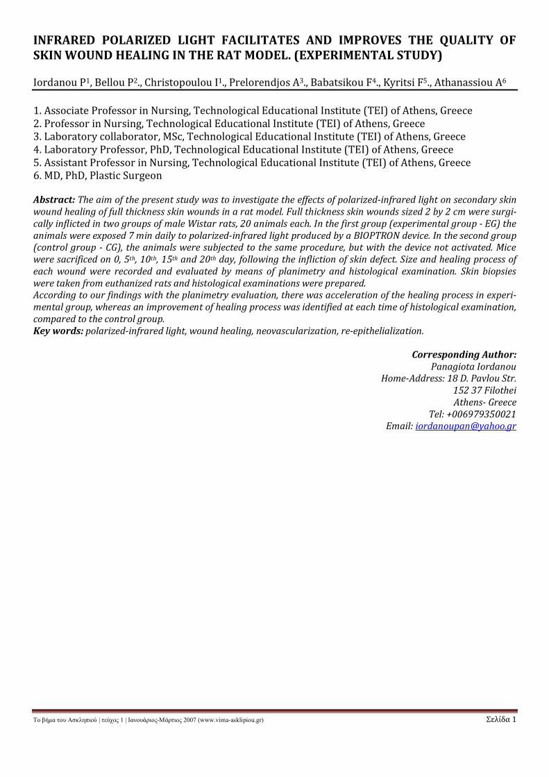
Το βήμα του Ασκληπιού | τεύχος 1 | Ιανουάριος-Μάρτιος 2007 (www.vima-asklipiou.gr) Σελίδα 1 INFRARED POLARIZED LIGHT FACILITATES AND IMPROVES THE QUALITY OF SKIN WOUND HEALING IN THE RAT MODEL. (EXPERIMENTAL STUDY) Iordanou P 1 , Bellou P 2 ., Christopoulou I 1 ., Prelorendjos A 3 ., Babatsikou F 4 ., Kyritsi F 5 ., Athanassiou A 6 1. Associate Professor in Nursing, Technological Educational Institute (TEI) of Athens, Greece 2. Professor in Nursing, Technological Educational Institute (TEI) of Athens, Greece 3. Laboratory collaborator, MSc, Technological Educational Institute (TEI) of Athens, Greece 4. Laboratory Professor, PhD, Technological Educational Institute (TEI) of Athens, Greece 5. Assistant Professor in Nursing, Technological Educational Institute (TEI) of Athens, Greece 6. MD, PhD, Plastic Surgeon Abstract: The aim of the present study was to investigate the effects of polarized-infrared light on secondary skin wound healing of full thickness skin wounds in a rat model. Full thickness skin wounds sized 2 by 2 cm were surgi- cally inflicted in two groups of male Wistar rats, 20 animals each. In the first group (experimental group - EG) the animals were exposed 7 min daily to polarized-infrared light produced by a BIOPTRON device. In the second group (control group - CG), the animals were subjected to the same procedure, but with the device not activated. Mice were sacrificed on 0, 5 th , 10 th , 15 th and 20 th day, following the infliction of skin defect. Size and healing process of each wound were recorded and evaluated by means of planimetry and histological examination. Skin biopsies were taken from euthanized rats and histological examinations were prepared. According to our findings with the planimetry evaluation, there was acceleration of the healing process in experi- mental group, whereas an improvement of healing process was identified at each time of histological examination, compared to the control group. Key words: polarized-infrared light, wound healing, neovascularization, re-epithelialization. Corresponding Author: Panagiota Iordanou Home-Address: 18 D. Pavlou Str. 152 37 Filothei Athens- Greece Tel: +006979350021 Email: [email protected]
Transcript of The effect of polarized light in the healing process of pressure … 01_07/VA_OP_1_06_0… · Το...
Το βήμα του Ασκληπιού | τεύχος 1 | Ιανουάριος-Μάρτιος 2007 (www.vima-asklipiou.gr) Σελίδα 1
INFRARED POLARIZED LIGHT FACILITATES AND IMPROVES THE QUALITY OFSKIN WOUND HEALING IN THE RAT MODEL. (EXPERIMENTAL STUDY)
Iordanou P1, Bellou P2., Christopoulou I1., Prelorendjos A3., Babatsikou F4., Kyritsi F5., Athanassiou A6
1. Associate Professor in Nursing, Technological Educational Institute (TEI) of Athens, Greece2. Professor in Nursing, Technological Educational Institute (TEI) of Athens, Greece3. Laboratory collaborator, MSc, Technological Educational Institute (TEI) of Athens, Greece4. Laboratory Professor, PhD, Technological Educational Institute (TEI) of Athens, Greece5. Assistant Professor in Nursing, Technological Educational Institute (TEI) of Athens, Greece6. MD, PhD, Plastic Surgeon
Abstract: The aim of the present study was to investigate the effects of polarized-infrared light on secondary skinwound healing of full thickness skin wounds in a rat model. Full thickness skin wounds sized 2 by 2 cm were surgi-cally inflicted in two groups of male Wistar rats, 20 animals each. In the first group (experimental group - EG) theanimals were exposed 7 min daily to polarized-infrared light produced by a BIOPTRON device. In the second group(control group - CG), the animals were subjected to the same procedure, but with the device not activated. Micewere sacrificed on 0, 5th, 10th, 15th and 20th day, following the infliction of skin defect. Size and healing process ofeach wound were recorded and evaluated by means of planimetry and histological examination. Skin biopsieswere taken from euthanized rats and histological examinations were prepared.According to our findings with the planimetry evaluation, there was acceleration of the healing process in experi-mental group, whereas an improvement of healing process was identified at each time of histological examination,compared to the control group.Key words: polarized-infrared light, wound healing, neovascularization, re-epithelialization.
Corresponding Author:Panagiota Iordanou
Home-Address: 18 D. Pavlou Str.152 37 FilotheiAthens- Greece
Tel: +006979350021Email: [email protected]
Το βήμα του Ασκληπιού | τεύχος 1 | Ιανουάριος-Μάρτιος 2007 (www.vima-asklipiou.gr) Σελίδα 2
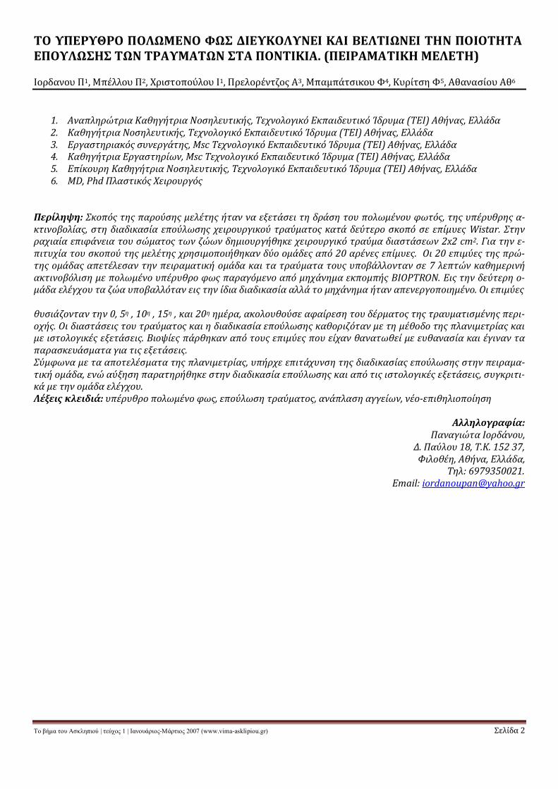
ΤΟ ΥΠΕΡΥΘΡΟ ΠΟΛΩΜΕΝΟ ΦΩΣ ΔΙΕΥΚΟΛΥΝΕΙ ΚΑΙ ΒΕΛΤΙΩΝΕΙ ΤΗΝ ΠΟΙΟΤΗΤΑΕΠΟΥΛΩΣΗΣ ΤΩΝ ΤΡΑΥΜΑΤΩΝ ΣΤΑ ΠΟΝΤΙΚΙΑ. (ΠΕΙΡΑΜΑΤΙΚΗ ΜΕΛΕΤΗ)
Ιορδανου Π1, Μπέλλου Π2, Χριστοπούλου Ι1, Πρελορέντζος Α3, Μπαμπάτσικου Φ4, Κυρίτση Φ5, Αθανασίου Αθ6
1. Αναπληρώτρια Καθηγήτρια Νοσηλευτικής, Τεχνολογικό Εκπαιδευτικό Ίδρυμα (ΤΕΙ) Αθήνας, Ελλάδα2. Καθηγήτρια Νοσηλευτικής, Τεχνολογικό Εκπαιδευτικό Ίδρυμα (ΤΕΙ) Αθήνας, Ελλάδα3. Εργαστηριακός συνεργάτης, Msc Τεχνολογικό Εκπαιδευτικό Ίδρυμα (ΤΕΙ) Αθήνας, Ελλάδα4. Καθηγήτρια Εργαστηρίων, Msc Τεχνολογικό Εκπαιδευτικό Ίδρυμα (ΤΕΙ) Αθήνας, Ελλάδα5. Επίκουρη Καθηγήτρια Νοσηλευτικής, Τεχνολογικό Εκπαιδευτικό Ίδρυμα (ΤΕΙ) Αθήνας, Ελλάδα6. MD, Phd Πλαστικός Χειρουργός
Περίληψη: Σκοπός της παρούσης μελέτης ήταν να εξετάσει τη δράση του πολωμένου φωτός, της υπέρυθρης α-κτινοβολίας, στη διαδικασία επούλωσης χειρουργικού τραύματος κατά δεύτερο σκοπό σε επίμυες Wistar. Στηνραχιαία επιφάνεια του σώματος των ζώων δημιουργήθηκε χειρουργικό τραύμα διαστάσεων 2x2 cm2. Για την ε-πιτυχία του σκοπού της μελέτης χρησιμοποιήθηκαν δύο ομάδες από 20 αρένες επίμυες. Οι 20 επιμύες της πρώ-της ομάδας απετέλεσαν την πειραματική ομάδα και τα τραύματα τους υποβάλλονταν σε 7 λεπτών καθημερινήακτινοβόλιση με πολωμένο υπέρυθρο φως παραγόμενο από μηχάνημα εκπομπής BIOPTRON. Εις την δεύτερη ο-μάδα ελέγχου τα ζώα υποβαλλόταν εις την ίδια διαδικασία αλλά το μηχάνημα ήταν απενεργοποιημένο. Οι επιμύες
θυσιάζονταν την 0, 5η , 10η , 15η , και 20η ημέρα, ακολουθούσε αφαίρεση του δέρματος της τραυματισμένης περι-οχής. Οι διαστάσεις του τραύματος και η διαδικασία επούλωσης καθοριζόταν με τη μέθοδο της πλανιμετρίας καιμε ιστολογικές εξετάσεις. Βιοψίες πάρθηκαν από τους επιμύες που είχαν θανατωθεί με ευθανασία και έγιναν ταπαρασκευάσματα για τις εξετάσεις.Σύμφωνα με τα αποτελέσματα της πλανιμετρίας, υπήρχε επιτάχυνση της διαδικασίας επούλωσης στην πειραμα-τική ομάδα, ενώ αύξηση παρατηρήθηκε στην διαδικασία επούλωσης και από τις ιστολογικές εξετάσεις, συγκριτι-κά με την ομάδα ελέγχου.Λέξεις κλειδιά: υπέρυθρο πολωμένο φως, επούλωση τραύματος, ανάπλαση αγγείων, νέο-επιθηλιοποίηση
Αλληλογραφία:Παναγιώτα Ιορδάνου,
Δ. Παύλου 18, Τ.Κ. 152 37,Φιλοθέη, Αθήνα, Ελλάδα,
Τηλ: 6979350021.Email: [email protected]
Το βήμα του Ασκληπιού | τεύχος 1 | Ιανουάριος-Μάρτιος 2007 (www.vima-asklipiou.gr) Σελίδα 3
INTRODUCTION
he Hungarian scientist Mester1,2 is one ofthe pioneers with large experimentaland clinical experience in the use of bios-
timulating effect of laser beams. A hypotheticalmodel based on experimental facts to explainthis effect was proposed. The substance of thatmodel showed that the polarized light reordersthe polar heads of the lipid bilayer (being neartransition phase) in the cell membrane. Thechange of the polar heads of the lipid bilayer inthe cell membrane has influenced all processesclosely connected with it. Another interestingfinding pointed out that while the incoherentirradiation of a thermal light source (630 nm)was ineffective, the result of irradiation by po-larized light showed about equal effectiveness(80%) of helium neon (HeNe) polarized laserlight.3,4 This phenomenon indicates that bios-timulasion might also be produced by polarizedthermal light source. Mester and Fenyo1-4 simi-larly suggested that a source of incoherent lightemitting polarized rays would induce biosti-mulative effects in living cells similar to lowlevel lasers. This means that lamps emittingpolarized light and low-laser light, when usedas wound treatment, both cause the phenome-non of biostimulation.
Several experiments in wounds with polarizedlight showed a stimulating and healing effect.1Kubasova5 used human embryo fibroblast cellcultures to examine the effect of HeNe laser po-larized light on the cell membrane. This studyshowed that 4j/cm2 was effective for irradia-tion and the duration of exposure was 7 mins.
Sommer and Franke6 supported that lowerenergy densities promote a significant biosti-mulation in human tissue. They introduced anew laser beam distribution system for medicalapplication, i.e. low energy densities appliedwith high–energy lasers in combination withlaser beam diverging lenses. They found thatthis system is effective for biostimulating hu-man tissue and also is of low cost. The lampemitting polarized light used in the presentstudy was also chosen because we assumed
that it has a similarly biostimulating effect withlow laser polarized light.
Mester et al.1-2 positive view of biostimulationwas a motivation for the present study, inwhich the aim was to evaluate what is the ef-fect of polarized infrared light on secondaryskin wound healing.
MATERIALS AND METHODS
Forty male Wistar rats, 4 months old andweighting 200 ± 30gr, were used and cared foraccording to Greek and European guidelinesregulating animal research. Animals were ac-climated for a period of 3 days, during whichthey were examined for any signs of disease.Throughout the entire study period, the ani-mals were housed individually, in order toavoid cannibalistic behavior, in plexiglas cagescovered with stainless steel lids. Their diet con-tained dried pellets and tap water was suppliedto them. The animals were kept in a room un-der stable conditions of temperature (22 o C)and humidity (between 30%-70%), and thelight cycle was 12/12-hours light/dark sche-dule. On day 0, anesthesia was induced byintraperitoneal injection of a mixture of Keta-mine and Midazolamin (3.5 mg and 7mg re-spectively per kg animal’s body weight). Thewhole surgical procedure was conducted underaseptic conditions. A square shaped area mea-suring 2 by 2 cm was marked on the dorsum ofeach animal, followed by a block excision of theskin and underlying panniculus carnosus. Theanimals were randomized in two groups of 20rats each. In the first group, those receivingtreatment were called Experimental Croup(EG) and those not receiving treatment, ControlGroup (CG).
The treatment consisted of a daily 7 min thera-py session with polarized infrared light. Thisstarted on day 0 and finished on day 20. Duringthe experiment, one rat at a time, was placedand immobilized in a special constructedwooden box with no metallic components, inorder to avoid any interference with the pola-rized light. The device was vertically centeredover the box at a constant distance of 10 cm
T
Το βήμα του Ασκληπιού | τεύχος 1 | Ιανουάριος-Μάρτιος 2007 (www.vima-asklipiou.gr) Σελίδα 4
from the wound surface, so that the woundarea was received the whole degree of polari-zation. The experiment was conducted usingthe non-invasive and non-thermal BioptronCompact III7 modified with a filter wrattennumber 25 (which absorbs the yellow light) asa result, the remaining light was infrared wave-length of 700-3400nm. Energies deliveredwere typically 2.4 joule/cm2 per minute, degreeof polarization >95%, wavelength 480-3400nm, power density 40 mW/cm2, using a20W 7.
After the experiment the rat was returned to itscage and housed separately without using anydressing or antibiotics on the wound.
On the 0, 5th, 10th and 15th day after wound cre-ation, four rats of each group were euthanized.Wound surface area was measured by tracingthe border of the wounds on transparent film(table 1). The area from these tracings wasmeasured with a polar planimeter (model No
317 E, Manufacturer of HAFF planimeters: W-Germany. The precision of this instrument iswithin 1mm2).8
Skin biopsies were taken from mice that weresacrificed on the 5th, 10th, 15th and 20th day af-ter skin infliction. The tissue specimens werefixed in 10% formalin solution, and bothwound and surrounding healthy tissue wereharvested for histological examination. All spe-cimens were paraffin-embedded, cut in 5mmthick blocks, perpendicularly to the skin sur-face, including the whole thickness of the skinwound.
The slides obtained were stained with Heama-toxylin-Eosin (HE), Vascular EndothelialGrowth Factor Ab-1 (VEGF Ab-1, dilution1:100, LAB VISION), α-Smooth Muscle ActinAb-1 (α-SMA Ab-1, dilution 1:200, LAB VI-SION), Vimentin Ab-2 (Vim Ab-2, dilution1:400, LAB VISION) and Van Gieson stain (VG).
The VEGF LAB VISION antigen is specific for theWistar rat. This antibody is a homodimeric,disulfide-linked glycoprotein involved in theangiogenesis. Its permeability to the vascularendothelium is specific and act as diffusibleagents especially in the smaller isoforms. The
α-SMA antibody stained the smooth musclecells in the newly formed vessel wall and it wasfound mainly intracytoplasmic. Vimentin wasthe main intermediate filament protein in me-senchymal cells and was therefore found ingreat quantity in the vascular smooth musclecells and its staining pattern was cytoplasmic.Van Gieson stain highlighted the collagen bun-dles formed by the fibroblasts and it was foundin great quantity in older granulation tissue,starting from the margins of the wound duringthe first days and finally spreading through outthe wound.9,10,11
We scored and evaluated 4 different parame-ters of wound healing:
a) Epithelizationb) Inflammationc) Newly-formed vesselsd) Collagen formation
We blindly scored the microscopic slides forthe two groups in question (EG and CG) in or-der to avoid any misguiding influence.
Exact measurements of the degree of epitheli-zation (both in the experimental and controlwound groups, EG and CG) were taken. Thiswas done by microscopically measuring the en-tire wound area and also the surface area at themargins of the wound, where epithelizationhad already started.
Epithelization scoring is shown below:12
0 Indicates no epithelization1 Indicates no epithelization2 Indicates ≤40% of com-
plete epithelization3 Indicates ≤60% of com-
plete epithelization4 Indicates ≤ 80% of com-
plete epithelization5 Complete epithelization
Following this, we evaluated the degree of in-flammation of the wound. In our study no acuteelements of inflammation where observed inthe tissue biopsies since specimens were re-ceived from the day 5. Polymorphonuclear gra-nulocytes (PMNGs) where seen in the blood
Το βήμα του Ασκληπιού | τεύχος 1 | Ιανουάριος-Μάρτιος 2007 (www.vima-asklipiou.gr) Σελίδα 5
clot above the wound since PMNGs help cleanthe wound from necrobiotic material duringthe first days.
The amount of newly formed vessels were alsoestimated and scored subjectively. We revealedthe endothelial cell lining of the newly formedvessels with VEGF stain. They were scoredfrom 0-3 according to their density in the gra-nulation tissue.
Finally, we evaluated the intensity of the colla-gen bundles, measured their distance from thesurface of the wound and the degree of colla-gen formation from the fibroblasts with theVan Gieson stain. Wounds which had collagenin the deepest area (>0.6mm from the surfaceof the wound) stained only weakly and scored1. Score 2 was given to those wounds with col-lagen which stained moderately and found upto the mid-portion of the wound (app. 0.5mm),and score 3 was given to those wounds withcollagen found in the whole thickness of thewound that stained intensely with the Van Gie-son stain.
All of the above listed parameters were scoredas follows:
0 = None1 = Mild2 = Moderate3 = Severe
STATISTICAL ANALYSIS
The t-test and one way analysis of variancewere used to evaluate the significance of differ-ences within and between groups, accepting5% (p < 0.05) as the level of significance. Thesignificance of the results obtained is sup-ported by histological evaluations.
RESULTS
Throughout the entire experiment, all rats inboth groups remained healthy. All wounds sitesshowed the normal wound healing processwith no signs of infection or purulent dis-charge. In the experiment the observed processin both groups is healing by secondary inten-tion.We observed that the experimental groupdeveloped earlier formation of newly formed
capillaries and in larger quantity (particularlyduring the first 5 days) compared to the controlgroup. Another finding was that after the 15th
day there was a decrease in neovascularizationand also the capillaries were more mature andwell established in EG group.
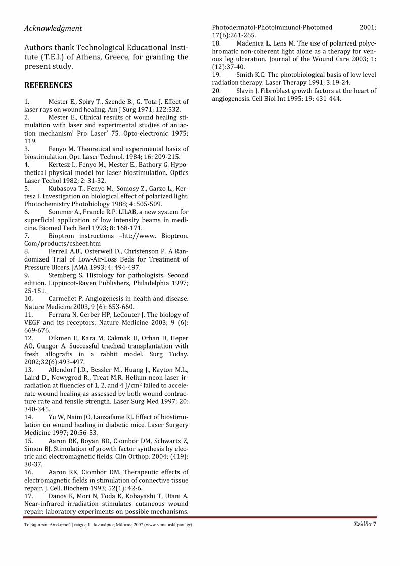
Referring to the planimetric evaluation, al-though we observed acceleration of woundhealing in the EG, in the overall experimentthere was no statistically significant differencesbetween the two groups (p=0.508). We ob-served though significant differences betweenthe two groups on 10th and 15th day (p=0.017and p=0.036 respectively). The differences be-tween those rates are clearly represented bybars given in Figure 1.
Histologically the following findings were ob-served:
Day 5: The histological results showed that inthe control group (CG) the epithelization andthe produced collagen was measured andscored 1.Underneath the blood clot there wasloose granulation tissue with few stimulatedfibroblasts producing collagen around the mar-gins of the wounds. The newly formed endo-thelial cells were scored 2 as only small andmoderately dense capillaries were seen. (Fig-ure 2A)
In the experimental group (EG) a slight epithe-lization was observed and scored 1. The pres-ence of several fibroblasts was evident andsome collagen bundles had already started toform at the base of the wound, 0.5mm from thesurface, scored also 1. The density of the newlyformed capillaries was noted and stained moreintensely in the EG than in the CG with VEGF,score 3 (Table 2 and Figure.2B). This findingwas also confirmed with Vim and a-SMA whichstained the pre- and post-capillary vascularsmooth muscle cells.
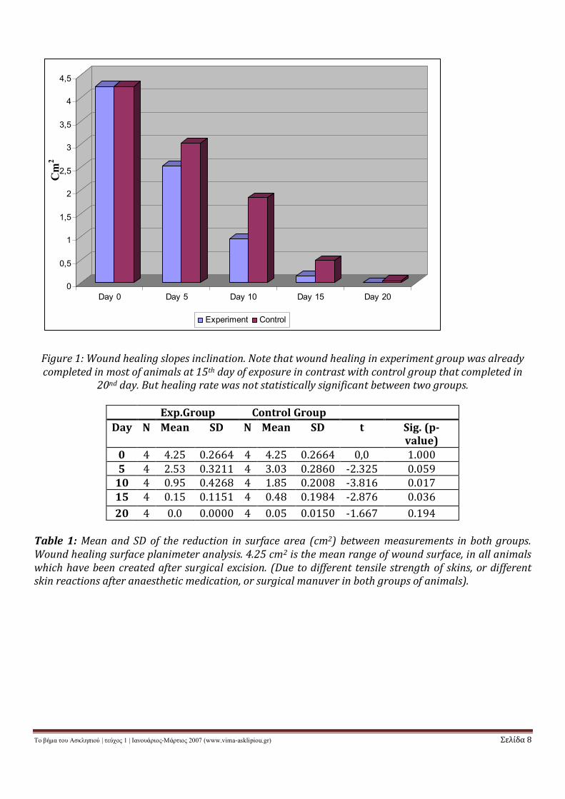
Day 10: The wound healing in the CG showedan epithelization which was measured andscored 1 whilst in the EG scored 3. The newlyformed capillary network, scored 2 both in theCG and the EG as it was demonstrated by VEGF,Vim and a-SMA Also there was an increase inthe formation of collagen by fibroblasts at the
Το βήμα του Ασκληπιού | τεύχος 1 | Ιανουάριος-Μάρτιος 2007 (www.vima-asklipiou.gr) Σελίδα 6
base of the wound, in the CG scored 1 and inthe EG scored 2, (Table 2 and Fig.3A,B ).
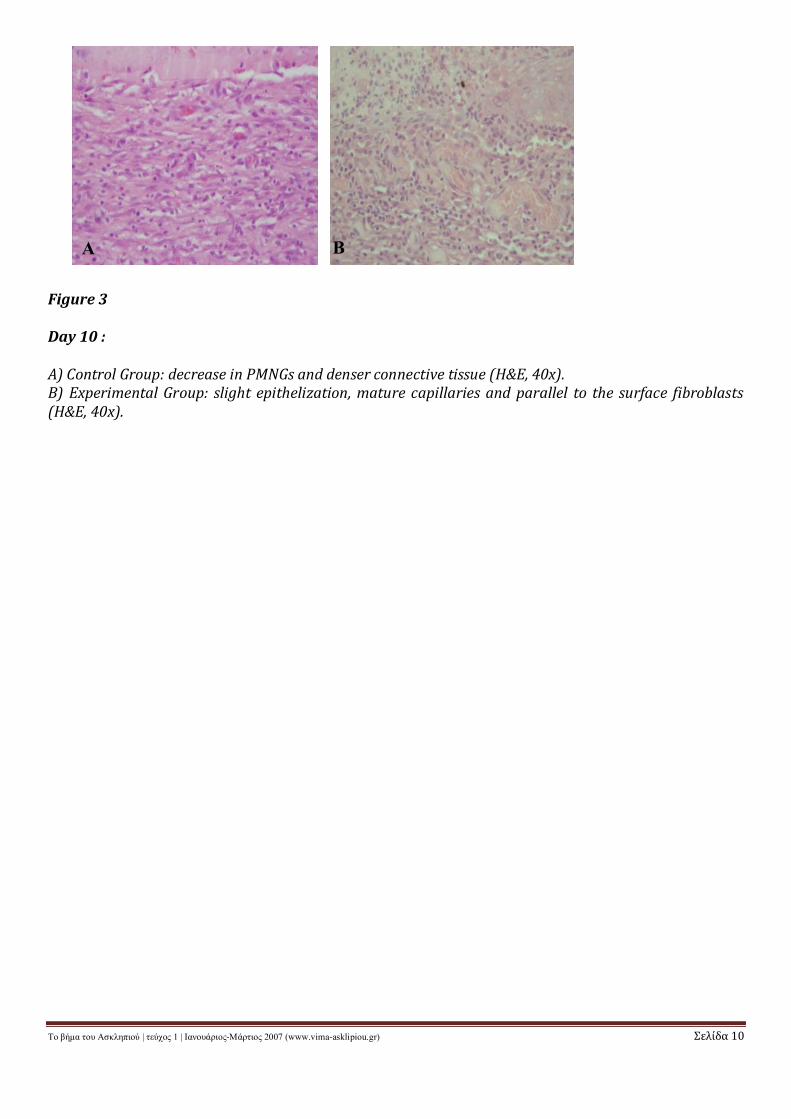
Day 15: The epithelization was still in an in-complete fashion and was measured andscored 3 in CG, whilst in the EG measured andscored 4. The same moderate amount of fibrob-lasts with abundant bundles of collagen werefound in the CG as well as in the EG on the 15th
day, scored 2. The capillary network was moremature than on the 10th day both in the CG andthe EG but the score was the same for bothgroups, 1, as no new capillaries were formed(Table 2 and Fig. 4A,B,C,D).
Day 20: In the final day of our experiment, sim-ilar findings were recorded, in both groups(Table 2). Although quantitive results are simi-lar, in the EG better epithelization and moremature fibroblasts creating a denser collagendeposit parallel to the surface was observedthus creating a better quality of wound healing(Figure 5A,B).
DISCUSSIONA number of studies have assessed the healingprocess of wounds treated by light using differ-ent wavelengths (i.e. low level laser light) butcontroversies appeared between them as somewere unable to show a beneficial effect.13,14
Controversies might be accounted for the dif-ferent criteria and from the variety of appear-ance of the wounds. Some other studies indi-cated that light therapy could be of great bene-fit in the treatment of chronic wounds althoughthe design of these studies was different fromthe present one.5, 6
In our work, although, we did not observe anystatistically significant differences in the over-all healing process between the two groups, theresults showed that most of the rats of EG hadnearly completed the wound healing processon the 15th day compared to the CG which hadcomplete healing on the 20th day. Thus accele-ration of wound healing with a non invasivemethod, such as infrared polarized light, maybe important in reducing bacteria accumula-tion, stimulating growth factor and reducingearly inflammation, creating an appropriateenvironment to facilitate tissue regenera-tion.15,16
Danos17 et al findings are very similar to thoseof the present study. They used cultural humankeratinocytes, endothelial cells and fibroblastsexposing them to the infrared light and theyconcluded that the infrared irradiation poten-tially enhances the wound healing process,supported by its biostimulatory effects.
Madenica,18 using the polarized polychromaticnon-coherent light for venous leg ulcerationsand from their immunochistological analysis,showed that all tissue specimens revealed sig-nificant histologic changes after three weeks oftreatment. Sections stained by Masson’ trich-rome showed significant proliferation of fibrot-ic tissue. All of these sections demonstrated ex-tensive neo-angiogenesis and increased vascu-lar density.
Smith19 suggested that the infrared spectruminitiates the response at the membrane level,through photo physical effects on Ca++ chan-nels.19
Whilst Slavin20 indicated in his study that lowpower laser light therapy stimulates release ofgrowth factors from irradiated cells. Growthfactors stimulate angiogenesis, extra cellularmatrix production and degradation.
The main finding from our study after the useof infrared polarized light was the acceleratedreduction of wound surface area. This findingwas also histologically proven by the fastertransformation and maturation of myofibrob-lasts, forming better quality of collagen bundlesparallel to the surface and faster re-epithelialization of the wound.
CONCLUSION
In conclusion, the findings of the present studyindicated that infrared polarized light seem tofacilitate and improve the quality of skinwound healing in the rat model. Nevertheless,further studies are required to define the op-timal spectrum of which will ensure a betterand faster pattern of wound healing.
Το βήμα του Ασκληπιού | τεύχος 1 | Ιανουάριος-Μάρτιος 2007 (www.vima-asklipiou.gr) Σελίδα 7
Acknowledgment
Authors thank Technological Educational Insti-tute (T.E.I.) of Athens, Greece, for granting thepresent study.
REFERENCES
1. Mester E., Spiry T., Szende B., G. Tota J. Effect oflaser rays on wound healing. Am J Surg 1971; 122:532.2. Mester E., Clinical results of wound healing sti-mulation with laser and experimental studies of an ac-tion mechanism’ Pro Laser’ 75. Opto-electronic 1975;119.3. Fenyo M. Theoretical and experimental basis ofbiostimulation. Opt. Laser Technol. 1984; 16: 209-215.4. Kertesz I., Fenyo M., Mester E., Bathory G. Hypo-thetical physical model for laser biostimulation. OpticsLaser Techol 1982; 2: 31-32.5. Kubasova T., Fenyo M., Somosy Z., Garzo L., Ker-tesz I. Investigation on biological effect of polarized light.Photochemistry Photobiology 1988; 4: 505-509.6. Sommer A., Francle R.P. LILAB, a new system forsuperficial application of low intensity beams in medi-cine. Biomed Tech Berl 1993; 8: 168-171.7. Bioptron instructions –htt://www. Bioptron.Com/products/csheet.htm8. Ferrell A.B., Osterweil D., Christenson P. A Ran-domized Trial of Low-Air-Loss Beds for Treatment ofPressure Ulcers. JAMA 1993; 4: 494-497.9. Stemberg S. Histology for pathologists. Secondedition. Lippincot-Raven Publishers, Philadelphia 1997;25-151.10. Carmeliet P. Angiogenesis in health and disease.Nature Medicine 2003, 9 (6): 653-660.11. Ferrara N, Gerber HP, LeCouter J. The biology ofVEGF and its receptors. Nature Medicine 2003; 9 (6):669-676.12. Dikmen E, Kara M, Cakmak H, Orhan D, HeperAO, Gungor A. Successful tracheal transplantation withfresh allografts in a rabbit model. Surg Today.2002;32(6):493-497.13. Allendorf J.D., Bessler M., Huang J., Kayton M.L.,Laird D., Nowygrod R., Treat M.R. Helium neon laser ir-radiation at fluencies of 1, 2, and 4 J/cm2 failed to accele-rate wound healing as assessed by both wound contrac-ture rate and tensile strength. Laser Surg Med 1997; 20:340-345.14. Yu W, Naim JO, Lanzafame RJ. Effect of biostimu-lation on wound healing in diabetic mice. Laser SurgeryMedicine 1997; 20:56-53.15. Aaron RK, Boyan BD, Ciombor DM, Schwartz Z,Simon BJ. Stimulation of growth factor synthesis by elec-tric and electromagnetic fields. Clin Orthop. 2004; (419):30-37.16. Aaron RK, Ciombor DM. Therapeutic effects ofelectromagnetic fields in stimulation of connective tissuerepair. J. Cell. Biochem 1993; 52(1): 42-6.17. Danos K, Mori N, Toda K, Kobayashi T, Utani A.Near-infrared irradiation stimulates cutaneous woundrepair: laboratory experiments on possible mechanisms.
Photodermatol-Photoimmunol-Photomed 2001;17(6):261-265.18. Madenica L, Lens M. The use of polarized polyc-hromatic non-coherent light alone as a therapy for ven-ous leg ulceration. Journal of the Wound Care 2003; 1:(12):37-40.19. Smith K.C. The photobiological basis of low levelradiation therapy. Laser Therapy 1991; 3:19-24.20. Slavin J. Fibroblast growth factors at the heart ofangiogenesis. Cell Biol Int 1995; 19: 431-444.
Το βήμα του Ασκληπιού | τεύχος 1 | Ιανουάριος-Μάρτιος 2007 (www.vima-asklipiou.gr) Σελίδα 8
Figure 1: Wound healing slopes inclination. Note that wound healing in experiment group was alreadycompleted in most of animals at 15th day of exposure in contrast with control group that completed in
20nd day. But healing rate was not statistically significant between two groups.
Exp.Group Control GroupDay N Mean SD N Mean SD t Sig. (p-
value)0 4 4.25 0.2664 4 4.25 0.2664 0,0 1.0005 4 2.53 0.3211 4 3.03 0.2860 -2.325 0.059
10 4 0.95 0.4268 4 1.85 0.2008 -3.816 0.01715 4 0.15 0.1151 4 0.48 0.1984 -2.876 0.03620 4 0.0 0.0000 4 0.05 0.0150 -1.667 0.194
Table 1: Mean and SD of the reduction in surface area (cm2) between measurements in both groups.Wound healing surface planimeter analysis. 4.25 cm2 is the mean range of wound surface, in all animalswhich have been created after surgical excision. (Due to different tensile strength of skins, or differentskin reactions after anaesthetic medication, or surgical manuver in both groups of animals).
0
0,5
1
1,5
2
2,5
3
3,5
4
4,5
Day 0 Day 5 Day 10 Day 15 Day 20
Experiment Control
Cm
2

Το βήμα του Ασκληπιού | τεύχος 1 | Ιανουάριος-Μάρτιος 2007 (www.vima-asklipiou.gr) Σελίδα 9
Table 2: Scoring of the Epithelization, Inflammation, Neovascularization and Collagen formation tissuein the EG and CC groups
Figure 2
Day 5 :
A) Control Group: immature endothelial cells and newly formed capillary network. (VEGF, 20X).B) Experimental Group: more mature endothelial cells and better formed capillary network are evidend.(VEGF, 20X).
Experimental GroupDay Epith.
ScoreInflam. Neo-
Vasc/tionScore
Collagenscore
5 1 (1/18mm) Negative 3 1 (0.5mm)10 3 (4/8mm) Negative 2 2 (<0.5mm)15 4 (3/5mm) Negative 1 2 (<0.5mm)20 5 (complete) Negative 1 3
Control GroupDay Epith.
ScoreInflam. Neo-
Vasc/tionScore
Collagenscore
5 1(1.5/16mm)
Negative 2 1 (>0.5mm)
10 1 (1/18mm) Negative 2 1 (>0.5mm)15 3 (4/6mm) Negative 1 2 (0.5mm)20 4 (complete) Negative 1 3
A B
Το βήμα του Ασκληπιού | τεύχος 1 | Ιανουάριος-Μάρτιος 2007 (www.vima-asklipiou.gr) Σελίδα 10
Figure 3
Day 10 :
A) Control Group: decrease in PMNGs and denser connective tissue (H&E, 40x).B) Experimental Group: slight epithelization, mature capillaries and parallel to the surface fibroblasts(H&E, 40x).
A B
Το βήμα του Ασκληπιού | τεύχος 1 | Ιανουάριος-Μάρτιος 2007 (www.vima-asklipiou.gr) Σελίδα 11
Figure 4
Day 15 :
A) Control Group: slight only epithelization and immature fibroblasts in the granulation tissue (SMA, 2x).B) Experimental Group: moderate surface epithelization and mature Fibroblasts (SMA, 2x).C) Control group: higher magnification focusing on our findings (SMA, 20x)D) Experiment Group (SMA, 20x).
A
C
B
D
Το βήμα του Ασκληπιού | τεύχος 1 | Ιανουάριος-Μάρτιος 2007 (www.vima-asklipiou.gr) Σελίδα 12
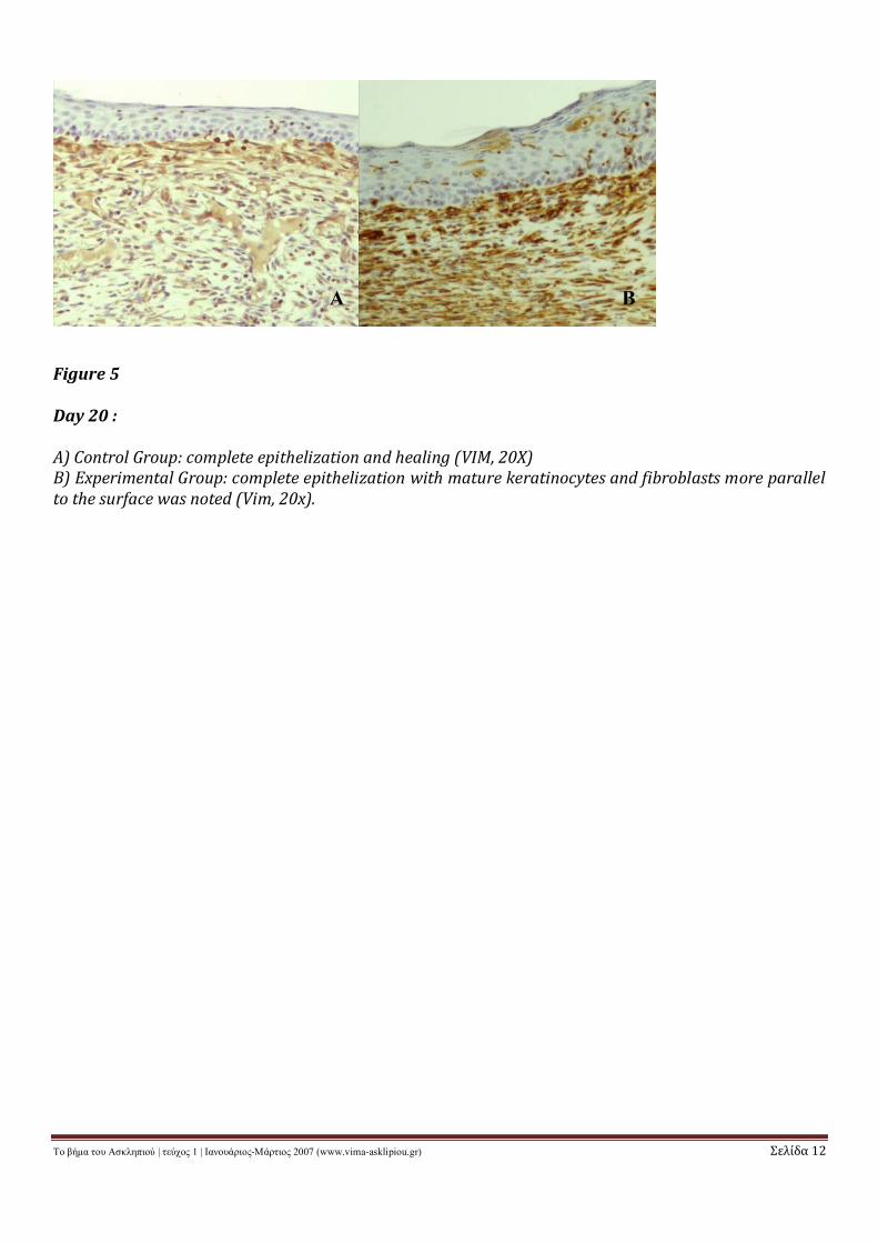
Figure 5
Day 20 :
A) Control Group: complete epithelization and healing (VIM, 20X)B) Experimental Group: complete epithelization with mature keratinocytes and fibroblasts more parallelto the surface was noted (Vim, 20x).
BA