TGF-β in inflammatory bowel disease: a key regulator of ... · REVIEW TGF-b in inflammatory bowel...
11
REVIEW TGF-b in inflammatory bowel disease: a key regulator of immune cells, epithelium, and the intestinal microbiota Sozaburo Ihara 1,2 • Yoshihiro Hirata 2 • Kazuhiko Koike 2 Received: 28 April 2017 / Accepted: 7 May 2017 / Published online: 22 May 2017 Ó Japanese Society of Gastroenterology 2017 Abstract Inflammatory bowel disease (IBD) is defined as chronic intestinal inflammation, and includes ulcerative colitis and Crohn’s disease. Multiple factors are involved in the pathogenesis of IBD, and the condition is characterized by aberrant mucosal immune reactions to intestinal microbes in genetically susceptible hosts. Transforming growth factor-b (TGF-b) is an immune-suppressive cyto- kine produced by many cell types and activated by inte- grins. Active TGF-b binds to its receptor and regulates mucosal immune reactions through the TGF-b signaling pathway. Dysregulated TGF-b signaling is observed in the intestines of IBD patients. TGF-b signal impairment in specific cell types, such as T-cells and dendritic cells, results in spontaneous colitis in mouse models. In addition, specific intestinal microbes contribute to immune home- ostasis by modulating TGF-b production. In this review, we describe the role of TGF-b in intestinal immunity, focusing on immune cells, epithelium, and intestinal microbes. In addition, we present potential therapeutic strategies for IBD that target TGF-b. Keywords Transforming growth factor-b Á Inflammatory bowel disease Á Dendritic cells Á Microbiota Á Adhesion molecules Abbreviations IBD Inflammatory bowel disease TGF-b Transforming growth factor-b UC Ulcerative colitis CD Crohn’s disease DCs Dendritic cells TGFbRI Transforming growth factor-b type I receptor TGFbRII Transforming growth factor-b type II receptor IELs Intraepithelial lymphocytes Introduction Inflammatory bowel disease (IBD) is defined as chronic intestinal inflammation and includes ulcerative colitis (UC) and Crohn’s disease (CD). IBD is believed to be caused by complex interactions among host genetic susceptibility, the immune response, environmental triggers, and the luminal microbiota [1, 2]. Genome-wide association studies have identified more than 160 loci associated with IBD sus- ceptibility [3], including genes related to intestinal mucosal immune responses, such as NOD2 and ATG16L1 [4, 5]. Regarding environmental triggers, frequent use of antibi- otics and improved sanitary conditions (leading to reduced contact with bacteria) are linked to IBD [6]. Changes in the intestinal microbiota are also involved in the pathogenesis of IBD [2, 7]. Inappropriate reactions to commensal intestinal bacteria, as well as an altered bacterial commu- nity, contribute to intestinal inflammation [7, 8]. However, the pathogenesis of IBD has not been fully elucidated, and the incidence and prevalence of IBD are increasing worldwide. Transforming growth factor-b (TGF-b) is a pleiotropic cytokine produced by many cell types, including immune & Yoshihiro Hirata [email protected] 1 Division of Gastroenterology, The Institute for Adult Diseases, Asahi Life Foundation, Tokyo, Japan 2 Department of Gastroenterology, Graduate School of Medicine, The University of Tokyo, 7-3-1 Hongo, Bunkyo- ku, Tokyo 113-8655, Japan 123 J Gastroenterol (2017) 52:777–787 DOI 10.1007/s00535-017-1350-1
Transcript of TGF-β in inflammatory bowel disease: a key regulator of ... · REVIEW TGF-b in inflammatory bowel...
REVIEW
TGF-b in inflammatory bowel disease: a key regulator of immunecells, epithelium, and the intestinal microbiota
Sozaburo Ihara1,2 • Yoshihiro Hirata2 • Kazuhiko Koike2
Received: 28 April 2017 / Accepted: 7 May 2017 / Published online: 22 May 2017
� Japanese Society of Gastroenterology 2017
Abstract Inflammatory bowel disease (IBD) is defined as
chronic intestinal inflammation, and includes ulcerative
colitis and Crohn’s disease. Multiple factors are involved in
the pathogenesis of IBD, and the condition is characterized
by aberrant mucosal immune reactions to intestinal
microbes in genetically susceptible hosts. Transforming
growth factor-b (TGF-b) is an immune-suppressive cyto-
kine produced by many cell types and activated by inte-
grins. Active TGF-b binds to its receptor and regulates
mucosal immune reactions through the TGF-b signaling
pathway. Dysregulated TGF-b signaling is observed in the
intestines of IBD patients. TGF-b signal impairment in
specific cell types, such as T-cells and dendritic cells,
results in spontaneous colitis in mouse models. In addition,
specific intestinal microbes contribute to immune home-
ostasis by modulating TGF-b production. In this review,
we describe the role of TGF-b in intestinal immunity,
focusing on immune cells, epithelium, and intestinal
microbes. In addition, we present potential therapeutic
strategies for IBD that target TGF-b.
Keywords Transforming growth factor-b � Inflammatory
bowel disease � Dendritic cells � Microbiota � Adhesionmolecules
Abbreviations
IBD Inflammatory bowel disease
TGF-b Transforming growth factor-bUC Ulcerative colitis
CD Crohn’s disease
DCs Dendritic cells
TGFbRI Transforming growth factor-b type I receptor
TGFbRII Transforming growth factor-b type II receptor
IELs Intraepithelial lymphocytes
Introduction
Inflammatory bowel disease (IBD) is defined as chronic
intestinal inflammation and includes ulcerative colitis (UC)
and Crohn’s disease (CD). IBD is believed to be caused by
complex interactions among host genetic susceptibility, the
immune response, environmental triggers, and the luminal
microbiota [1, 2]. Genome-wide association studies have
identified more than 160 loci associated with IBD sus-
ceptibility [3], including genes related to intestinal mucosal
immune responses, such as NOD2 and ATG16L1 [4, 5].
Regarding environmental triggers, frequent use of antibi-
otics and improved sanitary conditions (leading to reduced
contact with bacteria) are linked to IBD [6]. Changes in the
intestinal microbiota are also involved in the pathogenesis
of IBD [2, 7]. Inappropriate reactions to commensal
intestinal bacteria, as well as an altered bacterial commu-
nity, contribute to intestinal inflammation [7, 8]. However,
the pathogenesis of IBD has not been fully elucidated, and
the incidence and prevalence of IBD are increasing
worldwide.
Transforming growth factor-b (TGF-b) is a pleiotropic
cytokine produced by many cell types, including immune
& Yoshihiro Hirata
1 Division of Gastroenterology, The Institute for Adult
Diseases, Asahi Life Foundation, Tokyo, Japan
2 Department of Gastroenterology, Graduate School of
Medicine, The University of Tokyo, 7-3-1 Hongo, Bunkyo-
ku, Tokyo 113-8655, Japan
123
J Gastroenterol (2017) 52:777–787
DOI 10.1007/s00535-017-1350-1
cells and non-hematopoietic cells, and regulates multiple
cellular functions as a suppressor of the immune response,
cell proliferation, and oncogenesis. In intestinal immunity,
TGF-b suppresses inflammatory responses to luminal
bacterial antigens and contributes to the induction of
immune tolerance [9, 10]. Smad3, an intracellular signaling
protein in the canonical TGF-b pathway, is among the loci
associated with IBD susceptibility [11]. Impaired TGF-bsignaling is reported to be associated with the development
of intestinal inflammation in experimental models and IBD
patients, and compounds that restore TGF-b signaling are
considered candidate agents for IBD treatment.
Here, we provide an overview of the role of TGF-b in
IBD, focusing on TGF-b production, signaling, and func-
tions as revealed by murine experimental models. Finally,
we discuss potential therapeutic strategies for IBD that
target TGF-b.
TGF-b in IBD
TGF-b production
TGF-b is abundant in the mammalian intestine. There are
three TGF-b isoforms in mammals: TGF-b1, TGF-b2, andTGF-b3 [12]. Among them, TGF-b1 is the most abundant
isoform and its role in intestinal immunity has been
investigated extensively [9, 13]. TGF-b is produced by
many cell types; e.g., epithelial cells, immune cells, and
fibroblasts [13, 14] (Fig. 1a). Although the mechanism
underlying modulation of TGF-b production in the human
intestine remains to be elucidated, TGF-b production is
upregulated by various factors, such as bacteria, viruses,
cytokines, apoptotic cells, and the autocrine/paracrine loop
[15, 16]. A study using laser-captured micro-dissection
reported that TGF-b expression was higher in the lamina
propria than the epithelium in a healthy human colon [9].
TGF-b levels in IBD patients have also been evaluated.
Babyatsky et al. reported that there was no significant
difference in TGF-b1 expression in the colonic mucosa of
healthy individuals and inactive UC and CD patients [13].
Another study showed that TGF-b1 expression in unin-
volved mucosa of UC patients was lower than that in
normal mucosa [17]. In contrast, TGF-b expression was
reported to be elevated in active IBD patients, especially in
lamina propria lymphocytes [13, 18]. Del Zotto et al.
reported that lamina propria lymphocytes isolated from
inflamed mucosa of UC patients showed increased TGF-b1production compared to controls when stimulated with
CD2 and CD28 [19]. Kanazawa et al. found that TGF-b2and TGF-b3 expression was elevated in lamina propria
lymphocytes of active UC and CD patients [20]. Moreover,
the intestinal epithelium can produce TGF-b-containing
extracellular vesicles [21]. Therefore, TGF-b levels are
increased in IBD tissues, but this may not be sufficient to
counteract the ongoing inflammation.
The association between TGF-b level and intestinal
strictures in CD patients has also been investigated. Di
Sabatino et al. reported that TGF-b expression was ele-
vated in the intestinal mucosa overlying strictures in CD
patients [22]. This was due in part to elevated TGF-bproduction by myofibroblasts near the involved intestinal
stricture. Li et al. reported that the active TGF-b level was
higher in strictured intestinal muscle obtained from surgi-
cally resected ilea of CD patients than in adjacent normal
intestinal tissue [23]. IL-6 production by smooth muscle
cells was increased in strictured segments of CD patients.
Furthermore, IL-6 activated the STAT3 pathway and pro-
moted TGF-b1 production by intestinal smooth muscle
cells of CD patients [24].
Serum TGF-b levels in IBD patients have also been
evaluated. Sambuelli et al. reported that the serum TGF-b1level was higher in naıve active UC patients compared to
healthy controls. In addition, the serum TGF-b1 concen-
tration increased in response to conventional IBD treat-
ments, suggesting that TGF-b is required for suppression of
intestinal inflammation in active UC patients [19, 25].
Contini et al. reported that serum TGF-b1 and TGF-b-ex-pressing-neutrophils increased during granulocyte and
monocyte apheresis therapy in patients with active UC
[26].
Activation of latent TGF-b is important for proper TGF-
b function (Fig. 1a) [27]. First, TGF-b is produced and
secreted as an inactivate complex with latency-associated
peptide. Next, latent TGF-b complex is cleaved and acti-
vated by serine proteases or metalloproteinases in a TGF-
b-isoform-specific manner. In this process, integrins often
function as critical co-factors of TGF-b activation [28].
Another study reported that avb8 integrin was highly
expressed in dendritic cells (DCs) in the human intestine.
Indeed, avb8 integrin expression on DCs was upregulated
in IBD patients and by microbial stimuli [29]. These data
suggest that TGF-b production and activation by immune
cells, especially DCs, are necessary for inhibiting intestinal
inflammation in IBD patients. In contrast, another study
showed that TGF-b1 activation by integrin avb3 on smooth
muscle cells increased collagen production and develop-
ment of fibrosis in CD patients with stricture [23].
TGF-b targets and signaling
Activated TGF-b binds to the TGF-b type II receptor
(TGFbRII), which is followed by formation of a complex
with TGF-b type I receptor (TGFbRI). The resultant TGF-b receptor complex activates intracellular signaling
through the Smad-dependent canonical and Smad-
778 J Gastroenterol (2017) 52:777–787
123
independent non-canonical pathways. TGF-b receptors are
expressed by many cell types, including immune cells and
epithelial cells, and have multiple functions associated with
intestinal immune homeostasis (Fig. 1b). Some patients
with Loeys–Dietz syndrome, an autosomal dominant dis-
order caused by heterozygous mutations of the genes
encoding TGFbRI or TGFbRII (Tgfbr1 or Tgfbr2), develop
early-onset IBD [30, 31].
In the Smad-dependent canonical pathway, phosphory-
lated Smad2 and Smad3 form a complex with Smad4 and
enter the nucleus to regulate the transcription of target
genes [32] (Fig. 2). A previous study reported that in
healthy individuals, phosphorylation of Smad3 in intestinal
T-cells was upregulated compared to that in peripheral
T-cells, indicating a role for TGF-b signaling in intestinal
immune homeostasis [9]. Another study reported that
phosphorylation of Smad3 was downregulated in colonic
lamina propria mononuclear cells from IBD patients [10].
Smad7 is a downstream target of the TGF-b pathway that
binds to TGFbRI and acts in a negative-feedback manner
to inhibit the canonical TGF-b pathway. Smad7 levels were
reported to be elevated in the intestinal mucosa and lamina
propria lymphocytes of IBD patients. An elevated Smad7
level resulted in decreased Smad3 phosphorylation and
insufficient TGF-b signaling, and may be associated with
the pathogenesis of IBD [9, 10].
Role of TGF-b in intestinal immunity
The intestinal immune system balances immune responses
to commensal and harmful antigens in the intestinal lumen
to maintain homeostasis. Dysfunction of this system results
in intestinal inflammation. Several experimental models of
IBD facilitate evaluation of the role of the intestinal
immune system, and have identified key regulators and
pathways of IBD pathogenesis.
Some mouse strains with inactivation or disruption of
TGF-b signaling are susceptible to intestinal inflammation
and are used to study the pathogenesis of IBD. In 1993,
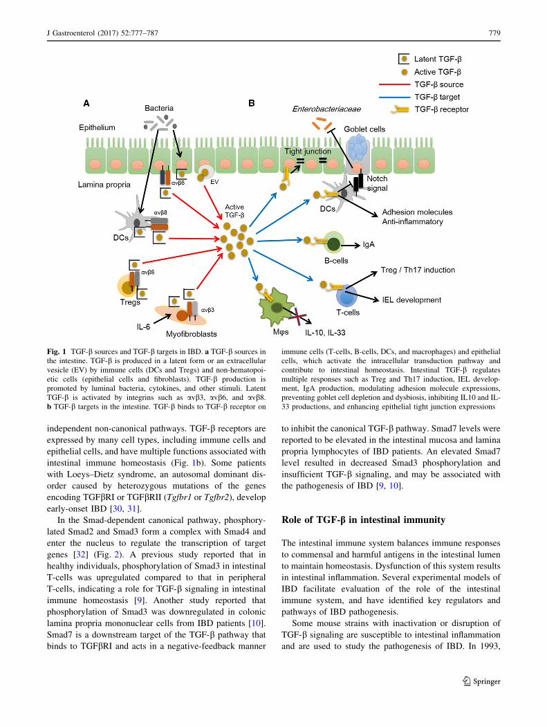
Fig. 1 TGF-b sources and TGF-b targets in IBD. a TGF-b sources in
the intestine. TGF-b is produced in a latent form or an extracellular
vesicle (EV) by immune cells (DCs and Tregs) and non-hematopoi-
etic cells (epithelial cells and fibroblasts). TGF-b production is
promoted by luminal bacteria, cytokines, and other stimuli. Latent
TGF-b is activated by integrins such as avb3, avb6, and avb8.b TGF-b targets in the intestine. TGF-b binds to TGF-b receptor on
immune cells (T-cells, B-cells, DCs, and macrophages) and epithelial
cells, which activate the intracellular transduction pathway and
contribute to intestinal homeostasis. Intestinal TGF-b regulates
multiple responses such as Treg and Th17 induction, IEL develop-
ment, IgA production, modulating adhesion molecule expressions,
preventing goblet cell depletion and dysbiosis, inhibiting IL10 and IL-
33 productions, and enhancing epithelial tight junction expressions
J Gastroenterol (2017) 52:777–787 779
123
TGF-b1 germline-null mice were reported to exhibit mas-
sive inflammatory lesions in multiple organs, including the
colon, at a few weeks of age [33, 34]. Thereafter, several
studies have focused on the specific cellular functions
mediated by TGF-b signaling. Mice with inactivated TGF-
b signaling due to the presence of a dominant-negative
mutant under the control of a cell-specific promoter, or
with cell-specific disruption of TGF-b signaling by a Cre-
lox recombination, have been used in these studies
(Table 1).
In this section, we review the role of TGF-b in immune
cells and the intestinal epithelium in the pathogenesis of
IBD, focusing on experimental mouse models. In addition,
we discuss the roles of TGF-b and the intestinal microbiota
in intestinal immunity.
Immune cells
T-cells
TGF-b regulates multiple immune processes of T-cells. A
major function of TGF-b signaling in T-cells is to suppress
T-cell proliferation and activation through Treg differen-
tiation. Mice with T-cell-targeted deletion of TGF-b sig-
naling (CD4-Cre Tgfbr2fl/fl) showed early onset of fatal
systemic autoimmunity at 3–5 weeks of age [35, 36].
Furthermore, mice with T-cell–targeted inactivation of
TGF-b signaling (CD4-dnTGFbRII) slowly developed
systemic autoimmunity with spontaneous severe colitis at
3–4 months of age [37]. Autoimmunity in both mouse
strains was characterized by massive infiltration of lym-
phocytes and the presence of activated T-cells in multiple
organs. CD4-Cre Tgfbr2fl/fl mice lack TGFbRII expressionon immature CD4? thymocytes and mature peripheral
CD4? T-cells, including CD4? Tregs [35]. As a result,
CD4-Cre Tgfbr2fl/fl mice showed a marked reduction in
peripheral CD4?Foxp3? Tregs. These results suggest that
TGF-b signaling in T-cells contributes to intestinal
immune tolerance, in part by maintenance of the peripheral
Treg cell population. Regarding the effect of TGF-b pro-
duction by CD4? T-cells on Treg differentiation, mice with
CD4? T-cell-targeted deletion of TGF-b1 production
(CD4-CreTgfb1fl/-) did not show a reduction in the num-
bers of peripheral CD4?Foxp3? Tregs, although TGF-b1-
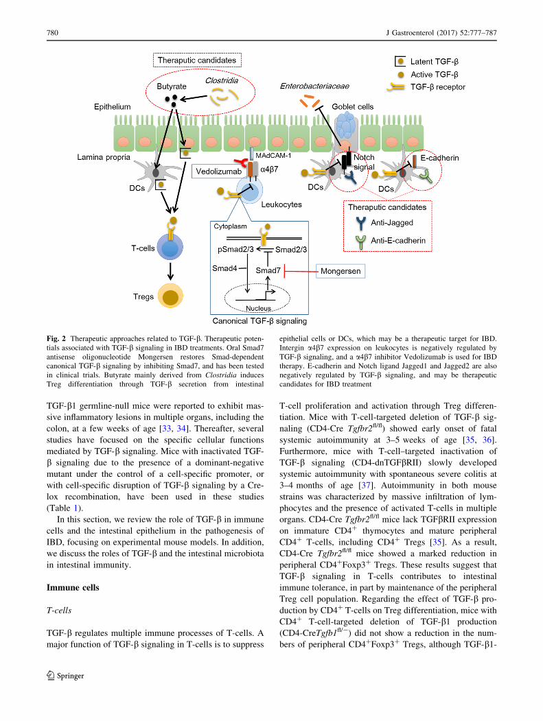
Fig. 2 Therapeutic approaches related to TGF-b. Therapeutic poten-
tials associated with TGF-b signaling in IBD treatments. Oral Smad7
antisense oligonucleotide Mongersen restores Smad-dependent
canonical TGF-b signaling by inhibiting Smad7, and has been tested
in clinical trials. Butyrate mainly derived from Clostridia induces
Treg differentiation through TGF-b secretion from intestinal
epithelial cells or DCs, which may be a therapeutic target for IBD.
Intergin a4b7 expression on leukocytes is negatively regulated by
TGF-b signaling, and a a4b7 inhibitor Vedolizumab is used for IBD
therapy. E-cadherin and Notch ligand Jagged1 and Jagged2 are also
negatively regulated by TGF-b signaling, and may be therapeutic
candidates for IBD treatment
780 J Gastroenterol (2017) 52:777–787
123
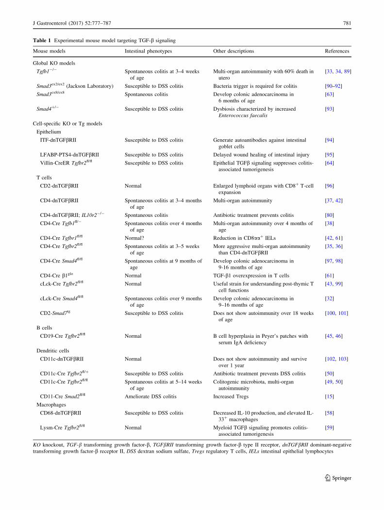
Table 1 Experimental mouse model targeting TGF-b signaling
Mouse models Intestinal phenotypes Other descriptions References
Global KO models
Tgfb1-/- Spontaneous colitis at 3–4 weeks
of age
Multi-organ autoimmunity with 60% death in
utero
[33, 34, 89]
Smad3ex2/ex2 (Jackson Laboratory) Susceptible to DSS colitis Bacteria trigger is required for colitis [90–92]
Smad3ex8/ex8 Spontaneous colitis Develop colonic adenocarcinoma in
6 months of age
[63]
Smad4?/- Susceptible to DSS colitis Dysbiosis characterized by increased
Enterococcus faecalis
[93]
Cell-specific KO or Tg models
Epithelium
ITF-dnTGFbRII Susceptible to DSS colitis Generate autoantibodies against intestinal
goblet cells
[94]
LFABP-PTS4-dnTGFbRII Susceptible to DSS colitis Delayed wound healing of intestinal injury [95]
Villin-CreER Tgfbr2fl/fl Susceptible to DSS colitis Epithelial TGFb signaling suppresses colitis-
associated tumorigenesis
[64]
T cells
CD2-dnTGFbRII Normal Enlarged lymphoid organs with CD8? T-cell
expansion
[96]
CD4-dnTGFbRII Spontaneous colitis at 3–4 months
of age
Multi-organ autoimmunity [37, 42]
CD4-dnTGFbRII; IL10r2-/- Spontaneous colitis Antibiotic treatment prevents colitis [80]
CD4-Cre Tgfb1fl/- Spontaneous colitis over 4 months
of age
Multi-organ autoimmunity over 4 months of
age
[38]
CD4-Cre Tgfbr1fl/fl Normal? Reduction in CD8aa? IELs [42, 61]
CD4-Cre Tgfbr2fl/fl Spontaneous colitis at 3–5 weeks
of age
More aggressive multi-organ autoimmunity
than CD4-dnTGFbRII[35, 36]
CD4-Cre Smad4fl/fl Spontaneous colitis at 9 months of
age
Develop colonic adenocarcinoma in
9-16 months of age
[97, 98]
CD4-Cre b1glo Normal TGF-b1 overexpression in T cells [61]
cLck-Cre Tgfbr2fl/fl Normal Useful strain for understanding post-thymic T
cell functions
[43, 99]
cLck-Cre Smad4fl/fl Spontaneous colitis over 9 months
of age
Develop colonic adenocarcinoma in
9–16 months of age
[32]
CD2-Smad7tg Susceptible to DSS colitis Does not show autoimmunity over 18 weeks
of age
[100, 101]
B cells
CD19-Cre Tgfbr2fl/fl Normal B cell hyperplasia in Peyer’s patches with
serum IgA deficiency
[45, 46]
Dendritic cells
CD11c-dnTGFbRII Normal Does not show autoimmunity and survive
over 1 year
[102, 103]
CD11c-Cre Tgfbr2fl/? Susceptible to DSS colitis Antibiotic treatment prevents DSS colitis [50]
CD11c-Cre Tgfbr2fl/fl Spontaneous colitis at 5–14 weeks
of age
Colitogenic microbiota, multi-organ
autoimmunity
[49, 50]
CD11-Cre Smad2fl/fl Ameliorate DSS colitis Increased Tregs [15]
Macrophages
CD68-dnTGFbRII Susceptible to DSS colitis Decreased IL-10 production, and elevated IL-
33? macrophages
[58]
Lysm-Cre Tgfbr2fl/fl Normal Myeloid TGFb signaling promotes colitis-
associated tumorigenesis
[59]
KO knockout, TGF-b transforming growth factor-b, TGFbRII transforming growth factor-b type II receptor, dnTGFbRII dominant-negative
transforming growth factor-b receptor II, DSS dextran sodium sulfate, Tregs regulatory T cells, IELs intestinal epithelial lymphocytes
J Gastroenterol (2017) 52:777–787 781
123
null mice showed lower numbers of Tregs, indicating that
TGF-b1 produced by cell types other than T-cells con-
tributes to peripheral Treg differentiation [38].
Another function of TGF-b in T-cells is Th17 differ-
entiation. TGF-b together with IL-6 was reported to induce
differentiation of Th17 cells from naıve CD4? T-cells.
Th17 cells produce IL-17 and IFN-c, which are necessary
for mucosal defense against bacteria, but tend to promote
intestinal inflammation [39, 40]. Previous studies showed
that Th17 development in the intestine was impaired in
TGF-b1-null mice and CD4-Cre Tgfb1fl/- mice [38, 41]. In
contrast, Th17 development was not impaired in CD4-Cre
Tgfbr1fl/fl mice or CD4-dnTGFbRII mice [42]. Moreover,
the role of Th17 cells in intestinal inflammation in TGF-b-mutant mice is unclear.
A novel function for TGF-b in memory CD8? T-cells
was reported recently. cLck-Cre Tgfbr2fl/fl mice showed
decreased retention of antigen-specific memory CD8?
T-cells in the intestine, partly due to the defective
expression of integrins [43].
B-cells
TGF-b in B-cells mediates IgA class-switch and promotes
IgA production [44, 45]. Although mice with deletion of
TGF-b signaling in B-cells (CD19-Cre Tgfbr2fl/fl) did not
show signs of autoimmunity or colitis, CD19-Cre Tgfbr2fl/fl
mice showed B-cell hyperplasia in Peyer’s patches and
decreased B-cell responsiveness with complete serum IgA
deficiency [45, 46]. IgA protects against luminal bacteria
by neutralization, enhancing phagocytosis and antigen
presentation by DCs. IgA also inhibits bacterial adhesion to
the epithelium by blocking surface epitopes of bacteria
[47]. IgA production was augmented by the interaction
between B-cells and DCs in Peyer’s patches through inte-
grin avb8-activated TGF-b [48]. This was confirmed by
the finding that IgA class-switch by B-cells in Peyer’s
patches was impaired in CD11c-Cre Itgb8fl/fl mice and by
treatment with an avb8-blocking antibody [48].
DCs
Mice with deletion of TGF-b signaling in DCs (CD11c-Cre
Tgfbr2fl/fl) developed spontaneous colitis with multiple
organ autoimmunity, similar to CD4-Cre Tgfbr2fl/fl and
TGF-b1-null mice [49, 50]. Spontaneous colitis in CD11c-
Cre Tgfbr2fl/fl mice was characterized by loss of goblet
cells with lymphocytic infiltration and systemic autoim-
munity due to altered Treg differentiation, activated T-cells
and B-cells, and increased secretion of inflammatory
cytokines such as TNF-a and IFN-c [49]. We also exam-
ined colitis in CD11c-Cre Tgfbr2fl/fl mice and found
enhanced expression of Notch ligands on DCs, goblet cell
depletion, a thinner mucus layer, and dysbiosis (Fig. 1b)
[50]. These results reveal the critical role played by TGF-bsignaling by DCs in colonic homeostasis.
DCs are also important as a source and activator of
TGF-b in the intestine. Intestinal DCs produce TGF-b and
IL-10, which are major suppressors of intestinal immunity
[51]. Previous reports showed that CD103? tolerogenic
DCs produce TGF-b and retinoic acid, which contributes to
Treg differentiation [52, 53]. Intestinal DCs also contribute
to TGF-b activation. Mice with DC-specific deletion of
integrin b8 (CD11c-Cre Itgb8fl/fl) developed spontaneous
colitis due to a lack of TGF-b activation by avb8 in DCs,
whereas T-cell-specific deletion of integrin b8 (CD4-Cre
Itgb8fl/fl) did not result in the development of colitis [54].
In contrast to CD103? tolerogenic DCs, E-cadherin?
inflammatory DCs promoted intestinal inflammation
through aberrant IL-17 production by CD4? T-cells [55].
E-cadherin is an adhesion molecule expressed in the
intestinal epithelium, and also by subsets of monocytes,
DCs, and macrophages [55–57]. E-cadherin? DCs were
increased in a T-cell-transfer murine colitis model, espe-
cially in the inflamed intestine. Indeed, adoptive transfer of
E-cadherin? BMDCs into T-cell-restored Rag1-/- mice
exacerbated colitis, with an increased Th17 response [55].
TGF-b-deficient mice (DO11.10 Tgfb1-/-) showed an
increased frequency of E-cadherin? DCs, indicating that
TGF-b limits the accumulation of E-cadherin on DCs [55].
However, the molecular mechanisms underlying exacer-
bation of colitis by E-cadherin? DCs are unclear, and data
regarding E-cadherin expression in intestinal DCs from
IBD patients are lacking.
Macrophages
Mice with TGF-b signaling inactivation in macrophages
(CD68-dnTGFbRII) did not develop spontaneous colitis,
but exhibited susceptibility to DSS-induced colitis with
reduced IL-10 production [58]. TGF-b signaling in mac-
rophages suppressed IL-33 production and protected
against intestinal inflammation [58, 59]. It has also been
reported that TGF-b downregulates the expression of
innate response receptors, such as that for LPS (CD14), on
human intestinal macrophages. This contributed to the
development of an ‘‘inflammatory anergy’’ macrophage
phenotype, which is characterized by a lack of proinflam-
matory cytokine production under inflammatory stimuli but
retention of phagocytic and bactericidal activity [60].
Intraepithelial lymphocytes (IELs)
TGF-b production and signaling by T-cells are important
for IEL development [61]. IELs reside in the intestinal
epithelial layer and play a role in mucosal defense. The
782 J Gastroenterol (2017) 52:777–787
123
majority of TCRab? IELs are divided into subsets
expressing CD8aa? or CD8ab? [62]. A previous
study demonstrated that Tgfb1-/-, Smad3ex8/ex8, and CD4-
Cre Tgfbr1fl/fl mice showed reduced numbers of
TCRab?CD8aa? IELs, whereas mice with TGF-b1-over-expressing T-cells (CD4-Cre b1glo) showed increased
numbers of TCRab?CD8aa? IELs, suggesting that TGF-bcontrols the generation and retention of CD8aa? IELs via
CD8a expression [61, 63]. Another study investigated
TGF-b production by TCRab?CD8ab? IELs. Upon
infection by Toxoplasma gondii, TGF-b produced by IELs
interacted with the lamina propria CD4? T-cells and
reduced intestinal inflammation by downregulating IFN-cproduction [62].
Epithelium and extracellular matrix
Previous studies have unraveled the roles of TGF-b in
intestinal epithelial homeostasis associated with mucosal
integrity, wound healing, and consequent fibrosis. Mouse
models with disruption of TGF-b signaling in the intestinal
epithelium (such as Villin-CreER Tgfbr2fl/fl mice) showed
increased susceptibility to DSS-induced colitis [64],
although spontaneous colitis did not occur in these strains,
in contrast to TGF-b1-null mice (Table 1). A recent study
revealed that TGF-b-containing extracellular vesicles
released by epithelial cells induced Treg differentiation and
inhibited colitis by binding to EpCAM? epithelial cells
[21]. Therefore, epithelial cells also play an important role
in immune homeostasis in a TGF-b-dependent manner.
TGF-b modulates the barrier function of the epithelium
by regulating the expression levels of tight-junction pro-
teins and adhesion molecules [65]. A previous in vitro
study using an intestinal monolayer cell line reported that
TGF-b enhanced intestinal epithelial barrier function by
inducing production of the tight junction protein Claudin-1,
and by preventing the pathogenic bacteria-induced reduc-
tion of levels of the tight-junction proteins Claudin-2,
Occludin, and ZO-1 [66].
As wound healing progresses in injured tissues, the
provisional extracellular matrix is replaced by a newly
formed matrix, which is rich in collagen synthesized by
fibroblasts migrating into the wound [67]. The extracellular
matrix is composed of collagens, non-collagenous glyco-
proteins (including fibronectin) and proteoglycans. In CD
patients, chronic transmural intestinal inflammation can
result in intestinal fibrosis and fistula, which require sur-
gical resection [68]. Intestinal strictures in CD patients,
which are usually caused by chronic inflammation and
healing, were associated with an increased TGF-b tran-
script level and excessive accumulation of extracellular
matrix proteins, such as collagens and fibronectin [22, 69].
Myofibroblasts isolated from intestinal strictures of CD
patients overexpressed collagen III, and TGF-b1 promoted
collagen III production by myofibroblasts [69]. Moreover,
pirfenidone, an anti-fibrogenic drug used for the treatment
of fibrotic diseases, suppressed intestinal fibrosis in a DSS-
induced colitis model by inhibiting TGF-b signaling
[70, 71].
Microbiota
The intestinal lumen harbors trillions of microbes of
diverse taxa, including both commensal and harmful bac-
teria; this microbial ecosystem is termed the microbiota.
The microbiota plays a mutualistic role in intestinal
homeostasis by modulating the host immune systems
through their own physiological processes and metabolites
[72]. Some commensal bacterial strains exert an
immunomodulatory effect in a manner involving TGF-b.
Clostridium
Clostridium is a major genus in the intestinal microbiota
and includes several commensal taxa. Microbial trans-
plantation experiments using germ-free mice showed that
some strains of Clostridium cluster IV and XIVa induced
TGF-b release from the intestinal epithelium and Treg
differentiation [73–75]. Clostridium cluster IV and XIVa
were reported to be less abundant in IBD patients than in
healthy controls [76]. Another study showed that admin-
istration of Clostridium butyricum (cluster I) as a probiotic
[77] promoted Treg differentiation through TGF-b1 pro-
duced by lamina propria DCs in a TGF-b-Smad and TLR-
ERK-AP1 pathway-dependent manner [15].
Bacteroides
Bacteroides is a Gram-negative, obligate anaerobic bacte-
rial genus that comprises a considerable proportion of the
normal intestinal flora. The abundance of the genus Bac-
teroides is decreased in IBD patients compared to healthy
controls [78]. Bacteroides fragilis was reported to induce
Treg differentiation [79]. B. fragilis monocolonization of
germ-free mice restored TGF-b2 and IL-10 production by
Tregs and elicited mucosal tolerance in the intestine. This
TGF-b2 production was dependent on polysaccharide A of
B. fragilis [79]. In contrast, another study reported that
luminal commensal bacteria, such as Bacteroides vulgatus
and Bacteroides thetaiotaomicron, are responsible for the
development of colitis in mice with T-cell-specific inacti-
vation of TGF-b and IL-10 signaling [80]. Importantly,
these two Bacteroides species did not induce colitis in
hosts with intact TGF-b signaling, suggesting that TGF-bsignaling suppresses the proinflammatory effects of com-
mensal bacteria.
J Gastroenterol (2017) 52:777–787 783
123
Enterobacteriaceae
The commensal Gram-negative Enterobacteriaceae com-
prise a minor proportion of the intestinal microbiota;
however, overgrowth of Enterobacteriaceae occurs in most
colitis models and IBD patients, and may be associated
with the promotion of intestinal inflammation [8, 72, 81].
The mechanisms underlying the induction and promotion
of colitis by Enterobacteriaceae are unclear; however,
increased oxidative stress due to, for example, increased
ROS and NOS generation caused by bacterial stimulation
of TLRs, contributes to dysbiosis [82, 83]. We previously
reported overgrowth of Enterobacteriaceae in CD11c-cre
Tgfbr2fl/fl mice. TGF-b signaling of DCs was essential for
control of the luminal Enterobacteriaceae through the
interactions with epithelial cells [50].
Potential therapeutic strategies targeting TGF-b
Anti-TNF-a antibodies have for more than 15 years been
used to treat IBD. Our increasing understanding of the role
of immune regulation in the pathogenesis of IBD has
resulted in the proposal of novel therapeutic options for
IBD [84]. As described above, the importance of TGF-bsignaling in IBD pathogenesis is becoming increasingly
apparent. In this section, we focus on the therapeutic
potential of TGF-b signaling.
An oral Smad7 antisense oligonucleotide (Mongersen,
GED-0301) was reported to restore TGF-b signaling in the
intestine of CD patients, an effect mediated by degradation
of Smad7 mRNA [85]. In a preclinical trial, Monteleone
et al. showed that inhibition of Smad7 by a specific anti-
sense oligonucleotide restored TGF-b1-induced Smad3
phosphorylation in lamina propria mononuclear cells of
UC and CD patients [10]. In a phase I trial of Mongersen,
15 patients with active CD received one of three doses of
Mongersen (40, 80, or 160 mg), and the results demon-
strated its safety and tolerability [86]. In a phase II trial,
166 patients with active CD were randomly assigned to
receive one of three doses of Mongersen (10, 40, or
160 mg) or placebo [85, 87]. The primary outcome of this
phase II trial, which was the clinical remission rate at day
15 of Mongersen treatment, was achieved in 55 and 65%
of patients in the 40 and 160 mg Mongersen groups,
respectively, compared with 10% for the placebo group.
Most adverse events were related to complications and
symptoms of CD. This trial demonstrated a benefit in terms
of the clinical remission rate, as well as the safety and
tolerability, of Mongersen in active CD patients [87].
Further studies involving a large population of CD patients
are needed to confirm the safety and efficacy of
Mongersen.
Other than Mongersen, no drug that targets TGF-b sig-
naling to ameliorate IBD has been subjected to a clinical
trial. However, therapeutic approaches related to TGF-bhave potential for IBD treatment (Fig. 2). Because
impaired TGF-b signaling in IBD patients is associated
with reduced Treg differentiation, approaches to promote
Treg differentiation should also be considered. A previous
study reported that butyrate, a short-chain fatty acid bac-
terial metabolite produced mainly by commensal
Clostridium species, was associated with Treg induction
[73]. Therefore, administration of butyrate or butyrate-
producing Clostridium species may have therapeutic
potential for IBD.
TGF-b regulates the expression of adhesion molecules,
such as integrins and E-cadherin, and Notch ligands in
lymphocytes, which are also therapeutic targets for IBD.
TGF-b downregulates the expression of integrin a4b7,E-cadherin, and Jagged1/2 [43, 50, 55]. Expression of these
factors may be upregulated in TGF-b-signaling-impaired
IBD patients, and so agents that suppress expression of
these factors may have therapeutic potential. Although
anti-E-cadherin and Jagged1/2 have not been subjected to
clinical trials, anti-a4b7 treatment has been used in clinical
practice. Vedolizumab is the first gut-selective humanized
a4b7 inhibitor, and functions by blocking the interaction
between a4b7 on gut-homing T-cells and MAdCAM-1 in
the epithelium [88]. Vedolizumab has been approved for
clinical IBD therapy in the USA and Europe. Therefore,
factors involved in TGF-b signaling may also have thera-
peutic potential, which should be the subject of further
work.
Concluding remarks
The studies included in this review suggest that TGF-b is
involved in the maintenance of intestinal homeostasis
through modulating the functions of immune cells, the
epithelium, and the luminal microbiota, which are associ-
ated with the pathogenesis of IBD. Studies using samples
from IBD patients have shown that the TGF-b level is
elevated in active IBD patients, but activation of TGF-bsignaling is insufficient to suppress active IBD. Studies
using mouse models have shown that TGF-b signaling by
T-cells or DCs is important for Treg differentiation and
epithelial homeostasis, to protect against the development
of spontaneous colitis. Further studies using such mouse
models will enhance our understanding of the role of TGF-
b signaling in IBD, and likely result in identification of
novel therapeutic targets associated with TGF-b signaling.
Recent clinical studies showing the efficacy of TGF-bsignal restoration in treatment of active CD will drive
targeting of TGF-b signaling in IBD treatment. Therapies
784 J Gastroenterol (2017) 52:777–787
123
that influence the microbiota to modulate intestinal TGF-bproduction, and those targeting TGF-b-related adhesion
molecules can be the next candidates for IBD treatments.
Acknowledgments This study was supported by a grant from Asahi
Life Foundation.
Compliance with ethical standards
Conflict of interest The authors declare that there are no conflicts of
interest.
References
1. Abraham C, Cho JH. Inflammatory bowel disease. N Engl J
Med. 2009;361:2066–78.
2. Xavier RJ, Podolsky DK. Unravelling the pathogenesis of
inflammatory bowel disease. Nature. 2007;448:427–34.
3. Jostins L, Ripke S, Weersma RK, et al. Host-microbe interac-
tions have shaped the genetic architecture of inflammatory
bowel disease. Nature. 2012;491:119–24.
4. Hugot JP, Chamaillard M, Zouali H, et al. Association of NOD2
leucine-rich repeat variants with susceptibility to Crohn’s dis-
ease. Nature. 2001;411:599–603.
5. Hampe J, Franke A, Rosenstiel P, et al. A genome-wide asso-
ciation scan of nonsynonymous SNPs identifies a susceptibility
variant for Crohn disease in ATG16L1. Nat Genet.
2007;39:207–11.
6. Bernstein CN, Shanahan F. Disorders of a modern lifestyle:
reconciling the epidemiology of inflammatory bowel diseases.
Gut. 2008;57:1185–91.
7. Sartor RB. Microbial influences in inflammatory bowel diseases.
Gastroenterology. 2008;134:577–94.
8. Garrett WS, Gallini CA, Yatsunenko T, et al. Enterobacteriaceae
act in concert with the gut microbiota to induce spontaneous and
maternally transmitted colitis. Cell Host Microbe.
2010;8:292–300.
9. Di Sabatino A, Pickard KM, Rampton D, et al. Blockade of
transforming growth factor beta upregulates T-box transcription
factor T-bet, and increases T helper cell type 1 cytokine and
matrix metalloproteinase-3 production in the human gut mucosa.
Gut. 2008;57:605–12.
10. Monteleone G, Kumberova A, Croft NM, et al. Blocking Smad7
restores TGF-beta1 signaling in chronic inflammatory bowel
disease. J Clin Investig. 2001;108:601–9.
11. Lees CW, Barrett JC, Parkes M, et al. New IBD genetics:
common pathways with other diseases. Gut. 2011;60:1739–53.
12. Massague J. The transforming growth factor-beta family. Annu
Rev Cell Biol. 1990;6:597–641.
13. Babyatsky MW, Rossiter G, Podolsky DK. Expression of
transforming growth factors alpha and beta in colonic mucosa in
inflammatory bowel disease. Gastroenterology.
1996;110:975–84.
14. Letterio JJ, Roberts AB. Regulation of immune responses by
TGF-beta. Annu Rev Immunol. 1998;16:137–61.
15. Kashiwagi I, Morita R, Schichita T, et al. Smad2 and Smad3
inversely regulate TGF-b autoinduction in Clostridium butyr-
icum-activated dendritic cells. Immunity. 2015;43:65–79.
16. Torchinsky MB, Garaude J, Martin AP, et al. Innate immune
recognition of infected apoptotic cells directs T(H)17 cell dif-
ferentiation. Nature. 2009;458:78–82.
17. Chowdhury A, Fukuda R, Fukumoto S. Growth factor mRNA
expression in normal colorectal mucosa and in uninvolved
mucosa from ulcerative colitis patients. J Gastroenterol.
1996;31:353–60.
18. McCabe RP, Secrist H, Botney M, et al. Cytokine mRNA
expression in intestine from normal and inflammatory bowel
disease patients. Clin Immunol Immunopathol. 1993;66:52–8.
19. Del Zotto B, Mumolo G, Pronio AM, et al. TGF-beta1 pro-
duction in inflammatory bowel disease: differing production
patterns in Crohn’s disease and ulcerative colitis. Clin Exp
Immunol. 2003;134:120–6.
20. Kanazawa S, Tsunoda T, Onuma E, et al. VEGF, basic-FGF, and
TGF-beta in Crohn’s disease and ulcerative colitis: a novel
mechanism of chronic intestinal inflammation. Am J Gastroen-
terol. 2001;96:822–8.
21. Jiang L, Shen Y, Guo D, et al. EpCAM-dependent extracellular
vesicles from intestinal epithelial cells maintain intestinal tract
immune balance. Nat Commun. 2016;7:13045.
22. Di Sabatino A, Jackson CL, Pickard KM, et al. Transforming
growth factor beta signalling and matrix metalloproteinases in
the mucosa overlying Crohn’s disease strictures. Gut.
2009;58:777–89.
23. Li C, Flynn RS, Grider JR, et al. Increased activation of latent
TGF-b1 by aVb3 in human Crohn’s disease and fibrosis in
TNBS colitis can be prevented by cilengitide. Inflamm Bowel
Dis. 2013;19:2829–39.
24. Li C, Iness A, Yoon J, et al. Noncanonical STAT3 activation
regulates excess TGF-b1 and collagen I expression in muscle of
stricturing Crohn’s disease. J Immunol. 2015;194:3422–31.
25. Sambuelli A, Diez RA, Sugai E, et al. Serum transforming
growth factor-beta1 levels increase in response to successful
anti-inflammatory therapy in ulcerative colitis. Aliment Phar-
macol Ther. 2000;14:1443–9.
26. Contini P, Negrini S, Bodini G, et al. Granulocytes and mono-
cytes apheresis induces upregulation of TGFb1 in patients with
active ulcerative colitis: a possible involvement of soluble HLA-
I. J Clin Apher. 2017;32:49–55.
27. Massague J. TGFb signalling in context. Nat Rev Mol Cell Biol.
2012;13:616–30.
28. Jenkins G. The role of proteases in transforming growth factor-
beta activation. Int J Biochem Cell Biol. 2008;40:1068–78.
29. Fenton TM, Kelly A, Shuttleworth EE, et al. Inflammatory cues
enhance TGFb activation by distinct subsets of human intestinal
dendritic cells via integrin avb8. Mucosal Immunol.
2017;10:624–34.
30. Naviglio S, Arrigo S, Martelossi S, et al. Severe inflammatory
bowel disease associated with congenital alteration of trans-
forming growth factor beta signaling. J Crohns Colitis.
2014;8:770–4.
31. Loeys BL, Schwarze U, Holm T, et al. Aneurysm syndromes
caused by mutations in the TGF-beta receptor. N Engl J Med.
2006;355:788–98.
32. Johnston CJ, Smyth DJ, Dresser DW, et al. TGF-b in tolerance,
development and regulation of immunity. Cell Immunol.
2016;299:14–22.
33. Shull MM, Ormsby I, Kier AB, et al. Targeted disruption of the
mouse transforming growth factor-beta 1 gene results in multi-
focal inflammatory disease. Nature. 1992;359:693–9.
34. Kulkarni AB, Huh CG, Becker D, et al. Transforming growth
factor beta 1 null mutation in mice causes excessive inflam-
matory response and early death. Proc Natl Acad Sci USA.
1993;90:770–4.
35. Li MO, Sanjabi S, Flavell RA. Transforming growth factor-beta
controls development, homeostasis, and tolerance of T cells by
regulatory T cell-dependent and -independent mechanisms.
Immunity. 2006;25:455–71.
36. Marie JC, Liggitt D, Rudensky AY. Cellular mechanisms of
fatal early-onset autoimmunity in mice with the T cell-specific
J Gastroenterol (2017) 52:777–787 785
123
targeting of transforming growth factor-beta receptor. Immunity.
2006;25:441–54.
37. Gorelik L, Flavell RA. Abrogation of TGFbeta signaling in T
cells leads to spontaneous T cell differentiation and autoimmune
disease. Immunity. 2000;12:171–81.
38. Li MO, Wan YY, Flavell RA. T cell-produced transforming
growth factor-beta1 controls T cell tolerance and regulates Th1-
and Th17-cell differentiation. Immunity. 2007;26:579–91.
39. Goto Y, Panea C, Nakato G, et al. Segmented filamentous
bacteria antigens presented by intestinal dendritic cells drive
mucosal Th17 cell differentiation. Immunity. 2014;40:594–607.
40. Ivanov II, Atarashi K, Manel N, et al. Induction of intestinal
Th17 cells by segmented filamentous bacteria. Cell.
2009;139:485–98.
41. Mangan PR, Harrington LE, O’Quinn DB, et al. Transforming
growth factor-beta induces development of the T(H)17 lineage.
Nature. 2006;441:231–4.
42. Ghoreschi K, Laurence A, Yang XP, et al. Generation of
pathogenic T(H)17 cells in the absence of TGF-b signalling.
Nature. 2010;467:967–71.
43. Zhang N, Bevan MJ. Transforming growth factor-b signaling
controls the formation and maintenance of gut-resident memory
T cells by regulating migration and retention. Immunity.
2013;39:687–96.
44. Ruane D, Chorny A, Lee H, et al. Microbiota regulate the ability
of lung dendritic cells to induce IgA class-switch recombination
and generate protective gastrointestinal immune responses.
J Exp Med. 2016;213:53–73.
45. Cazac BB, Roes J. TGF-beta receptor controls B cell respon-
siveness and induction of IgA in vivo. Immunity.
2000;13:443–51.
46. Roes J, Choi BK, Cazac BB. Redirection of B cell responsive-
ness by transforming growth factor beta receptor. Proc Natl
Acad Sci USA. 2003;100:7241–6.
47. Kubinak JL, Round JL. Do antibodies select a healthy micro-
biota? Nat Rev Immunol. 2016;16:767–74.
48. Reboldi A, Arnon TI, Rodda LB, et al. IgA production requires
B cell interaction with subepithelial dendritic cells in Peyer’s
patches. Science. 2016;352:aaf4822.
49. Ramalingam R, Larmonier CB, Thurston RD, et al. Dendritic
cell-specific disruption of TGF-b receptor II leads to altered
regulatory T cell phenotype and spontaneous multiorgan
autoimmunity. J Immunol. 2012;189:3878–93.
50. Ihara S, Hirata Y, Serizawa T, et al. TGF-b signaling in den-
dritic cells governs colonic homeostasis by controlling epithelial
differentiation and the luminal microbiota. J Immunol.
2016;196:4603–13.
51. Guery L, Hugues S. Tolerogenic and activatory plasmacytoid
dendritic cells in autoimmunity. Front Immunol. 2013;4:59.
52. Coombes JL, Siddiqui KR, Arancibia-Carcamo CV, et al. A
functionally specialized population of mucosal CD103? DCs
induces Foxp3? regulatory T cells via a TGF-beta and retinoic
acid-dependent mechanism. J Exp Med. 2007;204:1757–64.
53. Sun CM, Hall JA, Blank RB, et al. Small intestine lamina
propria dendritic cells promote de novo generation of Foxp3 T
reg cells via retinoic acid. J Exp Med. 2007;204:1775–85.
54. Travis MA, Reizis B, Melton AC, et al. Loss of integrin
alpha(v)beta8 on dendritic cells causes autoimmunity and colitis
in mice. Nature. 2007;449:361–5.
55. Siddiqui KR, Laffont S, Powrie F. E-cadherin marks a subset of
inflammatory dendritic cells that promote T cell-mediated col-
itis. Immunity. 2010;32:557–67.
56. Van den Bossche J, Malissen B, Mantovani A, et al. Regulation
and function of the E-cadherin/catenin complex in cells of the
monocyte-macrophage lineage and DCs. Blood. 2012;119:
1623–33.
57. Van den Bossche J, Van Ginderachter JA. E-cadherin: from
epithelial glue to immunological regulator. Eur J Immunol.
2013;43:34–7.
58. Rani R, Smulian AG, Greaves DR, et al. TGF-b limits IL-33
production and promotes the resolution of colitis through reg-
ulation of macrophage function. Eur J Immunol.
2011;41:2000–9.
59. Li J, Liu Y, Wang B, et al. Myeloid TGF-b signaling contributes
to colitis-associated tumorigenesis in mice. Carcinogenesis.
2013;34:2099–108.
60. Smythies LE, Sellers M, Clements RH, et al. Human intestinal
macrophages display profound inflammatory anergy despite
avid phagocytic and bacteriocidal activity. J Clin Investig.
2005;115:66–75.
61. Konkel JE, Maruyama T, Carpenter AC, et al. Control of the
development of CD8aa? intestinal intraepithelial lymphocytes
by TGF-b. Nat Immunol. 2011;12:312–9.
62. Mennechet FJ, Kasper LH, Rachinel N, et al. Intestinal
intraepithelial lymphocytes prevent pathogen-driven inflamma-
tion and regulate the Smad/T-bet pathway of lamina propria
CD4? T cells. Eur J Immunol. 2004;34:1059–67.
63. Yang X, Letterio JJ, Lechleider RJ, et al. Targeted disruption of
SMAD3 results in impaired mucosal immunity and diminished
T cell responsiveness to TGF-beta. EMBO J. 1999;18:1280–91.
64. Oshima H, Nakayama M, Han TS, et al. Suppressing TGFbsignaling in regenerating epithelia in an inflammatory
microenvironment is sufficient to cause invasive intestinal can-
cer. Cancer Res. 2015;75:766–76.
65. Biancheri P, Di Sabatino A, Corazza GR, et al. Proteases and the
gut barrier. Cell Tissue Res. 2013;351:269–80.
66. Howe KL, Reardon C, Wang A, et al. Transforming growth
factor-beta regulation of epithelial tight junction proteins
enhances barrier function and blocks enterohemorrhagic
Escherichia coli O157: H7-induced increased permeability. Am
J Pathol. 2005;167:1587–97.
67. Biancheri P, Giuffrida P, Docena GH, et al. The role of trans-
forming growth factor (TGF)-b in modulating the immune
response and fibrogenesis in the gut. Cytokine Growth Factor
Rev. 2014;25:45–55.
68. Burke JP, Mulsow JJ, O’Keane C, et al. Fibrogenesis in Crohn’s
disease. Am J Gastroenterol. 2007;102:439–48.
69. Stallmach A, Schuppan D, Riese HH, et al. Increased collagen
type III synthesis by fibroblasts isolated from strictures of
patients with Crohn’s disease. Gastroenterology. 1992;102:
1920–9.
70. Li G, Ren J, Hu Q, et al. Oral pirfenidone protects against fibrosis
by inhibiting fibroblast proliferation and TGF-b signaling in a
murine colitis model. Biochem Pharmacol. 2016;117:57–67.
71. Speca S, Rousseaux C, Dubuquoy C, et al. Novel PPARcmodulator GED-0507-34 levo ameliorates inflammation-driven
intestinal fibrosis. Inflamm Bowel Dis. 2016;22:279–92.
72. Kamada N, Seo SU, Chen GY, et al. Role of the gut microbiota
in immunity and inflammatory disease. Nat Rev Immunol.
2013;13:321–35.
73. Furusawa Y, Obata Y, Fukuda S, et al. Commensal microbe-
derived butyrate induces the differentiation of colonic regulatory
T cells. Nature. 2013;504:446–50.
74. Atarashi K, Tanoue T, Shima T, et al. Induction of colonic
regulatory T cells by indigenous Clostridium species. Science.
2011;331:337–41.
75. Atarashi K, Tanoue T, Oshima K, et al. Treg induction by a
rationally selected mixture of Clostridia strains from the human
microbiota. Nature. 2013;500:232–6.
76. Sokol H, Seksik P, Furet JP, et al. Low counts of Faecalibac-
terium prausnitzii in colitis microbiota. Inflamm Bowel Dis.
2009;15:1183–9.
786 J Gastroenterol (2017) 52:777–787
123
77. Seki H, Shiohara M, Matsumura T, et al. Prevention of antibi-
otic-associated diarrhea in children by Clostridium butyricum
MIYAIRI. Pediatr Int. 2003;45:86–90.
78. Frank DN, St Amand AL, Feldman RA, et al. Molecular-phy-
logenetic characterization of microbial community imbalances
in human inflammatory bowel diseases. Proc Natl Acad Sci
USA. 2007;104:13780–5.
79. Round JL, Mazmanian SK. Inducible Foxp3? regulatory T-cell
development by a commensal bacterium of the intestinal
microbiota. Proc Natl Acad Sci USA. 2010;107:12204–9.
80. Bloom SM, Bijanki VN, Nava GM, et al. Commensal Bac-
teroides species induce colitis in host-genotype-specific fashion
in a mouse model of inflammatory bowel disease. Cell Host
Microbe. 2011;9:390–403.
81. Carvalho FA, Koren O, Goodrich JK, et al. Transient inability to
manage proteobacteria promotes chronic gut inflammation in
TLR5-deficient mice. Cell Host Microbe. 2012;12:139–52.
82. Panday A, Sahoo MK, Osorio D, et al. NADPH oxidases: an
overview from structure to innate immunity-associated
pathologies. Cell Mol Immunol. 2015;12:5–23.
83. Kamdar K, Khakpour S, Chen J, et al. Genetic and metabolic
signals during acute enteric bacterial infection alter the micro-
biota and drive progression to chronic inflammatory disease.
Cell Host Microbe. 2016;19:21–31.
84. Hirata Y, Ihara S, Koike K. Targeting the complex interactions
between microbiota, host epithelial and immune cells in
inflammatory bowel disease. Pharmacol Res. 2016;113:574–84.
85. Monteleone G, Neurath MF, Ardizzone S, et al. Mongersen, an
oral SMAD7 antisense oligonucleotide, and Crohn’s disease.
N Engl J Med. 2015;372:1104–13.
86. Monteleone G, Fantini MC, Onali S, et al. Phase I clinical trial
of Smad7 knockdown using antisense oligonucleotide in patients
with active Crohn’s disease. Mol Ther. 2012;20:870–6.
87. Monteleone G, Di Sabatino A, Ardizzone S, et al. Impact of
patient characteristics on the clinical efficacy of Mongersen
(GED-0301), an oral Smad7 antisense oligonucleotide, in active
Crohn’s disease. Aliment Pharmacol Ther. 2016;43:717–24.
88. Feagan BG, Rutgeerts P, Sands BE, et al. Vedolizumab as
induction and maintenance therapy for ulcerative colitis. N Engl
J Med. 2013;369:699–710.
89. Kulkarni AB, Ward JM, Yaswen L, et al. Transforming growth
factor-beta 1 null mice. An animal model for inflammatory
disorders. Am J Pathol. 1995;146:264–75.
90. Seamons A, Treuting PM, Brabb T, Maggio-Price L. Charac-
terization of dextran sodium sulfate-induced inflammation and
colonic tumorigenesis in Smad3(-/-) mice with dysregulated
TGFb. PLoS One. 2013;8:e79182.
91. Maggio-Price L, Treuting P, Bielefeldt-Ohmann H, et al. Bac-
terial infection of Smad3/Rag2 double-null mice with trans-
forming growth factor-beta dysregulation as a model for
studying inflammation-associated colon cancer. Am J Pathol.
2009;174:317–29.
92. Maggio-Price L, Treuting P, Zeng W, Tsang M, Bielefeldt-
Ohmann H, Iritani BM. Helicobacter infection is required for
inflammation and colon cancer in SMAD3-deficient mice.
Cancer Res. 2006;66:828–38.
93. Szigeti R, Pangas SA, Nagy-Szakal D, et al. SMAD4 haploin-
sufficiency associates with augmented colonic inflammation in
select humans and mice. Ann Clin Lab Sci. 2012;42:401–8.
94. Hahm KB, Im YH, Parks TW, et al. Loss of transforming growth
factor beta signalling in the intestine contributes to tissue injury
in inflammatory bowel disease. Gut. 2001;49:190–8.
95. Beck PL, Rosenberg IM, Xavier RJ, Koh T, Wong JF, Podolsky
DK. Transforming growth factor-beta mediates intestinal heal-
ing and susceptibility to injury in vitro and in vivo through
epithelial cells. Am J Pathol. 2003;162:597–608.
96. Lucas PJ, Kim SJ, Melby SJ, Gress RE. Disruption of T cell
homeostasis in mice expressing a T cell-specific dominant
negative transforming growth factor beta II receptor. J Exp Med.
2000;191:1187–96.
97. Yang XO, Nurieva R, Martinez GJ, et al. Molecular antagonism
and plasticity of regulatory and inflammatory T cell programs.
Immunity. 2008;29:44–56.
98. Kim BG, Li C, Qiao W, et al. Smad4 signalling in T cells is
required for suppression of gastrointestinal cancer. Nature.
2006;441:1015–19.
99. Zhang N, Bevan MJ. TGF-b signaling to T cells inhibits
autoimmunity during lymphopenia-driven proliferation. Nat
Immunol. 2012;13:667–73
100. Fantini MC, Rizzo A, Fina D, et al. Smad7 controls resistance of
colitogenic T cells to regulatory T cell-mediated suppression.
Gastroenterology. 2009;136:1308–16. doi:10.1053/j.gastro.
2008.12.053.
101. Rizzo A, Waldner MJ, Stolfi C, et al. Smad7 expression in T
cells prevents colitis-associated cancer. Cancer Res.
2011;71:7423–32
102. Laouar Y, Sutterwala FS, Gorelik L, Flavell RA. Transforming
growth factor-beta controls T helper type 1 cell development
through regulation of natural killer cell interferon-gamma. Nat
Immunol. 2005;6:600–7
103. Sanjabi S, Flavell RA. Overcoming the hurdles in using mouse
genetic models that block TGF-beta signaling. J Immunol
Methods. 2010;353:111–4
J Gastroenterol (2017) 52:777–787 787
123