TGF-β and the TGF-Family βirm.ucsf.edu/derynck/documents/211 Derynck.pdf · sor segments (Derynck...
16
2 TGF-β and the TGF-β Family Rik Derynck 1 and Kohei Miyazono 2 1 Department of Cell and Tissue Biology University of California San Francisco, California 94143 2 Department of Molecular Pathology Graduate School of Medicine The University of Tokyo Tokyo 113-0033, Japan AS OUTLINED IN THE PREVIOUS CHAPTER, the biochemical characteri- zation of human transforming growth factor-β (TGF-β), now known as TGF-β1, and the determination of its sequence through cDNA cloning provided the basis for identification of TGF-β as structurally distinct from TGF-α. The most striking characteristic that set it apart from TGF-α at that time was that TGF-β was a 25-kD disulfide-linked dimer that was reduced to a 12.5-kD band on gel following treatment with β-mercaptoethanol (Roberts et al. 1983). Following its cDNA cloning (Derynck et al. 1985), it became apparent that TGF-β did not at all resemble TGF-α to which it had been functionally compared thus far and that its polypeptide sequence was unrelated to anything known before. The predicted polypeptide sequence also clearly showed that the mature TGF-β monomer corresponded to only the carboxy-terminal third of a much larger precursor, thus requiring proteolytic cleavage (see Fig. 3 of Chapter 1). Subsequent cDNA cloning demonstrated that the polypeptide chains that define the heteromeric disulfide-linked in- hibin are structurally related to TGF-β (Mason et al. 1985; Vale et al. 1986). These polypeptides are, similarly to TGF-β, encoded as carboxy- terminal polypeptides of larger precursors, and only the carboxy-termi- nal mature polypeptides show structural similarity with TGF-β. Thus was born the realization that there may be a family of secreted disulfide- The TGF-β Family ©2008 Cold Spring Harbor Laboratory Press 978-087969752-5 29 02_TGFb_029-044.qxd 8/15/07 9:45 AM Page 29
Transcript of TGF-β and the TGF-Family βirm.ucsf.edu/derynck/documents/211 Derynck.pdf · sor segments (Derynck...
2
TGF-β and the TGF-β Family
Rik Derynck1 and Kohei Miyazono2
1Department of Cell and Tissue BiologyUniversity of CaliforniaSan Francisco, California 94143
2Department of Molecular PathologyGraduate School of MedicineThe University of TokyoTokyo 113-0033, Japan
AS OUTLINED IN THE PREVIOUS CHAPTER, the biochemical characteri-zation of human transforming growth factor-β (TGF-β), now knownas TGF-β1, and the determination of its sequence through cDNAcloning provided the basis for identification of TGF-β as structurallydistinct from TGF-α. The most striking characteristic that set it apartfrom TGF-α at that time was that TGF-β was a 25-kD disulfide-linkeddimer that was reduced to a 12.5-kD band on gel following treatmentwith β-mercaptoethanol (Roberts et al. 1983). Following its cDNAcloning (Derynck et al. 1985), it became apparent that TGF-β did notat all resemble TGF-α to which it had been functionally compared thusfar and that its polypeptide sequence was unrelated to anything knownbefore. The predicted polypeptide sequence also clearly showed that themature TGF-β monomer corresponded to only the carboxy-terminalthird of a much larger precursor, thus requiring proteolytic cleavage(see Fig. 3 of Chapter 1). Subsequent cDNA cloning demonstrated thatthe polypeptide chains that define the heteromeric disulfide-linked in-hibin are structurally related to TGF-β (Mason et al. 1985; Vale et al.1986). These polypeptides are, similarly to TGF-β, encoded as carboxy-terminal polypeptides of larger precursors, and only the carboxy-termi-nal mature polypeptides show structural similarity with TGF-β. Thus wasborn the realization that there may be a family of secreted disulfide-
The TGF-β Family ©2008 Cold Spring Harbor Laboratory Press 978-087969752-5 29
02_TGFb_029-044.qxd 8/15/07 9:45 AM Page 29
linked dimeric polypeptides encoded as carboxy-terminal segments oflarger secreted polypeptides. This realization was further borne out bythe cDNA cloning of bone morphogenetic protein-2A (BMP-2A) andBMP-2B, now known as BMP-2 and BMP-4, respectively (Wozney et al.1988), of Müllerian inhibiting substance (MIS; also termed anti-Müller-ian hormone or AMH) (Cate et al. 1986), and an increasing number ofproteins that were identified for their roles in developmental processes.
As the number of TGF-β-related proteins rapidly increased, the des-ignation of a TGF-β “superfamily” was more frequently used. We elect notto define the TGF-β-related proteins as a “superfamily,” because superfam-ilies typically combine proteins with structural or sequence similaritiescomprised within differently organized polypeptides. For example, theimmunoglobulin superfamily combines secreted immunoglobulins, cell-surface receptors, and secreted polypeptide precursors. With the realiza-tion of the human and mouse genome sequence projects, it became ap-parent that mammalian genomes encode 33 TGF-β-related proteins. Allof these genes encode secreted proteins with similar organizations, that is,an amino-terminal signal peptide, a larger precursor segment or proseg-ment, and a carboxy-terminal polypeptide monomer that is cleaved fromthe precursor. The carboxy-terminal monomer sequences display and de-fine the structural similarity of TGF-β family proteins, and the long pre-cursor sequences are unrelated in sequence to one another. Glial-cell-de-rived neutrotrophic factor (GDNF) and related proteins, that is, artemin,persephin, and neurturin, may be considered distant members of the TGF-β family (Lin et al. 1993; Sariola and Saarma 2003). However, we do notdiscuss GDNF-related factors in this volume, because the sequence simi-larities between the TGF-β family proteins and the GDNF-related proteinsare low, and the latter transduce signals through the Ret tyrosine kinasereceptor (Sariola and Saarma 2003).
THE MAMMALIAN TGF-β FAMILY
Precursor Structures
Various approaches, based on purification, genetic analyses, or targetedcDNA cloning, have led to the identification of the currently knownmembers of the TGF-β family, both in humans and mice. Additionally,the human and mouse genome projects have explored whether there wereadditional genes for previously unknown TGF-β-related proteins, but ap-parently did not discover such novel genes. Figure 1 shows the 33 knownhuman TGF-β family proteins with evolutionary relationships calculatedon the basis of their sequences. Considering the multiple and often par-
30 R. Derynck and K. Miyazono
02_TGFb_029-044.qxd 8/13/07 11:19 AM Page 30
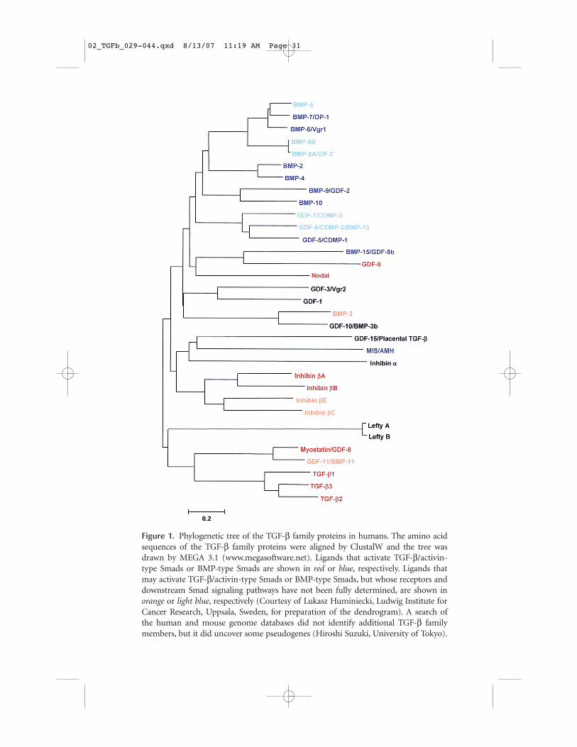
Figure 1. Phylogenetic tree of the TGF-β family proteins in humans. The amino acidsequences of the TGF-β family proteins were aligned by ClustalW and the tree wasdrawn by MEGA 3.1 (www.megasoftware.net). Ligands that activate TGF-β/activin-type Smads or BMP-type Smads are shown in red or blue, respectively. Ligands thatmay activate TGF-β/activin-type Smads or BMP-type Smads, but whose receptors anddownstream Smad signaling pathways have not been fully determined, are shown inorange or light blue, respectively (Courtesy of Lukasz Huminiecki, Ludwig Institute forCancer Research, Uppsala, Sweden, for preparation of the dendrogram). A search ofthe human and mouse genome databases did not identify additional TGF-β familymembers, but it did uncover some pseudogenes (Hiroshi Suzuki, University of Tokyo).
02_TGFb_029-044.qxd 8/13/07 11:19 AM Page 31
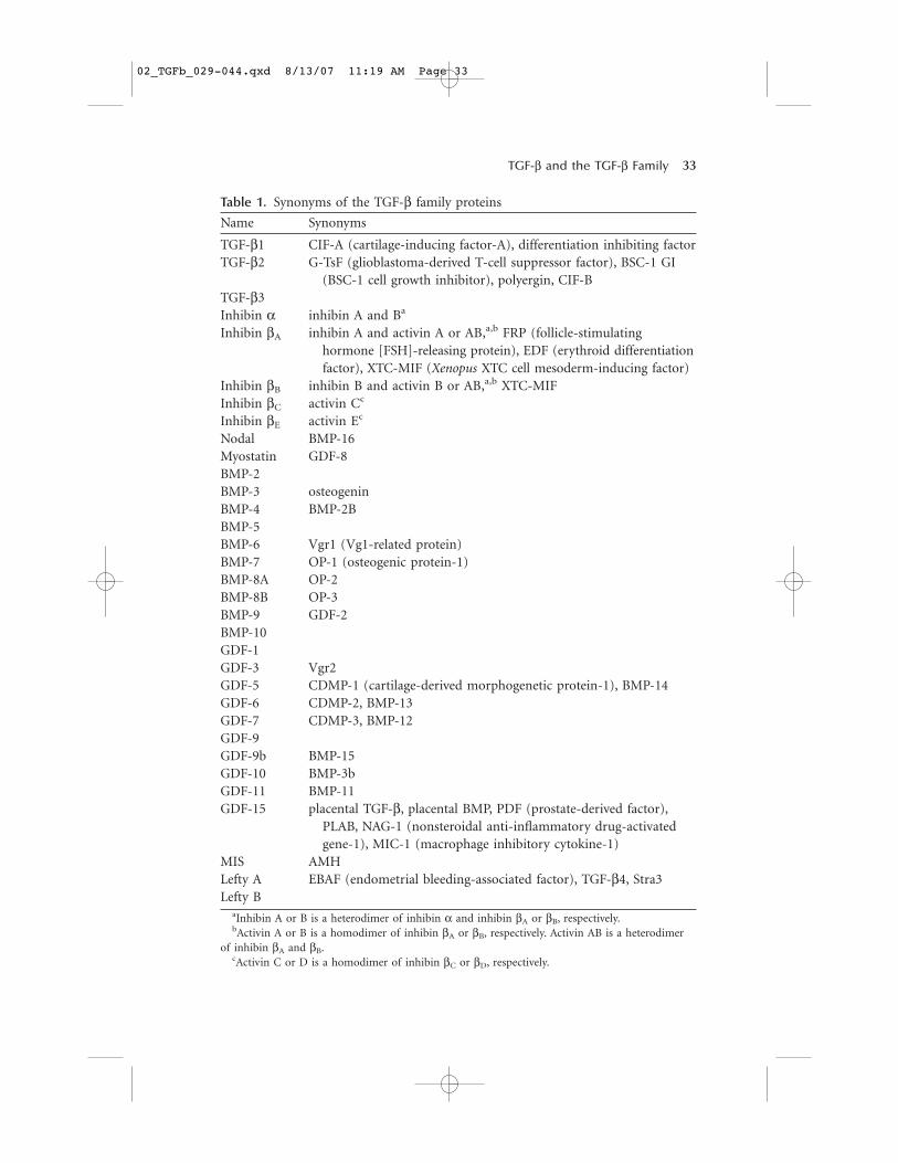
allel approaches that have led to the identification of TGF-β family pro-teins, it is not surprising that several ligands became known with multi-ple names (Table 1). However, most investigators have now settled on acommon name for each individual TGF-β family protein; this nomencla-ture is used in Figure 1 and throughout this volume.
All TGF-β family members are encoded by much larger precursor pro-teins, whose sequences have been deduced through cDNA cloning. They en-code an amino-terminal signal peptide that is removed during translocationof the protein into the lumen of the rough endoplasmic reticulum—a largeprecursor segment or prosegment that is often about twice the length of themonomer sequence for the active and fully mature TGF-β family protein—and the carboxy-terminal TGF-β family monomer polypeptide. The se-quence similarities among TGF-β-related proteins pertain exclusively to thesequences of the mature polypeptides. In the precursor, the mature TGF-βfamily polypeptide is immediately preceded by one to four basic residues,suggesting that this cleavage is regulated by intracellular KEX-like proteases,although other extracellular proteases have been invoked as well. The regu-lation of the cleavage of the mature protein from its larger precursor has re-ceived little attention and is discussed in Chapter 7.
The prosegments are remarkably unconserved between TGF-β fam-ily members. In fact, the structural conservation of the precursors islargely confined to the mature protein sequences. On the other hand,the sequence conservation of any given precursor segment among dif-ferent organisms strongly suggests highly conserved functions. This pre-cursor segment has several regulatory roles in the release and presenta-tion of the mature ligand, although most knowledge has been derivedfrom studies on TGF-β1. Thus, the prosegments may function as chap-erones of the mature bioactive proteins during intracellular biosynthe-sis, folding, and transport of the TGF-β family proteins. They have alsobeen implicated in targeting TGF-β proteins toward sites of storage oractivation. Such targeting could be achieved through direct interactionof the precursor segment with extracellular proteins in the cell environ-ment (Gregory et al. 2005) or through covalent interaction of the pre-cursor segment with one of four structurally related proteins known aslatent TGF-β-binding proteins (LTBPs) that in turn mediate targeting(see Chapter 7). Finally, the noncovalent interaction of the prosegmentswith the mature TGF-β family protein can confer “latency,” as shown inthe case of TGF-β, thereby preventing the bioactive protein from bind-ing to and activating the cognate receptors. The prosegments of theTGF-βs have therefore been termed latency-associated peptides (LAPs).This latency and the subsequent activation to release the active protein
32 R. Derynck and K. Miyazono
02_TGFb_029-044.qxd 8/13/07 11:19 AM Page 32
TGF-β and the TGF-β Family 33
Table 1. Synonyms of the TGF-β family proteins
Name Synonyms
TGF-β1 CIF-A (cartilage-inducing factor-A), differentiation inhibiting factorTGF-β2 G-TsF (glioblastoma-derived T-cell suppressor factor), BSC-1 GI
(BSC-1 cell growth inhibitor), polyergin, CIF-BTGF-β3Inhibin α inhibin A and Ba
Inhibin βA inhibin A and activin A or AB,a,b FRP (follicle-stimulating hormone [FSH]-releasing protein), EDF (erythroid differentiationfactor), XTC-MIF (Xenopus XTC cell mesoderm-inducing factor)
Inhibin βB inhibin B and activin B or AB,a,b XTC-MIFInhibin βC activin Cc
Inhibin βE activin Ec
Nodal BMP-16Myostatin GDF-8BMP-2BMP-3 osteogeninBMP-4 BMP-2BBMP-5BMP-6 Vgr1 (Vg1-related protein)BMP-7 OP-1 (osteogenic protein-1)BMP-8A OP-2BMP-8B OP-3BMP-9 GDF-2BMP-10GDF-1GDF-3 Vgr2GDF-5 CDMP-1 (cartilage-derived morphogenetic protein-1), BMP-14GDF-6 CDMP-2, BMP-13GDF-7 CDMP-3, BMP-12GDF-9GDF-9b BMP-15GDF-10 BMP-3bGDF-11 BMP-11GDF-15 placental TGF-β, placental BMP, PDF (prostate-derived factor),
PLAB, NAG-1 (nonsteroidal anti-inflammatory drug-activated gene-1), MIC-1 (macrophage inhibitory cytokine-1)
MIS AMHLefty A EBAF (endometrial bleeding-associated factor), TGF-β4, Stra3Lefty B
aInhibin A or B is a heterodimer of inhibin α and inhibin βA or βB, respectively.bActivin A or B is a homodimer of inhibin βA or βB, respectively. Activin AB is a heterodimer
of inhibin βA and βB.cActivin C or D is a homodimer of inhibin βC or βD, respectively.
02_TGFb_029-044.qxd 8/13/07 11:19 AM Page 33
have been primarily studied in the case of TGF-β1, with additionalknowledge gained on the activation of myostatin (Wolfman et al. 2003),but these have been poorly examined in the cases of other TGF-β fam-ily proteins, even the closely related TGF-β2 and -β3. The functions ofthe prosegments in the presentation of TGF-β have been discussed inChapter 7. The sequence divergence of the different prosegments thusallows highly specific regulation of presentation, deposition, and ligandactivation, even though the mature ligands may have very similar activ-ities, once they are released and activate the receptor. This is perhapsbest illustrated with TGF-β1, -β2, and -β3, which have very conservedsequences and exert similar biological activities by interacting with thesame receptor complexes. These three isoforms have divergent precur-sor segments (Derynck et al. 1988) that allow different regulation ofpresentation and the formation of different latent complexes that pre-sumably differentially regulate latent complex activation. The proseg-ment of MIS also noncovalently associates with the carboxy-terminalmature peptide. In contrast to TGF-βs and myostatin, however, the pro-segment of MIS has a role in enhancing the biological activity of thecarboxy-terminal peptide (Wilson et al. 1993).
The Mature TGF-β Family Polypeptides
On the basis of the structural and sequence features in the maturemonomer sequences, several subfamilies can be recognized within thelarger TGF-β family. Perhaps the most critical differences that set apartthese subfamilies are the number and location of the cysteines, whosespacing and conservation are the hallmark of the TGF-β family.
The mammalian genome encodes three different TGF-βs: TGF-β1,TGF-β2, and TGF-β3. The mature sequences align themselves in a highlyconserved manner and thereby identify the presence of nine alignedcysteines. The three-dimensional structure of TGF-β2 reveals that fourcysteine pairs are formed intramolecularly and that the sixth cysteine atposition 77 forms the single intermolecular disulfide bridge that resultsin dimer formation (Daopin et al. 1992; Schlunegger and Grütter 1992).The three-dimensional structures of TGF-β and TGF-β family membersare discussed in Chapter 13.
Another distinct subfamily is the inhibin β family. As discussed inChapter 4, inhibins are heterodimeric proteins consisting of an inhibin αchain and an inhibin β chain, whereas activins are homodimers ofinhibin β chains. The four known inhibin β chains are closely related toone another and have a corresponding set of nine cysteines that is appar-
34 R. Derynck and K. Miyazono
02_TGFb_029-044.qxd 8/13/07 11:19 AM Page 34
ent in the TGF-βs. Inhibin α, in contrast, is more divergent and has sevencysteines, corresponding to the seven carboxy-terminal cysteines seen inthe TGF-β sequences.
All other ligands in the TGF-β family, with five exceptions discussedfurther below, have a characteristic seven-cysteine pattern that corre-sponds to the cysteine pattern observed in TGF-β and inhibin β but with-out its two amino-terminal cysteines. In these cases, the disulfide-linkeddimerization is mediated by the fourth cysteine in each monomer. Manyof these proteins have been named BMP or GDF (growth and differen-tiation factor), followed by a number, based on their sequence relation-ship to other BMPs or GDFs that were often identified in the same labs.On the other hand, many of these have also remained largely uncharac-terized, thus requiring substantial caution to extrapolate one set of activ-ities associated with a BMP or GDF to another similarly named BMP orGDF, merely because they share a name. Nevertheless, within this largegroup of ligands with seven cysteines, there is a “core” BMP/GDF sub-family of proteins with similar or related activities (see Chapter 5). Theseare structurally closely related to one another and are shown as the top12 ligands in Figure 1. It also should be noted that some of the ligandsthat have a seven-cysteine pattern like the BMPs and GDFs do not sig-nal through the same receptors and intracellular mediators as BMPs andGDFs. Most notably, nodal signals through the same signaling effectorsas activins, whereas myostatin/GDF-8 signals through the pathways alsoactivated by activin or TGF-β.
Although in general TGF-β family proteins are considered and stud-ied as homodimers, heterodimers occur and may very well function asimportant signaling effectors in various physiological and tissue contexts.Accordingly, TGF-β1/2 and TGF-β2/3 heterodimers have been identifiedand shown to be biologically active polypeptides that function similarlyto the homodimers (Cheifetz et al. 1988; Ogawa et al. 1992). Similarly,BMP heterodimers have been identified and shown to be fully active. Infact, BMP-2/7, -4/7, and -2/6 heterodimers are more active than the cor-responding homodimers in ectopic bone formation assays (Aono et al.1995; Israel et al. 1996). In addition, BMP-2/4 and -2/7 heterodimers wereshown to potently induce mesoderm induction (Suzuki et al. 1997; Eimonand Harland 1999), and BMP-2/GDF-6 heterodimers were also able toregulate cell differentiation (Chang and Hemmati-Brivanlou 1999).
Finally, some TGF-β family proteins only have six cysteines, insteadof the uneven seven-cysteine or nine-cysteine patterns that allow theintermolecular disulfide bridge required for dimer formation. Specifically,lefty A and lefty B, BMP-15, GDF-9, and GDF-3 only have six cysteines
TGF-β and the TGF-β Family 35
02_TGFb_029-044.qxd 8/13/07 11:19 AM Page 35
and thus lack the fourth cysteine in the seven-cysteine pattern. Becausethis cysteine mediates the disulfide bond in ligand dimerization, oneshould assume that they either do not form disulfide-bonded dimers orhave alternative means of interacting with TGF-β family proteins. Ac-cordingly, lefty was found to associate with nodal and to inhibit nodalsignaling through a dual mechanism involving its interaction with nodaland with the EGF-CFC coreceptor required for nodal signaling (Chen andShen 2004; Tabibzadeh and Hemmati-Brivanlou 2006). A function simi-lar to that of inhibitor may also hold for GDF-3, which lacks the intermol-ecular disulfide-forming cysteine, similarly to lefty. Indeed, GDF-3 wasreported to function as a BMP antagonist (Levine and Brivanlou 2006),although it was also reported to act as a nodal-like ligand (Chen et al.2006). BMP-15 has been reported to bind to BMP receptors and phos-phorylate Smad1, Smad5, and Smad8, whereas GDF-9 has been reportedto activate Smad2 through TGF-β type I receptor ALK-5 (Moore et al.2003; Mazerbourg et al. 2004). Further characterization of the functions ofthis subfamily of TGF-β family proteins is expected.
SIGNALING BY TGF-β FAMILY PROTEINS
Members of the TGF-β family signal through a characteristic family ofcell-surface receptors (see Chapter 6). These transmembrane receptorsare kinases with specificity toward serine and threonine residues, althoughthey also phosphorylate on tyrosines, which is consistent with the fact thatthey share structural similarities with tyrosine kinase receptors (Manninget al. 2002). The TGF-β family receptors can be subdivided into two types,depending on the presence or absence of a glycine-serine-rich sequence(the GS region). This sequence is located upstream of the kinase domainin type I receptors and, once phosphorylated by the type II receptors,confers a conformational change that results in full activation of the typeI receptor kinases. The signaling receptor complex at the cell surface con-sists of two type II receptors and two type I receptors, and binding of theligand to this receptor complex allows the type II receptor to activate thetype I receptors.
The interactions of type II and I receptors in the receptor complexesallow for combinatorial interactions, whereby select type II and I recep-tors combine to give rise to different heteromeric receptor complexes. Thenumber of type II and I receptors is much more limited than the num-ber of ligands. Thus, the five type II receptors and seven type I receptorsencoded by mammalian genomes combine to provide all receptor com-plexes for the large number of TGF-β family ligands, and related ligands
36 R. Derynck and K. Miyazono
02_TGFb_029-044.qxd 8/13/07 11:19 AM Page 36
often signal through the same receptor complexes. The assembly of thereceptor complexes, the interactions of the ligands with the receptors, andthe functions and specificity of the receptor complexes are discussed inChapters 6 and 13.
The TGF-β family proteins are also characterized by their ability tosignal through Smads, a distinct class of intracellular signaling effectors.Distinct “receptor-activated” Smads are directly phosphorylated by theactivated type I receptors and commonly form trimeric complexes withSmad4. These heteromeric complexes then translocate into the nucleuswhere they regulate transcription of target genes through physical andfunctional interactions with DNA sequence-specific transcription factors,as well as transcriptional coactivators and corepressors. The mechanismsthat control the activation and complex formation of the Smads and theSmad-mediated transcription regulation are reviewed in Chapters 9 and10. In contrast to the large number of ligands, mammalian cells harbora small number of Smads. Among the eight Smads, Smad2 and Smad3are activated by TGF-βs, activins, myostatin, and nodal. In contrast,Smad1, Smad5, and Smad8 are activated by the commonly studied BMPsand some GDFs, as well as by MIS. Thus, the Smad signaling segregatesinto two branches, depending on the activation of Smad2 and Smad3 ver-sus Smad1, Smad5, and Smad8. Which Smads are activated by the ligandis in turn dictated by the nature of the type I receptors in the receptorcomplex (see Chapters 6 and 9). As a consequence, TGF-βs weakly phos-phorylate Smad1 and Smad5 in endothelial cells and certain other cellsthrough ALK-1, in addition to Smad2 and Smad3 through ALK-5(Goumans et al. 2003; see Chapter 24).
As shown in Figure 1, with the exception of the inhibitory ligands ofthe lefty subfamily and inhibin α, the TGF-β family can be subdividedinto two large classes of ligands, those that primarily activate Smad1,Smad5, and Smad8, that is, the BMP-GDF subfamily (shown in blue orlight blue), and the activin-nodal-TGF-β subfamily that primarily signalsthrough Smad2 and Smad3 (shown in red or orange). Which Smads relaythe signals for the less-characterized TGF-β family members remains tobe determined.
TGF-β FAMILY PROTEINS IN OTHER VERTEBRATES
The role of TGF-β family proteins in early differentiation has been wellexplored in embryos of the amphibian Xenopus, which allows a conven-ient analysis of TGF-β family proteins in the formation of mesoderm, ec-toderm, and endoderm, and in zebrafish, in which genetic analyses have
TGF-β and the TGF-β Family 37
02_TGFb_029-044.qxd 8/13/07 11:19 AM Page 37
linked inactivation of defined genes for TGF-β family members to pro-found developmental consequences. The roles of TGF-β family proteinsin early development of Xenopus and zebrafish are reviewed in Chap-ter 19.
A comprehensive survey or inventory of which TGF-β family pro-teins are made in these two animal species has not been made. However,through developmental studies, it has become apparent that Xenopusexpresses a panoply of TGF-β-related proteins corresponding to themajor subfamilies discerned within the TGF-β family in mammals.Thus, similarly to mammals, Xenopus has genes for TGF-βs, BMPs,GDFs, activins, and nodal proteins. Although for some TGF-β familyproteins the mammalian homolog is easily identifiable, the relationshipof others is less clear. For example, Xenopus embryos express the closelyrelated proteins Vg1 and derrière that have key roles in mesodermalspecification but do not seem to have clear mammalian homologs.ADMP (anti-dorsalizing morphogenetic protein) and ADMP2 areclosely related to each other and seem to be related to BMP-3 (Moos etal. 1995; Kumano et al. 2006). Xenopus also has six nodal-related TGF-β proteins named Xnr1–Xnr6, in contrast to the single nodal protein inmammals (Onuma et al. 2002). The expansion of some subdivisions ofthe TGF-β family may reflect not only the generation of functional di-vergence during development, but also the apparent genomictetraploidy of the Xenopus laevis genome. Finally, it should be notedthat Xnr3 and the closely related fugacin have six cysteines, in contrastto the seven cysteines in other nodal-related proteins and in BMPs, dueto the deletion of the seventh cysteine at the carboxyl terminus(Ecochard et al. 1995; Ezal et al. 2000). Unlike other Xnr proteins, Xnr3functions as a monomer and inhibits BMP signaling (Haramoto et al.2004, 2007). The similar cysteine configuration in the Xnr-related fu-gacin suggests that fugacin and Xnr3 share similarities in their modesof action. No homologs with such six-cysteine configurations have beenidentified in mammalian systems.
Genetic analyses of zebrafish development have identified genes forvarious TGF-β family proteins, indicating that like Xenopus and mam-mals, zebrafish express members of the different subclasses of the TGF-β family. However, the initial designation of a mutant developmental phe-notype with an acronym before the subsequent identification of the genemakes the nomenclature of TGF-β family proteins in zebrafish less ac-cessible. For example, antivin is a lefty homolog (Thisse and Thisse 1999),whereas swirl corresponds to BMP-2 and/or -4 (Kishimoto et al. 1997),
38 R. Derynck and K. Miyazono
02_TGFb_029-044.qxd 8/13/07 11:19 AM Page 38
and cyclops and squint are nodal proteins (Chen and Schier 2001; seealso Chapter 19).
TGF-β FAMILY PROTEINS IN INVERTEBRATE ORGANISMS
The fruit fly Drosophila melanogaster and the nematode Caenorhabditiselegans have served as convenient and elegant genetic systems to definethe roles of TGF-β family proteins and their signaling pathways in devel-opment. In fact, great credit should be given to these model systems forthe initial discovery of serine-threonine kinase receptors and Smads asmediators of TGF-β family signaling. In both organisms, the TGF-β fam-ily signaling system and the number of ligands are much simplified com-pared to vertebrate systems. Nevertheless, the two major Smad signalingsystems that govern BMP-type Smad signaling and TGF-β/activin-typeSmad signaling have been maintained, indicating their evolutionary con-servation in developmental processes of metazoans. The TGF-β family sig-naling systems in Drosophila and C. elegans are discussed in Chapters 17and 18, respectively.
In Drosophila, developmental and genetic studies have identifiedseven TGF-β family ligands but have primarily focused on the role ofDecapentaplegic (Dpp), a TGF-β family ligand that is considered as thehomolog of BMP-2 and -4. Two other ligands, Screw (Scw) and Glassbottom boat (Gbb), are also Dpp/BMP-like ligands that initiate theDpp/BMP-type Smad signaling pathway. In contrast, dActivin and daw-dle are activin-like ligands that signal through an activin-type Smadpathway that is much less defined. Finally, maverick, a TGF-β/activin-likeligand, and myoglianin, a ligand with some resemblance to myostatin,have largely remained uncharacterized.
In C. elegans, only two TGF-β family ligands have been functionallycharacterized (see Chapter 18). The BMP-like ligand DBL-1 activates theSmad-mediated Sma/Mab pathway, whereas the TGF-β/activin-like li-gand DAF-7 activates the parallel dauer pathway that acts through otherSmads. Three additional TGF-β family ligands, UNC-129, TIG-2, andTIG-3, are encoded by the C. elegans genome, but their functions havenot yet been characterized.
The same pattern of a small number of TGF-β family ligands thatactivate BMP-like or TGF-β/activin-like signaling in parallel Smad path-ways may very well hold for all invertebrate systems. For example, in seaurchins, an activin-like ligand named univin, a nodal-like ligand, alefty/antivin-like nodal antagonist, and a BMP-2/4-like ligand have beenshown to regulate patterning, including left–right asymmetry, dorsal–ven-
TGF-β and the TGF-β Family 39
02_TGFb_029-044.qxd 8/13/07 11:19 AM Page 39
tral patterning, and oral-aboral axis formation in the sea urchin embryo(Duboc et al. 2004; Duboc and Lepage 2006). Sequencing the genome ofthe sea urchin Strongylocentrotus purpuratus has demonstrated that itencodes members of nearly all subfamilies of TGF-β ligands identified invertebrates, including BMP, ADMP, GDF, activin, myostatin, nodal, andlefty, as well as TGF-β (Lapraz et al. 2006). Consistent with this notion,the genome of the primitive chordate Ciona intestinalis contains ten TGF-β family member genes, five TGF-β receptor genes, and five Smad genes(Hino et al. 2003). Finally, a TGF-β family signaling network may alsoexist in flatworms, such as the parasite Schistosoma mansonii, in whichan inhibin-activin-like TGF-β family protein is expressed in the adult fe-male reproductive system and is required for the development of eggsinto embryos (Freitas et al. 2007).
CONCLUSIONS
In vertebrates, the TGF-β family comprises a large number of secreted andstructurally related proteins with multiple roles in developmental pattern-ing, tissue differentiation and proliferation, and homeostasis. TGF-β familyproteins signal through an assigned family of transmembrane serine-threonine kinases that form heteromeric complexes at the cell surfaces andactivate intracellular transcription regulators, the Smads, that hitherto haveonly been shown to relay TGF-β family signals. TGF-β family proteins withtheir cognate receptors and signaling effectors are found in all vertebratesand are also operational, albeit with a lower level of complexity, in inverte-brates, where they have key roles in development. When during evolutionthe TGF-β family signaling system arose is unclear. Most likely it evolved asmetazoan organisms originated and required communication among dif-ferent cells, giving rise to cells with different functions and differentiationphenotypes. Because a better understanding of phylogenetic evolution isnow possible by combining developmental and molecular biology ap-proaches, we will gain better insight into the origins of the TGF-β familyligands, their receptors, and their signaling mechanisms.
REFERENCES
Aono A., Hazama M., Notoya K., Taketomi S., Yamasaki H., Tsukuda R., Sasaki S., andFujisawa Y. 1995. Potent ectopic bone-inducing activity of bone morphogeneticprotein-4/7 heterodimer. Biochem. Biophys. Res. Commun. 210: 670–677.
Cate R.L., Mattaliano R.J., Hession C., Tizard R., Farber N.M., Cheung A., Ninfa E.G.,Frey A.Z., Gash D.J., Chow E.P., et al. 1986. Isolation of the bovine and human genes
40 R. Derynck and K. Miyazono
02_TGFb_029-044.qxd 8/13/07 11:19 AM Page 40
for Müllerian inhibiting substance and expression of the human gene in animal cells.Cell 45: 685–698.
Chang C. and Hemmati-Brivanlou A. 1999. Xenopus GDF6, a new antagonist of nogginand a partner of BMPs. Development 126: 3347–3357.
Cheifetz S., Bassols A., Stanley K., Ohta M., Greenberger J., and Massagué J. 1988.Heterodimeric transforming growth factor β: Biological properties and interactionwith three types of cell surface receptors. J. Biol. Chem. 263: 10783–10789.
Chen C. and Shen M.M. 2004. Two modes by which Lefty proteins inhibit nodal signaling.Curr. Biol. 14: 618–624.
Chen C., Ware S.M., Sato A., Houston-Hawkins D.E., Habas R., Matzuk M.M., ShenM.M., and Brown C.W. 2006. The Vg1-related protein Gdf3 acts in a Nodal signalingpathway in the pre-gastrulation mouse embryo. Development 133: 319–329.
Chen Y. and Schier A.F. 2001. The zebrafish Nodal signal Squint functions as a mor-phogen. Nature 411: 607–610.
Daopin S., Piez K.A., Ogawa Y., and Davies D.R. 1992. Crystal structure of transforminggrowth factor-β2: An unusual fold for the superfamily. Science 257: 369–373.
Derynck R., Jarrett J.A., Chen E.Y., Eaton D.H., Bell J.R., Assoian R.K., Roberts A.B.,Sporn M.B., and Goeddel D.V. 1985. Human transforming growth factor-β comple-mentary DNA sequence and expression in normal and transformed cells. Nature 316:701–705.
Derynck R., Lindquist P.B., Lee A., Wen D., Tamm J., Graycar J.L., Rhee L., Mason A.J.,Miller D.A., Coffey R.J., et al. 1988. A new type of transforming growth factor-β,TGF-β3. EMBO J. 7: 3737–3743.
Duboc V. and Lepage T. 2006. A conserved role for the nodal signaling pathway in theestablishment of dorso-ventral and left–right axes in deuterostomes. J. Exp. Zoolog.(Mol. Dev. Evol.) 306B: 1–13.
Duboc V., Rottinger E., Besnardeau L., and Lepage T. 2004. Nodal and BMP2/4signaling organizes the oral-aboral axis of the sea urchin embryo. Dev. Cell 6:397–410.
Ecochard V., Cayrol C., Foulquier F., Zaraisky A., and Duprat A.M. 1995. A novelTGF-β-like gene, fugacin, specifically expressed in the Spemann organizer of Xenopus.Dev. Biol. 172: 699–703.
Eimon P.M. and Harland R.M. 1999. In Xenopus embryos, BMP heterodimers are notrequired for mesoderm induction, but BMP activity is necessary for dorsal/ventralpatterning. Dev. Biol. 216: 29–40.
Ezal C.H., Marion C.D., and Smith W.C. 2000. Primary structure requirements for Xenopusnodal-related 3 and a comparison with regions required by Xenopus nodal-related 2.J. Biol. Chem. 275: 14124–14131.
Freitas T.C., Jung E., and Pearce E.J. 2007. TGF-β signaling controls embryo developmentin the parasitic flatworm Schistosoma mansoni. PLoS Pathog. 3: e52.
Goumans M.J., Valdimarsdóttir G., Itoh S., Lebrin F., Larsson J., Mummery C., KarlssonS., and ten Dijke P. 2003. Activin receptor-like kinase (ALK)1 is an antagonistic me-diator of lateral TGFβ/ALK5 signaling. Mol. Cell 12: 817–828.
Gregory K.E., Ono R.N., Charbonneau N.L., Kuo C.L., Keene D.R., Bächinger H.P., andSakai L.Y. 2005. The prodomain of BMP-7 targets the BMP-7 complex to the extra-cellular matrix. J. Biol. Chem. 280: 27970–27980.
Haramoto Y., Takahashi S., and Asashima M. 2007. Monomeric mature protein of Nodal-related 3 activates Xbra expression. Dev. Genes Evol. 217: 29–37.
TGF-β and the TGF-β Family 41
02_TGFb_029-044.qxd 8/13/07 11:19 AM Page 41
Haramoto Y., Tanegashima K., Onuma Y., Takahashi S., Sekizaki H., and Asashima M.2004. Xenopus tropicalis nodal-related gene 3 regulates BMP signaling: An essential rolefor the pro-region. Dev. Biol. 265: 155–168.
Hino K., Satou Y., Yagi K., and Satoh N. 2003. A genomewide survey of developmentallyrelevant genes in Ciona intestinalis. VI. Genes for Wnt, TGFβ, Hedgehog andJAK/STAT signaling pathways. Dev. Genes Evol. 213: 264–272.
Israel D.I., Nove J., Kerns K.M., Kaufman R.J., Rosen V., Cox K.A., and Wozney J.M. 1996.Heterodimeric bone morphogenetic proteins show enhanced activity in vitro and invivo. Growth Factors 13: 291–300.
Kishimoto Y., Lee K.H., Zon L., Hammerschmidt M., and Schulte-Merker S. 1997. Themolecular nature of zebrafish swirl: BMP2 function is essential during early dorsoven-tral patterning. Development 124: 4457–4466.
Kumano G., Ezal C., and Smith W.C. 2006. ADMP2 is essential for primitive blood andheart development in Xenopus. Dev. Biol. 299: 411–423.
Lapraz F., Rottinger E., Duboc V., Range R., Duloquin L., Walton K., Wu S.Y., BradhamC., Loza M.A., Hibino T., et al. 2006. RTK and TGF-β signaling pathways genes in thesea urchin genome. Dev. Biol. 300: 132–152.
Levine A.J. and Brivanlou A.H. 2006. GDF3, a BMP inhibitor, regulates cell fate in stemcells and early embryos. Development 133: 209–216.
Lin L.F., Doherty D.H., Lile J.D., Bektesh S., and Collins F. 1993. GDNF: A glial cellline-derived neurotrophic factor for midbrain dopaminergic neurons. Science 260:1130–1132.
Manning G., Whyte D.B., Martinez R., Hunter T., and Sudarsanam S. 2002. The proteinkinase complement of the human genome. Science 298: 1912–1934.
Mason A.J., Hayflick J.S., Ling N., Esch F., Ueno N., Ying S.Y., Guillemin R., Niall H., andSeeburg P.H. 1985. Complementary DNA sequences of ovarian follicular fluid inhibinshow precursor structure and homology with transforming growth factor-β. Nature318: 659–663.
Mazerbourg S., Klein C., Roh J., Kaivo-Oja N., Mottershead D.G., Korchynskyi O., RitvosO., and Hsueh A.J. 2004. Growth differentiation factor-9 signaling is mediated by thetype I receptor, activin receptor-like kinase 5. Mol. Endocrinol. 18: 653–665.
Moore R.K., Otsuka F., and Shimasaki S. 2003. Molecular basis of bone morphogeneticprotein-15 signaling in granulosa cells. J. Biol. Chem. 278: 304–310.
Moos M., Wang S., and Krinks M. 1995. Anti-dorsalizing morphogenetic protein isa novel TGF-β homolog expressed in the Spemann organizer. Development 121:4293–4301.
Ogawa Y., Schmidt D.K., Dasch J.R., Chang R.J., and Glaser C.B. 1992. Purification andcharacterization of transforming growth factor-β2.3 and -β1.2 heterodimers frombovine bone. J. Biol. Chem. 267: 2325–2328.
Onuma Y., Takahashi S., Yokota C., and Asashima M. 2002. Multiple nodal-related genesact coordinately in Xenopus embryogenesis. Dev. Biol. 241: 94–105.
Roberts A.B., Anzano M.A., Meyers C.A., Wideman J., Blacher R., Pan Y.C., Stein S.,Lehrman, S.R., Smith J.M., Lamb L.C., et al. 1983. Purification and properties of atype β transforming growth factor from bovine kidney. Biochemistry 22: 5692-5698.
Sariola H. and Saarma M. 2003. Novel functions and signaling pathways for GDNF. J.Cell Sci. 116: 3855–3862.
Schlunegger M.P. and Grütter M.G. 1992. An unusual feature revealed by the crystal struc-ture at 2.2 Å resolution of human transforming growth factor-β2. Nature 358: 430–434.
42 R. Derynck and K. Miyazono
02_TGFb_029-044.qxd 8/13/07 11:19 AM Page 42
Suzuki A., Kaneko E., Maeda J., and Ueno N. 1997. Mesoderm induction by BMP-4 and -7heterodimers. Biochem. Biophys. Res. Commun. 232: 153–156.
Tabibzadeh S. and Hemmati-Brivanlou A. 2006. Lefty at the crossroads of “stemness” anddifferentiative events. Stem Cells 24: 1998–2006.
Thisse C. and Thisse B. 1999. Antivin, a novel and divergent member of the TGFβsuperfamily, negatively regulates mesoderm induction. Development 126: 229–240.
Vale W., Rivier J., Vaughan J., McClintock R., Corrigan A., Woo W., Karr D., and SpiessJ. 1986. Purification and characterization of an FSH releasing protein from porcineovarian follicular fluid. Nature 321: 776–779.
Wilson C.A., di Clemente N., Ehrenfels C., Pepinsky R.B., Josso N., Vigier B., and CateR.L. 1993. Müllerian inhibiting substance requires its N-terminal domain for mainte-nance of biological activity, a novel finding within the transforming growth factor-βsuperfamily. Mol. Endocrinol. 7: 247–257.
Wolfman N.M., McPherron A.C., Pappano W.N., Davies M.V., Song K., Tomkinson K.N.,Wright J.F., Zhao L., Sebald S.M., Greenspan D.S., et al. 2003. Activation of latent myo-statin by the BMP-1/tolloid family of metalloproteinases. Proc. Natl. Acad. Sci. 100:15842–15846.
Wozney J.M., Rosen V., Celeste A.J., Mitsock L.M., Whitters M.J., Kriz R.W., Hewick R.M.,and Wang E.A. 1988. Novel regulators of bone formation: Molecular clones andactivities. Science 242: 1528–1534.
TGF-β and the TGF-β Family 43
02_TGFb_029-044.qxd 8/13/07 11:19 AM Page 43

02_TGFb_029-044.qxd 8/13/07 11:19 AM Page 44


![sor 6 douze etudes - lievens.biz · Opus 6 --- 12 Études (Fernando Sor) 1. Allegro moderato (Segovia studie 4) 2. Andante allegro (Segovia studie 3) 3. [Andante] (Segovia studie](https://static.fdocument.org/doc/165x107/5b9377a809d3f232708dde9d/sor-6-douze-etudes-opus-6-12-etudes-fernando-sor-1-allegro-moderato.jpg)