![JPET #201616jpet.aspetjournals.org/content/jpet/early/2013/01/08/jpet.112.201616.full.pdf[D-Ala2, NMe-Phe4, Gly-ol5]-This article has not been copyedited and formatted. The final version](https://static.fdocument.org/doc/165x107/5e3960ce75216306724b28d2/jpet-d-ala2-nme-phe4-gly-ol5-this-article-has-not-been-copyedited-and-formatted.jpg)
Structure and stability of globular proteins.. Aliphatic amino acids and Gly.
40
Structure and stability of globular proteins.
-
Upload
adela-douglas -
Category
Documents
-
view
223 -
download
1
Transcript of Structure and stability of globular proteins.. Aliphatic amino acids and Gly.
Structure and stability of globular proteins.
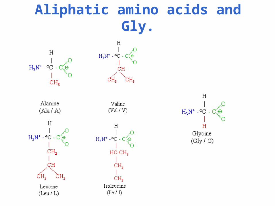
Aliphatic amino acids and Gly.

Cyclic Imino Acid: Proline.
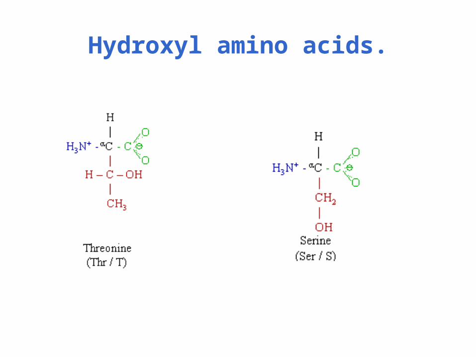
Hydroxyl amino acids.
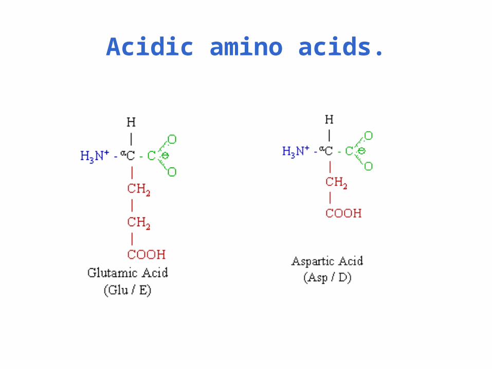
Acidic amino acids.
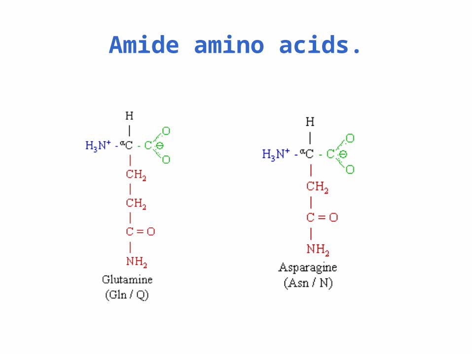
Amide amino acids.
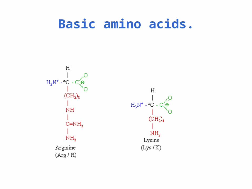
Basic amino acids.
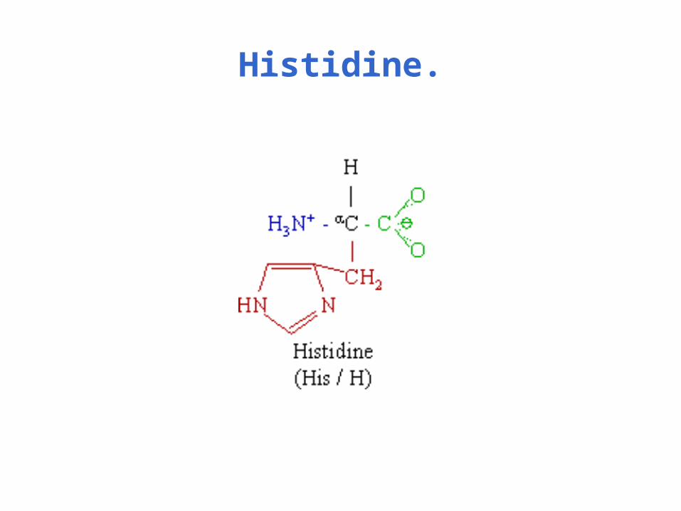
Histidine.

Sulfur-containing amino acids.
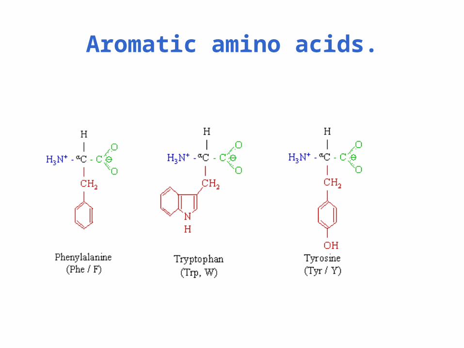
Aromatic amino acids.

Peptide bond is formed between two amino acids.

Backbone conformation is described by φ and ψ angles.
Picture from T. Przytycka, 2002
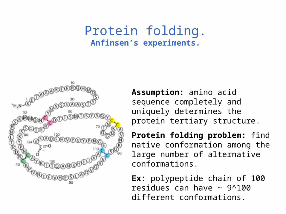
Protein folding.Anfinsen’s experiments.
Assumption: amino acid sequence completely and uniquely determines the protein tertiary structure.
Protein folding problem: find native conformation among the large number of alternative conformations.
Ex: polypeptide chain of 100 residues can have ~ 9^100 different conformations.
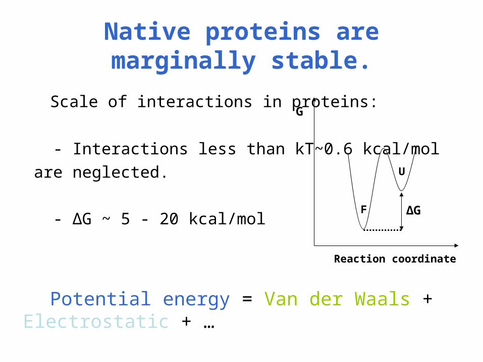
Native proteins are marginally stable.
Scale of interactions in proteins:
- Interactions less than kT~0.6 kcal/mol
are neglected.
- ΔG ~ 5 - 20 kcal/mol
Potential energy = Van der Waals + Electrostatic + …
U
F
G
Reaction coordinate
ΔG
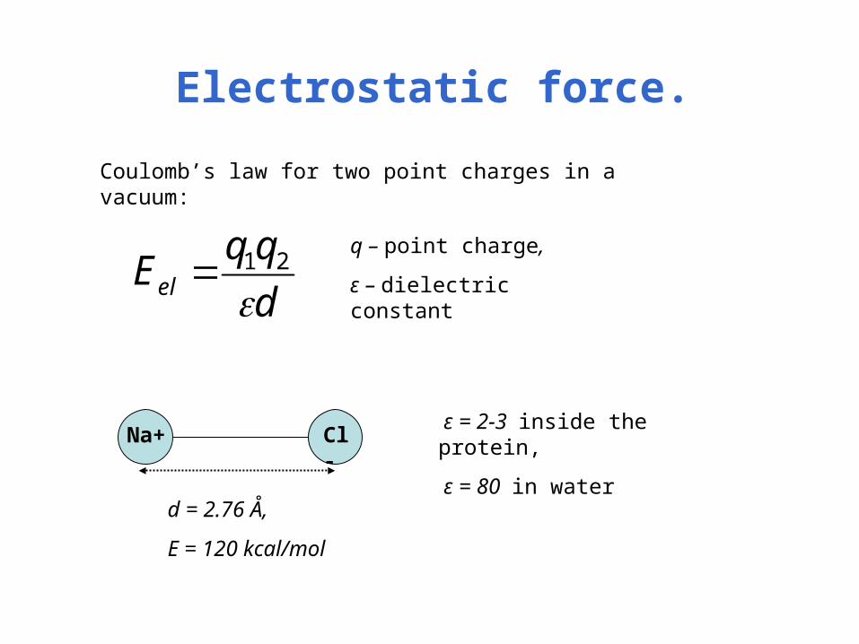
Electrostatic force.
d
qqEel
21
Coulomb’s law for two point charges in a vacuum:
q – point charge,
ε – dielectric constant
Na+ Cl-
d = 2.76 Å,
E = 120 kcal/mol
ε = 2-3 inside the protein,
ε = 80 in water

Dipolar interactions.
qdD
521
321 ))((3)(
r
rr
rE dd
Dipole moment:
Interaction energy of two dipoles separated by the vector r: N
C
O
H
- 0.42
+0.42
+0.20
-0.20
Peptide bond:
μ = 3.5D,
Water molecule:
μ = 1.85D.
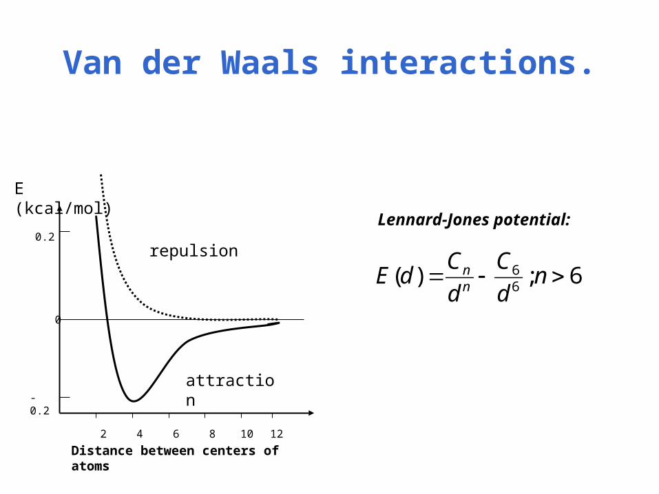
Van der Waals interactions.
0.2
12 10 8 6 4 2
- 0.2
repulsion
attraction
0
6;)(66 nd
C
d
CdE
nn
Lennard-Jones potential:
Distance between centers of atoms
E (kcal/mol)
Van der Waals interactions.
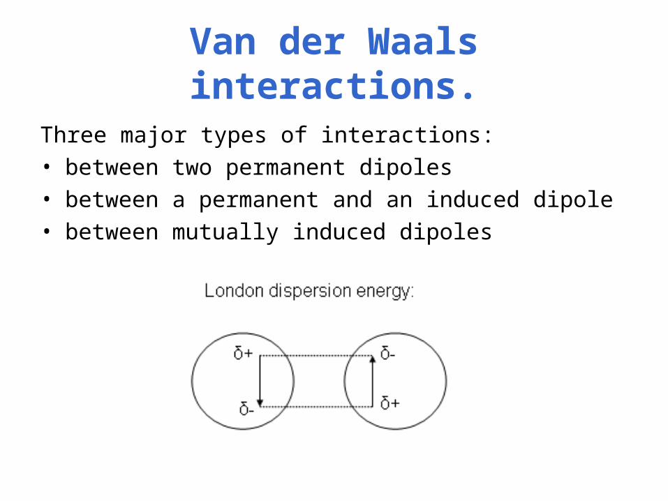
Three major types of interactions:• between two permanent dipoles• between a permanent and an induced dipole• between mutually induced dipoles

Hydrogen bonds.
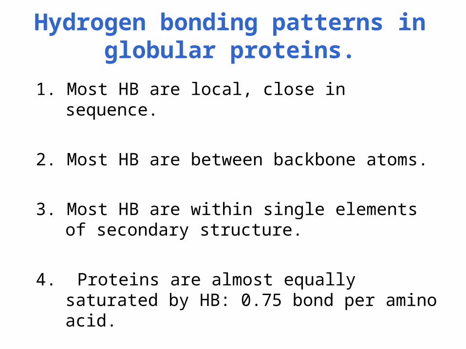
Hydrogen bonding patterns in globular proteins.
1. Most HB are local, close in sequence.
2. Most HB are between backbone atoms.
3. Most HB are within single elements of secondary structure.
4. Proteins are almost equally saturated by HB: 0.75 bond per amino acid.

Disulfide bonds.
PROTEIN + GS-SG PROTEIN + GSHPROTEIN + 2GSH
SH HS SH S-SG
- Breakdown and formation of S-S bonds are catalyzed by disulfide isomerase.
- In the cell S-S bonds are reversible, the energetic equilibrium is close to zero.
- Secreted proteins have a lot of S-S bonds since outside the cell the equilibrium is shifted towards their formation.
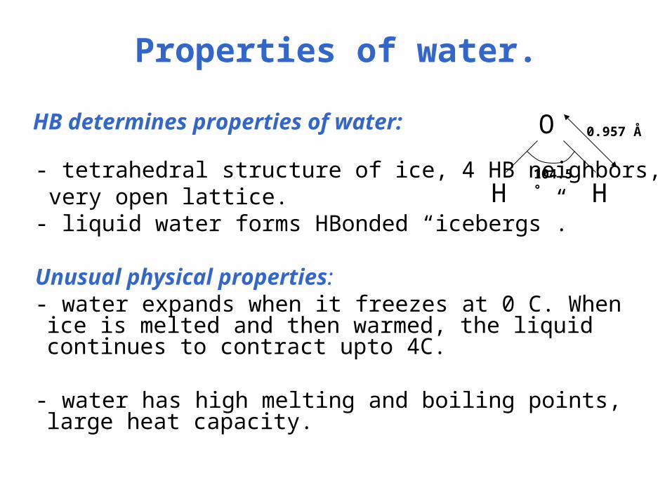
Properties of water.
HB determines properties of water:
- tetrahedral structure of ice, 4 HB neighbors, very open lattice. - liquid water forms HBonded “icebergs”. Unusual physical properties: - water expands when it freezes at 0 C. When ice is melted and
then warmed, the liquid continues to contract upto 4C.
- water has high melting and boiling points, large heat capacity.
104.5°
0.957 ÅO
H H
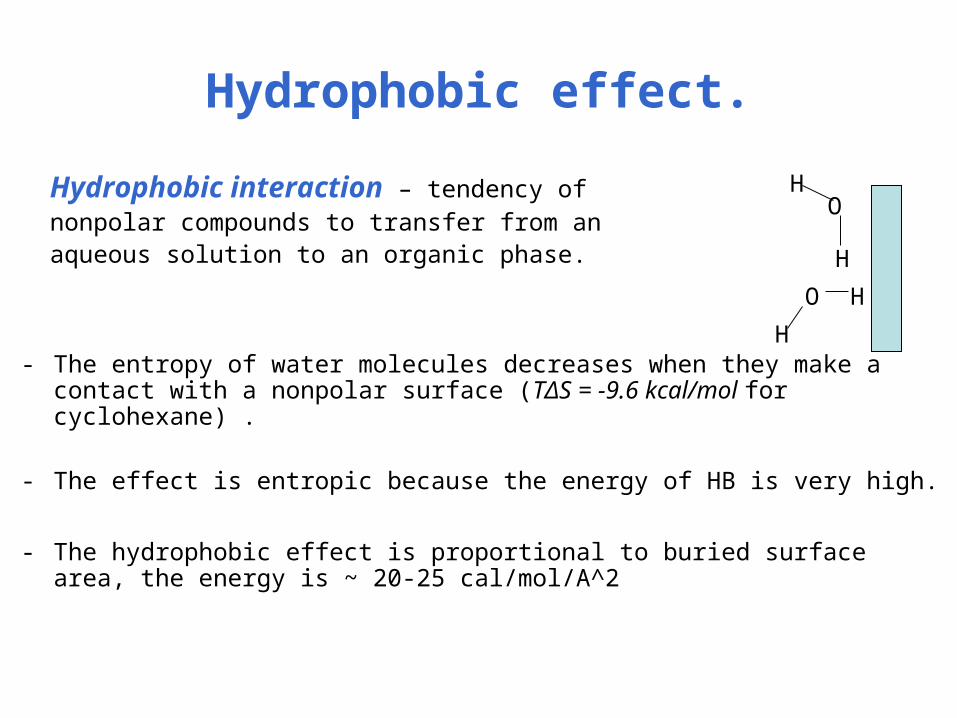
Hydrophobic effect.
Hydrophobic interaction – tendency of nonpolar compounds to transfer from an aqueous solution to an organic phase.
- The entropy of water molecules decreases when they make a contact with a nonpolar surface (TΔS = -9.6 kcal/mol for cyclohexane) .
- The effect is entropic because the energy of HB is very high.
- The hydrophobic effect is proportional to buried surface area, the energy is ~ 20-25 cal/mol/A^2
O
H
H
H
H
O
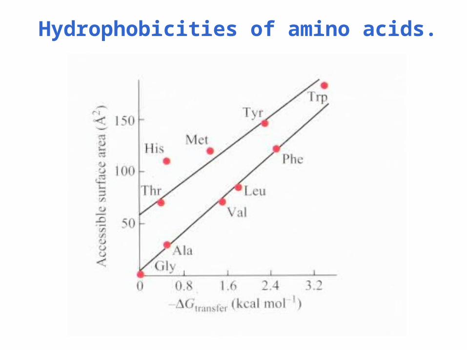
Hydrophobicities of amino acids.
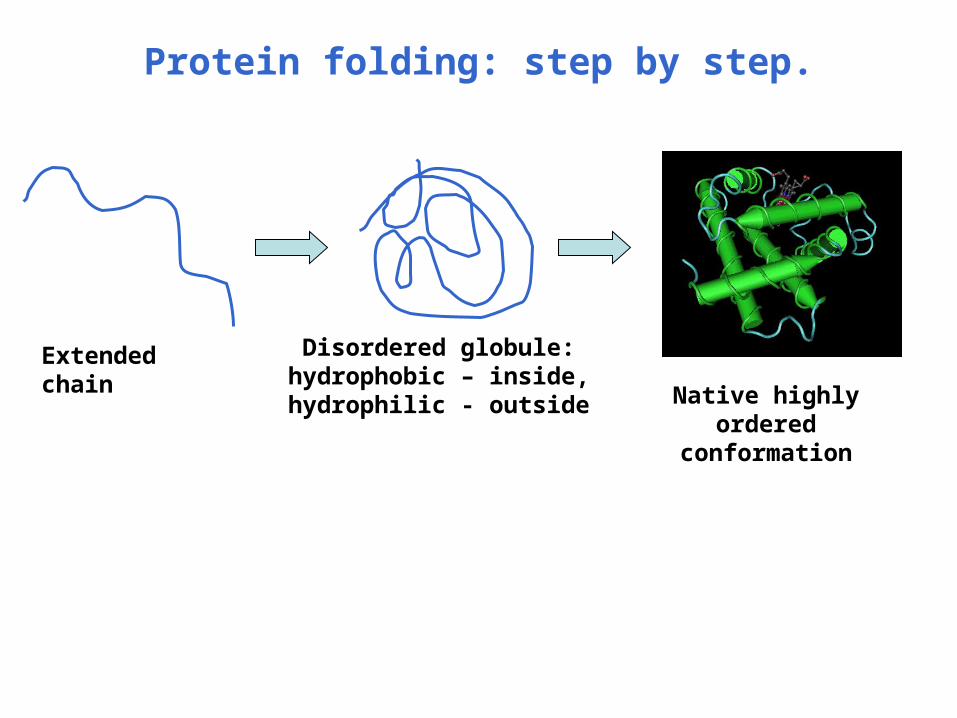
Protein folding: step by step.
Extended chain Disordered globule: hydrophobic – inside, hydrophilic - outside Native highly ordered
conformation

Hierarchy of protein structure.
1. Amino acid sequence
2. Secondary structure
3. Tertiary structure
4. Quaternary structure
Picture from Branden & Tooze “Introduction to protein structure”
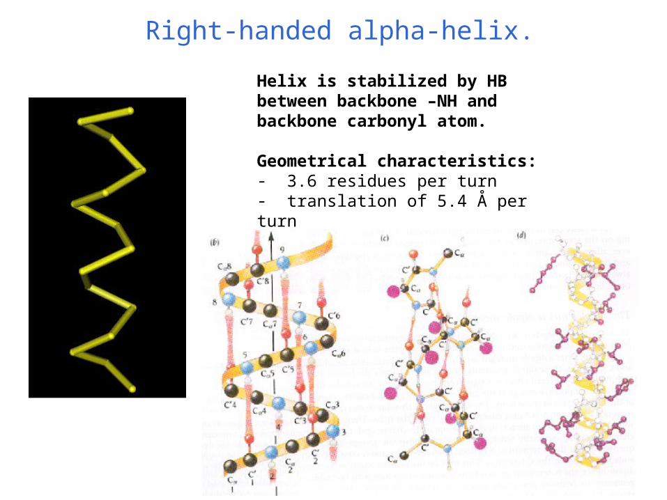
Right-handed alpha-helix.
Helix is stabilized by HB between backbone –NH and backbone carbonyl atom.
Geometrical characteristics:- 3.6 residues per turn- translation of 5.4 Å per turn- translation of 1.5 Å per residue

β-strand and β-sheet.
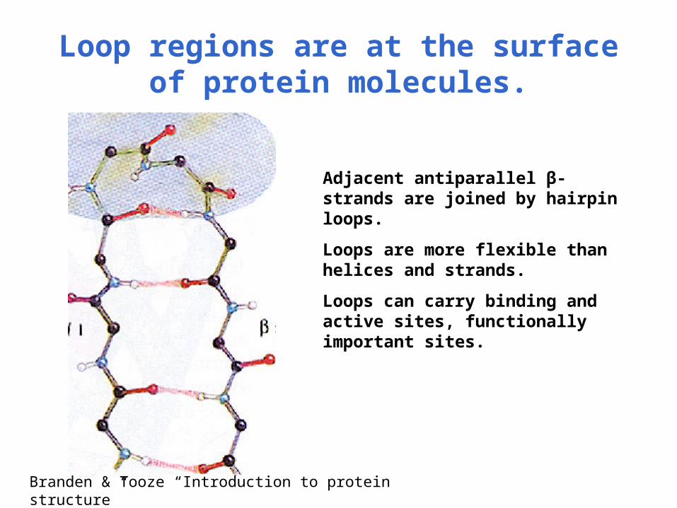
Loop regions are at the surface of protein molecules.
Adjacent antiparallel β-strands are joined by hairpin loops.
Loops are more flexible than helices and strands.
Loops can carry binding and active sites, functionally important sites.
Branden & Tooze “Introduction to protein structure”

Fibrous proteins.
• Have extended structures, regular conformations, insoluble.
• Basis for regularities in structure is in regularities in sequence.
• Coiled coils (heptad repeats in sequence).
• Collagen triple helix (every third residue is Gly).
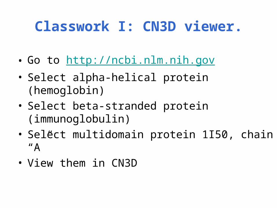
Classwork I: CN3D viewer.
• Go to http://ncbi.nlm.nih.gov• Select alpha-helical protein (hemoglobin)• Select beta-stranded protein (immunoglobulin)• Select multidomain protein 1I50, chain “A”• View them in CN3D

PDB databank.
• Archive of protein crystal structures was established in 1971 with several structures.
in 2002 – 17000 structures including NMR structures
• Data processing: data deposition, annotation and validation
• PDB code – nXYZ, n – integer, X, Y, Z -characters

Content of Data in the PDB.• Organism, species name
• Full protein sequence
• Chemical structure of cofactors and prosthetic groups
• Names of all components of the structure
• Qualitative description of the structural characteristics
• Literature citations
• Three-dimensional coordinates
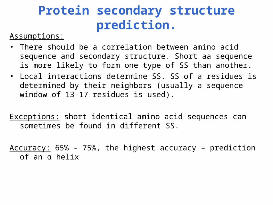
Protein secondary structure prediction.Assumptions: • There should be a correlation between amino acid sequence and
secondary structure. Short aa sequence is more likely to form one type of SS than another.
• Local interactions determine SS. SS of a residues is determined by their neighbors (usually a sequence window of 13-17 residues is used).
Exceptions: short identical amino acid sequences can sometimes be found in different SS.
Accuracy: 65% - 75%, the highest accuracy – prediction of an α helix

Methods of SS prediction.
• Chou-Fasman method
• GOR (Garnier,Osguthorpe and Robson)
• Neural network method

Chou-Fasman method.
Amino acid itself determines the type of secondary structure, which it is likely to adopt.
Analysis of frequences for all amino acids to be in different types of SS. Ala, Glu, Leu and Met – strong predictors of alpha-helices, Pro and Gly predict to break the helix.
Prediction of an alpha-helix - if four of six adjacent amino acids have high probability to be a helix.
Prediction of a beta-strand – if three of five adjacent amino acids have high probability to be a strand.
)(/),(log(),( SfSafSaScore ii
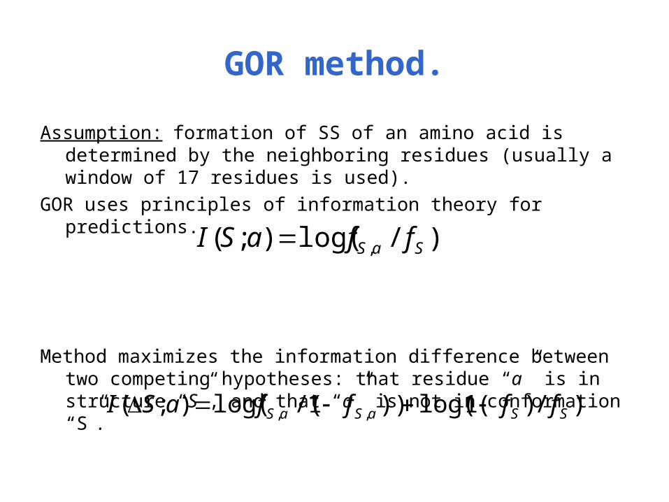
GOR method.
Assumption: formation of SS of an amino acid is determined by the neighboring residues (usually a window of 17 residues is used).
GOR uses principles of information theory for predictions.
Method maximizes the information difference between two competing hypotheses: that residue “a” is in structure “S”, and that “a” is not in conformation “S”.
)/log();( , SaS ffaSI
)/)1log(())1/(log();( ,, SSaSaS ffffaSI

Neural network method.
L
A
W
P
G
E
V
G
A
S
T
Y
P
0
0
0
0
0
1
0
0
0
0
0
0
0
0
0
0
0
0
0
0
0
HjOi
α
β
coil
1
0
0
Si
Hj Oi
Wij Sj
Input sequence window
Input layer
Hidden layer Output layer Predicted SS
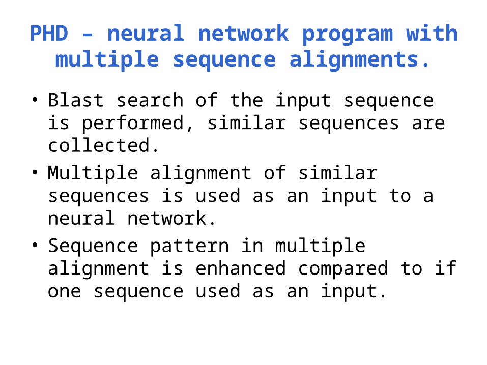
PHD – neural network program with multiple sequence alignments.
• Blast search of the input sequence is performed, similar sequences are collected.
• Multiple alignment of similar sequences is used as an input to a neural network.
• Sequence pattern in multiple alignment is enhanced compared to if one sequence used as an input.

Classwork
• Go to http://ncbi.nlm.nih.gov, search for protein “flavodoxin” in Entrez, retrieve its amino acid sequence.
• Go to http://cubic.bioc.columbia.edu/predictprotein and run PHD on the sequence.


![Evaluation of treatment response of cilengitide in an experimental ... · metastasis of breast cancer to bone [9, 10]. Cilengitide (EMD 121974) is a cyclic arginine-gly- cine-aspartic](https://static.fdocument.org/doc/165x107/5f02f2da7e708231d406ce8b/evaluation-of-treatment-response-of-cilengitide-in-an-experimental-metastasis.jpg)