Structural basis for λN-dependent processive transcription … · 2017-04-28 · In te format...
12
In the format provided by the authors and unedited. 4 Nelly Said 1 , Ferdinand Krupp 2,† , Ekaterina Anedchenko 1,†,‡ , Karine F. Santos 1,# , Olexandr 5 Dybkov 3 , Yong-Heng Huang 1 , Chung-Tien Lee 4,6 , Bernhard Loll 1 , Elmar Behrmann 2,ǁ , Jörg 6 Bürger 2,5 , Thorsten Mielke 5 , Justus Loerke 2 , Henning Urlaub 4,6 , Christian M. T. Spahn 2 , Gert 7 Weber 1,§ , Markus C. Wahl 1,7, * 8 9 1 Freie Universität Berlin, Laboratory of Structural Biochemistry, Takustraβe 6, D-14195 10 Berlin, Germany 11 2 Medizinische Physik und Biophysik, Charité – Universitätsmedizin Berlin, Charitéplatz 1, 12 D-10117 Berlin, Germany 13 3 Max Planck Institut für biophysikalische Chemie, Department of Cellular Biochemistry, Am 14 Fassberg 11, D-37077 Göttingen, Germany 15 4 Max Planck Institut für biophysikalische Chemie, Bioanalytical Mass Spectrometry, Am 16 Fassberg 11, D-37077 Göttingen, Germany 17 5 Max-Planck-Institut für Molekulare Genetik, Microscopy and Cryo-Electron Microscopy 18 Group, Ihnestraße 63-73, D14195 Berlin, Germany 19 6 Universitätsmedizin Göttingen, Institut für Klinische Chemie, Bioanalytik, Robert-Koch- 20 Straße 40, D-35075 Göttingen, Germany 21 7 Helmholtz-Zentrum Berlin für Materialien und Energie, Macromolecular Crystallography, 22 Albert-Einstein-Straße 15, D-12489 Berlin, Germany 23 24 † These authors contributed equally to this work. 25 ‡ Present address: Humboldt-Universität zu Berlin, Molecular Cell Biology, Philippstr. 13, D- 26 10099 Berlin, Germany 27 # Present address: moloX GmbH, Takustraße 6, D-14195 Berlin, Germany 28 ǁ Present address: Research Group Structural Dynamics of Proteins, Center of Advanced 29 European Studies and Research (Caesar), Ludwig-Erhard-Allee 2, D-53175 Bonn, 30 Germany 31 § Present address: Ernst-Moritz-Arndt-Universität Greifswald, Molekulare Strukturbiologie, 32 Felix-Hausdorff-Str. 4, D-17487 Greifswald, Germany 33 * Correspondence to: [email protected] 34 Structural basis for λN-dependent processive transcription antitermination © 2017 Macmillan Publishers Limited, part of Springer Nature. All rights reserved. SUPPLEMENTARY INFORMATION VOLUME: 2 | ARTICLE NUMBER: 17062 NATURE MICROBIOLOGY | DOI: 10.1038/nmicrobiol.2017.62 | www.nature.com/naturemicrobiology 1
Transcript of Structural basis for λN-dependent processive transcription … · 2017-04-28 · In te format...
In the format provided by the authors and unedited.
Supplementary Information 1
Structural basis for λN-dependent processive transcription 2
antitermination 3
4Nelly Said1, Ferdinand Krupp2,†, Ekaterina Anedchenko1,†,‡, Karine F. Santos1,#, Olexandr 5Dybkov3, Yong-Heng Huang1, Chung-Tien Lee4,6, Bernhard Loll1, Elmar Behrmann2,ǁ, Jörg 6Bürger2,5, Thorsten Mielke5, Justus Loerke2, Henning Urlaub4,6, Christian M. T. Spahn2, Gert 7Weber1,§, Markus C. Wahl1,7,* 8 91 Freie Universität Berlin, Laboratory of Structural Biochemistry, Takustraβe 6, D-14195 10
Berlin, Germany 112 Medizinische Physik und Biophysik, Charité – Universitätsmedizin Berlin, Charitéplatz 1, 12
D-10117 Berlin, Germany 133 Max Planck Institut für biophysikalische Chemie, Department of Cellular Biochemistry, Am 14
Fassberg 11, D-37077 Göttingen, Germany 154 Max Planck Institut für biophysikalische Chemie, Bioanalytical Mass Spectrometry, Am 16
Fassberg 11, D-37077 Göttingen, Germany 175 Max-Planck-Institut für Molekulare Genetik, Microscopy and Cryo-Electron Microscopy 18
Group, Ihnestraße 63-73, D14195 Berlin, Germany 196 Universitätsmedizin Göttingen, Institut für Klinische Chemie, Bioanalytik, Robert-Koch-20
Straße 40, D-35075 Göttingen, Germany 217 Helmholtz-Zentrum Berlin für Materialien und Energie, Macromolecular Crystallography, 22
Albert-Einstein-Straße 15, D-12489 Berlin, Germany 23 24† These authors contributed equally to this work. 25‡ Present address: Humboldt-Universität zu Berlin, Molecular Cell Biology, Philippstr. 13, D- 26
10099 Berlin, Germany 27# Present address: moloX GmbH, Takustraße 6, D-14195 Berlin, Germany 28ǁ Present address: Research Group Structural Dynamics of Proteins, Center of Advanced 29
European Studies and Research (Caesar), Ludwig-Erhard-Allee 2, D-53175 Bonn, 30Germany 31
§ Present address: Ernst-Moritz-Arndt-Universität Greifswald, Molekulare Strukturbiologie, 32Felix-Hausdorff-Str. 4, D-17487 Greifswald, Germany 33
* Correspondence to: [email protected] 34
Structural basis for λN-dependent processivetranscription antitermination
© 2017 Macmillan Publishers Limited, part of Springer Nature. All rights reserved.
SUPPLEMENTARY INFORMATIONVOLUME: 2 | ARTICLE NUMBER: 17062
NATURE MICROBIOLOGY | DOI: 10.1038/nmicrobiol.2017.62 | www.nature.com/naturemicrobiology 1
S2
Supplementary Discussion 35
Co-transcriptional TAC assembly. Our results delineate possible steps of TAC formation 36
during transcription elongation. NusA accompanies RNAP during elongation1 and would thus 37
allow early docking of λN. Initial docking might capitalize on the interaction of λN residues 34-38
47 with the NusA AR1 domain, which has repeatedly been observed2-5. Presence of a minor 39
complex, in which λN34-47 interacts with the NusA AR1 domain, is also seen in our present 40
cross-linking analyses (Supplementary Tables 2 and 3). Previous assays, which showed that 41
the NusA AR1 domain is dispensable for antitermination3,6, may not have been sensitive 42
enough to reveal a beneficial, albeit not essential, role of NusA AR1 in TAC assembly. λN 43
and NusA might then concomitantly recognize nut RNA via boxB and the boxA-boxB spacer, 44
as NusA is activated for RNA binding by RNAP αCTD-AR2 and/or λN-AR1 contacts7,8. As 45
NusA binding to a λN-nut RNP depends on NusA-RNA contacts9, boxB binding to λN might 46
induce conformational changes in neighboring λN regions to counteract the initial λN34-47-AR1 47
interaction, initiating repositioning of λN on NusA as suggested previously6. Indeed, in the 48
absence of nut RNA, where λN might be trapped on AR1, NusA inhibits λN function10. 49
Subsequent entry of the NusB-NusE heterodimer would then be facilitated by concomitant 50
contacts to boxA, NusA and λN, which are not stable individually, leading to accommodation 51
of λN47-52 in the NusA-NusE cavity. NusG might join the complex independently or aided by 52
weak NusG CTD-NusE11 and/or NusG NTD-NusA interactions12. 53
54
S3
Supplementary Figures 55
56
57
S4
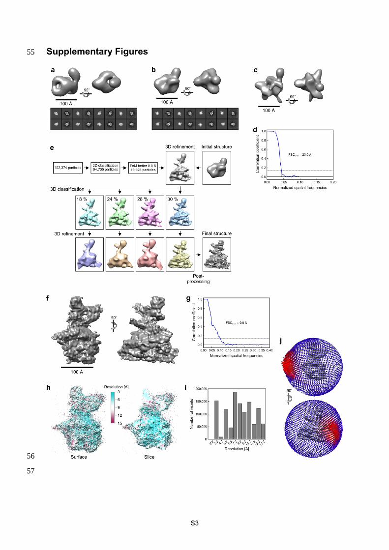
Supplementary Figure 1. EM analysis of the TAC. a, Top, orthogonal views of the initial 58
map based on negative stain data. Bottom, comparison of re-projections of the map (left) and 59
class averages (right). b, Top, orthogonal views of the map obtained after template matching 60
of all particle images against the initial model in (a). Bottom, comparison of re-projections of 61
the map (left) and class averages (right). c, Orthogonal views of the initial cryo-EM map. d, 62
Fourier shell correlation (FSC) indicating a resolution of 23.0 Å for the map in (c) according 63
to the FSC0.143 criterion. e, 3D sorting of the large cryo-EM data set. FoM, Figure of Merit. 3D 64
classification separated the data set into four classes, one of which (right) showed high-65
resolution features. f, Orthogonal views of the final cryo-EM map. g, FSC indicating an 66
overall resolution of 9.8 Å based on the FSC0.143 criterion. h, Unfiltered map colored 67
according to local resolution. Left, surface view. Right, cross section view. i, ResMap 68
analysis indicated higher local resolution than according to the FSC0.143 criterion, with a large 69
portion of voxels containing signal better than 9 Å resolution. j, Visualization of the 70
distribution of Euler angles associated with the particle images. 71
72
S5
73
S6
74
S7
75
76
S8
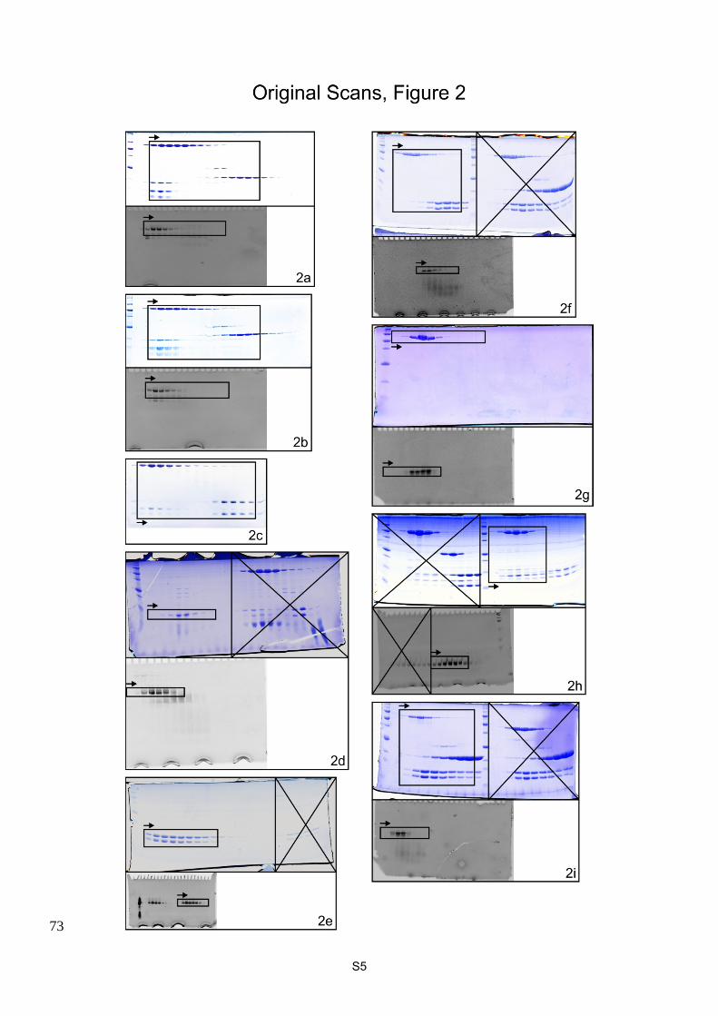
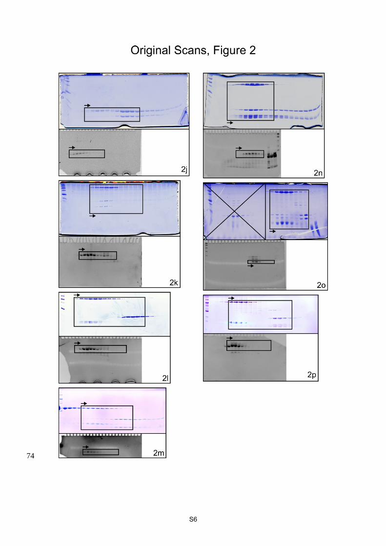
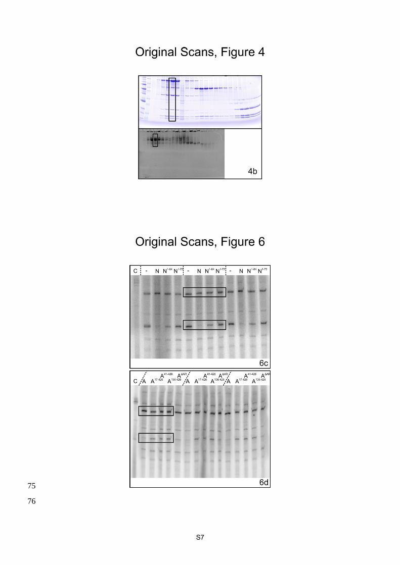
Supplementary Figure 2. Full scans of all gels shown in this work. Regions shown in the 77
respective figures are boxed. 78
79
S9
Supplementary Tables 80
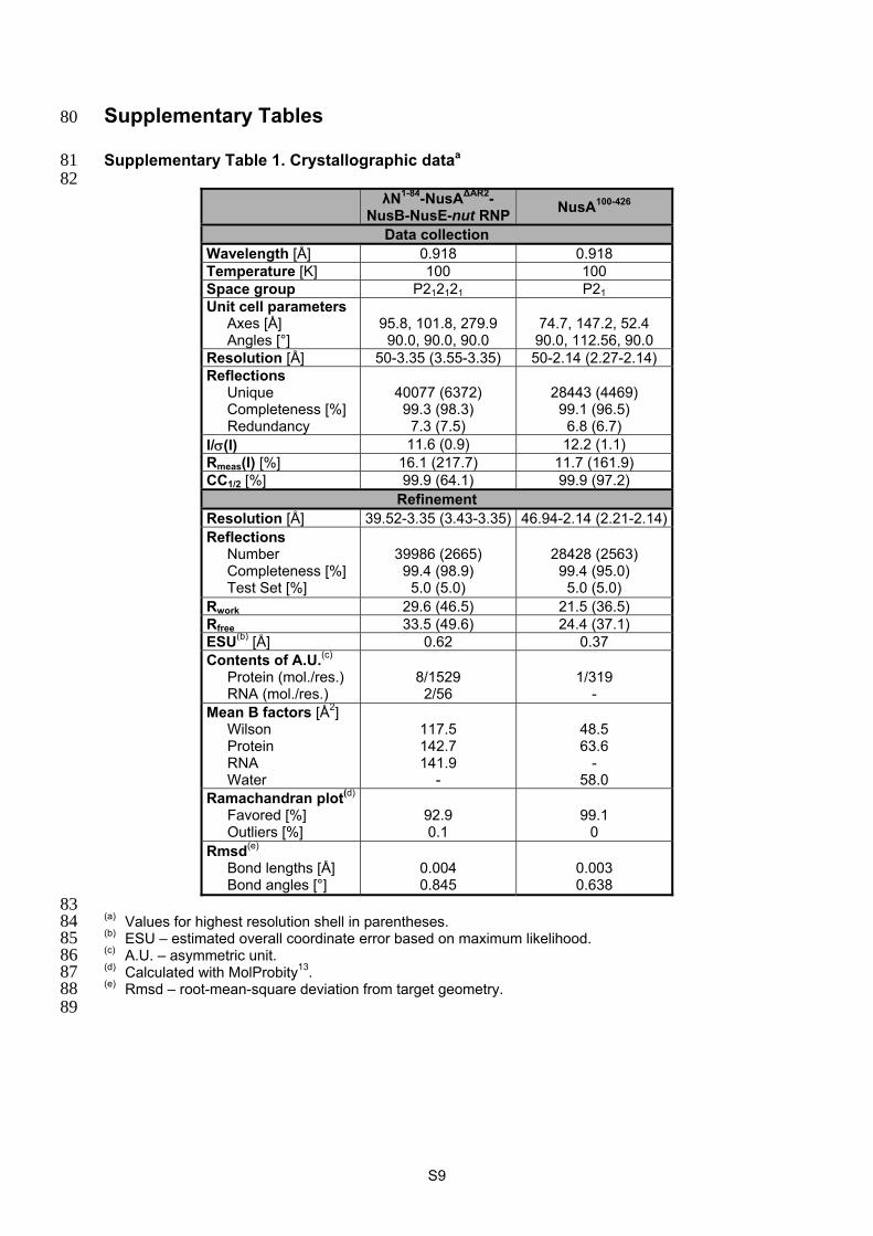
Supplementary Table 1. Crystallographic dataa 81 82
λN1-84-NusA∆AR2-
NusB-NusE-nut RNPNusA100-426
Data collection Wavelength [Å] 0.918 0.918 Temperature [K] 100 100 Space group P212121 P21 Unit cell parameters
Axes [Å] Angles [°]
95.8, 101.8, 279.9 90.0, 90.0, 90.0
74.7, 147.2, 52.4 90.0, 112.56, 90.0
Resolution [Å] 50-3.35 (3.55-3.35) 50-2.14 (2.27-2.14) Reflections
Unique Completeness [%] Redundancy
40077 (6372) 99.3 (98.3)
7.3 (7.5)
28443 (4469) 99.1 (96.5)
6.8 (6.7) I/(I) 11.6 (0.9) 12.2 (1.1) Rmeas(I) [%] 16.1 (217.7) 11.7 (161.9) CC1/2 [%] 99.9 (64.1) 99.9 (97.2)
Refinement Resolution [Å] 39.52-3.35 (3.43-3.35) 46.94-2.14 (2.21-2.14) Reflections
Number Completeness [%] Test Set [%]
39986 (2665) 99.4 (98.9)
5.0 (5.0)
28428 (2563) 99.4 (95.0)
5.0 (5.0) Rwork 29.6 (46.5) 21.5 (36.5) Rfree 33.5 (49.6) 24.4 (37.1) ESU(b) [Å] 0.62 0.37 Contents of A.U.(c)
Protein (mol./res.) RNA (mol./res.)
8/1529
2/56
1/319
- Mean B factors [Å2]
Wilson Protein RNA Water
117.5 142.7 141.9
-
48.5 63.6
- 58.0
Ramachandran plot(d) Favored [%] Outliers [%]
92.9 0.1
99.1
0 Rmsd(e)
Bond lengths [Å] Bond angles [°]
0.004 0.845
0.003 0.638
83 (a) Values for highest resolution shell in parentheses. 84 (b) ESU – estimated overall coordinate error based on maximum likelihood. 85 (c) A.U. – asymmetric unit. 86 (d) Calculated with MolProbity13. 87 (e) Rmsd – root-mean-square deviation from target geometry. 88 89
S10
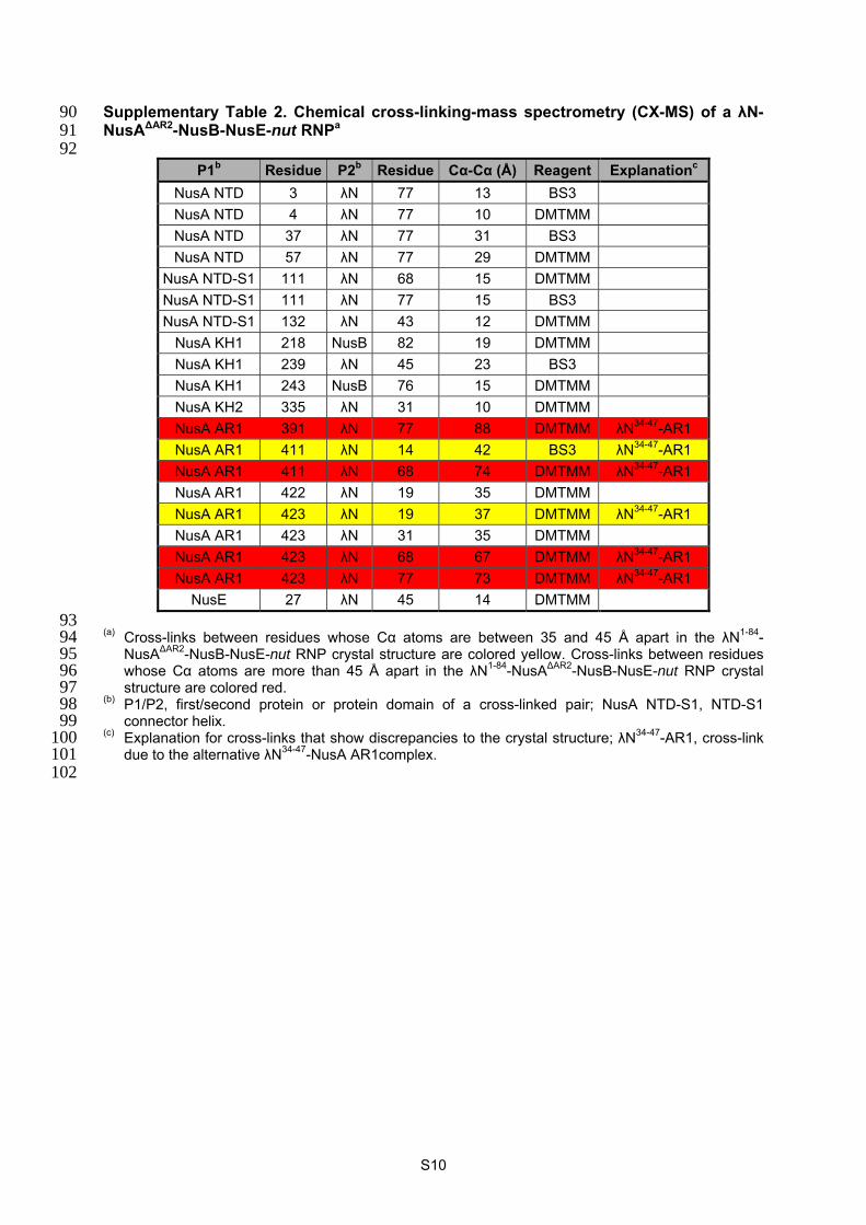
Supplementary Table 2. Chemical cross-linking-mass spectrometry (CX-MS) of a λN-90 NusA∆AR2-NusB-NusE-nut RNPa 91 92
P1b Residue P2b Residue Cα-Cα (Å) Reagent Explanationc
NusA NTD 3 λN 77 13 BS3
NusA NTD 4 λN 77 10 DMTMM
NusA NTD 37 λN 77 31 BS3
NusA NTD 57 λN 77 29 DMTMM
NusA NTD-S1 111 λN 68 15 DMTMM
NusA NTD-S1 111 λN 77 15 BS3
NusA NTD-S1 132 λN 43 12 DMTMM
NusA KH1 218 NusB 82 19 DMTMM
NusA KH1 239 λN 45 23 BS3
NusA KH1 243 NusB 76 15 DMTMM
NusA KH2 335 λN 31 10 DMTMM
NusA AR1 391 λN 77 88 DMTMM λN34-47-AR1
NusA AR1 411 λN 14 42 BS3 λN34-47-AR1
NusA AR1 411 λN 68 74 DMTMM λN34-47-AR1
NusA AR1 422 λN 19 35 DMTMM
NusA AR1 423 λN 19 37 DMTMM λN34-47-AR1
NusA AR1 423 λN 31 35 DMTMM
NusA AR1 423 λN 68 67 DMTMM λN34-47-AR1
NusA AR1 423 λN 77 73 DMTMM λN34-47-AR1
NusE 27 λN 45 14 DMTMM
93 (a) Cross-links between residues whose Cα atoms are between 35 and 45 Å apart in the λN1-84-94
NusA∆AR2-NusB-NusE-nut RNP crystal structure are colored yellow. Cross-links between residues 95 whose Cα atoms are more than 45 Å apart in the λN1-84-NusA∆AR2-NusB-NusE-nut RNP crystal 96 structure are colored red. 97
(b) P1/P2, first/second protein or protein domain of a cross-linked pair; NusA NTD-S1, NTD-S1 98 connector helix. 99
(c) Explanation for cross-links that show discrepancies to the crystal structure; λN34-47-AR1, cross-link 100 due to the alternative λN34-47-NusA AR1complex. 101
102
S11
Supplementary Table 3. Chemical cross-linking-mass spectrometry (CX-MS) of a λN-103 based transcription antitermination complex (TAC)a 104 105 (a) Cross-links between residues whose Cα atoms are between 35 and 45 Å apart in the cryo-EM 106
model are colored yellow. Cross-links between residues whose Cα atoms are more than 45 Å apart 107 in the cryo-EM model are colored red. 108
(b) P1/P2, first/second protein or protein domain of a cross-linked pair; NusA NTD-S1, NTD-S1 109 connector helix; NusA KH2-AR1, KH2-AR1 connector helix; NusG NTD-CTD, NTD-CTD linker. 110
(c) Explanation for cross-links that show discrepancies to the cryo-EM model; λN34-47-AR1, cross-link 111 due to the alternative λN34-47-NusA AR1 complex; flex, cross-link involving residues that reside in 112 highly flexible regions or in regions known to undergo conformational changes. Remaining 113 discrepancies most likely originate from a minor fraction of aggregated material. 114
115 116 Supplementary Table 4. Chemical cross-linking-mass spectrometry (CX-MS) of a 117 transcription elongation complex (TEC) lacking λNa 118 119 (a) Cross-links between residues whose Cα atoms are between 35 and 45 Å apart in the cryo-EM 120
model of the TAC are colored yellow. Cross-links between residues whose Cα atoms are more 121 than 45 Å apart in the cryo-EM model of the TAC are colored red. Cross-links differing between 122 TAC and TEC (lacking λN) are highlighted in green. 123
(b) P1/P2, first/second protein or protein domain of a cross-linked pair; NusA NTD-S1, NTD-S1 124 connector helix; NusG NTD-CTD, NTD-CTD linker. 125
(c) Cα-Cα distances are from the TAC structure. 126 127
S12
Supplementary References 128
1. Mooney, R. A. et al. Regulator Trafficking on Bacterial Transcription Units In Vivo. Mol. 129 Cell 33, 97-108, (2009). 130
2. Mogridge, J. et al. Independent ligand-induced folding of the RNA-binding domain and 131 two functionally distinct antitermination regions in the phage lambda N protein. Mol. Cell 132 1, 265-275, (1998). 133
3. Mah, T. F., Li, J., Davidson, A. R. & Greenblatt, J. Functional importance of regions in 134 Escherichia coli elongation factor NusA that interact with RNA polymerase, the 135 bacteriophage lambda N protein and RNA. Mol. Microbiol. 34, 523-537, (1999). 136
4. Bonin, I. et al. Structural basis for the interaction of Escherichia coli NusA with protein N 137 of phage lambda. Proc. Natl. Acad. Sci. USA 101, 13762-13767, (2004). 138
5. Prasch, S. et al. Interaction of the intrinsically unstructured phage lambda N Protein with 139 Escherichia coli NusA. Biochemistry 45, 4542-4549, (2006). 140
6. Mishra, S., Mohan, S., Godavarthi, S. & Sen, R. The interaction surface of a bacterial 141 transcription elongation factor required for complex formation with an antiterminator 142 during transcription antitermination. J. Biol. Chem. 288, 28089-28103, (2013). 143
7. Mah, T. F., Kuznedelov, K., Mushegian, A., Severinov, K. & Greenblatt, J. The alpha 144 subunit of E. coli RNA polymerase activates RNA binding by NusA. Genes Dev. 14, 145 2664-2675, (2000). 146
8. Schweimer, K. et al. NusA Interaction with the alpha Subunit of E-coli RNA Polymerase 147 Is via the UP Element Site and Releases Autoinhibition. Structure 19, 945-954, (2011). 148
9. Zhou, Y., Mah, T. F., Greenblatt, J. & Friedman, D. I. Evidence that the KH RNA-binding 149 domains influence the action of the E. coli NusA protein. J. Mol. Biol. 318, 1175-1188, 150 (2002). 151
10. Rees, W. A., Weitzel, S. E., Yager, T. D., Das, A. & von Hippel, P. H. Bacteriophage 152 lambda N protein alone can induce transcription antitermination in vitro. Proc. Natl. Acad. 153 Sci. USA 93, 342-346, (1996). 154
11. Burmann, B. M. et al. A NusE:NusG complex links transcription and translation. Science 155 328, 501-504, (2010). 156
12. Strauss, M. et al. Transcription is regulated by NusA:NusG interaction. Nucleic Acids 157 Res., (2016). 158
13. Chen, V. B., Wedell, J. R., Wenger, R. K., Ulrich, E. L. & Markley, J. L. MolProbity for the 159 masses-of data. J Biomol NMR 63, 77-83, (2015). 160
161