SSB SJ Pep CC Support R - Royal Society of Chemistry · Supporting Information 3 N N + N Scheme...
75
Supporting Information 1 Triazolyl-Donor/Acceptor Chromophore Decorated Unnatural Amino Acids and Peptides: FRET Event in β-Turn Conformation†† Subhendu Sekhar Bag,* , a Subhashis Jana, a Afsana Yashmeen, a K. Senthilkumar, b and Raghunath Bag a ††Dedicated to Professor Amit Basak on the occasion of his 62 nd birthday a Bio-organic Chemistry Laboratory, Department of Chenistry, Indian Institute of Technology Guwahati, Guwahati-781039, Assam, India. Fax: (+)91-361-258-2349. b Department of Physics, Bharathiar University, Coimbatore - 641 046, India. E-mail: [email protected] Topics Page 1. General Experimental Section (Materials and Methods) S2 2. Synthetic Schemes S2-S3 3. Full Chemical Structures of the Used or Synthesized Compounds S4 4. Exprimental Procedure and Characterization Data of the Synthesized UNAAs and Tripeptides S5-S12 5. Study of Photophysical Property and Summary Tables S13-S25 6. Study of Fluorescence Life Time S26-S29 7. Spectroscopic Evidences of Various Interactions and β-Turn Structures in the Peptides S29-S38 8. Study of Fluorescence Resonance Energy Transfer (FRET) in a Mixture of Two Monomeric Donor/Acceptor Amino Acids S39 9. Study of Fluorescence Resonance Energy Transfer (FRET) S40-41 10. Study of Solvent Assisted Modulation of FRET Emission S42 11. Macromodel Optimized Geometry S43 12. References S43 13. 1 H and 13 C Spectra of Synthesized Compounds. S44-S75 Electronic Supplementary Material (ESI) for Chemical Communications This journal is © The Royal Society of Chemistry 2013
Transcript of SSB SJ Pep CC Support R - Royal Society of Chemistry · Supporting Information 3 N N + N Scheme...
Supporting Information
1
Triazolyl-Donor/Acceptor Chromophore Decorated Unnatural Amino Acids
and Peptides: FRET Event in β-Turn Conformation††
Subhendu Sekhar Bag,*, a Subhashis Jana,a Afsana Yashmeen,a K. Senthilkumar, b and Raghunath Bag
a
††Dedicated to Professor Amit Basak on the occasion of his 62nd birthday
aBio-organic Chemistry Laboratory, Department of Chenistry, Indian Institute of Technology
Guwahati, Guwahati-781039, Assam, India. Fax: (+)91-361-258-2349. bDepartment of Physics, Bharathiar University, Coimbatore - 641 046, India.
E-mail: [email protected]
Topics Page
1. General Experimental Section (Materials and Methods) S2
2. Synthetic Schemes S2-S3
3. Full Chemical Structures of the Used or Synthesized Compounds S4
4. Exprimental Procedure and Characterization Data of the
Synthesized UNAAs and Tripeptides
S5-S12
5. Study of Photophysical Property and Summary Tables S13-S25
6. Study of Fluorescence Life Time S26-S29
7. Spectroscopic Evidences of Various Interactions and β-Turn
Structures in the Peptides
S29-S38
8. Study of Fluorescence Resonance Energy Transfer (FRET) in a
Mixture of Two Monomeric Donor/Acceptor Amino Acids S39
9. Study of Fluorescence Resonance Energy Transfer (FRET) S40-41
10. Study of Solvent Assisted Modulation of FRET Emission S42
11. Macromodel Optimized Geometry S43
12. References S43
13. 1H and 13
C Spectra of Synthesized Compounds. S44-S75
Electronic Supplementary Material (ESI) for Chemical CommunicationsThis journal is © The Royal Society of Chemistry 2013
Supporting Information
2
1. General Experimental Section (Materials and Methods)
All reactions except N-Boc protection of primary amine were carried out under nitrogen atmosphere in flame-dried glassware, using a nitrogen filled balloon. Organic extracts were dreid over anhydrous sodium sulfate. Solvents were removed in a rotary evaporator under reduced pressure. Silica gel (60- 120 mesh size) was used for the column chromatography. Reactions were monitored by TLC on silica gel 60 F254 (0.25). 1H NMR spectra were recorded at 400MHz and 13C NMR spectra were recorded at 100Mz. Coupling constants (J value) were reported in hertz. The chemical shift were shown in ppm downfield form tetramethylsilane, using residual chloroform (δ = 7.24 in 1H NMR, δ = 77.23 in 13C NMR), as an internal standard. Mass spectra were recorded with a HR mass spectrometer and data analysed by using built-in software. IR spectra were recorded in KBr on a FT-IR spectrometer.
2. Synthetic Schemes
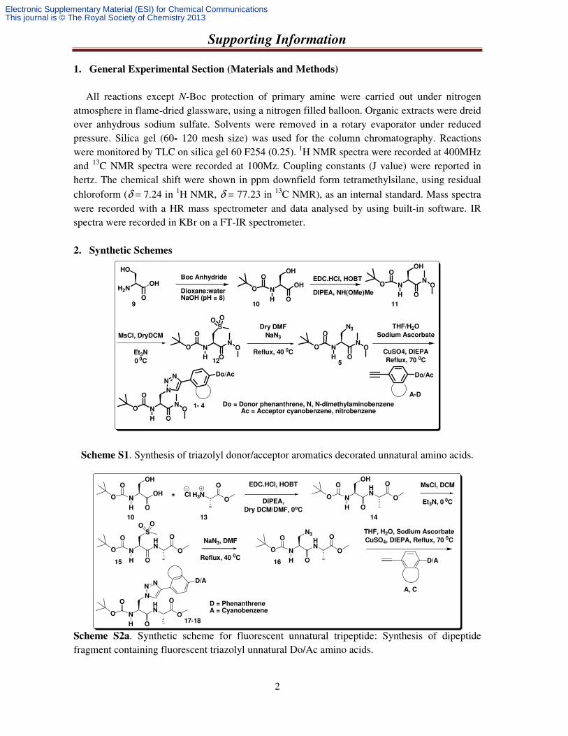
Scheme S1. Synthesis of triazolyl donor/acceptor aromatics decorated unnatural amino acids.
Scheme S2a. Synthetic scheme for fluorescent unnatural tripeptide: Synthesis of dipeptide fragment containing fluorescent triazolyl unnatural Do/Ac amino acids.
NN
OH
O
MsCl, DryDCM
Et3N
0 0C
Dry DMF
NaN3
Reflux, 40 0C
THF/H2O
Sodium Ascorbate
CuSO4, DIEPA
Reflux, 70 0C
Do/Ac
O
O
SO O
NN
OH
O
O
O
N3
NN
OH
O
O
O
N
NN
Do/Ac
1- 4 Do = Donor phenanthrene, N, N-dimethylaminobenzeneAc = Acceptor cyanobenzene, nitrobenzene
H2N
O
OH
HOBoc Anhydride
Dioxane:waterNaOH (pH = 8)
N
OH
OH
OH
O
O
EDC.HCl, HOBT
DIPEA, NH(OMe)MeN
OH
N
OH
O
O
O
9 10 11
12 5
A-D
N
OH
OH
H
O
O
O
H3NClO
O
+
EDC.HCl, HOBT
DIPEA,
Dry DCM/DMF, 0oC
N
OH
HN
H
O
O
O
O
O
MsCl, DCM
Et3N, 0 0C
N
HN
H
O
O
O
O
O
SO O
NaN3, DMF
Reflux, 40 0CN
N3
HN
H
O
O
O
O
O
THF, H2O, Sodium Ascorbate
CuSO4, DIEPA, Reflux, 70 0C
D/A
N
HN
H
O
O
O
O
O
N
NN
D/A
D = Phenanthrene A = Cyanobenzene
10 13 14
15 16
17-18
A, C
Electronic Supplementary Material (ESI) for Chemical CommunicationsThis journal is © The Royal Society of Chemistry 2013
Supporting Information
3
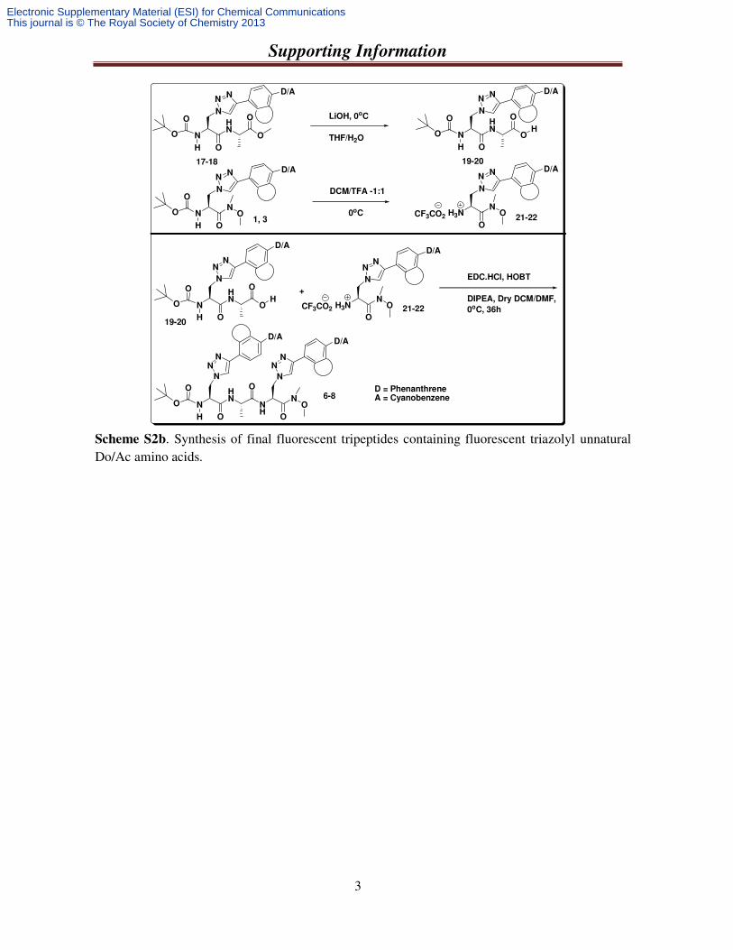
Scheme S2b. Synthesis of final fluorescent tripeptides containing fluorescent triazolyl unnatural Do/Ac amino acids.
N
HN
H
O
O
O
O
O
N
NN
D/A
17-18
EDC.HCl, HOBT
DIPEA, Dry DCM/DMF,
0oC, 36h
LiOH, 0oC
THF/H2O N
HN
H
O
O
O
O
OH
N
NN
D/A
19-20
N
H
O
O
O
N
NN
D/A
1, 3CF3CO2
DCM/TFA -1:1
0oC H3NN
O
O
N
NN
D/A
21-22
N
HN
H
O
O
O
O
OH
N
NN
D/A
19-20
+
CF3CO2 H3NN
O
O
N
NN
D/A
21-22
N
HN
H
O
O
O
O
N
NN
NH
N
NN
N
O
O6-8
D = PhenanthreneA = Cyanobenzene
NO
D/AD/A
Electronic Supplementary Material (ESI) for Chemical CommunicationsThis journal is © The Royal Society of Chemistry 2013

Supporting Information
4
3. Full Chemical Structures of the Used or Synthesized Compounds
Figure S1. Structure of synthesized unnatural amino acids (1-4).
Figure S2. Structures of the Synthesized Unnatural Donour-Acceptor Tripeptide.
C
N
N N+O–O
NN
OH
O
O
O
N
NN
NN
OH
O
O
O
N
NN
NN
OH
O
O
O
N
NN
NN
OH
O
O
O
N
NN
CNNMe2 NO2
A B C D
1 2 3 4
Used alkynes for synthesis of UNAA
TPhenAlaDo TNDMBAlaDo TCNBAlaAc TNBAlaAc
N
HN
H
O
O
O
O
O
N
NN
CN
N
HN
H
O
O
O
O
O
N
NN
CF3CO2 H3NN
O
O
N
NN
CF3CO2 H3NN
O
O
N
NN
CN
N
HN
H
O
O
O
O
NH
N
O
O
NN
NN
NN
N
HN
H
O
O
O
O
NH
N
O
O
NN
NN
NN
17 18 21 22
7
(TPhenAlaDo- Ala - TPhenAlaDo)
N
HN
H
O
O
O
O
NH
N
O
O
NN
NN
NN
CNCN
CN
6
(TPhenAlaDo- Ala - TCNBAlaAc)
8
(TCNBAlaAc - Ala - TCNBAlaAc)
Electronic Supplementary Material (ESI) for Chemical CommunicationsThis journal is © The Royal Society of Chemistry 2013
Supporting Information
5
4. Exprimental Procedure and Characterization Data of the Synthesized UNAAs and
Tripeptides
4.1. General Procedure for the Synthesis of Chiral Serine Azide: Four steps are discussed below:
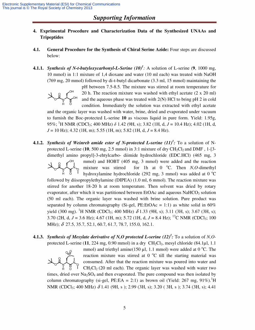
4.1.1. Synthesis of N-t-butyloxycarbonyl-L-Serine (10)1: A solution of L-serine (9, 1000 mg,
10 mmol) in 1:1 mixture of 1,4 dioxane and water (10 ml each) was treated with NaOH (769 mg, 20 mmol) followed by di-t-butyl dicarbonate (3.3 ml, 15 mmol) maintaining the
pH between 7.5-8.5. The mixture was stirred at room temperature for 20 h. The reaction mixture was washed with ethyl acetate (2 x 20 ml) and the aqueous phase was treated with 2(N) HCl to bring pH 2 in cold condition. Immediately the solution was extracted with ethyl acetate
and the organic layer was washed with water, brine, dried and evaporated under vacuum to furnish the Boc-protected L-serine 10 as viscous liquid in pure form. Yield: 1.95g, 95%; 1H NMR (CDCl3; 400 MHz) δ 1.42 (9H, s); 3.82 (1H, d, J = 10.4 Hz); 4.02 (1H, d, J = 10 Hz); 4.32 (1H, m); 5.55 (1H, m); 5.82 (1H, d, J = 8.4 Hz).
4.1.2. Synthesis of Weinreb amide ester of N-protected L-serine (11)1: To a solution of N-
protected L-serine (10, 500 mg, 2.5 mmol) in 3:1 mixture of dry CH2Cl2 and DMF , 1-[3-dimethyl amino propyl]-3-ehtylcarbo- diimide hydrochloride (EDC.HCl) (465 mg, 3
mmol) and HOBT (405 mg, 3 mmol) were added and the reaction mixture was stirred for 1h at 0 oC. Then N,O-dimethyl hydroxylamine hydrochloride (292 mg, 3 mmol) was added at 0 oC
followed by diisopropylethylamine (DIPEA) (1.0 ml, 6 mmol). The reaction mixture was stirred for another 18-20 h at room temperature. Then solvent was dried by rotary evaporator, after which it was partitioned between EtOAc and aqueous NaHCO3 solution (50 ml each). The organic layer was washed with brine solution. Pure product was separated by column chromatography (Si-gel, PE:EtOAc = 1:1) as white solid in 60% yield (300 mg). 1H NMR (CDCl3; 400 MHz) δ 1.33 (9H, s); 3.11 (3H, s); 3.67 (3H, s); 3.70 (2H, d, J = 3.6 Hz); 4.67 (1H, m); 5.72 (1H, d, J = 8.4 Hz); 13C NMR (CDCl3; 100 MHz); δ 27.5, 35.7, 52.1, 60.7, 61.7, 78.7, 155.0, 162.1.
4.1.3. Synthesis of Mesylate derivative of N,O protected L-serine (12)1: To a solution of N,O-
protected L-serine (11, 224 mg, 0.90 mmol) in a dry CH2Cl2, mesyl chloride (84.1µl, 1.1 mmol) and triethyl amine(150 µl, 1.1 mmol) were added at 0 oC. The reaction mixture was stirred at 0 oC till the starting material was consumed. After that the reaction mixture was poured into water and CH2Cl2 (20 ml each). The organic layer was washed with water two
times, dried over Na2SO4 and then evaporated. The pure compound was then isolated by column chromatography (si-gel, PE:EA = 2:1) as brown oil (Yield: 267 mg, 91%).1H NMR (CDCl3; 400 MHz) δ 1.41 (9H, s ); 2.99 (3H, s); 3.20 ( 3H, s ); 3.74 (3H, s); 4.41
N
OH
OH
OH
O
O
N
OH
N
OH
O
O
O
NN
OH
O
O
O
SO O
Electronic Supplementary Material (ESI) for Chemical CommunicationsThis journal is © The Royal Society of Chemistry 2013
Supporting Information
6
(2H, m); 4.90 (1H, m); 5.33 (1H, d, J = 8 Hz); 13C NMR (CDCl3; 100 MHz); δ 27.76, 31.5, 52.1, 60.9, 62.1, 66.4, 79.0, 155.2, 170.7.
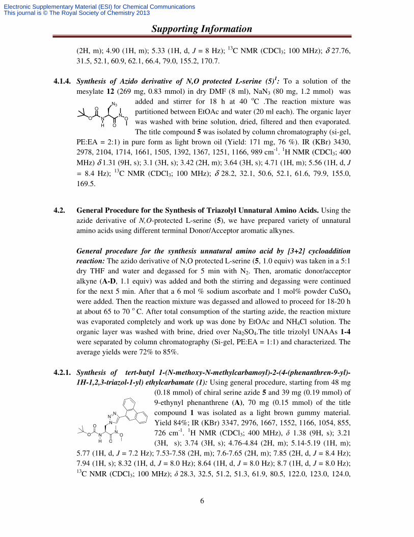
4.1.4. Synthesis of Azido derivative of N,O protected L-serine (5)1: To a solution of the
mesylate 12 (269 mg, 0.83 mmol) in dry DMF (8 ml), NaN3 (80 mg, 1.2 mmol) was added and stirrer for 18 h at 40 oC .The reaction mixture was partitioned between EtOAc and water (20 ml each). The organic layer was washed with brine solution, dried, filtered and then evaporated. The title compound 5 was isolated by column chromatography (si-gel,
PE:EA = 2:1) in pure form as light brown oil (Yield: 171 mg, 76 %). IR (KBr) 3430, 2978, 2104, 1714, 1661, 1505, 1392, 1367, 1251, 1166, 989 cm-1. 1H NMR (CDCl3; 400 MHz) δ 1.31 (9H, s); 3.1 (3H, s); 3.42 (2H, m); 3.64 (3H, s); 4.71 (1H, m); 5.56 (1H, d, J
= 8.4 Hz); 13C NMR (CDCl3; 100 MHz); δ 28.2, 32.1, 50.6, 52.1, 61.6, 79.9, 155.0, 169.5.
4.2. General Procedure for the Synthesis of Triazolyl Unnatural Amino Acids. Using the azide derivative of N,O-protected L-serine (5), we have prepared variety of unnatural amino acids using different terminal Donor/Acceptor aromatic alkynes.
General procedure for the synthesis unnatural amino acid by [3+2] cycloaddition
reaction: The azido derivative of N,O protected L-serine (5, 1.0 equiv) was taken in a 5:1 dry THF and water and degassed for 5 min with N2. Then, aromatic donor/acceptor alkyne (A-D, 1.1 equiv) was added and both the stirring and degassing were continued for the next 5 min. After that a 6 mol % sodium ascorbate and 1 mol% powder CuSO4
were added. Then the reaction mixture was degassed and allowed to proceed for 18-20 h at about 65 to 70 o C. After total consumption of the starting azide, the reaction mixture was evaporated completely and work up was done by EtOAc and NH4Cl solution. The organic layer was washed with brine, dried over Na2SO4.The title trizolyl UNAAs 1-4 were separated by column chromatography (Si-gel, PE:EA = 1:1) and characterized. The average yields were 72% to 85%.
4.2.1. Synthesis of tert-butyl 1-(N-methoxy-N-methylcarbamoyl)-2-(4-(phenanthren-9-yl)-
1H-1,2,3-triazol-1-yl) ethylcarbamate (1): Using general procedure, starting from 48 mg (0.18 mmol) of chiral serine azide 5 and 39 mg (0.19 mmol) of 9-ethynyl phenanthrene (A), 70 mg (0.15 mmol) of the title compound 1 was isolated as a light brown gummy material. Yield 84%; IR (KBr) 3347, 2976, 1667, 1552, 1166, 1054, 855, 726 cm-1. 1H NMR (CDCl3; 400 MHz), δ 1.38 (9H, s); 3.21 (3H, s); 3.74 (3H, s); 4.76-4.84 (2H, m); 5.14-5.19 (1H, m);
5.77 (1H, d, J = 7.2 Hz); 7.53-7.58 (2H, m); 7.6-7.65 (2H, m); 7.85 (2H, d, J = 8.4 Hz); 7.94 (1H, s); 8.32 (1H, d, J = 8.0 Hz); 8.64 (1H, d, J = 8.0 Hz); 8.7 (1H, d, J = 8.0 Hz); 13C NMR (CDCl3; 100 MHz); δ 28.3, 32.5, 51.2, 51.3, 61.9, 80.5, 122.0, 123.0, 124.0,
NN
OH
O
O
O
N
NN
NN
OH
O
O
O
N3
Electronic Supplementary Material (ESI) for Chemical CommunicationsThis journal is © The Royal Society of Chemistry 2013
Supporting Information
7
126.2, 126.9, 128.4, 128.9, 130.0, 130.2, 130.7, 131.3, 146.7, 155.2, 169.0; HRMS calcd. for C26H30N5O4 ([M + H]+) 476.2292, found 476.2294.
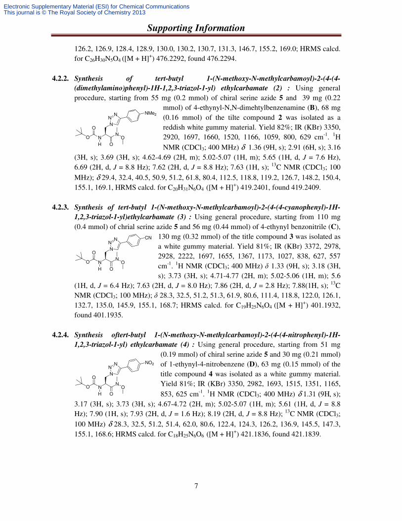
4.2.2. Synthesis of tert-butyl 1-(N-methoxy-N-methylcarbamoyl)-2-(4-(4-
(dimethylamino)phenyl)-1H-1,2,3-triazol-1-yl) ethylcarbamate (2) : Using general procedure, starting from 55 mg (0.2 mmol) of chiral serine azide 5 and 39 mg (0.22
mmol) of 4-ethynyl-N,N-dimehtylbenzenamine (B), 68 mg (0.16 mmol) of the tilte compound 2 was isolated as a reddish white gummy material. Yield 82%; IR (KBr) 3350, 2920, 1697, 1660, 1520, 1166, 1059, 800, 629 cm-1. 1H NMR (CDCl3; 400 MHz) δ 1.36 (9H, s); 2.91 (6H, s); 3.16
(3H, s); 3.69 (3H, s); 4.62-4.69 (2H, m); 5.02-5.07 (1H, m); 5.65 (1H, d, J = 7.6 Hz), 6.69 (2H, d, J = 8.8 Hz); 7.62 (2H, d, J = 8.8 Hz); 7.63 (1H, s); 13C NMR (CDCl3; 100 MHz); δ 29.4, 32.4, 40.5, 50.9, 51.2, 61.8, 80.4, 112.5, 118.8, 119.2, 126.7, 148.2, 150.4, 155.1, 169.1, HRMS calcd. for C20H31N6O4 ([M + H]+) 419.2401, found 419.2409.
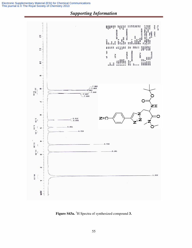
4.2.3. Synthesis of tert-butyl 1-(N-methoxy-N-methylcarbamoyl)-2-(4-(4-cyanophenyl)-1H-
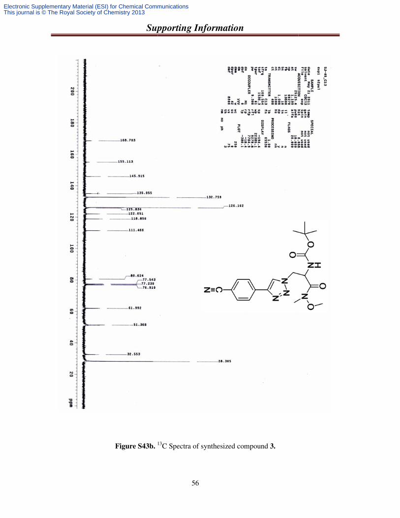
1,2,3-triazol-1-yl)ethylcarbamate (3) : Using general procedure, starting from 110 mg (0.4 mmol) of chrial serine azide 5 and 56 mg (0.44 mmol) of 4-ethynyl benzonitrile (C),
130 mg (0.32 mmol) of the title compound 3 was isolated as a white gummy material. Yield 81%; IR (KBr) 3372, 2978, 2928, 2222, 1697, 1655, 1367, 1173, 1027, 838, 627, 557 cm-1. 1H NMR (CDCl3; 400 MHz) δ 1.33 (9H, s); 3.18 (3H, s); 3.73 (3H, s); 4.71-4.77 (2H, m); 5.02-5.06 (1H, m); 5.6
(1H, d, J = 6.4 Hz); 7.63 (2H, d, J = 8.0 Hz); 7.86 (2H, d, J = 2.8 Hz); 7.88(1H, s); 13C NMR (CDCl3; 100 MHz); δ 28.3, 32.5, 51.2, 51.3, 61.9, 80.6, 111.4, 118.8, 122.0, 126.1, 132.7, 135.0, 145.9, 155.1, 168.7; HRMS calcd. for C19H25N6O4 ([M + H]+) 401.1932, found 401.1935.
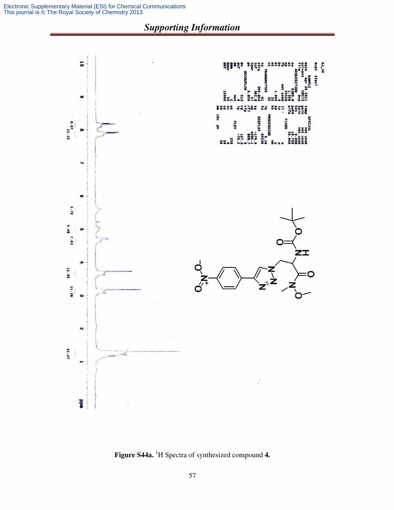
4.2.4. Synthesis oftert-butyl 1-(N-methoxy-N-methylcarbamoyl)-2-(4-(4-nitrophenyl)-1H-
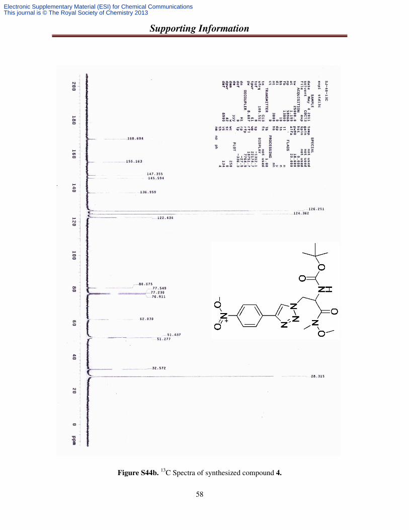
1,2,3-triazol-1-yl) ethylcarbamate (4) : Using general procedure, starting from 51 mg (0.19 mmol) of chiral serine azide 5 and 30 mg (0.21 mmol) of 1-ethynyl-4-nitrobenzene (D), 63 mg (0.15 mmol) of the title compound 4 was isolated as a white gummy material. Yield 81%; IR (KBr) 3350, 2982, 1693, 1515, 1351, 1165, 853, 625 cm-1. 1H NMR (CDCl3; 400 MHz) δ 1.31 (9Η, s);
3.17 (3H, s); 3.73 (3H, s); 4.67-4.72 (2H, m); 5.02-5.07 (1H, m); 5.61 (1H, d, J = 8.8 Hz); 7.90 (1H, s); 7.93 (2H, d, J = 1.6 Hz); 8.19 (2H, d, J = 8.8 Hz); 13C NMR (CDCl3; 100 MHz) δ 28.3, 32.5, 51.2, 51.4, 62.0, 80.6, 122.4, 124.3, 126.2, 136.9, 145.5, 147.3, 155.1, 168.6; HRMS calcd. for C18H25N6O6 ([M + H]+) 421.1836, found 421.1839.
NN
OH
O
O
O
N
NN
NMe2
NN
OH
O
O
O
N
NN
NO2
NN
OH
O
O
O
N
NN
CN
Electronic Supplementary Material (ESI) for Chemical CommunicationsThis journal is © The Royal Society of Chemistry 2013
Supporting Information
8
4.3. General procedure for the synthesis of azido derivative of dipeptide (Boc-NH-Ser.Ala-
OMe) : Three steps are discussed below:
4.3.1. Synthesis of L-Alanine methyl ester (13): To a degassed and cold solution of L-Alanine in dry methanol thionyl chloride (3 equivalent) was added at 0 oC. After 15 minutes the reaction mixture was refluxed at 75-80 oC, over night. The solvent was evaporated in vacuum. Material was washed with toluene three
times to remove excess thionyl chloride and the amine salt of methyl ester -L-alanine was isolated, washed with ether twice, dried and used for the next step.
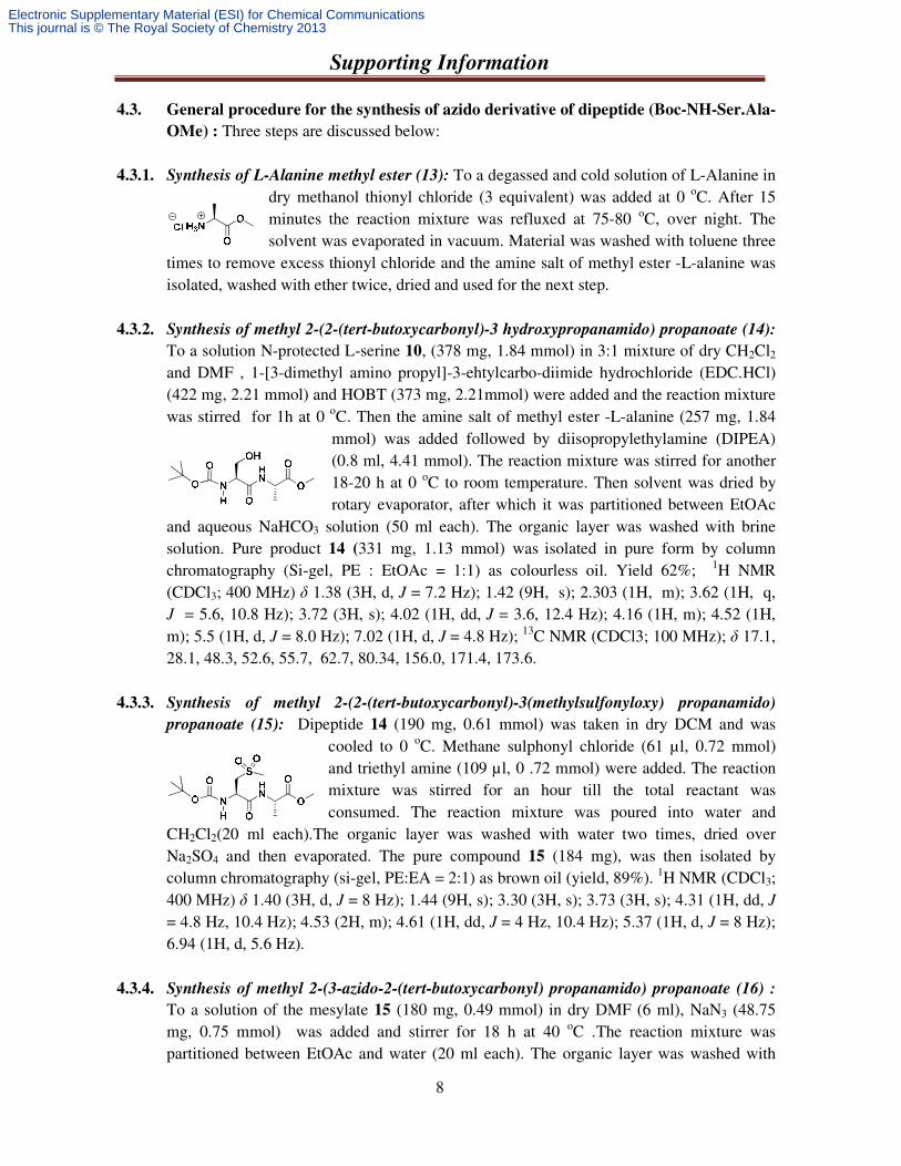
4.3.2. Synthesis of methyl 2-(2-(tert-butoxycarbonyl)-3 hydroxypropanamido) propanoate (14):
To a solution N-protected L-serine 10, (378 mg, 1.84 mmol) in 3:1 mixture of dry CH2Cl2
and DMF , 1-[3-dimethyl amino propyl]-3-ehtylcarbo-diimide hydrochloride (EDC.HCl) (422 mg, 2.21 mmol) and HOBT (373 mg, 2.21mmol) were added and the reaction mixture was stirred for 1h at 0 oC. Then the amine salt of methyl ester -L-alanine (257 mg, 1.84
mmol) was added followed by diisopropylethylamine (DIPEA) (0.8 ml, 4.41 mmol). The reaction mixture was stirred for another 18-20 h at 0 oC to room temperature. Then solvent was dried by rotary evaporator, after which it was partitioned between EtOAc
and aqueous NaHCO3 solution (50 ml each). The organic layer was washed with brine solution. Pure product 14 (331 mg, 1.13 mmol) was isolated in pure form by column chromatography (Si-gel, PE : EtOAc = 1:1) as colourless oil. Yield 62%; 1H NMR (CDCl3; 400 MHz) δ 1.38 (3H, d, J = 7.2 Hz); 1.42 (9H, s); 2.303 (1H, m); 3.62 (1H, q, J = 5.6, 10.8 Hz); 3.72 (3H, s); 4.02 (1H, dd, J = 3.6, 12.4 Hz); 4.16 (1H, m); 4.52 (1H, m); 5.5 (1H, d, J = 8.0 Hz); 7.02 (1H, d, J = 4.8 Hz); 13C NMR (CDCl3; 100 MHz); δ 17.1, 28.1, 48.3, 52.6, 55.7, 62.7, 80.34, 156.0, 171.4, 173.6.
4.3.3. Synthesis of methyl 2-(2-(tert-butoxycarbonyl)-3(methylsulfonyloxy) propanamido)
propanoate (15): Dipeptide 14 (190 mg, 0.61 mmol) was taken in dry DCM and was cooled to 0 oC. Methane sulphonyl chloride (61 µl, 0.72 mmol) and triethyl amine (109 µl, 0 .72 mmol) were added. The reaction mixture was stirred for an hour till the total reactant was consumed. The reaction mixture was poured into water and
CH2Cl2(20 ml each).The organic layer was washed with water two times, dried over Na2SO4 and then evaporated. The pure compound 15 (184 mg), was then isolated by column chromatography (si-gel, PE:EA = 2:1) as brown oil (yield, 89%). 1H NMR (CDCl3; 400 MHz) δ 1.40 (3H, d, J = 8 Hz); 1.44 (9H, s); 3.30 (3H, s); 3.73 (3H, s); 4.31 (1H, dd, J
= 4.8 Hz, 10.4 Hz); 4.53 (2H, m); 4.61 (1H, dd, J = 4 Hz, 10.4 Hz); 5.37 (1H, d, J = 8 Hz); 6.94 (1H, d, 5.6 Hz).
4.3.4. Synthesis of methyl 2-(3-azido-2-(tert-butoxycarbonyl) propanamido) propanoate (16) : To a solution of the mesylate 15 (180 mg, 0.49 mmol) in dry DMF (6 ml), NaN3 (48.75 mg, 0.75 mmol) was added and stirrer for 18 h at 40 oC .The reaction mixture was partitioned between EtOAc and water (20 ml each). The organic layer was washed with
Electronic Supplementary Material (ESI) for Chemical CommunicationsThis journal is © The Royal Society of Chemistry 2013
Supporting Information
9
brine solution, dried, filtered and then evaporated. The title compound 16 was isolated by column chromatography (Si-gel, PE:EA = 2:1), as light brown oil (yield, 78 %). IR (KBr) 3425, 2981, 2107, 1661, 1515, 1368,
1251, 1163, 1062, 859. 1H NMR (CDCl3; 400 MHz) δ 1.35 (3H, d, J = 7.2); 1.39 (1H, s); 3.49 (2H, dd, J = 2.8, 11.6 Hz); 3.69 (3H, s);
4.31(1H, m); 4.52 (1H, m); 5.52(1H, d, J = 5.2 Hz); 7.09 (1H, s); 13C NMR (CDCl3; 100 MHz); δ 18.2, 28.3, 48.4, 52.4, 52.6, 53.8, 80.8, 155.3, 169.2, 173.
4.4. General procedure for the synthesis triazolyl unnatural dipeptide: Using the azido derivative of dipeptide 16 (Boc-NH-Ser.Ala-OMe), we have prepared three different unnatural tri-peptide using Donor/Acceptor terminal alkynes.
General procedure for the synthesis triazolyl unnatural dipeptide (Boc-NH-Ser.Ala-
OMe) by [3+2] cycloaddition reaction: The azido derivative of dipeptide (Boc-NH-Ser.Ala-OMe) 16 (1.0 equiv) was taken in 5:1 dry THF and water and degassed for 5 min with N2. Then, alkyne (1.1 equiv) was added and both the stirring and degassing were continued for the next 5 min. After that a 6 mol % sodium ascorbate and 1 mol% powderd CuSO4 were added. Then the reaction mixture was degassed and allowed to proceed for 18-20 h about 65 to 70 oC. After total consumption of the starting azide, the reaction mixture was evaporated completely and work up was done by EtoAc and NH4Cl solution. The organic layer was washed with brine, dried over Na2SO4.The title trizolyl unnatural di-peptids 17 and 18 were separated by column chromatography and characterized.
4.4.1. Synthesis of methyl 2-(2-(tert-butoxycarbonyl)-3-(4-(phenanthren-9-yl)-1H-1,2,3-triazol-
1-yl) propanamido) propanoate (17) : Using general procedure, starting from 155 mg (0.49 mmol) of azido derivative of dipeptide 16 and 109 g (0.54 mmol) of 9- ethynylphenanthrene (A), 210 mg (0.41 mmol) of the title compound 17 was isolated as a
light brown gummy material. Yield 83%. 1H NMR (CDCl3; 400 MHz) δ =1.27 (3H, d, J = 6.8 Hz); 1.46 (9H, s); 3.69 (3H, s); 4.48 (1H, t, J = 7.6 Hz); 4.82 (2H, m); 5.03 (1H, m); 5.79 (1H, d, J = 7.60 Hz); 7.11 (1H, d, J = 6 Hz); 7.62 (4H, dd, J = 8.4, 24 Hz); 7.95 (1H, d, J = 5.2 Hz); 8.26 (2H, d, J = 6.8 Hz); 8.32 (1H, d, J = 7.6 Hz); 8.71 (2H, dd, J = 8 Hz, 25.6 Hz); 13C
NMR (CDCl3; 100 MHz); δ 17.7, 28.2, 48.4, 51.4, 52.4, 54.3, 80.7, 122.5, 122.9, 124.9, 126.1, 126.8, 128.4, 128.8, 129.9, 130.3, 130.6, 131.2, 146.5, 155.53, 169.0, 172.7.
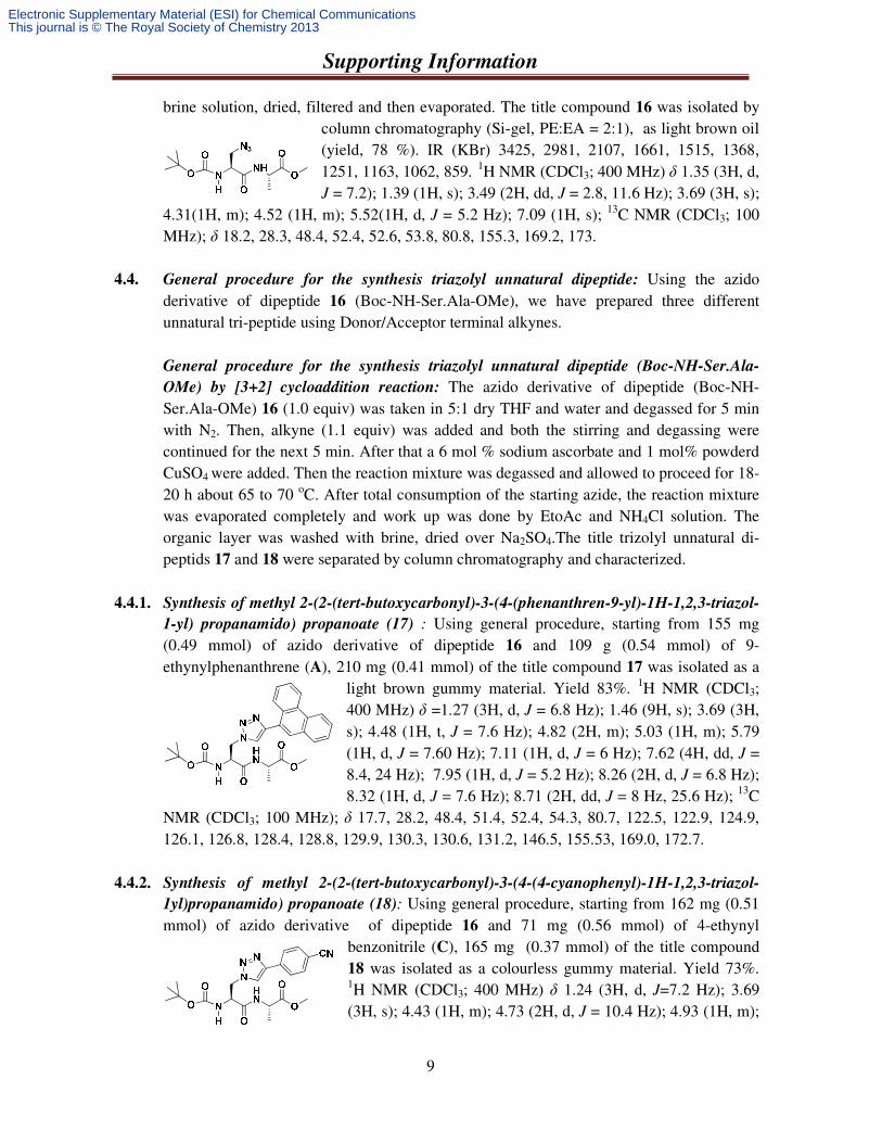
4.4.2. Synthesis of methyl 2-(2-(tert-butoxycarbonyl)-3-(4-(4-cyanophenyl)-1H-1,2,3-triazol-
1yl)propanamido) propanoate (18): Using general procedure, starting from 162 mg (0.51 mmol) of azido derivative of dipeptide 16 and 71 mg (0.56 mmol) of 4-ethynyl
benzonitrile (C), 165 mg (0.37 mmol) of the title compound 18 was isolated as a colourless gummy material. Yield 73%. 1H NMR (CDCl3; 400 MHz) δ 1.24 (3H, d, J=7.2 Hz); 3.69 (3H, s); 4.43 (1H, m); 4.73 (2H, d, J = 10.4 Hz); 4.93 (1H, m);
Electronic Supplementary Material (ESI) for Chemical CommunicationsThis journal is © The Royal Society of Chemistry 2013
Supporting Information
10
5.66 (1H, d, J = 5.2 Hz); 7.02 (1H, d, J = 6.4 Hz); 7.68 (2H, d, J = 7.6 Hz); 7.89 (2H, d, J = 7.2 Hz); 7.98 (1H, s); 13C NMR (CDCl3; 100 MHz); δ 18.0, 28.3, 48.5, 51.4, 52.7, 54.3, 81.3, 111.7, 118.8, 123.1, 126.1, 132.9, 134.8, 146.0, 155.5, 168.6, 172.8.
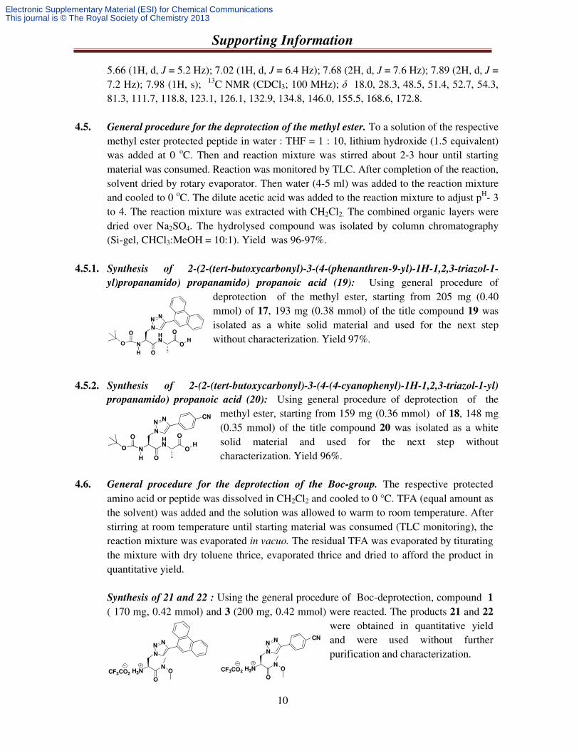
4.5. General procedure for the deprotection of the methyl ester. To a solution of the respective methyl ester protected peptide in water : THF = 1 : 10, lithium hydroxide (1.5 equivalent) was added at 0 oC. Then and reaction mixture was stirred about 2-3 hour until starting material was consumed. Reaction was monitored by TLC. After completion of the reaction, solvent dried by rotary evaporator. Then water (4-5 ml) was added to the reaction mixture and cooled to 0 oC. The dilute acetic acid was added to the reaction mixture to adjust pH- 3 to 4. The reaction mixture was extracted with CH2Cl2. The combined organic layers were dried over Na2SO4. The hydrolysed compound was isolated by column chromatography (Si-gel, CHCl3:MeOH = 10:1). Yield was 96-97%.
4.5.1. Synthesis of 2-(2-(tert-butoxycarbonyl)-3-(4-(phenanthren-9-yl)-1H-1,2,3-triazol-1-
yl)propanamido) propanamido) propanoic acid (19): Using general procedure of deprotection of the methyl ester, starting from 205 mg (0.40 mmol) of 17, 193 mg (0.38 mmol) of the title compound 19 was isolated as a white solid material and used for the next step without characterization. Yield 97%.
4.5.2. Synthesis of 2-(2-(tert-butoxycarbonyl)-3-(4-(4-cyanophenyl)-1H-1,2,3-triazol-1-yl)
propanamido) propanoic acid (20): Using general procedure of deprotection of the methyl ester, starting from 159 mg (0.36 mmol) of 18, 148 mg (0.35 mmol) of the title compound 20 was isolated as a white solid material and used for the next step without characterization. Yield 96%.
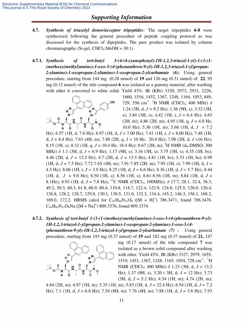
4.6. General procedure for the deprotection of the Boc-group. The respective protected amino acid or peptide was dissolved in CH2Cl2 and cooled to 0 °C. TFA (equal amount as the solvent) was added and the solution was allowed to warm to room temperature. After stirring at room temperature until starting material was consumed (TLC monitoring), the reaction mixture was evaporated in vacuo. The residual TFA was evaporated by titurating the mixture with dry toluene thrice, evaporated thrice and dried to afford the product in quantitative yield. Synthesis of 21 and 22 : Using the general procedure of Boc-deprotection, compound 1
( 170 mg, 0.42 mmol) and 3 (200 mg, 0.42 mmol) were reacted. The products 21 and 22 were obtained in quantitative yield and were used without further purification and characterization.
CF3CO2 H3NN
O
O
N
NN
CF3CO2 H3NN
O
O
N
NN
CN
N
HN
H
O
O
O
O
OH
N
NN
CN
N
HN
H
O
O
O
O
OH
N
NN
Electronic Supplementary Material (ESI) for Chemical CommunicationsThis journal is © The Royal Society of Chemistry 2013
Supporting Information
11
4.7. Synthesis of triazolyl donor/acceptor tripeptides: The target tripeptides 6-8 were synthesized following the general procedure of peptide coupling protocol as was discussed for the synthesis of dipeptides. The pure product was isolated by column chromatography (Si-gel, CHCl3:MeOH = 30:1).
4.7.1. Synthesis of tert-butyl 3-(4-(4-cyanophenyl)-1H-1,2,3-triazol-1-yl)-1-(1-(1-
(methoxy(methyl)amino)-1-oxo-3-(4-(phenanthren-9-yl)-1H-1,2,3-triazol-1-yl)propan-
2-ylamino)-1-oxopropan-2-ylamino)-1-oxopropan-2-ylcarbamate (6): Using general procedure, starting from 144 mg (0.28 mmol) of 19 and 126 mg (0.31 mmol) of 22, 95 mg (0.12 mmol) of the title compound 6 was isolated as a gummy material, after washing with ether it converted to white solid. Yield 43%. IR (KBr) 3320, 2973, 2931, 2226,
1660, 1516, 1452, 1367, 1248, 1164, 1053, 849, 729, 556 cm-1. 1H NMR (CDCl3; 400 MHz) δ 1.24 (3H, d, J = 9.2 Hz); 1.36 (9H, s); 3.32 (3H, s); 3.84 (3H, s); 4.42 (1H, t, J = 6.4 Hz); 4.63 (2H, m); 4.86 (2H, m); 4.95 (1H, q, J = 4.0 Hz, 10.0 Hz); 5.36 (1H, m); 5.68 (1H, d, J = 7.2
Hz); 6.57 (1H, d, 7.6 Hz); 6.97 (1H, d, J = 7.00 Hz); 7.41 (1H, t, J = 8.00 Hz); 7.48 (1H, d, J = 8.4 Hz); 7.63 (4H, m); 7.88 (2H, q, J = 10 Hz, 20.4 Hz); 7.98 (2H, d, J =16 Hz); 8.15 (1H, s); 8.32 (1H, q, J = 10.4 Hz, 18.4 Hz); 8.67 (2H, m); 1H NMR (d6-DMSO; 300 MHz) δ 1.1 (3H, d, J = 6.9 Hz); 1.17 (9H, s); 3.16 (3H, s); 3.75 (3H, s); 4.35 (1H, bs); 4.46 (2H, d, J = 13.2 Hz); 4.7 (2H, d, J = 13.5 Hz); 4.81 (1H, bs); 5.33 (1H, bs); 6.99 (1H, d, J = 7.5 Hz); 7.72-7.65 (4H, m); 7.91-7.85 (2H, m); 7.95 (1H, s); 7.99 (1H, d, J = 4.5 Hz); 8.06 (1H, t, J = 3.9 Hz); 8.25 (1H, d, J = 6.6 Hz); 8.36 (1H, d, J = 5.7 Hz); 8.44 (1H, d, J = 9.0 Hz); 8.50 (1H, s); 8.56 (1H, s); 8.61-8.56 (1H, m); 8.84 (1H, d, J = 8.1Hz); 8.93 (1H, d, J = 7.8 Hz); 13C NMR (CDCl3; 100MHz); δ 17.7, 28.1, 32.4, 36.3, 49.2, 50.3, 60.3, 61.8, 66.9, 80.4, 110.8, 118.7, 122.4, 122.9, 124.8, 125.5, 126.0, 126.4, 126.8, 128.2, 128.7, 129.8, 130.1, 130.5, 131.0, 132.3, 134.4, 145.2, 146.3, 156.1, 168.2, 169.0, 172.2. HRMS calcd for C41H44N11O6 ([M + H]+) 786.3471, found 786.3476. C41H43N11O6Na ([M + Na]+) 809.3376, found 809.3374.
4.7.2. Synthesis of tert-butyl 1-(1-(1-(methoxy(methyl)amino)-1-oxo-3-(4-(phenanthren-9-yl)-
1H-1,2,3-triazol-1-yl)propan-2-ylamino)-1-oxopropan-2-ylamino)-1-oxo-3-(4-
(phenanthren-9-yl)-1H-1,2,3-triazol-1-yl)propan-2-ylcarbamate (7) : Using general procedure, starting from 193 mg (0.37 mmol) of 19 and 182 mg (0.37 mmol) of 21, 147
mg (0.17 mmol) of the title compound 7 was isolated as a brown solid compound after washing with ether. Yield 45%. IR (KBr) 3327, 2979, 1655, 1519, 1451, 1367, 1248, 1165, 1054, 728 cm-1. 1H NMR (CDCl3; 400 MHz) δ 1.23 (3H, d, J = 13.2 Hz); 1.37 (9H, s), 3.20 ( 3H, d, J = 12 Hz); 3.73 (3H, d, J = 5.2 Hz); 4.54 (1H, m); 4.74 (2H, m);
4.84 (2H, m); 4.97 (1H, m); 5.35 (1H, m); 5.83 (1H, d, J = 22.4 Hz); 6.54 (1H, d, J = 7.2 Hz); 7.1 (1H, d, J = 6.8 Hz); 7.54 (8H, m); 7.76 (4H, m); 7.88 (1H, d, J = 3.6 Hz); 7.93
N
HN
H
O
O
O
O
NH
N
O
O
NN
NN
NN
CN
Electronic Supplementary Material (ESI) for Chemical CommunicationsThis journal is © The Royal Society of Chemistry 2013
Supporting Information
12
(1H, d, J = 11.6 Hz); 8.26 (2H, m); 8.59 (4H, m); 1H NMR (d6-DMSO; 300 MHz) δ 1.13 (3H, d, J = 6.9 Hz); 1.26 (9H, s); 3.15 (3H, s); 3.71 (3H, s); 4.39 (1H, bs);4.69- 4.53 (4H, m); 4.78 (1H, bs); 5.33 (1H, bs); 7.24-7.13 (1H, m); 7.72-7.66 (8H, m); 8.07-8.01 (4H, m); 8.34 (1H, d, J = 7.2 Hz); 8.45 (3H, d, J = 8.1 Hz); 8.52 (1H, s); 8.57 (1H, s); 8.87-8.82 (4H, dd, J = 7.8 Hz); 13C NMR (CDCl3; 100 MHz); δ 17.3, 28.3, 32.5, 49.5, 50.4, 51.0, 51.3, 54.9, 61.9, 80.8, 122.5, 122.9, 124.8, 126.8, 126.9, 128.4, 128.9, 130.0, 130.4, 130.7, 131.3, 146.7, 155.9, 168.2, 169.6, 172.1. HRMS calcd for C48H49N10O6 ([M + H]+) 861.3831, found 861.3837. C48H48N10O6Na ([M + Na]+) 884.3734, found 884.3731.
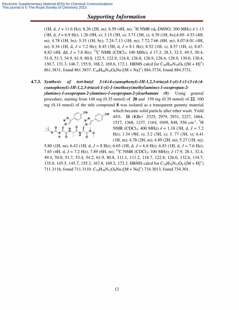
4.7.3. Synthesis of tert-butyl 3-(4-(4-cyanophenyl)-1H-1,2,3-triazol-1-yl)-1-(1-(3-(4-(4-
cyanophenyl)-1H-1,2,3-triazol-1-yl)-1-(methoxy(methyl)amino)-1-oxopropan-2-
ylamino)-1-oxopropan-2-ylamino)-1-oxopropan-2-ylcarbamate (8): Using general procedure, starting from 148 mg (0.35 mmol) of 20 and 159 mg (0.39 mmol) of 22, 100 mg (0.14 mmol) of the title compound 8 was isolated as a transparent gummy material
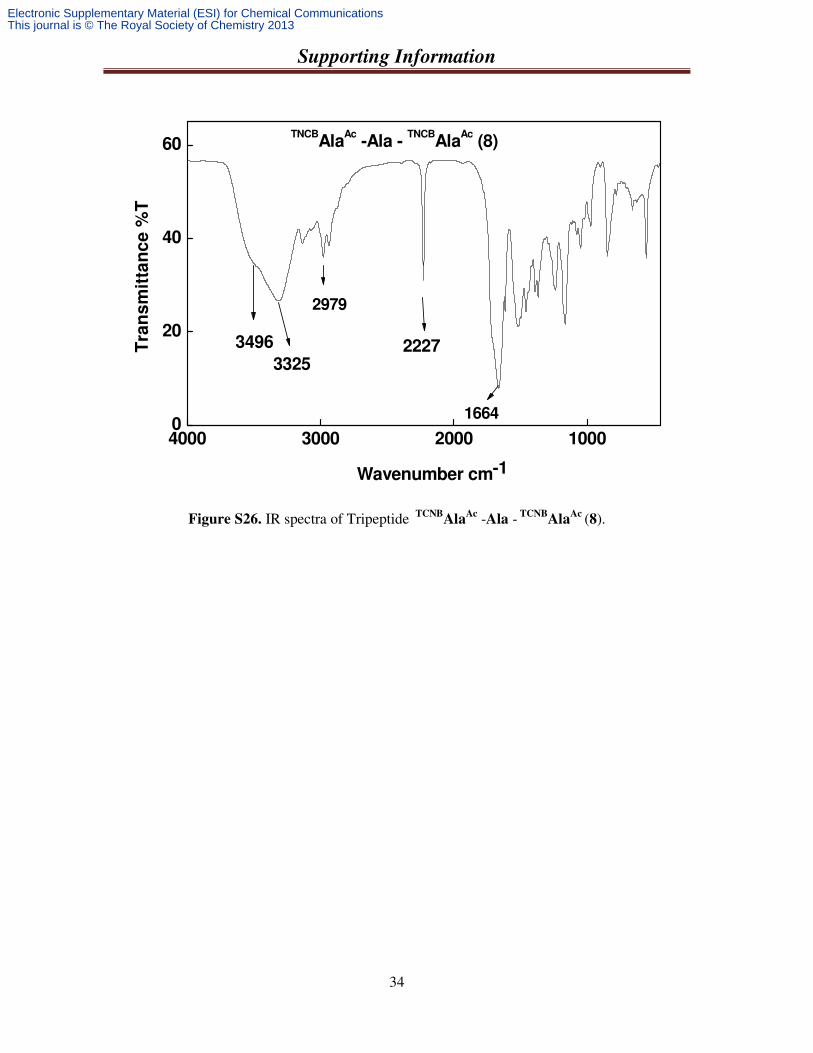
which became solid particle after ether wash. Yield 44%. IR (KBr) 3325, 2979, 2931, 2227, 1664, 1517, 1368, 1237, 1164, 1049, 848, 556 cm-1. 1H NMR (CDCl3; 400 MHz) δ = 1.18 (3H, d, J = 7.2 Hz); 1.34 (9H, s); 3.2 (3H, s); 3. 77 (3H, s); 4.41 (1H, m); 4.78 (2H, m); 4.89 (2H, m); 5.27 (1H, m);
5.80 (1H, m); 6.43 (1H, d, J = 8 Hz); 6.65 (1H, d, J = 6.8 Hz); 6.83 (1H, d, J = 7.6 Hz); 7.65 (4H, d, J = 7.2 Hz); 7.89 (6H, m); 13C NMR (CDCl3; 100 MHz); δ 17.9, 28.1, 32.4, 49.4, 50.0, 51.7, 53.4, 54.2, 61.9, 80.8, 111.1, 111.2, 118.7, 122.8, 126.0, 132.6, 134.7, 135.0, 145.5, 145.7, 155.3, 167.8, 169.2, 172.2. HRMS calcd for C34H39N12O6 ([M + H]+) 711.3116, found 711.3110. C34H38N12O6Na ([M + Na]+) 734.3013, found 734.301.
N
HN
H
O
O
O
O
NH
N
O
O
NN
NN
NN
CNCN
Electronic Supplementary Material (ESI) for Chemical CommunicationsThis journal is © The Royal Society of Chemistry 2013
Supporting Information
13
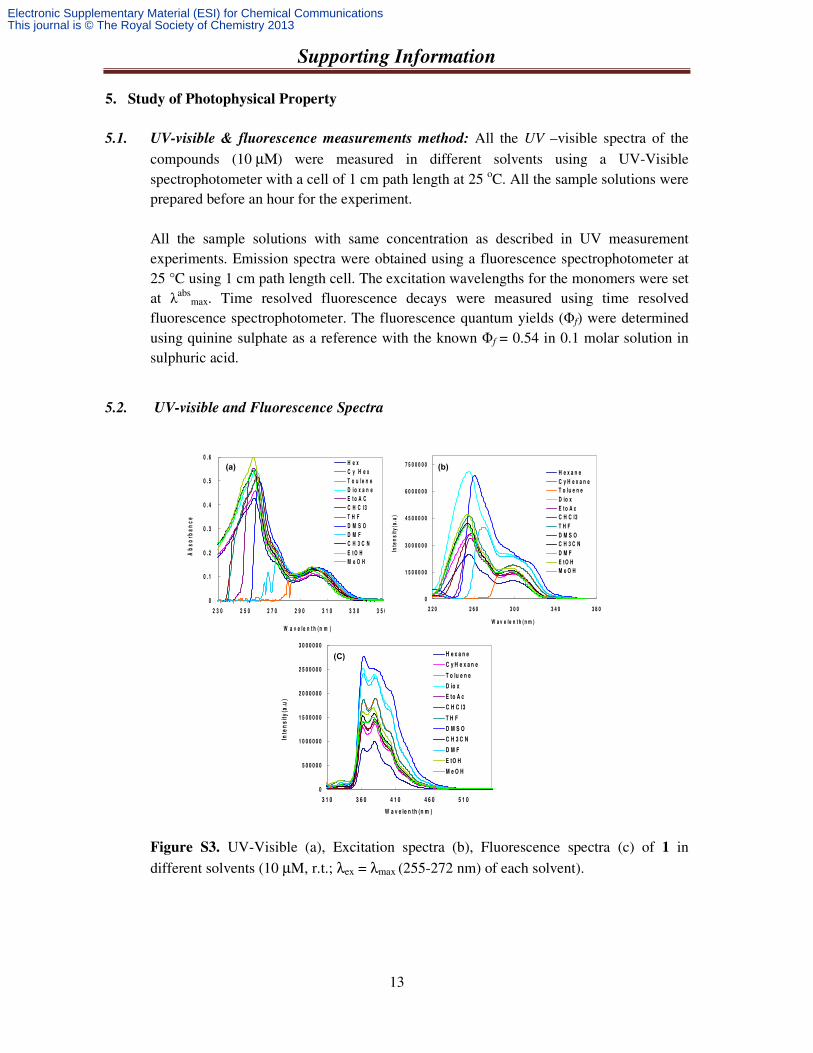
5. Study of Photophysical Property
5.1. UV-visible & fluorescence measurements method: All the UV –visible spectra of the compounds (10 µΜ) were measured in different solvents using a UV-Visible spectrophotometer with a cell of 1 cm path length at 25 oC. All the sample solutions were prepared before an hour for the experiment. All the sample solutions with same concentration as described in UV measurement experiments. Emission spectra were obtained using a fluorescence spectrophotometer at 25 °C using 1 cm path length cell. The excitation wavelengths for the monomers were set at λabs
max. Time resolved fluorescence decays were measured using time resolved fluorescence spectrophotometer. The fluorescence quantum yields (Φf) were determined using quinine sulphate as a reference with the known Φf = 0.54 in 0.1 molar solution in sulphuric acid.
5.2. UV-visible and Fluorescence Spectra
Figure S3. UV-Visible (a), Excitation spectra (b), Fluorescence spectra (c) of 1 in different solvents (10 µM, r.t.; λex = λmax (255-272 nm) of each solvent).
0
0 . 1
0 . 2
0 . 3
0 . 4
0 . 5
0 . 6
2 3 0 2 5 0 2 7 0 2 9 0 3 1 0 3 3 0 3 5 0
W a v e l e n t h ( n m )
Ab
so
rba
nc
e
H e x
C y H e x
T o u l e n e
D i o x a n e
E t o A C
C H C l 3
T H F
D M S O
D M F
C H 3 C N
E t O H
M e O H
0
1 5 0 0 0 0 0
3 0 0 0 0 0 0
4 5 0 0 0 0 0
6 0 0 0 0 0 0
7 5 0 0 0 0 0
2 2 0 2 6 0 3 0 0 3 4 0 3 8 0
W a v e le n t h ( n m )
Inte
ns
ity
(a.u
)
H e x a n e
C y H e x a n e
T o lu e n e
D io x
E t o A c
C H C l3
T H F
D M S O
C H 3 C N
D M F
E t O H
M e O H
0
5 0 0 0 0 0
1 0 0 0 0 0 0
1 5 0 0 0 0 0
2 0 0 0 0 0 0
2 5 0 0 0 0 0
3 0 0 0 0 0 0
3 1 0 3 6 0 4 1 0 4 6 0 5 1 0
W a v e le n th (n m )
Inte
ns
ity
(a.u
)
H e x a n e
C y H e x a n e
T o lu e n e
D io x
E to A c
C H C l3
T H F
D M S O
C H 3 C N
D M F
E tO H
M e O H
(a) (b)
(C)
Electronic Supplementary Material (ESI) for Chemical CommunicationsThis journal is © The Royal Society of Chemistry 2013
Supporting Information
14
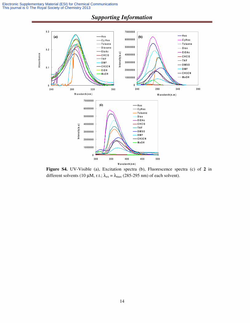
Figure S4. UV-Visible (a), Excitation spectra (b), Fluorescence spectra (c) of 2 in different solvents (10 µM, r.t.; λex = λmax (285-295 nm) of each solvent).
0
0 .1
0 .2
0 .3
2 4 0 2 8 0 3 2 0 3 6 0
W a v e le n th (n m )
Ab
so
rba
nc
eH e x
C y H e x
T o lu e n e
D io x a n e
E to A c
C H C l3
T H F
D M F
C H 3 C N
E tO H
M e O H
0
1 0 0 0 0 0 0
2 0 0 0 0 0 0
3 0 0 0 0 0 0
4 0 0 0 0 0 0
5 0 0 0 0 0 0
6 0 0 0 0 0 0
7 0 0 0 0 0 0
2 4 0 2 9 0 3 4 0 3 9 0
W a v e le n th (n .m )
Inte
ns
ity
(a.u
)
H e x
C y H e x
T o lu e n e
D io x
E tO A c
C H C l3
T H F
D M S O
D M F
C H 3 C N
M e O H
0
1 0 0 0 0 0 0
2 0 0 0 0 0 0
3 0 0 0 0 0 0
4 0 0 0 0 0 0
5 0 0 0 0 0 0
6 0 0 0 0 0 0
7 0 0 0 0 0 0
3 0 0 3 5 0 4 0 0 4 5 0 5 0 0
W a v e le n th (n m )
Inte
ns
ity
(a.u
)
H e x
C y H e x
T o lu e n e
D io x
E tO A c
C H C l3
T H F
D M S O
D M F
C H 3 C N
M e O H
Electronic Supplementary Material (ESI) for Chemical CommunicationsThis journal is © The Royal Society of Chemistry 2013
Supporting Information
15
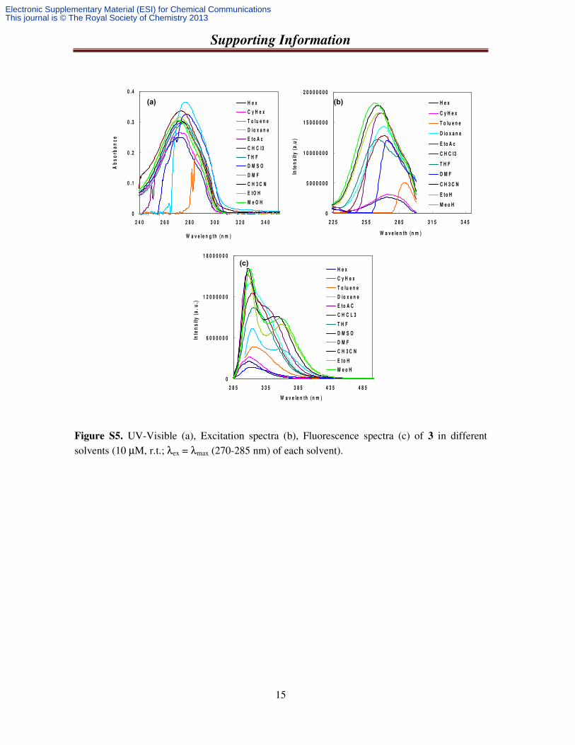
Figure S5. UV-Visible (a), Excitation spectra (b), Fluorescence spectra (c) of 3 in different solvents (10 µM, r.t.; λex = λmax (270-285 nm) of each solvent).
0
0 .1
0 .2
0 .3
0 .4
2 4 0 2 6 0 2 8 0 3 0 0 3 2 0 3 4 0
W a v e le n g t h ( n m )
Ab
so
rba
nc
e
H e x
C y H e x
T o lu e n e
D io x a n e
E t o A c
C H C l3
T H F
D M S O
D M F
C H 3 C N
E t O H
M e O H
0
5 0 0 0 0 0 0
1 0 0 0 0 0 0 0
1 5 0 0 0 0 0 0
2 0 0 0 0 0 0 0
2 2 5 2 5 5 2 8 5 3 1 5 3 4 5
W a v e le n th (n m )
Inte
ns
ity
(a
.u)
H e x
C y H e x
T o lu e n e
D io x a n e
E to A c
C H C l3
T H F
D M F
C H 3 C N
E to H
M e o H
0
6 0 0 0 0 0 0
1 2 0 0 0 0 0 0
1 8 0 0 0 0 0 0
2 8 5 3 3 5 3 8 5 4 3 5 4 8 5
W a v e le n th (n m )
Inte
ns
ity
(a
. u
.)
H e x
C y H e x
T o lu e n e
D io x a n e
E to A C
C H C L 3
T H F
D M S O
D M F
C H 3 C N
E to H
M e o H
(a) (b)
(c)
Electronic Supplementary Material (ESI) for Chemical CommunicationsThis journal is © The Royal Society of Chemistry 2013
Supporting Information
16
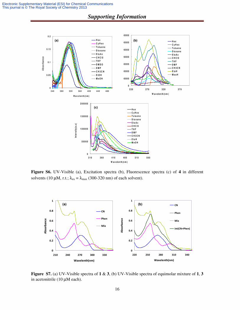
Figure S6. UV-Visible (a), Excitation spectra (b), Fluorescence spectra (c) of 4 in different solvents (10 µM, r.t.; λex = λmax (300-320 nm) of each solvent).
Figure S7. (a) UV-Visible spectra of 1 & 3, (b) UV-Visible spectra of equimolar mixture of 1, 3
in acetonitrile (10 µM each).
0
1 0 0 0 0 0
2 0 0 0 0 0
3 0 0 0 0 0
4 0 0 0 0 0
5 0 0 0 0 0
6 0 0 0 0 0
7 0 0 0 0 0
2 2 0 2 7 0 3 2 0 3 7 0
W a v e le n th (n m )
Inte
ns
ity
(a.u
)
H e x
C y H e x
T o lu e n e
D io x a n e
E to A c
C H C l3
T H F
D M F
C H 3 C N
E to H
M e o H
0
5 0 0 0 0
1 0 0 0 0 0
1 5 0 0 0 0
2 0 0 0 0 0
3 1 0 3 6 0 4 1 0 4 6 0 5 1 0 5 6 0
W a v e le n th (n m )
Inte
ns
ity
(a.u
)
H e x
C y H e x
T o lu e n e
D io x a n e
E to A c
C H C l3
T H F
D M F
C H 3 C N
E to H
M e O H
0
0 .0 5
0 .1
0 .1 5
0 .2
2 4 0 2 8 0 3 2 0 3 6 0 4 0 0 4 4 0 4 8 0
W a v e le n t h ( n m )
Ab
so
rba
nc
eH e x
C y H e x
T o lu e n e
D io x a n e
E to A c
C H C l3
T H F
D M S O
D M F
C H 3 C N
E tO H
M e O H
(a) (b)
(c)
0
0.2
0.4
0.6
0.8
1
220 250 280 310 340
Wavelenth(nm)
Ab
sorb
an
ce
CN
Phen
Mix
Int(CN+Phen)
0
0.2
0.4
0.6
0.8
1
210 240 270 300 330
Wavelenth(nm)
Ab
sorb
an
ce
CN
Phen
Mix
(a) (b)
Electronic Supplementary Material (ESI) for Chemical CommunicationsThis journal is © The Royal Society of Chemistry 2013
Supporting Information
17
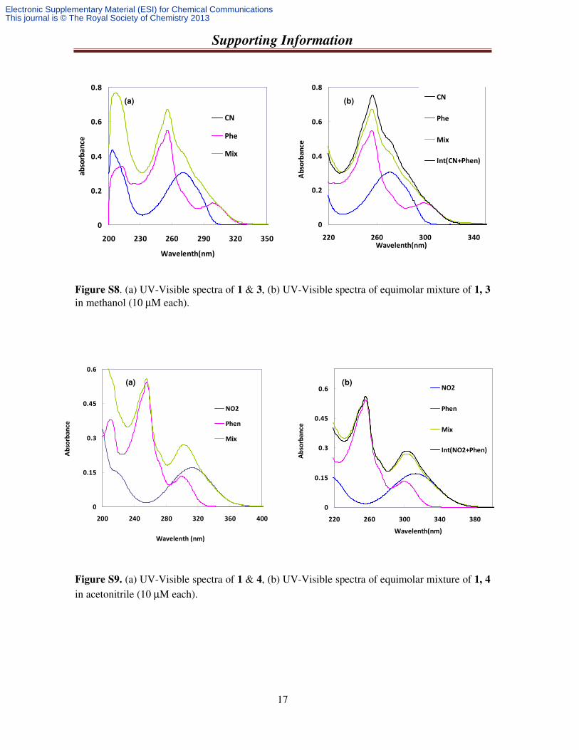
Figure S8. (a) UV-Visible spectra of 1 & 3, (b) UV-Visible spectra of equimolar mixture of 1, 3 in methanol (10 µM each).
Figure S9. (a) UV-Visible spectra of 1 & 4, (b) UV-Visible spectra of equimolar mixture of 1, 4 in acetonitrile (10 µM each).
0
0.2
0.4
0.6
0.8
220 260 300 340Wavelenth(nm)
Ab
sorb
an
ce
CN
Phe
Mix
Int(CN+Phen)
0
0.2
0.4
0.6
0.8
200 230 260 290 320 350
Wavelenth(nm)
ab
sorb
an
ce
CN
Phe
Mix
(a) (b)
0
0.15
0.3
0.45
0.6
200 240 280 320 360 400
Wavelenth (nm)
Ab
sorb
an
ce
NO2
Phen
Mix
0
0.15
0.3
0.45
0.6
220 260 300 340 380
Wavelenth(nm)
Ab
sorb
an
ce
NO2
Phen
Mix
Int(NO2+Phen)
(a) (b)
Electronic Supplementary Material (ESI) for Chemical CommunicationsThis journal is © The Royal Society of Chemistry 2013
Supporting Information
18
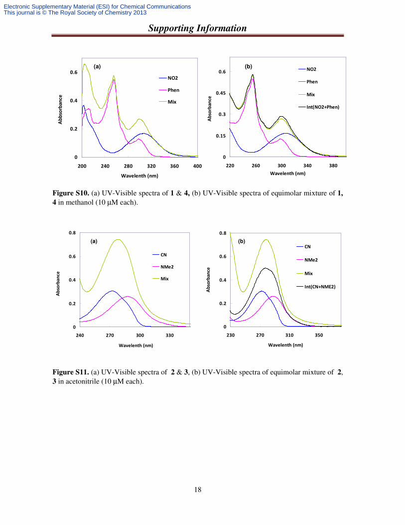
Figure S10. (a) UV-Visible spectra of 1 & 4, (b) UV-Visible spectra of equimolar mixture of 1, 4 in methanol (10 µM each).
Figure S11. (a) UV-Visible spectra of 2 & 3, (b) UV-Visible spectra of equimolar mixture of 2, 3 in acetonitrile (10 µM each).
0
0.2
0.4
0.6
0.8
240 270 300 330
Wavelenth (nm)
Ab
sorb
an
ce
CN
NMe2
Mix
0
0.2
0.4
0.6
0.8
230 270 310 350
Wavelenth (nm)
Ab
sorb
an
ce
CN
NMe2
Mix
Int(CN+NME2)
(a) (b)
0
0.2
0.4
0.6
200 240 280 320 360 400
Wavelenth (nm)
Ab
bso
rba
nce
NO2
Phen
Mix
0
0.15
0.3
0.45
0.6
220 260 300 340 380
Wavelenth (nm)
Ab
sorb
an
ce
NO2
Phen
Mix
Int(NO2+Phen)
(a) (b)
Electronic Supplementary Material (ESI) for Chemical CommunicationsThis journal is © The Royal Society of Chemistry 2013
Supporting Information
19
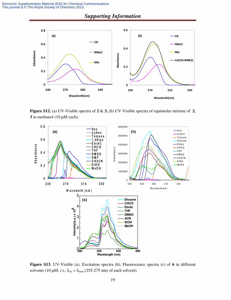
Figure S12. (a) UV-Visible spectra of 2 & 3, (b) UV-Visible spectra of equimolar mixture of 2, 3 in methanol (10 µM each).
Figure S13. UV-Visible (a), Excitation spectra (b), Fluorescence spectra (c) of 6 in different solvents (10 µM, r.t.; λex = λmax (255-275 nm) of each solvent).
0
0.2
0.4
0.6
230 270 310 350
Wavelenth(nm)
Ab
sorb
an
ce
CN
NMe2
Mix
Int(CN+NME2)
0
0.2
0.4
0.6
0.8
240 270 300 330
Wavelenth(nm)
Ab
sorb
an
ce
CN
NMe2
Mix
(a) (b)
0
0 .2
0 .4
0 .6
0 .8
2 3 0 2 7 0 3 1 0 3 5 0
W a v e le n t h ( n m )
Ab
so
rba
nc
e
H e xC y H e xT o lu e n e1 , 4 D io xE t o A CC H C l3T H FD M S OD M FC H 3 C NE t O HM e O H
0
1 0 0 0 0 0 0
2 0 0 0 0 0 0
3 0 0 0 0 0 0
4 0 0 0 0 0 0
5 0 0 0 0 0 0
6 0 0 0 0 0 0
2 2 5 2 5 5 2 8 5 3 1 5 3 4 5
W a v e le n t h ( n m )
Inte
ns
ity
(a.u
)
H e x
C y H e x
T o lu e n e
D io x a n e
E t o A c
C H C l3
T H F
D M S O
C H 3 C N
E t O H
M e O H
280 350 420 4900
1
2
3
4
5 Dioxane
CHCl3
EtoAc
THF
DMSO
ACN
EtOH
MeOH
Inte
nsit
y(a
.u.)
x 1
06
Wavelength (nm)
Electronic Supplementary Material (ESI) for Chemical CommunicationsThis journal is © The Royal Society of Chemistry 2013
Supporting Information
20
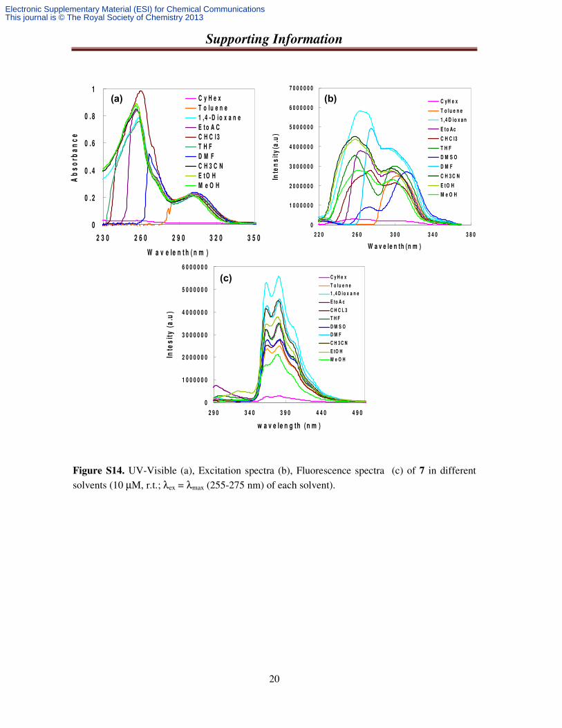
Figure S14. UV-Visible (a), Excitation spectra (b), Fluorescence spectra (c) of 7 in different solvents (10 µM, r.t.; λex = λmax (255-275 nm) of each solvent).
0
0 . 2
0 . 4
0 . 6
0 . 8
1
2 3 0 2 6 0 2 9 0 3 2 0 3 5 0
W a v e le n t h ( n m )
Ab
so
rba
nc
e
C y H e x
T o lu e n e
1 ,4 - D i o x a n e
E t o A C
C H C l 3
T H F
D M F
C H 3 C N
E t O H
M e O H
0
1 0 0 0 0 0 0
2 0 0 0 0 0 0
3 0 0 0 0 0 0
4 0 0 0 0 0 0
5 0 0 0 0 0 0
6 0 0 0 0 0 0
2 9 0 3 4 0 3 9 0 4 4 0 4 9 0
w a v e le n g th (n m )
Inte
sit
y (
a.u
)
C y H e x
T o l u e n e
1 , 4 D i o x a n e
E to A c
C H C L 3
T H F
D M S O
D M F
C H 3 C N
E tO H
M e O H
0
1 0 0 0 0 0 0
2 0 0 0 0 0 0
3 0 0 0 0 0 0
4 0 0 0 0 0 0
5 0 0 0 0 0 0
6 0 0 0 0 0 0
7 0 0 0 0 0 0
2 2 0 2 6 0 3 0 0 3 4 0 3 8 0
W a v e le n th (n m )
Inte
ns
ity
(a.u
)
C yH e x
T o lu e n e
1 ,4 D io x a n
E t o Ac
C H C l3
T H F
D M S O
D M F
C H 3 C N
E t O H
M e O H
(a) (b)
(c)
Electronic Supplementary Material (ESI) for Chemical CommunicationsThis journal is © The Royal Society of Chemistry 2013
Supporting Information
21
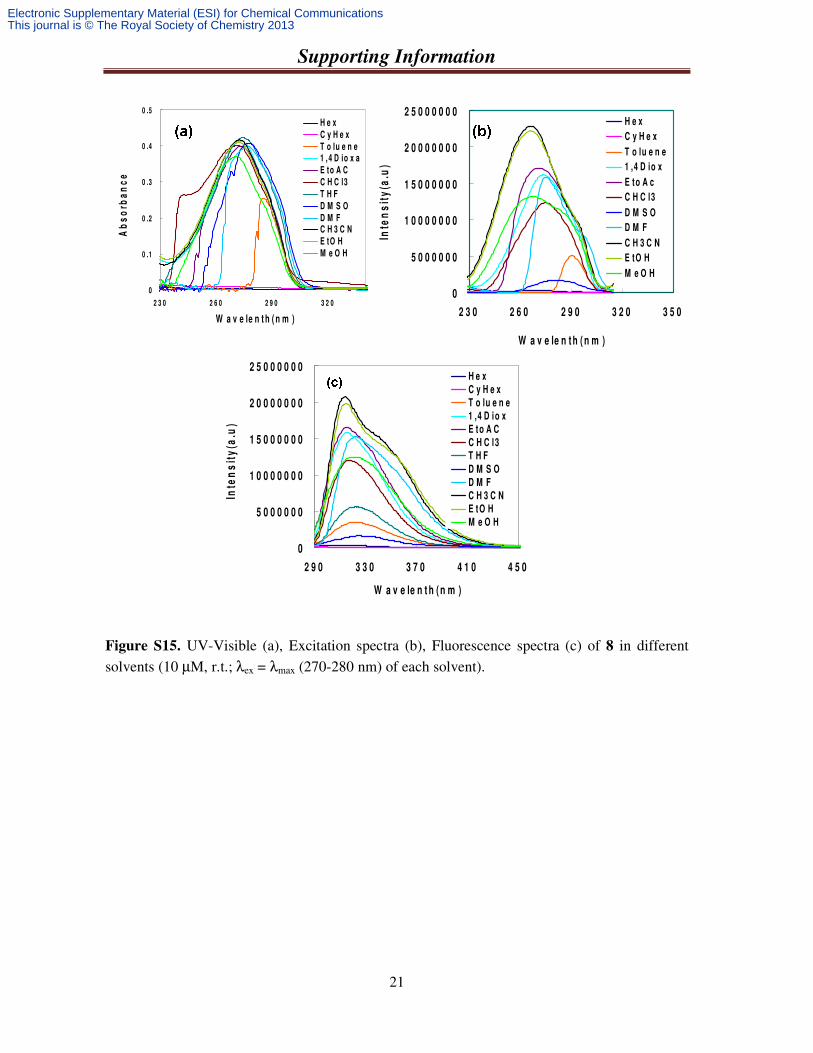
Figure S15. UV-Visible (a), Excitation spectra (b), Fluorescence spectra (c) of 8 in different solvents (10 µM, r.t.; λex = λmax (270-280 nm) of each solvent).
0
0 .1
0 .2
0 .3
0 .4
0 .5
2 3 0 2 6 0 2 9 0 3 2 0
W a v e le n t h ( n m )
Ab
so
rba
nc
eH e x
C y H e x
T o lu e n e1 ,4 D io x a
E to A C
C H C l3
T H F
D M S O
D M FC H 3 C N
E tO H
M e O H
0
5 0 0 0 0 0 0
1 0 0 0 0 0 0 0
1 5 0 0 0 0 0 0
2 0 0 0 0 0 0 0
2 5 0 0 0 0 0 0
2 3 0 2 6 0 2 9 0 3 2 0 3 5 0
W a v e le n t h ( n m )
Inte
ns
ity
(a.u
)
H e x
C y H e x
T o lu e n e
1 ,4 D io x
E to A c
C H C l3
D M S O
D M F
C H 3 C N
E tO H
M e O H
0
5 0 0 0 0 0 0
1 0 0 0 0 0 0 0
1 5 0 0 0 0 0 0
2 0 0 0 0 0 0 0
2 5 0 0 0 0 0 0
2 9 0 3 3 0 3 7 0 4 1 0 4 5 0
W a v e le n t h ( n m )
Inte
ns
ity
(a.u
)
H e x
C y H e x
T o lu e n e
1 ,4 D io x
E to A C
C H C l3
T H F
D M S O
D M F
C H 3 C N
E tO H
M e O H
Electronic Supplementary Material (ESI) for Chemical CommunicationsThis journal is © The Royal Society of Chemistry 2013
Supporting Information
22
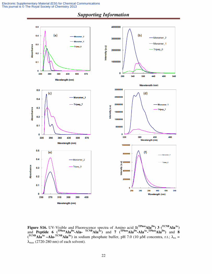
Figure S16. UV-Visible and Fluorescence spectra of Amino acid 1(TPhen
AlaDo
) 3 (TCNB
AlaAc
)
and Peptide 6 (TPhen
AlaDo
-Ala- TCNB
AlaAc
) and 7 (TPhen
AlaDo
-AlaAc
-TPhen
AlaDo
) and 8
(TCNB
AlaAc
–Ala-TCNB
AlaAc
) in sodium phosphate buffer, pH 7.0 (10 µM concentra, r.t.; λex = λmax (2720-280 nm) of each solvent).
Electronic Supplementary Material (ESI) for Chemical CommunicationsThis journal is © The Royal Society of Chemistry 2013
Supporting Information
23
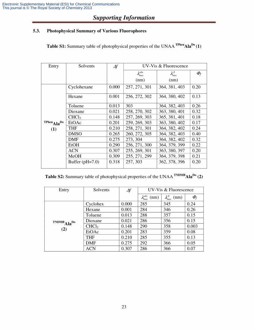
5.3. Photophysical Summary of Various Fluorophores
Table S1: Summary table of photophysical properties of the UNAA TPhen
AlaDo
(1)
Table S2: Summary table of photophysical properties of the UNAA TNDMB
AlaDo
(2)
Entry Solvents ∆f UV-Vis & Fluorescence abs
maxλ (nm)
fl
maxλ (nm)
Φf
TPhenAla
Do
(1)
Cyclohexane 0.000 257, 271, 301 364, 381, 403 0.20
Hexane 0.001 256, 272, 302 364, 380, 402 0.13
Toluene 0.013 303 364, 382, 403 0.26 Dioxane 0.021 258, 270, 302 363, 380, 401 0.32 CHCl3 0.148 257, 269, 303 365, 381, 401 0.18 EtOAc 0.201 259, 269, 303 363, 380, 402 0.17 THF 0.210 258, 271, 301 364, 382, 402 0.24 DMSO 0.265 260, 272, 305 364, 382, 403 0.40 DMF 0.275 273, 304 364, 382, 402 0.32 EtOH 0.290 256, 271, 300 364, 379, 399 0.22 ACN 0.307 255, 269, 301 363, 380, 397 0.20 MeOH 0.309 255, 271, 299 364, 379, 398 0.21 Buffer (pH=7.0) 0.318 257, 303 362, 378, 396 0.20
Entry Solvents ∆f UV-Vis & Fluorescence abs
maxλ (nm) fl
maxλ (nm) Φf
TNDMBAla
Do
(2)
Cyclohex 0.000 285 345 0.24 Hexane 0.001 284 346 0.26 Toluene 0.013 288 357 0.15 Dioxane 0.021 286 356 0.15 CHCl3 0.148 290 358 0.003 EtOAc 0.201 283 359 0.08 THF 0.210 285 355 0.13 DMF 0.275 292 366 0.05 ACN 0.307 286 366 0.07
Electronic Supplementary Material (ESI) for Chemical CommunicationsThis journal is © The Royal Society of Chemistry 2013
Supporting Information
24
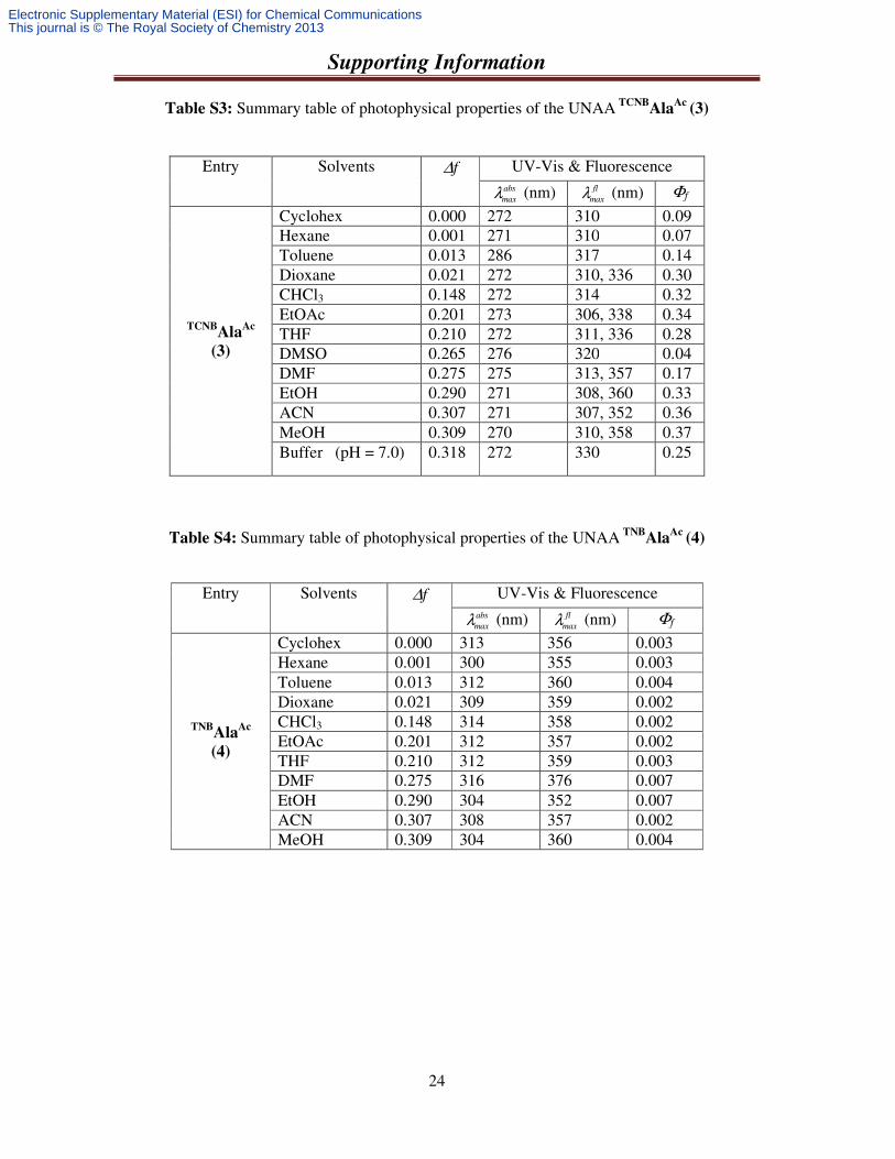
Table S3: Summary table of photophysical properties of the UNAA TCNBAla
Ac (3)
Table S4: Summary table of photophysical properties of the UNAA TNBAla
Ac (4)
Entry Solvents ∆f UV-Vis & Fluorescence abs
maxλ (nm) fl
maxλ (nm) Φf
TCNBAla
Ac
(3)
Cyclohex 0.000 272 310 0.09 Hexane 0.001 271 310 0.07 Toluene 0.013 286 317 0.14 Dioxane 0.021 272 310, 336 0.30 CHCl3 0.148 272 314 0.32 EtOAc 0.201 273 306, 338 0.34 THF 0.210 272 311, 336 0.28 DMSO 0.265 276 320 0.04 DMF 0.275 275 313, 357 0.17 EtOH 0.290 271 308, 360 0.33 ACN 0.307 271 307, 352 0.36 MeOH 0.309 270 310, 358 0.37 Buffer (pH = 7.0) 0.318 272 330 0.25
Entry Solvents ∆f UV-Vis & Fluorescence abs
maxλ (nm) fl
maxλ (nm) Φf
TNBAla
Ac
(4)
Cyclohex 0.000 313 356 0.003 Hexane 0.001 300 355 0.003 Toluene 0.013 312 360 0.004 Dioxane 0.021 309 359 0.002 CHCl3 0.148 314 358 0.002 EtOAc 0.201 312 357 0.002 THF 0.210 312 359 0.003 DMF 0.275 316 376 0.007 EtOH 0.290 304 352 0.007 ACN 0.307 308 357 0.002 MeOH 0.309 304 360 0.004
Electronic Supplementary Material (ESI) for Chemical CommunicationsThis journal is © The Royal Society of Chemistry 2013
Supporting Information
25
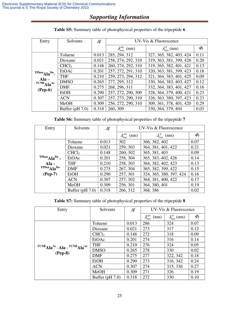
Table S5: Summary table of photophysical properties of the tripeptide 6
Table S6: Summary table of photophysical properties of the tripeptide 7
Table S7: Summary table of photophysical properties of the tripeptide 8
Entry Solvents ∆f UV-Vis & Fluorescence abs
maxλ (nm) fl
maxλ (nm) Φf
TPhen
AlaDo-
Ala –
TCNBAla
Ac (Pep-6)
Toluene 0.013 285, 294, 312 327, 365, 382, 403, 424 0.11 Dioxane 0.021 258, 274, 292, 310 319, 363, 381, 399, 426 0.20 CHCl3 0.148 260, 274, 292, 310 319, 365, 382, 401, 421 0.13 EtOAc 0.201 257, 272, 291, 310 320, 363, 381, 399, 423 0.18 THF 0.210 259, 273, 294, 312 321, 364, 383, 401, 425 0.09 DMSO 0.265 277, 295, 312 330, 364, 383, 403, 427 0.12 DMF 0.275 268, 296, 311 332, 364, 383, 401, 427 0.16 EtOH 0.290 257, 272, 290, 309 328, 364, 379, 400, 421 0.23 ACN 0.307 257, 273, 290, 310 326, 363, 380, 397, 423 0.23 MeOH 0.309 256, 272, 290, 310 309, 361, 378, 401, 420 0.29 Buffer (pH 7.0) 0.318 260, 309 330, 364, 379, 404 0.03
Entry Solvents ∆f UV-Vis & Fluorescence abs
maxλ (nm) fl
maxλ (nm) Φf
TPhenAla
Do -
Ala -
TPhenAla
Do
(Pep-7)
Toluene 0.013 302 366, 382, 402 0.07 Dioxane 0.021 259, 303 364, 381, 401, 422 0.21 CHCl3 0.148 260, 302 365, 381, 403 0.09 EtOAc 0.201 258, 304 365, 383, 402, 426 0.14 THF 0.210 258, 303 364, 382, 402, 423 0.13 DMF 0.275 267, 304 365, 382, 399, 422 0.15 EtOH 0.290 257, 301 324, 365, 380, 397, 424 0.16 ACN 0.307 257, 302 364, 381, 400, 422 0.17 MeOH 0.309 256, 301 364, 380, 401 0.19 Buffer (pH 7.0) 0.318 266, 312 368, 386 0.02
Entry Solvents ∆f UV-Vis & Fluorescence abs
maxλ (nm) fl
maxλ (nm) Φf
TCNBAla
Ac -Ala - TCNBAla
Ac
(Pep-8)
Toluene 0.013 286 324 0.07 Dioxane 0.021 273 317 0.12 CHCl3 0.148 272 318 0.09 EtOAc 0.201 274 316 0.14 THF 0.210 276 324 0.05 DMSO 0.265 278 330 0.02 DMF 0.275 277 322, 342 0.18 EtOH 0.290 273 316, 342 0.24 ACN 0.307 274 315, 336 0.27 MeOH 0.309 271 326 0.19 Buffer (pH 7.0) 0.318 272 330 0.10
Electronic Supplementary Material (ESI) for Chemical CommunicationsThis journal is © The Royal Society of Chemistry 2013
Supporting Information
26
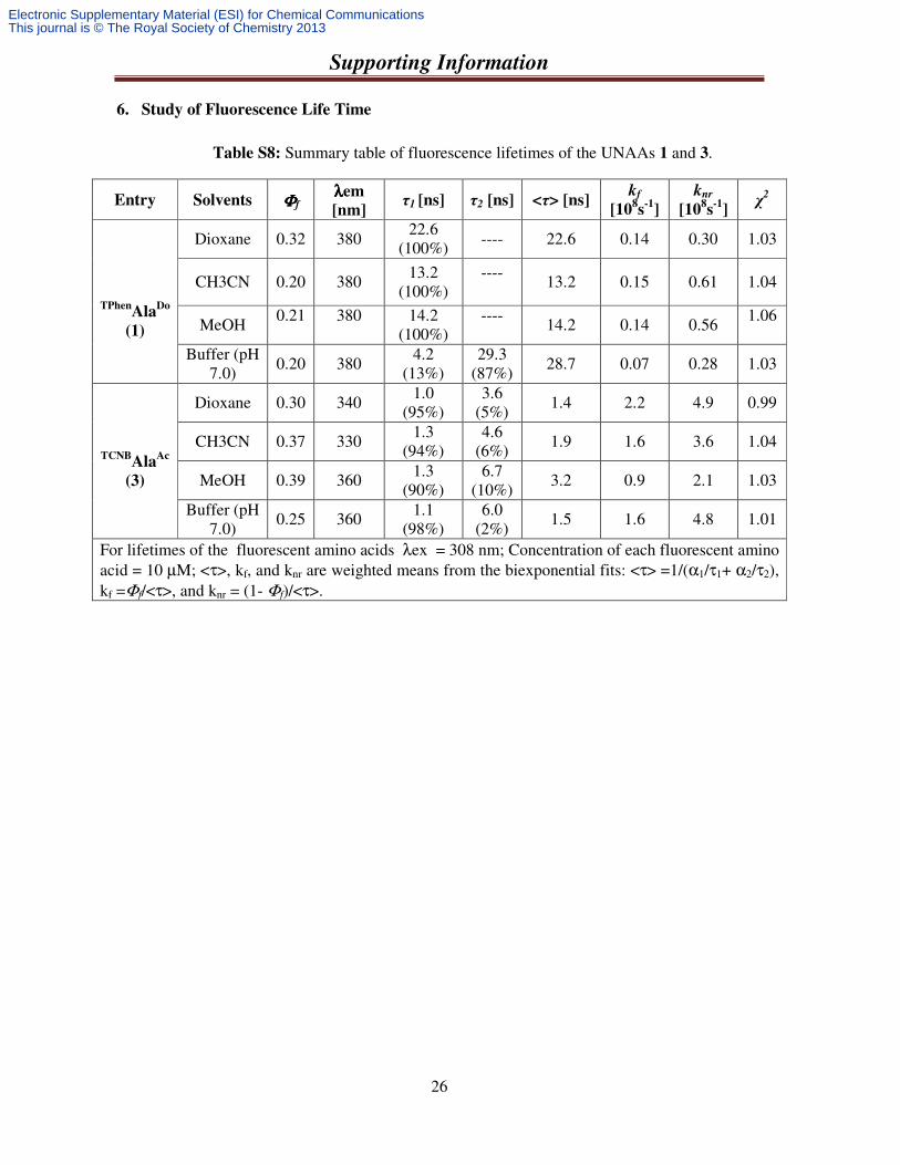
6. Study of Fluorescence Life Time
Table S8: Summary table of fluorescence lifetimes of the UNAAs 1 and 3.
Entry Solvents ΦΦΦΦf λλλλem
[nm] τ1 [ns] τ2 [ns] <τ> [ns]
kf
[108s
-1]
knr
[108s
-1]
χ2
TPhenAla
Do
(1)
Dioxane 0.32 380 22.6
(100%) ---- 22.6 0.14 0.30 1.03
CH3CN 0.20 380 13.2 (100%)
----
13.2 0.15 0.61 1.04
MeOH 0.21
380
14.2
(100%) ----
14.2 0.14 0.56
1.06
Buffer (pH 7.0)
0.20 380 4.2
(13%) 29.3
(87%) 28.7 0.07 0.28 1.03
TCNBAla
Ac
(3)
Dioxane 0.30 340 1.0
(95%) 3.6
(5%) 1.4 2.2 4.9 0.99
CH3CN 0.37 330 1.3
(94%) 4.6
(6%) 1.9 1.6 3.6 1.04
MeOH 0.39 360 1.3 (90%)
6.7 (10%)
3.2 0.9 2.1 1.03
Buffer (pH 7.0)
0.25 360 1.1 (98%)
6.0 (2%)
1.5 1.6 4.8 1.01
For lifetimes of the fluorescent amino acids λex = 308 nm; Concentration of each fluorescent amino acid = 10 µM; <τ>, kf, and knr are weighted means from the biexponential fits: <τ> =1/(α1/τ1+ α2/τ2), kf =Φf/<τ>, and knr = (1- Φf)/<τ>.
Electronic Supplementary Material (ESI) for Chemical CommunicationsThis journal is © The Royal Society of Chemistry 2013
Supporting Information
27
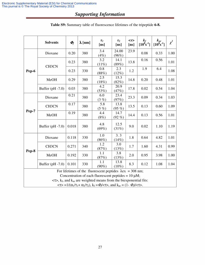
Table S9: Summary table of fluorescence lifetimes of the tripeptide 6-8.
Solvents ΦΦΦΦf λλλλ [nm]
τ1
[ns]
τ2
[ns]
<τ>
[ns]
kf
[108s
-1]
knr
[108s
-1]
χ2
Pep-6
Dioxane 0.20 380 3.4
(4%) 24.00 (96%)
23.9
0.08 0.33 1.00
CH3CN 0.23 380
3.2 (11%)
14.1 (89%)
13.8 0.16
0.56
1.01
0.23 330 0.8 (88%)
2.3 (12%)
1.2 1.9
6.4
1.08
MeOH 0.29 380 2.5 (18%)
15.3 (82%)
14.8 0.20 0.48 1.01
Buffer (pH -7.0) 0.03 380 4.2 (53%)
20.9 (47%)
17.8 0.02 0.54 1.04
Pep-7
Dioxane 0.21
380 6.0 (3 %)
23.4 (97%)
23.3 0.09 0.34 1.03
CH3CN 0.17
380
5.8 (5 %)
13.8 (95 %)
13.5 0.13 0.60 1.09
MeOH 0.19
380
4.4 (8%)
14.7 (92 %)
14.4 0.13 0.56 1.01
Buffer (pH -7.0) 0.018 380 4.8 (69%)
12.5 (31%)
9.0 0.02 1.10 1.19
Pep-8
Dioxane 0.118 330 1.0
(86%) 3. 3
(14%) 1.8 0.64 4.82 1.01
CH3CN 0.271 340 1.2
(87%) 3.0
(13%) 1.7 1.60 4.31 0.99
MeOH 0.192 330 1.1 (87%)
3.8 (13%)
2.0 0.95 3.98 1.00
Buffer (pH -7.0) 0.101 330 1.1 (90%)
13.8 (10%)
8.3 0.12 1.08 1.04
For lifetimes of the fluorescent peptides λex = 308 nm; Concentration of each fluorescent peptides = 10 µM;
<τ>, kf, and knr are weighted means from the biexponential fits: <τ> =1/(α1/τ1+ α2/τ2), kf =Φf/<τ>, and knr = (1- Φf)/<τ>.
Electronic Supplementary Material (ESI) for Chemical CommunicationsThis journal is © The Royal Society of Chemistry 2013
Supporting Information
28
0 .0 0 E + 0 0
2 .0 0 E + 0 2
4 .0 0 E + 0 2
6 .0 0 E + 0 2
8 .0 0 E + 0 2
1 .0 0 E + 0 3
1 .2 0 E + 0 3
3 8 1 3
T im e /n s
Co
un
ts
D io x a n e
C H 3 C N
M e O H
B u f fe r
0 .0 0 E + 0 0
5 .0 0 E + 0 2
1 .0 0 E + 0 3
1 .5 0 E + 0 3
2 .0 0 E + 0 3
0 2 0 4 0 6 0 8 0 1 0 0
T im e /n s
Co
un
tsD io x a n e
C H 3 C N
M e O H
B u f f e r
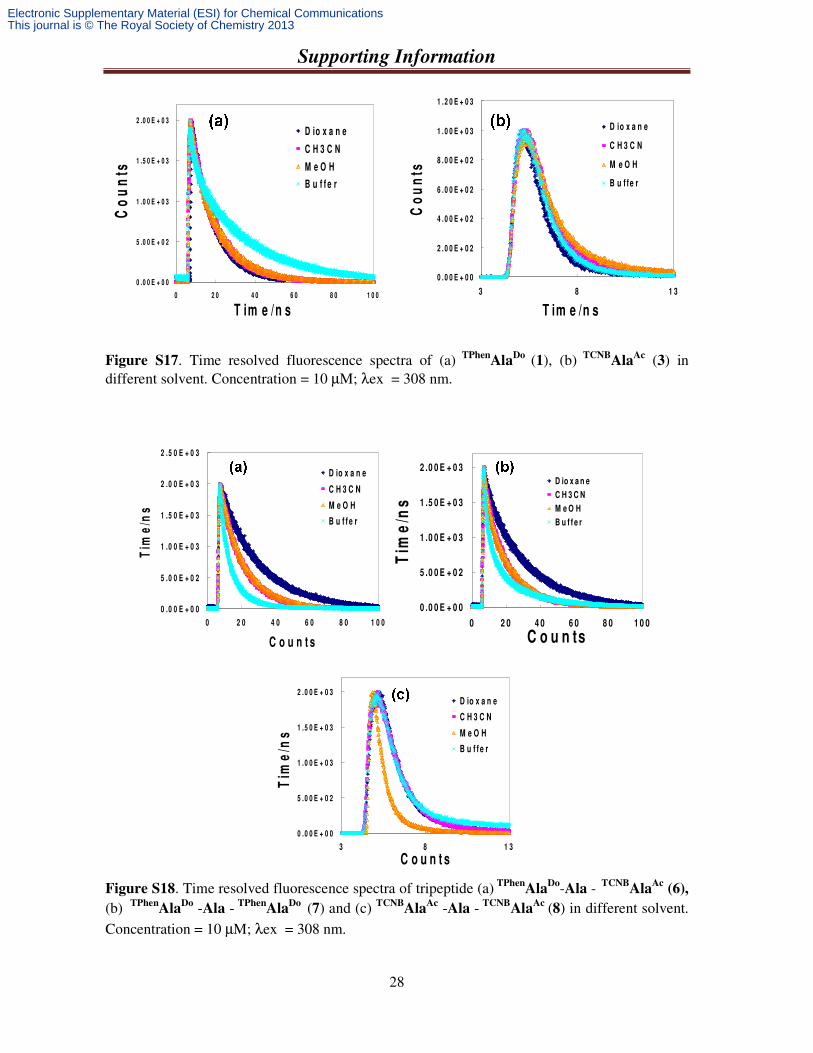
Figure S17. Time resolved fluorescence spectra of (a) TPhen
AlaDo (1), (b) TCNB
AlaAc (3) in
different solvent. Concentration = 10 µM; λex = 308 nm.
0 .0 0 E + 0 0
5 .0 0 E + 0 2
1 .0 0 E + 0 3
1 .5 0 E + 0 3
2 .0 0 E + 0 3
2 .5 0 E + 0 3
0 2 0 4 0 6 0 8 0 1 0 0
C o u n t s
Tim
e/n
s
D io x a n e
C H 3 C N
M e O H
B u f f e r
0 .0 0 E + 0 0
5 .0 0 E + 0 2
1 .0 0 E + 0 3
1 .5 0 E + 0 3
2 .0 0 E + 0 3
0 2 0 4 0 6 0 8 0 1 0 0
C o u n ts
Tim
e/n
s
D io x a n e
C H 3 C N
M e O H
B u f f e r
0 .0 0 E + 0 0
5 .0 0 E + 0 2
1 .0 0 E + 0 3
1 .5 0 E + 0 3
2 .0 0 E + 0 3
3 8 1 3
C o u n t s
Tim
e/n
s
D io x a n e
C H 3 C N
M e O H
B u f f e r
Figure S18. Time resolved fluorescence spectra of tripeptide (a) TPhen
AlaDo-Ala - TCNB
AlaAc (6),
(b) TPhenAla
Do -Ala - TPhenAla
Do (7) and (c) TCNBAla
Ac -Ala - TCNBAla
Ac (8) in different solvent. Concentration = 10 µM; λex = 308 nm.
Electronic Supplementary Material (ESI) for Chemical CommunicationsThis journal is © The Royal Society of Chemistry 2013
Supporting Information
29
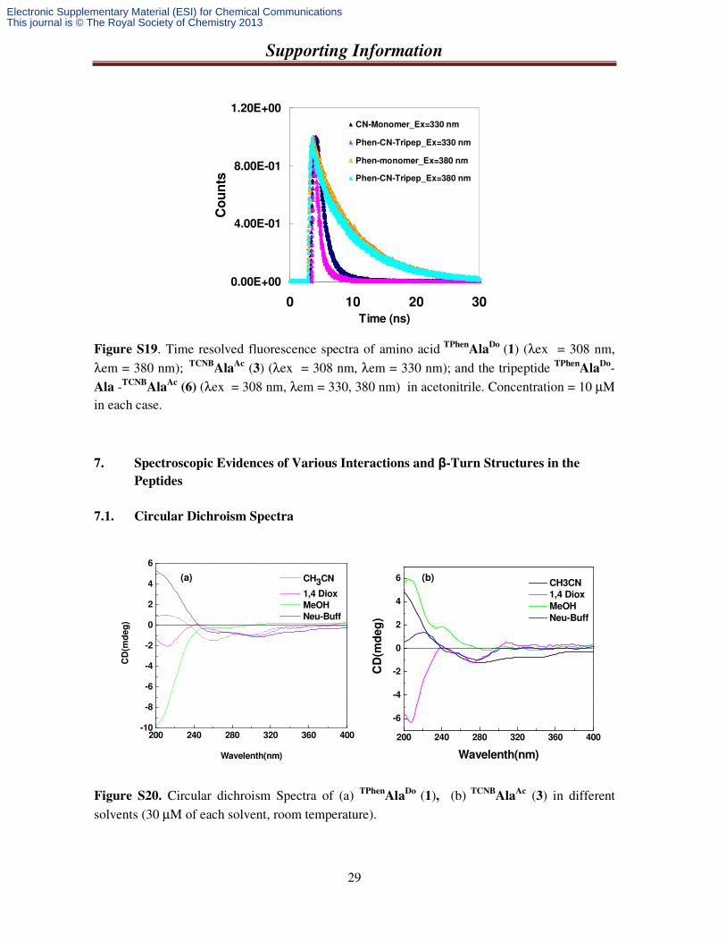
Figure S19. Time resolved fluorescence spectra of amino acid TPhenAla
Do (1) (λex = 308 nm, λem = 380 nm); TCNB
AlaAc (3) (λex = 308 nm, λem = 330 nm); and the tripeptide TPhen
AlaDo-
Ala -TCNBAla
Ac (6) (λex = 308 nm, λem = 330, 380 nm) in acetonitrile. Concentration = 10 µM in each case.
7. Spectroscopic Evidences of Various Interactions and β-Turn Structures in the
Peptides
7.1. Circular Dichroism Spectra
Figure S20. Circular dichroism Spectra of (a) TPhenAla
Do (1), (b) TCNBAla
Ac (3) in different solvents (30 µM of each solvent, room temperature).
200 240 280 320 360 400-10
-8
-6
-4
-2
0
2
4
6
CD
(md
eg
)
Wavelenth(nm)
CH3CN
1,4 Diox
MeOH
Neu-Buff
(a) (b)
200 240 280 320 360 400
-6
-4
-2
0
2
4
6
CD
(md
eg
)
Wavelenth(nm)
CH3CN
1,4 Diox
MeOH
Neu-Buff
0.00E+00
4.00E-01
8.00E-01
1.20E+00
0 10 20 30
Time (ns)
Co
un
ts
CN-Monomer_Ex=330 nm
Phen-CN-Tripep_Ex=330 nm
Phen-monomer_Ex=380 nm
Phen-CN-Tripep_Ex=380 nm
Electronic Supplementary Material (ESI) for Chemical CommunicationsThis journal is © The Royal Society of Chemistry 2013
Supporting Information
30
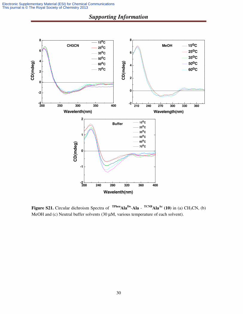
Figure S21. Circular dichroism Spectra of TPhenAla
Do-Ala - TCNBAla
Ac (10) in (a) CH3CN, (b) MeOH and (c) Neutral buffer solvents (30 µM, various temperature of each solvent).
200 250 300 350 400-4
-2
0
2
4
6
8
CD
(md
eg
)
Wavelenth(nm)
15oC
25oC
35oC
50oC
60oC
70oC
210 240 270 300 330 360-2
0
2
4
6
8
CD
(md
eg
)
Wavelength(nm)
15oC
25oC
35oC
50oC
60oC
200 240 280 320 360 400-2
-1
0
1
2
CD
(md
eg
)
Wavelenth(nm)
15o
C
25o
C
35o
C
50o
C
60o
C
70o
C
CH3CN MeOH
Buffer
Electronic Supplementary Material (ESI) for Chemical CommunicationsThis journal is © The Royal Society of Chemistry 2013
Supporting Information
31
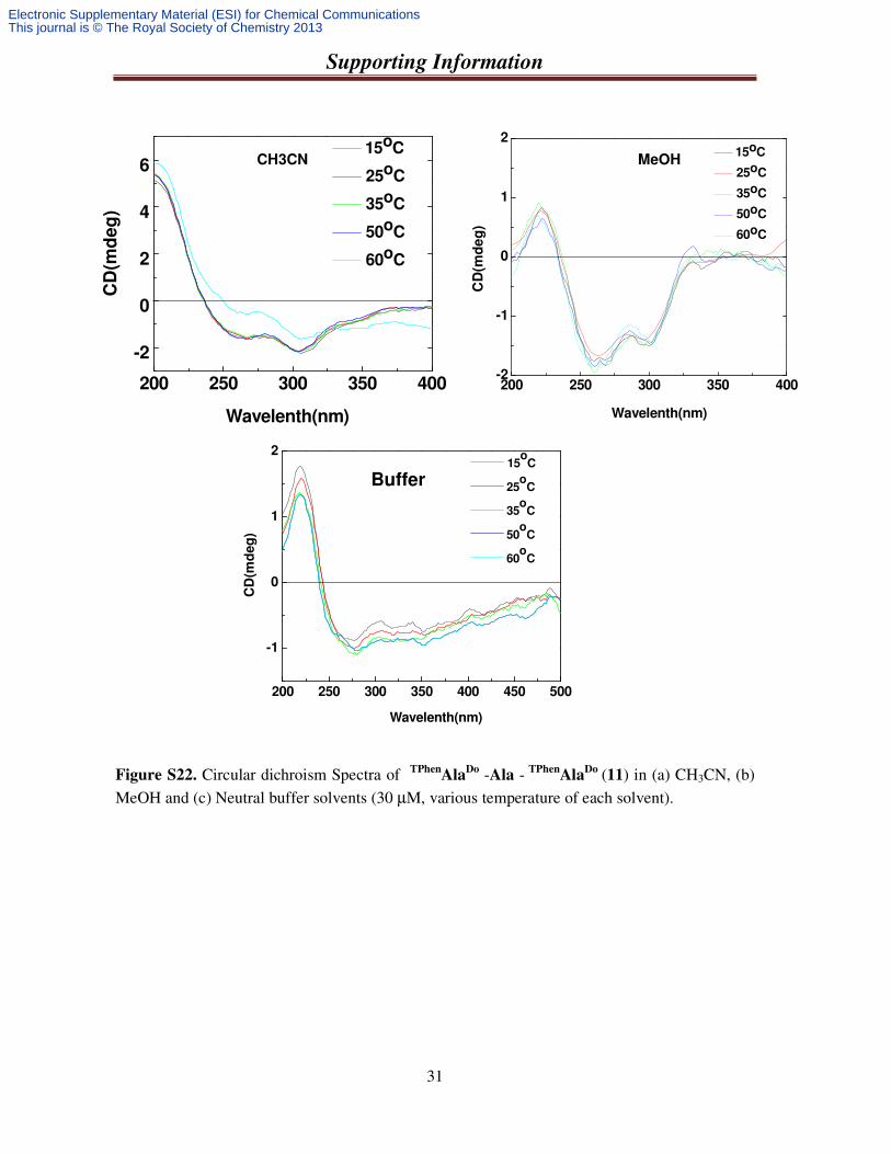
Figure S22. Circular dichroism Spectra of TPhenAla
Do -Ala - TPhenAla
Do (11) in (a) CH3CN, (b) MeOH and (c) Neutral buffer solvents (30 µM, various temperature of each solvent).
200 250 300 350 400
-2
0
2
4
6
C
D(m
de
g)
Wavelenth(nm)
15oC
25oC
35oC
50oC
60oC
200 250 300 350 400-2
-1
0
1
2
CD
(md
eg
)
Wavelenth(nm)
15oC
25oC
35oC
50oC
60oC
CH3CN MeOH
200 250 300 350 400 450 500
-1
0
1
2
CD
(md
eg
)
Wavelenth(nm)
15o
C
25o
C
35o
C
50o
C
60o
C
Buffer
Electronic Supplementary Material (ESI) for Chemical CommunicationsThis journal is © The Royal Society of Chemistry 2013
Supporting Information
32
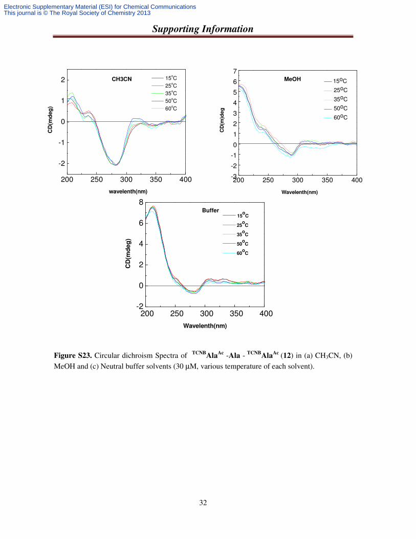
Figure S23. Circular dichroism Spectra of TCNBAla
Ac -Ala - TCNBAla
Ac (12) in (a) CH3CN, (b) MeOH and (c) Neutral buffer solvents (30 µM, various temperature of each solvent).
200 250 300 350 400
-2
-1
0
1
2 15oC
25oC
35oC
50oC
60oC
CD
(md
eg
)
wavelenth(nm)
200 250 300 350 400-3
-2
-1
0
1
2
3
4
5
6
7
CD
(m)d
eg
Wavelenth(nm)
15oC
25oC
35oC
50oC
60oC
200 250 300 350 400-2
0
2
4
6
8
15o
C
25o
C
35o
C
50o
C
60o
C
CD
(md
eg
)
Wavelenth(nm)
CH3CN MeOH
Buffer
Electronic Supplementary Material (ESI) for Chemical CommunicationsThis journal is © The Royal Society of Chemistry 2013
Supporting Information
33
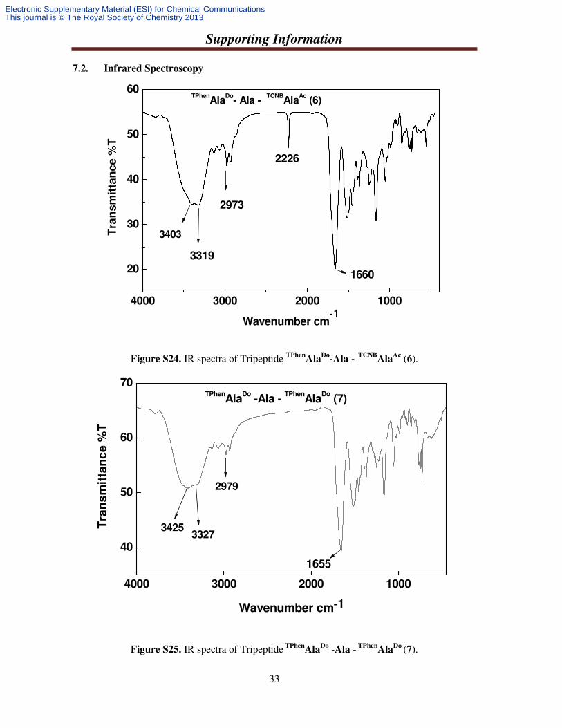
7.2. Infrared Spectroscopy
Figure S24. IR spectra of Tripeptide TPhenAla
Do-Ala -
TCNBAla
Ac (6).
Figure S25. IR spectra of Tripeptide TPhenAla
Do -Ala - TPhenAla
Do (7).
4000 3000 2000 1000
20
30
40
50
60
Tra
ns
mit
tan
ce %
T
Wavenumber cm-1
3403
3319
2973
2226
1660
TPhenAla
Do- Ala -
TCNBAla
Ac (6)
4000 3000 2000 1000
40
50
60
70
Tra
ns
mit
tan
ce
%T
Wavenumber cm-1
33273425
1655
2979
TPhenAla
Do -Ala -
TPhenAla
Do (7)
Electronic Supplementary Material (ESI) for Chemical CommunicationsThis journal is © The Royal Society of Chemistry 2013
Supporting Information
34
Figure S26. IR spectra of Tripeptide TCNBAla
Ac -Ala - TCNBAla
Ac (8).
4000 3000 2000 10000
20
40
60T
ran
sm
itta
nc
e %
T
Wavenumber cm-1
3325
3496 2227
1664
2979
TNCBAla
Ac -Ala -
TNCBAla
Ac (8)
Electronic Supplementary Material (ESI) for Chemical CommunicationsThis journal is © The Royal Society of Chemistry 2013
Supporting Information
35
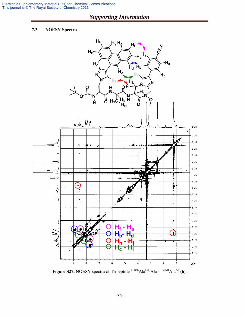
7.3. NOESY Spectra
Figure S27. NOESY spectra of Tripeptide TPhenAlaDo-Ala - TCNBAlaAc (6).
Hh - Hj
Hb- Hd
Hf - Ha
Hc - Hi
Electronic Supplementary Material (ESI) for Chemical CommunicationsThis journal is © The Royal Society of Chemistry 2013
Supporting Information
36
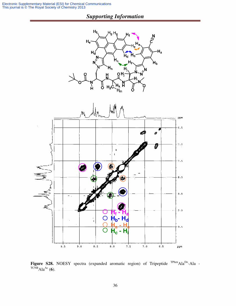
Figure S28. NOESY spectra (expanded aromatic region) of Tripeptide TPhenAlaDo-Ala -
TCNBAlaAc (6).
Ha - He
Hb- Hd
Hf - Ha
Hc - Hi
Electronic Supplementary Material (ESI) for Chemical CommunicationsThis journal is © The Royal Society of Chemistry 2013
Supporting Information
37
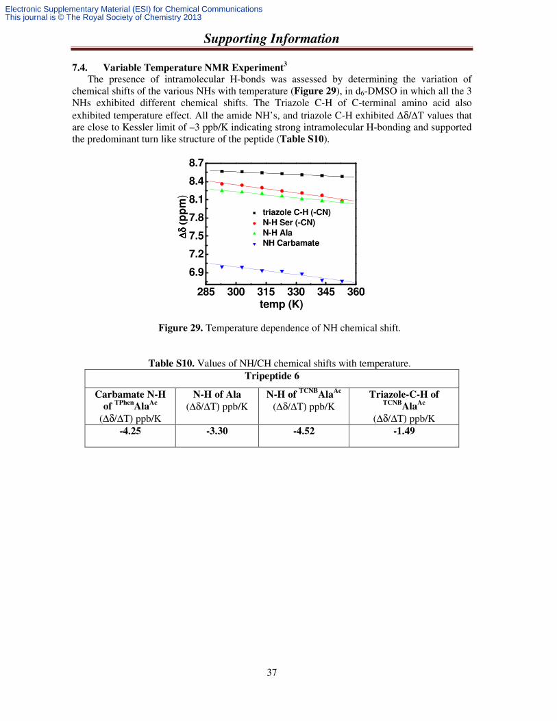
7.4. Variable Temperature NMR Experiment3
The presence of intramolecular H-bonds was assessed by determining the variation of
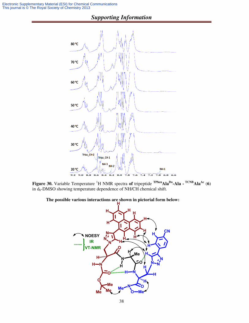
chemical shifts of the various NHs with temperature (Figure 29), in d6-DMSO in which all the 3 NHs exhibited different chemical shifts. The Triazole C-H of C-terminal amino acid also exhibited temperature effect. All the amide NH’s, and triazole C-H exhibited ∆δ/∆T values that are close to Kessler limit of –3 ppb/K indicating strong intramolecular H-bonding and supported the predominant turn like structure of the peptide (Table S10).
Figure 29. Temperature dependence of NH chemical shift.
Table S10. Values of NH/CH chemical shifts with temperature. Tripeptide 6
Carbamate N-H
of TPhen
AlaAc
(∆δ/∆Τ) ppb/K
N-H of Ala (∆δ/∆Τ) ppb/K
N-H of TCNB
AlaAc
(∆δ/∆Τ) ppb/K Triazole-C-H of
TCNBAla
Ac (∆δ/∆Τ) ppb/K
-4.25 -3.30 -4.52 -1.49
285 300 315 330 345 360
6.9
7.2
7.5
7.8
8.1
8.4
8.7
∆δ (
∆δ (
∆δ (
∆δ (p
pm
)
temp (K)
triazole C-H (-CN)
N-H Ser (-CN)
N-H Ala
NH Carbamate
Electronic Supplementary Material (ESI) for Chemical CommunicationsThis journal is © The Royal Society of Chemistry 2013
Supporting Information
38
Figure 30. Variable Temperature 1H NMR spectra of tripeptide TPhen
AlaDo
-Ala - TCNB
AlaAc (6)
in d6-DMSO showing temperature dependence of NH/CH chemical shift.
The possible various interactions are shown in pictorial form below:
O
O
NH
O
N
HO
NH
ONMe
Me Me
Me
NN
N
CN
HH
OMe
H
H
H
H
N
N
N
Me
H
H
H
H
H
H
H
H
NOESY HIR
VT-NMR
H
H
H
Electronic Supplementary Material (ESI) for Chemical CommunicationsThis journal is © The Royal Society of Chemistry 2013
Supporting Information
39
8. Study of Fluorescence Resonance Energy Transfer (FRET) in a Mixture of Two
Monomeric Donor/Acceptor Amino Acids
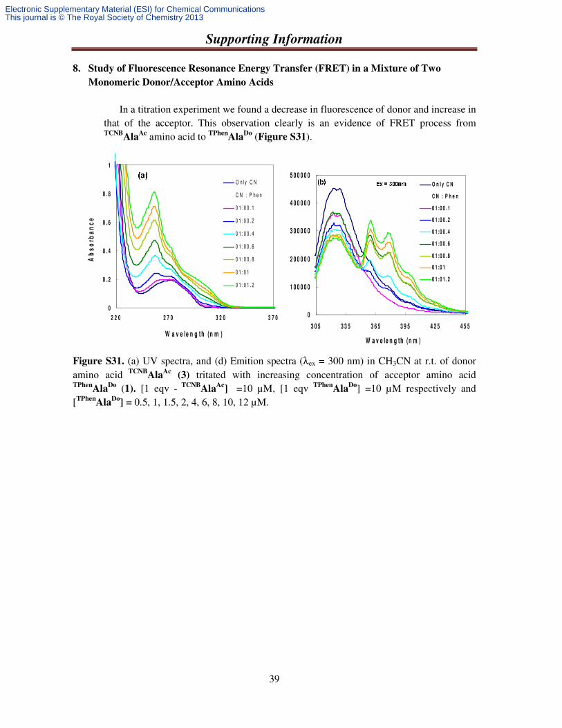
In a titration experiment we found a decrease in fluorescence of donor and increase in that of the acceptor. This observation clearly is an evidence of FRET process from TCNB
AlaAc amino acid to TPhen
AlaDo
(Figure S31).
Figure S31. (a) UV spectra, and (d) Emition spectra (λex = 300 nm) in CH3CN at r.t. of donor amino acid TCNB
AlaAc
(3) tritated with increasing concentration of acceptor amino acid TPhen
AlaDo (1). [1 eqv - TCNB
AlaAc
] =10 µM, [1 eqv TPhen
AlaDo] =10 µM respectively and
[TPhenAla
Do] = 0.5, 1, 1.5, 2, 4, 6, 8, 10, 12 µM.
0
1 0 0 0 0 0
2 0 0 0 0 0
3 0 0 0 0 0
4 0 0 0 0 0
5 0 0 0 0 0
3 0 5 3 3 5 3 6 5 3 9 5 4 2 5 4 5 5
W a v e le n g th (n m )
Inte
ns
ity
(a
.u)
O n l y C N
C N : P h e n
0 1 :0 0 . 1
0 1 :0 0 . 2
0 1 :0 0 . 4
0 1 :0 0 . 6
0 1 :0 0 . 8
0 1 :0 1
0 1 :0 1 . 2
0
0 . 2
0 . 4
0 . 6
0 . 8
1
2 2 0 2 7 0 3 2 0 3 7 0
W a v e le n g t h (n m )
Ab
so
rba
nc
e
O n ly C N
C N : P h e n
0 1 : 0 0 . 1
0 1 : 0 0 . 2
0 1 : 0 0 . 4
0 1 : 0 0 . 6
0 1 : 0 0 . 8
0 1 : 0 1
0 1 : 0 1 . 2
Electronic Supplementary Material (ESI) for Chemical CommunicationsThis journal is © The Royal Society of Chemistry 2013
Supporting Information
40
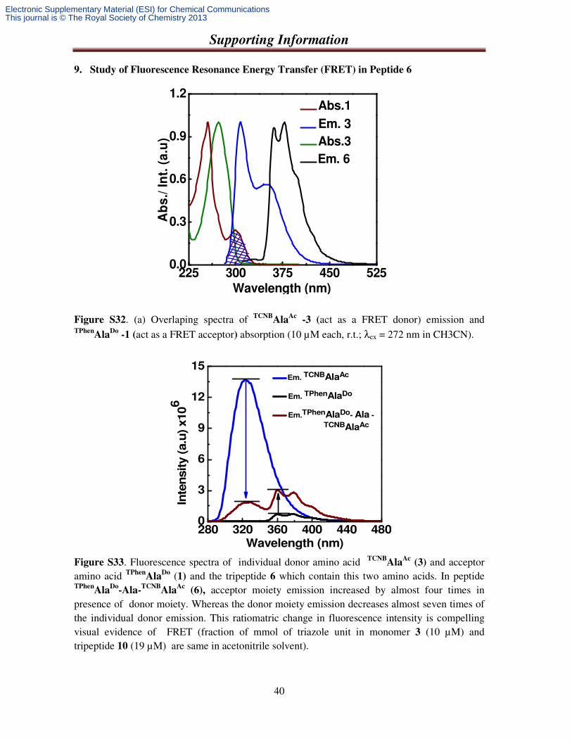
9. Study of Fluorescence Resonance Energy Transfer (FRET) in Peptide 6
Figure S32. (a) Overlaping spectra of TCNBAla
Ac -3 (act as a FRET donor) emission and
TPhenAla
Do -1 (act as a FRET acceptor) absorption (10 µM each, r.t.; λex = 272 nm in CH3CN).
Figure S33. Fluorescence spectra of individual donor amino acid TCNBAla
Ac (3) and acceptor
amino acid TPhenAla
Do (1) and the tripeptide 6 which contain this two amino acids. In peptide TPhen
AlaDo
-Ala-TCNB
AlaAc
(6), acceptor moiety emission increased by almost four times in presence of donor moiety. Whereas the donor moiety emission decreases almost seven times of the individual donor emission. This ratiomatric change in fluorescence intensity is compelling visual evidence of FRET (fraction of mmol of triazole unit in monomer 3 (10 µM) and tripeptide 10 (19 µM) are same in acetonitrile solvent).
225 300 375 450 5250.0
0.3
0.6
0.9
1.2
Ab
s./
In
t. (
a.u
)
Wavelength (nm)
Em. 3
Em. 6
Abs.3
Abs.1
280 320 360 400 440 4800
3
6
9
12
15 Em. TCNBAlaAc
Em. TPhenAlaDo
Em.TPhenAlaDo- Ala -
TCNBAlaAc
Inte
nsity (
a.u
) x106
Wavelength (nm)
Electronic Supplementary Material (ESI) for Chemical CommunicationsThis journal is © The Royal Society of Chemistry 2013
Supporting Information
41
Calculation of the Forster distance and FRET efficiency
The fluorescence resonance energy transfer (FRET) and the Förster distance for the FRET were calculated using the following three equations. The efficiency of energy transfer, E, was calculated using the equation (1)
60
6 60 0
E= 1R F
R r F= −
+……………………… (1)
where F and F0 are the fluorescence intensity of donor in the presence and absence of acceptor, r is the distance between donor and the acceptor and R0 is the critical distance when the energy transfer efficiency is 50% . The Förster distance R0 (Å) was calculated by the following equation (2)
5 2 4 1/ 60 [8.79 10 ( )]
DR n Jκ λ− −= × Φ …………………. (2)
where к2 is the orientation, n is the refractive index of the medium, ΦD is the quantum yield of the donor in the absence of acceptor J(λ) is the overlap integral of the fluorescence emission spectrum of the donor and the absorption spectrum of the acceptor given by the following equation (3)
…………………… (3)
where FD(λ) is the fluorescence intensity of the donor in the wavelength range λ to λ + ∆ λ with the total intensity normalized to unity. εA(λ) is the molar extinction coefficient of the acceptor as a function of wavelength (λ).
Using the values of к2= 2/3, n = 1.346, ΦD = 0.36, and the obtained overlap integral, J(λ) = 5.8643 x 10¹³ the R0 and r values were calculated which were found to be R0 = 20 Å and r = 15 Å. R0 is the critical distance when the energy transfer efficiency is 50 % and r is the distance between the donor and acceptor.
Energy Transfer efficiency (E) = 1- F/F0 = 85%. F and F0 are the fluorescence intensity of donor in the presence and absence of acceptor
( ) ( )
( )
4
0
0
D A
D
F dJ
F d
λ ε λ λ λ
λ λ
∞
∞=∫
∫
Electronic Supplementary Material (ESI) for Chemical CommunicationsThis journal is © The Royal Society of Chemistry 2013
Supporting Information
42
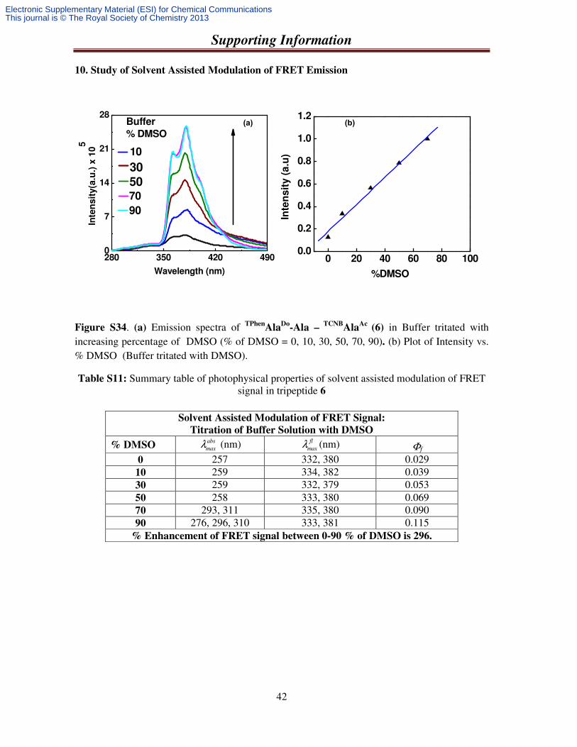
10. Study of Solvent Assisted Modulation of FRET Emission
Figure S34. (a) Emission spectra of TPhenAla
Do-Ala –
TCNBAla
Ac (6) in Buffer tritated with
increasing percentage of DMSO (% of DMSO = 0, 10, 30, 50, 70, 90). (b) Plot of Intensity vs. % DMSO (Buffer tritated with DMSO).
Table S11: Summary table of photophysical properties of solvent assisted modulation of FRET signal in tripeptide 6
Solvent Assisted Modulation of FRET Signal:
Titration of Buffer Solution with DMSO
% DMSO abs
maxλ (nm) fl
maxλ (nm) Φf
0 257 332, 380 0.029 10 259 334, 382 0.039 30 259 332, 379 0.053 50 258 333, 380 0.069 70 293, 311 335, 380 0.090 90 276, 296, 310 333, 381 0.115
% Enhancement of FRET signal between 0-90 % of DMSO is 296.
280 350 420 4900
7
14
21
28
90
70
50
30
10
% DMSO
Buffer
Inte
nsit
y(a
.u.)
x 1
05
Wavelength (nm)
(a) (b)
0 20 40 60 80 1000.0
0.2
0.4
0.6
0.8
1.0
1.2
Inte
ns
ity
(a
.u)
%DMSO
Electronic Supplementary Material (ESI) for Chemical CommunicationsThis journal is © The Royal Society of Chemistry 2013
Supporting Information
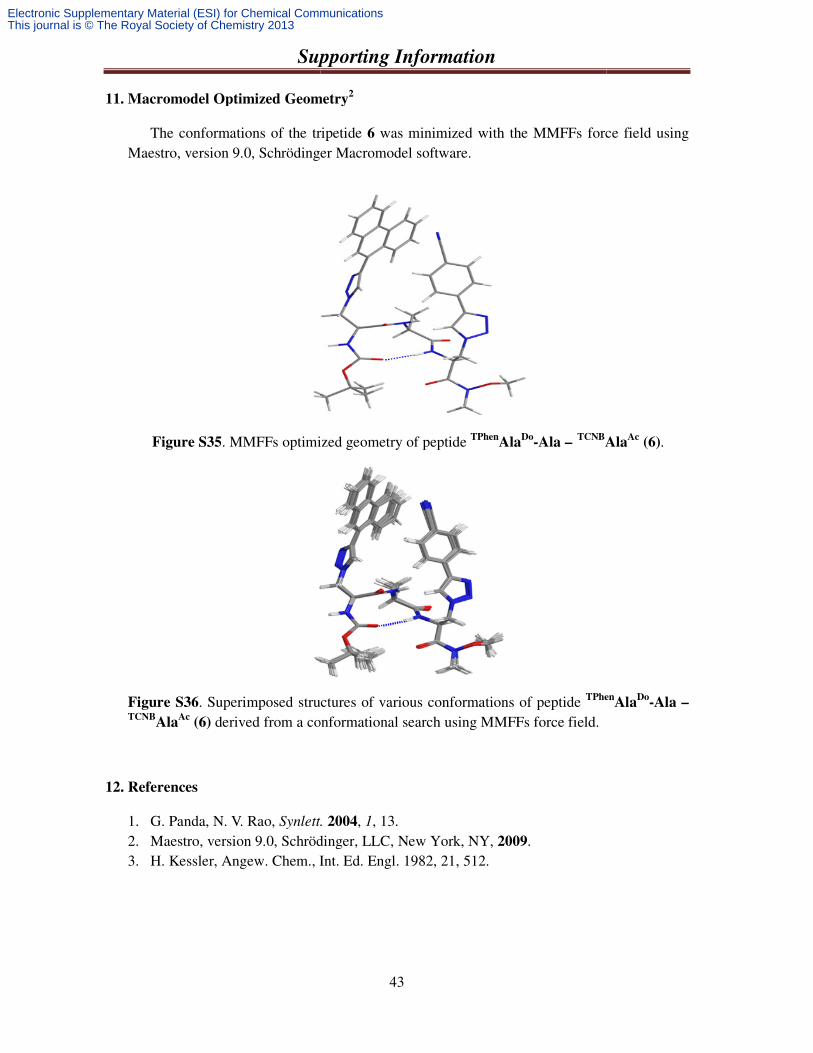
11. Macromodel Optimized Geometry
The conformations of the tripetide Maestro, version 9.0, Schrödinger Macromodel software.
Figure S35. MMFFs optimized geometry
Figure S36. Superimposed structures of various conformations of peptide TCNB
AlaAc
(6) derived from a conformational search using MMFFs
12. References
1. G. Panda, N. V. Rao, Synlett.
2. Maestro, version 9.0, Schrödinger, L3. H. Kessler, Angew. Chem., Int. Ed. Engl. 1982, 21, 512.
Supporting Information
43
model Optimized Geometry2
the tripetide 6 was minimized with the MMFFs force field using Maestro, version 9.0, Schrödinger Macromodel software.
optimized geometry of peptide TPhenAla
Do-Ala –
TCNBAla
Superimposed structures of various conformations of peptide TPhen
derived from a conformational search using MMFFs force field.
Synlett. 2004, 1, 13. Maestro, version 9.0, Schrödinger, LLC, New York, NY, 2009. H. Kessler, Angew. Chem., Int. Ed. Engl. 1982, 21, 512.
force field using
AlaAc
(6).
TPhenAla
Do-Ala –
Electronic Supplementary Material (ESI) for Chemical CommunicationsThis journal is © The Royal Society of Chemistry 2013
Supporting Information
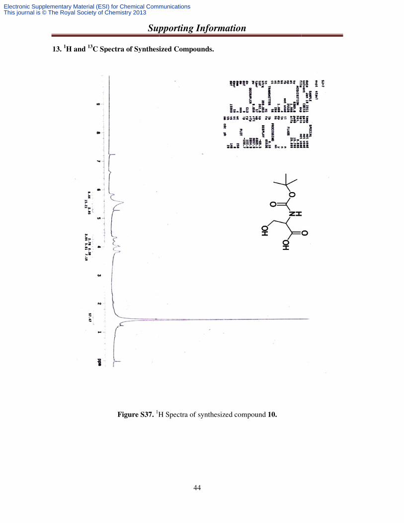
13. 1H and 13
C Spectra of Synthesized C
Figure S37. 1H Spectra of synthesized compound
Supporting Information
44
C Spectra of Synthesized Compounds.
H Spectra of synthesized compound 10.
Electronic Supplementary Material (ESI) for Chemical CommunicationsThis journal is © The Royal Society of Chemistry 2013

Supporting Information
Figure S38a. 1
Supporting Information
45
1H Spectra of synthesized compound 11.
Electronic Supplementary Material (ESI) for Chemical CommunicationsThis journal is © The Royal Society of Chemistry 2013
Supporting Information
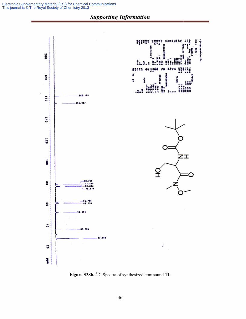
Figure S38b. 13
Supporting Information
46
13C Spectra of synthesized compound 11.
Electronic Supplementary Material (ESI) for Chemical CommunicationsThis journal is © The Royal Society of Chemistry 2013
Supporting Information
Figure S39a. 1
Supporting Information
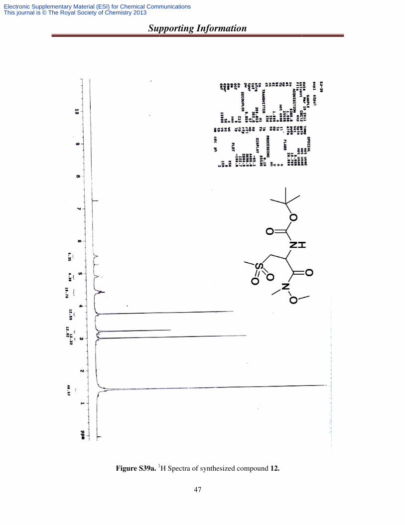
47
1H Spectra of synthesized compound 12.
Electronic Supplementary Material (ESI) for Chemical CommunicationsThis journal is © The Royal Society of Chemistry 2013
Supporting Information
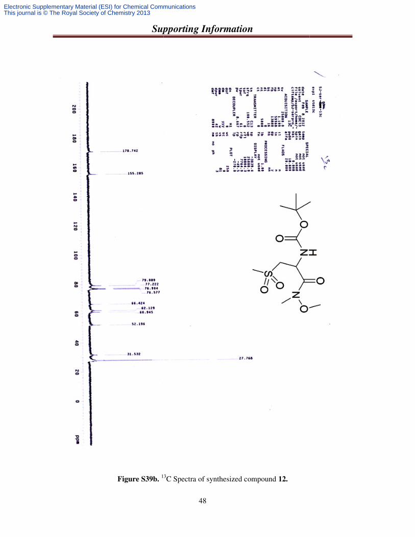
Figure S39b. 13
Supporting Information
48
13C Spectra of synthesized compound 12.
Electronic Supplementary Material (ESI) for Chemical CommunicationsThis journal is © The Royal Society of Chemistry 2013
Supporting Information
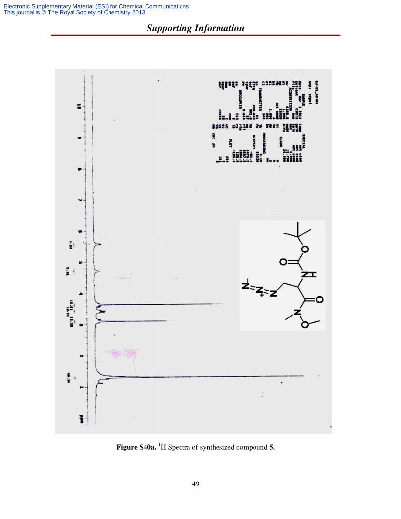
Figure S40a.
Supporting Information
49
. 1H Spectra of synthesized compound 5.
Electronic Supplementary Material (ESI) for Chemical CommunicationsThis journal is © The Royal Society of Chemistry 2013
Supporting Information
50
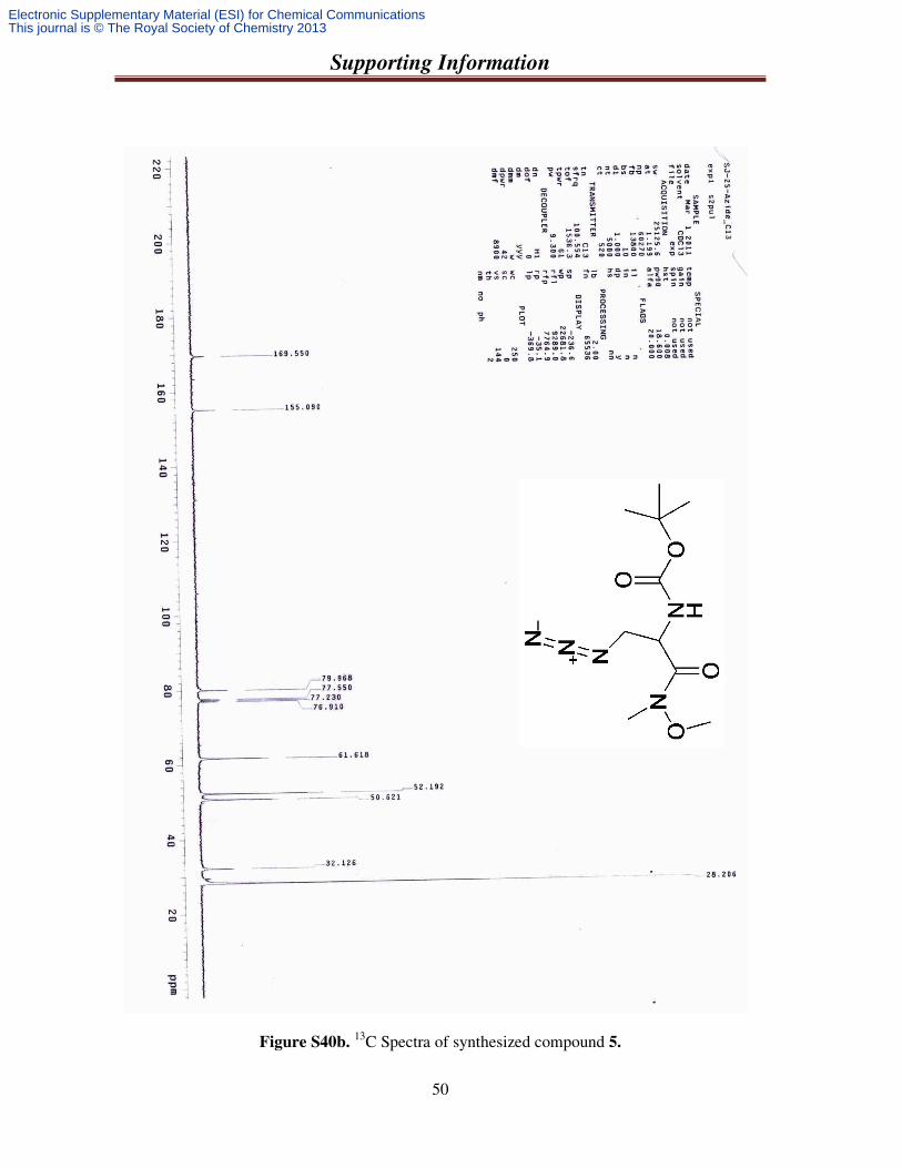
Figure S40b. 13C Spectra of synthesized compound 5.
Electronic Supplementary Material (ESI) for Chemical CommunicationsThis journal is © The Royal Society of Chemistry 2013
Supporting Information
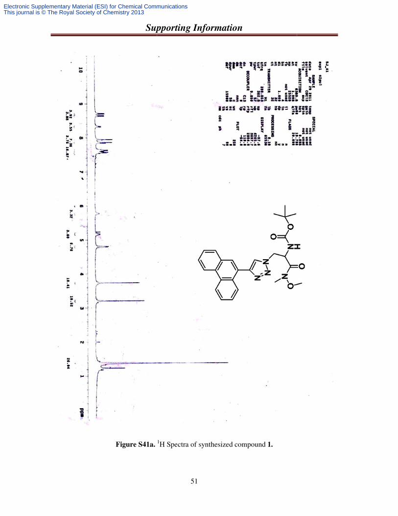
Figure S41a.
Supporting Information
51
. 1H Spectra of synthesized compound 1.
Electronic Supplementary Material (ESI) for Chemical CommunicationsThis journal is © The Royal Society of Chemistry 2013
Supporting Information
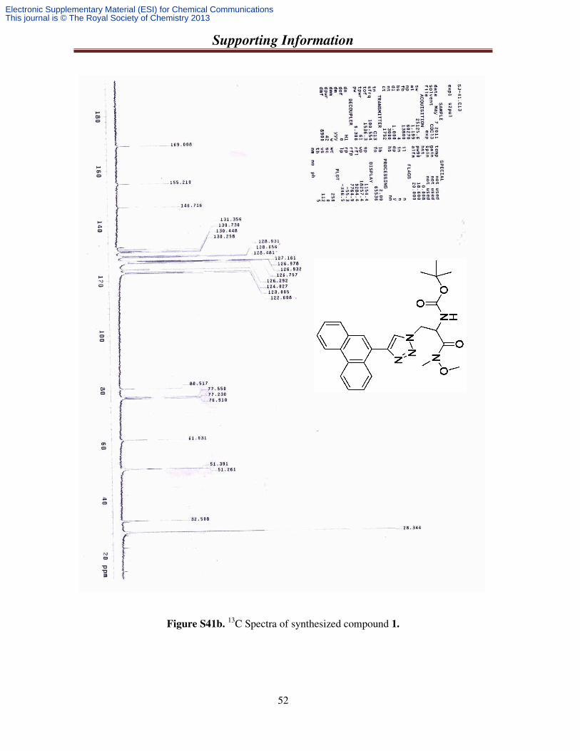
52
Figure S41b. 13C Spectra of synthesized compound 1.
Electronic Supplementary Material (ESI) for Chemical CommunicationsThis journal is © The Royal Society of Chemistry 2013
Supporting Information
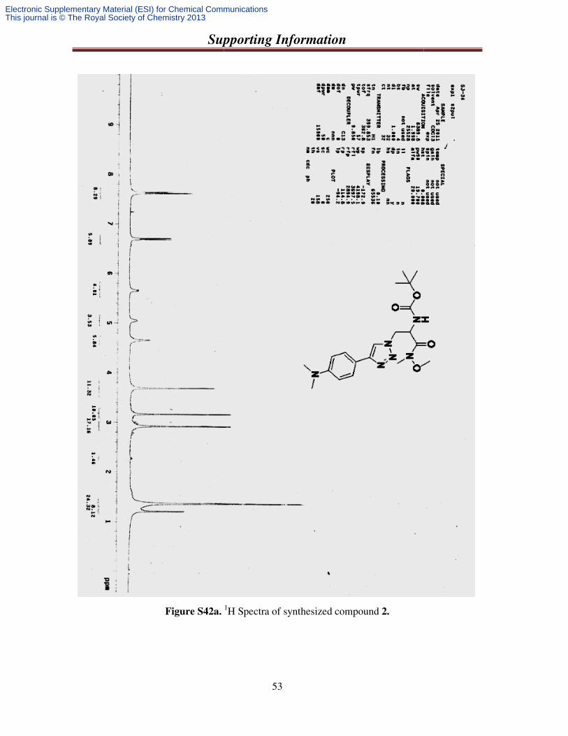
Figure S42a.
Supporting Information
53
. 1H Spectra of synthesized compound 2.
Electronic Supplementary Material (ESI) for Chemical CommunicationsThis journal is © The Royal Society of Chemistry 2013
Supporting Information
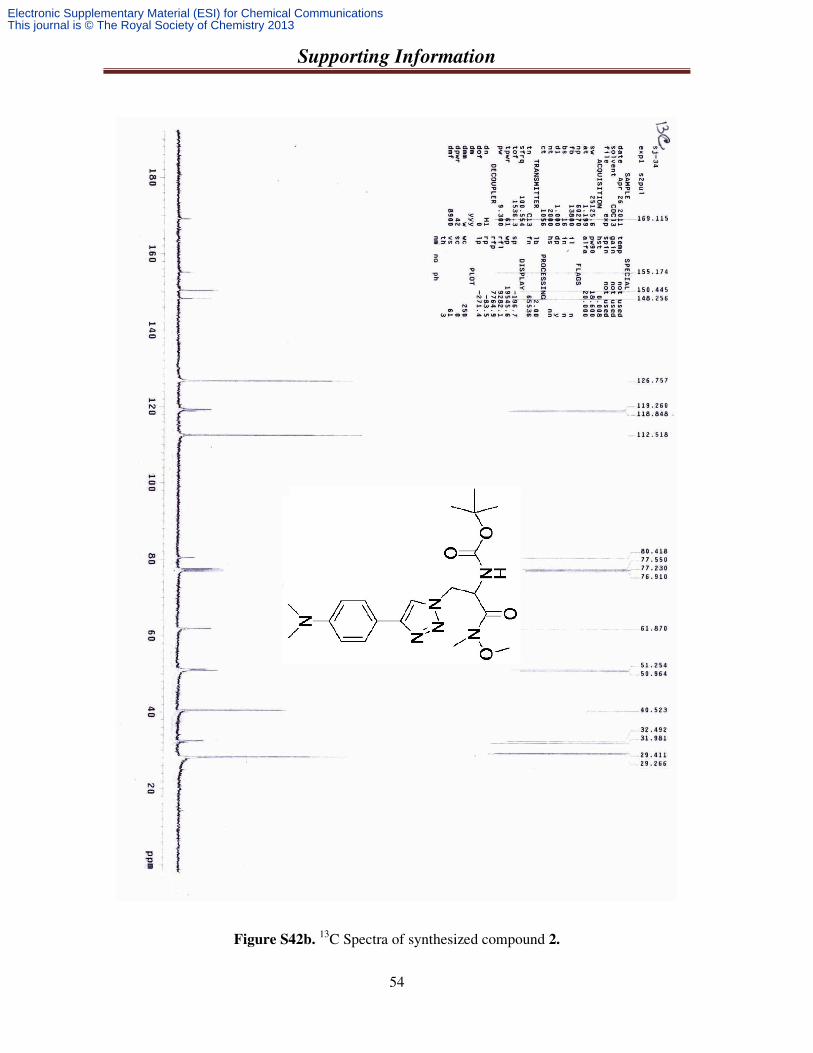
54
Figure S42b. 13C Spectra of synthesized compound 2.
Electronic Supplementary Material (ESI) for Chemical CommunicationsThis journal is © The Royal Society of Chemistry 2013
Supporting Information
55
Figure S43a. 1H Spectra of synthesized compound 3.
Electronic Supplementary Material (ESI) for Chemical CommunicationsThis journal is © The Royal Society of Chemistry 2013
Supporting Information
Figure S43b. 13
Supporting Information
56
13C Spectra of synthesized compound 3.
Electronic Supplementary Material (ESI) for Chemical CommunicationsThis journal is © The Royal Society of Chemistry 2013
Supporting Information
Figure S44a.
Supporting Information
57
. 1H Spectra of synthesized compound 4.
Electronic Supplementary Material (ESI) for Chemical CommunicationsThis journal is © The Royal Society of Chemistry 2013
Supporting Information
58
Figure S44b. 13C Spectra of synthesized compound 4.
Electronic Supplementary Material (ESI) for Chemical CommunicationsThis journal is © The Royal Society of Chemistry 2013
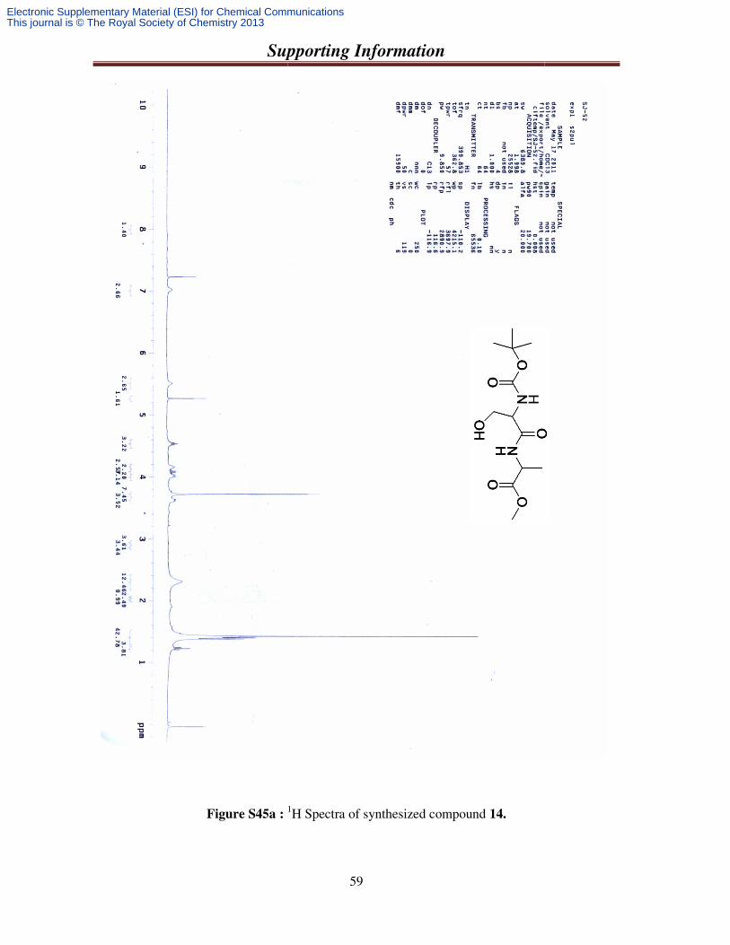
Supporting Information
Figure S45a : 1
Supporting Information
59
1H Spectra of synthesized compound 14.
Electronic Supplementary Material (ESI) for Chemical CommunicationsThis journal is © The Royal Society of Chemistry 2013
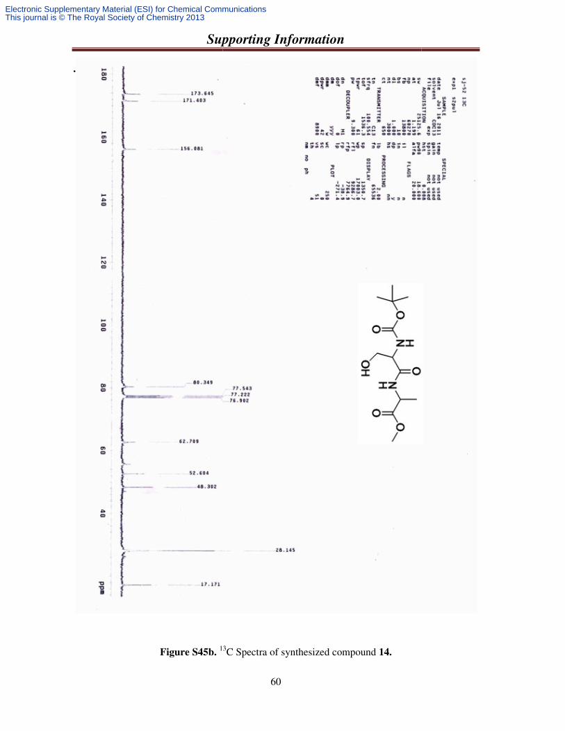
Supporting Information
.
Figure S45b. 13
Supporting Information
60
13C Spectra of synthesized compound 14.
Electronic Supplementary Material (ESI) for Chemical CommunicationsThis journal is © The Royal Society of Chemistry 2013
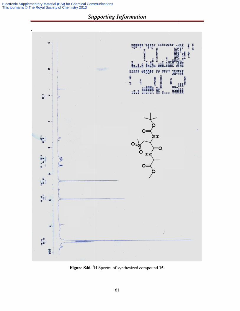
Supporting Information
.
Figure S46. 1H Spectra of sy
Supporting Information
61
H Spectra of synthesized compound 15.
Electronic Supplementary Material (ESI) for Chemical CommunicationsThis journal is © The Royal Society of Chemistry 2013
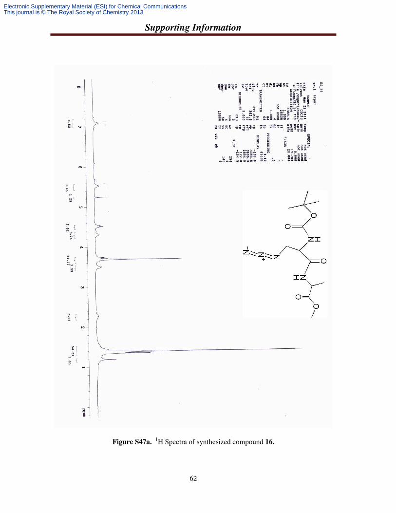
Supporting Information
62
Figure S47a. 1H Spectra of synthesized compound 16.
Electronic Supplementary Material (ESI) for Chemical CommunicationsThis journal is © The Royal Society of Chemistry 2013
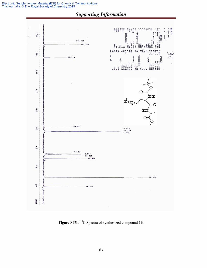
Supporting Information
63
Figure S47b. 13C Spectra of synthesized compound 16.
Electronic Supplementary Material (ESI) for Chemical CommunicationsThis journal is © The Royal Society of Chemistry 2013
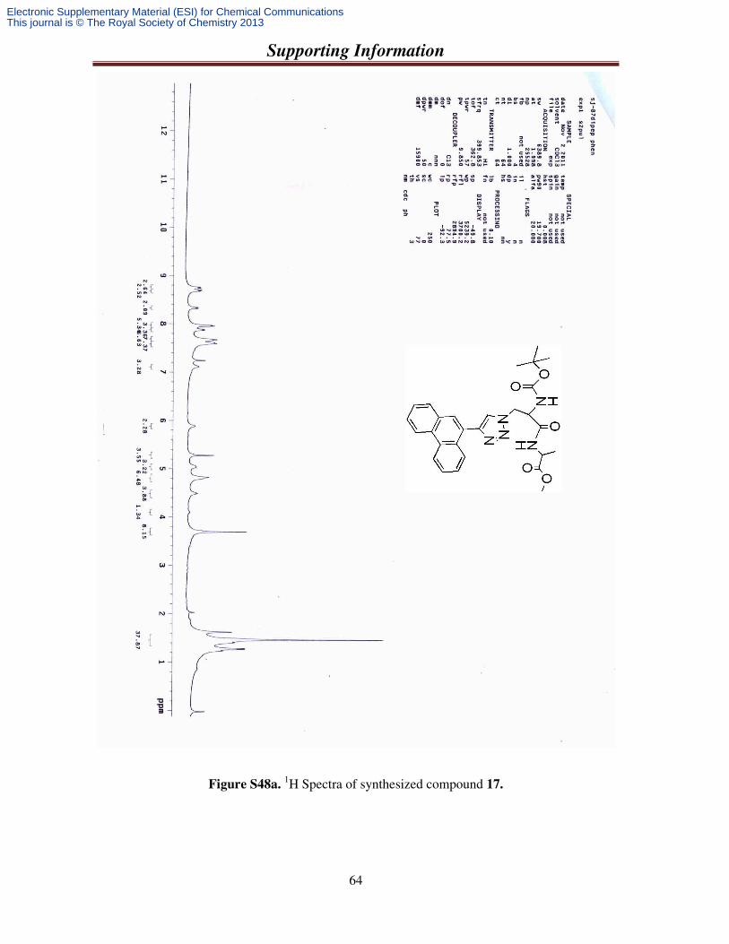
Supporting Information
64
Figure S48a. 1H Spectra of synthesized compound 17.
Electronic Supplementary Material (ESI) for Chemical CommunicationsThis journal is © The Royal Society of Chemistry 2013
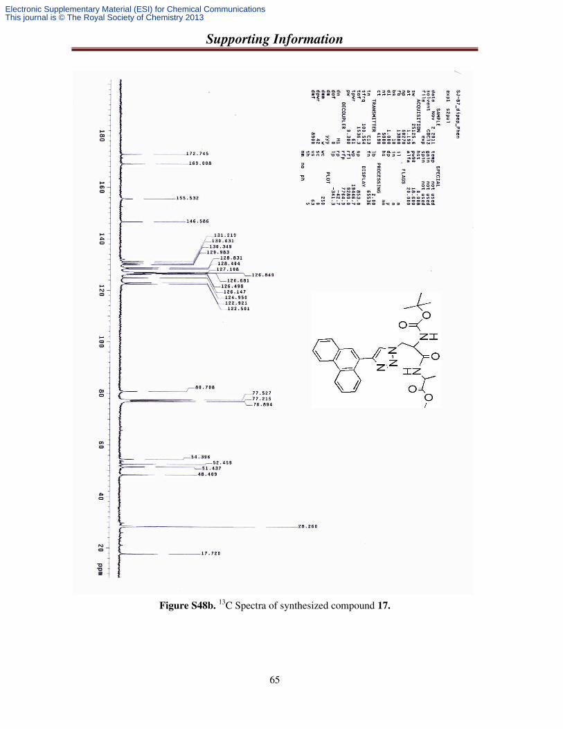
Supporting Information
65
Figure S48b. 13C Spectra of synthesized compound 17.
Electronic Supplementary Material (ESI) for Chemical CommunicationsThis journal is © The Royal Society of Chemistry 2013
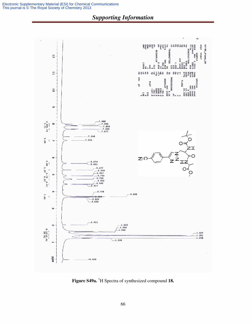
Supporting Information
66
Figure S49a. 1H Spectra of synthesized compound 18.
Electronic Supplementary Material (ESI) for Chemical CommunicationsThis journal is © The Royal Society of Chemistry 2013
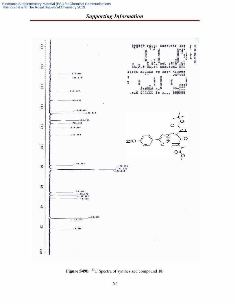
Supporting Information
67
Figure S49b. 13C Spectra of synthesized compound 18.
Electronic Supplementary Material (ESI) for Chemical CommunicationsThis journal is © The Royal Society of Chemistry 2013
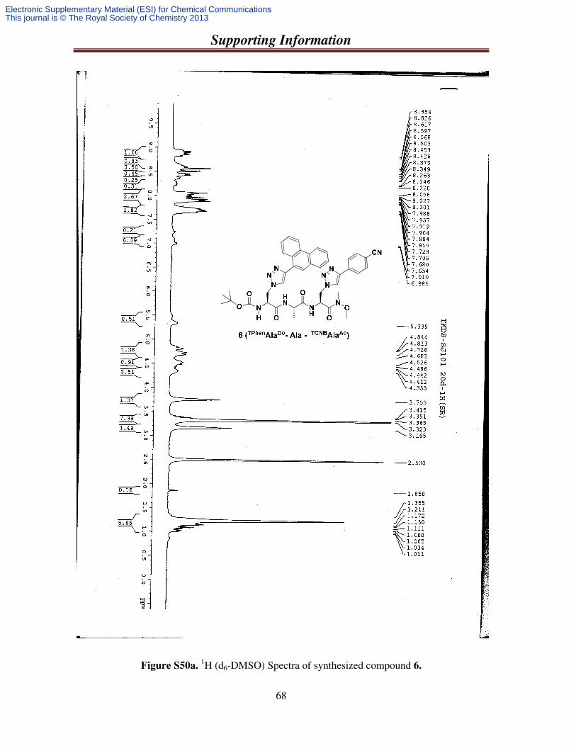
Supporting Information
68
Figure S50a. 1H (d6-DMSO) Spectra of synthesized compound 6.
Electronic Supplementary Material (ESI) for Chemical CommunicationsThis journal is © The Royal Society of Chemistry 2013
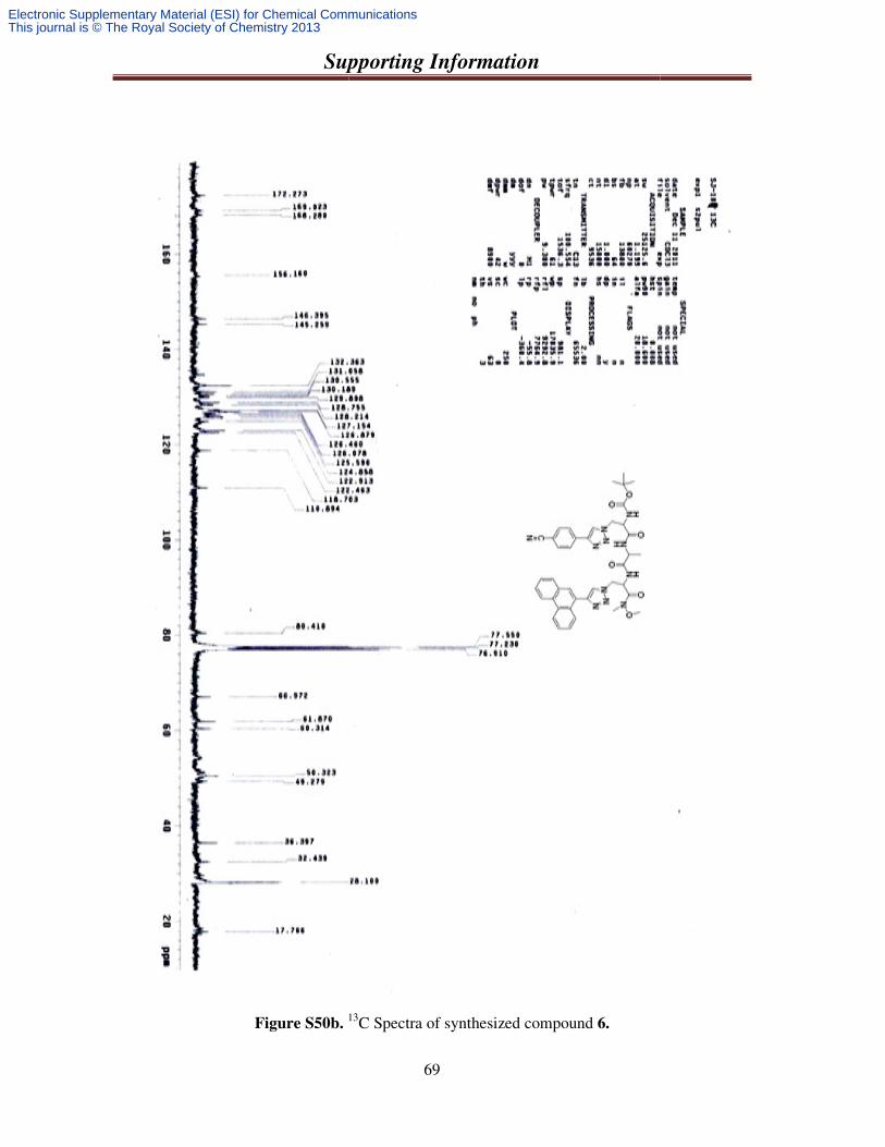
Supporting Information
Figure S50b. 13
Supporting Information
69
13C Spectra of synthesized compound 6.
Electronic Supplementary Material (ESI) for Chemical CommunicationsThis journal is © The Royal Society of Chemistry 2013
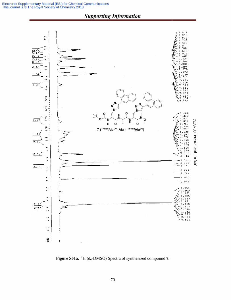
Supporting Information
Figure S51a. 1H (d6
Supporting Information
70
6-DMSO) Spectra of synthesized compound 7.
N
HN
H
O
O
O
O
NH
N
O
O
NN
NN
NN
7 (TPhenAlaDo- Ala - TPhenAlaDo)
Electronic Supplementary Material (ESI) for Chemical CommunicationsThis journal is © The Royal Society of Chemistry 2013
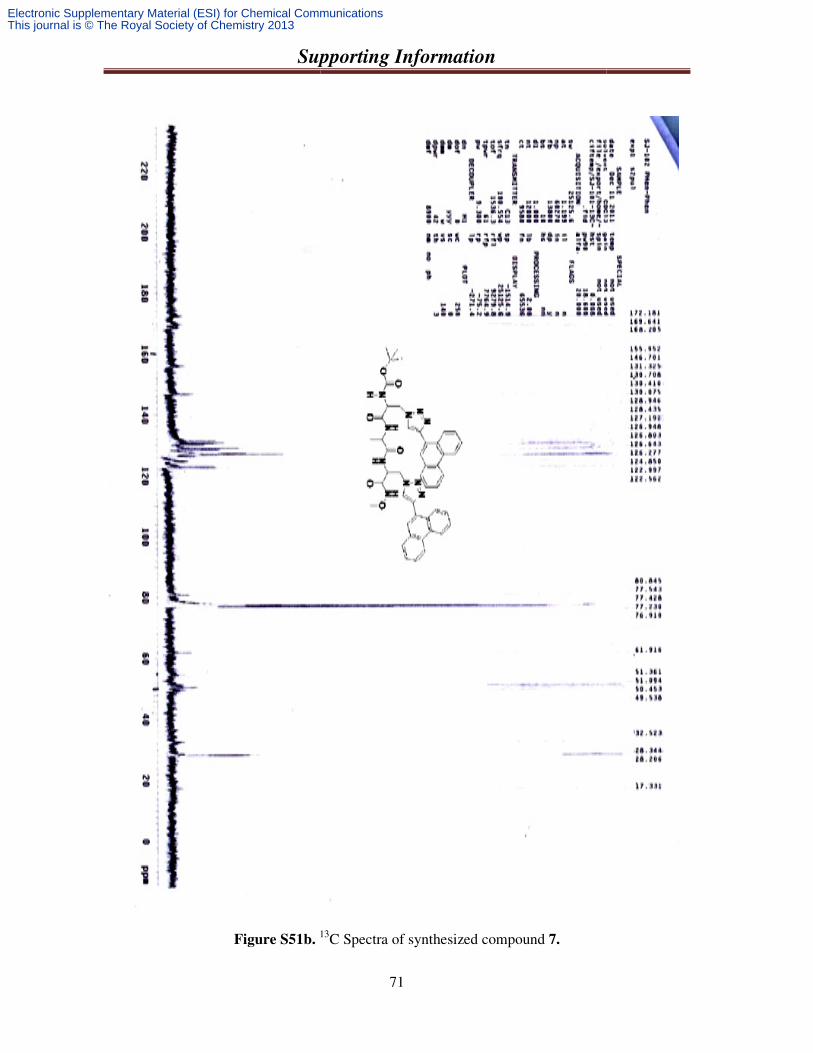
Supporting Information
Figure S51b. 13
Supporting Information
71
13C Spectra of synthesized compound 7.
Electronic Supplementary Material (ESI) for Chemical CommunicationsThis journal is © The Royal Society of Chemistry 2013
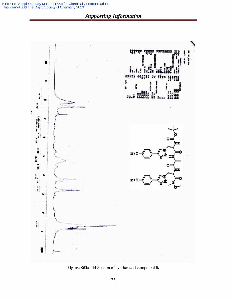
Supporting Information
Figure S52a.
Supporting Information
72
1H Spectra of synthesized compound 8.
Electronic Supplementary Material (ESI) for Chemical CommunicationsThis journal is © The Royal Society of Chemistry 2013
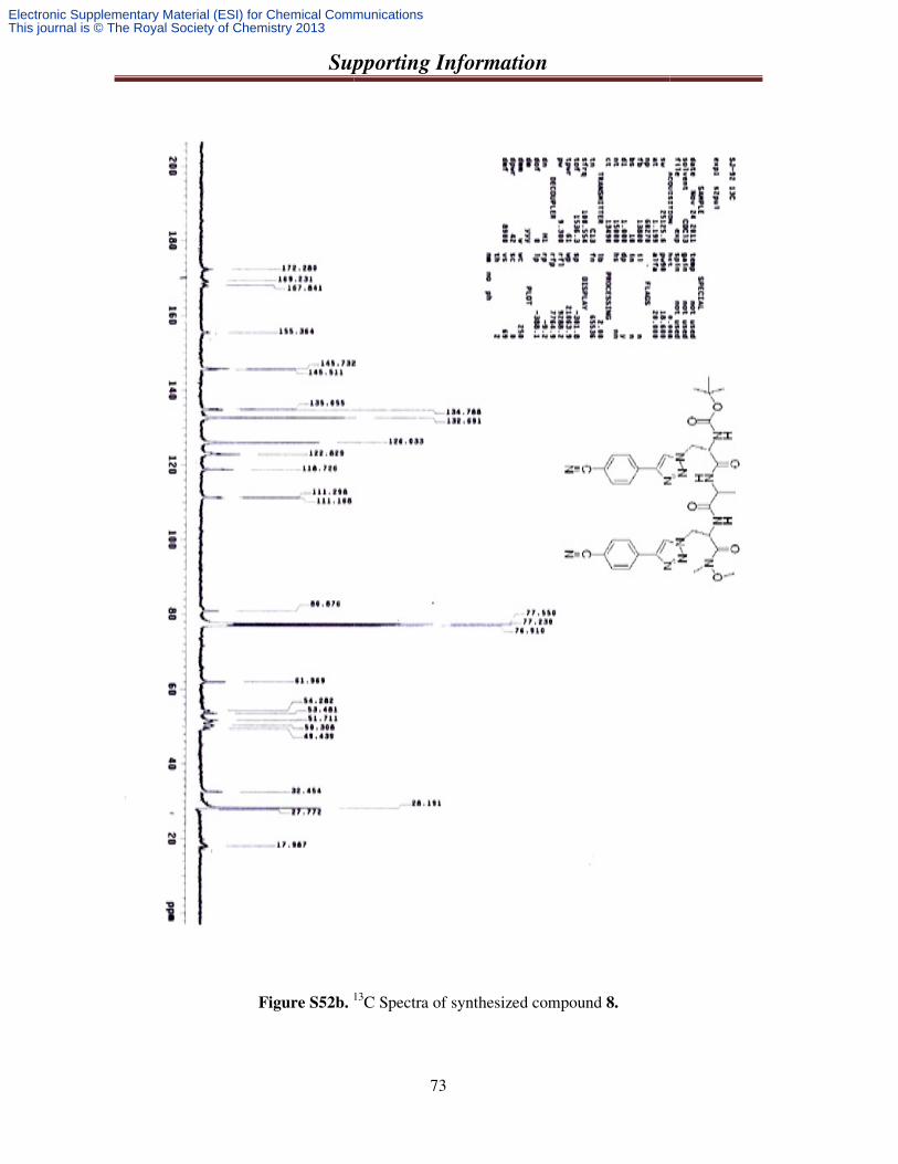
Supporting Information
Figure S52b. 13
Supporting Information
73
13C Spectra of synthesized compound 8.
Electronic Supplementary Material (ESI) for Chemical CommunicationsThis journal is © The Royal Society of Chemistry 2013
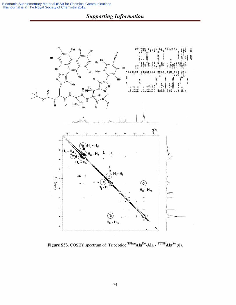
Supporting Information
74
Hc - Hd
Ha - Hb
Hc - Hd
Ha - Hb
Hj - Hl
Hj - HlHk - Hm
Hk - Hm
N
HN
H
O
O
O H2C
O
NH
N
O
O
N
N
N
NN
N
C
N
Hf
He
HfHg
He
Hd
Hc
Ha
Hb
Hb
Ha
Hi
HhHj
Hj
Hg
Hd
Hl
Hk
Hm
Figure S53. COSEY spectrum of Tripeptide TPhenAla
Do-Ala - TCNBAla
Ac (6).
Electronic Supplementary Material (ESI) for Chemical CommunicationsThis journal is © The Royal Society of Chemistry 2013
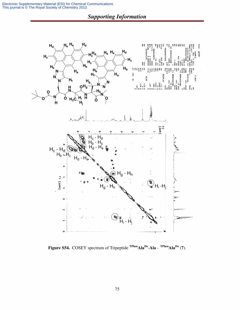
Supporting Information
75
N
HN
H
O
O O H2C
O
NH
N
OO
N
N
N
NN
N
Hd
Hc
HdHe
Hc
Hb
HaHf
Hf Hg
Hg
He
Hb
Hh
Hi
Hj
HeHd
Hc
HbHa
Hb
Hc
HdHe
Hc - Hd
Hc - Hd
Hb - Hc
Hb - Hc
Hd - He
Hd - He
Hg - Hh
Hg - Hh Hi -Hj
Hi - Hj
Figure S54. COSEY spectrum of Tripeptide TPhenAla
Do-Ala - TPhenAla
Do (7).
Electronic Supplementary Material (ESI) for Chemical CommunicationsThis journal is © The Royal Society of Chemistry 2013