Ultrafine Highly Magnetic Fluorescent γ-Fe2O3/NCD ... · Ultrafine Highly Magnetic Fluorescent...
7
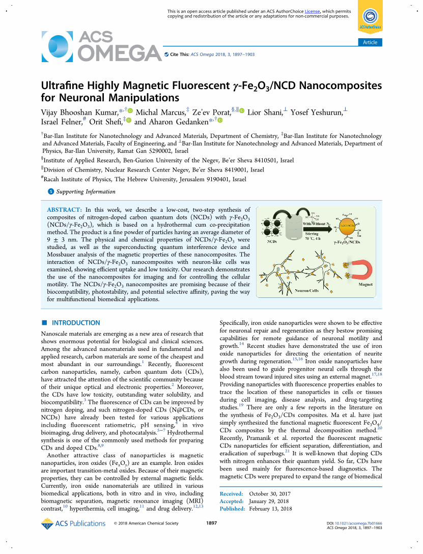
Ultrafine Highly Magnetic Fluorescent γ‑Fe 2 O 3 /NCD Nanocomposites for Neuronal Manipulations Vijay Bhooshan Kumar,* ,† Michal Marcus, ‡ Ze’ev Porat, §,∥ Lior Shani, ⊥ Yosef Yeshurun, ⊥ Israel Felner, # Orit Shefi, ‡ and Aharon Gedanken* ,† † Bar-Ilan Institute for Nanotechnology and Advanced Materials, Department of Chemistry, ‡ Bar-Ilan Institute for Nanotechnology and Advanced Materials, Faculty of Engineering, and ⊥ Bar-Ilan Institute for Nanotechnology and Advanced Materials, Department of Physics, Bar-Ilan University, Ramat Gan 5290002, Israel § Institute of Applied Research, Ben-Gurion University of the Negev, Be’er Sheva 8410501, Israel ∥ Division of Chemistry, Nuclear Research Center Negev, Be’er Sheva 8419001, Israel # Racah Institute of Physics, The Hebrew University, Jerusalem 9190401, Israel * S Supporting Information ABSTRACT: In this work, we describe a low-cost, two-step synthesis of composites of nitrogen-doped carbon quantum dots (NCDs) with γ-Fe 2 O 3 (NCDs/γ-Fe 2 O 3 ), which is based on a hydrothermal cum co-precipitation method. The product is a fine powder of particles having an average diameter of 9 ± 3 nm. The physical and chemical properties of NCDs/γ-Fe 2 O 3 were studied, as well as the superconducting quantum interference device and Mossbauer analysis of the magnetic properties of these nanocomposites. The interaction of NCDs/γ-Fe 2 O 3 nanocomposites with neuron-like cells was examined, showing efficient uptake and low toxicity. Our research demonstrates the use of the nanocomposites for imaging and for controlling the cellular motility. The NCDs/γ-Fe 2 O 3 nanocomposites are promising because of their biocompatibility, photostability, and potential selective affinity, paving the way for multifunctional biomedical applications. ■ INTRODUCTION Nanoscale materials are emerging as a new area of research that shows enormous potential for biological and clinical sciences. Among the advanced nanomaterials used in fundamental and applied research, carbon materials are some of the cheapest and most abundant in our surroundings. 1 Recently, fluorescent carbon nanoparticles, namely, carbon quantum dots (CDs), have attracted the attention of the scientific community because of their unique optical and electronic properties. 2 Moreover, the CDs have low toxicity, outstanding water solubility, and biocompatibility. 3 The fluorescence of CDs can be improved by nitrogen doping, and such nitrogen-doped CDs (N@CDs, or NCDs) have already been tested for various applications including fluorescent ratiometric, pH sensing, 4 in vivo bioimaging, drug delivery, and photocatalysis. 5−7 Hydrothermal synthesis is one of the commonly used methods for preparing CDs and doped CDs. 8,9 Another attractive class of nanoparticles is magnetic nanoparticles, iron oxides (Fe x O y ) are an example. Iron oxides are important transition-metal oxides. Because of their magnetic properties, they can be controlled by external magnetic fields. Currently, iron oxide nanomaterials are utilized in various biomedical applications, both in vitro and in vivo, including biomagnetic separation, magnetic resonance imaging (MRI) contrast, 10 hyperthermia, cell imaging, 11 and drug delivery. 12,13 Specifically, iron oxide nanoparticles were shown to be effective for neuronal repair and regeneration as they bestow promising capabilities for remote guidance of neuronal motility and growth. 14 Recent studies have demonstrated the use of iron oxide nanoparticles for directing the orientation of neurite growth during regeneration. 15,16 Iron oxide nanoparticles have also been used to guide progenitor neural cells through the blood stream toward injured sites using an external magnet. 17,18 Providing nanoparticles with fluorescence properties enables to trace the location of these nanoparticles in cells or tissues during cell imaging, disease analysis, and drug-targeting studies. 19 There are only a few reports in the literature on the synthesis of Fe 2 O 3 /CDs composites. Ma et al. have just simply synthesized the functional magnetic fluorescent Fe 3 O 4 / CDs composites by the thermal decomposition method. 20 Recently, Pramanik et al. reported the fluorescent magnetic CDs nanoparticles for efficient separation, differentiation, and eradication of superbugs. 21 It is well-known that doping CDs with nitrogen enhances their quantum yield. So far, CDs have been used mainly for fluorescence-based diagnostics. The magnetic CDs were prepared to expand the range of biomedical Received: October 30, 2017 Accepted: January 29, 2018 Published: February 13, 2018 Article Cite This: ACS Omega 2018, 3, 1897-1903 © 2018 American Chemical Society 1897 DOI: 10.1021/acsomega.7b01666 ACS Omega 2018, 3, 1897−1903 This is an open access article published under an ACS AuthorChoice License, which permits copying and redistribution of the article or any adaptations for non-commercial purposes.
Transcript of Ultrafine Highly Magnetic Fluorescent γ-Fe2O3/NCD ... · Ultrafine Highly Magnetic Fluorescent...
Ultrafine Highly Magnetic Fluorescent γ‑Fe2O3/NCD Nanocompositesfor Neuronal ManipulationsVijay Bhooshan Kumar,*,† Michal Marcus,‡ Ze’ev Porat,§,∥ Lior Shani,⊥ Yosef Yeshurun,⊥
Israel Felner,# Orit Shefi,‡ and Aharon Gedanken*,†
†Bar-Ilan Institute for Nanotechnology and Advanced Materials, Department of Chemistry, ‡Bar-Ilan Institute for Nanotechnologyand Advanced Materials, Faculty of Engineering, and ⊥Bar-Ilan Institute for Nanotechnology and Advanced Materials, Department ofPhysics, Bar-Ilan University, Ramat Gan 5290002, Israel§Institute of Applied Research, Ben-Gurion University of the Negev, Be’er Sheva 8410501, Israel∥Division of Chemistry, Nuclear Research Center Negev, Be’er Sheva 8419001, Israel#Racah Institute of Physics, The Hebrew University, Jerusalem 9190401, Israel
*S Supporting Information
ABSTRACT: In this work, we describe a low-cost, two-step synthesis ofcomposites of nitrogen-doped carbon quantum dots (NCDs) with γ-Fe2O3(NCDs/γ-Fe2O3), which is based on a hydrothermal cum co-precipitationmethod. The product is a fine powder of particles having an average diameter of9 ± 3 nm. The physical and chemical properties of NCDs/γ-Fe2O3 werestudied, as well as the superconducting quantum interference device andMossbauer analysis of the magnetic properties of these nanocomposites. Theinteraction of NCDs/γ-Fe2O3 nanocomposites with neuron-like cells wasexamined, showing efficient uptake and low toxicity. Our research demonstratesthe use of the nanocomposites for imaging and for controlling the cellularmotility. The NCDs/γ-Fe2O3 nanocomposites are promising because of theirbiocompatibility, photostability, and potential selective affinity, paving the wayfor multifunctional biomedical applications.
■ INTRODUCTION
Nanoscale materials are emerging as a new area of research thatshows enormous potential for biological and clinical sciences.Among the advanced nanomaterials used in fundamental andapplied research, carbon materials are some of the cheapest andmost abundant in our surroundings.1 Recently, fluorescentcarbon nanoparticles, namely, carbon quantum dots (CDs),have attracted the attention of the scientific community becauseof their unique optical and electronic properties.2 Moreover,the CDs have low toxicity, outstanding water solubility, andbiocompatibility.3 The fluorescence of CDs can be improved bynitrogen doping, and such nitrogen-doped CDs (N@CDs, orNCDs) have already been tested for various applicationsincluding fluorescent ratiometric, pH sensing,4 in vivobioimaging, drug delivery, and photocatalysis.5−7 Hydrothermalsynthesis is one of the commonly used methods for preparingCDs and doped CDs.8,9
Another attractive class of nanoparticles is magneticnanoparticles, iron oxides (FexOy) are an example. Iron oxidesare important transition-metal oxides. Because of their magneticproperties, they can be controlled by external magnetic fields.Currently, iron oxide nanomaterials are utilized in variousbiomedical applications, both in vitro and in vivo, includingbiomagnetic separation, magnetic resonance imaging (MRI)contrast,10 hyperthermia, cell imaging,11 and drug delivery.12,13
Specifically, iron oxide nanoparticles were shown to be effectivefor neuronal repair and regeneration as they bestow promisingcapabilities for remote guidance of neuronal motility andgrowth.14 Recent studies have demonstrated the use of ironoxide nanoparticles for directing the orientation of neuritegrowth during regeneration.15,16 Iron oxide nanoparticles havealso been used to guide progenitor neural cells through theblood stream toward injured sites using an external magnet.17,18
Providing nanoparticles with fluorescence properties enables totrace the location of these nanoparticles in cells or tissuesduring cell imaging, disease analysis, and drug-targetingstudies.19 There are only a few reports in the literature onthe synthesis of Fe2O3/CDs composites. Ma et al. have justsimply synthesized the functional magnetic fluorescent Fe3O4/CDs composites by the thermal decomposition method.20
Recently, Pramanik et al. reported the fluorescent magneticCDs nanoparticles for efficient separation, differentiation, anderadication of superbugs.21 It is well-known that doping CDswith nitrogen enhances their quantum yield. So far, CDs havebeen used mainly for fluorescence-based diagnostics. Themagnetic CDs were prepared to expand the range of biomedical
Received: October 30, 2017Accepted: January 29, 2018Published: February 13, 2018
Article
Cite This: ACS Omega 2018, 3, 1897−1903
© 2018 American Chemical Society 1897 DOI: 10.1021/acsomega.7b01666ACS Omega 2018, 3, 1897−1903
This is an open access article published under an ACS AuthorChoice License, which permitscopying and redistribution of the article or any adaptations for non-commercial purposes.
applications for which CDs can be used beyond bioimaging, forexample, being used in MRI.In this work, we report on a two-step synthesis of
multifunctional γ-Fe2O3/NCD hybrid nanocomposites. TheNCDs were synthesized from an aqueous solution of bovineserum albumin (BSA) by a hydrothermal reaction followed bythe second step of co-precipitation with Fe(CO)5. We proposea mechanism for the formation of these hybrid nanocompositesand investigate the long-lasting stability of their fluorescenceand magnetization. These properties were found to be stablefor more than four months. This remarkable stability isreflected in the fluorescence as well as in the absence ofprecipitation. We demonstrate the use of the nanocompositesfor labeling neuron-like cells and for controlling their motilityby applying an external magnetic field. These new NCDs/γ-Fe2O3 composites exhibit low toxicity and good biocompati-bility and may therefore be beneficial as theranostic agents invarious biomedical applications.
■ RESULTS AND DISCUSSION
The fine synthesized precipitates had a brownish red color andwere analyzed by various physicochemical techniques andsubsequently exploited for long-lasting stability examination aswell as for an assessment of their biocompatibility. The γ-Fe2O3/NCDs hybrid nanocomposites were used as a probe forfluorescence imaging of neuron-like cells (PC12 and SHSY-5Y), and their potential for assisting neuronal differentiationwas examined.Physical and Chemical Characterization of γ-Fe2O3/
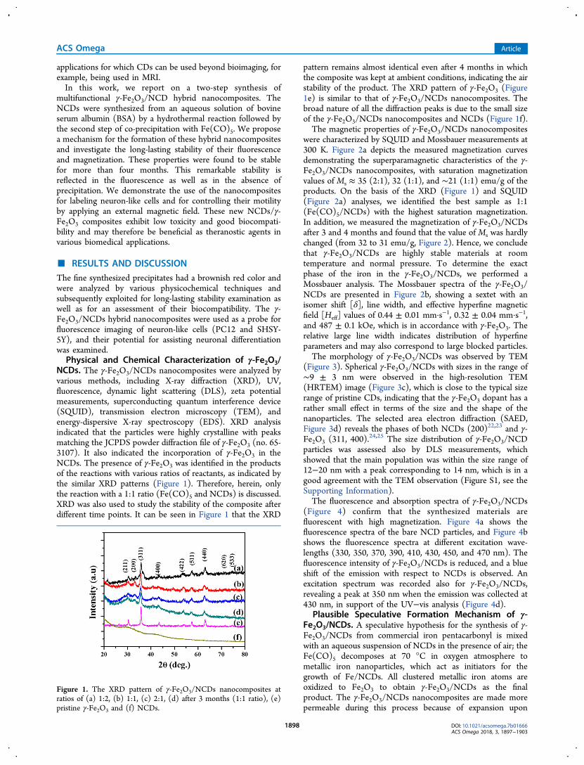
NCDs. The γ-Fe2O3/NCDs nanocomposites were analyzed byvarious methods, including X-ray diffraction (XRD), UV,fluorescence, dynamic light scattering (DLS), zeta potentialmeasurements, superconducting quantum interference device(SQUID), transmission electron microscopy (TEM), andenergy-dispersive X-ray spectroscopy (EDS). XRD analysisindicated that the particles were highly crystalline with peaksmatching the JCPDS powder diffraction file of γ-Fe2O3 (no. 65-3107). It also indicated the incorporation of γ-Fe2O3 in theNCDs. The presence of γ-Fe2O3 was identified in the productsof the reactions with various ratios of reactants, as indicated bythe similar XRD patterns (Figure 1). Therefore, herein, onlythe reaction with a 1:1 ratio (Fe(CO)5 and NCDs) is discussed.XRD was also used to study the stability of the composite afterdifferent time points. It can be seen in Figure 1 that the XRD
pattern remains almost identical even after 4 months in whichthe composite was kept at ambient conditions, indicating the airstability of the product. The XRD pattern of γ-Fe2O3 (Figure1e) is similar to that of γ-Fe2O3/NCDs nanocomposites. Thebroad nature of all the diffraction peaks is due to the small sizeof the γ-Fe2O3/NCDs nanocomposites and NCDs (Figure 1f).The magnetic properties of γ-Fe2O3/NCDs nanocomposites
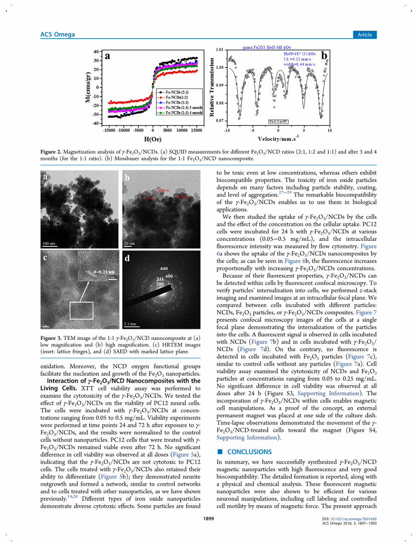
were characterized by SQUID and Mossbauer measurements at300 K. Figure 2a depicts the measured magnetization curvesdemonstrating the superparamagnetic characteristics of the γ-Fe2O3/NCDs nanocomposites, with saturation magnetizationvalues of Ms ≈ 35 (2:1), 32 (1:1), and ∼21 (1:1) emu/g of theproducts. On the basis of the XRD (Figure 1) and SQUID(Figure 2a) analyses, we identified the best sample as 1:1(Fe(CO)5/NCDs) with the highest saturation magnetization.In addition, we measured the magnetization of γ-Fe2O3/NCDsafter 3 and 4 months and found that the value of Ms was hardlychanged (from 32 to 31 emu/g, Figure 2). Hence, we concludethat γ-Fe2O3/NCDs are highly stable materials at roomtemperature and normal pressure. To determine the exactphase of the iron in the γ-Fe2O3/NCDs, we performed aMossbauer analysis. The Mossbauer spectra of the γ-Fe2O3/NCDs are presented in Figure 2b, showing a sextet with anisomer shift [δ], line width, and effective hyperfine magneticfield [Heff] values of 0.44 ± 0.01 mm·s−1, 0.32 ± 0.04 mm·s−1,and 487 ± 0.1 kOe, which is in accordance with γ-Fe2O3. Therelative large line width indicates distribution of hyperfineparameters and may also correspond to large blocked particles.The morphology of γ-Fe2O3/NCDs was observed by TEM
(Figure 3). Spherical γ-Fe2O3/NCDs with sizes in the range of∼9 ± 3 nm were observed in the high-resolution TEM(HRTEM) image (Figure 3c), which is close to the typical sizerange of pristine CDs, indicating that the γ-Fe2O3 dopant has arather small effect in terms of the size and the shape of thenanoparticles. The selected area electron diffraction (SAED,Figure 3d) reveals the phases of both NCDs (200)22,23 and γ-Fe2O3 (311, 400).
24,25 The size distribution of γ-Fe2O3/NCDparticles was assessed also by DLS measurements, whichshowed that the main population was within the size range of12−20 nm with a peak corresponding to 14 nm, which is in agood agreement with the TEM observation (Figure S1, see theSupporting Information).The fluorescence and absorption spectra of γ-Fe2O3/NCDs
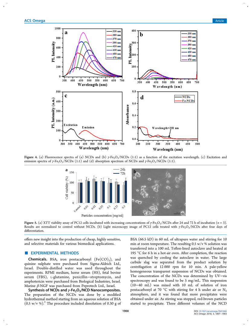
(Figure 4) confirm that the synthesized materials arefluorescent with high magnetization. Figure 4a shows thefluorescence spectra of the bare NCD particles, and Figure 4bshows the fluorescence spectra at different excitation wave-lengths (330, 350, 370, 390, 410, 430, 450, and 470 nm). Thefluorescence intensity of γ-Fe2O3/NCDs is reduced, and a blueshift of the emission with respect to NCDs is observed. Anexcitation spectrum was recorded also for γ-Fe2O3/NCDs,revealing a peak at 350 nm when the emission was collected at430 nm, in support of the UV−vis analysis (Figure 4d).
Plausible Speculative Formation Mechanism of γ-Fe2O3/NCDs. A speculative hypothesis for the synthesis of γ-Fe2O3/NCDs from commercial iron pentacarbonyl is mixedwith an aqueous suspension of NCDs in the presence of air; theFe(CO)5 decomposes at 70 °C in oxygen atmosphere tometallic iron nanoparticles, which act as initiators for thegrowth of Fe/NCDs. All clustered metallic iron atoms areoxidized to Fe2O3 to obtain γ-Fe2O3/NCDs as the finalproduct. The γ-Fe2O3/NCDs nanocomposites are made morepermeable during this process because of expansion upon
Figure 1. The XRD pattern of γ-Fe2O3/NCDs nanocomposites atratios of (a) 1:2, (b) 1:1, (c) 2:1, (d) after 3 months (1:1 ratio), (e)pristine γ-Fe2O3 and (f) NCDs.
ACS Omega Article
DOI: 10.1021/acsomega.7b01666ACS Omega 2018, 3, 1897−1903
1898
oxidation. Moreover, the NCD oxygen functional groupsfacilitate the nucleation and growth of the Fe2O3 nanoparticles.Interaction of γ-Fe2O3/NCD Nanocomposites with the
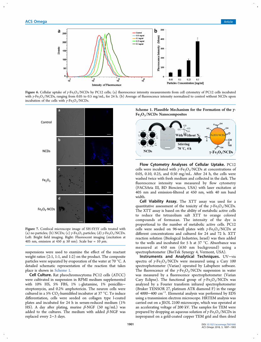
Living Cells. XTT cell viability assay was performed toexamine the cytotoxicity of the γ-Fe2O3/NCDs. We tested theeffect of γ-Fe2O3/NCDs on the viability of PC12 neural cells.The cells were incubated with γ-Fe2O3/NCDs at concen-trations ranging from 0.05 to 0.5 mg/mL. Viability experimentswere performed at time points 24 and 72 h after exposure to γ-Fe2O3/NCDs, and the results were normalized to the controlcells without nanoparticles. PC12 cells that were treated with γ-Fe2O3/NCDs remained viable even after 72 h. No significantdifference in cell viability was observed at all doses (Figure 5a),indicating that the γ-Fe2O3/NCDs are not cytotoxic to PC12cells. The cells treated with γ-Fe2O3/NCDs also retained theirability to differentiate (Figure 5b); they demonstrated neuriteoutgrowth and formed a network, similar to control networksand to cells treated with other nanoparticles, as we have shownpreviously.14,26 Different types of iron oxide nanoparticlesdemonstrate diverse cytotoxic effects. Some particles are found
to be toxic even at low concentrations, whereas others exhibitbiocompatible properties. The toxicity of iron oxide particlesdepends on many factors including particle stability, coating,and level of aggregation.27−29 The remarkable biocompatibilityof the γ-Fe2O3/NCDs enables us to use them in biologicalapplications.We then studied the uptake of γ-Fe2O3/NCDs by the cells
and the effect of the concentration on the cellular uptake. PC12cells were incubated for 24 h with γ-Fe2O3/NCDs at variousconcentrations (0.05−0.5 mg/mL), and the intracellularfluorescence intensity was measured by flow cytometry. Figure6a shows the uptake of the γ-Fe2O3/NCDs nanocomposites bythe cells; as can be seen in Figure 6b, the fluorescence increasesproportionally with increasing γ-Fe2O3/NCDs concentrations.Because of their fluorescent properties, γ-Fe2O3/NCDs can
be detected within cells by fluorescent confocal microscopy. Toverify particles’ internalization into cells, we performed z-stackimaging and examined images at an intracellular focal plane. Wecompared between cells incubated with different particles:NCDs, Fe2O3 particles, or γ-Fe2O3/NCDs composites. Figure 7presents confocal microscopy images of the cells at a singlefocal plane demonstrating the internalization of the particlesinto the cells. A fluorescent signal is observed in cells incubatedwith NCDs (Figure 7b) and in cells incubated with γ-Fe2O3/NCDs (Figure 7d). On the contrary, no fluorescence isdetected in cells incubated with Fe2O3 particles (Figure 7c),similar to control cells without any particles (Figure 7a). Cellviability assay examined the cytotoxicity of NCDs and Fe2O3particles at concentrations ranging from 0.05 to 0.25 mg/mL.No significant difference in cell viability was observed at alldoses after 24 h (Figure S3, Supporting Information). Theincorporation of γ-Fe2O3/NCDs within cells enables magneticcell manipulations. As a proof of the concept, an externalpermanent magnet was placed at one side of the culture dish.Time-lapse observations demonstrated the movement of the γ-Fe2O3/NCD-treated cells toward the magnet (Figure S4,Supporting Information).
■ CONCLUSIONSIn summary, we have successfully synthesized γ-Fe2O3/NCDmagnetic nanoparticles with high fluorescence and very goodbiocompatibility. The detailed formation is reported, along witha physical and chemical analysis. These fluorescent magneticnanoparticles were also shown to be efficient for variousneuronal manipulations, including cell labeling and controlledcell motility by means of magnetic force. The present approach
Figure 2. Magnetization analysis of γ-Fe2O3/NCDs. (a) SQUID measurements for different Fe2O3/NCD ratios (2:1, 1:2 and 1:1) and after 3 and 4months (for the 1:1 ratio). (b) Mossbauer analysis for the 1:1 Fe2O3/NCD nanocomposite.
Figure 3. TEM image of the 1:1 γ-Fe2O3/NCD nanocomposite at (a)low magnification and (b) high magnification. (c) HRTEM images(inset: lattice fringes), and (d) SAED with marked lattice plane.
ACS Omega Article
DOI: 10.1021/acsomega.7b01666ACS Omega 2018, 3, 1897−1903
1899
offers new insight into the production of cheap, highly sensitive,and selective materials for various biomedical applications.
■ EXPERIMENTAL METHODS
Chemicals. BSA, iron pentacarbonyl (Fe(CO)5), andquinine sulphate were purchased from Sigma-Aldrich Ltd.,Israel. Double-distilled water was used throughout theexperiments. RPMI medium, horse serum (HS), fetal bovineserum (FBS), L-glutamine, penicillin−streptomycin, andamphotericin were purchased from Biological Industries, Israel.Murine β-NGF was purchased from Peprotech Ltd., Israel.Synthesis of NCDs and γ-Fe2O3/NCD Nanocomposites.
The preparation of the NCDs was done by a modifiedhydrothermal method starting from an aqueous solution of BSA(0.5 w/v %).8 The procedure included dissolution of 0.30 g of
BSA (66.5 kD) in 60 mL of ultrapure water and stirring for 10min at room temperature. The resulting 0.5 w/v % solution wastransferred into a 100 mL Teflon-lined autoclave and heated at195 °C for 6 h in a hot-air oven. After completion, the reactionwas quenched by cooling the autoclave in water. The largecarbide slag was separated from the product solution bycentrifugation at 12 000 rpm for 10 min. A pale-yellowhomogeneous transparent suspension of NCDs was obtained.The concentration of the NCDs was determined by UV−visspectroscopy and was found to be 5 mg/mL. This suspension(10−40 mL) was mixed with 10 mL of solution of ironpentacarbonyl at 70 °C with stirring for 4 h under air or N2
atmosphere, and it was found that more precipitates wereobtained under air. As stirring was stopped, red-brown particlesstarted to precipitate. Three different volumes of the NCD
Figure 4. (a) Fluorescence spectra of (a) NCDs and (b) γ-Fe2O3/NCDs (1:1) as a function of the excitation wavelength. (c) Excitation andemission spectra of γ-Fe2O3/NCDs (1:1) and (d) absorption spectrum of NCDs and γ-Fe2O3/NCDs (1:1).
Figure 5. (a) XTT viability assay of PC12 cells incubated with increasing concentrations of γ-Fe2O3/NCDs after 24 and 72 h of incubation (n = 3).Results are normalized to control without NCDs. (b) Light microscopy image of PC12 cells treated with γ-Fe2O3/NCDs after four days ofdifferentiation.
ACS Omega Article
DOI: 10.1021/acsomega.7b01666ACS Omega 2018, 3, 1897−1903
1900
suspensions were used to examine the effect of the reactantweight ratios (2:1, 1:1, and 1:2) on the product. The compositeparticles were separated by evaporation of the water at 70 °C. Adetailed schematic representation of the reaction that takesplace is shown in Scheme 1.Cell Culture. Rat pheochromocytoma PC12 cells (ATCC)
were cultivated in suspension in RPMI medium supplementedwith 10% HS, 5% FBS, 1% L-glutamine, 1% penicillin−streptomycin, and 0.2% amphotericin. The neuron cells werecultured in a 5% CO2-humidified incubator at 37 °C. To inducedifferentiation, cells were seeded on collagen type l-coatedplates and incubated for 24 h in serum-reduced medium (1%HS). A day after plating, murine β-NGF (50 ng/mL) wasadded to the cultures. The medium with added β-NGF wasreplaced every 2−3 days.
Flow Cytometry Analyses of Cellular Uptake. PC12cells were incubated with γ-Fe2O3/NCDs at concentrations of0.05, 0.10, 0.25, and 0.50 mg/mL. After 24 h, the cells werewashed twice with fresh medium and collected in the dark. Thefluorescence intensity was measured by flow cytometry(FACSAria III, BD Bioscience, USA) with laser excitation at405 nm and emission-filtered at 450 nm, with 40 nm bandwidth.
Cell Viability Assay. The XTT assay was used for aquantitative assessment of the toxicity of the γ-Fe2O3/NCDs.The XTT assay is based on the ability of metabolic active cellsto reduce the tetrazolium salt XTT to orange coloredcompounds of formazan. The intensity of the dye isproportional to the number of metabolic active cells. PC12cells were seeded on 96-well plates with γ-Fe2O3/NCDs atdifferent concentrations and cultured for 24 and 72 h. XTTreaction solution (Biological Industries, Israel) was then addedto the wells and incubated for 5 h at 37 °C. Absorbance wasmeasured at 450 nm (630 nm background) using aspectrophotometer (BioTek Synergy 4, Vermont, USA).
Instruments and Analytical Techniques. UV−visspectra of γ-Fe2O3/NCDs were measured using a Cary 100spectrophotometer (Varian) operated by Labsphere software.The fluorescence of the γ-Fe2O3/NCDs suspension in waterwas measured by a fluorescence spectrophotometer (VarianCary Eclipse). The functional group of γ-Fe2O3/NCDs wasanalyzed by a Fourier transform infrared spectrophotometer(Bruker TENSOR 27, platinum ATR diamond F) in the rangeof 4000−400 cm−1. Elemental analysis was performed by EDSusing a transmission electron microscope. HRTEM analysis wascarried out on a JEOL 2100 microscope, which was operated atan accelerating voltage of 200 kV. The samples for TEM wereprepared by dropping an aqueous solution of γ-Fe2O3/NCDs inisopropanol on a gold-coated copper TEM grid and then dried
Figure 6. Cellular uptake of γ-Fe2O3/NCDs by PC12 cells; (a) fluorescence intensity measurements from cell cytometry of PC12 cells incubatedwith γ-Fe2O3/NCDs, ranging from 0.05 to 0.5 mg/mL, for 24 h. (b) Average of fluorescence intensity normalized to control without NCDs uponincubation of the cells with γ-Fe2O3/NCDs.
Figure 7. Confocal microscope image of SH-SY5Y cells treated with(a) no particles; (b) NCDs; (c) γ-Fe2O3 particles; (d) γ-Fe2O3/NCDs.Left: Bright field imaging. Right: Fluorescent imaging (excitation at405 nm, emission at 450 ± 50 nm). Scale bar = 10 μm.
Scheme 1. Plausible Mechanism for the Formation of the γ-Fe2O3/NCDs Nanocomposites
ACS Omega Article
DOI: 10.1021/acsomega.7b01666ACS Omega 2018, 3, 1897−1903
1901
under vacuum at room temperature overnight. The C, H, N, S,and O analyses were performed by CHNSO/O chromatogramspectroscopy. Zeta potential measurements of the particles, aswell as their size distribution studies were performed on aZetaSizer Nano-ZS (Malvern Instruments Ltd., Worcestershire,UK). The DLS was measured on the same instrument in anaqueous solution. X-ray powder diffractograms were obtainedusing a Bruker D8 ADVANCE diffractometer. Bragg−Bretanogeometry was adopted using Cu Kα radiation (λ = 1.541 Å)and a setting of 40 kV and 40 mA. The phases were identifiedusing the JCPDS database. The magnetization measurementswere conducted by a Quantum Design MPMS5 SQUIDmagnetometer. The γ-Fe2O3 phase analysis was done using aMossbauer spectrometer.
■ ASSOCIATED CONTENT*S Supporting InformationThe Supporting Information is available free of charge on theACS Publications website at DOI: 10.1021/acsomega.7b01666.
γ-Fe2O3/NCD magnetic nanoparticle interactions withneuron cells (AVI)DLS plot of NCDs and γ-Fe2O3/NCD magneticnanoparticles, TEM image of NCDs, and cell viabilityassay of NCDs and Fe2O3 particles at concentrationsranging from 0.05 to 0.25 mg/mL (PDF)
■ AUTHOR INFORMATIONCorresponding Authors*E-mail: [email protected] (V.B.K.).*E-mail: [email protected], [email protected](A.G.).ORCIDVijay Bhooshan Kumar: 0000-0001-7899-1463Ze’ev Porat: 0000-0002-9862-449XOrit Shefi: 0000-0001-6680-7390Aharon Gedanken: 0000-0002-1243-2957NotesThe authors declare no competing financial interest.
■ ACKNOWLEDGMENTSThe authors are grateful to Ortal Lidor-Shalev for help with theTEM measurements and to Dr. Ilana Perelshtein for theHRTEM measurements carried out at the Department ofChemistry of Bar-Ilan University, Israel.
■ ABBREVIATIONSBSA, bovine serum albumin; DDW, double distilled water;EDS, energy dispersive X-ray spectroscopy; FBS, fetal bovineserum; FWHM, full-width at half-maximum; HR-TEM, high-resolution transmission-electron microscopy; NTP, roomtemperature and normal pressure; NCDs, nitrogen-dopedcarbon-dots; PBS, phosphate buffered saline; TP, two-photon;XRD, X-ray diffraction
■ REFERENCES(1) Jariwala, D.; Sangwan, V. K.; Lauhon, L. J.; Marks, T. J.; Hersam,M. C. Carbon Nanomaterials for Electronics, Optoelectronics,Photovoltaics, and Sensing. Chem. Soc. Rev. 2013, 42, 2824−2860.(2) Wang, R.; Lu, K.-Q.; Tang, Z.-R.; Xu, Y.-J. Recent Progress inCarbon Quantum Dots: Synthesis, Properties and Applications inPhotocatalysis. J. Mater. Chem. A 2017, 5, 3717−3734.
(3) Hong, G.; Diao, S.; Antaris, A. L.; Dai, H. Carbon Nanomaterialsfor Biological Imaging and Nanomedicinal Therapy. Chem. Rev. 2015,115, 10816−10906.(4) Shi, W.; Li, X.; Ma, H. A Tunable Ratiometric Ph Sensor Basedon Carbon Nanodots for the Quantitative Measurement of theIntracellular pH of Whole Cells. Angew. Chem., Int. Ed. 2012, 51,6432−6435.(5) Ye, Z.; Tang, R.; Wu, H.; Wang, B.; Tan, M.; Yuan, J. Preparationof Europium Complex-Conjugated Carbon Dots for RatiometricFluorescence Detection of Copper(II) Ions. New J. Chem. 2014, 38,5721−5726.(6) Niu, J.; Gao, H. Synthesis and Drug Detection Performance ofNitrogen-Doped Carbon Dots. J. Lumin. 2014, 149, 159−162.(7) Amjadi, M.; Manzoori, J. L. Strong Enhancement of theChemiluminescence of the Cerium(IV)-Thiosulfate Reaction byCarbon Dots, and Its Application to the Sensitive Determination ofDopamine. Microchim. Acta 2014, 181, 671−677.(8) Kumar, V. B.; Sheinberger, J.; Porat, Z.; Shav-Tal, Y.; Gedanken,A. A Hydrothermal Reaction of an Aqueous Solution of BSA YieldsHighly Fluorescent N Doped C-Dots Used for Imaging of LiveMammalian Cells. J. Mater. Chem. B 2016, 4, 2913−2920.(9) Huang, L.; Yang, S.; Chen, L.; Chen, S. Hydrothermal Synthesisof Fluorescent Carbon Dots towards Ion Response and Silk ScreenPatterns. Chem. Lett. 2015, 44, 1251−1253.(10) Shen, Z.; Wu, A.; Chen, X. Iron Oxide Nanoparticle BasedContrast Agents for Magnetic Resonance Imaging. Mol. Pharm. 2017,14, 1352−1364.(11) Wolfbeis, O. S. An Overview of Nanoparticles Commonly Usedin Fluorescent Bioimaging. Chem. Soc. Rev. 2015, 44, 4743−4768.(12) Rodzinski, A.; Guduru, R.; Liang, P.; Hadjikhani, A.; Stewart, T.;Stimphil, E.; Runowicz, C.; Cote, R.; Altman, N.; Datar, R.; et al.Targeted and Controlled Anticancer Drug Delivery and Release withMagnetoelectric Nanoparticles. Sci. Rep. 2016, 6, 20867.(13) Sutradhar, K. B.; Amin, M. L. Nanotechnology in Cancer DrugDelivery and Selective Targeting. ISRN Nanotechnol. 2014, 2014,939378.(14) Marcus, M.; Karni, M.; Baranes, K.; Levy, I.; Alon, N.; Margel,S.; Shefi, O. Iron Oxide Nanoparticles for Neuronal Cell Applications:Uptake Study and Magnetic Manipulations. J. Nanobiotechnol. 2016,14, 37.(15) Polak, P.; Shefi, O. Nanometric Agents in the Service ofNeuroscience: Manipulation of Neuronal Growth and Activity UsingNanoparticles. Nanomed. Nanotechnol. Biol. Med. 2015, 11, 1467−1479.(16) Riggio, C.; Calatayud, M. P.; Giannaccini, M.; Sanz, B.; Torres,T. E.; Fernandez-Pacheco, R.; Ripoli, A.; Ibarra, M. R.; Dente, L.;Cuschieri, A.; et al. The Orientation of the Neuronal Growth ProcessCan Be Directed via Magnetic Nanoparticles under an AppliedMagnetic Field. Nanomed. Nanotechnol. Biol. Med. 2014, 10, 1549−1558.(17) Tewarie, R. S. N.; Hurtado, A.; Bartels, R. H.; Grotenhuis, A.;Oudega, M. Stem Cell-Based Therapies for Spinal Cord Injury. J.Spinal Cord Med. 2009, 32, 105−114.(18) Clarke, J. L.; Daniell, H. Plastid biotechnology for cropproduction: present status and future perspectives. Plant Mol. Biol.2011, 76, 211−220.(19) NDong, C.; Tate, J. A.; Kett, W. C.; Batra, J.; Demidenko, E.;Lewis, L. D.; Hoopes, P. J.; Gerngross, T. U.; Griswold, K. E. TumorCell Targeting by Iron Oxide Nanoparticles Is Dominated by DifferentFactors in Vitro versus in Vivo. PLoS One 2015, 10, No. e0115636.(20) Ma, Y.-j.; Liu, C.-y.; Zhou, X.-h.; Liu, Y. Preparation andCharacterisation of Multifunctional Magnetic-Fluorescent Fe3O4/carbon Dots/silica Composites. Micro Nano Lett. 2013, 8, 302−304.(21) Pramanik, A.; Jones, S.; Pedraza, F.; Vangara, A.; Sweet, C.;Williams, M. S.; Ruppa-Kasani, V.; Risher, S. E.; Sardar, D.; Ray, P. C.Fluorescent, Magnetic Multifunctional Carbon Dots for SelectiveSeparation, Identification, and Eradication of Drug-Resistant Super-bugs. ACS Omega 2017, 2, 554−562.
ACS Omega Article
DOI: 10.1021/acsomega.7b01666ACS Omega 2018, 3, 1897−1903
1902
(22) Kumar, V. B.; Sahu, A. K.; Mohsin, A. S. M.; Li, X.; Gedanken,A. Refractive-Index Tuning of Highly Fluorescent Carbon Dots. ACSAppl. Mater. Interfaces 2017, 9, 28930−28938.(23) Linehan, K.; Doyle, H. Size Controlled Synthesis of CarbonQuantum Dots Using Hydride Reducing Agents. J. Mater. Chem. C2014, 2, 6025−6031.(24) Kazeminezhad, I.; Mosivand, S. Phase Transition of Electro-oxidized Fe3O4 to γ And α-Fe2O3 Nanoparticles Using SinteringTreatment. Acta Phys. Pol., A 2014, 125, 1210−1214.(25) Wu, W.; Xiao, X. H.; Zhang, S. F.; Peng, T. C.; Zhou, J.; Ren, F.;Jiang, C. Z. Synthesis and Magnetic Properties of Maghemite (γ-Fe2O3) Short-Nanotubes. Nanoscale Res. Lett. 2010, 5, 1474−1479.(26) Marcus, M.; Skaat, H.; Alon, N.; Margel, S.; Shefi, O. NGF-Conjugated Iron Oxide Nanoparticles Promote Differentiation andOutgrowth of PC12 Cells. Nanoscale 2015, 7, 1058−1066.(27) Pisanic, T. R.; Blackwell, J. D.; Shubayev, V. I.; Finones, R. R.;Jin, S. Nanotoxicity of Iron Oxide Nanoparticle Internalization inGrowing Neurons. Biomaterials 2007, 28, 2572−2581.(28) Mahmoudi, M.; Hofmann, H.; Rothen-Rutishauser, B.; Petri-Fink, A. Assessing the in Vitro and in Vivo Toxicity of Super-paramagnetic Iron Oxide Nanoparticles. Chem. Rev. 2012, 112, 2323−2338.(29) Connell, J. J.; Patrick, P. S.; Yu, Y.; Lythgoe, M. F.; Kalber, T. L.Advanced Cell Therapies: Targeting, Tracking and Actuation of Cellswith Magnetic Particles. Regener. Med. 2015, 10, 757−772.
ACS Omega Article
DOI: 10.1021/acsomega.7b01666ACS Omega 2018, 3, 1897−1903
1903