Spike Protein and the Human 7 Nicotinic Receptor A ...
25
Page 1/25 A Functional Interaction Between the SARS-CoV-2 Spike Protein and the Human α 7 Nicotinic Receptor Juan Facundo Chrestia Universidad Nacional del Sur-Consejo Nacional de Investigaciones Cientícas y Técnicas (CONICET) Ana Soa Oliveira University of Bristol School of Chemistry Adrian J. Mulholland University of Bristol School of Chemistry Timothy Gallagher University of Bristol School of Chemistry Isabel Bermúdez Oxford Brookes University Cecilia Bouzat ( [email protected] ) Universidad Nacional del Sur-Consejo Nacional de Investigaciones Cientícas y Técnicas (CONICET) https://orcid.org/0000-0003-0388-6129 Research Article Keywords: nicotinic receptor, neurotransmitter receptors, single-channel recordings, patch-clamp, SARS- CoV-2 spike protein. Posted Date: October 6th, 2021 DOI: https://doi.org/10.21203/rs.3.rs-938911/v1 License: This work is licensed under a Creative Commons Attribution 4.0 International License. Read Full License
Transcript of Spike Protein and the Human 7 Nicotinic Receptor A ...
Page 1/25
A Functional Interaction Between the SARS-CoV-2Spike Protein and the Human α7 Nicotinic ReceptorJuan Facundo Chrestia
Universidad Nacional del Sur-Consejo Nacional de Investigaciones Cientí�cas y Técnicas (CONICET)Ana So�a Oliveira
University of Bristol School of ChemistryAdrian J. Mulholland
University of Bristol School of ChemistryTimothy Gallagher
University of Bristol School of ChemistryIsabel Bermúdez
Oxford Brookes UniversityCecilia Bouzat ( [email protected] )
Universidad Nacional del Sur-Consejo Nacional de Investigaciones Cientí�cas y Técnicas (CONICET)https://orcid.org/0000-0003-0388-6129
Research Article
Keywords: nicotinic receptor, neurotransmitter receptors, single-channel recordings, patch-clamp, SARS-CoV-2 spike protein.
Posted Date: October 6th, 2021
DOI: https://doi.org/10.21203/rs.3.rs-938911/v1
License: This work is licensed under a Creative Commons Attribution 4.0 International License. Read Full License
Page 2/25
AbstractCoronavirus disease 2019 (COVID-19) is caused by the severe acute respiratory syndrome coronavirus 2(SARS-CoV-2). Infection relies on the binding of the viral spike protein (S) to angiotensin-convertingenzyme 2 in host cells. The S protein has been suggested to interact with nicotinic acetylcholine receptors(nAChRs), and a potential contribution of nAChRs to COVID-19 pathophysiology has been proposed. α7nAChR is an interesting candidate target since it is present in neuronal and non-neuronal cells, includingimmune cells, and has anti-in�ammatory actions. We here identi�ed a novel direct functional interactionbetween the α7 nAChR and the Y674-R685 S region. The S fragment exerts a dual effect, acting as a low-e�cacy agonist and a non-competitive inhibitor. It activates the α7 nAChR, in line with our previousmolecular dynamics simulations showing favorable binding of this accessible region of the S protein tothe nAChR agonist binding pocket. However, activation requires the presence of positive allostericmodulators that enhance channel opening probability, indicating very low activation e�cacy. The Sfragment also induces an overlapped non-competitive inhibition, which may be the predominant effect onα7 responses. This study provides unequivocal evidence supporting a functional α7-S protein interaction,which opens doors for exploring the involvement of nAChRs in COVID-19 pathophysiology.
IntroductionThe emergence of the severe acute respiratory syndrome coronavirus-2 (SARS-CoV-2) in late 2019changed and reshaped the world in lasting ways. Even though several COVID-19 vaccines are nowavailable, the ongoing pandemic caused by this virus remains a global crisis with extensive negativeeconomic, social, and health impacts. Important efforts have been made to understand the nature of thisvirus, its mutations, host interactions and transmission routes in order to drive vaccine and therapeuticdiscovery and develop effective strategies to stop the current (and future) outbreaks.
The mechanism of SARS-CoV-2 infection starts with the virus-host cell recognition, which is mediated bythe viral spike (S) protein (Fig. 1A). The S protein is a homotrimeric type I fusion glycoprotein found onthe surface of the virion and is composed of two subunits, S1 and S2 (Supplementary Fig. 1) [1]. S1 bindswith high a�nity to the human angiotensin-converting enzyme 2 (ACE2) and induces membrane fusionallowing for the delivery of the viral RNA into the host cell [2, 3]. Infectivity is enhanced by additionalinteractions between S1 and the neuropilin 1 receptor [4, 5]. The S protein has been suggested tocontribute to COVID-19 pathophysiology also through direct or indirect interactions with other proteins,such as nicotinic acetylcholine receptors (nAChRs) [6–8], and epithelial sodium channels [9].
The potential interaction of SARS-CoV-2 with nAChRs, �rst postulated by Changeux and colleagues [6],was based on the fact that the sequence of the C-terminus region of the S1 subunit (region Y674-R685)has high sequence similarity with neurotoxins known to act as nAChR antagonists such as α-bungarotoxin from Bungarus multicinctus [6]. We recently examined the possible binding of the Y674-R685 region of the S protein to several nAChRs using molecular dynamics (MD) simulations [8]. Thesesimulations predicted favourable binding of the Y674-R685 region to the agonist binding site of the
Page 3/25
human α4β2 and α7 (Fig. 1B and C) nAChRs and the muscle-like αβγδ receptor from Tetronarcecalifornica [8]. Moreover, analyses of the MD simulations of the complete and fully glycosylated S proteinshowed that the Y674-R685 region is accessible for binding [8]. Among nAChRs, binding of Y674-R685 tothe α7 subtype is highly relevant to COVID-19 as nicotine, acting through this receptor, may regulate theexpression of ACE2 [10]. Also, activation of α7 nAChR reduces in�ammation and tissue damage bydownregulating pro-in�ammatory cytokines [11–13]. Thus, potentiation of α7 has emerged as animportant strategy for modulating in�ammation in different pathological contexts, including acuterespiratory distress syndrome [13, 14]. Hence, ligands that bind α7 nAChR may affect the SARS-CoV-2infectivity and the progression of COVID-19. Indeed, recently it has been shown that varenicline, a fullagonist at α7 nAChR [15], reduces infectivity and disease progression in a rhesus macaque model [16].
Here, we use whole-cell and single-channel patch clamp recordings to determine whether the Y674-R685region of the SARS-CoV-2 S protein can directly affect the human α7 nAChR. Our results reveal that thisregion acts as a very low-e�cacy agonist of α7 since it activates the receptor but only in the presence ofa potentiator. Additionally, the Y674-R685 fragment allosterically inhibits α7 responses at a wideconcentration range. This functional interaction may play a role in infectivity and/or disease progressionand its identi�cation provides the foundations for new therapeutic opportunities.
Materials And MethodsChemicals.
Acetylcholine (ACh) and 5-Hydroxyindole (5-HI) were purchased from Merck (USA). PNU-120596 (N-(5-Chloro-2,4-dimethoxyphenyl)-N’-(5-methyl-3-isoxazolyl)-urea) was obtained from Tocris Biosciences(Bristol, UK). Stock solutions were prepared in water (ACh, 5-HI) or DMSO (PNU-120596). The fragment ofthe S protein (S) of SARS-COV-2, called Y674-R685, contains 12 amino acids with the sequenceYQTQTNSPRRAR (MW 1477.60). The peptide was synthesized (90% purity) by Designer Bioscience Ltd.(Cambridge UK) and stored as 1 mM stock solutions (in water) at -20°C.
Expression of human α7 in mammalian cells:BOSC-23 cells, derived from HEK-293 cells (kindly provided by Dr. Sine, Mayo Clinic, USA), were culturedwith Dulbecco’s Modi�ed Eagle Medium (DMEM) culture medium (GIBCO, USA) supplemented with 100µg/mL streptomycin − 100 IU/mL penicillin (Invitrogen, USA), 10 % Fetal Bovine Serum (FBS,Internegocios, Argentina). Human α7 cDNA subunit (GenBank accession no X70297.1) was subclonedinto the pRBG4 expression vector [17, 18]. BOSC-23 cells were transfected by the calcium phosphateprocedure with α7 subunit cDNA together with the α7 chaperones Ric-3 and NACHO cDNAs [19, 20]. ThecDNA ratio was 1:2:1 for α7:Ric-3:NACHO, and the total amount was 4.2 µg/ 35 mm dish. Also, green�uorescence protein (GFP) cDNA plasmid was included to allow the identi�cation of transfected cells [21,22].
Page 4/25
All transfections were carried out for about 8–12 h in DMEM with 10 % FBS and terminated byexchanging the medium. Cells were used for experiments two to three days after transfection, at whichtime maximum functional expression levels were achieved [17, 23, 24].
Single-channel recordings in BOSC-23 cells.
Single channels were recorded in the cell-attached patch con�guration at -70 mV [24]. Each patchcorresponds to a different cell (n indicates the number of independent experiments). For each condition,at least three different cell transfections from distinct days were performed (N indicates the number ofcell transfections).
The bath and pipette solutions contained 142 mM KCl, 5.4 mM NaCl, 1.8 mM CaCl2, 1.7 mM MgCl2 and10 mM HEPES (pH 7.4). The peptide and 5-HI dissolved in water were added directly to the pipettesolution. PNU-120596 in DMSO was added either to the pipette solution or to the dish. The �nalconcentration of DMSO was lower than 0.1 % (v/v), which does not affect α7 activation properties [18].
Single-channel currents were digitized at 5–10 µs intervals and low-pass �ltered at a cut-off frequency of10 kHz using an Axopatch 200B patch-clamp ampli�er (Molecular Devices, CA, USA). Analysis wasperformed with the program TAC (Bruxton Corporation, Seattle, WA, USA) with the Gaussian digital �lterat 9 kHz (Final cut-off frequency 6.7 kHz) or at 3 kHz for recordings in the presence of PNU-120596.Events were detected by the half-amplitude threshold criterion [24].
Open-time histograms were �tted by the sum of exponential functions by maximum likelihood using theprogram TACFit (Bruxton Corporation, Seattle, WA, USA). The duration of the slowest open componentwas used for comparisons. Bursts of channel openings were identi�ed as a series of closely separatedopenings preceded and followed by closings longer than a critical duration, which was taken as the pointof intersection between closed components as previously described [23–25]. For α7 activated by ACh, thecritical duration for de�ning a burst was de�ned by the intersection between the �rst and second briefestcomponents in the closed-time histogram (∼200–400 µs). For de�ning bursts in the presence of 5-HI,critical times were selected between the second and third closed components (∼1–3 ms) [23, 25]. In thepresence of PNU-120596 and ACh, α7 openings are grouped in bursts, and several bursts form longclusters. Each cluster corresponds to the activation episode of the same receptor molecule. For bursts, thecritical time was set at 200–400 µs, and for clusters, the critical time was determined by the point ofintersection between the third and fourth closed components (∼30–60 ms) [23]. The burst and clusterdurations were taken from the slowest components of the corresponding histograms [23, 24].
Expression of α7 nAChR in Xenopus laevis oocytes.
Adult female Xenopus laevis were purchased from Xenopus One, MI, USA. Xenopus care and housing andcare followed the UK Home O�ce code of practice guidelines for the species. Stage V and VI Xenopusoocytes were prepared as previously described [26], and then injected with 2–6 ng of human α7 subunitcDNA into the nucleus in a volume of 23.0 nL, using a Nanoject Automatic Oocyte Injector (Drummond,
Page 5/25
Broomall, USA). To favour the expression of α7, its cDNA was co-injected with chaperone NACHO cDNA(GenBank accession no BC050273.1) [20, 27] at a ratio of 1 α7: 0.01 NACHO. Injected oocytes wereincubated until use at 18ºC in a solution (OR2) containing 82 mM NaCl, 2 mM KCl, 2 mM MgCl2, 5 mMHEPES; pH 7.5, supplemented with 0.1 mg/mL streptomycin, 1000 U/mL Penicillin and 100 µg/mLamikacin. Oocytes were used for electrophysiological recordings one to two days after injection [26, 27].
Electrophysiological recordings in Xenopus laevis oocytes.
Oocytes were impaled with two electrodes �lled with 3 M KCl, and the voltage-clamp was maintained at-60 mV throughout the experiment. All recordings were performed at 18°C, and cells were perfusedcontinuously with OR2 solution at pH 7.4. Currents were recorded using an automated platform equippedwith standard two electrode voltage-clamp con�guration (HiClamp; Multi Channel Systems, Reutlignen,Germany). This system differs from standard electrophysiology and other automated platforms because,instead of applying the compound in the perfusion, the oocyte is moved into a well from a 96-wellmicrotiter plate containing 230 µl of the desired solution, as previously described [26]. Our limit ofdetection for receptor-mediated activity is 0.05–0.08% of the maximal responses elicited by 1 mM ACh,since the mechanical transfer of the oocyte to the compound-application wells can cause currentartefacts. Experiments were carried out only if the resting potential of the impaled oocytes was greaterthan − 10 mV and the total holding current less than 0.2 µA. To assess the effects of Y674-R685 fragmentcompared to ACh-evoked responses of human α7 nAChR, baseline conditions were de�ned byapplications of 100 µM ACh (approx. EC50 concentration) made before and after co-applications of 1 µMY674-R685 and 100 µM ACh. Compounds were applied for 20 s and the washout period was 5 min.
Data were �ltered at 10 Hz, captured at 100 Hz using proprietary data acquisition software running underMatlab (Mathworks Inc., Natick, MA). Peak current responses were normalized to the peak ACh controlresponses and the data were expressed as means ± SEM from 5–6 oocytes obtained from at least threedifferent batches of oocytes. To estimate EC50 or IC50 and Imax/IAChcontrol values, concentration-responsedata were �tted with the Hill equation using GraphPad software version 5, as previously described [26].
Statistical analysis.Data are presented as mean ± SEM or mean ± SD as appropriate. Data sets that passed the Shapiro-Wilktest for normality and the Levene Median test for equal variance were analyzed using two-tailed Student’st-test for pairwise comparisons or Mann-Whitney rank sum test with SigmaPlot 12.0 (Sysat Software,Inc.). Statistically signi�cant differences between two groups of data were established at p values < 0.05.For each condition, n indicates the number of independent experiments, each from different cell patches,and N, the number of cell transfections, each from different days and cell batches.
Resultsα7 nAChR activation by the Y674-R685 fragment from the SARS-CoV-2 S protein in the presence ofpotentiators.

Page 6/25
Our previous MD simulations of the complex formed between the α7 nAChR and the Y674-R685 fragmentfrom the SARS-CoV-2 S protein suggested the potential of the Y674-R685 region to interact withconserved aromatic residues within the binding pocket of the receptor (Fig. 1C) [8]. To establishunequivocally the existence of molecular functional interactions between this region of SARS-CoV-2 Sprotein and the human α7 nAChR, we evaluated whether the synthetic fragment could elicit macroscopicand high-resolution single-channel currents.
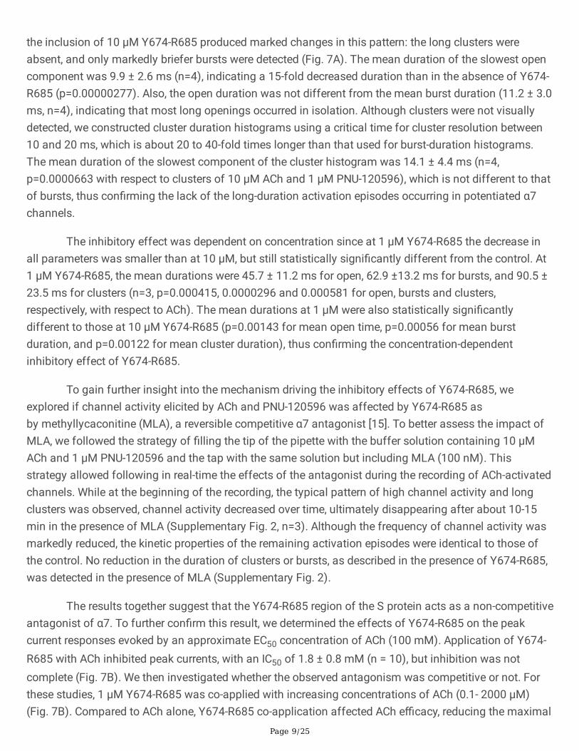
The macroscopic responses of the human α7 nAChR expressed in Xenopus oocytes to the applications ofthe Y674-R685 fragment at a range of concentrations (1 pM-10 mM) were examined along with controlACh-evoked responses from the same cells (Fig 2A). As shown in the �gure, Y674-R685 did not elicitdetectable currents in contrast to the robust responses elicited by 100 µM ACh. After 5 min wash,receptors remained responsive to subsequent control applications of ACh (Fig. 2A).
Single-channel currents from cell-attached patches from BOSC-23 cells expressing human α7 nAChRwere also recorded, thus allowing for more detailed mechanistic information. Recordings were carried outin parallel with control experiments with ACh as the agonist to con�rm the presence of functional α7nAChRs in the same batch of cells. ACh (10-100 µM) evoked isolated brief openings or less often shortbursts composed of a few openings in quick succession, which correspond to activation of the samereceptor molecule [18,21,22,24,25] (Fig. 2B). In contrast, channel activity was not detected at a range ofY674-R685 concentrations in a total of 21 patches from different cell transfections (1 pM, n=3; 1 nM, n=3;1 µM, n=8; 10 µM, n=3; 100 µM, n=4) (Fig. 2B).
Given that α7 nAChR activation in the presence of ACh occurs with low open probability as very briefopening events (Fig. 2B), we sought to amplify this effect using a potentiator. PNU-120596, a type IIpositive allosteric modulator (PAM), has been extensively used as a tool in α7 functional studies due toits ability to increase the probability of agonist-elicited channel opening and open-channel durations andto reduce desensitization [28]. We, therefore, examined whether Y674-R685 elicits α7 channel activity inthe presence of PNU-120596. Note that by itself, PNU-120596 cannot induce channel activation [29].
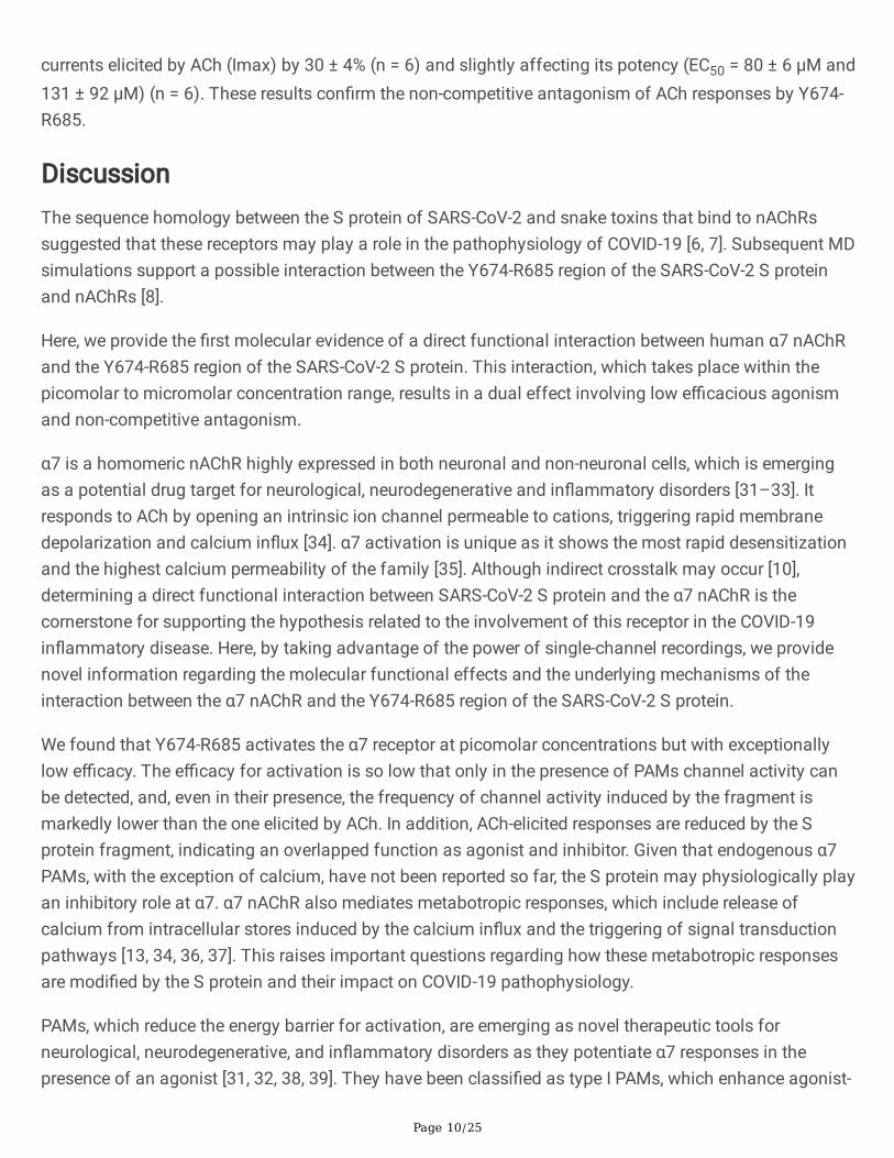
The macroscopic currents elicited by 1 µM Y674-R685 in the presence of 10 µM PNU-120596 wererecorded. Under these conditions, small currents in the low nA order were detected in 20% of the oocytestested (n=25) whereas neither Y674-R685 nor PNU-120596 on their own elicited currents (Fig. 3A).
To gain more insights into how Y674-R685 activates the α7 nAChR in the presence of PNU-120596, weexplored its effects at the single-channel level. ACh-elicited activity in the presence of 1 µM PNU-120596is profoundly different to that in its absence (Fig. 3B). Instead of the brief isolated openings, channelactivity shows long periods of high-frequency openings, named clusters, with a mean duration of about 1-3 s and an amplitude of 10 pA (-70 mV). A cluster corresponds to the activation episode of the samereceptor that recovers from desensitization and oscillates between open and closed states beforereaching again the more stable non-conducting desensitized state [28]. Clusters are composed of burstswith mean durations of ~300-500 ms, which comprise successive openings separated by very briefclosings (Figs. 3B and 4) [23,28].
Page 7/25
In the presence of 1 mM PNU-120596, Y674-R685 was capable of eliciting channel activity at a widerange of concentrations (1 pM to 10 mM), indicating that this region of the S protein can activate α7nAChRs (Fig. 3B). Since the frequency of channels is variable among patches due to variations inreceptor expression levels, parallel control recordings in the presence of ACh were made. When ACh andPNU-120596 were co-applied, >98% of patches showed channel activity (active patches), and the long-duration clusters described above appeared at high frequency as reported before [22] (Fig. 3B). In thepresence of PNU-120596 and Y674-R685, the percentage of active patches was lower than in thepresence of ACh: 65% (n=23, N=4; 1 pM Y674-R685), 40% (n=15, N=4; 1 nM Y674-R685), 67% (n=15, N=4;1 mM Y674-R685), and 62% (n=13, N=3; 10 mM Y674-R685). Also, channel activity evoked by Y674-R685was much more infrequent and interspaced by long silent periods when compared to that evoked by ACh(Fig. 3B). It is important to note that this type of experiments does not allow for a precise comparison ofchannel frequency since this parameter may be affected by the variability in the number of receptors ineach patch. Nevertheless, at 1 pM Y674-R685, the frequency of channel activation episodes was very low;albeit the active patches showed long clusters resembling those elicited by ACh and PNU-120596 (Figs.3B and 4).
The increase in Y674-R685 concentration resulted in profound changes in the channel activity pattern, asclearly illustrated in the recordings shown in Fig. 3B. The frequency of opening events appeared toincrease, but the duration of the openings and the activation episodes were reduced with increasingconcentrations. The typical long-duration clusters were completely absent at Y674-R685 concentrationshigher than 1 µM, at which activation occurred mainly as isolated openings or in short bursts (Fig. 3B and4).
To de�ne the properties of the activation episodes elicited by the Y674-R685 fragment at differentconcentrations, the mean durations of openings, bursts, and clusters in the presence of PNU-120596 weredetermined (Fig. 4). At 1 pM Y674-R685, the mean durations of the longest openings, bursts and clusterswere 141 ± 59 ms, 417 ± 113 ms, and 2330 ± 673 ms, respectively (n=3). These values were similar tothose determined in the presence of 10 µM ACh: 148 ± 12 ms for the slowest open component (p=0.859,n=3), 550 ± 38 ms for bursts (p=0.125, n=3), and 3048 ± 516 ms for clusters (p=0.217, n=3), and alsocomparable to those reported before for 100 µM ACh and 1 µM PNU-120596 [23,28]. Although the meanduration of clusters between 10 µM ACh and 1 pM Y674-R685 was similar, the relative area of thecomponent corresponding to clusters in the histogram was smaller when Y674-R685 was the agonist(relative areas were for ACh= 0.44 ± 0.09 and for Y674-R685= 0.21 ± 0.08; p=0.0264) (Fig. 4). With theincrease of Y674-R685 concentration, dwell time distributions for open, bursts and clusters were shiftedto briefer durations. The slowest component of each histogram became progressively briefer withincreasing Y674-R685 concentrations (Fig. 4). The mean durations were 48.2 ± 13.6 ms (1 µM, n=3)and 14.5 ± 4.3 ms (10 µM, n=4) for openings; 70.2 ± 19.7 ms (1 µM, n=3) and 20.7 ± 8.0 ms (10 µM, n=4)for bursts; and 104.2 ± 41.6 ms (1 µM, n=3) and 29.9 ± 14.8 ms (10 µM, n=4) for clusters. Mean valueswere statistically signi�cantly different to those determined in the presence of 10 µM ACh and 1 µM PNU-120596 (p=0.000665 and p=0.00000431 for open; p=0.0000403 and p= 0.00000106 for burst;p=0.000597 and p=0.0000681 for cluster, for 1 and 10 µM Y674-R685, respectively). At high Y674-R685
Page 8/25
concentrations (e.g., 10 µM), the mean open duration was similar to the mean burst and cluster durations(Fig. 4), indicating that openings occurred mostly in isolation and con�rming the lack of the typical long-duration clusters corresponding to potentiated α7 responses.
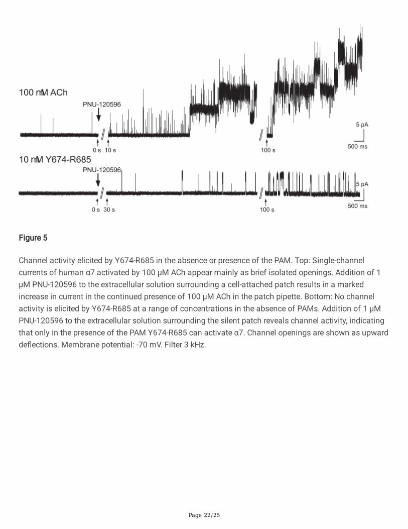
To further con�rm that channel activation can be elicited by Y674-R685 in the presence of PNU-120596but not in its absence, single-channel recordings in the presence of the Y674-R685 fragment alone in thepipette solution (1-10 µM) were made. Again, no channel activity was detected in all patches. However,the addition of 1 µM PNU-120596 to the dish during the course of the recording resulted in theappearance of single-channel activity in most of the silent patches (Fig. 5). Again, the frequency ofopening events was systematically markedly lower, and the durations were briefer than in the conditionsin which ACh was the agonist (Fig. 5).
Together, these results con�rm that Y674-R685 functionally interacts with the α7 nAChR. They also showthat Y674-R685 acts as a very low-e�cacy agonist since channel opening requires the presence of PNU-120596 and occurs with low probability, and that the increase in concentration is accompanied by adecrease in the duration of open channel lifetime and of the activation episodes.
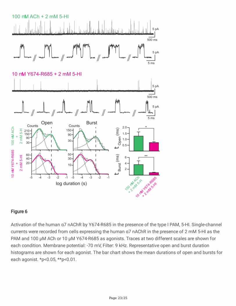
Because PNU-120596 is a highly e�cacious type II PAM with the capability of recovering receptors fromdesensitization, we also tested if Y674-R685 elicited channel activity in the presence of 5-hydroxyindole(5-HI), a type I PAM which induces clear potentiation but does not recover receptors from desensitization[21,23,30]. In the presence of 2 mM 5-HI, 100 µM ACh induced prolonged openings and bursts composedof successive openings which lasted about 4 ms (Fig. 6). The histograms showed that the duration of theslowest open component was ~4-fold longer (1.28 ± 0.35 ms, n=37, versus 0.30 ± 0.06 ms, n=38) and themean burst duration was 8-fold longer (3.60 ± 1.29 ms, n=37, versus 0.46 ± 0.12 ms, n=38) than in theabsence of the PAM. Replacing ACh by Y674-R685 (1 pM-10 µM) revealed α7 channel activity. However,the frequency of opening events was lower when compared to that elicited by ACh; only few events weredetected during a 15-min recording period (Fig. 6). At 10 µM Y674-R685, the mean open duration was0.73 ± 0.07 ms (n=3) and the mean burst duration was 0.85 ± 0.13 ms (n=3); both parameters werestatistically signi�cantly briefer than the corresponding mean durations in the presence of ACh and 2 mM5-HI (p=0.0114 and 0.005 for mean open and mean burst durations, respectively). Also, the observationthat in the presence of 10 µM Y674-R685 and 2 mM 5-HI the mean duration of openings was similar tothat of bursts indicates that channel opening occurred mainly as isolated events instead of in quicksuccession forming activation episodes, as described before for recordings with the type II PAM PNU-120596.
Inhibition of α7 activity by Y674-R685 from S protein
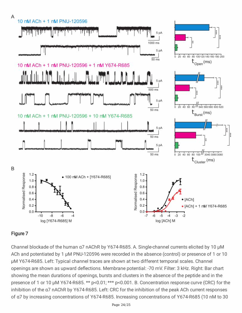
To understand the molecular mechanisms driving the inhibitory effects of Y674-R685 evidenced by thedramatic decrease in open durations, we evaluated Y674-R685 effects on α7 activated by 10 µM ACh(Fig. 7A). Given the very brief duration of α7 channels, which is close to the time resolution of our system,we included PNU-120596 to better quantify a decrease in open durations. Whereas in the presence of 1µM PNU-120596, 10 µM ACh led to an activation pattern composed of long clusters as described above,
Page 9/25
the inclusion of 10 µM Y674-R685 produced marked changes in this pattern: the long clusters wereabsent, and only markedly briefer bursts were detected (Fig. 7A). The mean duration of the slowest opencomponent was 9.9 ± 2.6 ms (n=4), indicating a 15-fold decreased duration than in the absence of Y674-R685 (p=0.00000277). Also, the open duration was not different from the mean burst duration (11.2 ± 3.0ms, n=4), indicating that most long openings occurred in isolation. Although clusters were not visuallydetected, we constructed cluster duration histograms using a critical time for cluster resolution between10 and 20 ms, which is about 20 to 40-fold times longer than that used for burst-duration histograms.The mean duration of the slowest component of the cluster histogram was 14.1 ± 4.4 ms (n=4,p=0.0000663 with respect to clusters of 10 µM ACh and 1 µM PNU-120596), which is not different to thatof bursts, thus con�rming the lack of the long-duration activation episodes occurring in potentiated α7channels.
The inhibitory effect was dependent on concentration since at 1 µM Y674-R685 the decrease inall parameters was smaller than at 10 µM, but still statistically signi�cantly different from the control. At1 µM Y674-R685, the mean durations were 45.7 ± 11.2 ms for open, 62.9 ±13.2 ms for bursts, and 90.5 ±23.5 ms for clusters (n=3, p=0.000415, 0.0000296 and 0.000581 for open, bursts and clusters,respectively, with respect to ACh). The mean durations at 1 µM were also statistically signi�cantlydifferent to those at 10 µM Y674-R685 (p=0.00143 for mean open time, p=0.00056 for mean burstduration, and p=0.00122 for mean cluster duration), thus con�rming the concentration-dependentinhibitory effect of Y674-R685.
To gain further insight into the mechanism driving the inhibitory effects of Y674-R685, weexplored if channel activity elicited by ACh and PNU-120596 was affected by Y674-R685 asby methyllycaconitine (MLA), a reversible competitive α7 antagonist [15]. To better assess the impact ofMLA, we followed the strategy of �lling the tip of the pipette with the buffer solution containing 10 µMACh and 1 µM PNU-120596 and the tap with the same solution but including MLA (100 nM). Thisstrategy allowed following in real-time the effects of the antagonist during the recording of ACh-activatedchannels. While at the beginning of the recording, the typical pattern of high channel activity and longclusters was observed, channel activity decreased over time, ultimately disappearing after about 10-15min in the presence of MLA (Supplementary Fig. 2, n=3). Although the frequency of channel activity wasmarkedly reduced, the kinetic properties of the remaining activation episodes were identical to those ofthe control. No reduction in the duration of clusters or bursts, as described in the presence of Y674-R685,was detected in the presence of MLA (Supplementary Fig. 2).
The results together suggest that the Y674-R685 region of the S protein acts as a non-competitiveantagonist of α7. To further con�rm this result, we determined the effects of Y674-R685 on the peakcurrent responses evoked by an approximate EC50 concentration of ACh (100 mM). Application of Y674-R685 with ACh inhibited peak currents, with an IC50 of 1.8 ± 0.8 mM (n = 10), but inhibition was notcomplete (Fig. 7B). We then investigated whether the observed antagonism was competitive or not. Forthese studies, 1 μM Y674-R685 was co-applied with increasing concentrations of ACh (0.1- 2000 μM)(Fig. 7B). Compared to ACh alone, Y674-R685 co-application affected ACh e�cacy, reducing the maximal
Page 10/25
currents elicited by ACh (Imax) by 30 ± 4% (n = 6) and slightly affecting its potency (EC50 = 80 ± 6 μM and131 ± 92 μM) (n = 6). These results con�rm the non-competitive antagonism of ACh responses by Y674-R685.
DiscussionThe sequence homology between the S protein of SARS-CoV-2 and snake toxins that bind to nAChRssuggested that these receptors may play a role in the pathophysiology of COVID-19 [6, 7]. Subsequent MDsimulations support a possible interaction between the Y674-R685 region of the SARS-CoV-2 S proteinand nAChRs [8].
Here, we provide the �rst molecular evidence of a direct functional interaction between human α7 nAChRand the Y674-R685 region of the SARS-CoV-2 S protein. This interaction, which takes place within thepicomolar to micromolar concentration range, results in a dual effect involving low e�cacious agonismand non-competitive antagonism.
α7 is a homomeric nAChR highly expressed in both neuronal and non-neuronal cells, which is emergingas a potential drug target for neurological, neurodegenerative and in�ammatory disorders [31–33]. Itresponds to ACh by opening an intrinsic ion channel permeable to cations, triggering rapid membranedepolarization and calcium in�ux [34]. α7 activation is unique as it shows the most rapid desensitizationand the highest calcium permeability of the family [35]. Although indirect crosstalk may occur [10],determining a direct functional interaction between SARS-CoV-2 S protein and the α7 nAChR is thecornerstone for supporting the hypothesis related to the involvement of this receptor in the COVID-19in�ammatory disease. Here, by taking advantage of the power of single-channel recordings, we providenovel information regarding the molecular functional effects and the underlying mechanisms of theinteraction between the α7 nAChR and the Y674-R685 region of the SARS-CoV-2 S protein.
We found that Y674-R685 activates the α7 receptor at picomolar concentrations but with exceptionallylow e�cacy. The e�cacy for activation is so low that only in the presence of PAMs channel activity canbe detected, and, even in their presence, the frequency of channel activity induced by the fragment ismarkedly lower than the one elicited by ACh. In addition, ACh-elicited responses are reduced by the Sprotein fragment, indicating an overlapped function as agonist and inhibitor. Given that endogenous α7PAMs, with the exception of calcium, have not been reported so far, the S protein may physiologically playan inhibitory role at α7. α7 nAChR also mediates metabotropic responses, which include release ofcalcium from intracellular stores induced by the calcium in�ux and the triggering of signal transductionpathways [13, 34, 36, 37]. This raises important questions regarding how these metabotropic responsesare modi�ed by the S protein and their impact on COVID-19 pathophysiology.
PAMs, which reduce the energy barrier for activation, are emerging as novel therapeutic tools forneurological, neurodegenerative, and in�ammatory disorders as they potentiate α7 responses in thepresence of an agonist [31, 32, 38, 39]. They have been classi�ed as type I PAMs, which enhance agonist-
Page 11/25
induced macroscopic currents, and type II PAMs, that also delay desensitization and recover receptorsfrom desensitized states [23, 40, 41]. At the single channel level, PAMs enhance open channel durationsand induce activation in episodes in which the channel oscillates between open and closedconformations, thus generating an activity pattern markedly different from the isolated, infrequentsubmillisecond-openings in the absence of PAMs [23]. Depending on the type and e�cacy of the PAM,the potentiated activation episodes can range from milliseconds (as shown here for 5-HI) to seconds (asdemonstrated here for PNU-120596). Despite the marked differences between 5-HI and PNU-120596regarding their e�cacy for potentiation and in�uence on receptor desensitization, Y674-R685 activatesthe α7 nAChR in the presence of both PAMs but not in their absence, indicating its action as a low-e�cacy agonist. Also, in the presence of both PAMs, reduction in the duration of the opening events withincreasing Y674-R685 concentration is observed, thus supporting the additional role of the fragment asan α7 inhibitor.
At 1 pM Y674-R685, channel activity in the presence of PNU-120596 is markedly infrequent but kineticallyindistinguishable from the ACh-elicited one. The open channel lifetime, and architecture and duration ofthe activation episodes (clusters) are similar in the presence of ACh or 1 pM Y674-R685. As the Y674-R685 concentration increases, the frequency of openings increases, but their durations becomeprogressively briefer, with long-duration clusters no longer present. In ACh-elicited channels, the Y674-R685 fragment also produces a concentration-dependent reduction of open and cluster durations, aneffect different to that produced by the competitive antagonist MLA. Thus, the type of single-channelinhibition together with the results from macroscopic current recordings support a mechanism of non-competitive inhibition.
The ability to act as a low-e�cacy agonist is consistent with data from our MD simulations predictingthat Y674-R685 can bind directly to the agonist binding site located in the extracellular domain of the α7nAChR, leading in some conformations to a semi-capped loop C [8]. Loop C is one of the three loops thatform the principal face of the orthosteric binding site [42]. Upon agonist binding, loop C closes to cap theagonist, an event associated with priming and channel opening [43]. Loop C capping aids the anchoringof the bound agonist to the orthosteric binding site. It has also been suggested that agonists induce morecompact loop conformations, while partial agonists and antagonists produce incomplete closure orprevent loop C capping [44, 46].
Our previous MD simulations of Y674-R685 bound to the α7 nAChR identi�ed the guanidinium group ofR682 in the SARS-COV-2 S protein as the key anchoring point to the α7 nAChR (Fig. 1C), forming stronginteractions with several residues lining the receptor's ligand binding pocket [8]. These simulations alsoshow that, when bound to α7, the Y674-R685 region of the SARS-CoV-2 S protein adopts many differentconformations within the binding pocket, ranging from highly compact to fully extended con�gurations(Supplementary Fig. 3). Some of these binding modes allow for the formation of the key interactionsnecessary to activate the receptor (Fig. 1C), while others do not (Supplementary Fig. 4) [8]. Although theα7 nAChR has �ve identical orthosteric binding sites, agonist occupancy of only one is required to elicitchannel activation [25]. Thus, it could be possible that the Y674-R685 fragment occupancy of the binding
Page 12/25
sites in multiple orientations, some of which cannot trigger activation, may contribute to its low e�cacy,as described before for partial agonists [45]. Additionally, the partial closure of loop C in the presence ofthe Y674-R685 [8] observed in some of our previous simulations may also reduce the e�cacy for channelopening (Supplementary Fig. 5).
As shown here for Y674-R685, several compounds of different structures have dual opposite effects onnAChRs, including α7, acting as agonists and non-competitive antagonists (also called negative allostericmodulators, NAMs) [22, 47, 48]. NAMs comprise a wide range of structurally different compounds thatinhibit receptor function by binding to sites distinct to the orthosteric sites (allosteric) [39]. Theseallosteric sites can be located in different receptor domains, including the extracellular [49] andtransmembrane domains [28, 50]. The site for the allosteric inhibition of Y674-R685 remains to bedetermined. The negative modulation may occur by different molecular mechanisms, comprising channelblock, enhanced desensitization, or decrease probability of opening. Given the complex kinetics of the α7receptor, distinguishing how the fragment exerts allosteric inhibition is not possible from our experiments.However, the type of concentration-dependent reduction of bursts and clusters suggests slow channelblockade since fast �ickering, typical of fast open-channel blockers, was not observed [51]. Due to thepresence of three arginine residues, the Y674-R685 fragment (sequence YQTQTNSPRRAR) is highlycharged, with an overall charge of + 3, which may be compatible with channel block.
Overall, our results reveal a functional interaction between the Y674-R685 region of the SARS-CoV-2 Sprotein and the α7 nAChR, with the predominant effect probably being inhibitory since activation cannotoccur in the absence of PAMs. Although too speculative at this stage, this �nding raises the possibilitythat the hyper-in�ammatory response observed in some COVID-19 patients may be partly due toinhibition of α7 nAChRs. It has been shown that activation of α7 nAChR in the immune system protectsfrom excessive production of pro-in�ammatory cytokines (e.g., TNF-α) [12]. Impairment of this receptorsubtype results in an overproduction of cytokines and enhanced tissue damage [52]. In this context,therapeutic interventions focused on α7 nAChR may be valuable to be explored.
AbbreviationsSARS-CoV-2: severe acute respiratory syndrome coronavirus 2
nAChR: Nicotinic Acetylcholine Receptor.
ACh: Acetylcholine.
MLA: methyllycaconitine
PAM: Positive Allosteric Modulator.
5-HI: 5-Hydroxyindole.
PNU-120596: (N-(5-Chloro-2,4-dimethoxyphenyl)- N’-(5-methyl-3-isoxazolyl)-urea)
Page 13/25
S protein: SARS-CoV-2 Spike protein
MD: Molecular dynamics
DeclarationsFunding: This work was supported by grants from Universidad Nacional del Sur (PGI 24/B298) andAgencia Nacional de Promoción Cientí�ca y Tecnológica (PICT 2017-1170) to CB; Oxford BrookesUniversity (REA-2020) to IB. AJM and ASFO thank EPSRC (grant number EP/M022609/1) and BBSRC(grant number BB/R016445/1) for support. We thank BrisSynBio, a BBSRC/EPSRC Synthetic BiologyResearch Centre (Grant Number:BB/L01386X/1), for funding ASFO and providing funds to purchase theY674-R685 peptide from Designer Bioscience Ltd. (Cambridge UK).
Con�ict of interest: The authors report no con�ict of interest.
Availability of data and material. The data that support the �ndings of this study are available within thearticle.
Ethics approval and consent to participate: not applicable
Consent for publication: not applicable
Acknowledgements: not applicable.
Code availability: Not applicable.
Author Contributions: JFC, ASFO, AJM, TG, IB and CB designed the research; JFC, ASFO, IB and CBperformed the research and analysed the data; CB, ASFO, AJM, TG, JFC and IB wrote, reviewed, and editedthe paper.
References1. Casalino L, Gaieb Z, Goldsmith JA et al (2020) Beyond shielding: the roles of glycans in the SARS-
CoV-2 spike protein. ACS Cent Sci 6 1722-1734. https://doi.org/10.1021/acscentsci.0c01056
2. Yan R, Zhang Y, Li Y et al (2020) Structural basis for the recognition of SARS-CoV-2 by full-lengthhuman ACE2. Science 367(6485) 1444–1448. https://doi.org/10.1126/science.abb2762
3. Hoffmann M, Kleine-Weber H, Schroeder S et al (2020) SARS-CoV-2 Cell Entry Depends on ACE2 andTMPRSS2 and Is Blocked by a Clinically Proven Protease Inhibitor. Cell 181(2) 271–280.e8. https://doi.org/10.1016/j.cell.2020.02.052
4. Daly JL, Simonetti B, Klein K et al (2020) Neuropilin-1 is a host factor for SARS-CoV-2 infection.Science 370(6518) 861–865. https://doi.org/10.1126/science.abd3072
Page 14/25
5. Cantuti-Castelvetri L, Ojha R, Pedro LD et al (2020) Neuropilin-1 facilitates SARS-CoV-2 cell entry andinfectivity. Science 370(6518) 856–860. https://doi.org/10.1126/science.abd2985
�. Changeux JP, Amoura Z, Rey FA, Miyara M (2020) A nicotinic hypothesis for Covid-19 with preventiveand therapeutic implications. Comptes rendus biologies 343(1) 33–39. https://doi.org/10.5802/crbiol.8
7. Farsalinos K, Eliopoulos E, Leonidas DD et al (2020) Nicotinic Cholinergic System and COVID-19: InSilico Identi�cation of an Interaction between SARS-CoV-2 and Nicotinic Receptors with PotentialTherapeutic Targeting Implications. International journal of molecular sciences 21(16)5807. https://doi.org/10.3390/ijms21165807
�. Oliveira A, Ibarra AA, Bermudez I et al (2021) A potential interaction between the SARS-CoV-2 spikeprotein and nicotinic acetylcholine receptors. Biophysical journal 120(6) 983–993. https://doi.org/10.1016/j.bpj.2021.01.037
9. Grant SN, Lester H (2021) Regulation of epithelial sodium channel activity by SARS-CoV-1 and SARS-CoV-2 proteins. Biophysical journal 120(14) 2805–2813. https://doi.org/10.1016/j.bpj.2021.06.005
10. Russo P, Bonassi S, Giacconi R et al (2020) COVID-19 and smoking: is nicotine the hidden link? TheEuropean respiratory journal 55(6) 2001116. https://doi.org/10.1183/13993003.01116-2020
11. Wang H, Yu M, Ochani M et al (2003) Nicotinic acetylcholine receptor alpha7 subunit is an essentialregulator of in�ammation. Nature 421(6921) 384–388. https://doi.org/10.1038/nature01339
12. Rosas-Ballina M, Ochani M, Parrish WR et al (2008) Splenic nerve is required for cholinergicantiin�ammatory pathway control of TNF in endotoxemia. Proceedings of the National Academy ofSciences of the United States of America 105(31) 11008–11013. https://doi.org/10.1073/pnas.0803237105
13. Egea J, Buendia I, Parada E et al (2015) Anti-in�ammatory role of microglial alpha7 nAChRs and itsrole in neuroprotection. Biochemical pharmacology 97(4) 463–472. https://doi.org/10.1016/j.bcp.2015.07.032
14. Pinheiro NM, Santana FP, Almeida RR et al (2017) Acute lung injury is reduced by the α7nAChRagonist PNU-282987 through changes in the macrophage pro�le. FASEB journal: o�cial publicationof the Federation of American Societies for Experimental Biology 31(1) 320–332. https://doi.org/10.1096/fj.201600431R
15. Wonnacott S (2014), Nicotinic ACh Receptors. Tocris Bioscience Scienti�c Reviews Series 1–31.
1�. Nau J, Luthra P, Lanzer K et al (2021) Varenicline prevents SARS-CoV-2 infection in vitro and inRhesus Macaques. bioRxiv [Preprint]. https://doi.org/10.1101/2021.06.29.450426 (accessed 20September 2021)
17. Bouzat C, Bren N, Sine SM (1994) Structural basis of the different gating kinetics of fetal and adultnicotinic acetylcholine receptors. Neuron 13 1395-1402. https://doi.org/10.1016/0896-6273(94)90424-3
1�. Chrestia JF, Bruzzone A, Esandi M, Bouzat C (2021) Tyrosine phosphorylation differentially �ne-tunesionotropic and metabotropic responses of human α7 nicotinic acetylcholine receptor. Cellular and

Page 15/25
molecular life sciences: CMLS, 78(13) 5381–5395. https://doi.org/10.1007/s00018-021-03853-3
19. Lansdell SJ, Gee VJ, Harkness PC et al (2005) RIC-3 enhances functional expression of multiplenicotinic acetylcholine receptor subtypes in mammalian cells. Molecular pharmacology 68(5) 1431–1438. https://doi.org/10.1124/mol.105.017459
20. Gu S, Matta JA, Lord B et al (2016) Brain α7 Nicotinic Acetylcholine Receptor Assembly RequiresNACHO. Neuron 89(5) 948–955. https://doi.org/10.1016/j.neuron.2016.01.018
21. Nielsen E, Bermudez I, Bouzat C (2019) Flavonoids as positive allosteric modulators of α7 nicotinicreceptors. Neuropharmacology 160 107794. https://doi.org/10.1016/j.neuropharm.2019.107794
22. Lasala M, Fabiani C, Corradi J, Antollini S, Bouzat C (2019) Molecular Modulation of Human α7Nicotinic Receptor by Amyloid-β Peptides. Frontiers in cellular neuroscience 1337. https://doi.org/10.3389/fncel.2019.00037
23. Andersen ND, Nielsen BE, Corradi J et al (2016) Exploring the positive allosteric modulation of humanα7 nicotinic receptors from a single-channel perspective. Neuropharmacology 107 189–200. https://doi.org/10.1016/j.neuropharm.2016.02.032
24. Bouzat C, Bartos M, Corradi J, Sine SM (2008) The interface between extracellular andtransmembrane domains of homomeric Cys-loop receptors governs open-channel lifetime and rateof desensitization. The Journal of neuroscience: the o�cial journal of the Society for Neuroscience28(31) 7808–7819. https://doi.org/10.1523/JNEUROSCI.0448-08.2008
25. Andersen N, Corradi J, Sine SM, Bouzat C (2013) Stoichiometry for activation of neuronal α7nicotinic receptors. Proceedings of the National Academy of Sciences of the United States ofAmerica 110(51) 20819–20824. https://doi.org/10.1073/pnas.1315775110
2�. Minguez-Viñas T, Nielsen BE, Shoemark DK et al (2021) A conserved arginine with non-conservedfunction is a key determinant of agonist selectivity in α7 nicotinic ACh receptors. British journal ofpharmacology 178(7) 1651–1668. https://doi.org/10.1111/bph.15389
27. Nielsen BE, Minguez T, Bermudez I, Bouzat C (2018) Molecular function of the novel α7β2 nicotinicreceptor. Cellular and molecular life sciences: CMLS 75(13) 2457–2471. https://doi.org/10.1007/s00018-017-2741-4
2�. daCosta CJ, Free CR, Corradi J, Bouzat C, Sine SM (2011) Single-channel and structural foundationsof neuronal α7 acetylcholine receptor potentiation. The Journal of neuroscience: the o�cial journalof the Society for Neuroscience, 31(39) 13870–13879. https://doi.org/10.1523/JNEUROSCI.2652-11.2011
29. Hurst RS, Hajós M, Raggenbass M et al (2005) A novel positive allosteric modulator of the alpha7neuronal nicotinic acetylcholine receptor: in vitro and in vivo characterization. The Journal ofneuroscience: the o�cial journal of the Society for Neuroscience 25(17) 4396–4405. https://doi.org/10.1523/JNEUROSCI.5269-04.2005
30. Zwart R, De Filippi G, Broad LM et al (2002) 5-Hydroxyindole potentiates human alpha 7 nicotinicreceptor-mediated responses and enhances acetylcholine-induced glutamate release in cerebellarslices. Neuropharmacology 43(3) 374–384. https://doi.org/10.1016/s0028-3908(02)00094-1
Page 16/25
31. Uteshev V (2014) The therapeutic promise of positive allosteric modulation of nicotinic receptors.European journal of pharmacology 727 181–185. https://doi.org/10.1016/j.ejphar.2014.01.072
32. Dineley KT, Pandya AA, Yakel JL (2015) Nicotinic ACh receptors as therapeutic targets in CNSdisorders. Trends in pharmacological sciences 36(2) 96–108(2015). https://doi.org/10.1016/j.tips.2014.12.002
33. Yang T, Xiao T, Sun Q, Wang K (2017) The current agonists and positive allosteric modulators of α7nAChR for CNS indications in clinical trials. Acta pharmaceutica Sinica. B 7(6) 611–622. https://doi.org/10.1016/j.apsb.2017.09.001
34. Bouzat C, Lasala M, Nielsen BE, Corradi J, Esandi M (2018) Molecular function of α7 nicotinicreceptors as drug targets. The Journal of physiology 596(10) 1847–1861. https://doi.org/10.1113/JP275101
35. Fucile S (2004) Ca2+ permeability of nicotinic acetylcholine receptors. Cell calcium 35(1) 1–8. https://doi.org/10.1016/j.ceca.2003.08.006
3�. Guan YZ, Jin XD, Guan LX et al (2015) Nicotine inhibits microglial proliferation and is neuroprotectivein global ischemia rats. Molecular neurobiology 51(3) 1480–1488. https://doi.org/10.1007/s12035-014-8825-3
37. Kabbani N, Nordman JC, Corgiat BA et al (2013) Are nicotinic acetylcholine receptors coupled to Gproteins? BioEssays: news and reviews in molecular, cellular and developmental biology 35(12),1025–1034. https://doi.org/10.1002/bies.201300082
3�. Changeux JP, Taly A (2008) Nicotinic receptors, allosteric proteins and medicine. Trends in molecularmedicine 14(3) 93–102. https://doi.org/10.1016/j.molmed.2008.01.001
39. Chatzidaki A, Millar NS (2015) Allosteric modulation of nicotinic acetylcholine receptors. Biochemicalpharmacology 97(4) 408–417. https://doi.org/10.1016/j.bcp.2015.07.028
40. Bertrand D, Gopalakrishnan M (2007) Allosteric modulation of nicotinic acetylcholine receptors.Biochemical pharmacology 74(8) 1155–1163. https://doi.org/10.1016/j.bcp.2007.07.011
41. Grønlien JH, Håkerud M, Ween H et al (2007) Distinct pro�les of alpha7 nAChR positive allostericmodulation revealed by structurally diverse chemotypes. Molecular pharmacology 72(3) 715–724. https://doi.org/10.1124/mol.107.035410
42. Noviello CM, Gharpure A, Mukhtasimova N et al (2021) Structure and gating mechanism of the α7nicotinic acetylcholine receptor. Cell 184(8) 2121–2134.e13. https://doi.org/10.1016/j.cell.2021.02.049
43. Mukhtasimova N, Lee WY, Wang HL, Sine SM (2009) Detection and trapping of intermediate statespriming nicotinic receptor channel opening. Nature 459 (7245) 451–454. https://doi.org/10.1038/nature07923
44. Hansen SB, Sulzenbacher G, Huxford T et al (2005) Structures of Aplysia AChBP complexes withnicotinic agonists and antagonists reveal distinctive binding interfaces and conformations. TheEMBO journal 24(20) 3635–3646. https://doi.org/10.1038/sj.emboj.7600828
Page 17/25
45. Hibbs RE, Sulzenbacher G, Shi J et al (2009) Structural determinants for interaction of partialagonists with acetylcholine binding protein and neuronal alpha7 nicotinic acetylcholine receptor. TheEMBO journal 28(19) 3040–3051. https://doi.org/10.1038/emboj.2009.227
4�. Brams M, Gay EA, Sáez JC et al (2011) Crystal structures of a cysteine-modi�ed mutant in loop D ofacetylcholine-binding protein. The Journal of biological chemistry 286(6) 4420–4428. https://doi.org/10.1074/jbc.M110.188730
47. Fabiani C, Murray AP, Corradi J, Antollini SS (2018) A novel pharmacological activity of caffeine inthe cholinergic system. Neuropharmacology 135 464–473. https://doi.org/10.1016/j.neuropharm.2018.03.041
4�. Pereira EF, Hilmas C, Santos MD et al (2002) Unconventional ligands and modulators of nicotinicreceptors. Journal of neurobiology 53(4) 479–500. https://doi.org/10.1002/neu.10146
49. Spurny R, Debaveye S, Farinha A et al (2015) Molecular blueprint of allosteric binding sites in ahomologue of the agonist-binding domain of the α7 nicotinic acetylcholine receptor. Proceedings ofthe National Academy of Sciences of the United States of America 112(19) E2543–E2552. https://doi.org/10.1073/pnas.1418289112
50. Young GT, Zwart R, Walker AS, Sher E, Millar NS (2008) Potentiation of alpha7 nicotinic acetylcholinereceptors via an allosteric transmembrane site. Proceedings of the National Academy of Sciences ofthe United States of America 105(38) 14686–14691. https://doi.org/10.1073/pnas.0804372105
51. Neher E, Steinbach JH (1978) Local anaesthetics transiently block currents through singleacetylcholine-receptor channels. The Journal of physiology 277 153–176. https://doi.org/10.1113/jphysiol.1978.sp012267
52. Parrish WR, Rosas-Ballina M, Gallowitsch-Puerta M et al (2008) Modulation of TNF release bycholine requires alpha7 subunit nicotinic acetylcholine receptor-mediated signaling. Molecularmedicine (Cambridge, Mass.) 14(9-10) 567–574. https://doi.org/10.2119/2008-00079.Parrish
Figures

Page 18/25
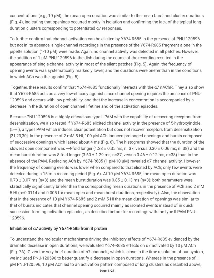
Figure 1
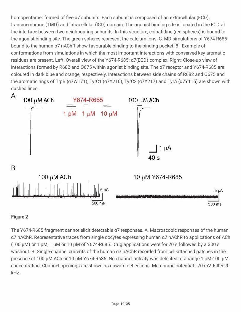
Three-dimensional structures of the SARS-CoV-2 S protein and the human α7 nAChR. A. The modelrepresents the complete, fully glycosylated S protein in the closed state after furin cleavage [1]. Theprotein is rendered as a blue cartoon with the glycans depicted in green. The receptor binding motifs(residues S438-Q506) and the Y674-R685 region are highlighted in yellow and red, respectively. The Y674-R685 region was shown to be accessible for binding in previous MD simulations of the fully glycosylatedS protein [8]. B. Cryo-EM structure of the human α7 nAChR (PDB code: 7KOX) [42]. This receptor is a
Page 19/25
homopentamer formed of �ve α7 subunits. Each subunit is composed of an extracellular (ECD),transmembrane (TMD) and intracellular (ICD) domain. The agonist binding site is located in the ECD atthe interface between two neighbouring subunits. In this structure, epibatidine (red spheres) is bound tothe agonist binding site. The green spheres represent the calcium ions. C. MD simulations of Y674-R685bound to the human α7 nAChR show favourable binding to the binding pocket [8]. Example ofconformations from simulations in which the most important interactions with conserved key aromaticresidues are present. Left: Overall view of the Y674-R685: α7(ECD) complex. Right: Close-up view ofinteractions formed by R682 and Q675 within agonist binding site. The α7 receptor and Y674-R685 arecoloured in dark blue and orange, respectively. Interactions between side chains of R682 and Q675 andthe aromatic rings of TrpB (α7W171), TyrC1 (α7Y210), TyrC2 (α7Y217) and TyrA (α7Y115) are shown withdashed lines.
Figure 2
The Y674-R685 fragment cannot elicit detectable α7 responses. A. Macroscopic responses of the humanα7 nAChR. Representative traces from single oocytes expressing human α7 nAChR to applications of ACh(100 µM) or 1 pM, 1 µM or 10 µM of Y674-R685. Drug applications were for 20 s followed by a 300 swashout. B. Single-channel currents of the human α7 nAChR recorded from cell-attached patches in thepresence of 100 µM ACh or 10 µM Y674-R685. No channel activity was detected at a range 1 pM-100 µMconcentration. Channel openings are shown as upward de�ections. Membrane potential: -70 mV. Filter: 9kHz.
Page 20/25
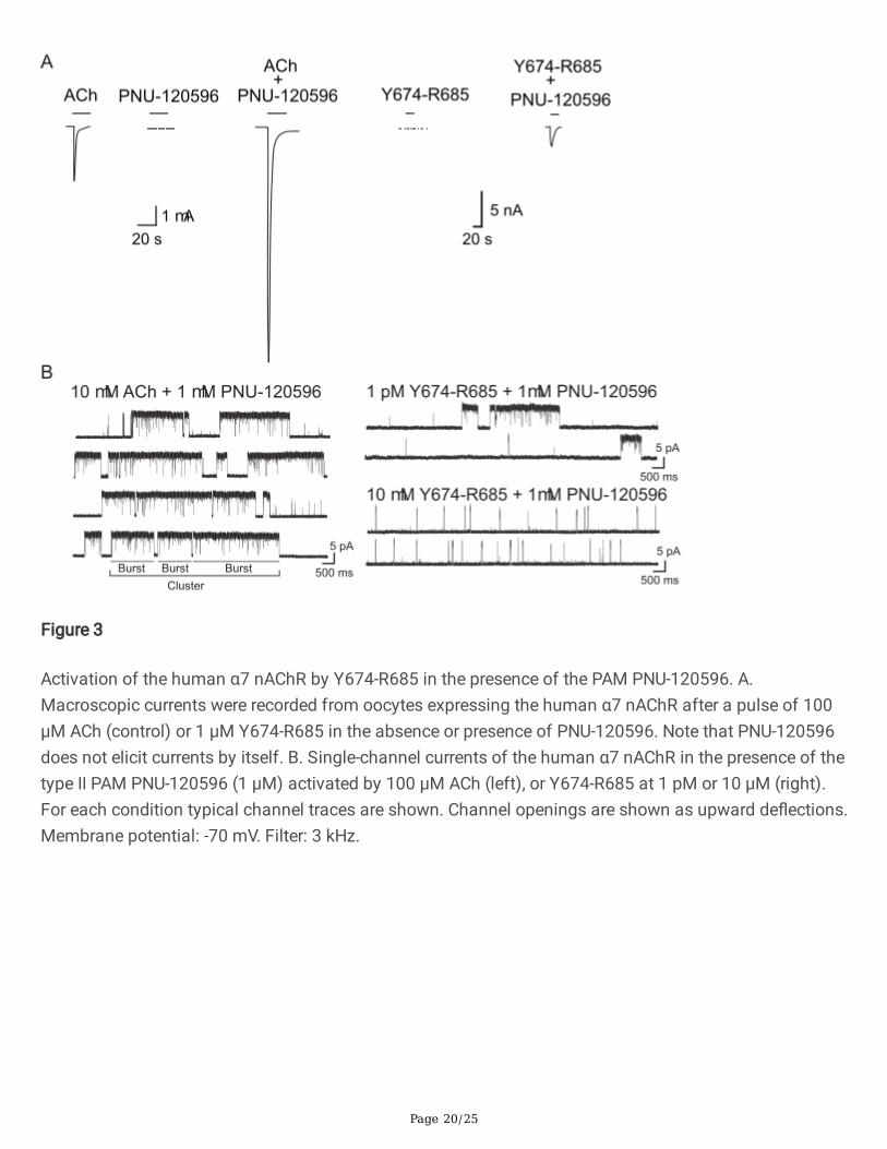
Figure 3
Activation of the human α7 nAChR by Y674-R685 in the presence of the PAM PNU-120596. A.Macroscopic currents were recorded from oocytes expressing the human α7 nAChR after a pulse of 100µM ACh (control) or 1 µM Y674-R685 in the absence or presence of PNU-120596. Note that PNU-120596does not elicit currents by itself. B. Single-channel currents of the human α7 nAChR in the presence of thetype II PAM PNU-120596 (1 µM) activated by 100 µM ACh (left), or Y674-R685 at 1 pM or 10 µM (right).For each condition typical channel traces are shown. Channel openings are shown as upward de�ections.Membrane potential: -70 mV. Filter: 3 kHz.
Page 21/25
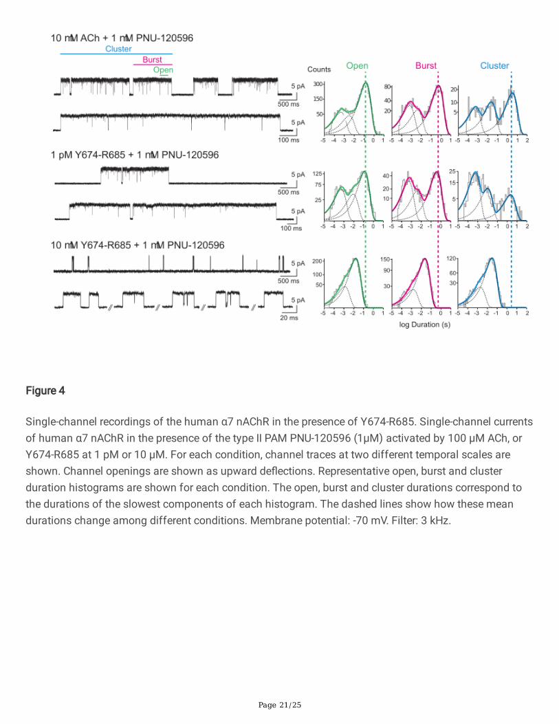
Figure 4
Single-channel recordings of the human α7 nAChR in the presence of Y674-R685. Single-channel currentsof human α7 nAChR in the presence of the type II PAM PNU-120596 (1µM) activated by 100 µM ACh, orY674-R685 at 1 pM or 10 µM. For each condition, channel traces at two different temporal scales areshown. Channel openings are shown as upward de�ections. Representative open, burst and clusterduration histograms are shown for each condition. The open, burst and cluster durations correspond tothe durations of the slowest components of each histogram. The dashed lines show how these meandurations change among different conditions. Membrane potential: -70 mV. Filter: 3 kHz.
Page 22/25
Figure 5
Channel activity elicited by Y674-R685 in the absence or presence of the PAM. Top: Single-channelcurrents of human α7 activated by 100 µM ACh appear mainly as brief isolated openings. Addition of 1µM PNU-120596 to the extracellular solution surrounding a cell-attached patch results in a markedincrease in current in the continued presence of 100 µM ACh in the patch pipette. Bottom: No channelactivity is elicited by Y674-R685 at a range of concentrations in the absence of PAMs. Addition of 1 µMPNU-120596 to the extracellular solution surrounding the silent patch reveals channel activity, indicatingthat only in the presence of the PAM Y674-R685 can activate α7. Channel openings are shown as upwardde�ections. Membrane potential: -70 mV. Filter 3 kHz.
Page 23/25
Figure 6
Activation of the human α7 nAChR by Y674-R685 in the presence of the type I PAM, 5-HI. Single-channelcurrents were recorded from cells expressing the human α7 nAChR in the presence of 2 mM 5-HI as thePAM and 100 µM ACh or 10 µM Y674-R685 as agonists. Traces at two different scales are shown foreach condition. Membrane potential: -70 mV, Filter: 9 kHz. Representative open and burst durationhistograms are shown for each agonist. The bar chart shows the mean durations of open and bursts foreach agonist. *p<0.05, **p<0.01.
Page 24/25
Figure 7
Channel blockade of the human α7 nAChR by Y674-R685. A. Single-channel currents elicited by 10 µMACh and potentiated by 1 µM PNU-120596 were recorded in the absence (control) or presence of 1 or 10µM Y674-R685. Left: Typical channel traces are shown at two different temporal scales. Channelopenings are shown as upward de�ections. Membrane potential: -70 mV. Filter: 3 kHz. Right: Bar chartshowing the mean durations of openings, bursts and clusters in the absence of the peptide and in thepresence of 1 or 10 µM Y674-R685. ** p<0.01; *** p<0.001. B. Concentration response curve (CRC) for theinhibition of the α7 nAChR by Y674-R685. Left: CRC for the inhibition of the peak ACh current responsesof α7 by increasing concentrations of Y674-R685. Increasing concentrations of Y674-R685 (10 nM to 30
Page 25/25
µM) were co-applied with control ACh (100 µM). Each data point represents the average normalizedresponse of six cells (± SEM). Right: Competition CRC data (red) for 1 μM Y674-R685 co-applied withdifferent ACh concentrations (0.1-2000 μM). For comparison, ACh CRC data alone (black) are shown atthe same concentrations. Data were �tted to the Hill equation.
Supplementary Files
This is a list of supplementary �les associated with this preprint. Click to download.
SupportingInformationChrestiaetalCMLS2021SUBMITTED.pdf