Structure-Function Studies of Nicotinic Acetylcholine...
162
Structure-Function Studies of Nicotinic Acetylcholine Receptors Using Unnatural Amino Acids Thesis by Nyssa Leigh Puskar In Partial Fulfillment of the Requirements for the Degree of Doctor of Philosophy California Institute of Technology Pasadena, CA 2012 (Defended January 20, 2012)
Transcript of Structure-Function Studies of Nicotinic Acetylcholine...
Structure-Function Studies of Nicotinic Acetylcholine Receptors Using Unnatural Amino Acids
Thesis by
Nyssa Leigh Puskar
In Partial Fulfillment of the
Requirements for the Degree of
Doctor of Philosophy
California Institute of Technology
Pasadena, CA
2012
(Defended January 20, 2012)

ii
© 2012
Nyssa Leigh Puskar
All Rights Reserved

iii
To my loving mother and father:
Cynthia Jean Puskar and James Michael Puskar

iv
ACKNOWLEDGMENTS
It feels like only yesterday when my mom and grandpa drove me across the
United States with everything I owned in the back of my grandpa’s pick-up truck.
During these past five-and-a-half years I have grown immensely not only as a scientist,
but also as a person. And for that, I would like to personally thank the special people
who helped me along the way.
First, I would like to thank my advisor, Professor Dennis Dougherty. I was
determined to join Dennis’ lab from the start and as such, I sat in his lab as if I was
already a member for several months (a tactic encouraged by Katie McMenimen and
Ariele Hanek). I remember feeling so victorious when Dennis eventually caved in and let
me join his lab. Dennis is an awesome advisor and has played a huge role in making my
time in graduate school a rewarding and positive experience. I really appreciate his
mentoring style, which fosters scientific and personal growth through giving his students
the flexibility to pursue multiple projects while keeping us focused on the end goal.
I have a great deal of respect and appreciation for Professor Henry Lester, a
longtime collaborator of the Dougherty lab. Henry has really been a coadvisor for
several of my projects. Henry has the ability to offer unique perspectives for our research
projects and I will always remember our many “hallway” chats about science. I also owe
many thanks to my thesis committee, Professor Peter Dervan, Professor Jacqueline
Barton, and Professor Harry Gray. Their words of encouragement and advice on life
after graduate school have greatly contributed to my positive experience at Caltech.
After moving cross-country and not knowing a single person, the Dougherty
group took me in and made me feel at home. The Dougherty group is a unique and
v
amazing group of people, and I have truly been fortunate to cross paths with each and
every one of them. I am greatly indebted to many of the older students who were
essential in getting me up to speed in the ways of the Dougherty lab. I especially express
gratitude to Joanne (Xinan) Xiu who was my early mentor in the lab and collaborator on
the α4β2 project. In addition to having an incredible work ethic, she is a sweet and
patient lady with the cutest babies.
I was initially drawn to the middle bay where I shared many laughs and
interesting conversations with Michael Torrice, Ariele Hanek, Katie McMenimen, Kiowa
Bower, and Jai Shanata. As my TAs in physical organic chemistry, Ariele and Katie
were my first friends in the lab. Ariele is one of the kindest people I have ever met, and I
cherish all of our talks (and swims) that we shared over the years. Katie is hilarious and I
will never forget her spot-on impressions of “classic” C&E News pictures. I also had the
privilege of being a part of the interesting and thought-provoking discussions regarding
zombies initiated by Mike, Kiowa, or Jai. These guys always made me laugh. Mike has
the best sense of humor, and his ability to deliver the punch line got me every single time.
I have so many fun memories with Kiowa in L.A., and he is one of the most interesting
people ever. I appreciate his sensitive heart and positive outlook on life.
I also spent a lot of time with Amy Eastwood and I truly miss her sweet
personality. She taught me how to screen print T-shirts and has the most adorable
hypoallergenic kitties. Erik Rodriguez is extremely focused and a very hardworker. I
know he will make a great professor. I became close to Kristin Gleitsman during the last
year she was at Caltech. She is truly an inspirational person, and I know she can achieve
anything she puts her mind to.
vi
Given that I was the only one in my class to join the Dougherty group, I always
felt a close kinship to the group above me – Angela Blum, Kay Limapichat, Sean
Kedrowski, and Jai Shanata. Angela and I quickly became friends in my first year and I
do not know what I would have done without her. We grew a special bond that is
difficult to explain. Though I do not have any siblings, I imagine it would be similar to
my relationship with Angela. Kay is a very unique individual and quite brilliant. I am
often inspired by her dedication to science and her love for salsa dancing. Sean is a very
talented and driven synthetic chemist, and I know he will do great things in life. Jai and I
became friends early on. He is a caring and enigmatic character who always challenges
you to think more.
Noah Duffy and Darren Nakamura joined the lab a year after me. Noah is a great
person and I am glad that we got to know each other. We share the joy for the simpler
things in life, like cowboy boots and open spaces. I have a soft spot for Darren as I
mentored him when he joined the lab. Darren is hilarious, ridiculous, and very smart.
We had lots of funny talks and sang together while the radio was on in lab (or at my
karaoke party).
The representatives of the following year are Kristina McCleary, Ethan Van
Arnam, Ximena Da Silva, and Maggie Thompson. I spent a lot of time with Kristina
during this last year as we collaborated on the mammalian project. She is very bright and
a joy to work with. Additionally, we both had gotten engaged around the same time so
we always had so much to talk about. Ethan is one smart cookie and an interesting
character who always keeps things lively with his impromptu voice impressions. Ximena
is a kind-hearted person whom I am lucky to call a friend. She is charismatic, honest, and
vii
very intelligent. I could always count on her to go shopping with or meet me in the pool
for a swim. I have a lot of respect for Maggie as she is a very strong and brave
individual. She will certainly make a fantastic physician.
I would also like to thank the newer members of the lab – Erin Lamb, Clint
Regan, Tim Miles, Fan Liu, Chris Marrota, and Oliver Shafaat – for continuing the
pursuits of the Dougherty lab and contributing to the general enjoyment of the lab.
While at Caltech, I have formed some deep friendships, and I am truly grateful for
Pam Sontz, Beverly Lu, and Heather Williamson. Pam is my rapping buddy; she always
makes me laugh and has an infectious personality. Beverly is my shopping buddy; she
has impeccable taste and has been a great friend to me over the years. Heather is my
line-dancing buddy and has been my roommate for the last five-and-a-half years.
Heather is just the sweetest thing, and I am so blessed to have her in my life.
Katrina McKay has been my best friend since we met at the University of Florida.
She has been a constant source of comfort and support through the years and I love her
dearly. From Florida to Vegas to California, we have shared many adventures together,
and I know we have so much more to come.
Last, but certainly not least, I want to thank my family – my mother, my father,
my Aunt Niki and Uncle Steve, and my loving husband. My mom is the best person in
the world. She and I are very close and always will be. I thank my mom for always
believing in me and teaching me that I can achieve anything. Her support and love has
provided a firm sense of stability that has allowed me to stand on my own two feet and
accomplish my goals. I love you, mom. My dad is my biggest fan from timing my swim
meets in high school to walking me down the aisle. He has always provided me with
viii
unconditional support and has taught me the importance of being an individual. Hugs and
kisses, dad. I cherish my relationship with my Aunt Niki and Uncle Steve. I thank them
for always praying for me and checking up on me. I will always remember singing
Prince in the car and feeding the baby ducks together. And last, I am completely
indebted to my loving husband, Cory Clark. He has been an amazing source of comfort
and peace during this last stretch of graduate school and I could not have done it without
him. Cory, you are the love of my life.

ix
ABSTRACT
This dissertation primarily describes structure-function studies of the nicotinic
acetylcholine receptors (nAChRs). These studies use a combination of unnatural amino
acid mutagenesis and electrophysiology to determine the specific molecular interactions
required for neurotransmitter binding to nAChRs.
Chapter 2 examines the mode of agonist activation for the α4β2 nAChR, the
receptor responsible for nicotine addiction. This study investigates the molecular
interactions that differentiate the α4β2 receptor from other receptor subtypes and endow
it with the ability to mediate nicotine addiction. We report that the high affinity for
nicotine at the α4β2 receptor is a result of a strong cation-π interaction and a
strengthened backbone hydrogen bond to a conserved tryptophan (TrpB) of this receptor.
We also establish that a point mutation just four residues away from TrpB appears to
influence the shape of the agonist binding site, such that it can differentiate the agonist
binding mode of the α4β2 and muscle-type receptors.
Chapter 3 extends studies of the point mutation near TrpB, termed the “loop B
glycine.” We examine the muscle-type, α4β2, and α7 subtypes and show that the
identity of this residue strongly correlates with agonist potency. Low-potency receptor
subtypes have a glycine at the loop B site, while high-potency receptors have a lysine at
this site. We establish that mutation of this residue can to convert a low-potency receptor
to a high-potency receptor and vice versa.
Chapter 4 investigates the agonist binding mechanism of the α4β4 receptor. We
show both ACh and nicotine make a strong cation-π interaction to TrpB, and nicotine
makes a strong hydrogen bond to the backbone carbonyl of TrpB. Additionally, chimeric
x
β subunits are used to examine the influence of the complementary binding component
on receptor pharmacology for the α4β2 and α4β4 receptors.
Last, chapter 5 is a methodology-based project focused on optimizing the
incorporation of unnatural amino acids into mammalian cells. Using HEK293T cells, we
successfully suppressed an amber stop codon using HSAS, an in vivo aminoacylated
tRNA. Additional studies will pursue the viability of in vitro aminoacylated tRNAs for
nonsense suppression in mammalian cells.
xi
TABLE OF CONTENTS
LIST OF FIGURES……………………………………………………………….xv
LIST OF TABLES………………………………………………………………...xviii
CHAPTER 1: Using Chemical Biology to Study the Brain……………………..1
1.1 Chemical Signaling in the Brain 1
1.2 Nicotinic Acetylcholine Receptors: The Longest Known and Best-Studied Neuroreceptor 3
1.3 The Nonsense Suppression Methodology: An Invaluable Tool 8
1.4 Electrophysiology: A Sensitive Assay of Receptor Function 12
1.5 Summary of Dissertation Work 14
1.6 References 16
CHAPTER 2: Nicotine Binding to Brain Receptors Requires a Strong Cation-π Interaction……………………………………………………….20
2.1 Abstract 20
2.2 Introduction 21
2.3 Results and Discussion 24
2.3.1 Challenges in Studying Neuronal nAChRs 24
2.3.2 TrpB Makes a Cation-π Interaction in the α4β2 Receptor 29
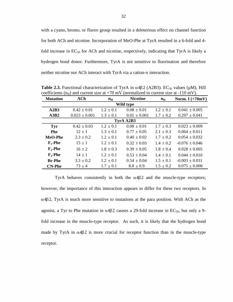
2.3.3 TyrA is a Hydrogen Bond Donor in the α4β2 Receptor 31
2.3.4 The Functions of TyrC1 and TyrC2 are Conserved in the α4β2 and Muscle-type Receptors 33
2.3.5 A Strong Hydrogen Bond in the α4β2 Receptor 34
xii
2.3.6 Studies with the Smoking Cessation Drug Varenicline at the α4β2 Receptor 35
2.3.7 A Residue Outside of the Aromatic Box Differentiates the
α4β2 and Muscle-type Receptors 38
2.3.8 Summary 41
2.4 Methods 41
2.5 Acknowledgements 44
2.6 References 45
CHAPTER 3: Probing the Effects of Residues Located Outside the Agonist Binding Site on Drug-Receptor Selectivity in the Nicotinic Receptor…………………………………………………………………48
3.1 Abstract 48
3.2 Introduction 49
3.3 Results and Discussion 52
3.3.1 Probing the G153 Site in the Low-Potency (α1)2β1γδ (Muscle-type) Receptor 52
3.3.2 Probing the G152 Site in the Low-Potency (α7)5 Receptor 56
3.3.3 Probing the K158 Site in the High-Potency (α4)2(β2)3 Receptor 59
3.3.4 Probing the Proposed Loop B-Loop C Hydrogen Bond 62
3.3.5 Implications for nAChR Function and Subtype Selectivity 64
3.4 Methods 67
3.5 Acknowledgements 71
3.6 References 72
xiii
CHAPTER 4: Contrasting Drug-Receptor Interactions at Neuronal vs. Muscle-Type Nicotinic Acetylcholine Receptors: The Neuronal α4β4 Receptor…………………………………………………….. 74
4.1 Abstract 74
4.2 Introduction 75
4.3 Results 78
4.3.1 Part 1: Using Unnatural Amino Acid Mutagenesis to Probe the Principal Binding Site of the Neuronal α4β4 Receptor 78
4.3.2 Part 2: Using Chimeric β Subunits to Examine the Contribution of the Complementary Binding Site to Subtype-Specific Receptor Pharmacology 87
4.4 Discussion 93
4.5 Methods 97
4.6 Acknowledgements 100
4.7 References 101
CHAPTER 5: Optimizing Techniques to Implement Nonsense Suppression in Mammalian Cells…………………………………………………104
5.1 Abstract 104
5.2 Introduction 105
5.3 Methods 107
5.3.1 Molecular Biology 107
5.3.2 Mammalian Cell Culture 107
5.3.3 The FlexStation 3 109
5.3.4 Fluorescent Dye Experiments 113
xiv
5.4 Results and Discussion 113
5.4.1 Determining the Optimal PMT Setting to Detect Small Fluorescence Signals 113
5.4.2 Transfection of Mammalian Cells 114
5.4.3 Possible Challenges for Nonsense Suppression in Mammalian Cells 133
5.5 Conclusions and Future Directions 138
5.6 Acknowledgments 140
5.7 References 141
xv
LIST OF FIGURES
Figure 1.1 Synaptic transmission 2
Figure 1.2 nAChR agonists studied in this dissertation 4
Figure 1.3 nAChR structure 5
Figure 1.4 nAChR subtypes studied in this dissertation 6
Figure 1.5 A model of the nAChR agonist binding site from AChBP 7
Figure 1.6 Mutation of tyrosine: Comparison of conventional mutagenesis and unnatural amino acid mutagenesis 9
Figure 1.7 Hijacking protein translation: Using nonsense suppression to incorporate unnatural amino acids 11
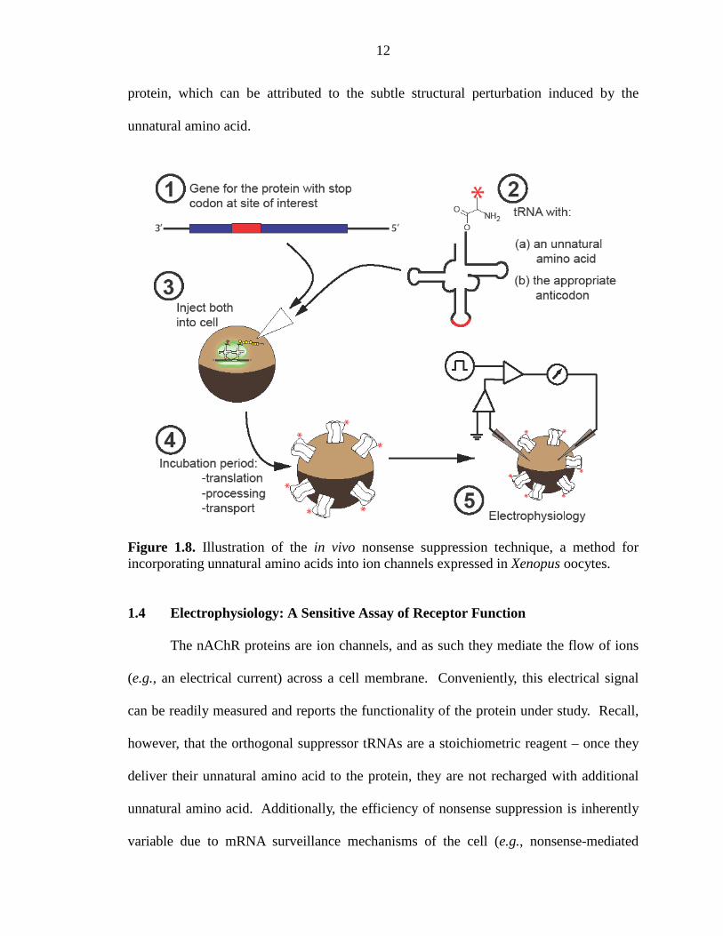
Figure 1.8 Illustration of the in vivo nonsense suppression technique, a method for incorporating unnatural amino acids into ion channels expressed in Xenopus oocytes 12
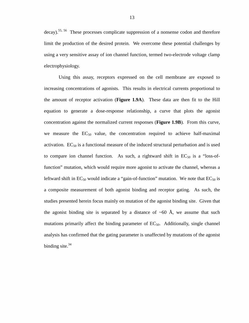
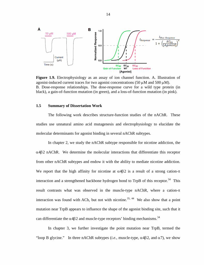
Figure 1.9 Electrophysiology as an assay of ion channel function 14
Figure 2.1 The binding site of AChBP, thought to resemble that of nAChRs 23
Figure 2.2 Sequence alignment for loops A, B, C, and D in the vicinity of the aromatic binding box 23
Figure 2.3 Agonists and unnatural amino acids considered here 25
Figure 2.4 Rectification behaviors of A2B3 and A3B2 α4L9’Aβ2 nAChRs 28
Figure 2.5 Nonsense suppression in the α4β2 receptor 29
Figure 2.6 Fluorination plots for ACh and nicotine at α4β2 and muscle-type receptors 30
Figure 2.7 Fluorination plot for varenicline at α4β2 (A2B3) 36
Figure 2.8 Electrostatic potential surfaces of indole and F4-indole, corresponding to the aromatic portions of the side chains of Trp and F4-Trp, respectively 37
Figure 2.9 Fluorination plot testing nicotine at the muscle-type receptor containing the G153K point mutation in the α1 subunit 40
Figure 3.1 Sequence alignment of the nAChR agonist binding site 49
xvi
Figure 3.2 nAChR agonist binding site, based on the structure of AChBP 51
Figure 3.3 Agonists and unnatural amino acids used in the present study 53
Figure 3.4 Fluorination plots probing the effect of the α1 G153K mutation on the muscle-type nAChR 54
Figure 3.5 Fluorination plots probing the effect of the α7 G152K mutation on the (α7)5 nAChR 58
Figure 3.6 Fluorination plots probing the effect of the α4 K158G
mutation on the (α4)2(β2)3 nAChR 61
Figure 3.7 Comparing the effect on agonist potency of mutating select residues located outside of the (α4)2(β2)3 agonist binding site 62
Figure 4.1 nAChR structure 76
Figure 4.2 Wild type recovery experiments for the α4β4 nAChR 81
Figure 4.3 Key structures employed in this study 82
Figure 4.4 Cation-π binding plots for α4β4 nAChR at position TrpB with ACh and nicotine 84
Figure 4.5 The backbone ester strategy for modulating a hydrogen bond; α-hydroxy acid incorporation 87
Figure 4.6. Design of chimeric β subunits 88
Figure 4.7 Bar graph comparing F3-Trp/Trp fold shifts for chimeric receptors 91
Figure 4.8 Cation-π binding (fluorination) plots monitor the relative
strength of a cation-π interaction at TrpB 92
Figure 4.9 Summary of ligand-receptor interactions present for the
muscle-type, α4β2, α4β4, and α7 nAChRs 94
Figure 5.1 Schematic of the FlexStation 3 microplate reader 110
Figure 5.2 Membrane potential assay 111
Figure 5.3 SOFTmax PRO data analysis of a 96-well plate 112
Figure 5.4 Confocal images of HEK293T cells expressing mouse α4β4 or α4β2 receptors containing GFP in the α4 subunit 115

xvii
Figure 5.5 Dose-reponse curves of human 5HT3A receptors expressed in HEK293T cells 117
Figure 5.6 Dose-response curves of 5HT3A W183A or W183Y mutant receptors expressed in HEK293T cells 118
Figure 5.7 Confocal images of HEK293T cells expressing eGFP 125
Figure 5.8 THG73-W in response to Neon transfection 134
Figure 5.9 THG73-W after TransIT transfection 136
Figure 5.10 Deprotection of THG73-W-NVOC 137
xviii
LIST OF TABLES
Table 2.1 Injection ratio of α4(L9’A):β2 mRNA controls receptor stoichiometry 26
Table 2.2 Functional characterization of TrpB in α4β2 (A2B3) 30
Table 2.3 Functional characterization of TyrA in α4β2 (A2B3) 32
Table 2.4 Functional characterization of TyrC1 in α4β2 (A2B3) 33
Table 2.5 Functional characterization of TyrC2 in α4β2 (A2B3) 34
Table 2.6 Functional characterization of Thr (TrpB + 1) in α4β2 (A2B3) 35
Table 2.7 Functional characterization of varenicline at TrpB in
α4β2 (A2B3) 36
Table 2.8 Evaluation of binding interactions in the α4β2 (A2B3) receptor 37
Table 2.9 Functional characterization of varenicline at Thr (TrpB + 1) in α4β2 (A2B3) 38
Table 2.10 Functional characterization of the muscle-type receptor containing the G153K point mutation in the α1 subunit 40
Table 3.1 Mutation of the loop B Gly/Lys site 52
Table 3.2 EC50 values (µM) and Hill coefficients for mutant
(α1)2β1γδ nAChRs 53
Table 3.3 Amide-to-ester experiments for the backbone carbonyl of TrpB residue for (α1)2β1γδ, (α7)5, and (α4)2(β2)3 55
Table 3.4 EC50 values (µM) and Hill coefficients for mutant (α7)5 nAChRs 57
Table 3.5 EC50 values (µM) and Hill coefficients for mutant
(α4)2(β2)3 nAChRs 60
Table 3.6 EC50 values (µM) and Hill coefficients for mutant (α4)2(β2)3 nAChRs probing the Loop B-Loop C hydrogen bond 63
Table 3.7 Injection ratios of α4 K158G:β2 mRNA used to control
α4β2 receptor stoichiometry in Xenopus oocytes 69
xix
Table 4.1 Stoichiometries of α4β4 nAChRs expressed in Xenopus oocytes injected with different ratios of human α4:β4 subunit mRNA 80
Table 4.2 Data for mutant α4β4 (A2B3) nAChRs 83
Table 4.3 EC50 values (µM) and Hill coefficients for α4β2/β4 chimeras 90
Table 4.4 EC50 values (µM) and Hill coefficients for chimera F 92
Table 4.5 Ratios of mutant EC50 to wild type EC50, such that ratios > 1 represent loss-of-function 93
Table 5.1 Optimization of HEK293T cell seeding for 100 mm dishes 108
Table 5.2 Optimization of HEK293T cell seeding for 35 mm dishes 108
Table 5.3 Comparison of RFU signal detected by the FlexStation 3 for each PMT settings for a serial dilution of rhodamine B 114
Table 5.4 Human α4β4 receptors expressed in HEK293T cells for
given ratios of α4:β4 DNA 116
Table 5.5 Conventional 5HT3A mutant receptors expressed in HEK293T cells 118
Table 5.6 Using ExpressFect to perform wild type recovery of tryptophan at position W183 in the 5HT3A receptor 120
Table 5.7 Using PEI to perform wild type recovery of tryptophan at position W183 in the 5HT3A receptor 120
Table 5.8 Optimizating wild type 5HT3A DNA transfection using the Neon Transfection System 121
Table 5.9 Optimization of Neon electroporation protocol 121
Table 5.10 Using Neon electroporation for wild type recovery incorporating tryptophan at position W183 in the 5HT3A receptor 122
Table 5.11 Using Neon electroporation for wild type recovery of a frameshift codon at position W183 in the 5HT3A receptor 123
Table 5.12 Comparison of Neon electroporation and PEI transfection using HSAS to incorporate serine at position W183 in the 5HT3A receptor 126
xx
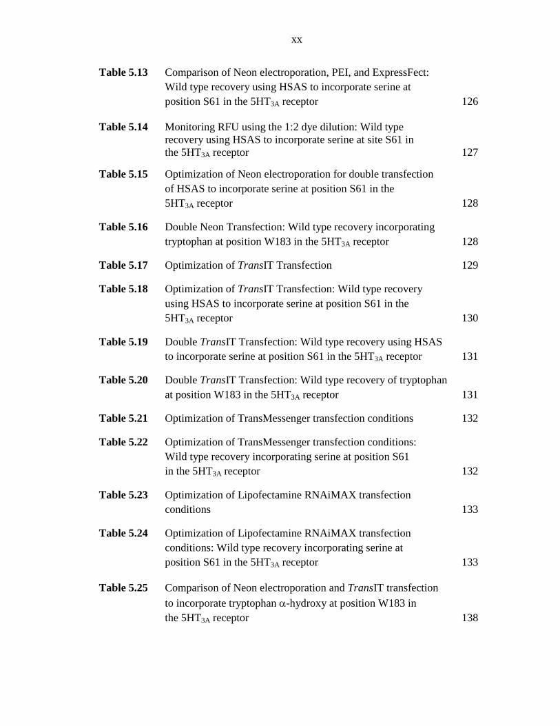
Table 5.13 Comparison of Neon electroporation, PEI, and ExpressFect: Wild type recovery using HSAS to incorporate serine at position S61 in the 5HT3A receptor 126
Table 5.14 Monitoring RFU using the 1:2 dye dilution: Wild type recovery using HSAS to incorporate serine at site S61 in the 5HT3A receptor 127
Table 5.15 Optimization of Neon electroporation for double transfection of HSAS to incorporate serine at position S61 in the 5HT3A receptor 128
Table 5.16 Double Neon Transfection: Wild type recovery incorporating tryptophan at position W183 in the 5HT3A receptor 128
Table 5.17 Optimization of TransIT Transfection 129
Table 5.18 Optimization of TransIT Transfection: Wild type recovery using HSAS to incorporate serine at position S61 in the 5HT3A receptor 130
Table 5.19 Double TransIT Transfection: Wild type recovery using HSAS to incorporate serine at position S61 in the 5HT3A receptor 131
Table 5.20 Double TransIT Transfection: Wild type recovery of tryptophan at position W183 in the 5HT3A receptor 131
Table 5.21 Optimization of TransMessenger transfection conditions 132
Table 5.22 Optimization of TransMessenger transfection conditions: Wild type recovery incorporating serine at position S61 in the 5HT3A receptor 132
Table 5.23 Optimization of Lipofectamine RNAiMAX transfection conditions 133
Table 5.24 Optimization of Lipofectamine RNAiMAX transfection conditions: Wild type recovery incorporating serine at position S61 in the 5HT3A receptor 133
Table 5.25 Comparison of Neon electroporation and TransIT transfection to incorporate tryptophan α-hydroxy at position W183 in the 5HT3A receptor 138
1
Chapter 1
Using Chemical Biology to Study the Brain 1.1 Chemical Signaling in the Brain
As scientists, we continually strive to understand complex biological and
chemical systems with the ultimate goal of comprehending the human condition and
improving human health. From Hippocrates and Aristotle to the modern scientist, we
have been fixated on studying the most complex organ in the human body – the brain.
The adult human brain contains approximately 1011 neurons, and each neuron forms
thousands of connections to other neurons through junctions called synapses. As such,
the resulting 1014 to 1015 synapses form the complex neural network responsible for the
intricacies of cognition and behavioral function. Efficient communication between
neurons is facilitated by neuroreceptors located at these synapses. Modern neurobiology
aims to understand the relationship between the properties of these fundamental brain
components and cognitive function/dysfunction.
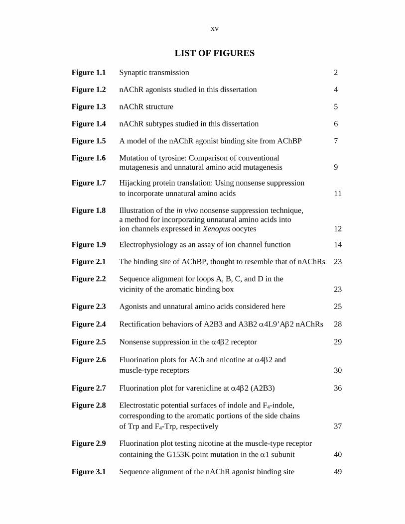
Neurons communicate via synaptic transmission; a process in which a presynaptic
neuron produces a signal and a postsynaptic neuron receives this signal (Figure 1.1A).
This process begins when the presynaptic nerve cell receives information from other
neurons via its dendrites. This information is processed and the presynaptic neuron fires
an electrical signal, called an action potential, which travels down the axon of the
presynaptic neuron. Upon reaching the axon terminal, the action potential triggers the
release of vesicles containing small-molecule neurotransmitters into the synaptic cleft,
the space between neurons. Neurotransmitters diffuse across the synaptic cleft and bind
to receptors embedded within the postsynaptic membrane, the so-called neurotransmitter-
2
gated ion channels. Upon neurotransmitter binding, the receptor undergoes a
conformational change from a closed (non-conducting) state to an open (ion-conducting)
state allowing the flow of ions across the postsynaptic membrane (Figure 1.1B). Thus,
an electrical signal (the action potential) is converted into a chemical signal (the
neurotransmitter) and subsequently back into an electrical signal (ion flow across the
membrane), thereby completing the transmission of information from one cell to another.
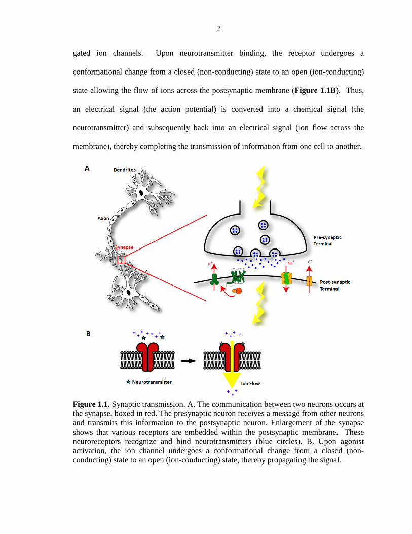
Figure 1.1. Synaptic transmission. A. The communication between two neurons occurs at the synapse, boxed in red. The presynaptic neuron receives a message from other neurons and transmits this information to the postsynaptic neuron. Enlargement of the synapse shows that various receptors are embedded within the postsynaptic membrane. These neuroreceptors recognize and bind neurotransmitters (blue circles). B. Upon agonist activation, the ion channel undergoes a conformational change from a closed (non-conducting) state to an open (ion-conducting) state, thereby propagating the signal.
3
These neuroreceptors are among the molecules of sensory perception, learning,
and memory, and can function at either the presynaptic or the postsynaptic neuron. If
located at the presynaptic terminal, the ligand-gated ion channel usually has a regulatory
function, such as facilitating neurotransmitter release, whereas postsynaptic receptors
propagate rapid electrical signal transmission between neurons.1-3 Regardless of synaptic
location of the ligand-gated ion channel, neurotransmitter binding and receptor activation
are chemical-scale events essential to proper receptor function. As chemists, we are
interested in developing chemical strategies to understand specific chemical interactions
that mediate the structure/function relationship of these complex proteins. We employ
chemical neurobiology to understand the process by which small-molecule
neurotransmitters activate these much larger neuroreceptors proteins.
1.2 Nicotinic Acetylcholine Receptors: The Longest Known and Best-Studied Neuroreceptor
The nicotinic acetylcholine receptor (nAChR) represents a class of
neurotransmitter-gated ion channels belonging to the Cys-loop superfamily of
neuroreceptors, which also includes the γ-aminobutyric acid type A and type C (GABAA
and GABAC), glycine (Gly), and serotonin type 3 (5-HT3) receptors.4 As a family of
complex transmembrane proteins, nAChRs are activated by the endogenous
neurotransmitter acetylcholine. Coincidentally, nAChRs are also activated by the
addictive lipophilic alkaloid, nicotine and other structurally related molecules (Figure
1.2). This class of neuroreceptors is essential to rapid synaptic transmission in the
mammalian central nervous system (CNS) and peripheral nervous system (PNS).4-6
4
Given the abundant source of nAChRs available from the Torpedo electroplax, the
nAChR has become the best-studied and prototypical Cys-loop receptor.4-8
S-Nicotine
N+
HNN+
O
ON+
N
ClH H
EpibatidineAcetylcholine
(ACh)
N
N
N+
Varenicline
H
H
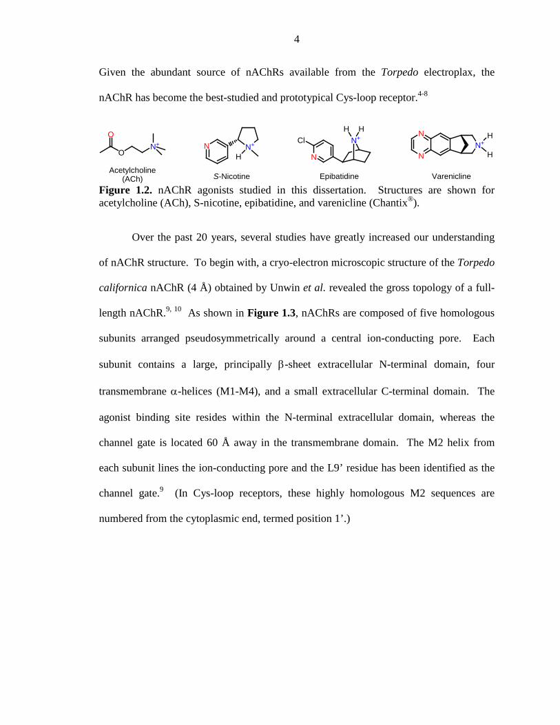
Figure 1.2. nAChR agonists studied in this dissertation. Structures are shown for acetylcholine (ACh), S-nicotine, epibatidine, and varenicline (Chantix®).
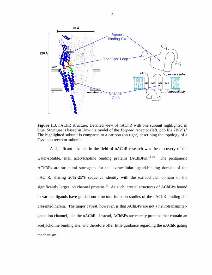
Over the past 20 years, several studies have greatly increased our understanding
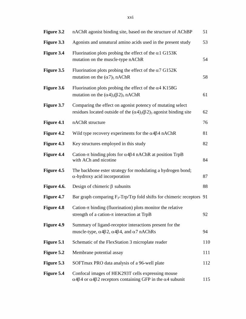
of nAChR structure. To begin with, a cryo-electron microscopic structure of the Torpedo
californica nAChR (4 Å) obtained by Unwin et al. revealed the gross topology of a full-
length nAChR.9, 10 As shown in Figure 1.3, nAChRs are composed of five homologous
subunits arranged pseudosymmetrically around a central ion-conducting pore. Each
subunit contains a large, principally β-sheet extracellular N-terminal domain, four
transmembrane α-helices (M1-M4), and a small extracellular C-terminal domain. The
agonist binding site resides within the N-terminal extracellular domain, whereas the
channel gate is located 60 Å away in the transmembrane domain. The M2 helix from
each subunit lines the ion-conducting pore and the L9’ residue has been identified as the
channel gate.9 (In Cys-loop receptors, these highly homologous M2 sequences are
numbered from the cytoplasmic end, termed position 1’.)
5
Figure 1.3. nAChR structure. Detailed view of nAChR with one subunit highlighted in blue. Structure is based in Unwin’s model of the Torpedo receptor (left, pdb file 2BG9).9 The highlighted subunit is compared to a cartoon (on right) describing the topology of a Cys-loop receptor subunit.
A significant advance in the field of nAChR research was the discovery of the
water-soluble, snail acetylcholine binding proteins (AChBPs).11-16 The pentameric
AChBPs are structural surrogates for the extracellular ligand-binding domain of the
nAChR, sharing 20%–25% sequence identity with the extracellular domain of the
significantly larger ion channel proteins.11 As such, crystal structures of AChBPs bound
to various ligands have guided our structure-function studies of the nAChR binding site
presented herein. The major caveat, however, is that AChBPs are not a neurotransmitter-
gated ion channel, like the nAChR. Instead, AChBPs are merely proteins that contain an
acetylcholine binding site, and therefore offer little guidance regarding the nAChR gating
mechanism.
extracellular
intracellular
Agonist Binding Site
Channel Gate
The “Cys” Loop
out
in membrane
120 Å
70 Å
6
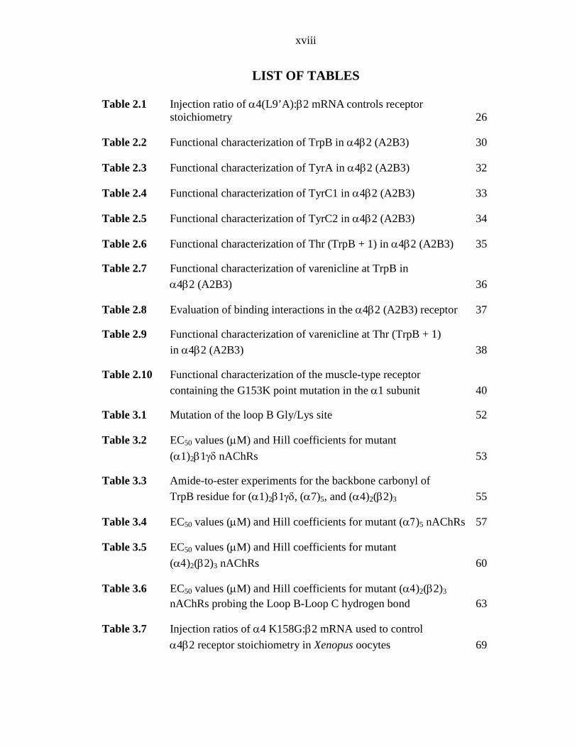
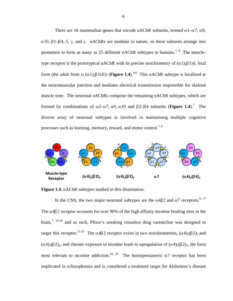
There are 16 mammalian genes that encode nAChR subunits, termed α1–α7, α9,
α10, β1–β4, δ, γ, and ε. nAChRs are modular in nature, so these subunits arrange into
pentamers to form as many as 25 different nAChR subtypes in humans.7, 8 The muscle-
type receptor is the prototypical nAChR with its precise stoichiometry of (α1)2β1γδ, fetal
form (the adult form is (α1)2β1εδ)) (Figure 1.4).4-6 This nAChR subtype is localized at
the neuromuscular junction and mediates electrical transmission responsible for skeletal
muscle tone. The neuronal nAChRs comprise the remaining nAChR subtypes, which are
formed by combinations of α2–α7, α9, α10 and β2–β4 subunits (Figure 1.4).7 The
diverse array of neuronal subtypes is involved in maintaining multiple cognitive
processes such as learning, memory, reward, and motor control.7, 8
Figure 1.4. nAChR subtypes studied in this dissertation.
In the CNS, the two major neuronal subtypes are the α4β2 and α7 receptors.8, 17
The α4β2 receptor accounts for over 90% of the high affinity nicotine binding sites in the
brain,7, 18-20 and as such, Pfizer’s smoking cessation drug varenicline was designed to
target this receptor.21-23 The α4β2 receptor exists in two stoichiometries, (α4)2(β2)3 and
(α4)3(β2)2, and chronic exposure to nicotine leads to upregulation of (α4)2(β2)3, the form
most relevant to nicotine addiction.24, 25 The homopentameric α7 receptor has been
implicated in schizophrenia and is considered a treatment target for Alzheimer’s disease
Muscle-typeReceptor (α4)2(β2)3 (α4)3(β2)2
α1
α1
β1
δ
γ/ε
α4
α4
α4α4
α4
β2
β2β2
β2
β2
+
+
++
++
-
-
--
-- α7
α7 α7
α7
α7 α4
α4 β4
β4
β4
(α4)2(β4)3α7
++
+
++
++
--
-
--
--
7
and other cognitive disorders.7 The last nAChR subtype discussed in this thesis is the
α4β4 subtype, which like α4β2, can arrange in variable stoichometries and is associated
with nicotine addiction.7, 26, 27
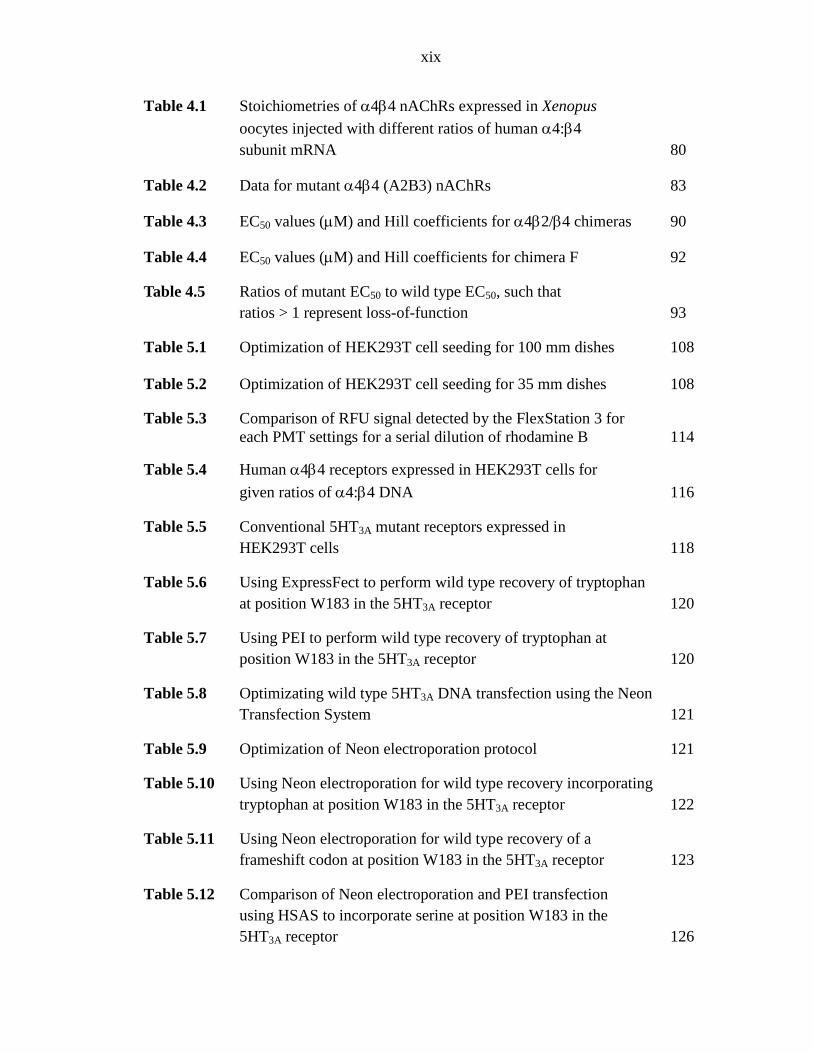
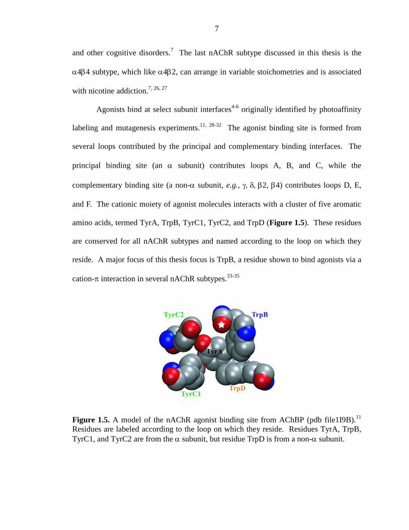
Agonists bind at select subunit interfaces4-6 originally identified by photoaffinity
labeling and mutagenesis experiments.11, 28-32 The agonist binding site is formed from
several loops contributed by the principal and complementary binding interfaces. The
principal binding site (an α subunit) contributes loops A, B, and C, while the
complementary binding site (a non-α subunit, e.g., γ, δ, β2, β4) contributes loops D, E,
and F. The cationic moiety of agonist molecules interacts with a cluster of five aromatic
amino acids, termed TyrA, TrpB, TyrC1, TyrC2, and TrpD (Figure 1.5). These residues
are conserved for all nAChR subtypes and named according to the loop on which they
reside. A major focus of this thesis focus is TrpB, a residue shown to bind agonists via a
cation-π interaction in several nAChR subtypes.33-35
Figure 1.5. A model of the nAChR agonist binding site from AChBP (pdb file1I9B).11 Residues are labeled according to the loop on which they reside. Residues TyrA, TrpB, TyrC1, and TyrC2 are from the α subunit, but residue TrpD is from a non-α subunit.
8
1.3 The Nonsense Suppression Methodology: An Invaluable Tool
Nonsense suppression is a broadly applicable technique that can dissect the
structure-function relationship of various complex proteins,33-36 and is especially useful
for understanding proteins in the absence of a crystal structure. The major advantage of
this approach is the ability to introduce minimal structural modifications to amino acid
side chains, thereby allowing for more accurate interpretations of the effects of a specific
perturbation. This strategy is complementary to conventional site-directed mutagenesis
which can more globally alter or completely abolish side chain functionality within the
confines of the naturally occurring amino acids.
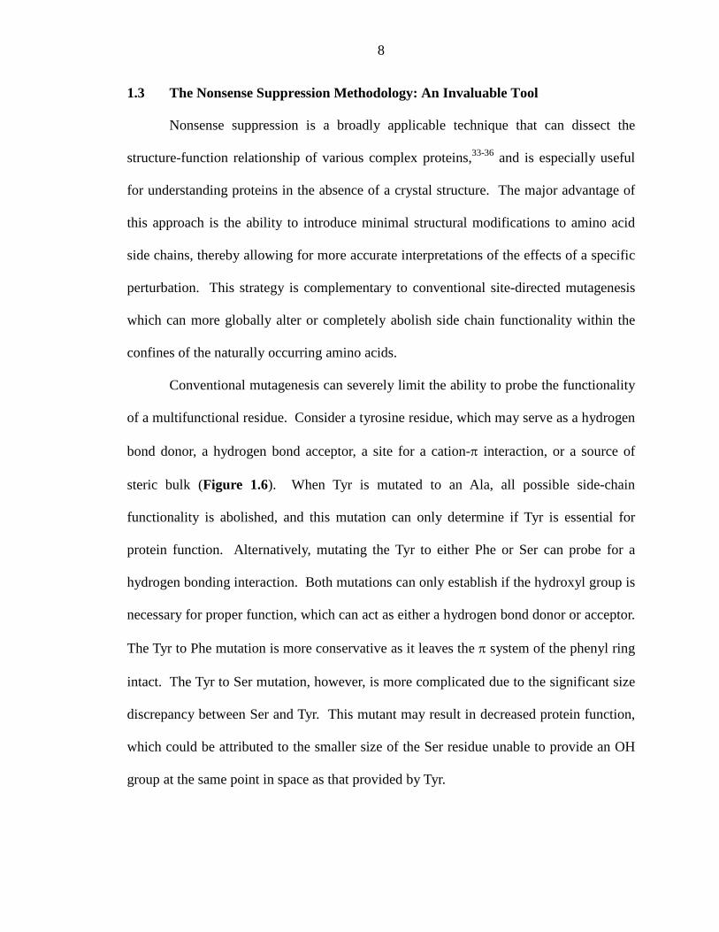
Conventional mutagenesis can severely limit the ability to probe the functionality
of a multifunctional residue. Consider a tyrosine residue, which may serve as a hydrogen
bond donor, a hydrogen bond acceptor, a site for a cation-π interaction, or a source of
steric bulk (Figure 1.6). When Tyr is mutated to an Ala, all possible side-chain
functionality is abolished, and this mutation can only determine if Tyr is essential for
protein function. Alternatively, mutating the Tyr to either Phe or Ser can probe for a
hydrogen bonding interaction. Both mutations can only establish if the hydroxyl group is
necessary for proper function, which can act as either a hydrogen bond donor or acceptor.
The Tyr to Phe mutation is more conservative as it leaves the π system of the phenyl ring
intact. The Tyr to Ser mutation, however, is more complicated due to the significant size
discrepancy between Ser and Tyr. This mutant may result in decreased protein function,
which could be attributed to the smaller size of the Ser residue unable to provide an OH
group at the same point in space as that provided by Tyr.
9
Figure 1.6. Mutation of tyrosine: Comparison of conventional mutagenesis and unnatural amino acid mutagenesis.
Unnatural amino acid mutagenesis, however, can address all of these issues by
expanding the repertoire of amino acids offered by nature (Figure 1.6). For instance,
incorporation of 4-methoxy phenylalanine (4-MeOPhe) allows one to determine whether
the residue of interest acts as a hydrogen bond acceptor. The issue of steric bulk can be
examined by incorporation of 4-methyl phenylalanine (4-MePhe), a residue that would
occupy the same relative space as Tyr. Incorporation of cyclohexylalanine (Cha) can
establish if the aromatic nature of Tyr is important to proper function. Additionally,
incorporation of fluorinated Phe derivatives can investigate the presence of a cation-π
interaction, a non-covalent interaction between the face of an electron-rich π system and
a cation.37-40
We use the nonsense suppression methodology, developed by Schultz in 1989,41
to site-specifically incorporate unnatural amino acids into proteins heterologously
H2NOH
O
HO
H2NOH
O
F
H2NOH
OH2N
OH
O
FF F
FF
H2NOH
O
H2NOH
O
H2NOH
O
CH3O
H2NOH
O
CH3
H2NOH
O
H2NOH
O
Conventional Mutagenesis
Unnatural Amino Acid Mutagenesis
HO
Ala
Phe Ser Cha
F1-Phe F2-Phe F3-Phe
4-MeO-Phe
4-Me-Phe
10
expressed in Xenopus laevis oocytes.42, 43 Using this method, our lab has successfully
determined the ligand binding mechanism and channel gating properties of numerous ion
channels and neuroreceptors.33-35, 44-52
In normal protein synthesis, the ribosome is a multisubunit complex of RNAs and
proteins that functions to decode a template mRNA strand and generate a specific protein
target. The mRNA sequence contains a series of codons that directs the succession of
“charged” tRNA molecules (i.e., tRNA with an amino acid appended to the 3’ end)
containing the appropriate anticodons. Amino acids are linked together via peptide bonds
to form the growing polypeptide chain. Termination of protein synthesis occurs when the
ribosome encounters a STOP or nonsense codon (e.g., UAA, UAG, or UGA), after
which, the polypeptide chain is released.
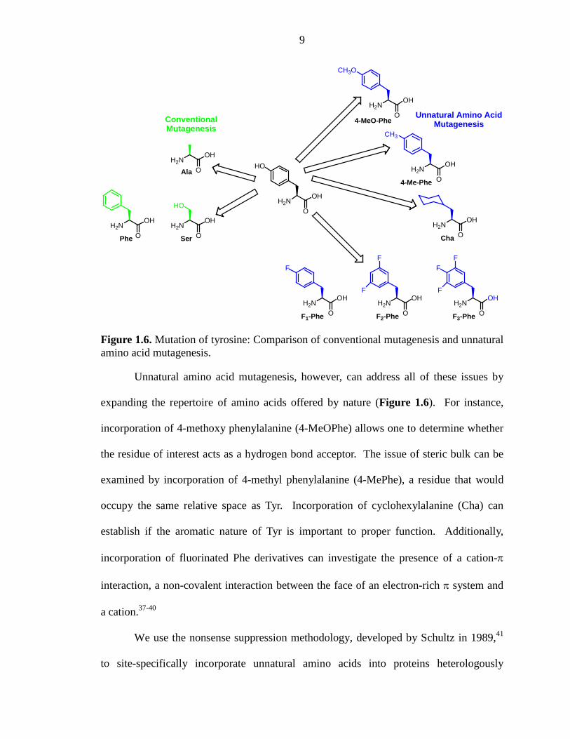
Nonsense suppression, however, “hijacks” the endogenous translational
machinery of the Xenopus oocyte (Figure 1.7). In this process, either a nonsense
codon35, 41 or four-base codon50-52 (e.g., TAG or GGGT) is placed at the amino acid
position of interest in DNA containing the subunit gene. Naturally occurring tRNAs do
not recognize these codons, and as such these codons would normally elicit termination
of protein synthesis or a frameshift mutation, respectively. Instead, we employ a special
suppressor tRNA that contains the correct anticodon and is charged with the unnatural
amino acid of choice linked through a highly reactive ester bond.51-53 The fidelity of this
method relies on the orthogonality of the suppressor tRNA, meaning that the tRNA is not
recognized by the endogenous aminoacyl-tRNA synthesizes of the cell and thereby
avoids recharging with natural amino acids.
11
Figure 1.7. Hijacking protein translation: Using nonsense suppression to incorporate unnatural amino acids.
A full-length, 76 nucleotide suppressor tRNA is made through a non-trivial
process using a combination of synthetic and molecular biology techniques.42, 43, 53, 54
First, the suppressor tRNA is in vitro transcribed as a 74-nucleotide fragment, which is
missing the last two nucleotides of the acceptor stem (cytosine; C and adenine; A). The
deoxy-C and A (dCA) dinucleotide is synthesized and chemically acylated with the
unnatural amino acid (UAA) of choice. Chemical ligation of the dCA- UAA to the
74mer tRNA produces a complete tRNA-UAA molecule.
In the last step of nonsense suppression, in vitro transcribed mRNA containing the
nonsense codon and the tRNA-UAA are coinjected into a Xenopus laevis oocyte, an
unfertilized frog egg (Figure 1.8). The endogenous translational machinery of the oocyte
completes this process by synthesizing, processing, and exporting the desired protein to
the surface of the cell membrane. We then examine the functional properties of the novel
12
protein, which can be attributed to the subtle structural perturbation induced by the
unnatural amino acid.
Figure 1.8. Illustration of the in vivo nonsense suppression technique, a method for incorporating unnatural amino acids into ion channels expressed in Xenopus oocytes.
1.4 Electrophysiology: A Sensitive Assay of Receptor Function
The nAChR proteins are ion channels, and as such they mediate the flow of ions
(e.g., an electrical current) across a cell membrane. Conveniently, this electrical signal
can be readily measured and reports the functionality of the protein under study. Recall,
however, that the orthogonal suppressor tRNAs are a stoichiometric reagent – once they
deliver their unnatural amino acid to the protein, they are not recharged with additional
unnatural amino acid. Additionally, the efficiency of nonsense suppression is inherently
variable due to mRNA surveillance mechanisms of the cell (e.g., nonsense-mediated
13
decay).55, 56 These processes complicate suppression of a nonsense codon and therefore
limit the production of the desired protein. We overcome these potential challenges by
using a very sensitive assay of ion channel function, termed two-electrode voltage clamp
electrophysiology.
Using this assay, receptors expressed on the cell membrane are exposed to
increasing concentrations of agonists. This results in electrical currents proportional to
the amount of receptor activation (Figure 1.9A). These data are then fit to the Hill
equation to generate a dose-response relationship, a curve that plots the agonist
concentration against the normalized current responses (Figure 1.9B). From this curve,
we measure the EC50 value, the concentration required to achieve half-maximal
activation. EC50 is a functional measure of the induced structural perturbation and is used
to compare ion channel function. As such, a rightward shift in EC50 is a “loss-of-
function” mutation, which would require more agonist to activate the channel, whereas a
leftward shift in EC50 would indicate a “gain-of-function” mutation. We note that EC50 is
a composite measurement of both agonist binding and receptor gating. As such, the
studies presented herein focus mainly on mutation of the agonist binding site. Given that
the agonist binding site is separated by a distance of ~60 Å, we assume that such
mutations primarily affect the binding parameter of EC50. Additionally, single channel
analysis has confirmed that the gating parameter is unaffected by mutations of the agonist
binding site.34
14
Figure 1.9. Electrophysiology as an assay of ion channel function. A. Illustration of agonist-induced current traces for two agonist concentrations (50 µM and 500 µM). B. Dose-response relationships. The dose-response curve for a wild type protein (in black), a gain-of-function mutation (in green), and a loss-of-function mutation (in pink).
1.5 Summary of Dissertation Work
The following work describes structure-function studies of the nAChR. These
studies use unnatural amino acid mutagenesis and electrophysiology to elucidate the
molecular determinants for agonist binding in several nAChR subtypes.
In chapter 2, we study the nAChR subtype responsible for nicotine addiction, the
α4β2 nAChR. We determine the molecular interactions that differentiate this receptor
from other nAChR subtypes and endow it with the ability to mediate nicotine addiction.
We report that the high affinity for nicotine at α4β2 is a result of a strong cation-π
interaction and a strengthened backbone hydrogen bond to TrpB of this receptor.34 This
result contrasts what was observed in the muscle-type nAChR, where a cation-π
interaction was found with ACh, but not with nicotine.35, 44 We also show that a point
mutation near TrpB appears to influence the shape of the agonist binding site, such that it
can differentiate the α4β2 and muscle-type receptors’ binding mechanisms.34
In chapter 3, we further investigate the point mutation near TrpB, termed the
“loop B glycine.” In three nAChR subtypes (i.e., muscle-type, α4β2, and α7), we show
15
that the correlation between agonist potency and this loop B site is strong. Low-potency
receptor subtypes have a glycine at the loop B site, while high-potency receptors have a
lysine at this site. We establish that mutation of this residue can to convert a low-potency
receptor to a high-potency receptor and vice versa.
Chapter 4 describes our efforts to understand the agonist binding mechanism of a
fourth nAChR subtype, the α4β4 receptor. We confirm that the α4β4 receptor, like
α4β2, utilizes a strong cation-π interaction to TrpB for both ACh and nicotine, and
nicotine makes a strong hydrogen bond to the backbone carbonyl of TrpB.33
Additionally, we use chimeric β subunits in an attempt to understand how the
complementary binding component can influence agonist binding and receptor
pharmacology in the α4β2 and α4β4 receptors. Together, chapters 2-4 identify structural
features of the nAChR that contribute to differential receptor pharmacology and hold
significant implications for drug discovery efforts seeking to selectively target nAChRs.
Last, chapter 5 takes a shift from the previous chapters and describes a
methodology-based project. This chapter focuses on the optimization of unnatural amino
acid incorporation into mammalian cells and its application to large-scale imaging
techniques, such as the FlexStation 3. We have successfully suppressed an amber stop
codon using HSAS, an in vivo aminoacylated tRNA, in HEK293T cells. Studies are
ongoing to achieve nonsense suppression using in vitro aminoacylated tRNAs.
16
1.6 REFERENCES 1. Boehm, S.; Kubista, H., Fine tuning of sympathetic transmitter release via
ionotropic and metabotropic presynaptic receptors. Pharmacol Rev 2002, 54, (1), 43-99.
2. Ghijsen, W. E.; Leenders, A. G., Differential signaling in presynaptic neurotransmitter release. Cell Mol Life Sci 2005, 62, (9), 937-54.
3. Wonnacott, S.; Barik, J.; Dickinson, J.; Jones, I. W., Nicotinic receptors modulate transmitter cross talk in the CNS: nicotinic modulation of transmitters. J Mol Neurosci 2006, 30, (1-2), 137-40.
4. Corringer, P. J.; Le Novere, N.; Changeux, J. P., Nicotinic receptors at the amino acid level. Annu Rev Pharmacol Toxicol 2000, 40, 431-58.
5. Grutter, T.; Changeux, J. P., Nicotinic receptors in wonderland. Trends Biochem Sci 2001, 26, (8), 459-63.
6. Karlin, A., Emerging structure of the nicotinic acetylcholine receptors. Nat Rev Neurosci 2002, 3, (2), 102-14.
7. Jensen, A. A.; Frolund, B.; Liljefors, T.; Krogsgaard-Larsen, P., Neuronal nicotinic acetylcholine receptors: structural revelations, target identifications, and therapeutic inspirations. J Med Chem 2005, 48, (15), 4705-45.
8. Romanelli, M. N.; Gratteri, P.; Guandalini, L.; Martini, E.; Bonaccini, C.; Gualtieri, F., Central Nicotinic Receptors: Structure, function, ligands, and therapeutic potential. ChemMedChem 2007, 2, (6), 746-767.
9. Unwin, N., Refined structure of the nicotinic acetylcholine receptor at 4A resolution. J Mol Biol 2005, 346, (4), 967-89.
10. Miyazawa, A.; Fujiyoshi, Y.; Unwin, N., Structure and gating mechanism of the acetylcholine receptor pore. Nature 2003, 423, (6943), 949-55.
11. Brejc, K.; van Dijk, W. J.; Klaassen, R. V.; Schuurmans, M.; van Der Oost, J.; Smit, A. B.; Sixma, T. K., Crystal structure of an ACh-binding protein reveals the ligand-binding domain of nicotinic receptors. Nature 2001, 411, (6835), 269-76.
12. Celie, P. H.; van Rossum-Fikkert, S. E.; van Dijk, W. J.; Brejc, K.; Smit, A. B.; Sixma, T. K., Nicotine and carbamylcholine binding to nicotinic acetylcholine receptors as studied in AChBP crystal structures. Neuron 2004, 41, (6), 907-14.
13. Hansen, S. B.; Sulzenbacher, G.; Huxford, T.; Marchot, P.; Bourne, Y.; Taylor, P., Structural characterization of agonist and antagonist-bound acetylcholine-binding protein from Aplysia californica. J Mol Neurosci 2006, 30, (1-2), 101-2.
14. Hansen, S. B.; Sulzenbacher, G.; Huxford, T.; Marchot, P.; Taylor, P.; Bourne, Y., Structures of Aplysia AChBP complexes with nicotinic agonists and antagonists reveal distinctive binding interfaces and conformations. EMBO J 2005, 24, (20), 3635-46.
15. Rucktooa, P.; Smit, A. B.; Sixma, T. K., Insight in nAChR subtype selectivity from AChBP crystal structures. Biochem Pharmacol 2009, 78, (7), 777-87.
16. Taylor, P.; Talley, T. T.; Radic, Z.; Hansen, S. B.; Hibbs, R. E.; Shi, J., Structure-guided drug design: conferring selectivity among neuronal nicotinic receptor and acetylcholine-binding protein subtypes. Biochem Pharmacol 2007, 74, (8), 1164-71.
17
17. Gotti, C.; Zoli, M.; Clementi, F., Brain nicotinic acetylcholine receptors: native subtypes and their relevance. Trends Pharmacol Sci 2006, 27, (9), 482-91.
18. Laviolette, S. R.; van der Kooy, D., The neurobiology of nicotine addiction: bridging the gap from molecules to behaviour. Nat Rev Neurosci 2004, 5, (1), 55-65.
19. Mansvelder, H. D.; Keath, J. R.; McGehee, D. S., Synaptic mechanisms underlie nicotine-induced excitability of brain reward areas. Neuron 2002, 33, (6), 905-19.
20. Mansvelder, H. D.; McGehee, D. S., Cellular and synaptic mechanisms of nicotine addiction. J Neurobiol 2002, 53, (4), 606-17.
21. Coe, J. W.; Brooks, P. R.; Vetelino, M. G.; Wirtz, M. C.; Arnold, E. P.; Huang, J.; Sands, S. B.; Davis, T. I.; Lebel, L. A.; Fox, C. B.; Shrikhande, A.; Heym, J. H.; Schaeffer, E.; Rollema, H.; Lu, Y.; Mansbach, R. S.; Chambers, L. K.; Rovetti, C. C.; Schulz, D. W.; Tingley, F. D., 3rd; O'Neill, B. T., Varenicline: an alpha4beta2 nicotinic receptor partial agonist for smoking cessation. J Med Chem 2005, 48, (10), 3474-7.
22. Gonzales, D.; Rennard, S. I.; Nides, M.; Oncken, C.; Azoulay, S.; Billing, C. B.; Watsky, E. J.; Gong, J.; Williams, K. E.; Reeves, K. R., Varenicline, an alpha4beta2 nicotinic acetylcholine receptor partial agonist, vs sustained-release bupropion and placebo for smoking cessation: a randomized controlled trial. JAMA 2006, 296, (1), 47-55.
23. Mihalak, K. B.; Carroll, F. I.; Luetje, C. W., Varenicline is a partial agonist at alpha4beta2 and a full agonist at alpha7 neuronal nicotinic receptors. Mol Pharmacol 2006, 70, (3), 801-5.
24. Moroni, M.; Bermudez, I., Stoichiometry and pharmacology of two human alpha4beta2 nicotinic receptor types. J Mol Neurosci 2006, 30, (1-2), 95-6.
25. Moroni, M.; Zwart, R.; Sher, E.; Cassels, B. K.; Bermudez, I., alpha4beta2 nicotinic receptors with high and low acetylcholine sensitivity: pharmacology, stoichiometry, and sensitivity to long-term exposure to nicotine. Mol Pharmacol 2006, 70, (2), 755-68.
26. Improgo, M. R.; Scofield, M. D.; Tapper, A. R.; Gardner, P. D., The nicotinic acetylcholine receptor CHRNA5/A3/B4 gene cluster: dual role in nicotine addiction and lung cancer. Prog Neurobiol 92, (2), 212-26.
27. Wu, J.; Liu, Q.; Yu, K.; Hu, J.; Kuo, Y. P.; Segerberg, M.; St John, P. A.; Lukas, R. J., Roles of nicotinic acetylcholine receptor beta subunits in function of human alpha4-containing nicotinic receptors. J Physiol 2006, 576, (Pt 1), 103-18.
28. Chabala, L. D.; Lester, H. A., Activation of acetylcholine receptor channels by covalently bound agonists in cultured rat myoballs. J Physiol 1986, 379, 83-108.
29. Czajkowski, C.; Kaufmann, C.; Karlin, A., Negatively charged amino acid residues in the nicotinic receptor delta subunit that contribute to the binding of acetylcholine. Proc Natl Acad Sci USA 1993, 90, (13), 6285-9.
30. Dennis, M.; Giraudat, J.; Kotzyba-Hibert, F.; Goeldner, M.; Hirth, C.; Chang, J. Y.; Lazure, C.; Chretien, M.; Changeux, J. P., Amino acids of the Torpedo marmorata acetylcholine receptor alpha subunit labeled by a photoaffinity ligand for the acetylcholine binding site. Biochemistry 1988, 27, (7), 2346-57.
31. Karlin, A., Chemical modification of the active site of the acetylcholine receptor. J Gen Physiol 1969, 54, (1), 245-64.
18
32. Silman, I.; Karlin, A., Acetylcholine receptor: covalent attachment of depolarizing groups at the active site. Science 1969, 164, (3886), 1420-1.
33. Puskar, N. L.; Xiu, X.; Lester, H. A.; Dougherty, D. A., Two neuronal nicotinic acetylcholine receptors, alpha4beta4 and alpha7, show differential agonist binding modes. J Biol Chem 286, (16), 14618-27.
34. Xiu, X.; Puskar, N. L.; Shanata, J. A.; Lester, H. A.; Dougherty, D. A., Nicotine binding to brain receptors requires a strong cation-pi interaction. Nature 2009, 458, (7237), 534-7.
35. Zhong, W.; Gallivan, J. P.; Zhang, Y.; Li, L.; Lester, H. A.; Dougherty, D. A., From ab initio quantum mechanics to molecular neurobiology: a cation-pi binding site in the nicotinic receptor. Proc Natl Acad Sci U S A 1998, 95, (21), 12088-93.
36. Dougherty, D. A., Unnatural amino acids as probes of protein structure and function. Curr Opin Chem Biol 2000, 4, (6), 645-52.
37. Dougherty, D. A., Cation-pi interactions in chemistry and biology: a new view of benzene, Phe, Tyr, and Trp. Science 1996, 271, (5246), 163-8.
38. Gallivan, J. P.; Dougherty, D. A., Cation-pi interactions in structural biology. Proc Natl Acad Sci U S A 1999, 96, (17), 9459-64.
39. Ma, J. C.; Dougherty, D. A., The Cation-pi Interaction. Chem Rev 1997, 97, (5), 1303-1324.
40. Zacharias, N.; Dougherty, D. A., Cation-pi interactions in ligand recognition and catalysis. Trends Pharmacol Sci 2002, 23, (6), 281-7.
41. Noren, C. J.; Anthony-Cahill, S. J.; Griffith, M. C.; Schultz, P. G., A general method for site-specific incorporation of unnatural amino acids into proteins. Science 1989, 244, (4901), 182-8.
42. Nowak, M. W.; Gallivan, J. P.; Silverman, S. K.; Labarca, C. G.; Dougherty, D. A.; Lester, H. A., In vivo incorporation of unnatural amino acids into ion channels in Xenopus oocyte expression system. Methods Enzymol 1998, 293, 504-29.
43. Nowak, M. W.; Kearney, P. C.; Sampson, J. R.; Saks, M. E.; Labarca, C. G.; Silverman, S. K.; Zhong, W.; Thorson, J.; Abelson, J. N.; Davidson, N.; et al., Nicotinic receptor binding site probed with unnatural amino acid incorporation in intact cells. Science 1995, 268, (5209), 439-42.
44. Beene, D. L.; Brandt, G. S.; Zhong, W.; Zacharias, N. M.; Lester, H. A.; Dougherty, D. A., Cation-pi interactions in ligand recognition by serotonergic (5-HT3A) and nicotinic acetylcholine receptors: the anomalous binding properties of nicotine. Biochemistry 2002, 41, (32), 10262-9.
45. Cashin, A. L.; Petersson, E. J.; Lester, H. A.; Dougherty, D. A., Using physical chemistry to differentiate nicotinic from cholinergic agonists at the nicotinic acetylcholine receptor. J Am Chem Soc 2005, 127, (1), 350-6.
46. Gallivan, J. P.; Lester, H. A.; Dougherty, D. A., Site-specific incorporation of biotinylated amino acids to identify surface-exposed residues in integral membrane proteins. Chem Biol 1997, 4, (10), 739-49.
47. Lummis, S. C.; Beene, D. L.; Lee, L. W.; Lester, H. A.; Broadhurst, R. W.; Dougherty, D. A., Cis-trans isomerization at a proline opens the pore of a neurotransmitter-gated ion channel. Nature 2005, 438, (7065), 248-52.
19
48. Lummis, S. C.; D, L. B.; Harrison, N. J.; Lester, H. A.; Dougherty, D. A., A cation-pi binding interaction with a tyrosine in the binding site of the GABAC receptor. Chem Biol 2005, 12, (9), 993-7.
49. Mu, T. W.; Lester, H. A.; Dougherty, D. A., Different binding orientations for the same agonist at homologous receptors: a lock and key or a simple wedge? J Am Chem Soc 2003, 125, (23), 6850-1.
50. Rodriguez, E. A.; Lester, H. A.; Dougherty, D. A., In vivo incorporation of multiple unnatural amino acids through nonsense and frameshift suppression. Proc Natl Acad Sci U S A 2006, 103, (23), 8650-5.
51. Rodriguez, E. A.; Lester, H. A.; Dougherty, D. A., Improved amber and opal suppressor tRNAs for incorporation of unnatural amino acids in vivo. Part 1: minimizing misacylation. RNA 2007, 13, (10), 1703-14.
52. Rodriguez, E. A.; Lester, H. A.; Dougherty, D. A., Improved amber and opal suppressor tRNAs for incorporation of unnatural amino acids in vivo. Part 2: evaluating suppression efficiency. RNA 2007, 13, (10), 1715-22.
53. Saks, M. E.; Sampson, J. R.; Nowak, M. W.; Kearney, P. C.; Du, F.; Abelson, J. N.; Lester, H. A.; Dougherty, D. A., An engineered Tetrahymena tRNAGln for in vivo incorporation of unnatural amino acids into proteins by nonsense suppression. J Biol Chem 1996, 271, (38), 23169-75.
54. Dougherty, D. A., Physical organic chemistry on the brain. J Org Chem 2008, 73, (10), 3667-73.
55. Nicholson, P.; Yepiskoposyan, H.; Metze, S.; Zamudio Orozco, R.; Kleinschmidt, N.; Muhlemann, O., Nonsense-mediated mRNA decay in human cells: mechanistic insights, functions beyond quality control and the double-life of NMD factors. Cell Mol Life Sci 67, (5), 677-700.
56. Whitfield, T. T.; Sharpe, C. R.; Wylie, C. C., Nonsense-mediated mRNA decay in Xenopus oocytes and embryos. Dev Biol 1994, 165, (2), 731-4.
20
Chapter 2
Nicotine Binding to Brain Receptors Requires a Strong Cation–π Interaction*
*This chapter is adapted in part from: Xiu, X.†; Puskar, N. L.†; Shanata, J. A. P.; Lester, H. A.; Dougherty, D. A. Nicotine binding to brain receptors requires a strong cation–π interaction. Nature. 2009; 458: 534-538. © Nature Publishing Group, a division of Macmillan Publishers Limited. The work described in this chapter concerning varenicline was done in collaboration with Ximena Da Silva Tavares Bongoll, Dr. Angela P. Blum, Darren T. Nakamura, and Dr. Jai A. P. Shanata. †Denotes equal contribution.
2.1 ABSTRACT
Nicotine addiction begins with high-affinity binding of nicotine to acetylcholine
(ACh) receptors in the brain. The end result is over 4,000,000 smoking-related deaths
annually worldwide and the largest source of preventable mortality in developed
countries. Stress reduction, pleasure, improved cognition and other central nervous
system effects are strongly associated with smoking. However, if nicotine activated ACh
receptors found in muscle as potently as it does brain ACh receptors, smoking would
cause intolerable and perhaps fatal muscle contractions. Despite extensive
pharmacological, functional, and structural studies of ACh receptors, the basis for the
differential action of nicotine on brain compared with muscle ACh receptors has not been
determined. Here we show that at the α4β2 brain receptors thought to underlie nicotine
addiction, the high affinity for nicotine binding is the result of a strong cation–π
interaction to a specific aromatic amino acid of the receptor, TrpB. In contrast, the low
affinity for nicotine at the muscle-type ACh receptor is largely due to the fact that this
key interaction is absent, even though the immediate binding site residues, including the
21
key amino acid TrpB, are identical in the brain and muscle receptors. At the same time a
hydrogen bond from nicotine to the backbone carbonyl of TrpB is enhanced in the
neuronal receptor relative to the muscle-type. The cation-π interaction and hydrogen
bond are also present between TrpB and the smoking cessation compound varenicline
(Chantix®) in the α4β2 receptor. Additionally, a point mutation near TrpB that
differentiates α4β2 and muscle-type receptors seems to influence the shape of the
binding site, allowing nicotine to interact more strongly with TrpB in the neuronal
receptor. ACh receptors are established therapeutic targets for Alzheimer’s disease,
schizophrenia, Parkinson’s disease, smoking cessation, pain, attention-deficit
hyperactivity disorder, epilepsy, autism, and depression.1 Along with solving a chemical
mystery in nicotine addiction, our results provide guidance for efforts to develop drugs
that target specific types of nicotinic receptors.
2.2 INTRODUCTION
Nicotinic acetylcholine receptors (nAChRs) comprise a family of ≥20
homologous subtypes that mediate fast synaptic transmission throughout the central and
peripheral nervous systems.2 The neuronal nAChRs are found in the central nervous
system (CNS) and autonomic ganglia. Of these, the subtype most strongly associated
with nicotine addiction and the target of recently developed smoking cessation drugs is
termed α4β2.3-7 The high nicotine affinity of α4β2 receptors, when combined with the
ability of nicotine to cross the blood–brain barrier and its favourable pharmacokinetics,
allows nicotine at the submicromolar concentrations in tobacco smoke to activate acutely
these receptors, providing reward, cognitive sensitization, and perhaps other effects. In
addition, the high-affinity interaction allows smoked nicotine to act as an intracellular
22
pharmacological chaperone of α4β2 receptors, leading to the upregulation of receptors
thought to underlie effects of chronic exposure.6-8
In previous studies of the nAChR of the neuromuscular junction (muscle-type),
we showed that an important contributor to ACh binding is a cation-π interaction to a
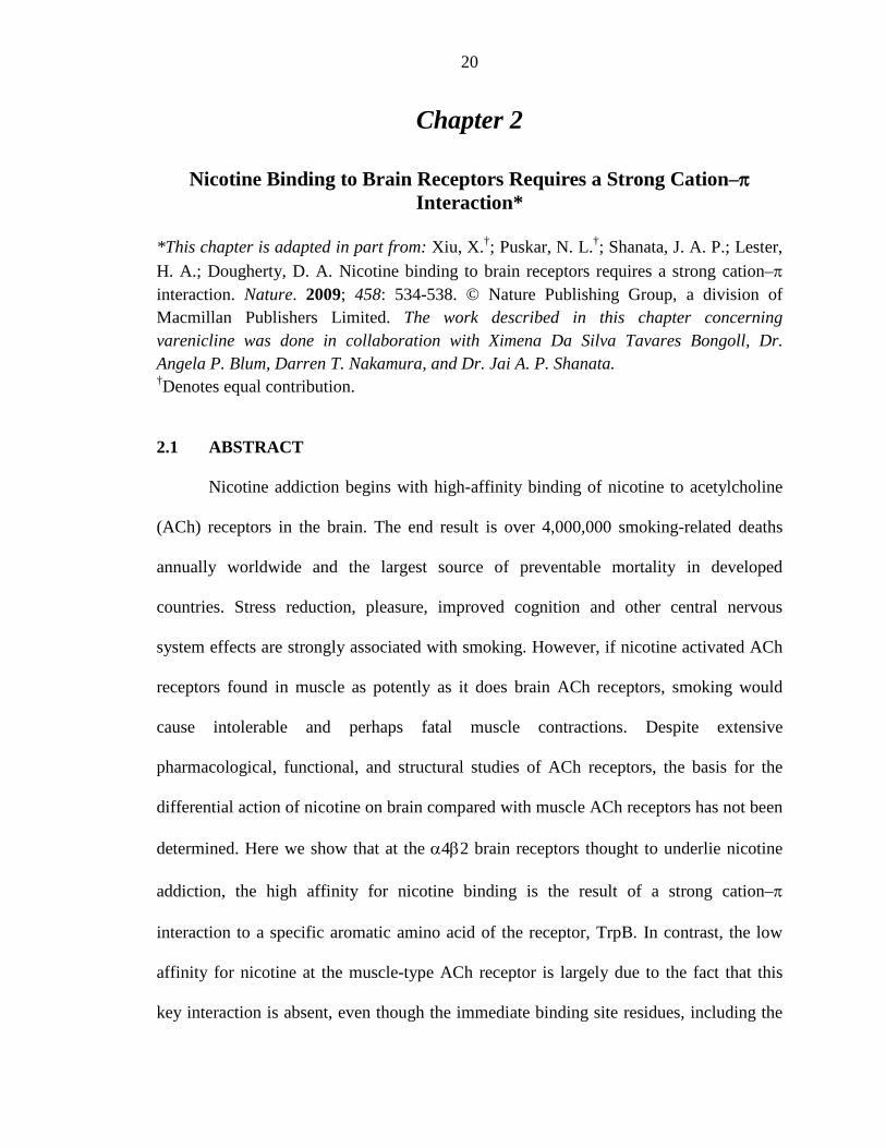
specific tryptophan (called TrpB, residue 149, Figure 2.1).9 These results were
subsequently supported by the important series of crystal structures of ACh binding
proteins (AChBP).10, 11 These structures revealed the “aromatic box” structural motif of
Figure 2.1, and the aligning residues are predominantly aromatic throughout the Cys-
loop family of neurotransmitter-gated ion channels. In other Cys-loop receptors, a cation–
π interaction between the natural agonist and one of the aromatics is always seen,
although its precise location varies.12 Interestingly, when nicotine activates the muscle-
type nAChR, there is no cation–π interaction,13 consistent with its relatively low affinity
for this receptor. This suggested that a cation–π interaction could discriminate between
high-affinity neuronal receptors and low-affinity muscle-type receptors. However, subtle
effects must be involved, as the nAChRs of the CNS and neuromuscular junction are
homologous throughout most regions of sequence and are essentially identical in the
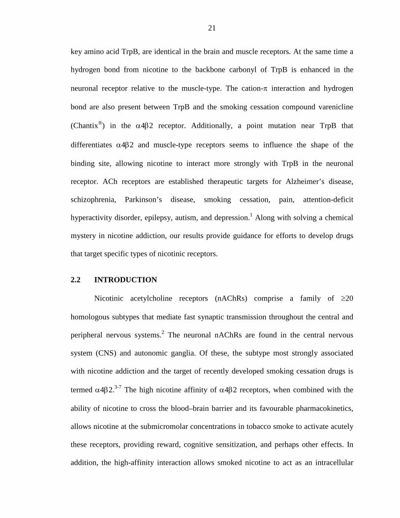
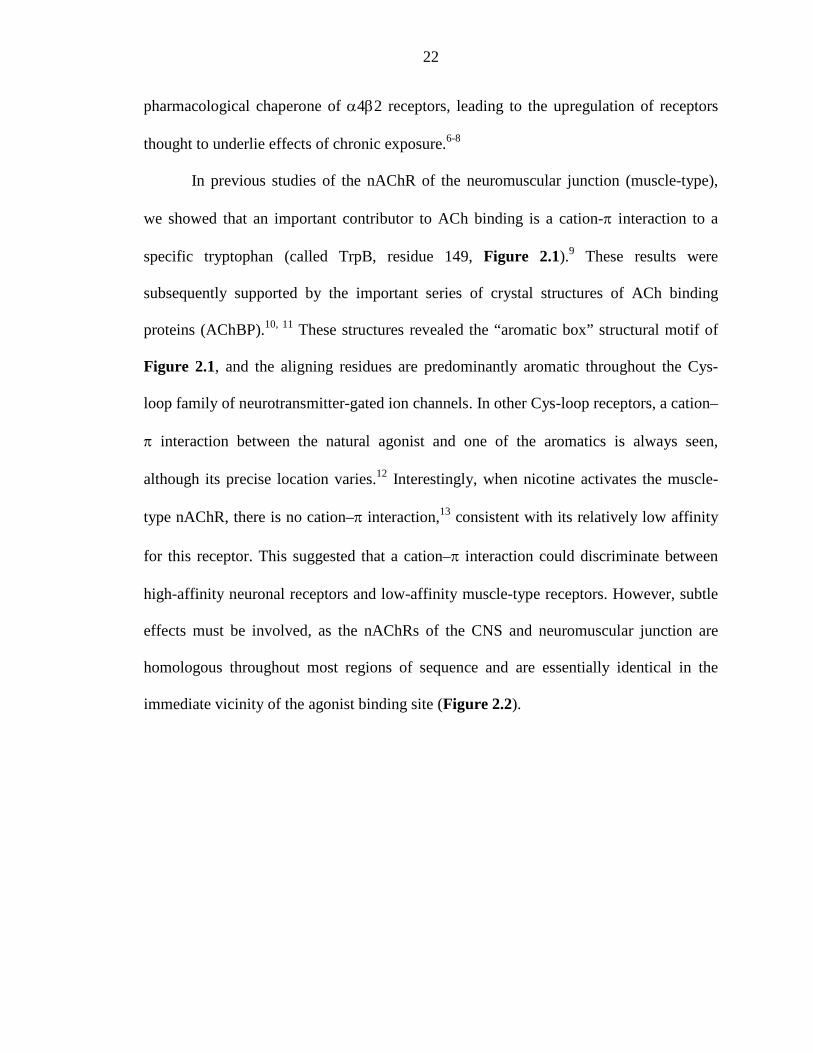
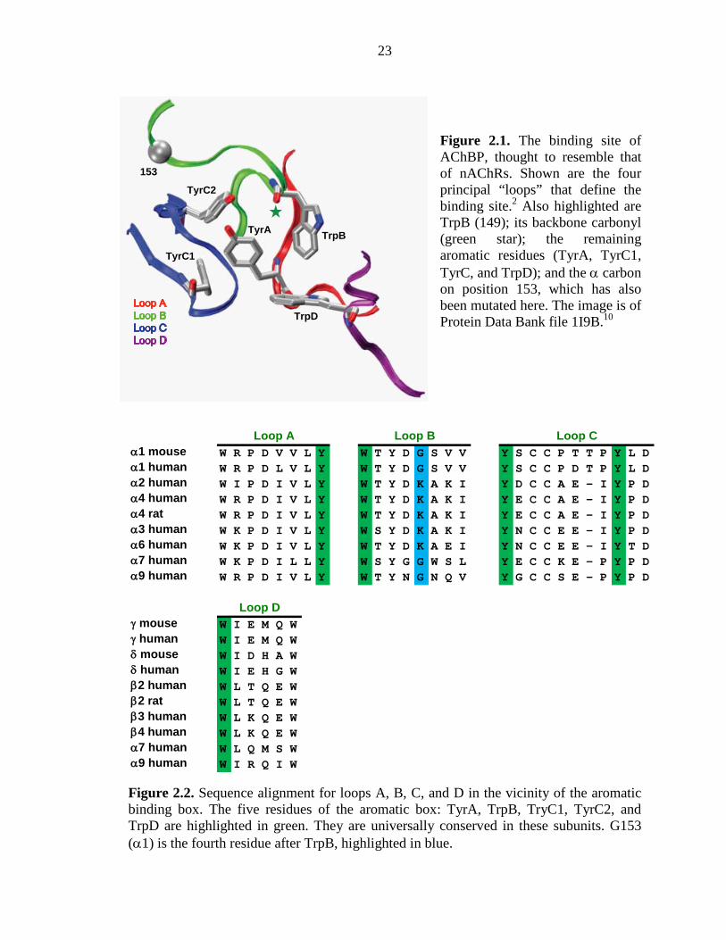
immediate vicinity of the agonist binding site (Figure 2.2).
23
Figure 2.1. The binding site of AChBP, thought to resemble that of nAChRs. Shown are the four principal “loops” that define the binding site.2 Also highlighted are TrpB (149); its backbone carbonyl (green star); the remaining aromatic residues (TyrA, TyrC1, TyrC, and TrpD); and the α carbon on position 153, which has also been mutated here. The image is of Protein Data Bank file 1I9B.10
Figure 2.2. Sequence alignment for loops A, B, C, and D in the vicinity of the aromatic binding box. The five residues of the aromatic box: TyrA, TrpB, TryC1, TyrC2, and TrpD are highlighted in green. They are universally conserved in these subunits. G153 (α1) is the fourth residue after TrpB, highlighted in blue.
α1 mouse W R P D V V L Y W T Y D G S V V Y S C C P T T P Y L Dα1 human W R P D L V L Y W T Y D G S V V Y S C C P D T P Y L Dα2 human W I P D I V L Y W T Y D K A K I Y D C C A E - I Y P Dα4 human W R P D I V L Y W T Y D K A K I Y E C C A E - I Y P Dα4 rat W R P D I V L Y W T Y D K A K I Y E C C A E - I Y P Dα3 human W K P D I V L Y W S Y D K A K I Y N C C E E - I Y P Dα6 human W K P D I V L Y W T Y D K A E I Y N C C E E - I Y T Dα7 human W K P D I L L Y W S Y G G W S L Y E C C K E - P Y P Dα9 human W R P D I V L Y W T Y N G N Q V Y G C C S E - P Y P D
γ mouse W I E M Q Wγ human W I E M Q Wδ mouse W I D H A Wδ human W I E H G Wβ2 human W L T Q E Wβ2 rat W L T Q E Wβ3 human W L K Q E Wβ4 human W L K Q E Wα7 human W L Q M S Wα9 human W I R Q I W
Loop A Loop B Loop C
Loop D
TrpB
153
TyrA
TrpD
TyrC1
TyrC2
24
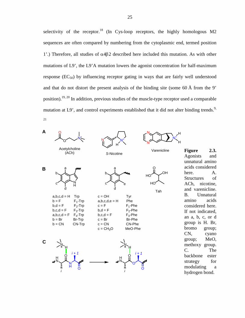
Here we describe studies of the neuronal α4β2 receptor. We find a remarkable
distinction in binding behaviour: both the endogenous neurotransmitter ACh and the
addictive nicotine molecule make a strong cation–π interaction to TrpB. In addition, a
hydrogen bond from nicotine to the backbone carbonyl of TrpB that is weak in the
muscle-type is much stronger in the α4β2 receptor. The smoking cessation drug
varenicline (marketed as Chantix® in the U.S.) was designed to target α4β2 receptors,3, 14,
15 and in fact makes the cation-π interaction and hydrogen bond. Taken together, these
two noncovalent interactions fully rationalize the differential affinity of nicotine in the
brain vs. the neuromuscular junction.
2.3 RESULTS AND DISCUSSION
2.3.1 Challenges in Studying Neuronal nAChRs
A cation–π interaction between a drug and a receptor can be revealed by
incorporation of a series of fluorinated amino acid analogues (Figure 2.3); a consistent
trend in receptor response indicates a binding interaction.9 Such an experiment is enabled
by the nonsense suppression methodology for incorporation of unnatural amino acids into
receptors and channels expressed in Xenopus oocytes. Although we have found the
nonsense suppression methodology to be broadly applicable,16, 17 implementing the
methodology for study of the α4β2 neuronal nAChRs proved to be especially
challenging, requiring new strategies. The α4β2 receptors are expressed in Xenopus
oocytes at inadequately low levels for nonsense suppression experiments. However,
recent studies showed that the Leu9’Ala (L9’A) mutation in the M2 transmembrane helix
of the α4 subunit greatly improves expression without altering the pharmacological
25
selectivity of the receptor.18 (In Cys-loop receptors, the highly homologous M2
sequences are often compared by numbering from the cytoplasmic end, termed position
1’.) Therefore, all studies of α4β2 described here included this mutation. As with other
mutations of L9’, the L9’A mutation lowers the agonist concentration for half-maximum
response (EC50) by influencing receptor gating in ways that are fairly well understood
and that do not distort the present analysis of the binding site (some 60 Å from the 9’
position).19, 20 In addition, previous studies of the muscle-type receptor used a comparable
mutation at L9’, and control experiments established that it did not alter binding trends.9,
21
Figure 2.3. Agonists and unnatural amino acids considered here. A. Structures of ACh, nicotine, and varenicline. B. Unnatural amino acids considered here. If not indicated, an a, b, c, or d group is H. Br, bromo group; CN, cyano group; MeO, methoxy group. C. The backbone ester strategy for modulating a hydrogen bond.
S-Nicotine
N+
HNN+
O
O
Acetylcholine(ACh)
N
N
N+
Varenicline
H
H
NH O
OHN
NH
i
i + 1
OO
OHN
NH
i
i + 1
A
B
C
NH
ab
cd
a,b,c,d = H Trpb = F F1-Trpb,d = F F2-Trpb,c,d = F F3-Trpa,b,c,d = F F4-Trpb = Br Br-Trpb = CN CN-Trp
ab
cd
e
c = OH Tyra,b,c,d,e = H Phec = F F1-Pheb,d = F F2-Pheb,c,d = F F3-Phec = Br Br-Phec = CN CN-Phec = CH3O MeO-Phe
HO
HO
OOH
Tah
26
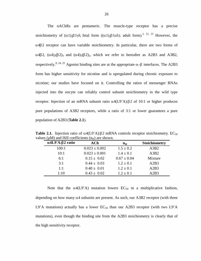
The nAChRs are pentameric. The muscle-type receptor has a precise
stoichiometry of (α1)2β1γδ, fetal form ((α1)2β1εδ); adult form).2, 22, 23 However, the
α4β2 receptor can have variable stoichiometry. In particular, there are two forms of
α4β2, (α4)2(β2)3 and (α4)3(β2)2, which we refer to hereafter as A2B3 and A3B2,
respectively.8, 24, 25 Agonist binding sites are at the appropriate α–β interfaces. The A2B3
form has higher sensitivity for nicotine and is upregulated during chronic exposure to
nicotine; our studies have focused on it. Controlling the ratios of messenger RNAs
injected into the oocyte can reliably control subunit stoichiometry in the wild type
receptor. Injection of an mRNA subunit ratio α4(L9’A):β2 of 10:1 or higher produces
pure populations of A3B2 receptors, while a ratio of 3:1 or lower guarantees a pure
population of A2B3 (Table 2.1).
Table 2.1. Injection ratio of α4(L9’A):β2 mRNA controls receptor stoichiometry. EC50 values (µM) and Hill coefficients (nH) are shown.
Note that the α4(L9’A) mutation lowers EC50 in a multiplicative fashion,
depending on how many α4 subunits are present. As such, our A3B2 receptor (with three
L9’A mutations) actually has a lower EC50 than our A2B3 receptor (with two L9’A
mutations), even though the binding site from the A2B3 stoichiometry is clearly that of
the high sensitivity receptor.
α4L9'A:β2 ratio ACh nH Stoichiometry100:1 0.023 ± 0.002 1.5 ± 0.2 A3B210:1 0.023 ± 0.001 1.4 ± 0.1 A3B26:1 0.15 ± 0.02 0.67 ± 0.04 Mixture3:1 0.44 ± 0.03 1.2 ± 0.1 A2B31:1 0.40 ± 0.01 1.2 ± 0.1 A2B3
1:10 0.43 ± 0.02 1.2 ± 0.1 A2B3
27
In a nonsense suppression experiment, however, the subunit that contains the stop
codon where the unnatural amino acid has been incorporated can show low and variable
expression levels. Therefore we sought a second, independent indicator of the
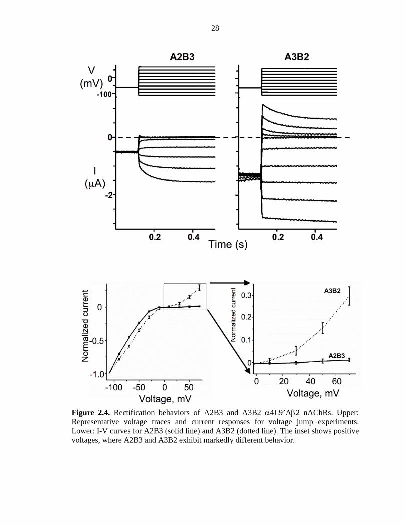
stoichiometry of the α4β2 receptor. We now report that the A2B3 and A3B2 forms of the
α4(L9’A)β2 receptor show markedly different rectification behaviours. As indicated by
either voltage ramp or voltage jump experiments, A2B3 is substantially more inward
rectifying than A3B2 (Figure 2.4). Thus, in all our experiments with unnatural amino
acids, the stoichiometries of mutant receptors are monitored by measuring current–
voltage relations with voltage jumps. For each mutant receptor studied, we determined
the fraction (outward current at +70 mV/inward current at –110 mV), and a value ≤0.1
establishes the desired A2B3 stoichiometry (Tables 2.2-2.7). With these methodological
developments in hand, incorporation of unnatural amino acids into the α4β2 receptor
becomes feasible (Figure 2.5).
28
Figure 2.4. Rectification behaviors of A2B3 and A3B2 α4L9’Aβ2 nAChRs. Upper: Representative voltage traces and current responses for voltage jump experiments. Lower: I-V curves for A2B3 (solid line) and A3B2 (dotted line). The inset shows positive voltages, where A2B3 and A3B2 exhibit markedly different behavior.
29
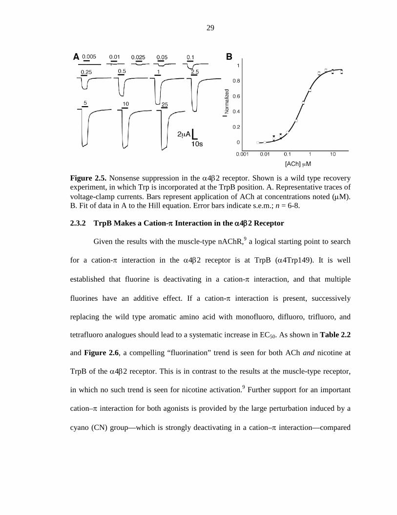
Figure 2.5. Nonsense suppression in the α4β2 receptor. Shown is a wild type recovery experiment, in which Trp is incorporated at the TrpB position. A. Representative traces of voltage-clamp currents. Bars represent application of ACh at concentrations noted (µM). B. Fit of data in A to the Hill equation. Error bars indicate s.e.m.; n = 6-8.
2.3.2 TrpB Makes a Cation-π Interaction in the α4β2 Receptor
Given the results with the muscle-type nAChR,9 a logical starting point to search
for a cation-π interaction in the α4β2 receptor is at TrpB (α4Trp149). It is well
established that fluorine is deactivating in a cation-π interaction, and that multiple
fluorines have an additive effect. If a cation-π interaction is present, successively
replacing the wild type aromatic amino acid with monofluoro, difluoro, trifluoro, and
tetrafluoro analogues should lead to a systematic increase in EC50. As shown in Table 2.2
and Figure 2.6, a compelling “fluorination” trend is seen for both ACh and nicotine at
TrpB of the α4β2 receptor. This is in contrast to the results at the muscle-type receptor,
in which no such trend is seen for nicotine activation.9 Further support for an important
cation–π interaction for both agonists is provided by the large perturbation induced by a
cyano (CN) group—which is strongly deactivating in a cation–π interaction—compared
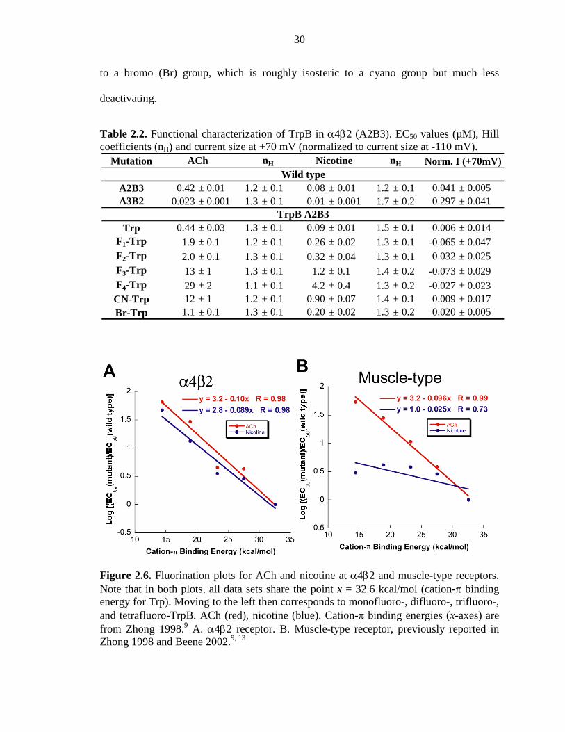
30
to a bromo (Br) group, which is roughly isosteric to a cyano group but much less
deactivating.
Table 2.2. Functional characterization of TrpB in α4β2 (A2B3). EC50 values (µM), Hill coefficients (nH) and current size at +70 mV (normalized to current size at -110 mV).
Figure 2.6. Fluorination plots for ACh and nicotine at α4β2 and muscle-type receptors. Note that in both plots, all data sets share the point x = 32.6 kcal/mol (cation-π binding energy for Trp). Moving to the left then corresponds to monofluoro-, difluoro-, trifluoro-, and tetrafluoro-TrpB. ACh (red), nicotine (blue). Cation-π binding energies (x-axes) are from Zhong 1998.9 A. α4β2 receptor. B. Muscle-type receptor, previously reported in Zhong 1998 and Beene 2002.9, 13
Mutation
A2B3 0.42 ± 0.01 1.2 ± 0.1 0.08 ± 0.01 1.2 ± 0.1 0.041 ± 0.005A3B2 0.023 ± 0.001 1.3 ± 0.1 0.01 ± 0.001 1.7 ± 0.2 0.297 ± 0.041
Trp 0.44 ± 0.03 1.3 ± 0.1 0.09 ± 0.01 1.5 ± 0.1 0.006 ± 0.014F1-Trp 1.9 ± 0.1 1.2 ± 0.1 0.26 ± 0.02 1.3 ± 0.1 -0.065 ± 0.047F2-Trp 2.0 ± 0.1 1.3 ± 0.1 0.32 ± 0.04 1.3 ± 0.1 0.032 ± 0.025F3-Trp 13 ± 1 1.3 ± 0.1 1.2 ± 0.1 1.4 ± 0.2 -0.073 ± 0.029F4-Trp 29 ± 2 1.1 ± 0.1 4.2 ± 0.4 1.3 ± 0.2 -0.027 ± 0.023
CN-Trp 12 ± 1 1.2 ± 0.1 0.90 ± 0.07 1.4 ± 0.1 0.009 ± 0.017Br-Trp 1.1 ± 0.1 1.3 ± 0.1 0.20 ± 0.02 1.3 ± 0.2 0.020 ± 0.005
Wild type
TrpB A2B3
ACh nH Nicotine nH Norm. I (+70mV)
31
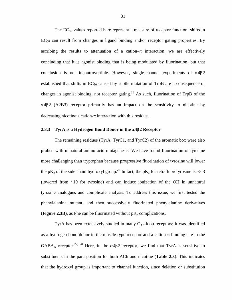
The EC50 values reported here represent a measure of receptor function; shifts in
EC50 can result from changes in ligand binding and/or receptor gating properties. By
ascribing the results to attenuation of a cation–π interaction, we are effectively
concluding that it is agonist binding that is being modulated by fluorination, but that
conclusion is not incontrovertible. However, single-channel experiments of α4β2
established that shifts in EC50 caused by subtle mutation of TrpB are a consequence of
changes in agonist binding, not receptor gating.26 As such, fluorination of TrpB of the
α4β2 (A2B3) receptor primarily has an impact on the sensitivity to nicotine by
decreasing nicotine’s cation-π interaction with this residue.
2.3.3 TyrA is a Hydrogen Bond Donor in the α4β2 Receptor
The remaining residues (TyrA, TyrC1, and TyrC2) of the aromatic box were also
probed with unnatural amino acid mutagenesis. We have found fluorination of tyrosine
more challenging than tryptophan because progressive fluorination of tyrosine will lower
the pKa of the side chain hydroxyl group.27 In fact, the pKa for tetrafluorotyrosine is ~5.3
(lowered from ~10 for tyrosine) and can induce ionization of the OH in unnatural
tyrosine analogues and complicate analysis. To address this issue, we first tested the
phenylalanine mutant, and then successively fluorinated phenylalanine derivatives
(Figure 2.3B), as Phe can be fluorinated without pKa complications.
TyrA has been extensively studied in many Cys-loop receptors; it was identified
as a hydrogen bond donor in the muscle-type receptor and a cation-π binding site in the
GABAA receptor.27, 28 Here, in the α4β2 receptor, we find that TyrA is sensitive to
substituents in the para position for both ACh and nicotine (Table 2.3). This indicates
that the hydroxyl group is important to channel function, since deletion or substitution
32
with a cyano, bromo, or fluoro group resulted in a deleterious effect on channel function
for both ACh and nicotine. Incorporation of MeO-Phe at TyrA resulted in a 6-fold and 4-
fold increase in EC50 for ACh and nicotine, respectively, indicating that TyrA is likely a
hydrogen bond donor. Furthermore, TyrA is not sensitive to fluorination and therefore
neither nicotine nor ACh interact with TyrA via a cation-π interaction.
Table 2.3. Functional characterization of TyrA in α4β2 (A2B3). EC50 values (µM), Hill coefficients (nH) and current size at +70 mV (normalized to current size at -110 mV).
TyrA behaves consistently in both the α4β2 and the muscle-type receptors;
however, the importance of this interaction appears to differ for these two receptors. In
α4β2, TyrA is much more sensitive to mutations at the para position. With ACh as the
agonist, a Tyr to Phe mutation in α4β2 causes a 29-fold increase in EC50, but only a 9-
fold increase in the muscle-type receptor. As such, it is likely that the hydrogen bond
made by TyrA in α4β2 is more crucial for receptor function than in the muscle-type
receptor.
Mutation
A2B3 0.42 ± 0.01 1.2 ± 0.1 0.08 ± 0.01 1.2 ± 0.1 0.041 ± 0.005A3B2 0.023 ± 0.001 1.3 ± 0.1 0.01 ± 0.001 1.7 ± 0.2 0.297 ± 0.041
Tyr 0.42 ± 0.03 1.2 ± 0.1 0.08 ± 0.01 1.7 ± 0.3 0.023 ± 0.009Phe 12 ± 1 1.3 ± 0.1 0.77 ± 0.05 2.1 ± 0.3 0.064 ± 0.011
MeO-Phe 2.3 ± 0.2 1.2 ± 0.1 0.40 ± 0.02 1.7 ± 0.2 0.054 ± 0.032F1-Phe 15 ± 1 1.2 ± 0.1 0.32 ± 0.03 1.4 ± 0.2 -0.076 ± 0.046F2-Phe 16 ± 2 1.8 ± 0.3 0.39 ± 0.05 1.8 ± 0.4 0.028 ± 0.005F3-Phe 14 ± 1 1.2 ± 0.1 0.53 ± 0.04 1.4 ± 0.1 0.044 ± 0.010Br-Phe 3.3 ± 0.2 1.2 ± 0.1 0.54 ± 0.04 1.5 ± 0.1 -0.003 ± 0.031CN-Phe 73 ± 4 1.7 ± 0.1 8.8 ± 0.9 1.5 ± 0.2 0.075 ± 0.008
ACh nH Nicotine nH Norm. I (+70mV)Wild type
TyrA A2B3
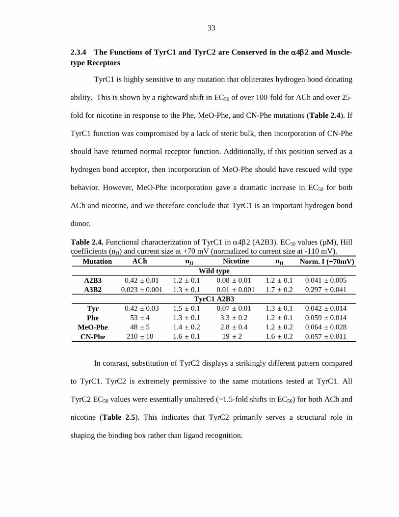
33
2.3.4 The Functions of TyrC1 and TyrC2 are Conserved in the α4β2 and Muscle-type Receptors
TyrC1 is highly sensitive to any mutation that obliterates hydrogen bond donating
ability. This is shown by a rightward shift in EC50 of over 100-fold for ACh and over 25-
fold for nicotine in response to the Phe, MeO-Phe, and CN-Phe mutations (Table 2.4). If
TyrC1 function was compromised by a lack of steric bulk, then incorporation of CN-Phe
should have returned normal receptor function. Additionally, if this position served as a
hydrogen bond acceptor, then incorporation of MeO-Phe should have rescued wild type
behavior. However, MeO-Phe incorporation gave a dramatic increase in EC50 for both
ACh and nicotine, and we therefore conclude that TyrC1 is an important hydrogen bond
donor.
Table 2.4. Functional characterization of TyrC1 in α4β2 (A2B3). EC50 values (µM), Hill coefficients (nH) and current size at +70 mV (normalized to current size at -110 mV).
In contrast, substitution of TyrC2 displays a strikingly different pattern compared
to TyrC1. TyrC2 is extremely permissive to the same mutations tested at TyrC1. All
TyrC2 EC50 values were essentially unaltered (~1.5-fold shifts in EC50) for both ACh and
nicotine (Table 2.5). This indicates that TyrC2 primarily serves a structural role in
shaping the binding box rather than ligand recognition.
Mutation
A2B3 0.42 ± 0.01 1.2 ± 0.1 0.08 ± 0.01 1.2 ± 0.1 0.041 ± 0.005A3B2 0.023 ± 0.001 1.3 ± 0.1 0.01 ± 0.001 1.7 ± 0.2 0.297 ± 0.041
Tyr 0.42 ± 0.03 1.5 ± 0.1 0.07 ± 0.01 1.3 ± 0.1 0.042 ± 0.014Phe 53 ± 4 1.3 ± 0.1 3.3 ± 0.2 1.2 ± 0.1 0.059 ± 0.014
MeO-Phe 48 ± 5 1.4 ± 0.2 2.8 ± 0.4 1.2 ± 0.2 0.064 ± 0.028CN-Phe 210 ± 10 1.6 ± 0.1 19 ± 2 1.6 ± 0.2 0.057 ± 0.011
ACh nH Nicotine nH Norm. I (+70mV)Wild type
TyrC1 A2B3
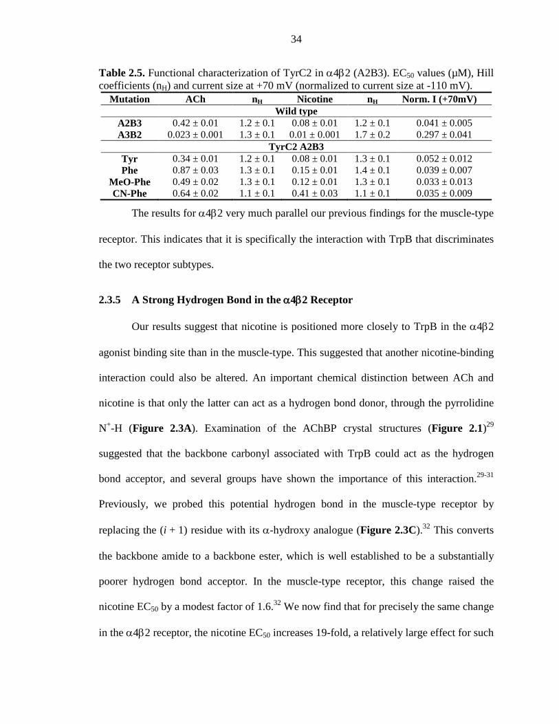
34
Table 2.5. Functional characterization of TyrC2 in α4β2 (A2B3). EC50 values (µM), Hill coefficients (nH) and current size at +70 mV (normalized to current size at -110 mV).
Mutation ACh nH Nicotine nH Norm. I (+70mV) Wild type
A2B3 0.42 ± 0.01 1.2 ± 0.1 0.08 ± 0.01 1.2 ± 0.1 0.041 ± 0.005 A3B2 0.023 ± 0.001 1.3 ± 0.1 0.01 ± 0.001 1.7 ± 0.2 0.297 ± 0.041
TyrC2 A2B3 Tyr 0.34 ± 0.01 1.2 ± 0.1 0.08 ± 0.01 1.3 ± 0.1 0.052 ± 0.012 Phe 0.87 ± 0.03 1.3 ± 0.1 0.15 ± 0.01 1.4 ± 0.1 0.039 ± 0.007
MeO-Phe 0.49 ± 0.02 1.3 ± 0.1 0.12 ± 0.01 1.3 ± 0.1 0.033 ± 0.013 CN-Phe 0.64 ± 0.02 1.1 ± 0.1 0.41 ± 0.03 1.1 ± 0.1 0.035 ± 0.009
The results for α4β2 very much parallel our previous findings for the muscle-type
receptor. This indicates that it is specifically the interaction with TrpB that discriminates
the two receptor subtypes.
2.3.5 A Strong Hydrogen Bond in the α4β2 Receptor
Our results suggest that nicotine is positioned more closely to TrpB in the α4β2
agonist binding site than in the muscle-type. This suggested that another nicotine-binding
interaction could also be altered. An important chemical distinction between ACh and
nicotine is that only the latter can act as a hydrogen bond donor, through the pyrrolidine
N+-H (Figure 2.3A). Examination of the AChBP crystal structures (Figure 2.1)29
suggested that the backbone carbonyl associated with TrpB could act as the hydrogen
bond acceptor, and several groups have shown the importance of this interaction.29-31
Previously, we probed this potential hydrogen bond in the muscle-type receptor by
replacing the (i + 1) residue with its α-hydroxy analogue (Figure 2.3C).32 This converts
the backbone amide to a backbone ester, which is well established to be a substantially
poorer hydrogen bond acceptor. In the muscle-type receptor, this change raised the
nicotine EC50 by a modest factor of 1.6.32 We now find that for precisely the same change
in the α4β2 receptor, the nicotine EC50 increases 19-fold, a relatively large effect for such
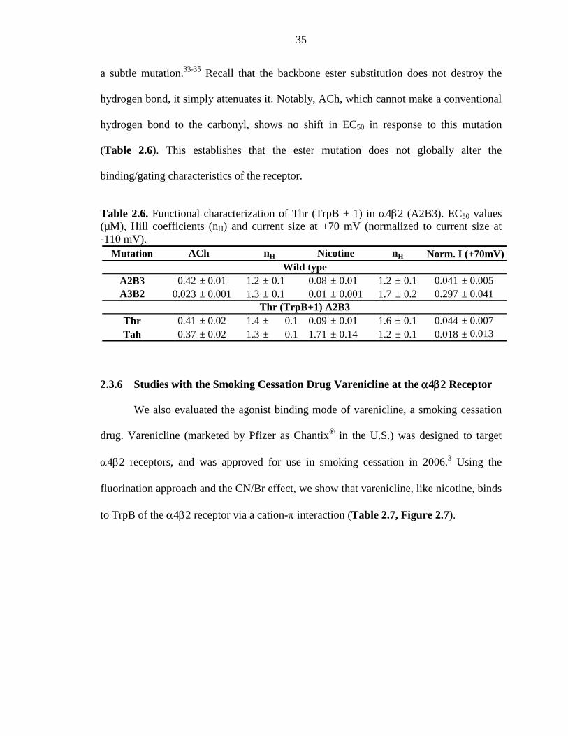
35
a subtle mutation.33-35 Recall that the backbone ester substitution does not destroy the
hydrogen bond, it simply attenuates it. Notably, ACh, which cannot make a conventional
hydrogen bond to the carbonyl, shows no shift in EC50 in response to this mutation
(Table 2.6). This establishes that the ester mutation does not globally alter the
binding/gating characteristics of the receptor.
Table 2.6. Functional characterization of Thr (TrpB + 1) in α4β2 (A2B3). EC50 values (µM), Hill coefficients (nH) and current size at +70 mV (normalized to current size at -110 mV).
2.3.6 Studies with the Smoking Cessation Drug Varenicline at the α4β2 Receptor
We also evaluated the agonist binding mode of varenicline, a smoking cessation
drug. Varenicline (marketed by Pfizer as Chantix® in the U.S.) was designed to target
α4β2 receptors, and was approved for use in smoking cessation in 2006.3 Using the
fluorination approach and the CN/Br effect, we show that varenicline, like nicotine, binds
to TrpB of the α4β2 receptor via a cation-π interaction (Table 2.7, Figure 2.7).
Mutation
A2B3 0.42 ± 0.01 1.2 ± 0.1 0.08 ± 0.01 1.2 ± 0.1 0.041 ± 0.005A3B2 0.023 ± 0.001 1.3 ± 0.1 0.01 ± 0.001 1.7 ± 0.2 0.297 ± 0.041
Thr 0.41 ± 0.02 1.4 ± 0.1 0.09 ± 0.01 1.6 ± 0.1 0.044 ± 0.007Tah 0.37 ± 0.02 1.3 ± 0.1 1.71 ± 0.14 1.2 ± 0.1 0.018 ± 0.013
Norm. I (+70mV)Wild type
Thr (TrpB+1) A2B3
ACh nH Nicotine nH
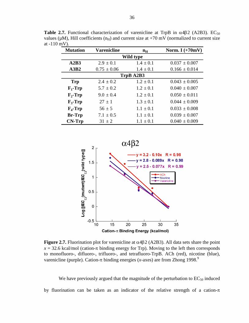
36
Table 2.7. Functional characterization of varenicline at TrpB in α4β2 (A2B3). EC50 values (µM), Hill coefficients (nH) and current size at +70 mV (normalized to current size at -110 mV).
Figure 2.7. Fluorination plot for varenicline at α4β2 (A2B3). All data sets share the point x = 32.6 kcal/mol (cation-π binding energy for Trp). Moving to the left then corresponds to monofluoro-, difluoro-, trifluoro-, and tetrafluoro-TrpB. ACh (red), nicotine (blue), varenicline (purple). Cation-π binding energies (x-axes) are from Zhong 1998.9
We have previously argued that the magnitude of the perturbation to EC50 induced
by fluorination can be taken as an indicator of the relative strength of a cation-π
Mutation nH
A2B3 2.9 ± 0.1 1.4 ± 0.1 0.037 ± 0.007A3B2 0.75 ± 0.06 1.4 ± 0.1 0.166 ± 0.014
Trp 2.4 ± 0.2 1.2 ± 0.1 0.043 ± 0.005F1-Trp 5.7 ± 0.2 1.2 ± 0.1 0.040 ± 0.007F2-Trp 9.0 ± 0.4 1.2 ± 0.1 0.050 ± 0.011F3-Trp 27 ± 1 1.3 ± 0.1 0.044 ± 0.009F4-Trp 56 ± 5 1.1 ± 0.1 0.033 ± 0.008Br-Trp 7.1 ± 0.5 1.1 ± 0.1 0.039 ± 0.007CN-Trp 31 ± 2 1.1 ± 0.1 0.040 ± 0.009
Varenicline Norm. I (+70mV)Wild type
TrpB A2B3
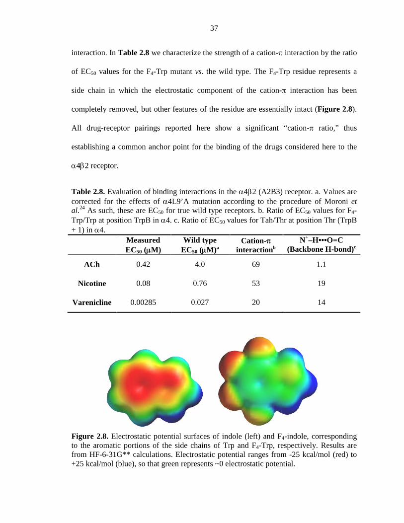
37
interaction. In Table 2.8 we characterize the strength of a cation-π interaction by the ratio
of EC50 values for the F4-Trp mutant vs. the wild type. The F4-Trp residue represents a
side chain in which the electrostatic component of the cation-π interaction has been
completely removed, but other features of the residue are essentially intact (Figure 2.8).
All drug-receptor pairings reported here show a significant “cation-π ratio,” thus
establishing a common anchor point for the binding of the drugs considered here to the
α4β2 receptor.
Table 2.8. Evaluation of binding interactions in the α4β2 (A2B3) receptor. a. Values are corrected for the effects of α4L9’A mutation according to the procedure of Moroni et al.24 As such, these are EC50 for true wild type receptors. b. Ratio of EC50 values for F4-Trp/Trp at position TrpB in α4. c. Ratio of EC50 values for Tah/Thr at position Thr (TrpB + 1) in α4.
Measured EC50 (µM)
Wild type EC50 (µM)a
Cation-π interactionb
N+–H•••O=C (Backbone H-bond)c
ACh 0.42 4.0 69 1.1
Nicotine 0.08 0.76 53 19
Varenicline 0.00285 0.027 20 14
Figure 2.8. Electrostatic potential surfaces of indole (left) and F4-indole, corresponding to the aromatic portions of the side chains of Trp and F4-Trp, respectively. Results are from HF-6-31G** calculations. Electrostatic potential ranges from -25 kcal/mol (red) to +25 kcal/mol (blue), so that green represents ~0 electrostatic potential.
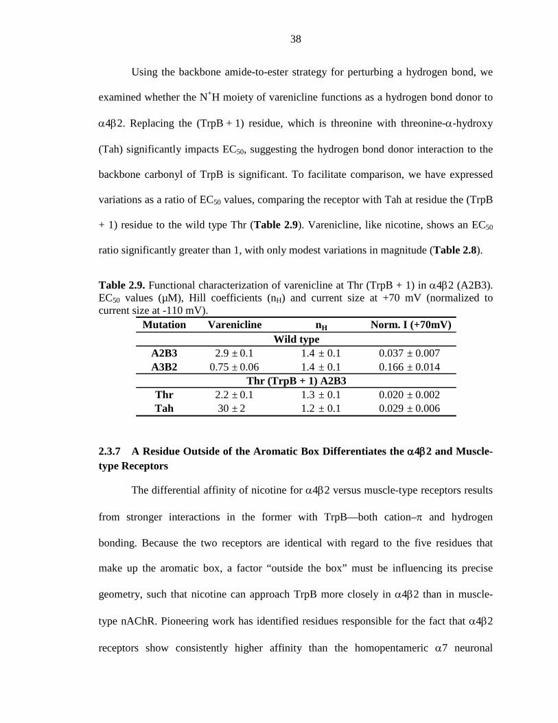
38
Using the backbone amide-to-ester strategy for perturbing a hydrogen bond, we
examined whether the N+H moiety of varenicline functions as a hydrogen bond donor to
α4β2. Replacing the (TrpB + 1) residue, which is threonine with threonine-α-hydroxy
(Tah) significantly impacts EC50, suggesting the hydrogen bond donor interaction to the
backbone carbonyl of TrpB is significant. To facilitate comparison, we have expressed
variations as a ratio of EC50 values, comparing the receptor with Tah at residue the (TrpB
+ 1) residue to the wild type Thr (Table 2.9). Varenicline, like nicotine, shows an EC50
ratio significantly greater than 1, with only modest variations in magnitude (Table 2.8).
Table 2.9. Functional characterization of varenicline at Thr (TrpB + 1) in α4β2 (A2B3). EC50 values (µM), Hill coefficients (nH) and current size at +70 mV (normalized to current size at -110 mV).
2.3.7 A Residue Outside of the Aromatic Box Differentiates the α4β2 and Muscle-type Receptors
The differential affinity of nicotine for α4β2 versus muscle-type receptors results
from stronger interactions in the former with TrpB—both cation–π and hydrogen
bonding. Because the two receptors are identical with regard to the five residues that
make up the aromatic box, a factor “outside the box” must be influencing its precise
geometry, such that nicotine can approach TrpB more closely in α4β2 than in muscle-
type nAChR. Pioneering work has identified residues responsible for the fact that α4β2
receptors show consistently higher affinity than the homopentameric α7 neuronal
Mutation nH
A2B3 2.9 ± 0.1 1.4 ± 0.1 0.037 ± 0.007A3B2 0.75 ± 0.06 1.4 ± 0.1 0.166 ± 0.014
Thr 2.2 ± 0.1 1.3 ± 0.1 0.020 ± 0.002Tah 30 ± 2 1.2 ± 0.1 0.029 ± 0.006
Varenicline Norm. I (+70mV)Wild type
Thr (TrpB + 1) A2B3
39
receptors.36 At a particular residue in loop B—position 153, just four residues from
TrpB—mutations strongly influence affinity. In high-affinity α4β2 receptors this residue
is a Lys, and this residue is proposed to help shape the aromatic box by forming a
backbone hydrogen bond between loops B and C (Figure 2.1). In the lower affinity α7
neuronal receptor, residue 153 is a Gly, and molecular dynamics simulations of α7
suggest that a Gly at 153 discourages the formation of the hydrogen bond between loops
B and C.36 Interestingly, the aligning residue in the muscle-type receptor is also Gly, and
a naturally occurring G153S mutation is gain-of-function and associated with a
congenital myasthenic syndrome.37 We now report that the muscle-type α1 G153K
mutant shows much higher affinity for nicotine, and that, when this mutation is present,
the cation–π interaction to TrpB is strong. The data are summarized in Table 2.10 and
Figure 2.9. As expected, the ACh cation–π interaction is maintained in the muscle-type
receptor with the G153K mutation. These data indicate that the loop B–loop C hydrogen
bond that is naturally present in α4β2 shapes the aromatic box so that nicotine can make
a closer contact to TrpB, and that this structural feature is absent or weaker in the muscle-
type receptor.
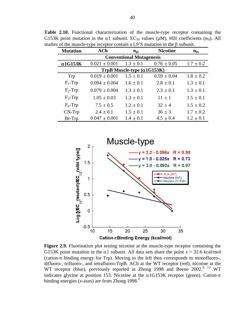
40
Table 2.10. Functional characterization of the muscle-type receptor containing the G153K point mutation in the α1 subunit. EC50 values (µM), Hill coefficients (nH). All studies of the muscle-type receptor contain a L9’S mutation in the β subunit.
Figure 2.9. Fluorination plot testing nicotine at the muscle-type receptor containing the G153K point mutation in the α1 subunit. All data sets share the point x = 32.6 kcal/mol (cation-π binding energy for Trp). Moving to the left then corresponds to monofluoro-, difluoro-, trifluoro-, and tetrafluoro-TrpB. ACh at the WT receptor (red), nicotine at the WT receptor (blue), previously reported in Zhong 1998 and Beene 2002.9, 13 WT indicates glycine at position 153. Nicotine at the α1G153K receptor (green). Cation-π binding energies (x-axes) are from Zhong 1998.9
Mutation
α1G153K 0.021 ± 0.001 1.3 ± 0.1 0.76 ± 0.05 1.7 ± 0.2
Trp 0.019 ± 0.001 1.5 ± 0.1 0.59 ± 0.04 1.8 ± 0.2F1-Trp 0.094 ± 0.004 1.6 ± 0.1 2.8 ± 0.1 1.3 ± 0.1
F2-Trp 0.079 ± 0.004 1.3 ± 0.1 2.3 ± 0.1 1.3 ± 0.1
F3-Trp 1.05 ± 0.03 1.3 ± 0.1 11 ± 1 1.5 ± 0.1
F4-Trp 7.5 ± 0.5 1.2 ± 0.1 32 ± 4 1.5 ± 0.2CN-Trp 2.4 ± 0.1 1.5 ± 0.1 36 ± 3 1.7 ± 0.2Br-Trp 0.047 ± 0.001 1.4 ± 0.1 4.5 ± 0.4 1.2 ± 0.1
Conventional Mutagenesis
TrpB Muscle-type (α1G153K)
ACh nH Nicotine nH
41
2.3.8 Summary
Taken together, the present results indicate that the higher affinity of nicotine in
the brain relative to the neuromuscular junction is a consequence of enhanced interactions
with TrpB. A cation–π interaction that is absent in the muscle-type receptor is quite
strong in α4β2. In addition, a hydrogen bond to a backbone carbonyl that is weak in the
muscle-type is enhanced in α4β2. Varenicline, the smoking cessation drug designed to
target α4β2 receptors and block nicotine binding, also employ the same cation-π and
hydrogen bonding interactions. Both effects are quite substantial, and in combination
they are more than adequate to account fully for the differential sensitivity to nicotine of
the two receptors. The side chain of residue 153 in loop B distinguishes the two receptor
types and apparently influences the shape of the binding site aromatic box, allowing a
stronger interaction between nicotine and TrpB in high-affinity receptors.
2.4 METHODS
mRNA Synthesis and Mutagenesis
All nAChR subunit genes were in the pAMV vector (rat α4 and β2; mouse α1,
β1, γ and δ). nAChR subunit mRNA was obtained from NotI linearizations of the
expression vector pAMV, followed by in vitro transcription using the mMessage
mMachine T7 kit (Ambion, Austin, TX). The mutations for each subunit were introduced
according to the QuikChange mutagenesis protocol (Stratagene, La Jolla, CA).
Ion Channel Expression
To express the ion channels with a wild type ligand binding box, α4L9’A mRNA
was coinjected with β2 mRNA at various ratios, see Table 2.1 (total mRNA 10-
42
25 ng/cell). For muscle-type nAChR experiments, α1:β1:γ:δ mRNA was injected at a
ratio of 2:1:1:1 by mass for wild type protein. Note that for all experiments reported, we
use a previously reported L9’S mutation in the β1 subunit to increase receptor
sensitivity.9 For nonsense suppression experiments of the muscle-type receptor, an
mRNA ratio of 10:1:1:1 was employed. For wild type and nonsense suppression
experiments, the total mRNA injected was 30-65 ng/oocyte. Stage V-VI oocytes
Xenopus laevis were injected and incubated in ND96 solution (96 mM NaCl, 2 mM KCl,
1 mM MgCl2, 1.8 mM CaCl2, and 5 mM HEPES, pH 7.5) with 0.005% (w/v) gentamycin
and 2% (v/v) horse serum at 18 °C for 24-48 hours.
Unnatural Amino Acid / α-hydroxy Acid Incorporation
Nitroveratryloxycarbonyl (NVOC) protected cyanomethyl ester form of unnatural
amino acids and α-hydroxythreonine cyanomethyl ester were synthesized, coupled to the
dinucleotide dCA, and enzymatically ligated to 74-mer THG73 tRNACUA.17, 38 The
unnatural amino acid-conjugated tRNA was deprotected by photolysis immediately prior
to coinjection with mRNA containing the UAG mutation at the site of interest.
Approximately 10-25 ng mRNA and 25 ng tRNA-amino acid or tRNA-hydroxy acid
were injected into stage V–VI oocytes in a total volume of 70 nL.
The fidelity of unnatural amino acid incorporation was confirmed at each site with
a “wild type recovery” experiment and a “read-through/reaminoacylation” test. In the
“wild type recovery” experiment, UAG mutant mRNA was coinjected with tRNA
charged with the amino acid that is present at this residue in the wild type protein.
Generation of receptors that were indistinguishable from the wild type protein indicated
that the residue carried by the suppressor tRNA was successfully and exclusively
43
integrated into the protein. In the “read-through/reaminoacylation” test, the UAG mutant
mRNA was introduced with (1) no tRNA, (2) tRNA THG73 that was not charged with
any amino acid or (3) tRNA THG73 enzymatically ligated with dinucleotide dCA. Lack
of currents in these experiments validated the reliability of the nonsense suppression
experiments.
Whole-Cell Electrophysiological Characterization of Ion Channels
Agonist-induced currents were recorded in two-electrode voltage clamp mode
using the OpusXpress 6000A (Molecular Devices Axon Instruments) at a holding
potential of -60 mV. Agonists were prepared in Ca2+-free ND96 solution and applied for
12 seconds followed by a 2 minute wash with Ca2+-free ND96 solution between each
concentration. Acetylcholine chloride and (-)-nicotine tartrate were purchased from
Sigma/Aldrich/RBI (St. Louis, MO). Varenicline tartrate was obtained from Targacept.
Dose-response data were obtained for at least 6 concentrations of agonists and for a
minimum of 5 oocytes. Mutants with Imax of at least 100 nA of current were defined as
functional. EC50 and Hill coefficient were calculated by fitting the dose-response relation
to the Hill equation. All data are reported as mean ± SE.
Voltage jump experiments were performed in the absence of ACh and at EC50
concentration of ACh. The membrane potential was held at -60 mV, and stepped to 10
test potentials at 20 mV increments between +70 mV and -110 mV for 400 ms each.
600 ms at the -60 mV holding potential was allowed between each test potential.
Background traces (no ACh) were subtracted from data traces, which were used to
measure the steady-state amplitudes of the ACh-induced currents approaching the end of
the test pulses. Normalized I-V curves were generated using current amplitudes
44
normalized to that at -110 mV. For each α4L9’Aβ2 mutant, normalized I+70 mV ± SE from
at least 5 cells was reported.
2.5 ACKNOWLEDGEMENTS
We thank B. N. Cohen for advice on single-channel recording and analysis. This
work was supported by the NIH (NS 34407; NS 11756) and the California Tobacco-
Related Disease Research Program of the University of California, grant number 16RT-
0160. J.A.P.S. was partially supported by an NRSA training grant. Varenicline tartrate
was a generous gift from Targacept.
45
2.6 REFERENCES 1. Romanelli, M. N.; Gratteri, P.; Guandalini, L.; Martini, E.; Bonaccini, C.;
Gualtieri, F., Central Nicotinic Receptors: Structure, Function, Ligands, and Therapeutic Potential. ChemMedChem 2007, 2, (6), 746-767.
2. Corringer, P. J.; Le Novere, N.; Changeux, J. P., Nicotinic receptors at the amino acid level. Annu Rev Pharmacol Toxicol 2000, 40, 431-58.
3. Coe, J. W.; Brooks, P. R.; Vetelino, M. G.; Wirtz, M. C.; Arnold, E. P.; Huang, J.; Sands, S. B.; Davis, T. I.; Lebel, L. A.; Fox, C. B.; Shrikhande, A.; Heym, J. H.; Schaeffer, E.; Rollema, H.; Lu, Y.; Mansbach, R. S.; Chambers, L. K.; Rovetti, C. C.; Schulz, D. W.; Tingley, F. D., 3rd; O'Neill, B. T., Varenicline: an alpha4beta2 nicotinic receptor partial agonist for smoking cessation. J Med Chem 2005, 48, (10), 3474-7.
4. Gotti, C.; Zoli, M.; Clementi, F., Brain nicotinic acetylcholine receptors: native subtypes and their relevance. Trends Pharmacol Sci 2006, 27, (9), 482-91.
5. Mansvelder, H. D.; Keath, J. R.; McGehee, D. S., Synaptic mechanisms underlie nicotine-induced excitability of brain reward areas. Neuron 2002, 33, (6), 905-19.
6. Tapper, A. R.; McKinney, S. L.; Nashmi, R.; Schwarz, J.; Deshpande, P.; Labarca, C.; Whiteaker, P.; Marks, M. J.; Collins, A. C.; Lester, H. A., Nicotine activation of alpha4* receptors: sufficient for reward, tolerance, and sensitization. Science 2004, 306, (5698), 1029-32.
7. Nashmi, R.; Xiao, C.; Deshpande, P.; McKinney, S.; Grady, S. R.; Whiteaker, P.; Huang, Q.; McClure-Begley, T.; Lindstrom, J. M.; Labarca, C.; Collins, A. C.; Marks, M. J.; Lester, H. A., Chronic nicotine cell specifically upregulates functional alpha 4* nicotinic receptors: basis for both tolerance in midbrain and enhanced long-term potentiation in perforant path. J Neurosci 2007, 27, (31), 8202-18.
8. Kuryatov, A.; Luo, J.; Cooper, J.; Lindstrom, J., Nicotine acts as a pharmacological chaperone to up-regulate human alpha4beta2 acetylcholine receptors. Mol Pharmacol 2005, 68, (6), 1839-51.
9. Zhong, W.; Gallivan, J. P.; Zhang, Y.; Li, L.; Lester, H. A.; Dougherty, D. A., From ab initio quantum mechanics to molecular neurobiology: a cation-pi binding site in the nicotinic receptor. Proc Natl Acad Sci U S A 1998, 95, (21), 12088-93.
10. Brejc, K.; van Dijk, W. J.; Klaassen, R. V.; Schuurmans, M.; van Der Oost, J.; Smit, A. B.; Sixma, T. K., Crystal structure of an ACh-binding protein reveals the ligand-binding domain of nicotinic receptors. Nature 2001, 411, (6835), 269-76.
11. Sixma, T. K.; Smit, A. B., Acetylcholine binding protein (AChBP): a secreted glial protein that provides a high-resolution model for the extracellular domain of pentameric ligand-gated ion channels. Annu Rev Biophys Biomol Struct 2003, 32, 311-34.
12. Dougherty, D. A., Cys-loop neuroreceptors: structure to the rescue? Chem Rev 2008, 108, (5), 1642-53.
13. Beene, D. L.; Brandt, G. S.; Zhong, W.; Zacharias, N. M.; Lester, H. A.; Dougherty, D. A., Cation-pi interactions in ligand recognition by serotonergic (5-HT3A) and nicotinic acetylcholine receptors: the anomalous binding properties of nicotine. Biochemistry 2002, 41, (32), 10262-9.
46
14. Gonzales, D.; Rennard, S. I.; Nides, M.; Oncken, C.; Azoulay, S.; Billing, C. B.; Watsky, E. J.; Gong, J.; Williams, K. E.; Reeves, K. R., Varenicline, an alpha4beta2 nicotinic acetylcholine receptor partial agonist, vs sustained-release bupropion and placebo for smoking cessation: a randomized controlled trial. JAMA 2006, 296, (1), 47-55.
15. Mihalak, K. B.; Carroll, F. I.; Luetje, C. W., Varenicline is a partial agonist at alpha4beta2 and a full agonist at alpha7 neuronal nicotinic receptors. Mol Pharmacol 2006, 70, (3), 801-5.
16. Dougherty, D. A., Physical organic chemistry on the brain. J Org Chem 2008, 73, (10), 3667-73.
17. Nowak, M. W.; Gallivan, J. P.; Silverman, S. K.; Labarca, C. G.; Dougherty, D. A.; Lester, H. A., In vivo incorporation of unnatural amino acids into ion channels in Xenopus oocyte expression system. Methods Enzymol 1998, 293, 504-29.
18. Fonck, C.; Cohen, B. N.; Nashmi, R.; Whiteaker, P.; Wagenaar, D. A.; Rodrigues-Pinguet, N.; Deshpande, P.; McKinney, S.; Kwoh, S.; Munoz, J.; Labarca, C.; Collins, A. C.; Marks, M. J.; Lester, H. A., Novel seizure phenotype and sleep disruptions in knock-in mice with hypersensitive alpha 4* nicotinic receptors. J Neurosci 2005, 25, (49), 11396-411.
19. Filatov, G. N.; White, M. M., The role of conserved leucines in the M2 domain of the acetylcholine receptor in channel gating. Mol Pharmacol 1995, 48, (3), 379-84.
20. Labarca, C.; Nowak, M. W.; Zhang, H.; Tang, L.; Deshpande, P.; Lester, H. A., Channel gating governed symmetrically by conserved leucine residues in the M2 domain of nicotinic receptors. Nature 1995, 376, (6540), 514-6.
21. Kearney, P. C.; Nowak, M. W.; Zhong, W.; Silverman, S. K.; Lester, H. A.; Dougherty, D. A., Dose-response relations for unnatural amino acids at the agonist binding site of the nicotinic acetylcholine receptor: tests with novel side chains and with several agonists. Mol Pharmacol 1996, 50, (5), 1401-12.
22. Grutter, T.; Changeux, J. P., Nicotinic receptors in wonderland. Trends Biochem Sci 2001, 26, (8), 459-63.
23. Karlin, A., Emerging structure of the nicotinic acetylcholine receptors. Nat Rev Neurosci 2002, 3, (2), 102-14.
24. Moroni, M.; Zwart, R.; Sher, E.; Cassels, B. K.; Bermudez, I., alpha4beta2 nicotinic receptors with high and low acetylcholine sensitivity: pharmacology, stoichiometry, and sensitivity to long-term exposure to nicotine. Mol Pharmacol 2006, 70, (2), 755-68.
25. Nelson, M. E.; Kuryatov, A.; Choi, C. H.; Zhou, Y.; Lindstrom, J., Alternate stoichiometries of alpha4beta2 nicotinic acetylcholine receptors. Mol Pharmacol 2003, 63, (2), 332-41.
26. Xiu, X.; Puskar, N. L.; Shanata, J. A.; Lester, H. A.; Dougherty, D. A., Nicotine binding to brain receptors requires a strong cation-pi interaction. Nature 2009, 458, (7237), 534-7.
27. Nowak, M. W.; Kearney, P. C.; Sampson, J. R.; Saks, M. E.; Labarca, C. G.; Silverman, S. K.; Zhong, W.; Thorson, J.; Abelson, J. N.; Davidson, N.; et al., Nicotinic receptor binding site probed with unnatural amino acid incorporation in intact cells. Science 1995, 268, (5209), 439-42.
47
28. Padgett, C. L.; Hanek, A. P.; Lester, H. A.; Dougherty, D. A.; Lummis, S. C., Unnatural amino acid mutagenesis of the GABA(A) receptor binding site residues reveals a novel cation-pi interaction between GABA and beta 2Tyr97. J Neurosci 2007, 27, (4), 886-92.
29. Celie, P. H.; van Rossum-Fikkert, S. E.; van Dijk, W. J.; Brejc, K.; Smit, A. B.; Sixma, T. K., Nicotine and carbamylcholine binding to nicotinic acetylcholine receptors as studied in AChBP crystal structures. Neuron 2004, 41, (6), 907-14.
30. Hansen, S. B.; Sulzenbacher, G.; Huxford, T.; Marchot, P.; Taylor, P.; Bourne, Y., Structures of Aplysia AChBP complexes with nicotinic agonists and antagonists reveal distinctive binding interfaces and conformations. EMBO J 2005, 24, (20), 3635-46.
31. Talley, T. T.; Yalda, S.; Ho, K. Y.; Tor, Y.; Soti, F. S.; Kem, W. R.; Taylor, P., Spectroscopic analysis of benzylidene anabaseine complexes with acetylcholine binding proteins as models for ligand-nicotinic receptor interactions. Biochemistry 2006, 45, (29), 8894-902.
32. Cashin, A. L.; Petersson, E. J.; Lester, H. A.; Dougherty, D. A., Using physical chemistry to differentiate nicotinic from cholinergic agonists at the nicotinic acetylcholine receptor. J Am Chem Soc 2005, 127, (1), 350-6.
33. Deechongkit, S.; Nguyen, H.; Powers, E. T.; Dawson, P. E.; Gruebele, M.; Kelly, J. W., Context-dependent contributions of backbone hydrogen bonding to beta-sheet folding energetics. Nature 2004, 430, (6995), 101-5.
34. England, P. M.; Zhang, Y.; Dougherty, D. A.; Lester, H. A., Backbone mutations in transmembrane domains of a ligand-gated ion channel: implications for the mechanism of gating. Cell 1999, 96, (1), 89-98.
35. Koh, J. T.; Cornish, V. W.; Schultz, P. G., An experimental approach to evaluating the role of backbone interactions in proteins using unnatural amino acid mutagenesis. Biochemistry 1997, 36, (38), 11314-22.
36. Grutter, T.; Prado de Carvalho, L.; Le Novere, N.; Corringer, P. J.; Edelstein, S.; Changeux, J. P., An H-bond between two residues from different loops of the acetylcholine binding site contributes to the activation mechanism of nicotinic receptors. EMBO J 2003, 22, (9), 1990-2003.
37. Sine, S. M.; Ohno, K.; Bouzat, C.; Auerbach, A.; Milone, M.; Pruitt, J. N.; Engel, A. G., Mutation of the acetylcholine receptor alpha subunit causes a slow-channel myasthenic syndrome by enhancing agonist binding affinity. Neuron 1995, 15, (1), 229-39.
38. Saks, M. E.; Sampson, J. R.; Nowak, M. W.; Kearney, P. C.; Du, F.; Abelson, J. N.; Lester, H. A.; Dougherty, D. A., An engineered Tetrahymena tRNAGln for in vivo incorporation of unnatural amino acids into proteins by nonsense suppression. J Biol Chem 1996, 271, (38), 23169-75.
48
Chapter 3
Probing the Effects of Residues Located Outside the Agonist Binding Site on Drug-Receptor Selectivity in the Nicotinic Receptor
3.1 ABSTRACT
The nicotinic acetylcholine receptors (nAChRs) are a family of closely related but
pharmacologically distinct neurotransmitter-gated ion channels. They are therapeutic
targets for a wide range of neurological disorders, and a key issue in drug development is
selective targeting among the greater than 20 subtypes of nAChRs that are known. The
present work evaluates a proposed hydrogen bonding interaction involving a residue
known as the “loop B glycine” that distinguishes receptors that are highly responsive to
ACh and nicotine from those that are much less so. We have performed structure-
function studies on the loop B site, including unnatural amino acid mutagenesis, in three
different nAChR subtypes and found that the correlation between agonist potency and
this residue is strong. Low-potency receptor subtypes have a glycine at this key site, and
mutation to a residue with a side chain converts a low-potency receptor to a high-potency
receptor. Innately high-potency receptors have a lysine at the loop B site and show a
decrease in potency for the reverse mutation (i.e., introducing a glycine). This residue lies
outside of the agonist binding site, and the details of how changes at the site impact
agonist potency vary for differing receptor subtypes. This suggests a model in which the
loop B residue influences the global shape of the agonist binding site rather than
modulating any specific interaction.
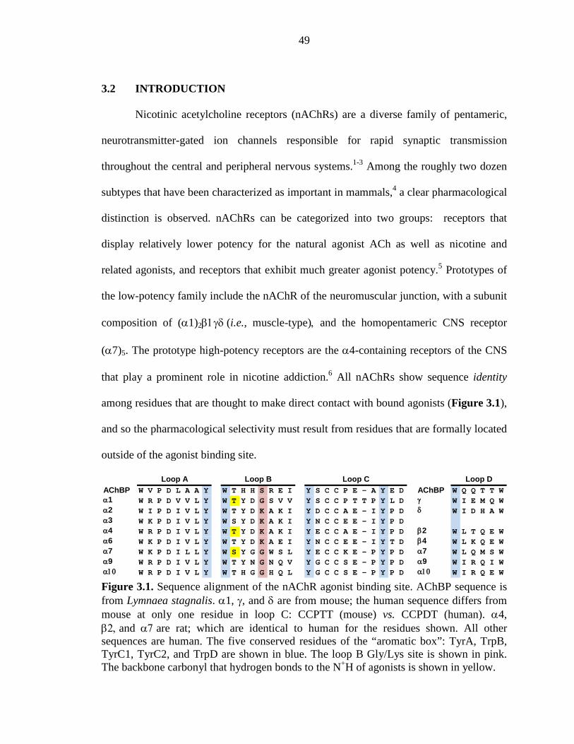
49
3.2 INTRODUCTION
Nicotinic acetylcholine receptors (nAChRs) are a diverse family of pentameric,
neurotransmitter-gated ion channels responsible for rapid synaptic transmission
throughout the central and peripheral nervous systems.1-3 Among the roughly two dozen
subtypes that have been characterized as important in mammals,4 a clear pharmacological
distinction is observed. nAChRs can be categorized into two groups: receptors that
display relatively lower potency for the natural agonist ACh as well as nicotine and
related agonists, and receptors that exhibit much greater agonist potency.5 Prototypes of
the low-potency family include the nAChR of the neuromuscular junction, with a subunit
composition of (α1)2β1γδ (i.e., muscle-type), and the homopentameric CNS receptor
(α7)5. The prototype high-potency receptors are the α4-containing receptors of the CNS
that play a prominent role in nicotine addiction.6 All nAChRs show sequence identity
among residues that are thought to make direct contact with bound agonists (Figure 3.1),
and so the pharmacological selectivity must result from residues that are formally located
outside of the agonist binding site.
AChBP W V P D L A A Y W T H H S R E I Y S C C P E - A Y E D AChBP W Q Q T T Wα1 W R P D V V L Y W T Y D G S V V Y S C C P T T P Y L D γ W I E M Q Wα2 W I P D I V L Y W T Y D K A K I Y D C C A E - I Y P D δ W I D H A Wα3 W K P D I V L Y W S Y D K A K I Y N C C E E - I Y P Dα4 W R P D I V L Y W T Y D K A K I Y E C C A E - I Y P D β2 W L T Q E Wα6 W K P D I V L Y W T Y D K A E I Y N C C E E - I Y T D β4 W L K Q E Wα7 W K P D I L L Y W S Y G G W S L Y E C C K E - P Y P D α7 W L Q M S Wα9 W R P D I V L Y W T Y N G N Q V Y G C C S E - P Y P D α9 W I R Q I Wα10 W R P D I V L Y W T H G G H Q L Y G C C S E - P Y P D α10 W I R Q E W
Loop DLoop A Loop B Loop C
Figure 3.1. Sequence alignment of the nAChR agonist binding site. AChBP sequence is from Lymnaea stagnalis. α1, γ, and δ are from mouse; the human sequence differs from mouse at only one residue in loop C: CCPTT (mouse) vs. CCPDT (human). α4, β2, and α7 are rat; which are identical to human for the residues shown. All other sequences are human. The five conserved residues of the “aromatic box”: TyrA, TrpB, TyrC1, TyrC2, and TrpD are shown in blue. The loop B Gly/Lys site is shown in pink. The backbone carbonyl that hydrogen bonds to the N+H of agonists is shown in yellow.
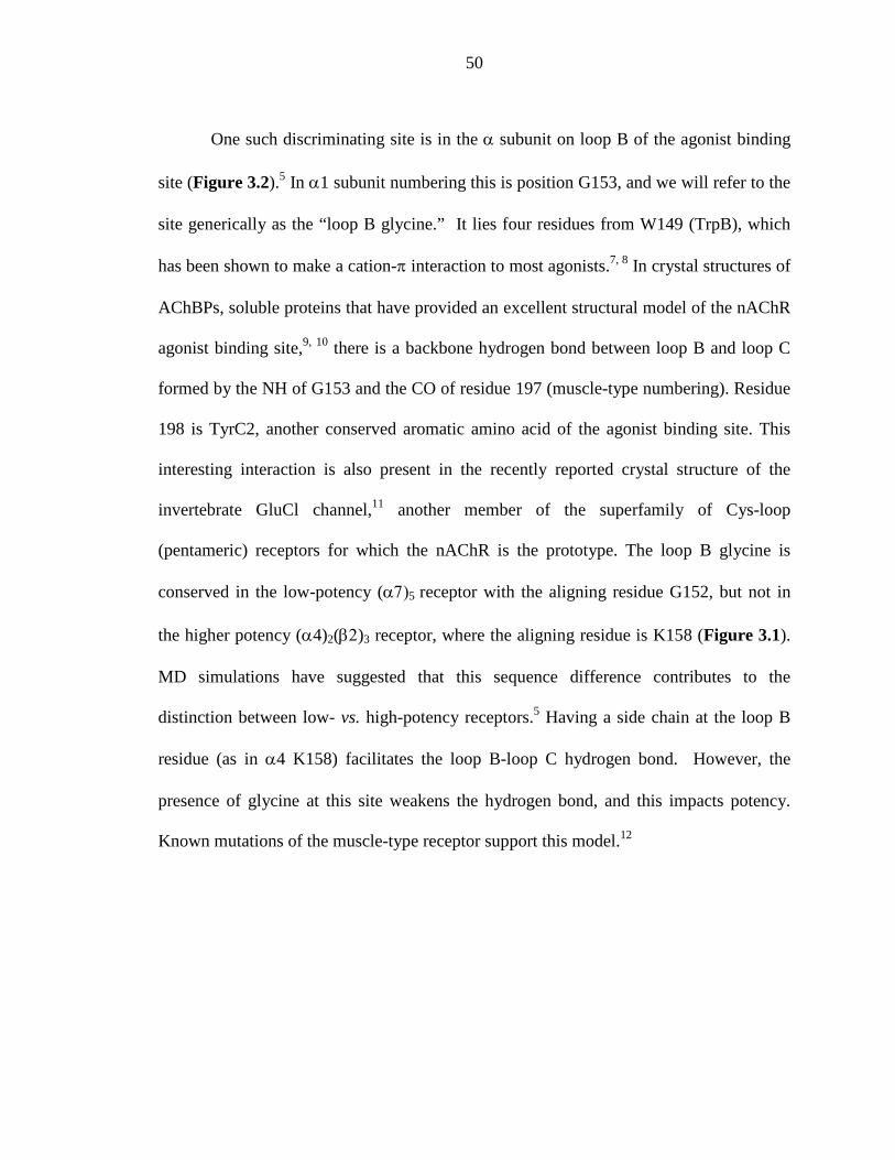
50
One such discriminating site is in the α subunit on loop B of the agonist binding
site (Figure 3.2).5 In α1 subunit numbering this is position G153, and we will refer to the
site generically as the “loop B glycine.” It lies four residues from W149 (TrpB), which
has been shown to make a cation-π interaction to most agonists.7, 8 In crystal structures of
AChBPs, soluble proteins that have provided an excellent structural model of the nAChR
agonist binding site,9, 10 there is a backbone hydrogen bond between loop B and loop C
formed by the NH of G153 and the CO of residue 197 (muscle-type numbering). Residue
198 is TyrC2, another conserved aromatic amino acid of the agonist binding site. This
interesting interaction is also present in the recently reported crystal structure of the
invertebrate GluCl channel,11 another member of the superfamily of Cys-loop
(pentameric) receptors for which the nAChR is the prototype. The loop B glycine is
conserved in the low-potency (α7)5 receptor with the aligning residue G152, but not in
the higher potency (α4)2(β2)3 receptor, where the aligning residue is K158 (Figure 3.1).
MD simulations have suggested that this sequence difference contributes to the
distinction between low- vs. high-potency receptors.5 Having a side chain at the loop B
residue (as in α4 K158) facilitates the loop B-loop C hydrogen bond. However, the
presence of glycine at this site weakens the hydrogen bond, and this impacts potency.
Known mutations of the muscle-type receptor support this model.12
51
Figure 3.2. nAChR agonist binding site, based on the structure of AChBP (pdb: 1UW6).13 Loop B is in green, loop C is in blue, nicotine is in purple, three of the five residues that form the “aromatic box” of the agonist binding site are highlighted. The backbone carbonyl (green star) that hydrogen bonds to the N+H of agonists, and the proposed loop B–loop C hydrogen bond is highlighted.
We recently showed that, indeed, a G153K mutation in the muscle-type receptor
greatly increased potency of both ACh and nicotine.7 The cause of the increased potency
was a cation-π interaction to TrpB that was absent or weak in the wild type muscle-type
receptor, but was strong in both α4-containing receptors and in the muscle-type receptor
with the G153K mutation.
In the present work we evaluate the role of the loop B glycine in both the muscle-
type and (α7)5 receptors. G-to-K mutations enhance potency substantially, but the details
of how the potency is enhanced differ in the two subtypes. We also evaluate whether the
reverse K-to-G mutation has the opposite effect on the high-potency (α4)2(β2)3 receptor.
Finally, we use unnatural amino acid mutagenesis to disrupt the proposed hydrogen bond
in the (α4)2(β2)3 receptor and evaluate the functional consequences.
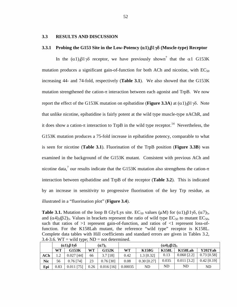
52
3.3 RESULTS AND DISCUSSION
3.3.1 Probing the G153 Site in the Low-Potency (α1)2β1γδ (Muscle-type) Receptor
In the (α1)2β1γδ receptor, we have previously shown7 that the α1 G153K
mutation produces a significant gain-of-function for both ACh and nicotine, with EC50
increasing 44- and 74-fold, respectively (Table 3.1). We also showed that the G153K
mutation strengthened the cation-π interaction between each agonist and TrpB. We now
report the effect of the G153K mutation on epibatidine (Figure 3.3A) at (α1)2β1γδ. Note
that unlike nicotine, epibatidine is fairly potent at the wild type muscle-type nAChR, and
it does show a cation-π interaction to TrpB in the wild type receptor.14 Nevertheless, the
G153K mutation produces a 75-fold increase in epibatidine potency, comparable to what
is seen for nicotine (Table 3.1). Fluorination of the TrpB position (Figure 3.3B) was
examined in the background of the G153K mutant. Consistent with previous ACh and
nicotine data,7 our results indicate that the G153K mutation also strengthens the cation-π
interaction between epibatidine and TrpB of the receptor (Table 3.2). This is indicated
by an increase in sensitivity to progressive fluorination of the key Trp residue, as
illustrated in a “fluorination plot” (Figure 3.4).
Table 3.1. Mutation of the loop B Gly/Lys site. EC50 values (µM) for (α1)2β1γδ, (α7)5, and (α4)2(β2)3. Values in brackets represent the ratio of wild type EC50 to mutant EC50, such that ratios of >1 represent gain-of-function, and ratios of <1 represent loss-of-function. For the K158Lah mutant, the reference “wild type” receptor is K158L. Complete data tables with Hill coefficients and standard errors are given in Tables 3.2, 3.4-3.6. WT = wild type; ND = not determined.
(α1)2β1γδ (α7)5 (α4)2(β2)3 WT G153K WT G152K WT K158G K158L K158Lah Y202Yah
ACh 1.2 0.027 [44] 66 3.7 [18] 0.42 1.3 [0.32] 0.13 0.060 [2.2] 0.73 [0.58]
Nic 56 0.76 [74] 23 0.76 [30] 0.08 0.30 [0.27] 0.035 0.011 [3.2] 0.42 [0.19]
Epi 0.83 0.011 [75] 0.26 0.016 [16] 0.00035 ND ND ND ND
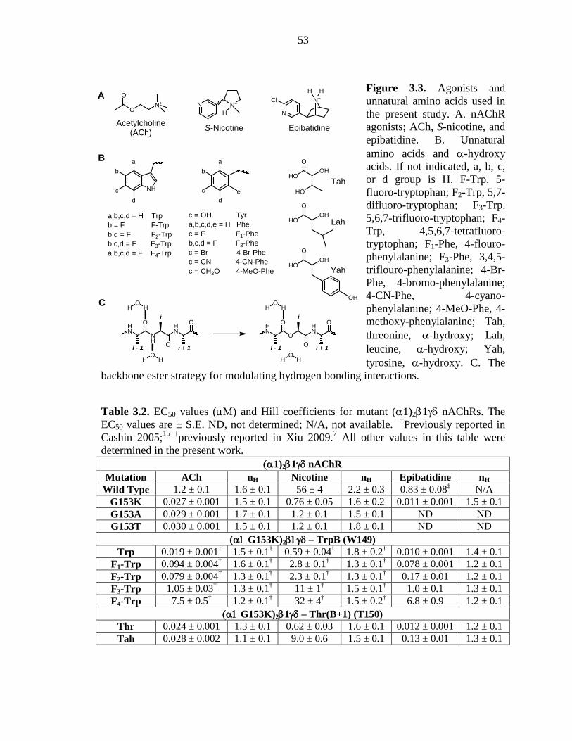
53
Figure 3.3. Agonists and unnatural amino acids used in the present study. A. nAChR agonists; ACh, S-nicotine, and epibatidine. B. Unnatural amino acids and α-hydroxy acids. If not indicated, a, b, c, or d group is H. F-Trp, 5-fluoro-tryptophan; F2-Trp, 5,7-difluoro-tryptophan; F3-Trp, 5,6,7-trifluoro-tryptophan; F4-Trp, 4,5,6,7-tetrafluoro-tryptophan; F1-Phe, 4-flouro-phenylalanine; F3-Phe, 3,4,5-triflouro-phenylalanine; 4-Br-Phe, 4-bromo-phenylalanine; 4-CN-Phe, 4-cyano-phenylalanine; 4-MeO-Phe, 4-methoxy-phenylalanine; Tah, threonine, α-hydroxy; Lah, leucine, α-hydroxy; Yah, tyrosine, α-hydroxy. C. The
backbone ester strategy for modulating hydrogen bonding interactions.
Table 3.2. EC50 values (µM) and Hill coefficients for mutant (α1)2β1γδ nAChRs. The EC50 values are ± S.E. ND, not determined; N/A, not available. ‡Previously reported in Cashin 2005;15 †previously reported in Xiu 2009.7 All other values in this table were determined in the present work.
(α1)2β1γδ nAChR Mutation ACh nH Nicotine nH Epibatidine nH
Wild Type 1.2 ± 0.1 1.6 ± 0.1 56 ± 4 2.2 ± 0.3 0.83 ± 0.08‡ N/A G153K 0.027 ± 0.001 1.5 ± 0.1 0.76 ± 0.05 1.6 ± 0.2 0.011 ± 0.001 1.5 ± 0.1 G153A 0.029 ± 0.001 1.7 ± 0.1 1.2 ± 0.1 1.5 ± 0.1 ND ND G153T 0.030 ± 0.001 1.5 ± 0.1 1.2 ± 0.1 1.8 ± 0.1 ND ND
(α1 G153K)2β1γδ – TrpB (W149) Trp 0.019 ± 0.001† 1.5 ± 0.1† 0.59 ± 0.04† 1.8 ± 0.2† 0.010 ± 0.001 1.4 ± 0.1
F1-Trp 0.094 ± 0.004† 1.6 ± 0.1† 2.8 ± 0.1† 1.3 ± 0.1† 0.078 ± 0.001 1.2 ± 0.1 F2-Trp 0.079 ± 0.004† 1.3 ± 0.1† 2.3 ± 0.1† 1.3 ± 0.1† 0.17 ± 0.01 1.2 ± 0.1 F3-Trp 1.05 ± 0.03† 1.3 ± 0.1† 11 ± 1† 1.5 ± 0.1† 1.0 ± 0.1 1.3 ± 0.1 F4-Trp 7.5 ± 0.5† 1.2 ± 0.1† 32 ± 4† 1.5 ± 0.2† 6.8 ± 0.9 1.2 ± 0.1
(α1 G153K)2β1γδ – Thr(B+1) (T150) Thr 0.024 ± 0.001 1.3 ± 0.1 0.62 ± 0.03 1.6 ± 0.1 0.012 ± 0.001 1.2 ± 0.1 Tah 0.028 ± 0.002 1.1 ± 0.1 9.0 ± 0.6 1.5 ± 0.1 0.13 ± 0.01 1.3 ± 0.1
S-Nicotine
N+
HNN+
O
ON+
N
ClH H
EpibatidineAcetylcholine
(ACh)
NH
HN
O
OHN
OH H
O
i - 1
i
i + 1
O
HN
O
OHN
OH H
O
i - 1
i
i + 1O
H HO
H H
A
C
B
NH
a
b
c
d
a,b,c,d = H Trpb = F F-Trpb,d = F F2-Trpb,c,d = F F3-Trpa,b,c,d = F F4-Trp
a
b
c
de
c = OH Tyra,b,c,d,e = H Phec = F F1-Pheb,c,d = F F3-Phec = Br 4-Br-Phec = CN 4-CN-Phec = CH3O 4-MeO-Phe
HO
OOH
HO
Tah
HO
OOH
HO
OOH
OH
Yah
Lah
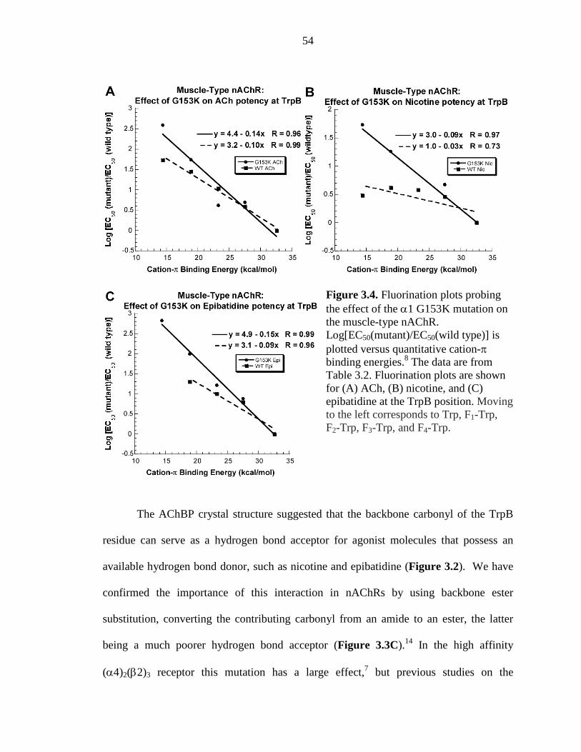
54
Figure 3.4. Fluorination plots probing the effect of the α1 G153K mutation on the muscle-type nAChR. Log[EC50(mutant)/EC50(wild type)] is plotted versus quantitative cation-π binding energies.8 The data are from Table 3.2. Fluorination plots are shown for (A) ACh, (B) nicotine, and (C) epibatidine at the TrpB position. Moving to the left corresponds to Trp, F1-Trp, F2-Trp, F3-Trp, and F4-Trp.
The AChBP crystal structure suggested that the backbone carbonyl of the TrpB
residue can serve as a hydrogen bond acceptor for agonist molecules that possess an
available hydrogen bond donor, such as nicotine and epibatidine (Figure 3.2). We have
confirmed the importance of this interaction in nAChRs by using backbone ester
substitution, converting the contributing carbonyl from an amide to an ester, the latter
being a much poorer hydrogen bond acceptor (Figure 3.3C).14 In the high affinity
(α4)2(β2)3 receptor this mutation has a large effect,7 but previous studies on the
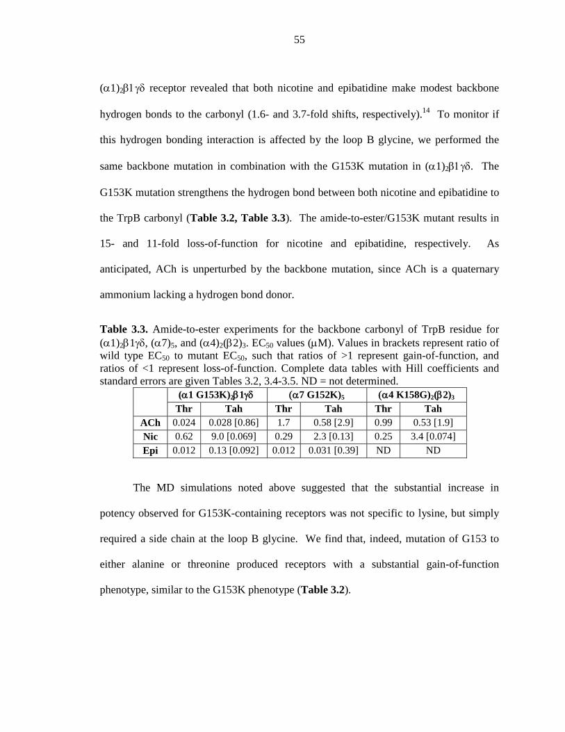
55
(α1)2β1γδ receptor revealed that both nicotine and epibatidine make modest backbone
hydrogen bonds to the carbonyl (1.6- and 3.7-fold shifts, respectively).14 To monitor if
this hydrogen bonding interaction is affected by the loop B glycine, we performed the
same backbone mutation in combination with the G153K mutation in (α1)2β1γδ. The
G153K mutation strengthens the hydrogen bond between both nicotine and epibatidine to
the TrpB carbonyl (Table 3.2, Table 3.3). The amide-to-ester/G153K mutant results in
15- and 11-fold loss-of-function for nicotine and epibatidine, respectively. As
anticipated, ACh is unperturbed by the backbone mutation, since ACh is a quaternary
ammonium lacking a hydrogen bond donor.
Table 3.3. Amide-to-ester experiments for the backbone carbonyl of TrpB residue for (α1)2β1γδ, (α7)5, and (α4)2(β2)3. EC50 values (µM). Values in brackets represent ratio of wild type EC50 to mutant EC50, such that ratios of >1 represent gain-of-function, and ratios of <1 represent loss-of-function. Complete data tables with Hill coefficients and standard errors are given Tables 3.2, 3.4-3.5. ND = not determined.
(α1 G153K)2β1γδ (α7 G152K)5 (α4 K158G)2(β2)3
Thr Tah Thr Tah Thr Tah ACh 0.024 0.028 [0.86] 1.7 0.58 [2.9] 0.99 0.53 [1.9] Nic 0.62 9.0 [0.069] 0.29 2.3 [0.13] 0.25 3.4 [0.074] Epi 0.012 0.13 [0.092] 0.012 0.031 [0.39] ND ND
The MD simulations noted above suggested that the substantial increase in
potency observed for G153K-containing receptors was not specific to lysine, but simply
required a side chain at the loop B glycine. We find that, indeed, mutation of G153 to
either alanine or threonine produced receptors with a substantial gain-of-function
phenotype, similar to the G153K phenotype (Table 3.2).
56
3.3.2 Probing the G152 Site in the Low-Potency (α7)5 Receptor
The homopentameric (α7)5 receptor is distinctive in many ways, including its
mode of agonist binding. While most nAChR subtypes employ a cation-π interaction
between TrpB and the agonist, the (α7)5 receptor eschews this common ligand binding
mechanism.16 Instead, in (α7)5, ACh forms a cation-π interaction to TyrA, and
epibatidine forms a cation-π interaction to TyrC2 of the agonist binding site. It is a “low-
potency” receptor, with a G152 aligning at the loop B glycine site. Given the unusual
binding pattern observed in (α7)5, it was interesting to investigate whether changing the
side chain of the loop B glycine would increase agonist potency through a mechanism
similar to that observed in (α1)2β1γδ. Introduction of lysine at this position (G152K) in
(α7)5 did result in a significant gain-of-function when tested with ACh, nicotine, and
epibatidine (Table 3.1).
To determine whether an amplification of the cation-π interaction analogous to
that seen in the muscle-type receptor would occur in (α7)5, we incorporated either F3Trp
at TrpB or F3Phe at TyrA and TyrC2 in the background of the G152K mutant. In contrast
to (α1)2β1γδ, the consequences of fluorination at TrpB in the G152K mutant did not
differ from wild type (α7)5 for ACh, nicotine, or epibatidine (Table 3.4). Similarly,
introduction of G152K in (α7)5 does not enhance the naturally occurring cation-π
interaction between ACh and TyrA.
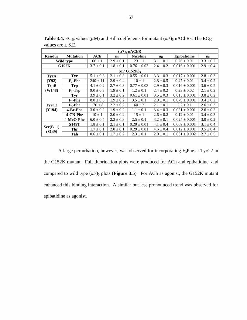
57
Table 3.4. EC50 values (µM) and Hill coefficients for mutant (α7)5 nAChRs. The EC50 values are ± S.E.
(α7)5 nAChR Residue Mutation ACh nH Nicotine nH Epibatidine nH
Wild type 66 ± 1 2.9 ± 0.1 23 ± 1 3.1 ± 0.1 0.26 ± 0.01 3.3 ± 0.2 G152K 3.7 ± 0.1 1.8 ± 0.1 0.76 ± 0.03 2.4 ± 0.2 0.016 ± 0.001 2.9 ± 0.4
(α7 G152K)5 TyrA (Y92)
Tyr 5.1 ± 0.3 2.1 ± 0.3 0.55 ± 0.01 3.3 ± 0.3 0.017 ± 0.001 2.8 ± 0.3 F3-Phe 240 ± 11 2.9 ± 0.4 10 ± 1 2.8 ± 0.5 0.47 ± 0.01 3.4 ± 0.2
TrpB (W148)
Trp 4.1 ± 0.2 2.7 ± 0.3 0.77 ± 0.03 2.9 ± 0.3 0.016 ± 0.001 3.6 ± 0.5 F3-Trp 9.0 ± 0.3 1.9 ± 0.1 1.2 ± 0.1 2.4 ± 0.2 0.23 ± 0.02 2.1 ± 0.2
TyrC2 (Y194)
Tyr 3.9 ± 0.1 3.2 ± 0.2 0.61 ± 0.01 3.5 ± 0.3 0.015 ± 0.001 3.8 ± 0.2 F1-Phe 8.0 ± 0.5 1.9 ± 0.2 3.5 ± 0.1 2.9 ± 0.1 0.079 ± 0.001 3.4 ± 0.2 F3-Phe 170 ± 8 2.2 ± 0.2 60 ± 2 2.1 ± 0.1 2.2 ± 0.1 2.6 ± 0.3
4-Br-Phe 3.0 ± 0.2 1.9 ± 0.2 1.1 ± 0.1 3.4 ± 0.3 0.021 ± 0.001 2.6 ± 0.2 4-CN-Phe 10 ± 1 2.0 ± 0.2 15 ± 1 2.6 ± 0.2 0.12 ± 0.01 3.4 ± 0.3
4-MeO-Phe 6.0 ± 0.4 2.3 ± 0.3 2.5 ± 0.1 3.2 ± 0.1 0.025 ± 0.001 3.0 ± 0.2
Ser(B+1) (S149)
S149T 1.8 ± 0.1 2.1 ± 0.1 0.29 ± 0.01 4.1 ± 0.4 0.009 ± 0.001 3.1 ± 0.4 Thr 1.7 ± 0.1 2.0 ± 0.1 0.29 ± 0.01 4.6 ± 0.4 0.012 ± 0.001 3.5 ± 0.4 Tah 0.6 ± 0.1 1.7 ± 0.2 2.3 ± 0.1 2.0 ± 0.1 0.031 ± 0.002 2.7 ± 0.5
A large perturbation, however, was observed for incorporating F3Phe at TyrC2 in
the G152K mutant. Full fluorination plots were produced for ACh and epibatidine, and
compared to wild type (α7)5 plots (Figure 3.5). For ACh as agonist, the G152K mutant
enhanced this binding interaction. A similar but less pronounced trend was observed for
epibatidine as agonist.
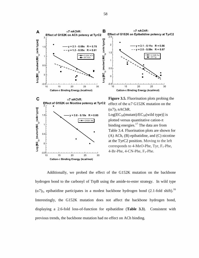
58
Figure 3.5. Fluorination plots probing the effect of the α7 G152K mutation on the
(α7)5 nAChR. Log[EC50(mutant)/EC50(wild type)] is plotted versus quantitative cation-π binding energies.17 The data are from Table 3.4. Fluorination plots are shown for (A) ACh, (B) epibatidine, and (C) nicotine at the TyrC2 position. Moving to the left corresponds to 4-MeO-Phe, Tyr, F1-Phe, 4-Br-Phe, 4-CN-Phe, F3-Phe.
Additionally, we probed the effect of the G152K mutation on the backbone
hydrogen bond to the carbonyl of TrpB using the amide-to-ester strategy. In wild type
(α7)5, epibatidine participates in a modest backbone hydrogen bond (2.1-fold shift).16
Interestingly, the G152K mutation does not affect the backbone hydrogen bond,
displaying a 2.6-fold loss-of-function for epibatidine (Table 3.3). Consistent with
previous trends, the backbone mutation had no effect on ACh binding.
59
Nicotine is not very potent at the (α7)5 receptor, which limits the kinds of studies
we have been able to perform in the past. Introduction of the G152K mutation in (α7)5
produced hypersensitive receptors sufficient for examining nicotine as the agonist. As
such, (α7G152K)5 mutant receptors allowed accurate measurement of nicotine-induced
currents even after incorporation of highly fluorinated Phe derivatives. For such
receptors, nicotine participates in a cation-π interaction with TyrC2, as indicated by a
fluorination plot (Figure 3.5C). For the backbone hydrogen bond, nicotine displayed a
7.8-fold loss-of-function in the background of the G152K mutation (Table 3.3).
3.3.3 Probing the K158 Site in the High-Potency (α4)2(β2)3 Receptor
A G-to-K mutation significantly increases potency of agonists in both the muscle-
type and (α7)5 receptors. It seemed possible that the reverse mutation, K158G, in the
high-potency (α4)2(β2)3 receptor would diminish potency. We now report that the
K158G mutation in (α4)2(β2)3 receptors is, indeed, a loss-of-function mutation (Table
3.1). However, the magnitude of the impact is significantly less than that seen for the G-
to-K mutations in the low-potency receptors.
For (α4)2(β2)3 receptors containing the K158G mutation, fluorinated tryptophan
derivatives were incorporated at TrpB, the site of a cation-π interaction for ACh and
nicotine (Table 3.5).7 Note that epibatidine also participates in a cation-π interaction and
hydrogen bond at TrpB in α4β2 (Table 3.5), but we chose not to test the effect of the
K158G mutation on epibatidine binding as it would likely mimic that of nicotine. As
such, the K158G mutation attenuated but did not completely abolish the existing cation-π
interaction for both ACh and nicotine (Figure 3.6).
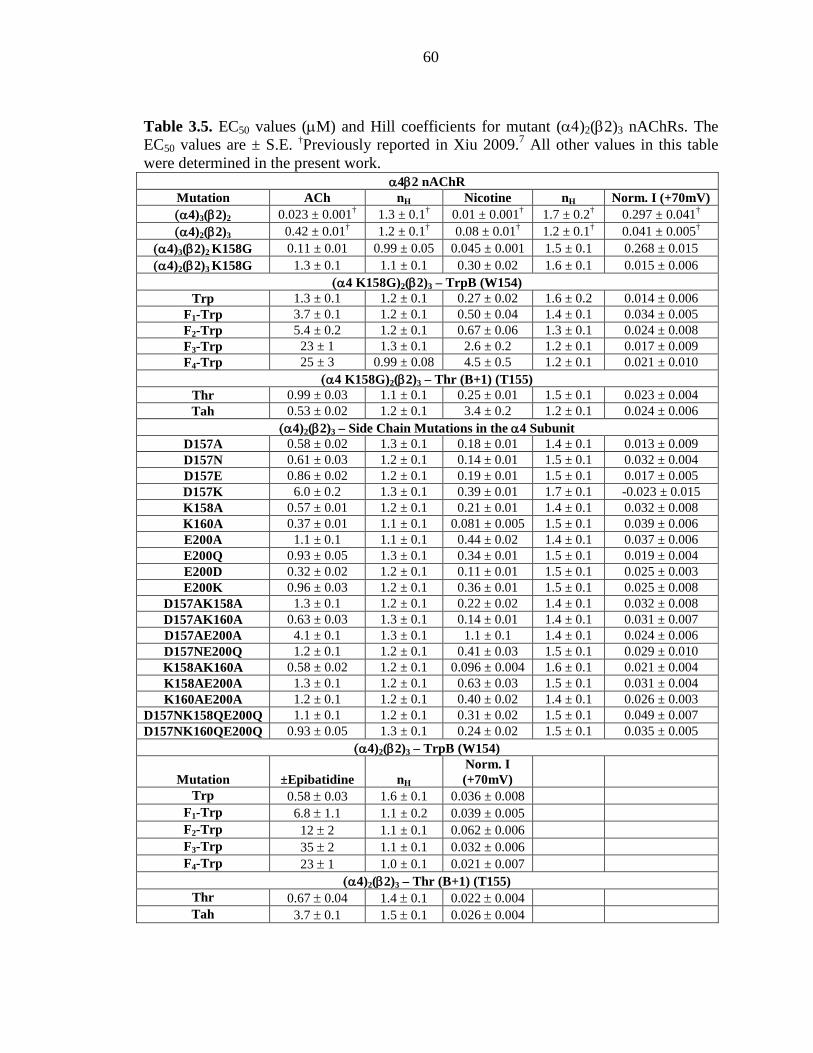
60
Table 3.5. EC50 values (µM) and Hill coefficients for mutant (α4)2(β2)3 nAChRs. The EC50 values are ± S.E. †Previously reported in Xiu 2009.7 All other values in this table were determined in the present work.
α4β2 nAChR Mutation ACh nH Nicotine nH Norm. I (+70mV) (α4)3(β2)2 0.023 ± 0.001† 1.3 ± 0.1† 0.01 ± 0.001† 1.7 ± 0.2† 0.297 ± 0.041† (α4)2(β2)3 0.42 ± 0.01† 1.2 ± 0.1† 0.08 ± 0.01† 1.2 ± 0.1† 0.041 ± 0.005†
(α4)3(β2)2 K158G 0.11 ± 0.01 0.99 ± 0.05 0.045 ± 0.001 1.5 ± 0.1 0.268 ± 0.015 (α4)2(β2)3 K158G 1.3 ± 0.1 1.1 ± 0.1 0.30 ± 0.02 1.6 ± 0.1 0.015 ± 0.006
(α4 K158G)2(β2)3 – TrpB (W154) Trp 1.3 ± 0.1 1.2 ± 0.1 0.27 ± 0.02 1.6 ± 0.2 0.014 ± 0.006
F1-Trp 3.7 ± 0.1 1.2 ± 0.1 0.50 ± 0.04 1.4 ± 0.1 0.034 ± 0.005 F2-Trp 5.4 ± 0.2 1.2 ± 0.1 0.67 ± 0.06 1.3 ± 0.1 0.024 ± 0.008 F3-Trp 23 ± 1 1.3 ± 0.1 2.6 ± 0.2 1.2 ± 0.1 0.017 ± 0.009 F4-Trp 25 ± 3 0.99 ± 0.08 4.5 ± 0.5 1.2 ± 0.1 0.021 ± 0.010
(α4 K158G)2(β2)3 – Thr (B+1) (T155) Thr 0.99 ± 0.03 1.1 ± 0.1 0.25 ± 0.01 1.5 ± 0.1 0.023 ± 0.004 Tah 0.53 ± 0.02 1.2 ± 0.1 3.4 ± 0.2 1.2 ± 0.1 0.024 ± 0.006
(α4)2(β2)3 – Side Chain Mutations in the α4 Subunit D157A 0.58 ± 0.02 1.3 ± 0.1 0.18 ± 0.01 1.4 ± 0.1 0.013 ± 0.009 D157N 0.61 ± 0.03 1.2 ± 0.1 0.14 ± 0.01 1.5 ± 0.1 0.032 ± 0.004 D157E 0.86 ± 0.02 1.2 ± 0.1 0.19 ± 0.01 1.5 ± 0.1 0.017 ± 0.005 D157K 6.0 ± 0.2 1.3 ± 0.1 0.39 ± 0.01 1.7 ± 0.1 -0.023 ± 0.015 K158A 0.57 ± 0.01 1.2 ± 0.1 0.21 ± 0.01 1.4 ± 0.1 0.032 ± 0.008 K160A 0.37 ± 0.01 1.1 ± 0.1 0.081 ± 0.005 1.5 ± 0.1 0.039 ± 0.006 E200A 1.1 ± 0.1 1.1 ± 0.1 0.44 ± 0.02 1.4 ± 0.1 0.037 ± 0.006 E200Q 0.93 ± 0.05 1.3 ± 0.1 0.34 ± 0.01 1.5 ± 0.1 0.019 ± 0.004 E200D 0.32 ± 0.02 1.2 ± 0.1 0.11 ± 0.01 1.5 ± 0.1 0.025 ± 0.003 E200K 0.96 ± 0.03 1.2 ± 0.1 0.36 ± 0.01 1.5 ± 0.1 0.025 ± 0.008
D157AK158A 1.3 ± 0.1 1.2 ± 0.1 0.22 ± 0.02 1.4 ± 0.1 0.032 ± 0.008 D157AK160A 0.63 ± 0.03 1.3 ± 0.1 0.14 ± 0.01 1.4 ± 0.1 0.031 ± 0.007 D157AE200A 4.1 ± 0.1 1.3 ± 0.1 1.1 ± 0.1 1.4 ± 0.1 0.024 ± 0.006 D157NE200Q 1.2 ± 0.1 1.2 ± 0.1 0.41 ± 0.03 1.5 ± 0.1 0.029 ± 0.010 K158AK160A 0.58 ± 0.02 1.2 ± 0.1 0.096 ± 0.004 1.6 ± 0.1 0.021 ± 0.004 K158AE200A 1.3 ± 0.1 1.2 ± 0.1 0.63 ± 0.03 1.5 ± 0.1 0.031 ± 0.004 K160AE200A 1.2 ± 0.1 1.2 ± 0.1 0.40 ± 0.02 1.4 ± 0.1 0.026 ± 0.003
D157NK158QE200Q 1.1 ± 0.1 1.2 ± 0.1 0.31 ± 0.02 1.5 ± 0.1 0.049 ± 0.007 D157NK160QE200Q 0.93 ± 0.05 1.3 ± 0.1 0.24 ± 0.02 1.5 ± 0.1 0.035 ± 0.005
(α4)2(β2)3 – TrpB (W154)
Mutation ±Epibatidine nH Norm. I (+70mV)
Trp 0.58 ± 0.03 1.6 ± 0.1 0.036 ± 0.008 F1-Trp 6.8 ± 1.1 1.1 ± 0.2 0.039 ± 0.005 F2-Trp 12 ± 2 1.1 ± 0.1 0.062 ± 0.006 F3-Trp 35 ± 2 1.1 ± 0.1 0.032 ± 0.006 F4-Trp 23 ± 1 1.0 ± 0.1 0.021 ± 0.007
(α4)2(β2)3 – Thr (B+1) (T155) Thr 0.67 ± 0.04 1.4 ± 0.1 0.022 ± 0.004 Tah 3.7 ± 0.1 1.5 ± 0.1 0.026 ± 0.004
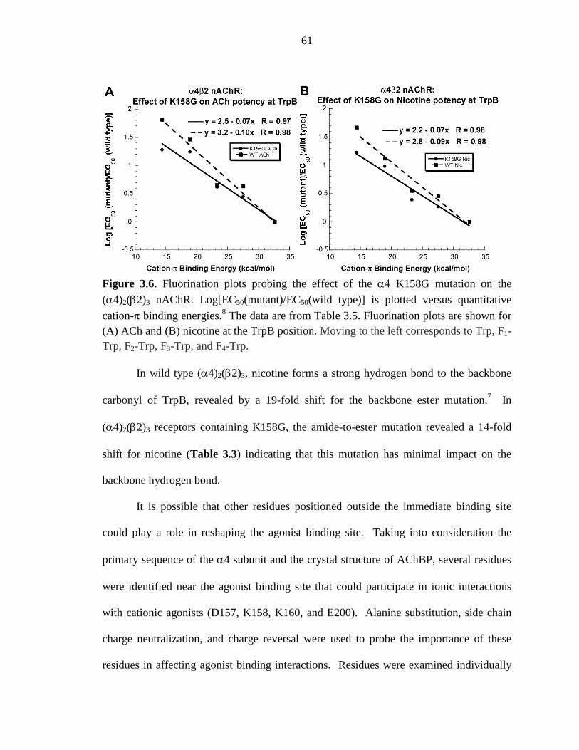
61
Figure 3.6. Fluorination plots probing the effect of the α4 K158G mutation on the
(α4)2(β2)3 nAChR. Log[EC50(mutant)/EC50(wild type)] is plotted versus quantitative
cation-π binding energies.8 The data are from Table 3.5. Fluorination plots are shown for (A) ACh and (B) nicotine at the TrpB position. Moving to the left corresponds to Trp, F1-Trp, F2-Trp, F3-Trp, and F4-Trp.
In wild type (α4)2(β2)3, nicotine forms a strong hydrogen bond to the backbone
carbonyl of TrpB, revealed by a 19-fold shift for the backbone ester mutation.7 In
(α4)2(β2)3 receptors containing K158G, the amide-to-ester mutation revealed a 14-fold
shift for nicotine (Table 3.3) indicating that this mutation has minimal impact on the
backbone hydrogen bond.
It is possible that other residues positioned outside the immediate binding site
could play a role in reshaping the agonist binding site. Taking into consideration the
primary sequence of the α4 subunit and the crystal structure of AChBP, several residues
were identified near the agonist binding site that could participate in ionic interactions
with cationic agonists (D157, K158, K160, and E200). Alanine substitution, side chain
charge neutralization, and charge reversal were used to probe the importance of these
residues in affecting agonist binding interactions. Residues were examined individually
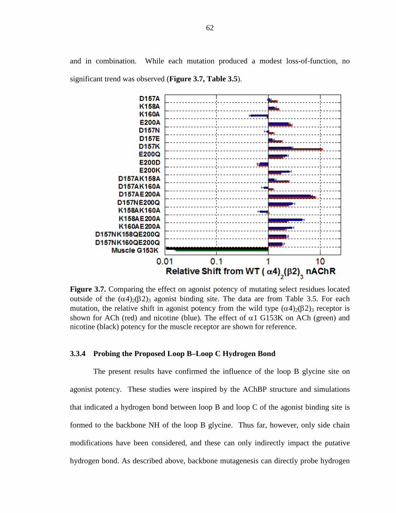
62
and in combination. While each mutation produced a modest loss-of-function, no
significant trend was observed (Figure 3.7, Table 3.5).
Figure 3.7. Comparing the effect on agonist potency of mutating select residues located outside of the (α4)2(β2)3 agonist binding site. The data are from Table 3.5. For each mutation, the relative shift in agonist potency from the wild type (α4)2(β2)3 receptor is shown for ACh (red) and nicotine (blue). The effect of α1 G153K on ACh (green) and nicotine (black) potency for the muscle receptor are shown for reference.
3.3.4 Probing the Proposed Loop B–Loop C Hydrogen Bond
The present results have confirmed the influence of the loop B glycine site on
agonist potency. These studies were inspired by the AChBP structure and simulations
that indicated a hydrogen bond between loop B and loop C of the agonist binding site is
formed to the backbone NH of the loop B glycine. Thus far, however, only side chain
modifications have been considered, and these can only indirectly impact the putative
hydrogen bond. As described above, backbone mutagenesis can directly probe hydrogen
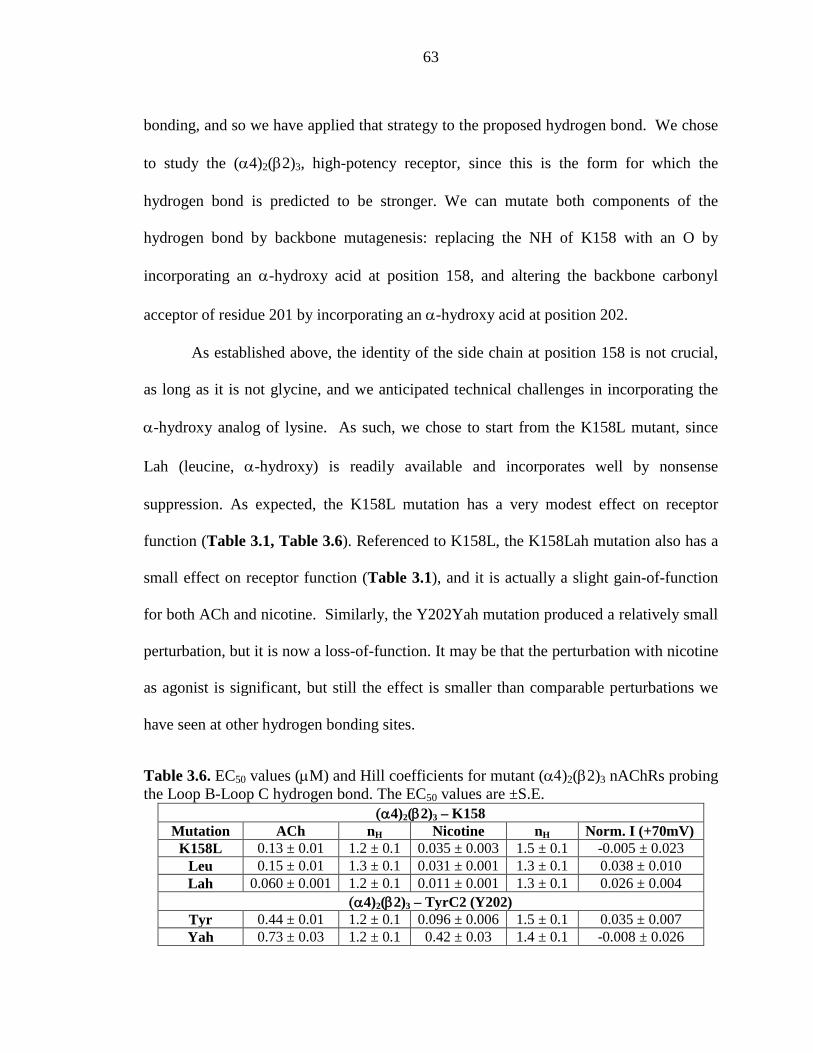
63
bonding, and so we have applied that strategy to the proposed hydrogen bond. We chose
to study the (α4)2(β2)3, high-potency receptor, since this is the form for which the
hydrogen bond is predicted to be stronger. We can mutate both components of the
hydrogen bond by backbone mutagenesis: replacing the NH of K158 with an O by
incorporating an α-hydroxy acid at position 158, and altering the backbone carbonyl
acceptor of residue 201 by incorporating an α-hydroxy acid at position 202.
As established above, the identity of the side chain at position 158 is not crucial,
as long as it is not glycine, and we anticipated technical challenges in incorporating the
α-hydroxy analog of lysine. As such, we chose to start from the K158L mutant, since
Lah (leucine, α-hydroxy) is readily available and incorporates well by nonsense
suppression. As expected, the K158L mutation has a very modest effect on receptor
function (Table 3.1, Table 3.6). Referenced to K158L, the K158Lah mutation also has a
small effect on receptor function (Table 3.1), and it is actually a slight gain-of-function
for both ACh and nicotine. Similarly, the Y202Yah mutation produced a relatively small
perturbation, but it is now a loss-of-function. It may be that the perturbation with nicotine
as agonist is significant, but still the effect is smaller than comparable perturbations we
have seen at other hydrogen bonding sites.
Table 3.6. EC50 values (µM) and Hill coefficients for mutant (α4)2(β2)3 nAChRs probing the Loop B-Loop C hydrogen bond. The EC50 values are ±S.E.
(α4)2(β2)3 – K158 Mutation ACh nH Nicotine nH Norm. I (+70mV)
K158L 0.13 ± 0.01 1.2 ± 0.1 0.035 ± 0.003 1.5 ± 0.1 -0.005 ± 0.023 Leu 0.15 ± 0.01 1.3 ± 0.1 0.031 ± 0.001 1.3 ± 0.1 0.038 ± 0.010 Lah 0.060 ± 0.001 1.2 ± 0.1 0.011 ± 0.001 1.3 ± 0.1 0.026 ± 0.004
(α4)2(β2)3 – TyrC2 (Y202) Tyr 0.44 ± 0.01 1.2 ± 0.1 0.096 ± 0.006 1.5 ± 0.1 0.035 ± 0.007 Yah 0.73 ± 0.03 1.2 ± 0.1 0.42 ± 0.03 1.4 ± 0.1 -0.008 ± 0.026
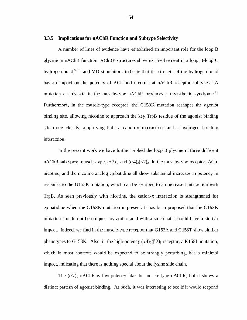
64
3.3.5 Implications for nAChR Function and Subtype Selectivity
A number of lines of evidence have established an important role for the loop B
glycine in nAChR function. AChBP structures show its involvement in a loop B-loop C
hydrogen bond,9, 10 and MD simulations indicate that the strength of the hydrogen bond
has an impact on the potency of ACh and nicotine at nAChR receptor subtypes.5 A
mutation at this site in the muscle-type nAChR produces a myasthenic syndrome.12
Furthermore, in the muscle-type receptor, the G153K mutation reshapes the agonist
binding site, allowing nicotine to approach the key TrpB residue of the agonist binding
site more closely, amplifying both a cation-π interaction7 and a hydrogen bonding
interaction.
In the present work we have further probed the loop B glycine in three different
nAChR subtypes: muscle-type, (α7)5, and (α4)2(β2)3. In the muscle-type receptor, ACh,
nicotine, and the nicotine analog epibatidine all show substantial increases in potency in
response to the G153K mutation, which can be ascribed to an increased interaction with
TrpB. As seen previously with nicotine, the cation-π interaction is strengthened for
epibatidine when the G153K mutation is present. It has been proposed that the G153K
mutation should not be unique; any amino acid with a side chain should have a similar
impact. Indeed, we find in the muscle-type receptor that G153A and G153T show similar
phenotypes to G153K. Also, in the high-potency (α4)2(β2)3 receptor, a K158L mutation,
which in most contexts would be expected to be strongly perturbing, has a minimal
impact, indicating that there is nothing special about the lysine side chain.
The (α7)5 nAChR is low-potency like the muscle-type nAChR, but it shows a
distinct pattern of agonist binding. As such, it was interesting to see if it would respond
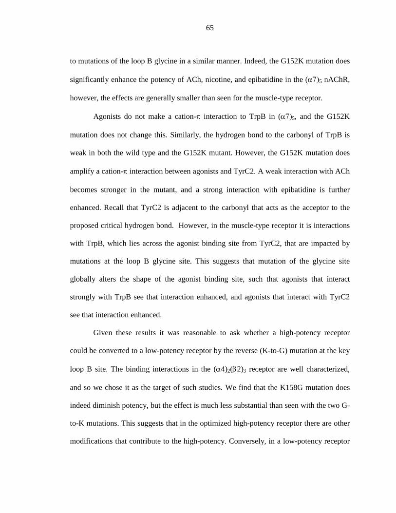
65
to mutations of the loop B glycine in a similar manner. Indeed, the G152K mutation does
significantly enhance the potency of ACh, nicotine, and epibatidine in the (α7)5 nAChR,
however, the effects are generally smaller than seen for the muscle-type receptor.
Agonists do not make a cation-π interaction to TrpB in (α7)5, and the G152K
mutation does not change this. Similarly, the hydrogen bond to the carbonyl of TrpB is
weak in both the wild type and the G152K mutant. However, the G152K mutation does
amplify a cation-π interaction between agonists and TyrC2. A weak interaction with ACh
becomes stronger in the mutant, and a strong interaction with epibatidine is further
enhanced. Recall that TyrC2 is adjacent to the carbonyl that acts as the acceptor to the
proposed critical hydrogen bond. However, in the muscle-type receptor it is interactions
with TrpB, which lies across the agonist binding site from TyrC2, that are impacted by
mutations at the loop B glycine site. This suggests that mutation of the glycine site
globally alters the shape of the agonist binding site, such that agonists that interact
strongly with TrpB see that interaction enhanced, and agonists that interact with TyrC2
see that interaction enhanced.
Given these results it was reasonable to ask whether a high-potency receptor
could be converted to a low-potency receptor by the reverse (K-to-G) mutation at the key
loop B site. The binding interactions in the (α4)2(β2)3 receptor are well characterized,
and so we chose it as the target of such studies. We find that the K158G mutation does
indeed diminish potency, but the effect is much less substantial than seen with the two G-
to-K mutations. This suggests that in the optimized high-potency receptor there are other
modifications that contribute to the high-potency. Conversely, in a low-potency receptor
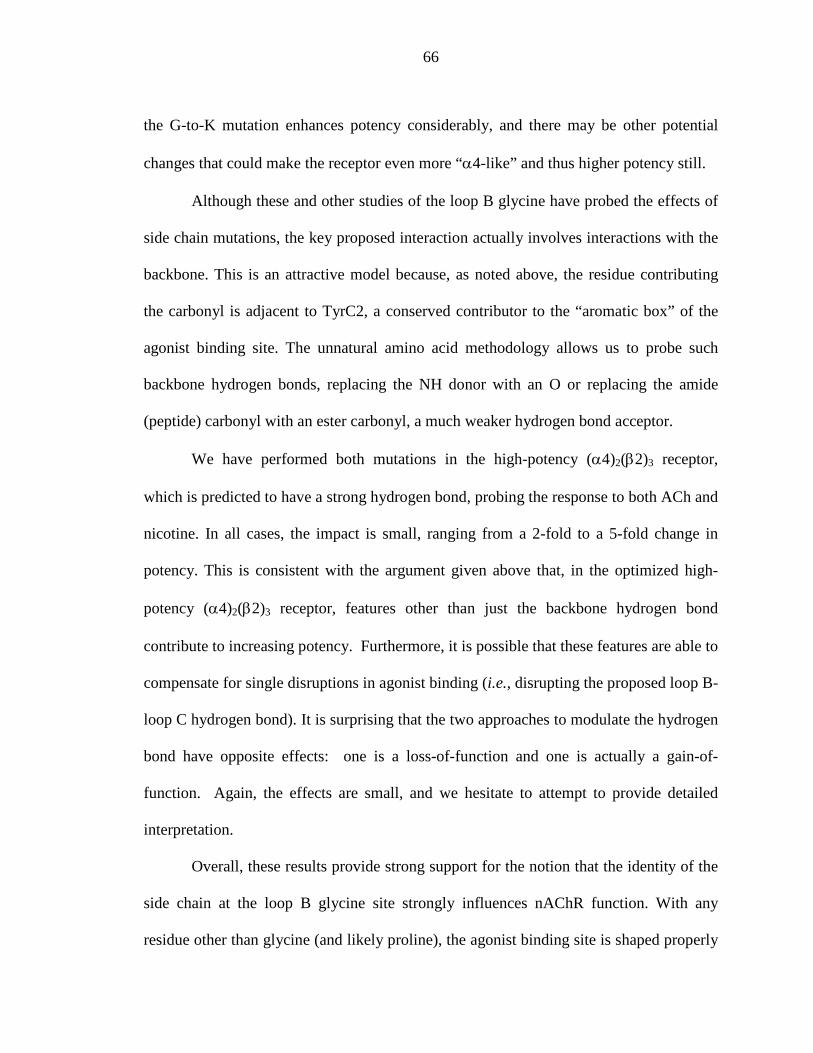
66
the G-to-K mutation enhances potency considerably, and there may be other potential
changes that could make the receptor even more “α4-like” and thus higher potency still.
Although these and other studies of the loop B glycine have probed the effects of
side chain mutations, the key proposed interaction actually involves interactions with the
backbone. This is an attractive model because, as noted above, the residue contributing
the carbonyl is adjacent to TyrC2, a conserved contributor to the “aromatic box” of the
agonist binding site. The unnatural amino acid methodology allows us to probe such
backbone hydrogen bonds, replacing the NH donor with an O or replacing the amide
(peptide) carbonyl with an ester carbonyl, a much weaker hydrogen bond acceptor.
We have performed both mutations in the high-potency (α4)2(β2)3 receptor,
which is predicted to have a strong hydrogen bond, probing the response to both ACh and
nicotine. In all cases, the impact is small, ranging from a 2-fold to a 5-fold change in
potency. This is consistent with the argument given above that, in the optimized high-
potency (α4)2(β2)3 receptor, features other than just the backbone hydrogen bond
contribute to increasing potency. Furthermore, it is possible that these features are able to
compensate for single disruptions in agonist binding (i.e., disrupting the proposed loop B-
loop C hydrogen bond). It is surprising that the two approaches to modulate the hydrogen
bond have opposite effects: one is a loss-of-function and one is actually a gain-of-
function. Again, the effects are small, and we hesitate to attempt to provide detailed
interpretation.
Overall, these results provide strong support for the notion that the identity of the
side chain at the loop B glycine site strongly influences nAChR function. With any
residue other than glycine (and likely proline), the agonist binding site is shaped properly
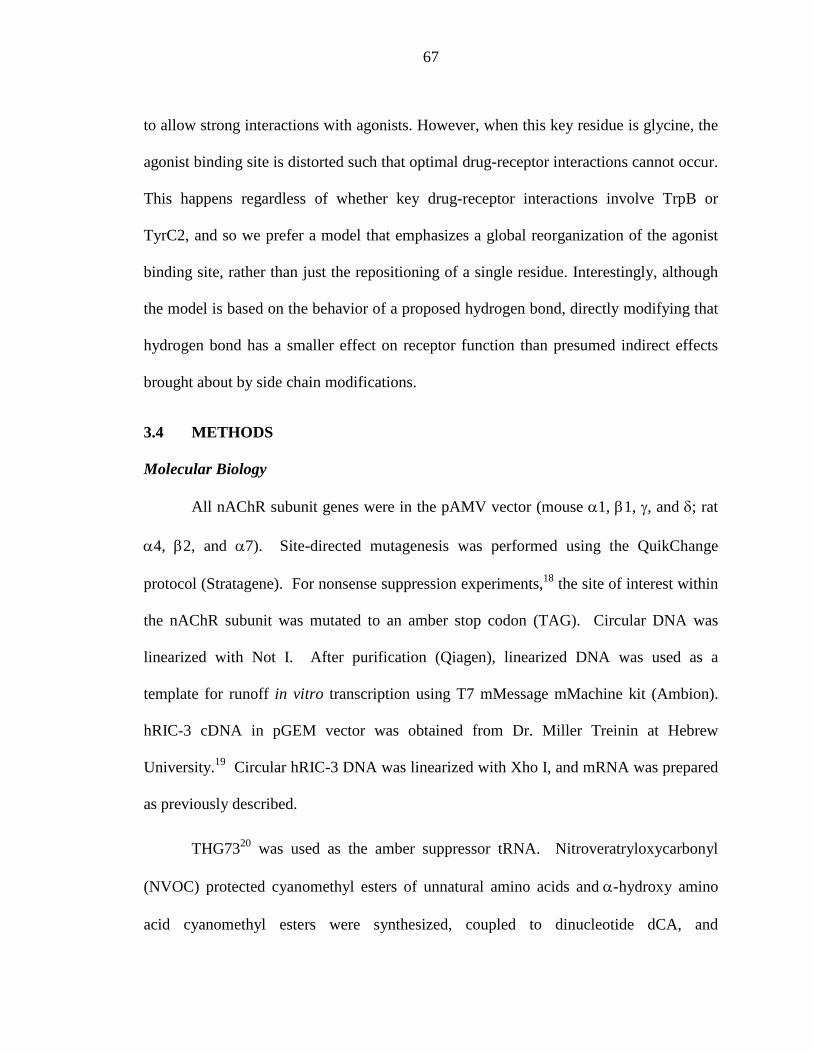
67
to allow strong interactions with agonists. However, when this key residue is glycine, the
agonist binding site is distorted such that optimal drug-receptor interactions cannot occur.
This happens regardless of whether key drug-receptor interactions involve TrpB or
TyrC2, and so we prefer a model that emphasizes a global reorganization of the agonist
binding site, rather than just the repositioning of a single residue. Interestingly, although
the model is based on the behavior of a proposed hydrogen bond, directly modifying that
hydrogen bond has a smaller effect on receptor function than presumed indirect effects
brought about by side chain modifications.
3.4 METHODS
Molecular Biology
All nAChR subunit genes were in the pAMV vector (mouse α1, β1, γ, and δ; rat
α4, β2, and α7). Site-directed mutagenesis was performed using the QuikChange
protocol (Stratagene). For nonsense suppression experiments,18 the site of interest within
the nAChR subunit was mutated to an amber stop codon (TAG). Circular DNA was
linearized with Not I. After purification (Qiagen), linearized DNA was used as a
template for runoff in vitro transcription using T7 mMessage mMachine kit (Ambion).
hRIC-3 cDNA in pGEM vector was obtained from Dr. Miller Treinin at Hebrew
University.19 Circular hRIC-3 DNA was linearized with Xho I, and mRNA was prepared
as previously described.
THG7320 was used as the amber suppressor tRNA. Nitroveratryloxycarbonyl
(NVOC) protected cyanomethyl esters of unnatural amino acids and α-hydroxy amino
acid cyanomethyl esters were synthesized, coupled to dinucleotide dCA, and
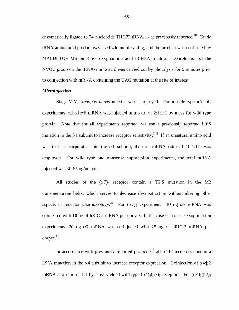
68
enzymatically ligated to 74-nucleotide THG73 tRNACUA as previously reported.18 Crude
tRNA-amino acid product was used without desalting, and the product was confirmed by
MALDI-TOF MS on 3-hydroxypicolinic acid (3-HPA) matrix. Deprotection of the
NVOC group on the tRNA-amino acid was carried out by photolysis for 5 minutes prior
to coinjection with mRNA containing the UAG mutation at the site of interest.
Microinjection
Stage V-VI Xenopus laevis oocytes were employed. For muscle-type nAChR
experiments, α1:β1:γ:δ mRNA was injected at a ratio of 2:1:1:1 by mass for wild type
protein. Note that for all experiments reported, we use a previously reported L9’S
mutation in the β1 subunit to increase receptor sensitivity.7, 8 If an unnatural amino acid
was to be incorporated into the α1 subunit, then an mRNA ratio of 10:1:1:1 was
employed. For wild type and nonsense suppression experiments, the total mRNA
injected was 30-65 ng/oocyte.
All studies of the (α7)5 receptor contain a T6’S mutation in the M2
transmembrane helix, which serves to decrease desensitization without altering other
aspects of receptor pharmacology.21 For (α7)5 experiments, 10 ng α7 mRNA was
coinjected with 10 ng of hRIC-3 mRNA per oocyte. In the case of nonsense suppression
experiments, 20 ng α7 mRNA was co-injected with 25 ng of hRIC-3 mRNA per
oocyte.16
In accordance with previously reported protocols,7 all α4β2 receptors contain a
L9’A mutation in the α4 subunit to increase receptor expression. Coinjection of α4:β2
mRNA at a ratio of 1:1 by mass yielded wild type (α4)2(β2)3 receptors. For (α4)2(β2)3
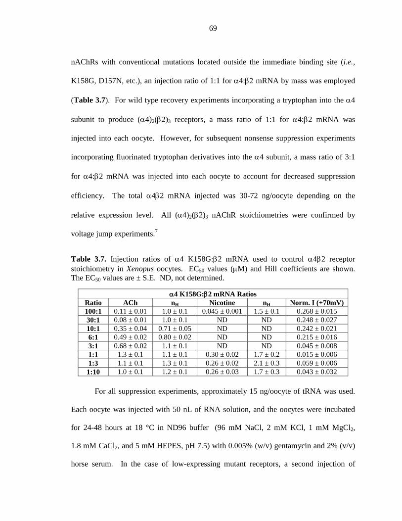
69
nAChRs with conventional mutations located outside the immediate binding site (i.e.,
K158G, D157N, etc.), an injection ratio of 1:1 for α4:β2 mRNA by mass was employed
(Table 3.7). For wild type recovery experiments incorporating a tryptophan into the α4
subunit to produce (α4)2(β2)3 receptors, a mass ratio of 1:1 for α4:β2 mRNA was
injected into each oocyte. However, for subsequent nonsense suppression experiments
incorporating fluorinated tryptophan derivatives into the α4 subunit, a mass ratio of 3:1
for α4:β2 mRNA was injected into each oocyte to account for decreased suppression
efficiency. The total α4β2 mRNA injected was 30-72 ng/oocyte depending on the
relative expression level. All (α4)2(β2)3 nAChR stoichiometries were confirmed by
voltage jump experiments.7
Table 3.7. Injection ratios of α4 K158G:β2 mRNA used to control α4β2 receptor stoichiometry in Xenopus oocytes. EC50 values (µM) and Hill coefficients are shown. The EC50 values are ± S.E. ND, not determined.
α4 K158G:β2 mRNA Ratios Ratio ACh nH Nicotine nH Norm. I (+70mV) 100:1 0.11 ± 0.01 1.0 ± 0.1 0.045 ± 0.001 1.5 ± 0.1 0.268 ± 0.015 30:1 0.08 ± 0.01 1.0 ± 0.1 ND ND 0.248 ± 0.027 10:1 0.35 ± 0.04 0.71 ± 0.05 ND ND 0.242 ± 0.021 6:1 0.49 ± 0.02 0.80 ± 0.02 ND ND 0.215 ± 0.016 3:1 0.68 ± 0.02 1.1 ± 0.1 ND ND 0.045 ± 0.008 1:1 1.3 ± 0.1 1.1 ± 0.1 0.30 ± 0.02 1.7 ± 0.2 0.015 ± 0.006 1:3 1.1 ± 0.1 1.3 ± 0.1 0.26 ± 0.02 2.1 ± 0.3 0.059 ± 0.006 1:10 1.0 ± 0.1 1.2 ± 0.1 0.26 ± 0.03 1.7 ± 0.3 0.043 ± 0.032
For all suppression experiments, approximately 15 ng/oocyte of tRNA was used.
Each oocyte was injected with 50 nL of RNA solution, and the oocytes were incubated
for 24-48 hours at 18 °C in ND96 buffer (96 mM NaCl, 2 mM KCl, 1 mM MgCl2,
1.8 mM CaCl2, and 5 mM HEPES, pH 7.5) with 0.005% (w/v) gentamycin and 2% (v/v)
horse serum. In the case of low-expressing mutant receptors, a second injection of
70
mRNA/tRNA was required 24 hour after the first injection. As a negative control for all
suppression experiments, 76-nucleotide tRNA (dCA ligated to 74-nucleotide tRNA) was
coinjected with mRNA in the same manner as fully charged tRNA.
Electrophysiology
Acetylcholine chloride and (-)-nicotine tartrate were purchased from
Sigma/Aldrich/RBI (St. Louis, MO) and drug dilutions were prepared from 1M aq stock
solutions. (±)-Epibatidine was purchased from Tocris and drug dilutions were prepared
from a 50 mM stock solution (1:1 H2O:EtOH). For (α1)2β1γδ and (α4)2(β2)3
experiments, drug dilutions were prepared in Ca2+-free ND96 buffer. For (α7)5
experiments, drug dilutions were prepared in Ca2+-containing ND96 buffer.
Ion channel function was assayed using the OpusXpress 6000A (Molecular
Devices Axon Instruments) in two-electrode voltage clamp mode. Oocytes were clamped
at a holding potential of -60mV. For (α1)2β1γδ and (α4)2(β2)3 receptors, 1 mL of each
drug solution was applied to the clamped oocytes for 12 seconds, followed by a 2 minute
wash with Ca2+-free ND96 buffer between each concentration. In the case of
hypersensitive (α1G153K)2β1γδ receptors, a 1.5 minute drug application was used to
ensure maximum peak response when using very low concentrations of agonist.
For (α7)5 receptors, 1 mL of each drug solution was applied for 30 seconds, followed by
a 5 minute wash step with Ca2+-containing ND96 buffer between each concentration.
Data were sampled at 50 Hz and filtered at 20 Hz. Voltage jump experiments were
sampled at 5000 Hz and filtered at 180 Hz.
71
Data Analysis
Dose-response data were obtained for at least 6 concentrations of agonists and for
a minimum of 5 oocytes (from two different batches). Mutants with Imax of at least 100
nA of current were defined as functional. EC50 and Hill coefficients (nH) were calculated
by fitting the averaged, normalized dose-response relation to the Hill equation. All data
are reported as mean ±S.E.
3.5 ACKNOWLEDGEMENTS
This work was supported by the NIH (NS 34407 and NS 11756).
72
3.6 REFERENCES
1. Corringer, P. J.; Le Novere, N.; Changeux, J. P., Nicotinic receptors at the amino acid level. Annu Rev Pharmacol Toxicol 2000, 40, 431-58. 2. Grutter, T.; Changeux, J. P., Nicotinic receptors in wonderland. Trends Biochem Sci 2001, 26, (8), 459-63. 3. Karlin, A., Emerging structure of the nicotinic acetylcholine receptors. Nat Rev Neurosci 2002, 3, (2), 102-14. 4. Jensen, A. A.; Frolund, B.; Lijefors, T.; Krogsgaard-Larsen, P., Neuronal nicotinic acetylcholine receptors: Structural revelations, target identifications, and therapeutic inspirations. J. Med. Chem. 2005, 48, (15), 4705-4745. 5. Grutter, T.; de Carvalho, L. P.; Le Novere, N.; Corringer, P. J.; Edelstein, S.; Changeux, J. P., An H-bond between two residues from different loops of the acetylcholine binding site contributes to the activation mechanism of nicotinic receptors. EMBO J. 2003, 22, (9), 1990-2003. 6. Gotti, C.; Zoli, M.; Clementi, F., Brain nicotinic acetylcholine receptors: native subtypes and their relevance. Trends Pharm. Sci. 2006, 27, (9), 482-491. 7. Xiu, X.; Puskar, N. L.; Shanata, J. A.; Lester, H. A.; Dougherty, D. A., Nicotine binding to brain receptors requires a strong cation-pi interaction. Nature 2009, 458, (7237), 534-7. 8. Zhong, W.; Gallivan, J. P.; Zhang, Y.; Li, L.; Lester, H. A.; Dougherty, D. A., From ab initio quantum mechanics to molecular neurobiology: a cation-pi binding site in the nicotinic receptor. Proc Natl Acad Sci U S A 1998, 95, (21), 12088-93. 9. Brejc, K.; van Dijk, W. J.; Klaassen, R. V.; Schuurmans, M.; van Der Oost, J.; Smit, A. B.; Sixma, T. K., Crystal structure of an ACh-binding protein reveals the ligand-binding domain of nicotinic receptors. Nature 2001, 411, (6835), 269-76. 10. Sixma, T. K.; Smit, A. B., Acetylcholine binding protein (AChBP): a secreted glial protein that provides a high-resolution model for the extracellular domain of pentameric ligand-gated ion channels. Annu Rev Biophys Biomol Struct 2003, 32, 311-34. 11. Hibbs, R. E.; Gouaux, E., Principles of activation and permeation in an anion-selective Cys-loop receptor. Nature 2011, 474, (7349), 54-U80. 12. Sine, S. M.; Ohno, K.; Bouzat, C.; Auerbach, A.; Milone, M.; Pruitt, J. N.; Engel, A. G., Mutation of the acetylcholine receptor alpha subunit causes a slow-channel myasthenic syndrome by enhancing agonist binding affinity. Neuron 1995, 15, (1), 229-239. 13. Celie, P. H.; van Rossum-Fikkert, S. E.; van Dijk, W. J.; Brejc, K.; Smit, A. B.; Sixma, T. K., Nicotine and carbamylcholine binding to nicotinic acetylcholine receptors as studied in AChBP crystal structures. Neuron 2004, 41, (6), 907-914. 14. Cashin, A. L.; Petersson, E. J.; Lester, H. A.; Dougherty, D. A., Using physical chemistry to differentiate nicotinic from cholinergic agonists at the nicotinic acetylcholine receptor. J. Am. Chem. Soc. 2005, 127, (1), 350-356. 15. Cashin, A. L.; Petersson, E. J.; Lester, H. A.; Dougherty, D. A., Using physical chemistry to differentiate nicotinic from cholinergic agonists at the nicotinic acetylcholine receptor. J Am Chem Soc 2005, 127, (1), 350-6.
73
16. Puskar, N. L.; Xiu, X.; Lester, H. A.; Dougherty, D. A., Two Neuronal Nicotinic Acetylcholine Receptors, alpha 4 beta 4 and alpha 7, Show Differential Agonist Binding Modes. Journal of Biological Chemistry 2011, 286, (16), 14618-14627. 17. Lummis, S. C.; D, L. B.; Harrison, N. J.; Lester, H. A.; Dougherty, D. A., A cation-pi binding interaction with a tyrosine in the binding site of the GABAC receptor. Chem Biol 2005, 12, (9), 993-7. 18. Nowak, M. W.; Gallivan, J. P.; Silverman, S. K.; Labarca, C. G.; Dougherty, D. A.; Lester, H. A., In vivo incorporation of unnatural amino acids into ion channels in a Xenopus oocyte expression system. Methods Enzymol 1998, 293, 504-529. 19. Williams, M. E.; Burton, B.; Urrutia, A.; Shcherbatko, A.; Chavez-Noriega, L. E.; Cohen, C. J.; Aiyar, J., Ric-3 promotes functional expression of the nicotinic acetylcholine receptor alpha7 subunit in mammalian cells. J Biol Chem 2005, 280, (2), 1257-63. 20. Saks, M. E.; Sampson, J. R.; Nowak, M. W.; Kearney, P. C.; Du, F.; Abelson, J. N.; Lester, H. A.; Dougherty, D. A., An engineered Tetrahymena tRNAGln for in vivo incorporation of unnatural amino acids into proteins by nonsense suppression. J. Biol. Chem 1996, 271, 23169-75. 21. Placzek, A. N.; Grassi, F.; Meyer, E. M.; Papke, R. L., An alpha7 nicotinic acetylcholine receptor gain-of-function mutant that retains pharmacological fidelity. Mol Pharmacol 2005, 68, (6), 1863-76.
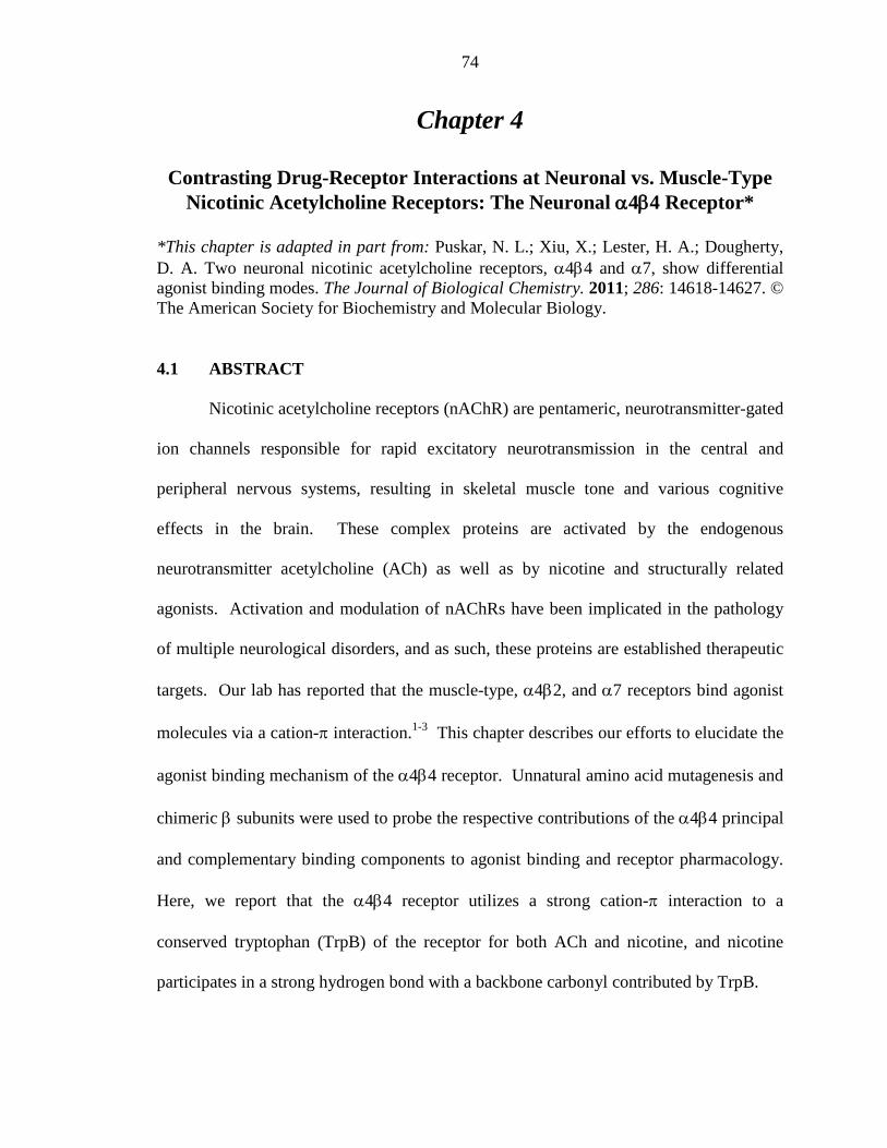
74
Chapter 4
Contrasting Drug-Receptor Interactions at Neuronal vs. Muscle-Type Nicotinic Acetylcholine Receptors: The Neuronal α4β4 Receptor*
*This chapter is adapted in part from: Puskar, N. L.; Xiu, X.; Lester, H. A.; Dougherty, D. A. Two neuronal nicotinic acetylcholine receptors, α4β4 and α7, show differential agonist binding modes. The Journal of Biological Chemistry. 2011; 286: 14618-14627. © The American Society for Biochemistry and Molecular Biology.
4.1 ABSTRACT
Nicotinic acetylcholine receptors (nAChR) are pentameric, neurotransmitter-gated
ion channels responsible for rapid excitatory neurotransmission in the central and
peripheral nervous systems, resulting in skeletal muscle tone and various cognitive
effects in the brain. These complex proteins are activated by the endogenous
neurotransmitter acetylcholine (ACh) as well as by nicotine and structurally related
agonists. Activation and modulation of nAChRs have been implicated in the pathology
of multiple neurological disorders, and as such, these proteins are established therapeutic
targets. Our lab has reported that the muscle-type, α4β2, and α7 receptors bind agonist
molecules via a cation-π interaction.1-3 This chapter describes our efforts to elucidate the
agonist binding mechanism of the α4β4 receptor. Unnatural amino acid mutagenesis and
chimeric β subunits were used to probe the respective contributions of the α4β4 principal
and complementary binding components to agonist binding and receptor pharmacology.
Here, we report that the α4β4 receptor utilizes a strong cation-π interaction to a
conserved tryptophan (TrpB) of the receptor for both ACh and nicotine, and nicotine
participates in a strong hydrogen bond with a backbone carbonyl contributed by TrpB.

75
4.2 INTRODUCTION
Nicotinic acetylcholine receptors (nAChRs) belong to the Cys-loop superfamily
of neurotransmitter-gated ion channels, which includes γ-aminobutyric acid (GABAA and
GABAC), glycine (Gly), and serotonin type 3 (5-HT3) receptors. These transmembrane
proteins are critical to proper rapid synaptic transmission in the central and peripheral
nervous systems.4 In fact, several nAChRs have been implicated in pathophysiology
and/or therapy of multiple neurological and psychiatric disorders including addiction,
schizophrenia, Parkinson’s disease, Alzheimer’s disease, pain, ADHD, epilepsy,
depression, and congenital myasthenic syndromes. 5, 6
The nAChR is the longest-known, most-studied neuroreceptor. nAChRs are
pentameric, integral membrane proteins whose overall structure has been roughly
determined by cryo-electron microscopy images of the Torpedo californica nAChR
(Figure 4.1).7 Each subunit contains a large, principally β-sheet extracellular N-terminal
domain, four transmembrane α-helices (M1-M4), and a small extracellular C-terminal
domain. Five homologous subunits are arranged pseudosymmetrically around a central
ion conducting pore formed by the M2 helices of each subunit.8 To date, 16 mammalian
genes have been identified that encode nAChR subunits, termed α1-α7, α9, α10, β1-β4,
δ, γ, and ε.
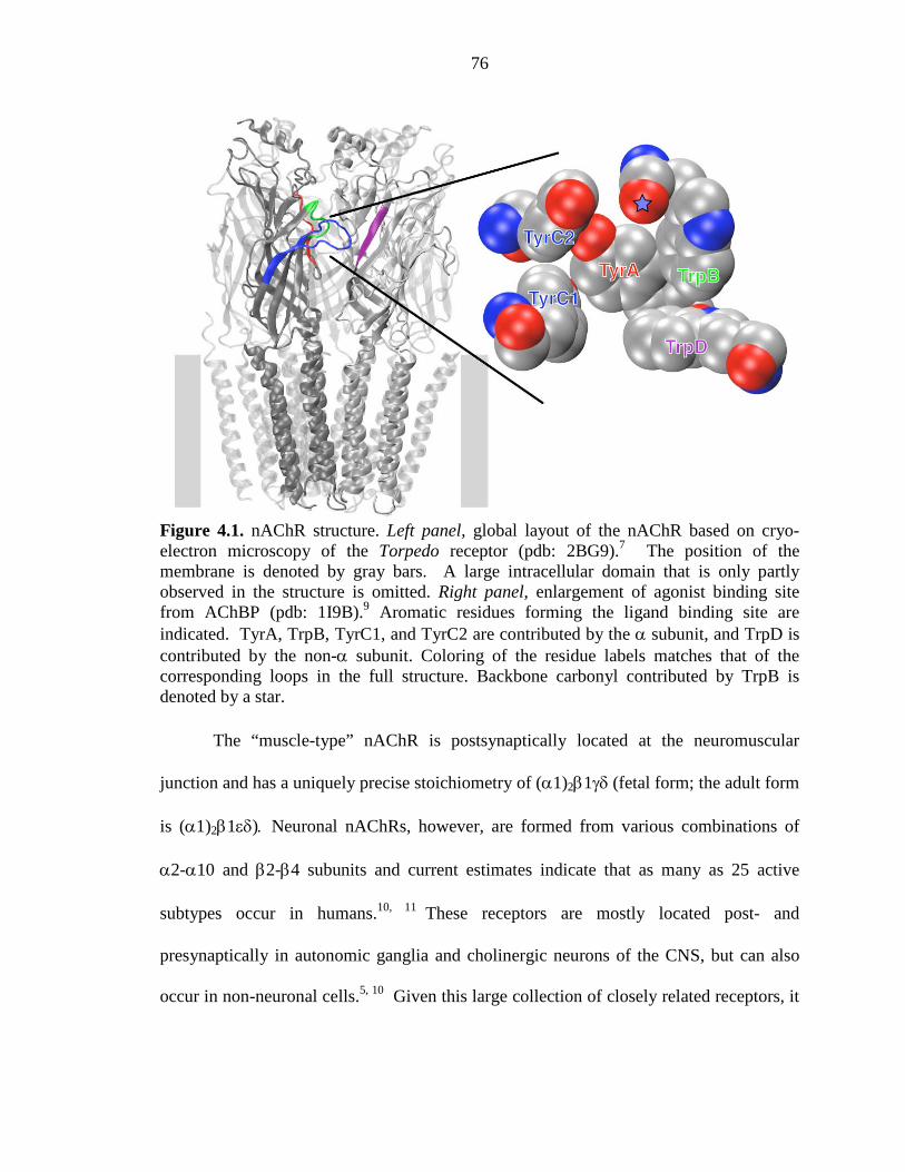
76
Figure 4.1. nAChR structure. Left panel, global layout of the nAChR based on cryo-electron microscopy of the Torpedo receptor (pdb: 2BG9).7 The position of the membrane is denoted by gray bars. A large intracellular domain that is only partly observed in the structure is omitted. Right panel, enlargement of agonist binding site from AChBP (pdb: 1I9B).9 Aromatic residues forming the ligand binding site are indicated. TyrA, TrpB, TyrC1, and TyrC2 are contributed by the α subunit, and TrpD is contributed by the non-α subunit. Coloring of the residue labels matches that of the corresponding loops in the full structure. Backbone carbonyl contributed by TrpB is denoted by a star.
The “muscle-type” nAChR is postsynaptically located at the neuromuscular
junction and has a uniquely precise stoichiometry of (α1)2β1γδ (fetal form; the adult form
is (α1)2β1εδ). Neuronal nAChRs, however, are formed from various combinations of
α2-α10 and β2-β4 subunits and current estimates indicate that as many as 25 active
subtypes occur in humans.10, 11 These receptors are mostly located post- and
presynaptically in autonomic ganglia and cholinergic neurons of the CNS, but can also
occur in non-neuronal cells.5, 10 Given this large collection of closely related receptors, it

77
seems certain that therapeutics directed toward specific neurological disorders will
require selectivity in terms of which nAChR subtype(s) is targeted.
Early work established a nicotinic pharmacophore comprised of a cationic N and
a hydrogen bond accepting group separated by an appropriate distance.12, 13 This report
focuses on critical drug-receptor interactions occurring at the agonist binding site. The
cationic moiety of ACh interacts with a cluster of aromatic amino acids first identified by
photoaffinity labeling and mutagenesis experiments of the full receptor.4, 9 Crystal
structures of the acetylcholine binding proteins (AChBP) provided a structural template
for the N-terminal, extracellular, ligand binding domain (LBD) of the nAChR, as it shares
20%-25% sequence identity with the extracellular domain of the much larger ion channel
protein.9, 14 Five aromatic residues (labeled according to their respective loop) form the
agonist binding site, and these five aromatics are completely conserved across the
nAChR family (Figure 4.1). The principal binding site contributes loop A (TyrA), loop
B (TrpB), and loop C (TyrC1 and TyrC2), and the complementary binding site contributes
loop D (TrpD), loop E, and loop F. In recent work, it was confirmed that the hydrogen
bond acceptor of the agonist interacts with residues from the complementary subunits (β
in neuronal nAChRs; γ, δ, ε in the muscle-type nAChR).15
In the Dougherty group, we have used the nonsense suppression methodology to
probe the molecular determinants for agonist binding in several nAChR subtypes, such as
the muscle-type and neuronal α4β2 and α7 receptors.1-3, 16, 17 From these studies, the
cation-π interaction proved a common component of agonist affinity in each of the
aforementioned receptors. Preliminary studies of the neuronal α4β4 nAChR, however,
proved inconclusive.18 Interestingly, the α4 and β4 subunits colocalize in brain regions
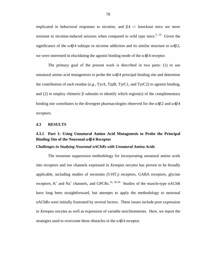
78
implicated in behavioral responses to nicotine, and β4 -/- knockout mice are more
resistant to nicotine-induced seizures when compared to wild type mice.5, 19 Given the
significance of the α4β4 subtype in nicotine addiction and its similar structure to α4β2,
we were interested in elucidating the agonist binding mode of the α4β4 receptor.
The primary goal of the present work is described in two parts: (1) to use
unnatural amino acid mutagenesis to probe the α4β4 principal binding site and determine
the contribution of each residue (e.g., TyrA, TrpB, TyrC1, and TyrC2) to agonist binding,
and (2) to employ chimeric β subunits to identify which region(s) of the complementary
binding site contributes to the divergent pharmacologies observed for the α4β2 and α4β4
receptors.
4.3 RESULTS
4.3.1 Part 1: Using Unnatural Amino Acid Mutagenesis to Probe the Principal Binding Site of the Neuronal α4β4 Receptor
Challenges in Studying Neuronal nAChRs with Unnatural Amino Acids
The nonsense suppression methodology for incorporating unnatural amino acids
into receptors and ion channels expressed in Xenopus oocytes has proven to be broadly
applicable, including studies of serotonin (5-HT3) receptors, GABA receptors, glycine
receptors, K+ and Na+ channels, and GPCRs.16, 20-24 Studies of the muscle-type nAChR
have long been straightforward, but attempts to apply the methodology to neuronal
nAChRs were initially frustrated by several factors. These issues include poor expression
in Xenopus oocytes as well as expression of variable stoichiometries. Here, we report the
strategies used to overcome these obstacles in the α4β4 receptor.
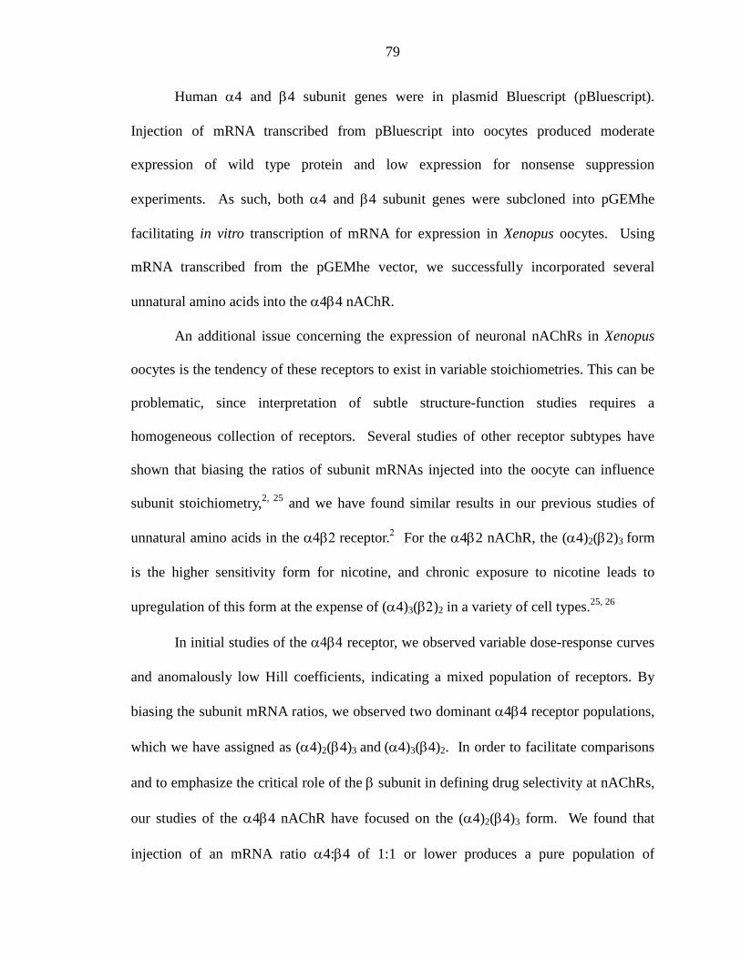
79
Human α4 and β4 subunit genes were in plasmid Bluescript (pBluescript).
Injection of mRNA transcribed from pBluescript into oocytes produced moderate
expression of wild type protein and low expression for nonsense suppression
experiments. As such, both α4 and β4 subunit genes were subcloned into pGEMhe
facilitating in vitro transcription of mRNA for expression in Xenopus oocytes. Using
mRNA transcribed from the pGEMhe vector, we successfully incorporated several
unnatural amino acids into the α4β4 nAChR.
An additional issue concerning the expression of neuronal nAChRs in Xenopus
oocytes is the tendency of these receptors to exist in variable stoichiometries. This can be
problematic, since interpretation of subtle structure-function studies requires a
homogeneous collection of receptors. Several studies of other receptor subtypes have
shown that biasing the ratios of subunit mRNAs injected into the oocyte can influence
subunit stoichiometry,2, 25 and we have found similar results in our previous studies of
unnatural amino acids in the α4β2 receptor.2 For the α4β2 nAChR, the (α4)2(β2)3 form
is the higher sensitivity form for nicotine, and chronic exposure to nicotine leads to
upregulation of this form at the expense of (α4)3(β2)2 in a variety of cell types.25, 26
In initial studies of the α4β4 receptor, we observed variable dose-response curves
and anomalously low Hill coefficients, indicating a mixed population of receptors. By
biasing the subunit mRNA ratios, we observed two dominant α4β4 receptor populations,
which we have assigned as (α4)2(β4)3 and (α4)3(β4)2. In order to facilitate comparisons
and to emphasize the critical role of the β subunit in defining drug selectivity at nAChRs,
our studies of the α4β4 nAChR have focused on the (α4)2(β4)3 form. We found that
injection of an mRNA ratio α4:β4 of 1:1 or lower produces a pure population of
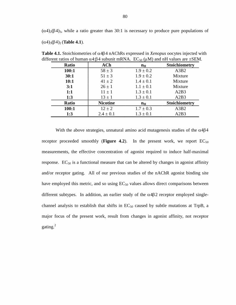
80
(α4)2(β4)3, while a ratio greater than 30:1 is necessary to produce pure populations of
(α4)3(β4)2 (Table 4.1).
Table 4.1. Stoichiometries of α4β4 nAChRs expressed in Xenopus oocytes injected with different ratios of human α4:β4 subunit mRNA. EC50 (µM) and nH values are ±SEM.
Ratio ACh nH Stoichiometry 100:1 58 ± 3 1.9 ± 0.2 A3B2 30:1 51 ± 3 1.9 ± 0.2 Mixture 10:1 41 ± 2 1.4 ± 0.1 Mixture 3:1 26 ± 1 1.1 ± 0.1 Mixture 1:1 11 ± 1 1.3 ± 0.1 A2B3 1:3 13 ± 1 1.3 ± 0.1 A2B3
Ratio Nicotine nH Stoichiometry 100:1 12 ± 2 1.7 ± 0.3 A3B2 1:3 2.4 ± 0.1 1.3 ± 0.1 A2B3
With the above strategies, unnatural amino acid mutagenesis studies of the α4β4
receptor proceeded smoothly (Figure 4.2). In the present work, we report EC50
measurements, the effective concentration of agonist required to induce half-maximal
response. EC50 is a functional measure that can be altered by changes in agonist affinity
and/or receptor gating. All of our previous studies of the nAChR agonist binding site
have employed this metric, and so using EC50 values allows direct comparisons between
different subtypes. In addition, an earlier study of the α4β2 receptor employed single-
channel analysis to establish that shifts in EC50 caused by subtle mutations at TrpB, a
major focus of the present work, result from changes in agonist affinity, not receptor
gating.2
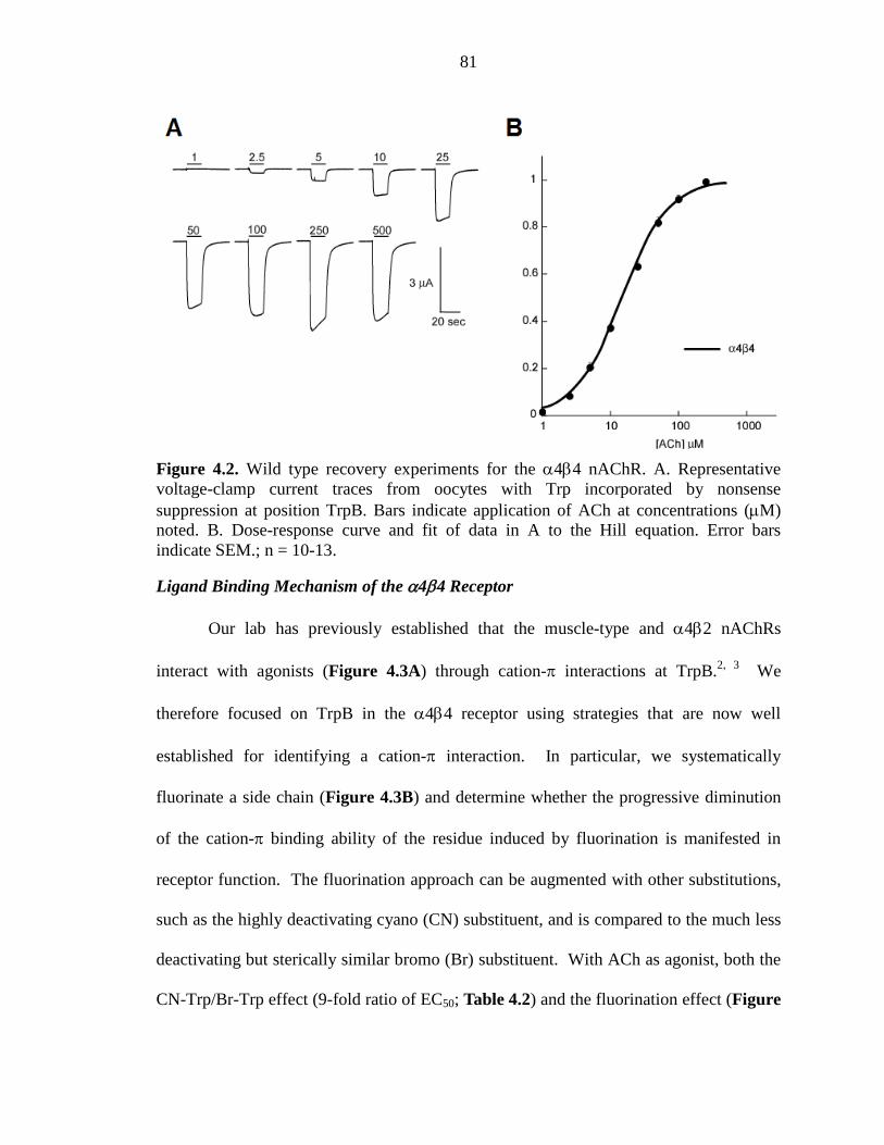
81
Figure 4.2. Wild type recovery experiments for the α4β4 nAChR. A. Representative voltage-clamp current traces from oocytes with Trp incorporated by nonsense suppression at position TrpB. Bars indicate application of ACh at concentrations (µM) noted. B. Dose-response curve and fit of data in A to the Hill equation. Error bars indicate SEM.; n = 10-13.
Ligand Binding Mechanism of the α4β4 Receptor
Our lab has previously established that the muscle-type and α4β2 nAChRs
interact with agonists (Figure 4.3A) through cation-π interactions at TrpB.2, 3 We
therefore focused on TrpB in the α4β4 receptor using strategies that are now well
established for identifying a cation-π interaction. In particular, we systematically
fluorinate a side chain (Figure 4.3B) and determine whether the progressive diminution
of the cation-π binding ability of the residue induced by fluorination is manifested in
receptor function. The fluorination approach can be augmented with other substitutions,
such as the highly deactivating cyano (CN) substituent, and is compared to the much less
deactivating but sterically similar bromo (Br) substituent. With ACh as agonist, both the
CN-Trp/Br-Trp effect (9-fold ratio of EC50; Table 4.2) and the fluorination effect (Figure
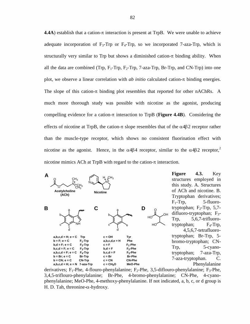
82
4.4A) establish that a cation-π interaction is present at TrpB. We were unable to achieve
adequate incorporation of F3-Trp or F4-Trp, so we incorporated 7-aza-Trp, which is
structurally very similar to Trp but shows a diminished cation-π binding ability. When
all the data are combined (Trp, F1-Trp, F2-Trp, 7-aza-Trp, Br-Trp, and CN-Trp) into one
plot, we observe a linear correlation with ab initio calculated cation-π binding energies.
The slope of this cation-π binding plot resembles that reported for other nAChRs. A
much more thorough study was possible with nicotine as the agonist, producing
compelling evidence for a cation-π interaction to TrpB (Figure 4.4B). Considering the
effects of nicotine at TrpB, the cation-π slope resembles that of the α4β2 receptor rather
than the muscle-type receptor, which shows no consistent fluorination effect with
nicotine as the agonist. Hence, in the α4β4 receptor, similar to the α4β2 receptor,2
nicotine mimics ACh at TrpB with regard to the cation-π interaction.
Figure 4.3. Key structures employed in this study. A. Structures of ACh and nicotine. B. Tryptophan derivatives; F1-Trp, 5-fluoro-tryptophan; F2-Trp, 5,7-difluoro-tryptophan; F3-Trp, 5,6,7-trifluoro-tryptophan; F4-Trp,
4,5,6,7-tetrafluoro-tryptophan; Br-Trp, 5-bromo-tryptophan; CN-Trp, 5-cyano-tryptophan; 7-aza-Trp, 7-aza-tryptophan. C.
Phenylalanine derivatives; F1-Phe, 4-flouro-phenylalanine; F2-Phe, 3,5-diflouro-phenylalanine; F3-Phe, 3,4,5-triflouro-phenylalanine; Br-Phe, 4-bromo-phenylalanine; CN-Phe, 4-cyano-phenylalanine; MeO-Phe, 4-methoxy-phenylalanine. If not indicated, a, b, c, or d group is H. D. Tah, threonine-α-hydroxy.
ON
O CH3
CH3CH3Acetylcholine
(ACh)
NN
H CH3
Nicotine
A
e NH
a
b
c
d
a,b,c,d = H; e = C Trpb = F; e = C F1-Trpb,d = F; e = C F2-Trpb,c,d = F; e = C F3-Trpa,b,c,d = F; e = C F4-Trpb = Br; e = C Br-Trpb = CN; e = C CN-Trpa,b,c,d = H; e = N 7-aza-Trp
a
b
c
de
c = OH Tyra,b,c,d,e = H Phec = F F1-Pheb,d = F F2-Pheb,c,d = F F3-Phec = Br Br-Phec = CN CN-Phec = CH3O MeO-Phe
B C
HO
HO
OOH
D
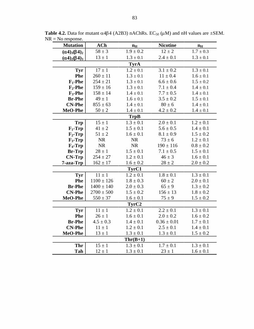
83
Table 4.2. Data for mutant α4β4 (A2B3) nAChRs. EC50 (µM) and nH values are ±SEM. NR = No response.
Mutation ACh nH Nicotine nH (α4)3(β4)2 58 ± 3 1.9 ± 0.2 12 ± 2 1.7 ± 0.3 (α4)2(β4)3 13 ± 1 1.3 ± 0.1 2.4 ± 0.1 1.3 ± 0.1
TyrA Tyr 17 ± 1 1.2 ± 0.1 3.1 ± 0.2 1.3 ± 0.1 Phe 260 ± 11 1.3 ± 0.1 11 ± 0.4 1.6 ± 0.1
F1-Phe 254 ± 21 1.3 ± 0.1 6.6 ± 0.6 1.5 ± 0.2 F2-Phe 159 ± 16 1.3 ± 0.1 7.1 ± 0.4 1.4 ± 0.1 F3-Phe 158 ± 14 1.4 ± 0.1 7.7 ± 0.5 1.4 ± 0.1 Br-Phe 49 ± 1 1.6 ± 0.1 3.5 ± 0.2 1.5 ± 0.1
CN-Phe 855 ± 63 1.4 ± 0.1 80 ± 6 1.4 ± 0.1 MeO-Phe 50 ± 2 1.4 ± 0.1 4.2 ± 0.2 1.4 ± 0.1
TrpB Trp 15 ± 1 1.3 ± 0.1 2.0 ± 0.1 1.2 ± 0.1
F1-Trp 41 ± 2 1.5 ± 0.1 5.6 ± 0.5 1.4 ± 0.1 F2-Trp 51 ± 2 1.6 ± 0.1 8.1 ± 0.9 1.5 ± 0.2 F3-Trp NR NR 73 ± 6 1.2 ± 0.1 F4-Trp NR NR 190 ± 116 0.8 ± 0.2 Br-Trp 28 ± 1 1.5 ± 0.1 7.1 ± 0.5 1.5 ± 0.1
CN-Trp 254 ± 27 1.2 ± 0.1 46 ± 3 1.6 ± 0.1 7-aza-Trp 162 ± 17 1.6 ± 0.2 28 ± 2 2.0 ± 0.2
TyrC1 Tyr 11 ± 1 1.2 ± 0.1 1.8 ± 0.1 1.3 ± 0.1 Phe 1100 ± 126 1.8 ± 0.3 60 ± 2 2.0 ± 0.1
Br-Phe 1400 ± 140 2.0 ± 0.3 65 ± 9 1.3 ± 0.2 CN-Phe 2700 ± 500 1.5 ± 0.2 156 ± 13 1.8 ± 0.2
MeO-Phe 550 ± 37 1.6 ± 0.1 75 ± 9 1.5 ± 0.2 TyrC2
Tyr 11 ± 1 1.2 ± 0.1 2.2 ± 0.1 1.3 ± 0.1 Phe 26 ± 1 1.6 ± 0.1 2.0 ± 0.2 1.6 ± 0.2
Br-Phe 4.5 ± 0.3 1.4 ± 0.1 0.36 ± 0.01 1.7 ± 0.1 CN-Phe 11 ± 1 1.2 ± 0.1 2.5 ± 0.1 1.4 ± 0.1
MeO-Phe 13 ± 1 1.3 ± 0.1 1.3 ± 0.1 1.5 ± 0.2 Thr(B+1)
Thr 15 ± 1 1.3 ± 0.1 1.7 ± 0.1 1.3 ± 0.1 Tah 12 ± 1 1.3 ± 0.1 23 ± 1 1.6 ± 0.1
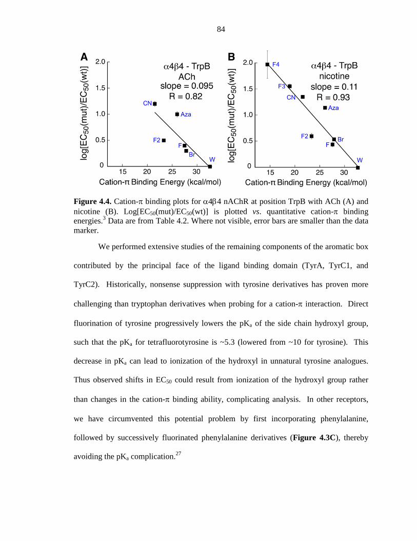
84
Figure 4.4. Cation-π binding plots for α4β4 nAChR at position TrpB with ACh (A) and nicotine (B). Log[EC50(mut)/EC50(wt)] is plotted vs. quantitative cation-π binding energies.3 Data are from Table 4.2. Where not visible, error bars are smaller than the data marker.
We performed extensive studies of the remaining components of the aromatic box
contributed by the principal face of the ligand binding domain (TyrA, TyrC1, and
TyrC2). Historically, nonsense suppression with tyrosine derivatives has proven more
challenging than tryptophan derivatives when probing for a cation-π interaction. Direct
fluorination of tyrosine progressively lowers the pKa of the side chain hydroxyl group,
such that the pKa for tetrafluorotyrosine is ~5.3 (lowered from ~10 for tyrosine). This
decrease in pKa can lead to ionization of the hydroxyl in unnatural tyrosine analogues.
Thus observed shifts in EC50 could result from ionization of the hydroxyl group rather
than changes in the cation-π binding ability, complicating analysis. In other receptors,
we have circumvented this potential problem by first incorporating phenylalanine,
followed by successively fluorinated phenylalanine derivatives (Figure 4.3C), thereby
avoiding the pKa complication.27
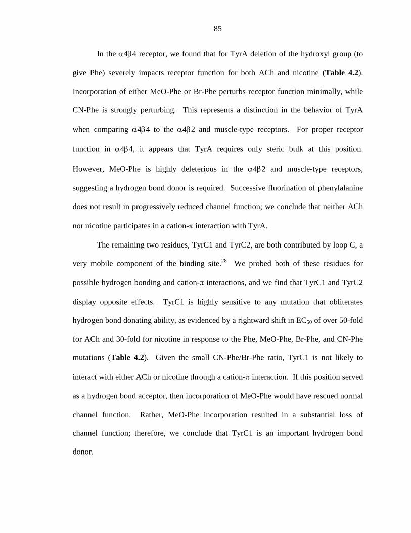
85
In the α4β4 receptor, we found that for TyrA deletion of the hydroxyl group (to
give Phe) severely impacts receptor function for both ACh and nicotine (Table 4.2).
Incorporation of either MeO-Phe or Br-Phe perturbs receptor function minimally, while
CN-Phe is strongly perturbing. This represents a distinction in the behavior of TyrA
when comparing α4β4 to the α4β2 and muscle-type receptors. For proper receptor
function in α4β4, it appears that TyrA requires only steric bulk at this position.
However, MeO-Phe is highly deleterious in the α4β2 and muscle-type receptors,
suggesting a hydrogen bond donor is required. Successive fluorination of phenylalanine
does not result in progressively reduced channel function; we conclude that neither ACh
nor nicotine participates in a cation-π interaction with TyrA.
The remaining two residues, TyrC1 and TyrC2, are both contributed by loop C, a
very mobile component of the binding site.28 We probed both of these residues for
possible hydrogen bonding and cation-π interactions, and we find that TyrC1 and TyrC2
display opposite effects. TyrC1 is highly sensitive to any mutation that obliterates
hydrogen bond donating ability, as evidenced by a rightward shift in EC50 of over 50-fold
for ACh and 30-fold for nicotine in response to the Phe, MeO-Phe, Br-Phe, and CN-Phe
mutations (Table 4.2). Given the small CN-Phe/Br-Phe ratio, TyrC1 is not likely to
interact with either ACh or nicotine through a cation-π interaction. If this position served
as a hydrogen bond acceptor, then incorporation of MeO-Phe would have rescued normal
channel function. Rather, MeO-Phe incorporation resulted in a substantial loss of
channel function; therefore, we conclude that TyrC1 is an important hydrogen bond
donor.
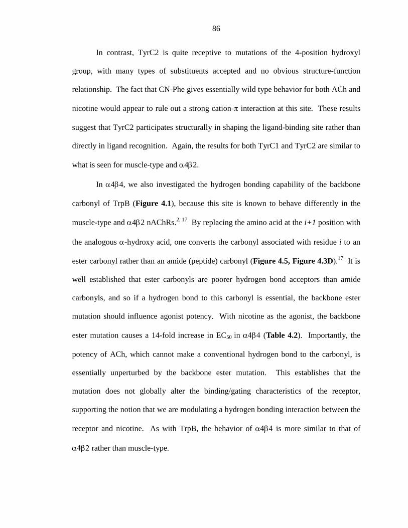
86
In contrast, TyrC2 is quite receptive to mutations of the 4-position hydroxyl
group, with many types of substituents accepted and no obvious structure-function
relationship. The fact that CN-Phe gives essentially wild type behavior for both ACh and
nicotine would appear to rule out a strong cation-π interaction at this site. These results
suggest that TyrC2 participates structurally in shaping the ligand-binding site rather than
directly in ligand recognition. Again, the results for both TyrC1 and TyrC2 are similar to
what is seen for muscle-type and α4β2.
In α4β4, we also investigated the hydrogen bonding capability of the backbone
carbonyl of TrpB (Figure 4.1), because this site is known to behave differently in the
muscle-type and α4β2 nAChRs.2, 17 By replacing the amino acid at the i+1 position with
the analogous α-hydroxy acid, one converts the carbonyl associated with residue i to an
ester carbonyl rather than an amide (peptide) carbonyl (Figure 4.5, Figure 4.3D).17 It is
well established that ester carbonyls are poorer hydrogen bond acceptors than amide
carbonyls, and so if a hydrogen bond to this carbonyl is essential, the backbone ester
mutation should influence agonist potency. With nicotine as the agonist, the backbone
ester mutation causes a 14-fold increase in EC50 in α4β4 (Table 4.2). Importantly, the
potency of ACh, which cannot make a conventional hydrogen bond to the carbonyl, is
essentially unperturbed by the backbone ester mutation. This establishes that the
mutation does not globally alter the binding/gating characteristics of the receptor,
supporting the notion that we are modulating a hydrogen bonding interaction between the
receptor and nicotine. As with TrpB, the behavior of α4β4 is more similar to that of
α4β2 rather than muscle-type.
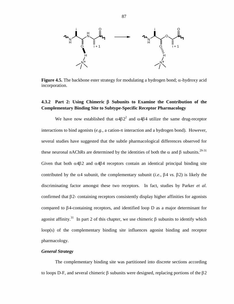
87
NH
HN
i
O
O
i + 1
N
H
NH
O
i
O
O
i + 1
N
H
Figure 4.5. The backbone ester strategy for modulating a hydrogen bond; α-hydroxy acid incorporation.
4.3.2 Part 2: Using Chimeric β Subunits to Examine the Contribution of the Complementary Binding Site to Subtype-Specific Receptor Pharmacology
We have now established that α4β22 and α4β4 utilize the same drug-receptor
interactions to bind agonists (e.g., a cation-π interaction and a hydrogen bond). However,
several studies have suggested that the subtle pharmacological differences observed for
these neuronal nAChRs are determined by the identities of both the α and β subunits.29-31
Given that both α4β2 and α4β4 receptors contain an identical principal binding site
contributed by the α4 subunit, the complementary subunit (i.e., β4 vs. β2) is likely the
discriminating factor amongst these two receptors. In fact, studies by Parker et al.
confirmed that β2- containing receptors consistently display higher affinities for agonists
compared to β4-containing receptors, and identified loop D as a major determinant for
agonist affinity.31 In part 2 of this chapter, we use chimeric β subunits to identify which
loop(s) of the complementary binding site influences agonist binding and receptor
pharmacology.
General Strategy
The complementary binding site was partitioned into discrete sections according
to loops D-F, and several chimeric β subunits were designed, replacing portions of the β2
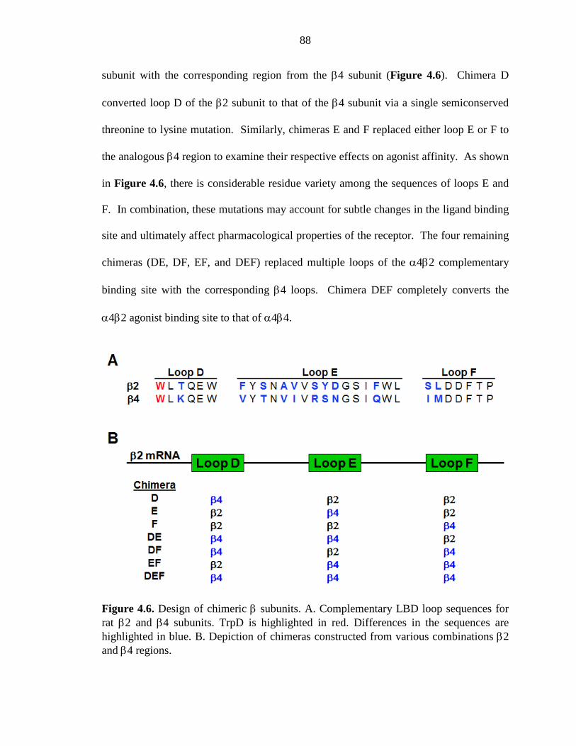
88
subunit with the corresponding region from the β4 subunit (Figure 4.6). Chimera D
converted loop D of the β2 subunit to that of the β4 subunit via a single semiconserved
threonine to lysine mutation. Similarly, chimeras E and F replaced either loop E or F to
the analogous β4 region to examine their respective effects on agonist affinity. As shown
in Figure 4.6, there is considerable residue variety among the sequences of loops E and
F. In combination, these mutations may account for subtle changes in the ligand binding
site and ultimately affect pharmacological properties of the receptor. The four remaining
chimeras (DE, DF, EF, and DEF) replaced multiple loops of the α4β2 complementary
binding site with the corresponding β4 loops. Chimera DEF completely converts the
α4β2 agonist binding site to that of α4β4.
Figure 4.6. Design of chimeric β subunits. A. Complementary LBD loop sequences for rat β2 and β4 subunits. TrpD is highlighted in red. Differences in the sequences are highlighted in blue. B. Depiction of chimeras constructed from various combinations β2 and β4 regions.
89
All chimeric β subunits were coexpressed with the α4 subunit. In accordance
with previous studies of α4β2 and α4β4,1, 2 a α4:chimerc β mRNA ratio of 1:3 ratio was
coinjected into oocytes to ensure a homogeneous population of (α4)2(β2/β4)3 receptors.
In both α4β2 and α4β4, TrpB is the key residue that interacts with both ACh and nicotine
via a cation-π interaction, a key component of agonist binding.2 We therefore
incorporated F3-Trp at TrpB in each chimeric receptor and evaluated the impact on the
cation-π interaction. For chimeras that experienced a functional change in response F3-
Trp incorporation when compared to α4β2, additional tryptophan derivatives were
incorporated at TrpB (Figure 4.3B).
Functional Scan of Chimeric α4β2/β4 Receptors
EC50 was used to evaluate receptor function as discussed in Part 1. Although
EC50 is composed of both binding and gating parameters, we anticipate that shifts in EC50
caused by subtle mutations at TrpB result from changes in agonist binding, not receptor
gating, based on previous single-channel analysis of α4β2.2 Using either ACh or nicotine
as the agonist, a dose-response relationship was determined for each chimera. EC50
values of chimeric receptors were compared to α4β2 values (Table 4.3). Each chimera
was functional and EC50 values were slightly shifted from α4β2, with chimera DE
experiencing the greatest perturbation.
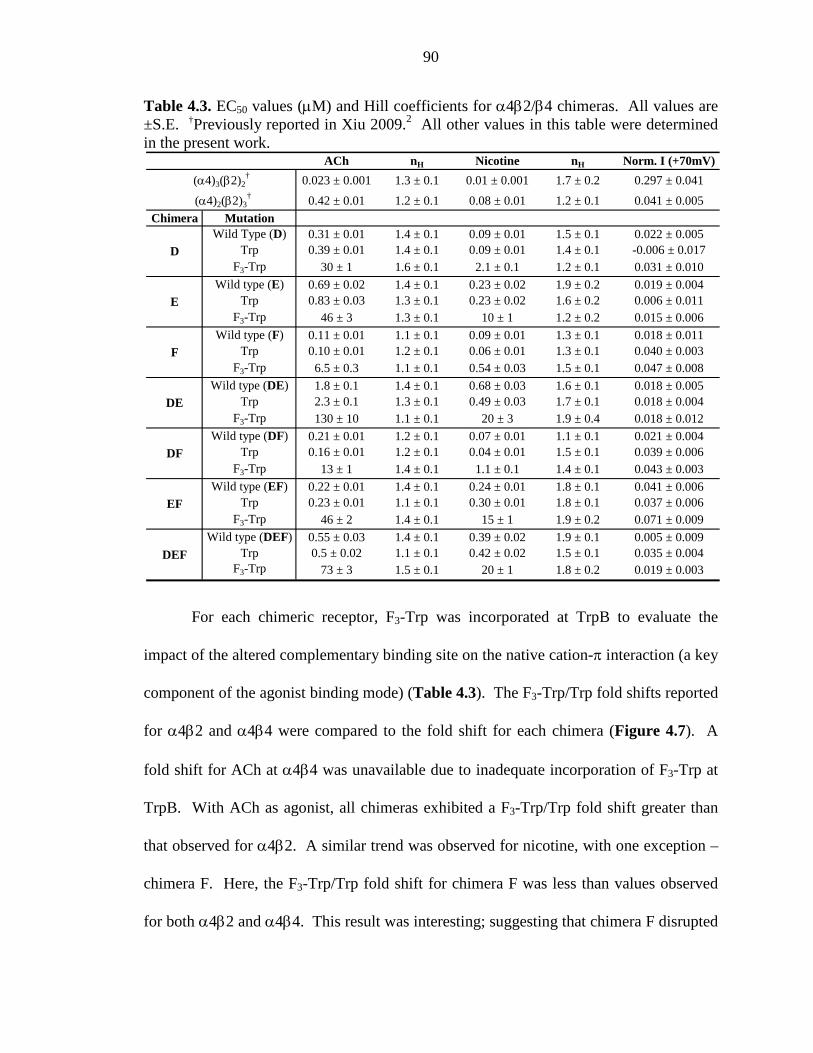
90
Table 4.3. EC50 values (µM) and Hill coefficients for α4β2/β4 chimeras. All values are ±S.E. †Previously reported in Xiu 2009.2 All other values in this table were determined in the present work.
For each chimeric receptor, F3-Trp was incorporated at TrpB to evaluate the
impact of the altered complementary binding site on the native cation-π interaction (a key
component of the agonist binding mode) (Table 4.3). The F3-Trp/Trp fold shifts reported
for α4β2 and α4β4 were compared to the fold shift for each chimera (Figure 4.7). A
fold shift for ACh at α4β4 was unavailable due to inadequate incorporation of F3-Trp at
TrpB. With ACh as agonist, all chimeras exhibited a F3-Trp/Trp fold shift greater than
that observed for α4β2. A similar trend was observed for nicotine, with one exception –
chimera F. Here, the F3-Trp/Trp fold shift for chimera F was less than values observed
for both α4β2 and α4β4. This result was interesting; suggesting that chimera F disrupted
ACh nH Nicotine nH Norm. I (+70mV)
0.023 ± 0.001 1.3 ± 0.1 0.01 ± 0.001 1.7 ± 0.2 0.297 ± 0.041
0.42 ± 0.01 1.2 ± 0.1 0.08 ± 0.01 1.2 ± 0.1 0.041 ± 0.005
Chimera MutationWild Type (D) 0.31 ± 0.01 1.4 ± 0.1 0.09 ± 0.01 1.5 ± 0.1 0.022 ± 0.005
Trp 0.39 ± 0.01 1.4 ± 0.1 0.09 ± 0.01 1.4 ± 0.1 -0.006 ± 0.017F3-Trp 30 ± 1 1.6 ± 0.1 2.1 ± 0.1 1.2 ± 0.1 0.031 ± 0.010
Wild type (E) 0.69 ± 0.02 1.4 ± 0.1 0.23 ± 0.02 1.9 ± 0.2 0.019 ± 0.004Trp 0.83 ± 0.03 1.3 ± 0.1 0.23 ± 0.02 1.6 ± 0.2 0.006 ± 0.011
F3-Trp 46 ± 3 1.3 ± 0.1 10 ± 1 1.2 ± 0.2 0.015 ± 0.006Wild type (F) 0.11 ± 0.01 1.1 ± 0.1 0.09 ± 0.01 1.3 ± 0.1 0.018 ± 0.011
Trp 0.10 ± 0.01 1.2 ± 0.1 0.06 ± 0.01 1.3 ± 0.1 0.040 ± 0.003F3-Trp 6.5 ± 0.3 1.1 ± 0.1 0.54 ± 0.03 1.5 ± 0.1 0.047 ± 0.008
Wild type (DE) 1.8 ± 0.1 1.4 ± 0.1 0.68 ± 0.03 1.6 ± 0.1 0.018 ± 0.005Trp 2.3 ± 0.1 1.3 ± 0.1 0.49 ± 0.03 1.7 ± 0.1 0.018 ± 0.004
F3-Trp 130 ± 10 1.1 ± 0.1 20 ± 3 1.9 ± 0.4 0.018 ± 0.012Wild type (DF) 0.21 ± 0.01 1.2 ± 0.1 0.07 ± 0.01 1.1 ± 0.1 0.021 ± 0.004
Trp 0.16 ± 0.01 1.2 ± 0.1 0.04 ± 0.01 1.5 ± 0.1 0.039 ± 0.006F3-Trp 13 ± 1 1.4 ± 0.1 1.1 ± 0.1 1.4 ± 0.1 0.043 ± 0.003
Wild type (EF) 0.22 ± 0.01 1.4 ± 0.1 0.24 ± 0.01 1.8 ± 0.1 0.041 ± 0.006Trp 0.23 ± 0.01 1.1 ± 0.1 0.30 ± 0.01 1.8 ± 0.1 0.037 ± 0.006
F3-Trp 46 ± 2 1.4 ± 0.1 15 ± 1 1.9 ± 0.2 0.071 ± 0.009Wild type (DEF) 0.55 ± 0.03 1.4 ± 0.1 0.39 ± 0.02 1.9 ± 0.1 0.005 ± 0.009
Trp 0.5 ± 0.02 1.1 ± 0.1 0.42 ± 0.02 1.5 ± 0.1 0.035 ± 0.004F3-Trp 73 ± 3 1.5 ± 0.1 20 ± 1 1.8 ± 0.2 0.019 ± 0.003
DE
DF
EF
DEF
(α4)3(β2)2†
(α4)2(β2)3†
E
D
F
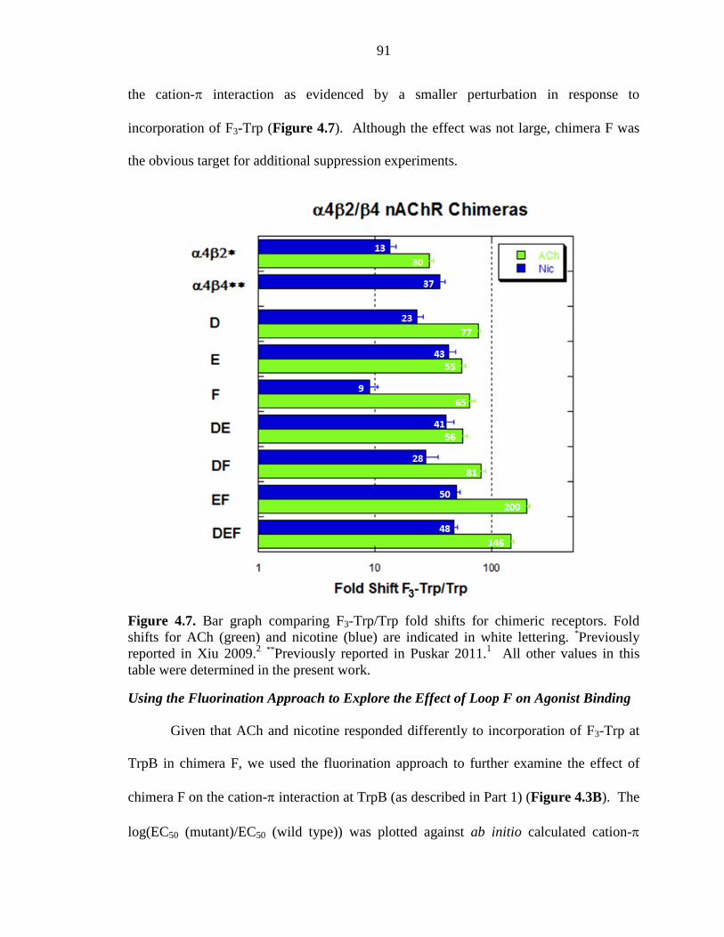
91
the cation-π interaction as evidenced by a smaller perturbation in response to
incorporation of F3-Trp (Figure 4.7). Although the effect was not large, chimera F was
the obvious target for additional suppression experiments.
Figure 4.7. Bar graph comparing F3-Trp/Trp fold shifts for chimeric receptors. Fold shifts for ACh (green) and nicotine (blue) are indicated in white lettering. *Previously reported in Xiu 2009.2 **Previously reported in Puskar 2011.1 All other values in this table were determined in the present work.
Using the Fluorination Approach to Explore the Effect of Loop F on Agonist Binding
Given that ACh and nicotine responded differently to incorporation of F3-Trp at
TrpB in chimera F, we used the fluorination approach to further examine the effect of
chimera F on the cation-π interaction at TrpB (as described in Part 1) (Figure 4.3B). The
log(EC50 (mutant)/EC50 (wild type)) was plotted against ab initio calculated cation-π
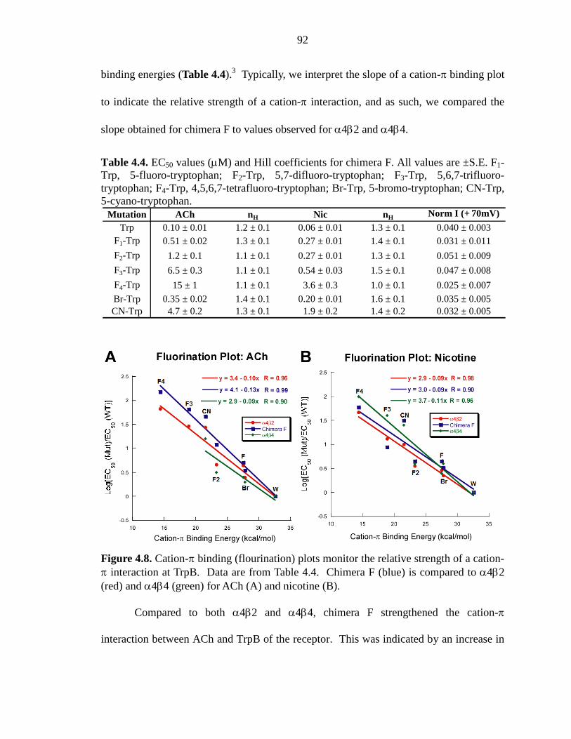
92
binding energies (Table 4.4).3 Typically, we interpret the slope of a cation-π binding plot
to indicate the relative strength of a cation-π interaction, and as such, we compared the
slope obtained for chimera F to values observed for α4β2 and α4β4.
Table 4.4. EC50 values (µM) and Hill coefficients for chimera F. All values are ±S.E. F1-Trp, 5-fluoro-tryptophan; F2-Trp, 5,7-difluoro-tryptophan; F3-Trp, 5,6,7-trifluoro-tryptophan; F4-Trp, 4,5,6,7-tetrafluoro-tryptophan; Br-Trp, 5-bromo-tryptophan; CN-Trp, 5-cyano-tryptophan.
Figure 4.8. Cation-π binding (flourination) plots monitor the relative strength of a cation-π interaction at TrpB. Data are from Table 4.4. Chimera F (blue) is compared to α4β2 (red) and α4β4 (green) for ACh (A) and nicotine (B).
Compared to both α4β2 and α4β4, chimera F strengthened the cation-π
interaction between ACh and TrpB of the receptor. This was indicated by an increase in
Mutation ACh nH Nic nH Norm I (+ 70mV)
Trp 0.10 ± 0.01 1.2 ± 0.1 0.06 ± 0.01 1.3 ± 0.1 0.040 ± 0.003F1-Trp 0.51 ± 0.02 1.3 ± 0.1 0.27 ± 0.01 1.4 ± 0.1 0.031 ± 0.011
F2-Trp 1.2 ± 0.1 1.1 ± 0.1 0.27 ± 0.01 1.3 ± 0.1 0.051 ± 0.009
F3-Trp 6.5 ± 0.3 1.1 ± 0.1 0.54 ± 0.03 1.5 ± 0.1 0.047 ± 0.008
F4-Trp 15 ± 1 1.1 ± 0.1 3.6 ± 0.3 1.0 ± 0.1 0.025 ± 0.007
Br-Trp 0.35 ± 0.02 1.4 ± 0.1 0.20 ± 0.01 1.6 ± 0.1 0.035 ± 0.005CN-Trp 4.7 ± 0.2 1.3 ± 0.1 1.9 ± 0.2 1.4 ± 0.2 0.032 ± 0.005
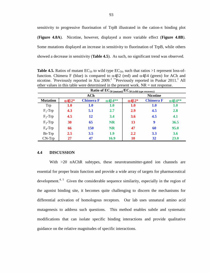
93
sensitivity to progressive fluorination of TrpB illustrated in the cation-π binding plot
(Figure 4.8A). Nicotine, however, displayed a more variable effect (Figure 4.8B).
Some mutations displayed an increase in sensitivity to fluorination of TrpB, while others
showed a decrease in sensitivity (Table 4.5). As such, no significant trend was observed.
Table 4.5. Ratios of mutant EC50 to wild type EC50, such that ratios >1 represent loss-of-function. Chimera F (blue) is compared to α4β2 (red) and α4β4 (green) for ACh and nicotine. *Previously reported in Xiu 2009.2 **Previously reported in Puskar 2011.1 All other values in this table were determined in the present work. NR = not response.
4.4 DISCUSSION
With >20 nAChR subtypes, these neurotransmitter-gated ion channels are
essential for proper brain function and provide a wide array of targets for pharmaceutical
development.4, 5 Given the considerable sequence similarity, especially in the region of
the agonist binding site, it becomes quite challenging to discern the mechanisms for
differential activation of homologous receptors. Our lab uses unnatural amino acid
mutagenesis to address such questions. This method enables subtle and systematic
modifications that can isolate specific binding interactions and provide qualitative
guidance on the relative magnitudes of specific interactions.
Mutation α4β2* Chimera F α4β4** α4β2* Chimera F α4β4**Trp 1.0 1.0 1.0 1.0 1.0 1.0
F1-Trp 4.3 5.1 2.7 2.9 4.5 2.8
F2-Trp 4.5 12 3.4 3.6 4.5 4.1
F3-Trp 30 65 NR 13 9 36.5
F4-Trp 66 150 NR 47 60 95.0Br-Trp 2.5 3.5 1.9 2.2 3.3 3.6CN-Trp 27 47 16.9 10 32 23.0
Ratio of EC50 (mutant)/EC50 (wild type recovery)
ACh Nicotine
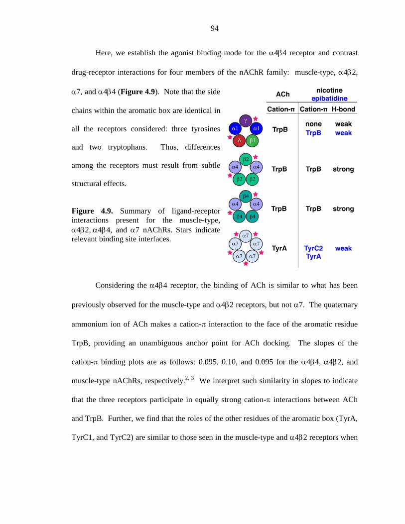
94
Here, we establish the agonist binding mode for the α4β4 receptor and contrast
drug-receptor interactions for four members of the nAChR family: muscle-type, α4β2,
α7, and α4β4 (Figure 4.9). Note that the side
chains within the aromatic box are identical in
all the receptors considered: three tyrosines
and two tryptophans. Thus, differences
among the receptors must result from subtle
structural effects.
Figure 4.9. Summary of ligand-receptor interactions present for the muscle-type, α4β2, α4β4, and α7 nAChRs. Stars indicate relevant binding site interfaces.
Considering the α4β4 receptor, the binding of ACh is similar to what has been
previously observed for the muscle-type and α4β2 receptors, but not α7. The quaternary
ammonium ion of ACh makes a cation-π interaction to the face of the aromatic residue
TrpB, providing an unambiguous anchor point for ACh docking. The slopes of the
cation-π binding plots are as follows: 0.095, 0.10, and 0.095 for the α4β4, α4β2, and
muscle-type nAChRs, respectively.2, 3 We interpret such similarity in slopes to indicate
that the three receptors participate in equally strong cation-π interactions between ACh
and TrpB. Further, we find that the roles of the other residues of the aromatic box (TyrA,
TyrC1, and TyrC2) are similar to those seen in the muscle-type and α4β2 receptors when
95
binding ACh. Results for α4β4 are strikingly different from the α7 receptor, which uses
agonist specific cation-π interactions at TyrA and TyrC2.
An interesting result is observed when nicotine is the agonist; now the neuronal
α4β4 receptor acts similarly to the α4β2 receptor rather than to the muscle-type receptor.
In α4β4, nicotine makes the same cation-π interaction to TrpB as ACh, consistent with
the long-accepted nicotinic pharmacophore, but an interaction that is absent in the
muscle-type receptor. Interestingly, the slope of the cation-π binding plot for α4β4 is
0.11, which could suggest a moderately stronger cation-π interaction at this position than
observed for α4β2 (slope = 0.089).2 Thus, a cation-π interaction to TrpB serves as a
discriminator between receptors with higher sensitivity to nicotine (α4β4 and α4β2) and
those with lower sensitivity (muscle-type).
Concerning the hydrogen bond to the backbone carbonyl associated with TrpB,
α4β4 also behaves like α4β2, not muscle-type or α7. At α4β4, nicotine displays a 14-
fold decrease in receptor function in response to the backbone ester mutation, comparable
to 19-fold for α4β2.2 This contrasts the 1.6-fold shift for muscle-type or 2.1-fold shift for
epibatidine at α7.1, 17 Note that when the agonist is ACh – a molecule unable to make a
conventional hydrogen bond to a carbonyl – essentially wild type receptor behavior is
observed. This indicates that the backbone mutation did not alter receptor function
downstream from binding, i.e., gating. We conclude that nicotine is able to make a
hydrogen bond to the carbonyl in question in all three receptors considered, but that the
interaction is much stronger in α4β4 and α4β2. This is an additional contributor to the
enhanced potency of nicotine at the neuronal α4β4 and α4β2 receptors. Previous studies
96
of neuronal nAChRs have indicated that large differences in agonist affinity are primarily
determined by the nature of the complementary subunit.31 Our results provide a
molecular rationale indicating that both α4-containing neuronal receptors make the same
ligand-receptor interactions, but the magnitudes of the two interactions examined differ
depending on the receptor, reflecting the nature of the β subunit. For nicotine, the cation-
π interaction is stronger in the α4β4 receptor, whereas the hydrogen bond interaction is
stronger in the α4β2 nAChR.
Chimeric β subunits were used to further examine the role of the complementary
binding site in defining subtype-specific receptor pharmacology. Using the cation-π
interaction at TrpB as a probe for agonist binding, we found that most chimeras
experienced a modest increase in the F3-Trp/Trp ratio compared to α4β2 and α4β4. One
exception, however, was nicotine at chimera F, which showed a decreased F3-Trp/Trp
ratio compared to α4β2. Further analysis revealed that chimera F experienced a variable
effect in response to additional tryptophan derivatives, and no significant trend was
observed. Given that the core residues of the principal binding site are conserved in all
nAChRs and chimera studies of the complementary binding site appear less informative,
the question remains as to what features of the nAChR are responsible for differences in
receptor pharmacology. It is possible that the variable residues flanking the conserved
core residues may be the key to understanding the pharmacological diversity of nAChRs.
Further experiments are underway to probe both the non-α subunits and residues within
the α subunit that are located outside the aromatic box (Chapter 3).
Here we identify structural features of the nAChR that discriminate among these
four receptors and are likely to contribute to differential receptor pharmacology. In the
97
muscle-type receptor, TrpB makes a cation-π interaction to ACh and to epibatidine, but
not to nicotine.3, 16, 17 In the neuronal α4β4 and α4β2 receptors, the TrpB cation-π
interaction to ACh remains, but now nicotine also makes a strong cation-π interaction.2
The α7 receptor eschews the cation-π interaction to TrpB, as agonists have moved their
cationic center across the aromatic box to TyrA and TyrC2.1 The nAChR family also
uses a backbone hydrogen bonding interaction as a second discriminating feature for
drug-receptor interactions. This interaction is modest in the muscle-type and α7
receptors,1, 17 but it is much stronger in α4β4 and α4β22 – the higher sensitivity receptors.
Taken as a whole, the data support the view that the energy of the cation-π and hydrogen
bond interactions studied here underlies the higher sensitivity of both α4β2 and α4β4.
4.5 METHODS
Molecular Biology
For ligand binding studies of the α4β4 nAChR (Part 1), human α4 and β4 subunit
genes were in pGEMhe. For chimera experiments (Part 2), rat α4 and β2 nAChR subunit
genes were in the pAMV vector, and as such, chimeras replaced loop regions of the rat
β2 subunit with the corresponding regions of the rat β4 subunit. In accordance with
previously reported protocols,2 all α4β2/β4 chimeric receptors contained a L9’A
mutation in the α4 subunit to increase receptor expression. Site-directed mutagenesis
was performed using the QuikChange protocol (Stratagene). For nonsense suppression
experiments, the site of interest within the nAChR subunit was mutated to an amber stop
codon. Circular DNA for nAChR subunit genes in pAMV were linearized with Not I and
nAChR subunit genes in pGEMhe were linearized with Nhe I. After purification
98
(Qiagen), linearized DNA was used as a template for runoff in vitro transcription using
T7 mMessage mMachine kit (Ambion).
THG7332 was used as the amber suppressor tRNA. Nitroveratryloxycarbonyl
(NVOC) protected cyanomethyl ester form of unnatural amino acids and α-
hydroxythreonine cyanomethyl ester were synthesized, coupled to dinucleotide dCA, and
enzymatically ligated to 74-nucleotide THG73 tRNACUA as previously reported.20 Crude
tRNA-amino acid product was used without desalting, and the product was confirmed by
MALDI-TOF MS on 3-hydroxypicolinic acid (3-HPA) matrix. Deprotection of the
NVOC group on the tRNA-amino acid was carried out by photolysis for 5 minutes prior
to coinjection with mRNA containing the UAG mutation at the site of interest.
Microinjection
Stage V-VI Xenopus laevis oocytes were employed. For experiments in Part 1,
coinjection of α4:β4 mRNA at a ratio of 1:1 by mass or lower yielded wild type
(α4)2(β4)3 receptors, while a ratio greater than 30:1 by mass produced pure populations
of (α4)3(β4)2. If an unnatural amino acid was to be incorporated into the α4 subunit to
produce a (α4)2(β4)3 receptor, then a mass ratio of 2:1 for α4:β4 mRNA was injected
into each oocyte.
In Part 2, α4β2/β4 chimeric receptors were expressed using a co injection of
α4:β2/β4 chimera mRNA at a ratio of 1:3 by mass. This ratio consistently produced
chimeric receptors with the stoichiometry of (α4)2(β2/β4)3. For nonsense suppression
experiments incorporating an unnatural amino acid into the α4 subunit, a mass ratio of
3:1 for α4:β2/β4 chimera mRNA was injected into each oocyte to produce (α4)2(β2/β4)3
99
receptors. To ensure (α4)2(β2/β4)3 stoichiometry, all receptor stoichiometries were
confirmed by voltage jump experiments.2
For all experiments (Part 1 and 2), the total mRNA injected was 25-65 ng/oocyte
depending on the relative expression level, and approximately 15 ng of tRNA per cell
was used for suppression experiments. Each oocyte was injected with 50 nL of RNA
solution, and the oocytes were incubated for 24-48 hours at 18 °C in ND96 buffer (96
mM NaCl, 2 mM KCl, 1 mM MgCl2, 1.8 mM CaCl2, and 5 mM HEPES, pH 7.5) with
0.005% (w/v) gentamycin and 2% (v/v) horse serum. In the case of low-expressing
mutant receptors, a second injection was required 24 hours after the first injection. As a
negative control for all suppression experiments, 76-nucleotide tRNA (dCA ligated to 74-
nucleotide tRNA) was coinjected with mRNA in the same manner as fully charged
tRNA.
Electrophysiology
Acetylcholine chloride and (-)-nicotine tartrate were purchased from
Sigma/Aldrich/RBI (St. Louis, MO) and drug dilutions were prepared from 1M aq stock
solutions. Drug dilutions were prepared in calcium-free ND96 buffer. Ion channel
function was assayed using the OpusXpress 6000A (Molecular Devices Axon
Instruments) in two-electrode voltage clamp mode. Oocytes were clamped at a holding
potential of -60 mV. One mL of each drug solution was applied to the clamped oocytes
for 12 sec and followed by a 2 minute wash with calcium-free ND96 buffer between each
concentration. Data were sampled at 125 Hz and filtered at 50 Hz. Voltage jump
experiments were sampled at 5000 Hz and filtered at 180 Hz.
100
Data Analysis
Dose-response data were obtained for at least 6 concentrations of agonists and for
a minimum of 5 oocytes (from two different batches). Mutants with Imax of at least 100
nA of current were defined as functional. EC50 and Hill coefficient (nH) were calculated
by fitting the averaged, normalized dose-response relation to the Hill equation. All data
are reported as means ±S.E.
4.6 ACKNOWLEDGEMENTS
We thank Ariele P. Hanek helpful discussions. This work was supported, in
whole or in part, by National Institutes of Health Grants NS34407 and NS11756 and by
Grant MH086383 (to H. A. L.). This work was also supported by California Tobacco-
Related Disease Research Program Grant 19XT-0102 from the University of California
(to D. A. D.).
101
4.7 REFERENCES
1. Puskar, N. L.; Xiu, X.; Lester, H. A.; Dougherty, D. A., Two neuronal nicotinic acetylcholine receptors, alpha4beta4 and alpha7, show differential agonist binding modes. J Biol Chem 286, (16), 14618-27.
2. Xiu, X.; Puskar, N. L.; Shanata, J. A.; Lester, H. A.; Dougherty, D. A., Nicotine binding to brain receptors requires a strong cation-pi interaction. Nature 2009, 458, (7237), 534-7.
3. Zhong, W.; Gallivan, J. P.; Zhang, Y.; Li, L.; Lester, H. A.; Dougherty, D. A., From ab initio quantum mechanics to molecular neurobiology: a cation-pi binding site in the nicotinic receptor. Proc Natl Acad Sci U S A 1998, 95, (21), 12088-93.
4. Corringer, P. J.; Le Novere, N.; Changeux, J. P., Nicotinic receptors at the amino acid level. Annu Rev Pharmacol Toxicol 2000, 40, 431-58.
5. Jensen, A. A.; Frolund, B.; Liljefors, T.; Krogsgaard-Larsen, P., Neuronal nicotinic acetylcholine receptors: structural revelations, target identifications, and therapeutic inspirations. J Med Chem 2005, 48, (15), 4705-45.
6. Romanelli, M. N.; Gratteri, P.; Guandalini, L.; Martini, E.; Bonaccini, C.; Gualtieri, F., Central nicotinic receptors: structure, function, ligands, and therapeutic potential. ChemMedChem 2007, 2, (6), 746-67.
7. Unwin, N., Refined structure of the nicotinic acetylcholine receptor at 4A resolution. J Mol Biol 2005, 346, (4), 967-89.
8. Miyazawa, A.; Fujiyoshi, Y.; Unwin, N., Structure and gating mechanism of the acetylcholine receptor pore. Nature 2003, 423, (6943), 949-55.
9. Brejc, K.; van Dijk, W. J.; Klaassen, R. V.; Schuurmans, M.; van Der Oost, J.; Smit, A. B.; Sixma, T. K., Crystal structure of an ACh-binding protein reveals the ligand-binding domain of nicotinic receptors. Nature 2001, 411, (6835), 269-76.
10. Gotti, C.; Clementi, F., Neuronal nicotinic receptors: From structure to pathology. Prog Neurobiol 2004, 74, (6), 363-96.
11. Romanelli, M. N.; Gratteri, P.; Guandalini, L.; Martini, E.; Bonaccini, C.; Gualtieri, F., Central Nicotinic Receptors: Structure, Function, Ligands, and Therapeutic Potential. ChemMedChem 2007, 2, (6), 746-767.
12. Beers, W. H.; Reich, E., Structure and activity of acetylcholine. Nature 1970, 228, (5275), 917-22.
13. Glennon, R. A.; Dukat, M., Central nicotinic receptor ligands and pharmacophores. Pharm Acta Helv 2000, 74, (2-3), 103-14.
14. Sixma, T. K.; Smit, A. B., Acetylcholine binding protein (AChBP): a secreted glial protein that provides a high-resolution model for the extracellular domain of pentameric ligand-gated ion channels. Annu Rev Biophys Biomol Struct 2003, 32, 311-34.
15. Blum, A. P.; Lester, H. A.; Dougherty, D. A., Inaugural article: Nicotinic pharmacophore: the pyridine N of nicotine and carbonyl of acetylcholine hydrogen bond across a subunit interface to a backbone NH. Proc Natl Acad Sci U S A 107, (30), 13206-11.
16. Beene, D. L.; Brandt, G. S.; Zhong, W.; Zacharias, N. M.; Lester, H. A.; Dougherty, D. A., Cation-pi interactions in ligand recognition by serotonergic (5-
102
HT3A) and nicotinic acetylcholine receptors: the anomalous binding properties of nicotine. Biochemistry 2002, 41, (32), 10262-9.
17. Cashin, A. L.; Petersson, E. J.; Lester, H. A.; Dougherty, D. A., Using physical chemistry to differentiate nicotinic from cholinergic agonists at the nicotinic acetylcholine receptor. J Am Chem Soc 2005, 127, (1), 350-6.
18. Xiu, J. Structure-Function Studies of Nicotinic Acetylcholine Receptors Using Unnatural Amino Acids. The California Institute of Technology, Ph.D. Thesis, Pasadena, 2007.
19. Wu, J.; Liu, Q.; Yu, K.; Hu, J.; Kuo, Y. P.; Segerberg, M.; St John, P. A.; Lukas, R. J., Roles of nicotinic acetylcholine receptor beta subunits in function of human alpha4-containing nicotinic receptors. J Physiol 2006, 576, (Pt 1), 103-18.
20. Nowak, M. W.; Gallivan, J. P.; Silverman, S. K.; Labarca, C. G.; Dougherty, D. A.; Lester, H. A., In vivo incorporation of unnatural amino acids into ion channels in Xenopus oocyte expression system. Methods Enzymol 1998, 293, 504-29.
21. Padgett, C. L.; Hanek, A. P.; Lester, H. A.; Dougherty, D. A.; Lummis, S. C., Unnatural amino acid mutagenesis of the GABA(A) receptor binding site residues reveals a novel cation-pi interaction between GABA and beta 2Tyr97. J Neurosci 2007, 27, (4), 886-92.
22. Lummis, S. C.; D, L. B.; Harrison, N. J.; Lester, H. A.; Dougherty, D. A., A cation-pi binding interaction with a tyrosine in the binding site of the GABAC receptor. Chem Biol 2005, 12, (9), 993-7.
23. Mu, T. W.; Lester, H. A.; Dougherty, D. A., Different binding orientations for the same agonist at homologous receptors: a lock and key or a simple wedge? J Am Chem Soc 2003, 125, (23), 6850-1.
24. Torrice, M. M.; Bower, K. S.; Lester, H. A.; Dougherty, D. A., Probing the role of the cation-pi interaction in the binding sites of GPCRs using unnatural amino acids. Proc Natl Acad Sci U S A 2009, 106, (29), 11919-24.
25. Moroni, M.; Zwart, R.; Sher, E.; Cassels, B. K.; Bermudez, I., alpha4beta2 nicotinic receptors with high and low acetylcholine sensitivity: pharmacology, stoichiometry, and sensitivity to long-term exposure to nicotine. Mol Pharmacol 2006, 70, (2), 755-68.
26. Nelson, M. E.; Kuryatov, A.; Choi, C. H.; Zhou, Y.; Lindstrom, J., Alternate stoichiometries of alpha4beta2 nicotinic acetylcholine receptors. Mol Pharmacol 2003, 63, (2), 332-41.
27. Nowak, M. W.; Kearney, P. C.; Sampson, J. R.; Saks, M. E.; Labarca, C. G.; Silverman, S. K.; Zhong, W.; Thorson, J.; Abelson, J. N.; Davidson, N.; et al., Nicotinic receptor binding site probed with unnatural amino acid incorporation in intact cells. Science 1995, 268, (5209), 439-42.
28. Espinoza-Fonseca, L. M.; Trujillo-Ferrara, J. G., Fully flexible docking models of the complex between alpha7 nicotinic receptor and a potent heptapeptide inhibitor of the beta-amyloid peptide binding. Bioorg Med Chem Lett 2006, 16, (13), 3519-23.
29. Hussy, N.; Ballivet, M.; Bertrand, D., Agonist and antagonist effects of nicotine on chick neuronal nicotinic receptors are defined by alpha and beta subunits. J Neurophysiol 1994, 72, (3), 1317-26.
103
30. Luetje, C. W.; Patrick, J., Both alpha- and beta-subunits contribute to the agonist sensitivity of neuronal nicotinic acetylcholine receptors. J Neurosci 1991, 11, (3), 837-45.
31. Parker, M. J.; Beck, A.; Luetje, C. W., Neuronal nicotinic receptor beta2 and beta4 subunits confer large differences in agonist binding affinity. Mol Pharmacol 1998, 54, (6), 1132-9.
32. Saks, M. E.; Sampson, J. R.; Nowak, M. W.; Kearney, P. C.; Du, F.; Abelson, J. N.; Lester, H. A.; Dougherty, D. A., An engineered Tetrahymena tRNAGln for in vivo incorporation of unnatural amino acids into proteins by nonsense suppression. J Biol Chem 1996, 271, (38), 23169-75.
104
Chapter 5
Optimizing Techniques to Implement Nonsense Suppression in Mammalian Cells*
*This work was done in collaboration with Kristina McCleary.
5.1 ABSTRACT
Our lab previously used microelectroporation to incorporate unnatural amino
acids into ion channels expressed in mammalian cells. While this work represented
major progress, this method still faced several limitations. Specifically,
microelectroporation is not robust enough for large-scale cellular imaging techniques and
requires significant amounts of aminoacylated tRNA. This chapter describes our efforts
to further develop transfection techniques for mammalian cells to increase the expression
of mutant protein such that functional analysis of several thousand cells simultaneously
can be achieved using the FlexStation3. Using HEK293T cells, we have optimized two
protocols to efficiently cotransfect DNA and tRNA. These protocols are termed double
electroporation using the Neon transfection system and single transfection using TransIT
transfection reagent. Both methods successfully suppress an amber stop codon using an
in vivo aminoacylated tRNA, but fail to achieve suppression using in vitro aminoacylated
tRNAs. Studies are ongoing to evaluate the cause of the failed suppression experiments
using in vitro aminoacylated tRNAs. Possible causes include the amino acid falling off
the tRNA inside the cell and translational incompetence of the tRNA molecule itself.
105
5.2 INTRODUCTION
The nonsense suppression methodology, developed by Schultz in 1989,1 has
emerged as a powerful biophysical tool facilitating site-specific incorporation of virtually
any unnatural amino acid - as long as it can be synthesized in the chemistry lab - into
biologically relevant proteins. This method is particularly attractive because it can
conceivably generate an infinite number of proteins with novel chemical properties as
well as probe protein structure/function relationships.
Our lab has adapted the nonsense suppression methodology to incorporate
unnatural amino acids into proteins expressed in Xenopus laevis oocytes.2, 3 In doing so,
we have successfully determined the ligand binding mechanism and channel gating
properties of numerous ion channels and neuroreceptors.4-9 While heterologous
expression in Xenopus oocytes has proven an optimal model system to efficiently express
hundreds of mutant proteins, we are currently interested in expanding this technology to a
mammalian expression system. One advantage of implementing a mammalian
expression system is that one can study mammalian proteins in a biologically relevant
context (i.e., one can study a human protein in a human cell line). Additionally, using a
mammalian system will allow for the study of cell-specific signal transduction pathways,
and one can imagine designing experiments using unnatural amino acids (e.g.,
photocaged amino acids) to exert temporal control of various signaling cascades.10
Progress towards Unnatural Amino Acid Mutagenesis in Mammalian Cells
One of the key challenges in mammalian protein expression is determining the
appropriate transfection method that will maximize protein expression, limit cell toxicity,
and consume the least amount of nucleic acid. Several transfection techniques have been
106
developed to effectively introduce nucleic acids into cells, such as chemical-based
methods (e.g., calcium phosphate, cationic polymers, cationic liposomes, and cationic
activated dendrimers), non-chemical methods (e.g., electroporation), particle-based
methods (e.g., gene gun), and viral transduction. Each transfection technique is
accompanied by inherent advantages and disadvantages depending on the cell line of
interest and nucleic acid material being delivered.
In recent years, many researchers have contributed major advances towards the
types of transfection techniques amenable to introducing both DNA and tRNA into
mammalian cells. For example, RajBhandary and workers successfully suppressed a
serine site with tyrosine in chloramphenicol acetyltransferase.11 This experiment utilized
a cationic lipid (Effectene) to co-transfect in vitro aminoacylated tRNA with DNA into
COS cells.11 Alternatively, Schultz and workers took a different approach employing
directed evolution of suppressor tRNA-aminoacyl synthetase pairs to site-specifically
incorporate unnatural amino acids into several types of mammalian cells.12-14 And in
2003, our lab used microelectroporation to cotransfect mRNA and tRNA that had been
aminoacylated in vitro with an unnatural amino acid to express mutant ion channels in
CHO cells and cultured neurons.15 Whole-cell patch clamp electrophysiology confirmed
the presence of functional mutant protein.15 While this work represents major progress in
this field, these methods do not yield robust amounts of mutant protein and require
functional analysis on a single cell scale.
We are interested in further developing techniques to increase the expression of
mutant protein in mammalian cells to a level that allows functional analysis of several
thousand cells simultaneously. This chapter describes our efforts to determine the
107
optimal transfection protocol for incorporating unnatural amino acids into ion channels
expressed in mammalian cells.
5.3 METHODS
5.3.1 Molecular Biology
Human 5HT3A subunit gene in pcDNA3.1(+) vector was purchased from Missouri
S & T cDNA Research Center. Mouse α4, α4GFP, and β2 genes were in pciNEO vector,
and the mouse β4 gene was in pcDNA3.1(+) vector. All mouse constructs were obtained
from the Lester lab. All eGFP constructs used in these studies are from Monahan et al.15
Site-directed mutagenesis was performed using the QuikChange protocol
(Stratagene) to incorporate either an amber stop codon (TAG) or a frameshift stop codon
(GTTT) at the site of interest within the subunit DNA. THG7316 was used as the amber
suppressor tRNA. Human serine amber suppressor tRNA (HSAS) was used to
incorporate serine opposite an amber stop codon.17 For suppression of the frameshift
codon (GTTT), the yeast phenylalanine frameshift suppressor tRNA (YFaFS) was used.18
Nitroveratryloxycarbonyl (NVOC) protected cyanomethyl esters of natural or
unnatural amino acids and α-hydroxy acid cyanomethyl esters were prepared as
previously reported.2 Aminoacyl tRNA was confirmed by MALDI-TOF MS on 3-
hydroxypicolinic acid (3-HPA) matrix. Prior to transfection, the NVOC protecting group
on the aminoacyl tRNA was deprotected by photolysis for 5 minutes.
5.3.2 Mammalian Cell Culture
Human embryonic kidney cells (HEK293T) cells were purchased from American
Tissue Culture Collection (ATCC). The HEK293T cell line is optimized for increased
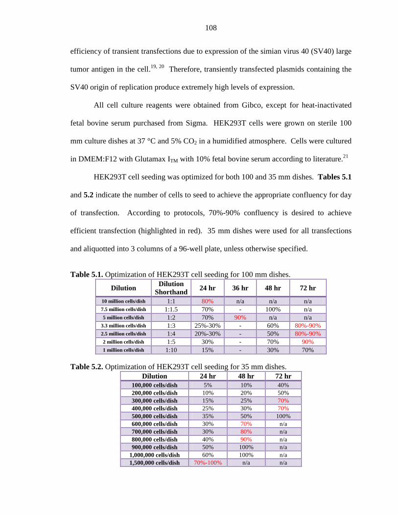
108
efficiency of transient transfections due to expression of the simian virus 40 (SV40) large
tumor antigen in the cell.19, 20 Therefore, transiently transfected plasmids containing the
SV40 origin of replication produce extremely high levels of expression.
All cell culture reagents were obtained from Gibco, except for heat-inactivated
fetal bovine serum purchased from Sigma. HEK293T cells were grown on sterile 100
mm culture dishes at 37 °C and 5% CO2 in a humidified atmosphere. Cells were cultured
in DMEM:F12 with Glutamax ITM with 10% fetal bovine serum according to literature.21
HEK293T cell seeding was optimized for both 100 and 35 mm dishes. Tables 5.1
and 5.2 indicate the number of cells to seed to achieve the appropriate confluency for day
of transfection. According to protocols, 70%-90% confluency is desired to achieve
efficient transfection (highlighted in red). 35 mm dishes were used for all transfections
and aliquotted into 3 columns of a 96-well plate, unless otherwise specified.
Table 5.1. Optimization of HEK293T cell seeding for 100 mm dishes.
Dilution Dilution
Shorthand 24 hr 36 hr 48 hr 72 hr
10 million cells/dish 1:1 80% n/a n/a n/a 7.5 million cells/dish 1:1.5 70% - 100% n/a 5 million cells/dish 1:2 70% 90% n/a n/a
3.3 million cells/dish 1:3 25%-30% - 60% 80%-90% 2.5 million cells/dish 1:4 20%-30% - 50% 80%-90% 2 million cells/dish 1:5 30% - 70% 90% 1 million cells/dish 1:10 15% - 30% 70%
Table 5.2. Optimization of HEK293T cell seeding for 35 mm dishes. Dilution 24 hr 48 hr 72 hr
100,000 cells/dish 5% 10% 40% 200,000 cells/dish 10% 20% 50% 300,000 cells/dish 15% 25% 70% 400,000 cells/dish 25% 30% 70% 500,000 cells/dish 35% 50% 100% 600,000 cells/dish 30% 70% n/a 700,000 cells/dish 30% 80% n/a 800,000 cells/dish 40% 90% n/a 900,000 cells/dish 50% 100% n/a
1,000,000 cells/dish 60% 100% n/a 1,500,000 cells/dish 70%-100% n/a n/a
109
Cells were transiently transfected using various protocols, such as
polyethylenimine (PEI), ExpressFect transfection reagent (Denville Scientific), Neon
Transfection System (Life Technologies), TransIT-mRNA transfection kit (Mirus),
TransMessenger transfection reagent (Qiagen), and Lipofectamine RNAiMAX
transfection reagent (Life Technologies). Following transfection, cells were plated (~3 x
104 cells/well) onto black 96-well plates with clear bottoms (BD Falcon), incubated for 1-
2 days, and then assayed using the Flexstation 3. Unless otherwise indicated,
manufacturers’ protocols were followed for each transfection method. For PEI
transfections in 35 mm plates, 1.5 µg of DNA was transfected with 0.3 mL of DMEM
and 9 µL of PEI (in 3 mL DMEM total). For confocal experiments, ExpressFect
transfections were performed in 35 mm plates and 1 µg of DNA was transfected with 4
µL of ExpressFect for 4 hr.
5.3.3 The FlexStation 3
The FlexStation 3 is a 96-well fluorescent plate reader used to analyze the
functional properties of neurotransmitter-gated ion channels (e.g., human 5HT3A and
human α4β4 receptors). This device is comprised of two modules - a fluidics module
and a detection module (Figure 5.1). The fluidics module contains an 8-channel pipette
that delivers solutions of neurotransmitter from the source plate to the 96-well sample
plate containing HEK293T cells expressing ion channels. The detection module contains
an excitation source and a PMT detector that detects the change in fluorescence in
response to neurotransmitter activation.
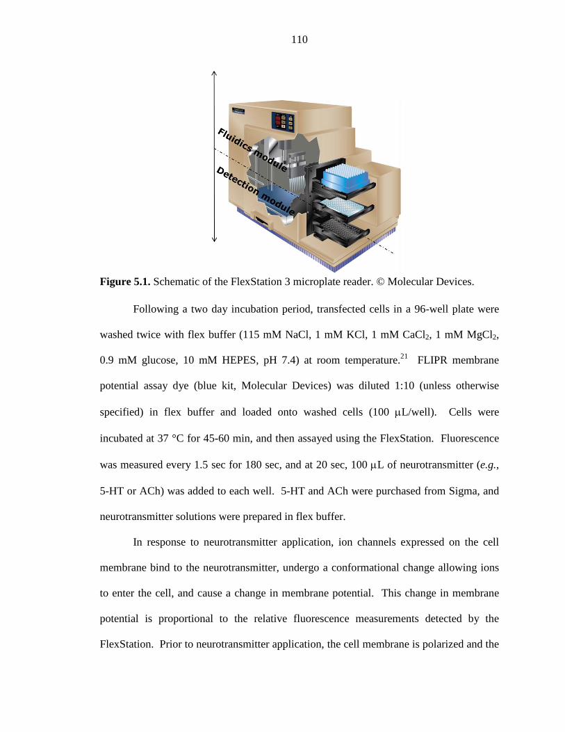
110
Figure 5.1. Schematic of the FlexStation 3 microplate reader. © Molecular Devices.
Following a two day incubation period, transfected cells in a 96-well plate were
washed twice with flex buffer (115 mM NaCl, 1 mM KCl, 1 mM CaCl2, 1 mM MgCl2,
0.9 mM glucose, 10 mM HEPES, pH 7.4) at room temperature.21 FLIPR membrane
potential assay dye (blue kit, Molecular Devices) was diluted 1:10 (unless otherwise
specified) in flex buffer and loaded onto washed cells (100 µL/well). Cells were
incubated at 37 °C for 45-60 min, and then assayed using the FlexStation. Fluorescence
was measured every 1.5 sec for 180 sec, and at 20 sec, 100 µL of neurotransmitter (e.g.,
5-HT or ACh) was added to each well. 5-HT and ACh were purchased from Sigma, and
neurotransmitter solutions were prepared in flex buffer.
In response to neurotransmitter application, ion channels expressed on the cell
membrane bind to the neurotransmitter, undergo a conformational change allowing ions
to enter the cell, and cause a change in membrane potential. This change in membrane
potential is proportional to the relative fluorescence measurements detected by the
FlexStation. Prior to neurotransmitter application, the cell membrane is polarized and the
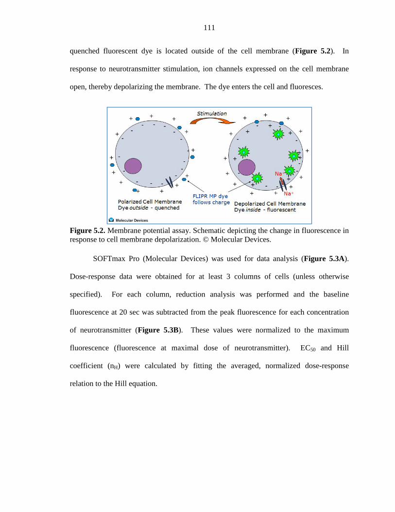
111
quenched fluorescent dye is located outside of the cell membrane (Figure 5.2). In
response to neurotransmitter stimulation, ion channels expressed on the cell membrane
open, thereby depolarizing the membrane. The dye enters the cell and fluoresces.
Figure 5.2. Membrane potential assay. Schematic depicting the change in fluorescence in response to cell membrane depolarization. © Molecular Devices.
SOFTmax Pro (Molecular Devices) was used for data analysis (Figure 5.3A).
Dose-response data were obtained for at least 3 columns of cells (unless otherwise
specified). For each column, reduction analysis was performed and the baseline
fluorescence at 20 sec was subtracted from the peak fluorescence for each concentration
of neurotransmitter (Figure 5.3B). These values were normalized to the maximum
fluorescence (fluorescence at maximal dose of neurotransmitter). EC50 and Hill
coefficient (nH) were calculated by fitting the averaged, normalized dose-response
relation to the Hill equation.
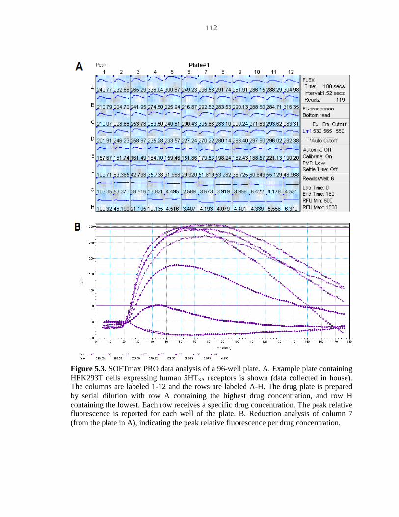
112
Figure 5.3. SOFTmax PRO data analysis of a 96-well plate. A. Example plate containing HEK293T cells expressing human 5HT3A receptors is shown (data collected in house). The columns are labeled 1-12 and the rows are labeled A-H. The drug plate is prepared by serial dilution with row A containing the highest drug concentration, and row H containing the lowest. Each row receives a specific drug concentration. The peak relative fluorescence is reported for each well of the plate. B. Reduction analysis of column 7 (from the plate in A), indicating the peak relative fluorescence per drug concentration.
113
5.3.4 Fluorescent Dye Experiments
Several concentrations of rhodamine B (Sigma) were prepared in a drug plate by
1:1 serial dilutions of rhodamine B:water starting with a 50 mM stock of rhodamine B.
100 µL of dye was added to 100 µL of water in the assay plate for each concentration and
the area under the curve of the emission spectrum was measured. This was repeated for
each PMT setting (low, medium, high).
Note that eosin Y and fluorescein (purchased from City Chemical Company and
Sigma, respectively) were also tested with the low PMT setting. Interestingly, both dyes
displayed a maximal RFU of 50,000 indicating that the FlexStation is capable of
detecting very high fluorescence signals.
5.4 RESULTS AND DISCUSSION
5.4.1 Determining the Optimal PMT Setting to Detect Small Fluorescence Signals
It is possible that even under optimal transfection conditions, the fluorescence
signal of mutant ion channels containing unnatural amino acids will be significantly
lower than for wild type ion channels. Thus, we needed to determine the appropriate
FlexStation PMT setting to maximize the relative fluorescence unit (RFU) signal
detected. The RFU dependence on PMT setting was measured for a serial dilution of
rhodamine B at each of the PMT settings (low, medium, high).
Using the low PMT setting, the FlexStation was unable to detect fluorescence for
high rhodamine B concentrations (50 – 0.39 mM), which is likely due to self-quenching
of the dye. A maximal signal of 10,000 RFU was detected for 0.19 mM rhodamine B and
each subsequent serial dilution produced a fluorescence signal decreased by
approximately half (Table 5.3). A similar trend was observed for the medium PMT
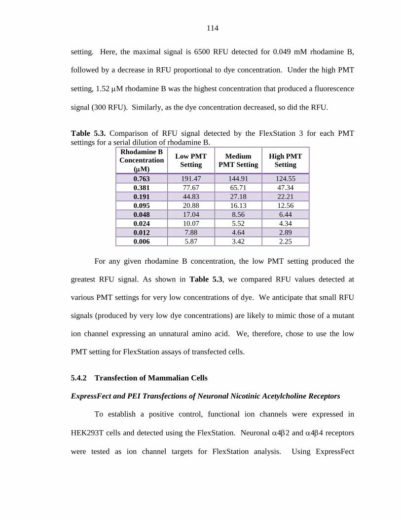
114
setting. Here, the maximal signal is 6500 RFU detected for 0.049 mM rhodamine B,
followed by a decrease in RFU proportional to dye concentration. Under the high PMT
setting, 1.52 µM rhodamine B was the highest concentration that produced a fluorescence
signal (300 RFU). Similarly, as the dye concentration decreased, so did the RFU.
Table 5.3. Comparison of RFU signal detected by the FlexStation 3 for each PMT settings for a serial dilution of rhodamine B.
Rhodamine B Concentration
(µM)
Low PMT Setting
Medium PMT Setting
High PMT Setting
0.763 191.47 144.91 124.55 0.381 77.67 65.71 47.34 0.191 44.83 27.18 22.21 0.095 20.88 16.13 12.56 0.048 17.04 8.56 6.44 0.024 10.07 5.52 4.34 0.012 7.88 4.64 2.89 0.006 5.87 3.42 2.25
For any given rhodamine B concentration, the low PMT setting produced the
greatest RFU signal. As shown in Table 5.3, we compared RFU values detected at
various PMT settings for very low concentrations of dye. We anticipate that small RFU
signals (produced by very low dye concentrations) are likely to mimic those of a mutant
ion channel expressing an unnatural amino acid. We, therefore, chose to use the low
PMT setting for FlexStation assays of transfected cells.
5.4.2 Transfection of Mammalian Cells
ExpressFect and PEI Transfections of Neuronal Nicotinic Acetylcholine Receptors
To establish a positive control, functional ion channels were expressed in
HEK293T cells and detected using the FlexStation. Neuronal α4β2 and α4β4 receptors
were tested as ion channel targets for FlexStation analysis. Using ExpressFect
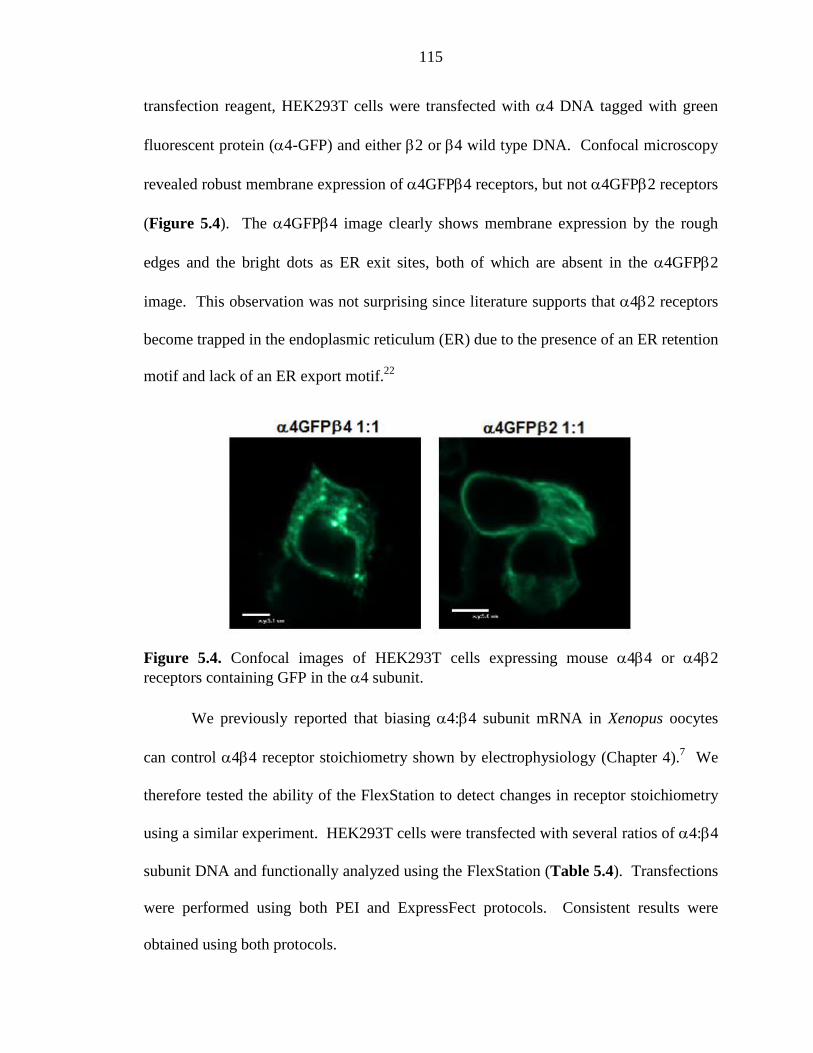
115
transfection reagent, HEK293T cells were transfected with α4 DNA tagged with green
fluorescent protein (α4-GFP) and either β2 or β4 wild type DNA. Confocal microscopy
revealed robust membrane expression of α4GFPβ4 receptors, but not α4GFPβ2 receptors
(Figure 5.4). The α4GFPβ4 image clearly shows membrane expression by the rough
edges and the bright dots as ER exit sites, both of which are absent in the α4GFPβ2
image. This observation was not surprising since literature supports that α4β2 receptors
become trapped in the endoplasmic reticulum (ER) due to the presence of an ER retention
motif and lack of an ER export motif.22
Figure 5.4. Confocal images of HEK293T cells expressing mouse α4β4 or α4β2 receptors containing GFP in the α4 subunit.
We previously reported that biasing α4:β4 subunit mRNA in Xenopus oocytes
can control α4β4 receptor stoichiometry shown by electrophysiology (Chapter 4).7 We
therefore tested the ability of the FlexStation to detect changes in receptor stoichiometry
using a similar experiment. HEK293T cells were transfected with several ratios of α4:β4
subunit DNA and functionally analyzed using the FlexStation (Table 5.4). Transfections
were performed using both PEI and ExpressFect protocols. Consistent results were
obtained using both protocols.
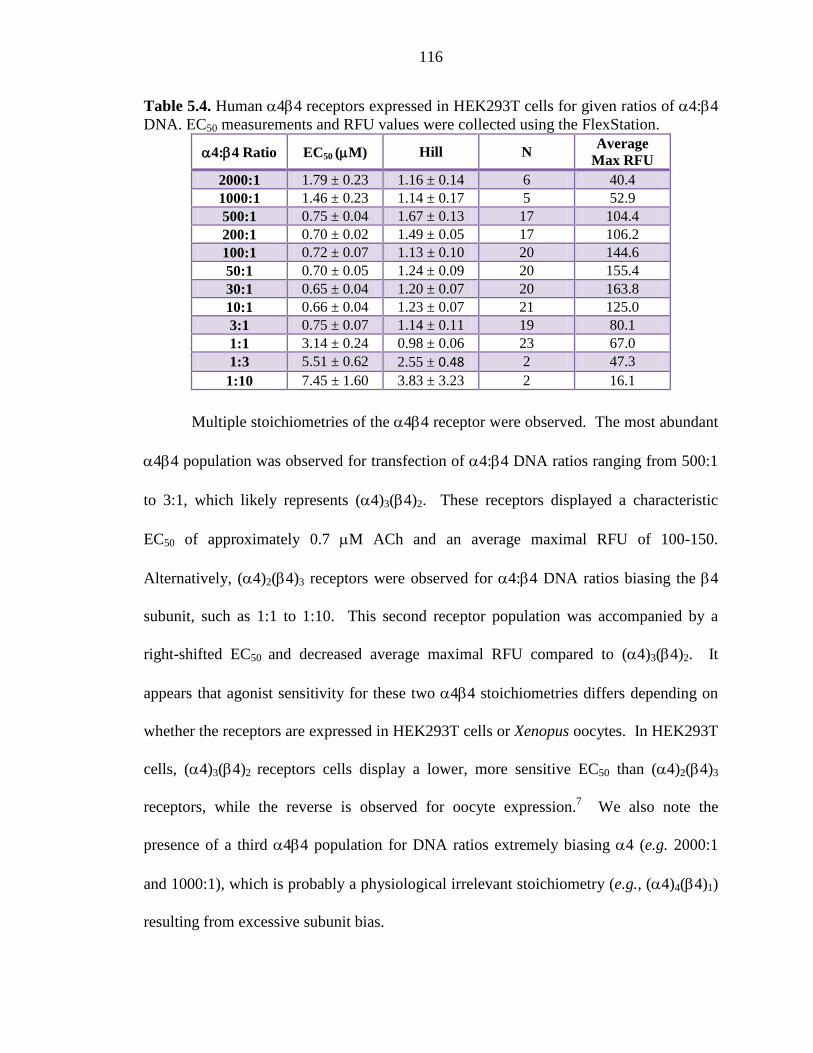
116
Table 5.4. Human α4β4 receptors expressed in HEK293T cells for given ratios of α4:β4 DNA. EC50 measurements and RFU values were collected using the FlexStation.
α4:β4 Ratio EC50 (µM) Hill N Average
Max RFU 2000:1 1.79 ± 0.23 1.16 ± 0.14 6 40.4 1000:1 1.46 ± 0.23 1.14 ± 0.17 5 52.9 500:1 0.75 ± 0.04 1.67 ± 0.13 17 104.4 200:1 0.70 ± 0.02 1.49 ± 0.05 17 106.2 100:1 0.72 ± 0.07 1.13 ± 0.10 20 144.6 50:1 0.70 ± 0.05 1.24 ± 0.09 20 155.4 30:1 0.65 ± 0.04 1.20 ± 0.07 20 163.8 10:1 0.66 ± 0.04 1.23 ± 0.07 21 125.0 3:1 0.75 ± 0.07 1.14 ± 0.11 19 80.1 1:1 3.14 ± 0.24 0.98 ± 0.06 23 67.0 1:3 5.51 ± 0.62 2.55 ± 0.48 2 47.3 1:10 7.45 ± 1.60 3.83 ± 3.23 2 16.1
Multiple stoichiometries of the α4β4 receptor were observed. The most abundant
α4β4 population was observed for transfection of α4:β4 DNA ratios ranging from 500:1
to 3:1, which likely represents (α4)3(β4)2. These receptors displayed a characteristic
EC50 of approximately 0.7 µM ACh and an average maximal RFU of 100-150.
Alternatively, (α4)2(β4)3 receptors were observed for α4:β4 DNA ratios biasing the β4
subunit, such as 1:1 to 1:10. This second receptor population was accompanied by a
right-shifted EC50 and decreased average maximal RFU compared to (α4)3(β4)2. It
appears that agonist sensitivity for these two α4β4 stoichiometries differs depending on
whether the receptors are expressed in HEK293T cells or Xenopus oocytes. In HEK293T
cells, (α4)3(β4)2 receptors cells display a lower, more sensitive EC50 than (α4)2(β4)3
receptors, while the reverse is observed for oocyte expression.7 We also note the
presence of a third α4β4 population for DNA ratios extremely biasing α4 (e.g. 2000:1
and 1000:1), which is probably a physiological irrelevant stoichiometry (e.g., (α4)4(β4)1)
resulting from excessive subunit bias.
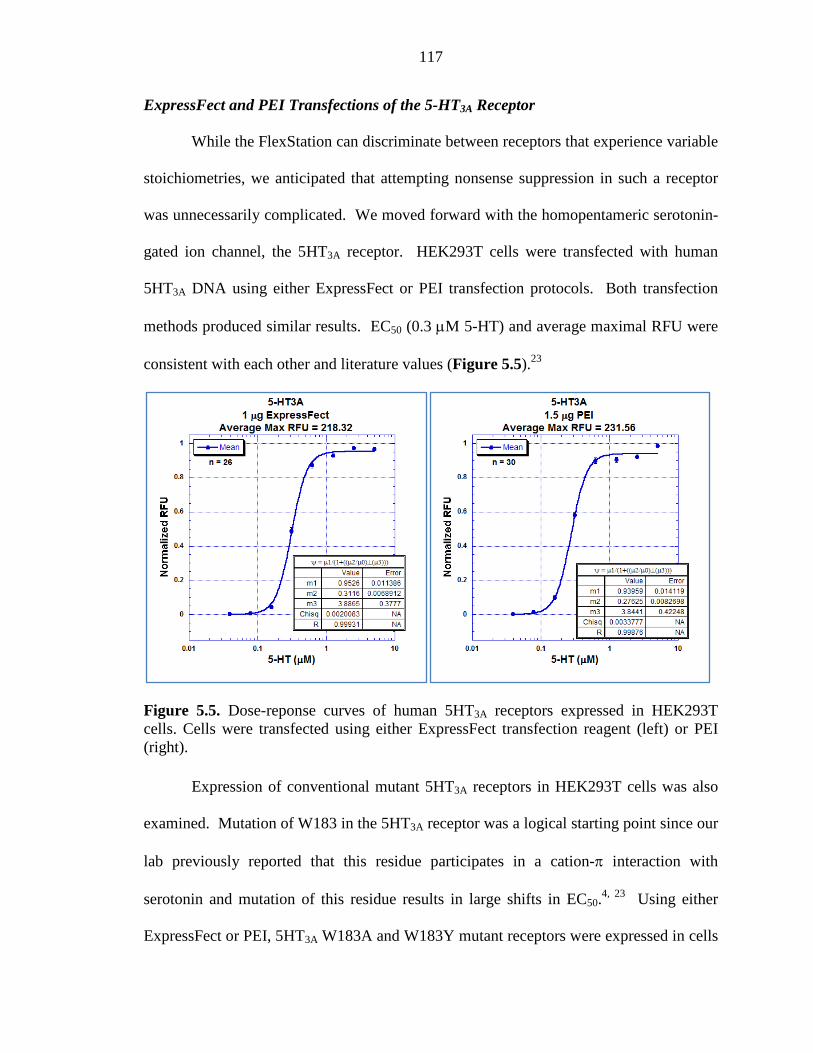
117
ExpressFect and PEI Transfections of the 5-HT3A Receptor
While the FlexStation can discriminate between receptors that experience variable
stoichiometries, we anticipated that attempting nonsense suppression in such a receptor
was unnecessarily complicated. We moved forward with the homopentameric serotonin-
gated ion channel, the 5HT3A receptor. HEK293T cells were transfected with human
5HT3A DNA using either ExpressFect or PEI transfection protocols. Both transfection
methods produced similar results. EC50 (0.3 µM 5-HT) and average maximal RFU were
consistent with each other and literature values (Figure 5.5).23
Figure 5.5. Dose-reponse curves of human 5HT3A receptors expressed in HEK293T cells. Cells were transfected using either ExpressFect transfection reagent (left) or PEI (right). Expression of conventional mutant 5HT3A receptors in HEK293T cells was also
examined. Mutation of W183 in the 5HT3A receptor was a logical starting point since our
lab previously reported that this residue participates in a cation-π interaction with
serotonin and mutation of this residue results in large shifts in EC50.4, 23 Using either
ExpressFect or PEI, 5HT3A W183A and W183Y mutant receptors were expressed in cells
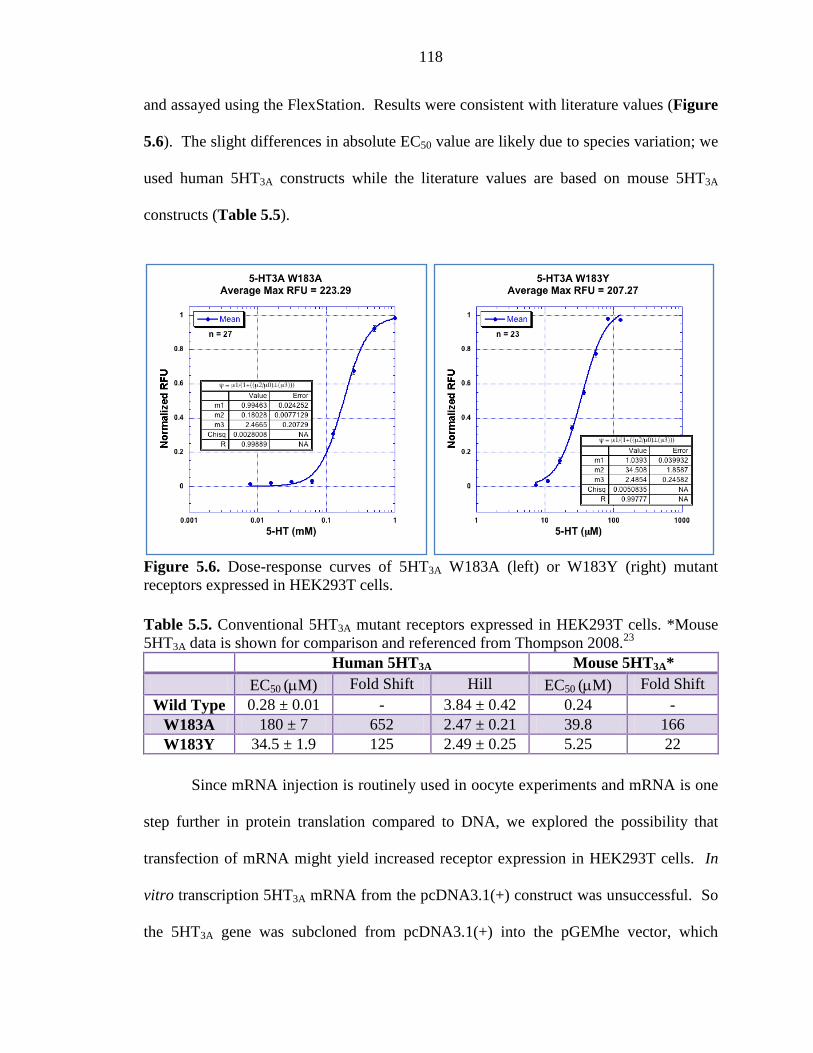
118
and assayed using the FlexStation. Results were consistent with literature values (Figure
5.6). The slight differences in absolute EC50 value are likely due to species variation; we
used human 5HT3A constructs while the literature values are based on mouse 5HT3A
constructs (Table 5.5).
Figure 5.6. Dose-response curves of 5HT3A W183A (left) or W183Y (right) mutant receptors expressed in HEK293T cells. Table 5.5. Conventional 5HT3A mutant receptors expressed in HEK293T cells. *Mouse 5HT3A data is shown for comparison and referenced from Thompson 2008.23
Human 5HT3A Mouse 5HT3A* EC50 (µM) Fold Shift Hill EC50 (µM) Fold Shift
Wild Type 0.28 ± 0.01 - 3.84 ± 0.42 0.24 - W183A 180 ± 7 652 2.47 ± 0.21 39.8 166 W183Y 34.5 ± 1.9 125 2.49 ± 0.25 5.25 22
Since mRNA injection is routinely used in oocyte experiments and mRNA is one
step further in protein translation compared to DNA, we explored the possibility that
transfection of mRNA might yield increased receptor expression in HEK293T cells. In
vitro transcription 5HT3A mRNA from the pcDNA3.1(+) construct was unsuccessful. So
the 5HT3A gene was subcloned from pcDNA3.1(+) into the pGEMhe vector, which
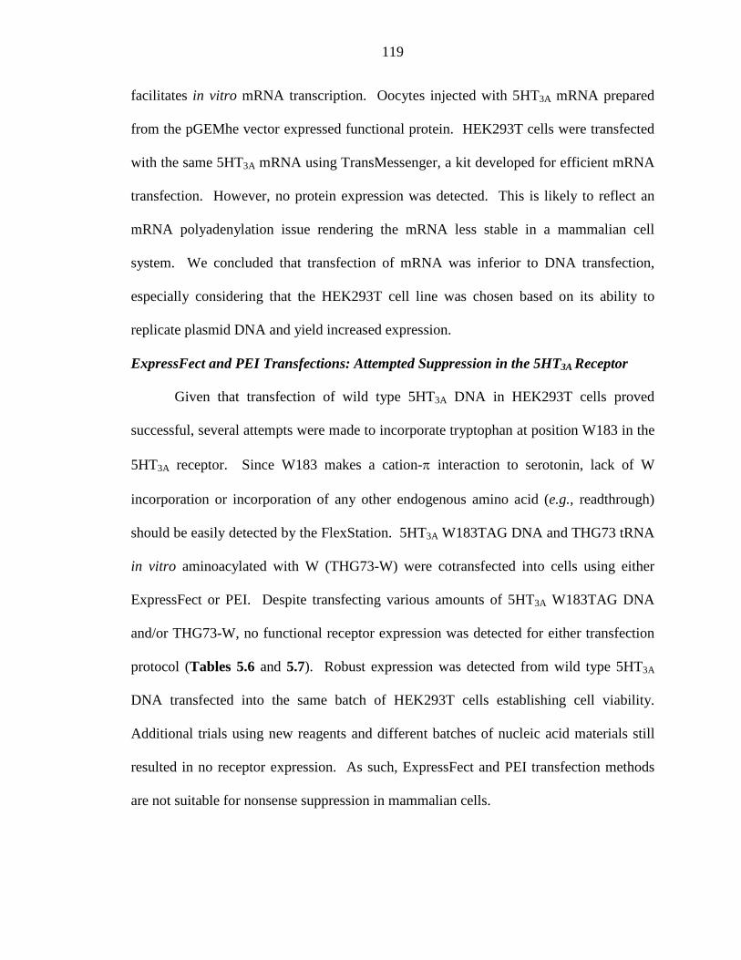
119
facilitates in vitro mRNA transcription. Oocytes injected with 5HT3A mRNA prepared
from the pGEMhe vector expressed functional protein. HEK293T cells were transfected
with the same 5HT3A mRNA using TransMessenger, a kit developed for efficient mRNA
transfection. However, no protein expression was detected. This is likely to reflect an
mRNA polyadenylation issue rendering the mRNA less stable in a mammalian cell
system. We concluded that transfection of mRNA was inferior to DNA transfection,
especially considering that the HEK293T cell line was chosen based on its ability to
replicate plasmid DNA and yield increased expression.
ExpressFect and PEI Transfections: Attempted Suppression in the 5HT3A Receptor
Given that transfection of wild type 5HT3A DNA in HEK293T cells proved
successful, several attempts were made to incorporate tryptophan at position W183 in the
5HT3A receptor. Since W183 makes a cation-π interaction to serotonin, lack of W
incorporation or incorporation of any other endogenous amino acid (e.g., readthrough)
should be easily detected by the FlexStation. 5HT3A W183TAG DNA and THG73 tRNA
in vitro aminoacylated with W (THG73-W) were cotransfected into cells using either
ExpressFect or PEI. Despite transfecting various amounts of 5HT3A W183TAG DNA
and/or THG73-W, no functional receptor expression was detected for either transfection
protocol (Tables 5.6 and 5.7). Robust expression was detected from wild type 5HT3A
DNA transfected into the same batch of HEK293T cells establishing cell viability.
Additional trials using new reagents and different batches of nucleic acid materials still
resulted in no receptor expression. As such, ExpressFect and PEI transfection methods
are not suitable for nonsense suppression in mammalian cells.
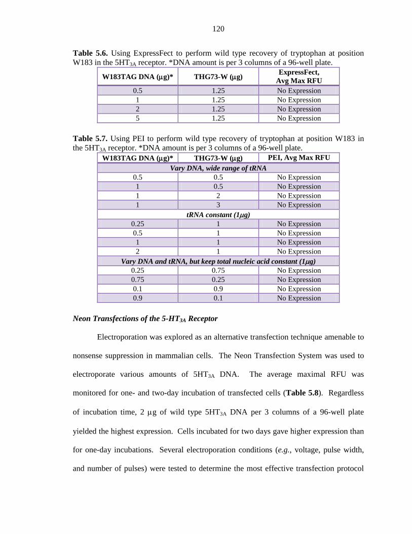
120
Table 5.6. Using ExpressFect to perform wild type recovery of tryptophan at position W183 in the 5HT3A receptor. *DNA amount is per 3 columns of a 96-well plate.
W183TAG DNA (µg)* THG73-W (µg) ExpressFect,
Avg Max RFU 0.5 1.25 No Expression 1 1.25 No Expression 2 1.25 No Expression 5 1.25 No Expression
Table 5.7. Using PEI to perform wild type recovery of tryptophan at position W183 in the 5HT3A receptor. *DNA amount is per 3 columns of a 96-well plate.
W183TAG DNA (µg)* THG73-W (µg) PEI, Avg Max RFU
Vary DNA, wide range of tRNA 0.5 0.5 No Expression 1 0.5 No Expression 1 2 No Expression 1 3 No Expression
tRNA constant (1µg) 0.25 1 No Expression 0.5 1 No Expression 1 1 No Expression 2 1 No Expression
Vary DNA and tRNA, but keep total nucleic acid constant (1µg) 0.25 0.75 No Expression 0.75 0.25 No Expression 0.1 0.9 No Expression 0.9 0.1 No Expression
Neon Transfections of the 5-HT3A Receptor
Electroporation was explored as an alternative transfection technique amenable to
nonsense suppression in mammalian cells. The Neon Transfection System was used to
electroporate various amounts of 5HT3A DNA. The average maximal RFU was
monitored for one- and two-day incubation of transfected cells (Table 5.8). Regardless
of incubation time, 2 µg of wild type 5HT3A DNA per 3 columns of a 96-well plate
yielded the highest expression. Cells incubated for two days gave higher expression than
for one-day incubations. Several electroporation conditions (e.g., voltage, pulse width,
and number of pulses) were tested to determine the most effective transfection protocol
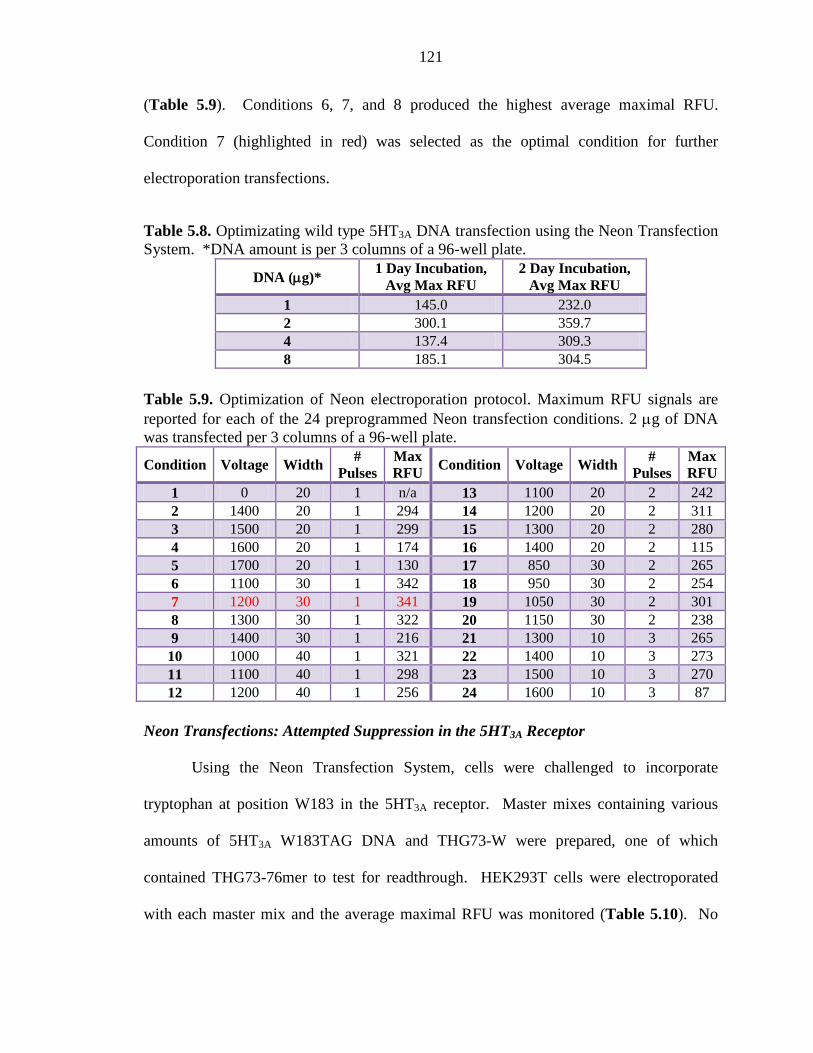
121
(Table 5.9). Conditions 6, 7, and 8 produced the highest average maximal RFU.
Condition 7 (highlighted in red) was selected as the optimal condition for further
electroporation transfections.
Table 5.8. Optimizating wild type 5HT3A DNA transfection using the Neon Transfection System. *DNA amount is per 3 columns of a 96-well plate.
DNA (µg)* 1 Day Incubation,
Avg Max RFU 2 Day Incubation,
Avg Max RFU 1 145.0 232.0 2 300.1 359.7 4 137.4 309.3 8 185.1 304.5
Table 5.9. Optimization of Neon electroporation protocol. Maximum RFU signals are reported for each of the 24 preprogrammed Neon transfection conditions. 2 µg of DNA was transfected per 3 columns of a 96-well plate.
Condition Voltage Width #
Pulses Max RFU Condition Voltage Width
# Pulses
Max RFU
1 0 20 1 n/a 13 1100 20 2 242 2 1400 20 1 294 14 1200 20 2 311 3 1500 20 1 299 15 1300 20 2 280 4 1600 20 1 174 16 1400 20 2 115 5 1700 20 1 130 17 850 30 2 265 6 1100 30 1 342 18 950 30 2 254 7 1200 30 1 341 19 1050 30 2 301 8 1300 30 1 322 20 1150 30 2 238 9 1400 30 1 216 21 1300 10 3 265 10 1000 40 1 321 22 1400 10 3 273 11 1100 40 1 298 23 1500 10 3 270 12 1200 40 1 256 24 1600 10 3 87
Neon Transfections: Attempted Suppression in the 5HT3A Receptor
Using the Neon Transfection System, cells were challenged to incorporate
tryptophan at position W183 in the 5HT3A receptor. Master mixes containing various
amounts of 5HT3A W183TAG DNA and THG73-W were prepared, one of which
contained THG73-76mer to test for readthrough. HEK293T cells were electroporated
with each master mix and the average maximal RFU was monitored (Table 5.10). No
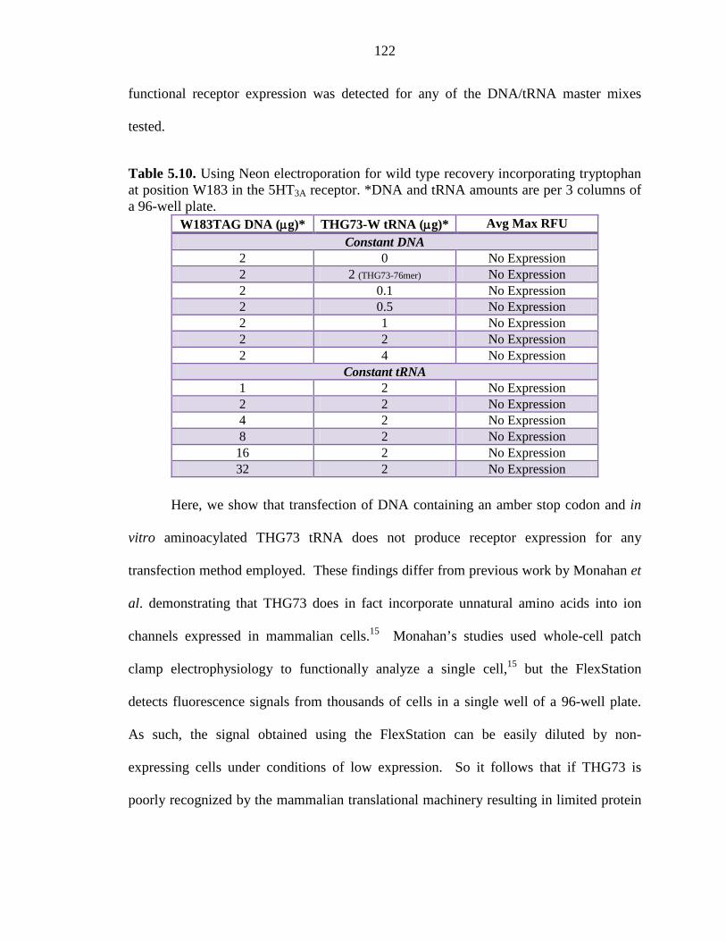
122
functional receptor expression was detected for any of the DNA/tRNA master mixes
tested.
Table 5.10. Using Neon electroporation for wild type recovery incorporating tryptophan at position W183 in the 5HT3A receptor. *DNA and tRNA amounts are per 3 columns of a 96-well plate.
W183TAG DNA (µg)* THG73-W tRNA (µg)* Avg Max RFU
Constant DNA 2 0 No Expression 2 2 (THG73-76mer) No Expression 2 0.1 No Expression 2 0.5 No Expression 2 1 No Expression 2 2 No Expression 2 4 No Expression
Constant tRNA 1 2 No Expression 2 2 No Expression 4 2 No Expression 8 2 No Expression 16 2 No Expression 32 2 No Expression
Here, we show that transfection of DNA containing an amber stop codon and in
vitro aminoacylated THG73 tRNA does not produce receptor expression for any
transfection method employed. These findings differ from previous work by Monahan et
al. demonstrating that THG73 does in fact incorporate unnatural amino acids into ion
channels expressed in mammalian cells.15 Monahan’s studies used whole-cell patch
clamp electrophysiology to functionally analyze a single cell,15 but the FlexStation
detects fluorescence signals from thousands of cells in a single well of a 96-well plate.
As such, the signal obtained using the FlexStation can be easily diluted by non-
expressing cells under conditions of low expression. So it follows that if THG73 is
poorly recognized by the mammalian translational machinery resulting in limited protein
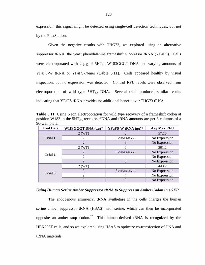
123
expression, this signal might be detected using single-cell detection techniques, but not
by the FlexStation.
Given the negative results with THG73, we explored using an alternative
suppressor tRNA, the yeast phenylalanine frameshift suppressor tRNA (YFaFS). Cells
were electroporated with 2 µg of 5HT3A W183GGGT DNA and varying amounts of
YFaFS-W tRNA or YFaFS-76mer (Table 5.11). Cells appeared healthy by visual
inspection, but no expression was detected. Control RFU levels were observed from
electroporation of wild type 5HT3A DNA. Several trials produced similar results
indicating that YFaFS tRNA provides no additional benefit over THG73 tRNA.
Table 5.11. Using Neon electroporation for wild type recovery of a frameshift codon at position W183 in the 5HT3A receptor. *DNA and tRNA amounts are per 3 columns of a 96-well plate.
Trial Date W183GGGT DNA (µg)* YFaFS-W tRNA (µg)* Avg Max RFU
Trial 1 2 (WT) 0 572.6
2 8 (YFaFS-76mer) No Expression 2 8 No Expression
Trial 2
2 (WT) 0 301.2 2 8 (YFaFS-76mer) No Expression 2 4 No Expression 2 8 No Expression
Trial 3
2 (WT) 0 443.7 2 8 (YFaFS-76mer) No Expression 2 4 No Expression 2 8 No Expression
Using Human Serine Amber Suppressor tRNA to Suppress an Amber Codon in eGFP
The endogenous aminoacyl tRNA synthetase in the cells charges the human
serine amber suppressor tRNA (HSAS) with serine, which can then be incorporated
opposite an amber stop codon.17 This human-derived tRNA is recognized by the
HEK293T cells, and so we explored using HSAS to optimize co-transfection of DNA and
tRNA materials.
124
We targeted residue A37 in the enhanced green fluorescent protein (eGFP) since
this site is located well before the chromophore. If suppression does not occur, then even
expression of a truncated protein will not give a fluorescent signal. Both electroporation
and ExpressFect were used to transfect cells with eGFP A37TAG DNA and HSAS, and
suppression efficiency was monitored by confocal microscopy. Wild type recovery of
eGFP A37TAG using THG73-Ala was attempted, but results were indistinguishable from
76mer control experiments. Cells transfected with eGPF A37TAG and HSAS revealed
improved eGFP expression relative to wild type recovery, however approximate
expression was <1% of wild type expression (Figure 5.7). Electroporation gave slightly
lower expression than ExpressFect. These experiments provided the initial confirmation
that successful transfection of tRNA had occurred, but that protein expression was
significantly hindered when using a stoichiometric tRNA (e.g., THG73 tRNA).
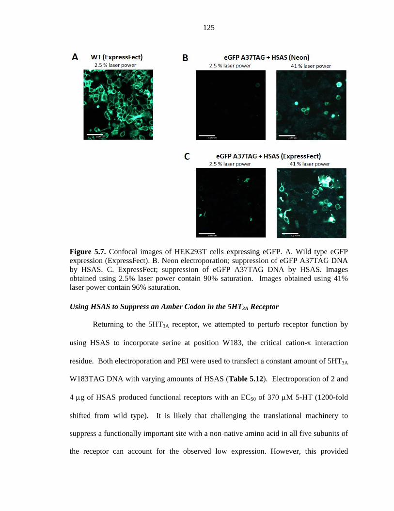
125
Figure 5.7. Confocal images of HEK293T cells expressing eGFP. A. Wild type eGFP expression (ExpressFect). B. Neon electroporation; suppression of eGFP A37TAG DNA by HSAS. C. ExpressFect; suppression of eGFP A37TAG DNA by HSAS. Images obtained using 2.5% laser power contain 90% saturation. Images obtained using 41% laser power contain 96% saturation. Using HSAS to Suppress an Amber Codon in the 5HT3A Receptor
Returning to the 5HT3A receptor, we attempted to perturb receptor function by
using HSAS to incorporate serine at position W183, the critical cation-π interaction
residue. Both electroporation and PEI were used to transfect a constant amount of 5HT3A
W183TAG DNA with varying amounts of HSAS (Table 5.12). Electroporation of 2 and
4 µg of HSAS produced functional receptors with an EC50 of 370 µM 5-HT (1200-fold
shifted from wild type). It is likely that challenging the translational machinery to
suppress a functionally important site with a non-native amino acid in all five subunits of
the receptor can account for the observed low expression. However, this provided
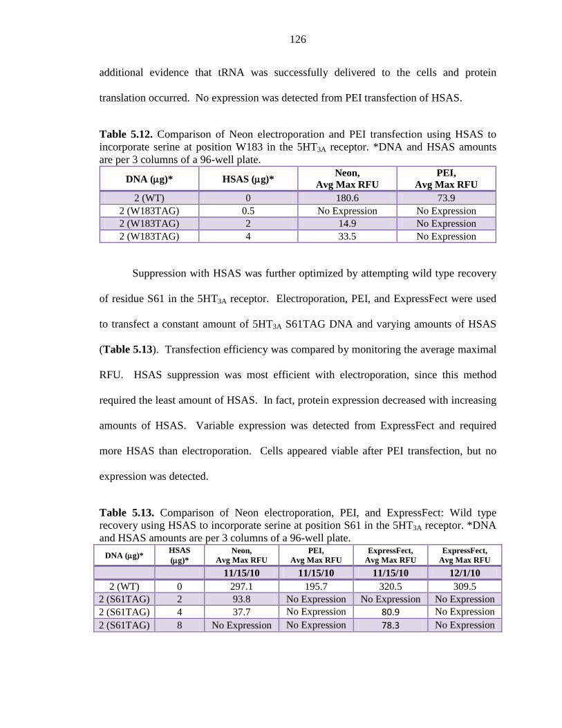
126
additional evidence that tRNA was successfully delivered to the cells and protein
translation occurred. No expression was detected from PEI transfection of HSAS.
Table 5.12. Comparison of Neon electroporation and PEI transfection using HSAS to incorporate serine at position W183 in the 5HT3A receptor. *DNA and HSAS amounts are per 3 columns of a 96-well plate.
DNA (µg)* HSAS (µg)* Neon,
Avg Max RFU PEI,
Avg Max RFU 2 (WT) 0 180.6 73.9
2 (W183TAG) 0.5 No Expression No Expression 2 (W183TAG) 2 14.9 No Expression 2 (W183TAG) 4 33.5 No Expression
Suppression with HSAS was further optimized by attempting wild type recovery
of residue S61 in the 5HT3A receptor. Electroporation, PEI, and ExpressFect were used
to transfect a constant amount of 5HT3A S61TAG DNA and varying amounts of HSAS
(Table 5.13). Transfection efficiency was compared by monitoring the average maximal
RFU. HSAS suppression was most efficient with electroporation, since this method
required the least amount of HSAS. In fact, protein expression decreased with increasing
amounts of HSAS. Variable expression was detected from ExpressFect and required
more HSAS than electroporation. Cells appeared viable after PEI transfection, but no
expression was detected.
Table 5.13. Comparison of Neon electroporation, PEI, and ExpressFect: Wild type recovery using HSAS to incorporate serine at position S61 in the 5HT3A receptor. *DNA and HSAS amounts are per 3 columns of a 96-well plate.
DNA (µg)* HSAS (µg)*
Neon, Avg Max RFU
PEI, Avg Max RFU
ExpressFect, Avg Max RFU
ExpressFect, Avg Max RFU
11/15/10 11/15/10 11/15/10 12/1/10 2 (WT) 0 297.1 195.7 320.5 309.5
2 (S61TAG) 2 93.8 No Expression No Expression No Expression 2 (S61TAG) 4 37.7 No Expression 80.9 No Expression 2 (S61TAG) 8 No Expression No Expression 78.3 No Expression
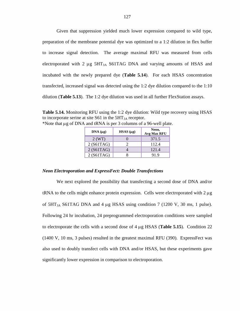
127
Given that suppression yielded much lower expression compared to wild type,
preparation of the membrane potential dye was optimized to a 1:2 dilution in flex buffer
to increase signal detection. The average maximal RFU was measured from cells
electroporated with 2 µg 5HT3A S61TAG DNA and varying amounts of HSAS and
incubated with the newly prepared dye (Table 5.14). For each HSAS concentration
transfected, increased signal was detected using the 1:2 dye dilution compared to the 1:10
dilution (Table 5.13). The 1:2 dye dilution was used in all further FlexStation assays.
Table 5.14. Monitoring RFU using the 1:2 dye dilution: Wild type recovery using HSAS to incorporate serine at site S61 in the 5HT3A receptor. *Note that µg of DNA and tRNA is per 3 columns of a 96-well plate.
DNA (µg) HSAS (µg) Neon,
Avg Max RFU
2 (WT) 0 371.5 2 (S61TAG) 2 112.4 2 (S61TAG) 4 121.4 2 (S61TAG) 8 91.9
Neon Electroporation and ExpressFect: Double Transfections
We next explored the possibility that transfecting a second dose of DNA and/or
tRNA to the cells might enhance protein expression. Cells were electroporated with 2 µg
of 5HT3A S61TAG DNA and 4 µg HSAS using condition 7 (1200 V, 30 ms, 1 pulse).
Following 24 hr incubation, 24 preprogrammed electroporation conditions were sampled
to electroporate the cells with a second dose of 4 µg HSAS (Table 5.15). Condition 22
(1400 V, 10 ms, 3 pulses) resulted in the greatest maximal RFU (390). ExpressFect was
also used to doubly transfect cells with DNA and/or HSAS, but these experiments gave
significantly lower expression in comparison to electroporation.
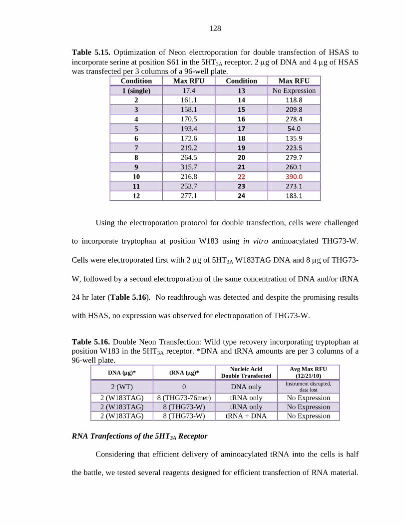
128
Table 5.15. Optimization of Neon electroporation for double transfection of HSAS to incorporate serine at position S61 in the 5HT3A receptor. 2 µg of DNA and 4 µg of HSAS was transfected per 3 columns of a 96-well plate.
Condition Max RFU Condition Max RFU 1 (single) 17.4 13 No Expression
2 161.1 14 118.8 3 158.1 15 209.8 4 170.5 16 278.4 5 193.4 17 54.0 6 172.6 18 135.9 7 219.2 19 223.5 8 264.5 20 279.7 9 315.7 21 260.1
10 216.8 22 390.0 11 253.7 23 273.1 12 277.1 24 183.1
Using the electroporation protocol for double transfection, cells were challenged
to incorporate tryptophan at position W183 using in vitro aminoacylated THG73-W.
Cells were electroporated first with 2 µg of 5HT3A W183TAG DNA and 8 µg of THG73-
W, followed by a second electroporation of the same concentration of DNA and/or tRNA
24 hr later (Table 5.16). No readthrough was detected and despite the promising results
with HSAS, no expression was observed for electroporation of THG73-W.
Table 5.16. Double Neon Transfection: Wild type recovery incorporating tryptophan at position W183 in the 5HT3A receptor. *DNA and tRNA amounts are per 3 columns of a 96-well plate.
DNA (µg)* tRNA (µg)* Nucleic Acid
Double Transfected Avg Max RFU
(12/21/10)
2 (WT) 0 DNA only Instrument disrupted, data lost
2 (W183TAG) 8 (THG73-76mer) tRNA only No Expression 2 (W183TAG) 8 (THG73-W) tRNA only No Expression 2 (W183TAG) 8 (THG73-W) tRNA + DNA No Expression
RNA Tranfections of the 5HT3A Receptor
Considering that efficient delivery of aminoacylated tRNA into the cells is half
the battle, we tested several reagents designed for efficient transfection of RNA material.
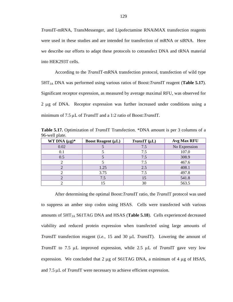
129
TransIT-mRNA, TransMessenger, and Lipofectamine RNAiMAX transfection reagents
were used in these studies and are intended for transfection of mRNA or siRNA. Here
we describe our efforts to adapt these protocols to cotransfect DNA and tRNA material
into HEK293T cells.
According to the TransIT-mRNA transfection protocol, transfection of wild type
5HT3A DNA was performed using various ratios of Boost:TransIT reagent (Table 5.17).
Significant receptor expression, as measured by average maximal RFU, was observed for
2 µg of DNA. Receptor expression was further increased under conditions using a
minimum of 7.5 µL of TransIT and a 1:2 ratio of Boost:TransIT.
Table 5.17. Optimization of TransIT Transfection. *DNA amount is per 3 columns of a 96-well plate.
WT DNA (µg)* Boost Reagent (µL) TransIT (µL) Avg Max RFU 0.02 5 7.5 No Expression 0.1 5 7.5 107.0 0.5 5 7.5 308.9 2 5 7.5 467.6 2 1.25 2.5 408.1 2 3.75 7.5 497.8 2 7.5 15 541.8 2 15 30 563.5
After determining the optimal Boost:TransIT ratio, the TransIT protocol was used
to suppress an amber stop codon using HSAS. Cells were transfected with various
amounts of 5HT3A S61TAG DNA and HSAS (Table 5.18). Cells experienced decreased
viability and reduced protein expression when transfected using large amounts of
TransIT transfection reagent (i.e., 15 and 30 µL TransIT). Lowering the amount of
TransIT to 7.5 µL improved expression, while 2.5 µL of TransIT gave very low
expression. We concluded that 2 µg of S61TAG DNA, a minimum of 4 µg of HSAS,
and 7.5 µL of TransIT were necessary to achieve efficient expression.
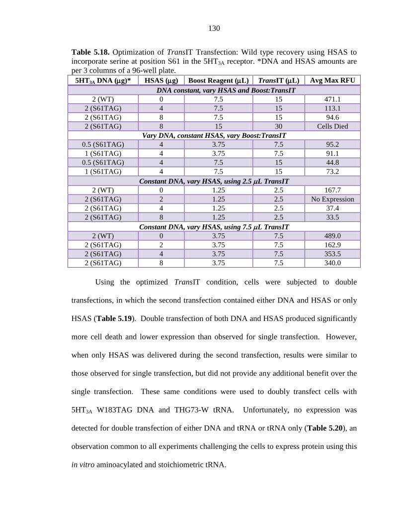
130
Table 5.18. Optimization of TransIT Transfection: Wild type recovery using HSAS to incorporate serine at position S61 in the 5HT3A receptor. *DNA and HSAS amounts are per 3 columns of a 96-well plate.
5HT3A DNA (µg)* HSAS (µg) Boost Reagent (µL) TransIT (µL) Avg Max RFU DNA constant, vary HSAS and Boost:TransIT
2 (WT) 0 7.5 15 471.1 2 (S61TAG) 4 7.5 15 113.1 2 (S61TAG) 8 7.5 15 94.6 2 (S61TAG) 8 15 30 Cells Died
Vary DNA, constant HSAS, vary Boost:TransIT 0.5 (S61TAG) 4 3.75 7.5 95.2 1 (S61TAG) 4 3.75 7.5 91.1
0.5 (S61TAG) 4 7.5 15 44.8 1 (S61TAG) 4 7.5 15 73.2
Constant DNA, vary HSAS, using 2.5 µL TransIT 2 (WT) 0 1.25 2.5 167.7
2 (S61TAG) 2 1.25 2.5 No Expression 2 (S61TAG) 4 1.25 2.5 37.4 2 (S61TAG) 8 1.25 2.5 33.5
Constant DNA, vary HSAS, using 7.5 µL TransIT 2 (WT) 0 3.75 7.5 489.0
2 (S61TAG) 2 3.75 7.5 162.9 2 (S61TAG) 4 3.75 7.5 353.5 2 (S61TAG) 8 3.75 7.5 340.0
Using the optimized TransIT condition, cells were subjected to double
transfections, in which the second transfection contained either DNA and HSAS or only
HSAS (Table 5.19). Double transfection of both DNA and HSAS produced significantly
more cell death and lower expression than observed for single transfection. However,
when only HSAS was delivered during the second transfection, results were similar to
those observed for single transfection, but did not provide any additional benefit over the
single transfection. These same conditions were used to doubly transfect cells with
5HT3A W183TAG DNA and THG73-W tRNA. Unfortunately, no expression was
detected for double transfection of either DNA and tRNA or tRNA only (Table 5.20), an
observation common to all experiments challenging the cells to express protein using this
in vitro aminoacylated and stoichiometric tRNA.
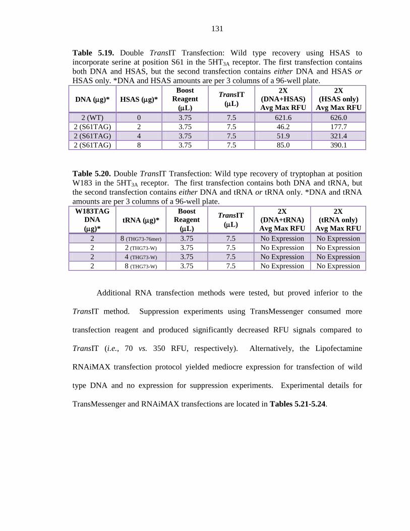
131
Table 5.19. Double TransIT Transfection: Wild type recovery using HSAS to incorporate serine at position S61 in the 5HT3A receptor. The first transfection contains both DNA and HSAS, but the second transfection contains either DNA and HSAS or HSAS only. *DNA and HSAS amounts are per 3 columns of a 96-well plate.
DNA (µg)* HSAS (µg)* Boost
Reagent (µL)
TransIT (µL)
2X (DNA+HSAS) Avg Max RFU
2X (HSAS only)
Avg Max RFU 2 (WT) 0 3.75 7.5 621.6 626.0
2 (S61TAG) 2 3.75 7.5 46.2 177.7 2 (S61TAG) 4 3.75 7.5 51.9 321.4 2 (S61TAG) 8 3.75 7.5 85.0 390.1
Table 5.20. Double TransIT Transfection: Wild type recovery of tryptophan at position W183 in the 5HT3A receptor. The first transfection contains both DNA and tRNA, but the second transfection contains either DNA and tRNA or tRNA only. *DNA and tRNA amounts are per 3 columns of a 96-well plate. W183TAG
DNA (µg)*
tRNA (µg)* Boost
Reagent (µL)
TransIT (µL)
2X (DNA+tRNA) Avg Max RFU
2X (tRNA only)
Avg Max RFU 2 8 (THG73-76mer) 3.75 7.5 No Expression No Expression 2 2 (THG73-W) 3.75 7.5 No Expression No Expression 2 4 (THG73-W) 3.75 7.5 No Expression No Expression 2 8 (THG73-W) 3.75 7.5 No Expression No Expression
Additional RNA transfection methods were tested, but proved inferior to the
TransIT method. Suppression experiments using TransMessenger consumed more
transfection reagent and produced significantly decreased RFU signals compared to
TransIT (i.e., 70 vs. 350 RFU, respectively). Alternatively, the Lipofectamine
RNAiMAX transfection protocol yielded mediocre expression for transfection of wild
type DNA and no expression for suppression experiments. Experimental details for
TransMessenger and RNAiMAX transfections are located in Tables 5.21-5.24.
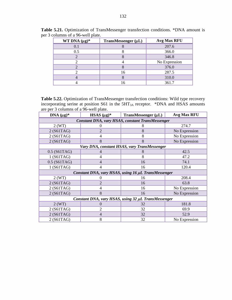
132
Table 5.21. Optimization of TransMessenger transfection conditions. *DNA amount is per 3 columns of a 96-well plate.
WT DNA (µg)* TransMessenger (µL) Avg Max RFU 0.1 8 207.6 0.5 8 366.0 2 8 346.8 2 4 No Expression 2 8 376.0 2 16 287.5 4 8 310.0 4 16 361.7
Table 5.22. Optimization of TransMessenger transfection conditions: Wild type recovery incorporating serine at position S61 in the 5HT3A receptor. *DNA and HSAS amounts are per 3 columns of a 96-well plate.
DNA (µg)* HSAS (µg)* TransMessenger (µL) Avg Max RFU
Constant DNA, vary HSAS, constant TransMessenger 2 (WT) 0 8 274.7
2 (S61TAG) 2 8 No Expression 2 (S61TAG) 4 8 No Expression 2 (S61TAG) 8 8 No Expression
Vary DNA, constant HSAS, vary TransMessenger 0.5 (S61TAG) 4 8 42.5 1 (S61TAG) 4 8 47.2
0.5 (S61TAG) 4 16 74.1 1 (S61TAG) 4 16 120.4
Constant DNA, vary HSAS, using 16 µL TransMessenger 2 (WT) 0 16 208.4
2 (S61TAG) 2 16 63.8 2 (S61TAG) 4 16 No Expression 2 (S61TAG) 8 16 No Expression
Constant DNA, vary HSAS, using 32 µL TransMessenger 2 (WT) 0 32 181.8
2 (S61TAG) 2 32 69.9 2 (S61TAG) 4 32 52.9 2 (S61TAG) 8 32 No Expression
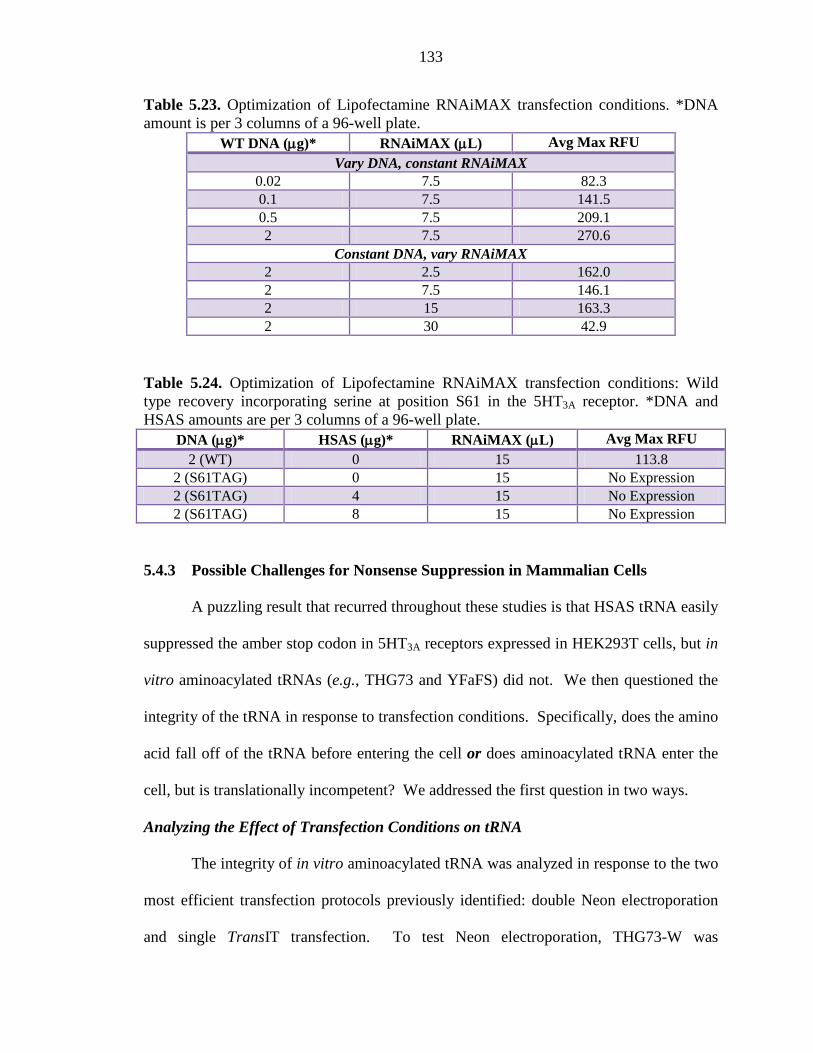
133
Table 5.23. Optimization of Lipofectamine RNAiMAX transfection conditions. *DNA amount is per 3 columns of a 96-well plate.
WT DNA (µg)* RNAiMAX (µL) Avg Max RFU
Vary DNA, constant RNAiMAX 0.02 7.5 82.3 0.1 7.5 141.5 0.5 7.5 209.1 2 7.5 270.6
Constant DNA, vary RNAiMAX 2 2.5 162.0 2 7.5 146.1 2 15 163.3 2 30 42.9
Table 5.24. Optimization of Lipofectamine RNAiMAX transfection conditions: Wild type recovery incorporating serine at position S61 in the 5HT3A receptor. *DNA and HSAS amounts are per 3 columns of a 96-well plate.
DNA (µg)* HSAS (µg)* RNAiMAX (µL) Avg Max RFU 2 (WT) 0 15 113.8
2 (S61TAG) 0 15 No Expression 2 (S61TAG) 4 15 No Expression 2 (S61TAG) 8 15 No Expression
5.4.3 Possible Challenges for Nonsense Suppression in Mammalian Cells
A puzzling result that recurred throughout these studies is that HSAS tRNA easily
suppressed the amber stop codon in 5HT3A receptors expressed in HEK293T cells, but in
vitro aminoacylated tRNAs (e.g., THG73 and YFaFS) did not. We then questioned the
integrity of the tRNA in response to transfection conditions. Specifically, does the amino
acid fall off of the tRNA before entering the cell or does aminoacylated tRNA enter the
cell, but is translationally incompetent? We addressed the first question in two ways.
Analyzing the Effect of Transfection Conditions on tRNA
The integrity of in vitro aminoacylated tRNA was analyzed in response to the two
most efficient transfection protocols previously identified: double Neon electroporation
and single TransIT transfection. To test Neon electroporation, THG73-W was
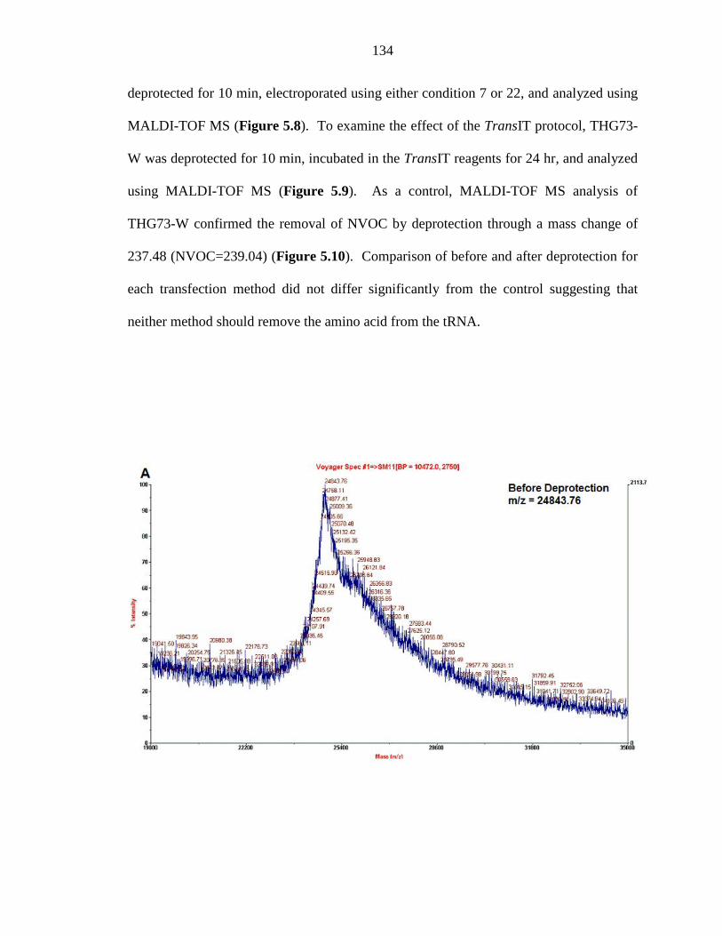
134
deprotected for 10 min, electroporated using either condition 7 or 22, and analyzed using
MALDI-TOF MS (Figure 5.8). To examine the effect of the TransIT protocol, THG73-
W was deprotected for 10 min, incubated in the TransIT reagents for 24 hr, and analyzed
using MALDI-TOF MS (Figure 5.9). As a control, MALDI-TOF MS analysis of
THG73-W confirmed the removal of NVOC by deprotection through a mass change of
237.48 (NVOC=239.04) (Figure 5.10). Comparison of before and after deprotection for
each transfection method did not differ significantly from the control suggesting that
neither method should remove the amino acid from the tRNA.
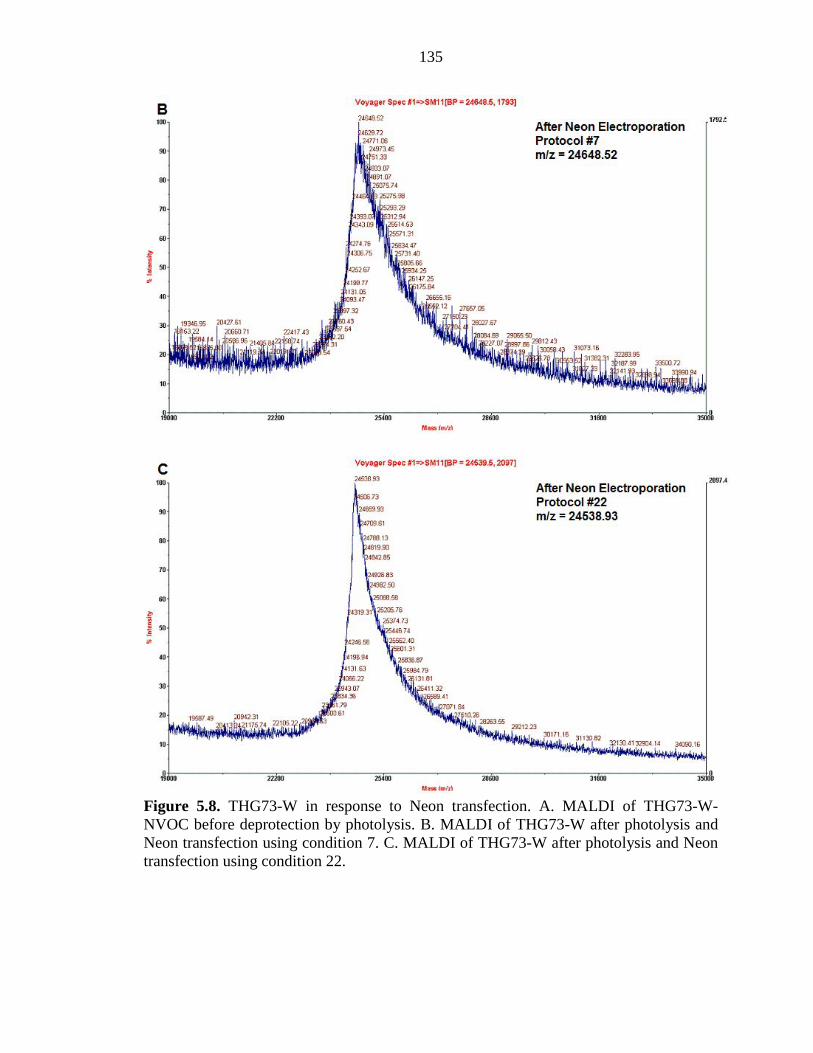
135
Figure 5.8. THG73-W in response to Neon transfection. A. MALDI of THG73-W-NVOC before deprotection by photolysis. B. MALDI of THG73-W after photolysis and Neon transfection using condition 7. C. MALDI of THG73-W after photolysis and Neon transfection using condition 22.
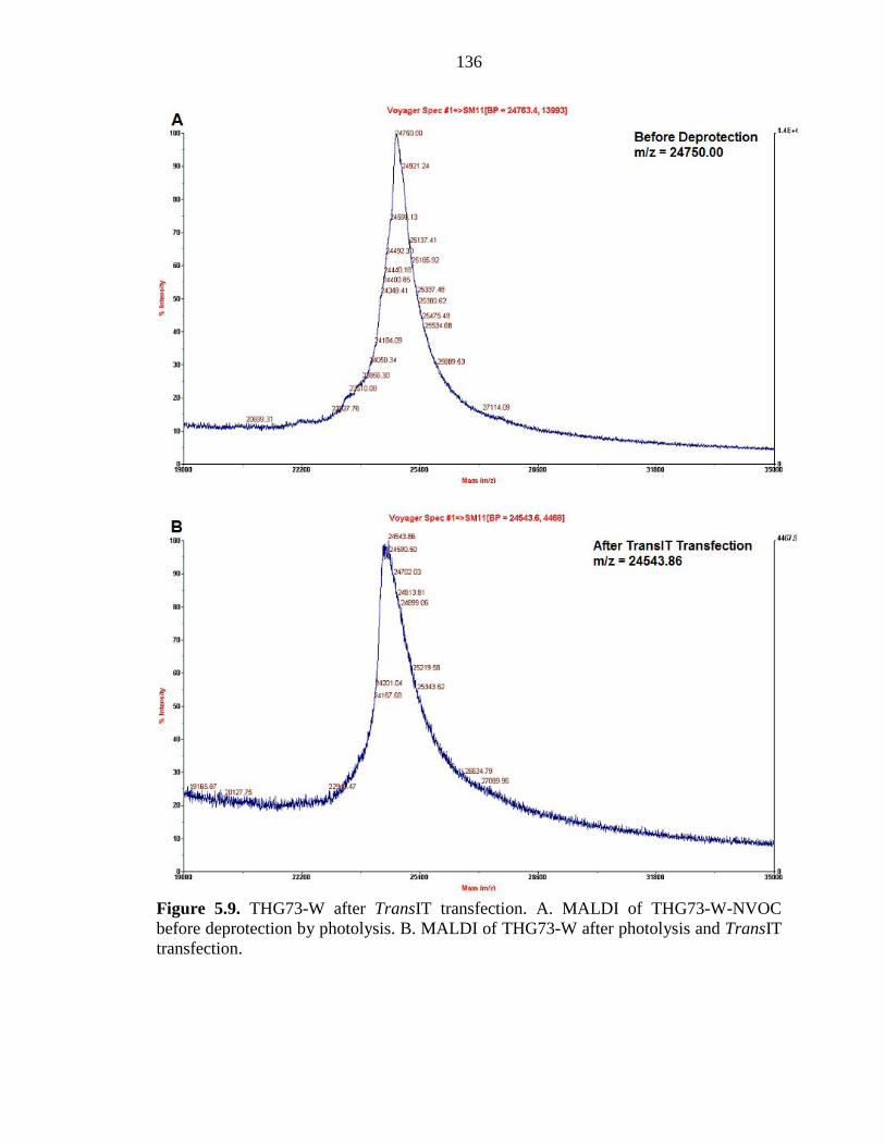
136
Figure 5.9. THG73-W after TransIT transfection. A. MALDI of THG73-W-NVOC before deprotection by photolysis. B. MALDI of THG73-W after photolysis and TransIT transfection.
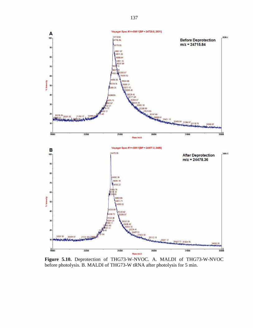
137
Figure 5.10. Deprotection of THG73-W-NVOC. A. MALDI of THG73-W-NVOC before photolysis. B. MALDI of THG73-W tRNA after photolysis for 5 min.
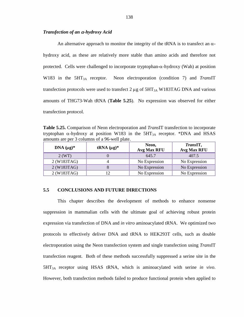
138
Transfection of an α-hydroxy Acid
An alternative approach to monitor the integrity of the tRNA is to transfect an α-
hydroxy acid, as these are relatively more stable than amino acids and therefore not
protected. Cells were challenged to incorporate tryptophan-α-hydroxy (Wah) at position
W183 in the 5HT3A receptor. Neon electroporation (condition 7) and TransIT
transfection protocols were used to transfect 2 µg of 5HT3A W183TAG DNA and various
amounts of THG73-Wah tRNA (Table 5.25). No expression was observed for either
transfection protocol.
Table 5.25. Comparison of Neon electroporation and TransIT transfection to incorporate tryptophan α-hydroxy at position W183 in the 5HT3A receptor. *DNA and HSAS amounts are per 3 columns of a 96-well plate.
DNA (µg)* tRNA (µg)* Neon,
Avg Max RFU TransIT,
Avg Max RFU 2 (WT) 0 645.7 407.5
2 (W183TAG) 4 No Expression No Expression 2 (W183TAG) 8 No Expression No Expression 2 (W183TAG) 12 No Expression No Expression
5.5 CONCLUSIONS AND FUTURE DIRECTIONS
This chapter describes the development of methods to enhance nonsense
suppression in mammalian cells with the ultimate goal of achieving robust protein
expression via transfection of DNA and in vitro aminoacylated tRNA. We optimized two
protocols to effectively deliver DNA and tRNA to HEK293T cells, such as double
electroporation using the Neon transfection system and single transfection using TransIT
transfection reagent. Both of these methods successfully suppressed a serine site in the
5HT3A receptor using HSAS tRNA, which is aminoacylated with serine in vivo.
However, both transfection methods failed to produce functional protein when applied to
139
suppression of an amber stop codon utilizing in vitro aminoacylated tRNAs, such as
THG73 and YFaFS.
Successful suppression experiments with HSAS tRNA indicated that tRNA is in
fact delivered to the cells. Possible issues limiting the functionality of in vitro
aminoacylated tRNAs could be their stability during the transfection processes and
subsequent recognition by the mammalian translational machinery. MALDI-TOF MS
analysis of deprotected THG73-W exposed to either transfection method suggested that
only the NVOC protecting group was removed and the aminoacyl tRNA remained intact
during transfection. These data support the idea that tRNA is delivered to the cell, but
that either the amino acid falls off the tRNA inside the cell or the tRNA is translationally
incompetent.
Translational incompetence may be attributed to the tRNA identity problem. This
concept refers to the specific interactions between the tRNA molecule and its synthetase
required for proper activity.24 The success of nonsense suppression experiments rely on
the principle that the mammalian synthetases will not recognize and aminoacylate the
tRNA with an endogenous amino acid. One can imagine future experiments designed to
bypass the tRNA identity problem entirely. This could be achieved by engineering a
novel synthetase that retains tRNA recognition elements specific to the in vitro
aminoacylated tRNA of interest, but does not recharge this tRNA with any endogenous
amino acid. Essentially, this would create a “dummy” synthetase capable of binding the
tRNA and “handing it off” to the ribosome. Complementary studies might include
engineering the tRNA component to bind the synthetase. Mutagenesis would target the
140
identity element regions of tRNA, such as the distal ends of the tRNA and the anticodon
loop.
Additionally, the possibility that the tRNA is not recognized by essential
associated proteins (e.g., EF-Tu) could contribute to hindered translational activity. As
such, it may be useful to overexpress the mammalian elongation factor, EF-1α, which is
responsible for delivering aminoacyl tRNA to the ribosome. However, we note that
several reports have confirmed the high conservation and abundant expression of EF-
1α in mammaliam cells.25
5.6 ACKNOWLEDGMENTS
Special thanks to Shawna Frazier for assistance with passaging techniques and
cell counting as well as Crystal Dilworth for assistance with the confocal microscope.
141
5.7 REFERENCES 1. Noren, C. J.; Anthony-Cahill, S. J.; Griffith, M. C.; Schultz, P. G., A general
method for site-specific incorporation of unnatural amino acids into proteins. Science 1989, 244, (4901), 182-8.
2. Nowak, M. W.; Gallivan, J. P.; Silverman, S. K.; Labarca, C. G.; Dougherty, D. A.; Lester, H. A., In vivo incorporation of unnatural amino acids into ion channels in Xenopus oocyte expression system. Methods Enzymol 1998, 293, 504-29.
3. Nowak, M. W.; Kearney, P. C.; Sampson, J. R.; Saks, M. E.; Labarca, C. G.; Silverman, S. K.; Zhong, W.; Thorson, J.; Abelson, J. N.; Davidson, N.; et al., Nicotinic receptor binding site probed with unnatural amino acid incorporation in intact cells. Science 1995, 268, (5209), 439-42.
4. Beene, D. L.; Brandt, G. S.; Zhong, W.; Zacharias, N. M.; Lester, H. A.; Dougherty, D. A., Cation-pi interactions in ligand recognition by serotonergic (5-HT3A) and nicotinic acetylcholine receptors: the anomalous binding properties of nicotine. Biochemistry 2002, 41, (32), 10262-9.
5. Lummis, S. C.; D, L. B.; Harrison, N. J.; Lester, H. A.; Dougherty, D. A., A cation-pi binding interaction with a tyrosine in the binding site of the GABAC receptor. Chem Biol 2005, 12, (9), 993-7.
6. Mu, T. W.; Lester, H. A.; Dougherty, D. A., Different binding orientations for the same agonist at homologous receptors: a lock and key or a simple wedge? J Am Chem Soc 2003, 125, (23), 6850-1.
7. Puskar, N. L.; Xiu, X.; Lester, H. A.; Dougherty, D. A., Two neuronal nicotinic acetylcholine receptors, alpha4beta4 and alpha7, show differential agonist binding modes. J Biol Chem 286, (16), 14618-27.
8. Xiu, X.; Puskar, N. L.; Shanata, J. A.; Lester, H. A.; Dougherty, D. A., Nicotine binding to brain receptors requires a strong cation-pi interaction. Nature 2009, 458, (7237), 534-7.
9. Zhong, W.; Gallivan, J. P.; Zhang, Y.; Li, L.; Lester, H. A.; Dougherty, D. A., From ab initio quantum mechanics to molecular neurobiology: a cation-pi binding site in the nicotinic receptor. Proc Natl Acad Sci U S A 1998, 95, (21), 12088-93.
10. Werry, T. D.; Gregory, K. J.; Sexton, P. M.; Christopoulos, A., Characterization of serotonin 5-HT2C receptor signaling to extracellular signal-regulated kinases 1 and 2. J Neurochem 2005, 93, (6), 1603-15.
11. Kohrer, C.; Xie, L.; Kellerer, S.; Varshney, U.; RajBhandary, U. L., Import of amber and ochre suppressor tRNAs into mammalian cells: a general approach to site-specific insertion of amino acid analogues into proteins. Proc Natl Acad Sci U S A 2001, 98, (25), 14310-5.
12. Liu, C. C.; Schultz, P. G., Adding new chemistries to the genetic code. Annu Rev Biochem 79, 413-44.
13. Liu, W.; Brock, A.; Chen, S.; Schultz, P. G., Genetic incorporation of unnatural amino acids into proteins in mammalian cells. Nat Methods 2007, 4, (3), 239-44.
14. Zhang, Z.; Alfonta, L.; Tian, F.; Bursulaya, B.; Uryu, S.; King, D. S.; Schultz, P. G., Selective incorporation of 5-hydroxytryptophan into proteins in mammalian cells. Proc Natl Acad Sci U S A 2004, 101, (24), 8882-7.
142
15. Monahan, S. L.; Lester, H. A.; Dougherty, D. A., Site-specific incorporation of unnatural amino acids into receptors expressed in mammalian cells. Chem Biol 2003, 10, (6), 573-80.
16. Saks, M. E.; Sampson, J. R.; Nowak, M. W.; Kearney, P. C.; Du, F.; Abelson, J. N.; Lester, H. A.; Dougherty, D. A., An engineered Tetrahymena tRNAGln for in vivo incorporation of unnatural amino acids into proteins by nonsense suppression. J Biol Chem 1996, 271, (38), 23169-75.
17. Capone, J. P.; Sharp, P. A.; RajBhandary, U. L., Amber, ochre and opal suppressor tRNA genes derived from a human serine tRNA gene. EMBO J 1985, 4, (1), 213-21.
18. Rodriguez, E. A.; Lester, H. A.; Dougherty, D. A., In vivo incorporation of multiple unnatural amino acids through nonsense and frameshift suppression. Proc Natl Acad Sci U S A 2006, 103, (23), 8650-5.
19. DuBridge, R. B.; Tang, P.; Hsia, H. C.; Leong, P. M.; Miller, J. H.; Calos, M. P., Analysis of mutation in human cells by using an Epstein-Barr virus shuttle system. Mol Cell Biol 1987, 7, (1), 379-87.
20. Pear, W. S.; Nolan, G. P.; Scott, M. L.; Baltimore, D., Production of high-titer helper-free retroviruses by transient transfection. Proc Natl Acad Sci U S A 1993, 90, (18), 8392-6.
21. Price, K. L.; Lummis, S. C., FlexStation examination of 5-HT3 receptor function using Ca2+ and membrane potential-sensitive dyes: advantages and potential problems. J Neurosci Methods 2005, 149, (2), 172-7.
22. Srinivasan, R.; Pantoja, R.; Moss, F. J.; Mackey, E. D.; Son, C. D.; Miwa, J.; Lester, H. A., Nicotine up-regulates alpha4beta2 nicotinic receptors and ER exit sites via stoichiometry-dependent chaperoning. J Gen Physiol 137, (1), 59-79.
23. Thompson, A. J.; Lochner, M.; Lummis, S. C., Loop B is a major structural component of the 5-HT3 receptor. Biophys J 2008, 95, (12), 5728-36.
24. Giege, R.; Sissler, M.; Florentz, C., Universal rules and idiosyncratic features in tRNA identity. Nucleic Acids Res 1998, 26, (22), 5017-35.
25. Lee, S.; LeBlanc, A.; Duttaroy, A.; Wang, E., Terminal differentiation-dependent alteration in the expression of translation elongation factor-1 alpha and its sister gene, S1, in neurons. Exp Cell Res 1995, 219, (2), 589-97.


![α-Conotoxin PeIA[S9H,V10A,E14N] potently and selectively ...molpharm.aspetjournals.org/content/molpharm/early/2012/08/22/mol… · 22/8/2012 · Nicotinic acetylcholine receptors](https://static.fdocument.org/doc/165x107/5ff86836422ebe55ca6ae52c/-conotoxin-peias9hv10ae14n-potently-and-selectively-2282012-nicotinic.jpg)