Role of interplay between IL-4 and IFN-γ in the in regulating M1 ... · Role of interplay between...
10
Role of interplay between IL-4 and IFN-γ in the in regulating M1 macrophage polarization induced by Nattectin Edson Kiyotaka Ishizuka a , Marcio José Ferreira a , Lidiane Zito Grund a , Erica Maria Martins Coutinho a , Evilin Naname Komegae a , Alexandra Anjos Cassado b , Karina Ramalho Bortoluci b , Monica Lopes-Ferreira a , Carla Lima a, ⁎ a Immunoregulation Unit, Special Laboratory of Applied Toxinology, Instituto Butantan, São Paulo, Brazil b Molecular Immunology of Innate Recognition Unit, Department of Biological Sciences, Campus Diadema and CTC-Mol, Federal University of São Paulo, São Paulo, Brazil abstract article info Article history: Received 23 April 2012 Received in revised form 9 August 2012 Accepted 14 August 2012 Available online 28 August 2012 Keywords: Nattectin C-type lectin M1 activation Macrophages Th1/Th2 cytokines Recently our group described that Nattectin, a C-type lectin of the venom of Thalassophryne nattereri shows a potent pro-inflammatory capacity. Here, we demonstrated that Nattectin is able to induce M1 macrophage marker iNOS, and up-regulate the expression of MHC class II, CD80, CD86 and CD40 molecules. The increase in MHC class II and CD49a integrin expression with MMP-9 production and endocytic capacity depend on lec- tin function of Nattectin. Moreover, the polarization of peritoneal and bone marrow-derived macrophages in- duced by Nattectin to M1 profile is dependent on Th1 cytokines (IL-12 and IFN-γ), and negatively regulated by Th2 cytokines (IL-4, IL-10 and IL-13). Also we reveal that IL-4 play a dual role in this polarization: a regular action of IL-4 was seen in the negative regulation of the CD40 expression, but an unexpected positive regu- lation was seen in the expression of CCR7 and MHC class II. Finally, our in vivo studies showed that the influx of neutrophils and small peritoneal macrophage - F4/80 low MHCII hi induced by Nattectin is totally dependent on IL-4 and IFN-γ cytokines. Furthermore, the induction of IL-6 release is negatively regulated by IL-4 and positively regulated by IL-12 and IFN-γ. Together, the results allowed us to expand the knowledge about the regulation of macrophage activation, as well as confirmed the ability of Nattectin, a fish C-type lectin, as an important immunomodulatory agent. © 2012 Elsevier B.V. 1. Introduction Macrophages are important innate immune cells that play a crit- ical role in sensing pathogens, directly or indirectly, and integrating this information to regulate the adaptive immune response. Het- erogeneity and plasticity are hallmarks of cells belonging to the monocyte–macrophage lineage. On the basis of Th1/Th2 polariza- tion concepts [1], phenotypically polarized macrophages are now generally termed pro-inflammatory M1 or classically activated, and anti-inflammatory M2 or alternatively activated [2,3]. The existence of multiple subsets of macrophages raises the ques- tion of whether they are functionally specialized to promote distinct immune responses. M1 macrophages, whose prototypical activating stimuli are IFN-γ and LPS, exhibit potent microbicidal properties and promote strong IL-12‐mediated T helper 1 responses [4]. In con- trast, M2 macrophages support T helper 2‐associated effector func- tions and may play a role in resolution of inflammation through endocytic clearance and trophic factor synthesis [3]. However, the M2 designation has rapidly expanded to include other types of mac- rophages: M2a, induced by IL-4 or IL-13; M2b, induced by immune complexes and agonists of TLRs or IL-1 receptors; and M2c, induced by IL-10 and glucocorticoid hormones [5]. While the classification of the M1 and M2 macrophage functions has provided an important tool for understanding the regulation of the inflammatory process, at present the molecular mechanisms that govern M1/M2 polariza- tion remain incompletely understood. Cytokines and growth factors have been implicated in the reprogramming of M1 and M2 macrophages. Activation of macro- phages by either Th1 (IFN-γ) or Th2 (IL-4 and IL-13) cytokines leads to dramatic changes in their physiology, including alterations in the expression of surface proteins as co-stimulatory molecules and the production of cytokines and pro-inflammatory mediators that influence T cell differentiation. Thus, adjuvants that modulate macrophage function, enhancing macrophages survival, inducing the expression of co-stimulatory molecules, and controlling the re- lease of pro-inflammatory cytokines may be particularly effective in inducing T cell expansion. Soluble C-type lectins, including molecules with specificity for galac- tose, have been identified in several teleost fish [6]. Recently our group International Immunopharmacology 14 (2012) 513–522 ⁎ Corresponding author at: Unidade Imunorregulação, Laboratório Especial de Toxinologia Aplicada, Instituto Butantan, Av. Vital Brazil, 1500. Butantan, 05503–900, Sao Paulo, Brazil. Tel./fax: +55 11 3676 1392. E-mail address: [email protected] (C. Lima). 1567-5769 © 2012 Elsevier B.V. http://dx.doi.org/10.1016/j.intimp.2012.08.009 Contents lists available at SciVerse ScienceDirect International Immunopharmacology journal homepage: www.elsevier.com/locate/intimp Open access under the Elsevier OA license. Open access under the Elsevier OA license.
-
Upload
phungkhanh -
Category
Documents
-
view
218 -
download
0
Transcript of Role of interplay between IL-4 and IFN-γ in the in regulating M1 ... · Role of interplay between...
International Immunopharmacology 14 (2012) 513–522
Contents lists available at SciVerse ScienceDirect
International Immunopharmacology
j ourna l homepage: www.e lsev ie r .com/ locate / in t imp
Role of interplay between IL-4 and IFN-γ in the in regulating M1 macrophagepolarization induced by Nattectin
Edson Kiyotaka Ishizuka a, Marcio José Ferreira a, Lidiane Zito Grund a, Erica Maria Martins Coutinho a,Evilin Naname Komegae a, Alexandra Anjos Cassado b, Karina Ramalho Bortoluci b,Monica Lopes-Ferreira a, Carla Lima a,⁎a Immunoregulation Unit, Special Laboratory of Applied Toxinology, Instituto Butantan, São Paulo, Brazilb Molecular Immunology of Innate Recognition Unit, Department of Biological Sciences, Campus Diadema and CTC-Mol, Federal University of São Paulo, São Paulo, Brazil
⁎ Corresponding author at: Unidade ImunorregulaToxinologia Aplicada, Instituto Butantan, Av. Vital BrazSao Paulo, Brazil. Tel./fax: +55 11 3676 1392.
E-mail address: [email protected] (C. Lima)
1567-5769 © 2012 Elsevier B.V.http://dx.doi.org/10.1016/j.intimp.2012.08.009
Open access under the Els
a b s t r a c t
a r t i c l e i n f oArticle history:Received 23 April 2012Received in revised form 9 August 2012Accepted 14 August 2012Available online 28 August 2012
Keywords:NattectinC-type lectinM1 activationMacrophagesTh1/Th2 cytokines
Recently our group described that Nattectin, a C-type lectin of the venom of Thalassophryne nattereri shows apotent pro-inflammatory capacity. Here, we demonstrated that Nattectin is able to induce M1 macrophagemarker iNOS, and up-regulate the expression of MHC class II, CD80, CD86 and CD40 molecules. The increasein MHC class II and CD49a integrin expression with MMP-9 production and endocytic capacity depend on lec-tin function of Nattectin. Moreover, the polarization of peritoneal and bone marrow-derived macrophages in-duced by Nattectin to M1 profile is dependent on Th1 cytokines (IL-12 and IFN-γ), and negatively regulatedby Th2 cytokines (IL-4, IL-10 and IL-13). Also we reveal that IL-4 play a dual role in this polarization: a regularaction of IL-4 was seen in the negative regulation of the CD40 expression, but an unexpected positive regu-lation was seen in the expression of CCR7 and MHC class II. Finally, our in vivo studies showed that the influxof neutrophils and small peritoneal macrophage - F4/80lowMHCIIhi induced by Nattectin is totally dependenton IL-4 and IFN-γ cytokines. Furthermore, the induction of IL-6 release is negatively regulated by IL-4 andpositively regulated by IL-12 and IFN-γ. Together, the results allowed us to expand the knowledge aboutthe regulation of macrophage activation, as well as confirmed the ability of Nattectin, a fish C-type lectin,as an important immunomodulatory agent.
© 2012 Elsevier B.V. Open access under the Elsevier OA license.
1. Introduction
Macrophages are important innate immune cells that play a crit-ical role in sensing pathogens, directly or indirectly, and integratingthis information to regulate the adaptive immune response. Het-erogeneity and plasticity are hallmarks of cells belonging to themonocyte–macrophage lineage. On the basis of Th1/Th2 polariza-tion concepts [1], phenotypically polarized macrophages are nowgenerally termed pro-inflammatory M1 or classically activated,and anti-inflammatory M2 or alternatively activated [2,3].
The existence of multiple subsets of macrophages raises the ques-tion of whether they are functionally specialized to promote distinctimmune responses. M1 macrophages, whose prototypical activatingstimuli are IFN-γ and LPS, exhibit potent microbicidal propertiesand promote strong IL-12‐mediated T helper 1 responses [4]. In con-trast, M2 macrophages support T helper 2‐associated effector func-tions and may play a role in resolution of inflammation through
ção, Laboratório Especial deil, 1500. Butantan, 05503–900,
.
evier OA license.
endocytic clearance and trophic factor synthesis [3]. However, theM2 designation has rapidly expanded to include other types of mac-rophages: M2a, induced by IL-4 or IL-13; M2b, induced by immunecomplexes and agonists of TLRs or IL-1 receptors; and M2c, inducedby IL-10 and glucocorticoid hormones [5]. While the classification ofthe M1 and M2 macrophage functions has provided an importanttool for understanding the regulation of the inflammatory process,at present the molecular mechanisms that govern M1/M2 polariza-tion remain incompletely understood.
Cytokines and growth factors have been implicated in thereprogramming of M1 and M2 macrophages. Activation of macro-phages by either Th1 (IFN-γ) or Th2 (IL-4 and IL-13) cytokinesleads to dramatic changes in their physiology, including alterationsin the expression of surface proteins as co-stimulatory moleculesand the production of cytokines and pro-inflammatory mediatorsthat influence T cell differentiation. Thus, adjuvants that modulatemacrophage function, enhancing macrophages survival, inducingthe expression of co-stimulatory molecules, and controlling the re-lease of pro-inflammatory cytokines may be particularly effectivein inducing T cell expansion.
Soluble C-type lectins, includingmoleculeswith specificity for galac-tose, have been identified in several teleost fish [6]. Recently our group
514 E.K. Ishizuka et al. / International Immunopharmacology 14 (2012) 513–522
described that Nattectin, a C-type lectin of the venom of Thalassophrynenattereri [7] shows a potent pro-inflammatory capacity, acting inducinga Th1 response through the control of recruitment and activation ofmonocytes/macrophages, licensing these cells to differentiate intocells exhibiting typical DC functions. These macrophage-derived DCsexhibit functional attributes: augmented Ag presentation capacitywith avid phagocytosis; enhanced migration to the T cell–rich area oflymph nodes, through its effect on production and release of activeMMP-9; high T cell co-stimulatorymolecules expression, and enhancedcapacity to activate T cells by the production of IFN-γ through thehigh-level of bioactive IL-12 p70 secretion [8].
Considering the role of macrophages in homeostasis as well as inhost defense and in view of the possibility to obtain natural adjuvantsable to modulate the immune system, specifically macrophage func-tions, the investigations presented here were undertaken to assesswhether Nattectin modulates the process of macrophage activationand M1/M2 polarization, and to evaluate the role of T helper derivedcytokines in the regulation of this process using IL-4, IL-12, andIFN-γ-deficient mice.
2. Materials and methods
2.1. Animals
Female BALB/c and male C57BL/6 mice were obtained from a col-ony at the Butantan Institute. IL-4−/−, IL-12−/− and IFN-γ−/− malemice on C57BL/6 background were purchased from Institute of Bio-medical Sciences at São Paulo University. All mice, 6 to 8 weeks old,were kept under pathogen-free conditions and given water andfood ad libitum. They were housed under controlled temperature, hu-midity and lighting conditions. All the procedures involving micewere in accordance with the guidelines provided by Brazilian Collegeof Animal Experimentation and approved by Animal Ethics Commit-tee of Butantan Institute (634/09) and Institute of Biomedical Sci-ences (106/91-2).
2.2. Reagents and Abs
Nattectin obtained by Reversed-phase HPLC (Amersham Biosci-ences) from T. nattereri venom (IBAMA 16221–1) was confirmedby MALDI-TOF mass spectrometry (Amersham Biosciences, Bucking-hamshire, U.K.) according to Lopes-Ferreira and coworkers [7]. En-dotoxin Detoxi-GelTM (Pierce Chemical Co.) was used according tothe manufacturer's instructions to remove >99% of the contaminat-ing LPS in the Nattectin solution (resulting in a total dose b0.8 pgLPS), which was measured by Limulus Amoebocyte Lysate (LonzaWalkersville Inc., Walkersville, MD). Dulbecco´s Modified eaglemedium (DMEM), and fluorescein 40,000 MW dextran were pur-chased from Invitrogen (Carlsbad, CA). IL-10 and IL-13 wereobtained from eBiosciences (San Diego, CA). Total iNOS/NOS II(ABN26) anti-RELM-α (AB3365P, resistin-like molecule α) werepurchased from Millipore (Concord Road, Billerica, MA) and β actinand horseradish peroxidase-labeled anti-rabbit IgG were purchasedfrom Santa Cruz (Santa Cruz, California). ELISA kits for quantitativemeasurement of murine cytokines and MMPs were purchased fromBD Pharmingen (San Jose, CA). FITC-, PE-, PE-Cy5-, PerCP-Cy5.5- orbiotin-conjugated mAbs used to detect the expression of CD11c(HL3), Ly-6C (AL-21), TLR2 (203325), CD14 (Sa2.8), CD18 (C71/16),CD29 (eBioHMb1-1), CD40 (3/23), CD49b (Hma2), CD80 (16-10A1),CD86 (GL-1), I-A/I-E (M5/114.15.2), CD11b (M1/70), CCR7 (4B12),CD44 (IM7), purified F4/80R (6F12), purified CD16 (275003), purifiedCD49a (Ha31/8), purified CD49e (HM25-1), as well as isotype-matchedcontrol mAbs were obtained from BD Pharmingen, eBiosciences andR&D Systems (Minneapolis, MN). Donkey anti-rat IgG PE conjugated(polyclonal) and anti-rat IgG FITC conjugated (polyclonal) were pur-chased from eBiosciences. Mouse anti-Armenian and Syrian hamster
IgG cocktail FITC conjugated (G70-204, G94-56) was obtained from BDPharmingen.
2.3. Generation of bone marrow-derived macrophages (BMDM)
Bonemarrow-derivedmacrophages isolated fromBALB/c or C57BL/6micewere obtained according to Falk and coworkers [9] andMuller andcoworkers [10]. Initially, bonemarrowwas flushed from the femurs andtibias of mice, and cells (2×10⁶/mL) were cultured in Petri dishes inDulbecco's Minimal Essential Medium (DMEM) supplemented with 2%horse serum, 1% vitamin, 1% sodium pyruvate, 1% non-essential aminoacids, 1% L-glutamine, 1% penicillin/streptomycin-glutamine, and 30%L929 cell-conditioned medium as a source of macrophage colony-stimulating factor (M-CSF) under a humidified atmosphere of 5% CO2
at 37 ° C. Cells were fed on day 2 and half of the old medium wasreplaced on day 6. After culture for 7 days, the homogeneous populationof macrophages was determined by flow cytometry (97–99% ofCD11bhighF4/80int-highCD14highLy6Clow).
2.4. Stimulation of macrophages with Nattectin
After BMDM generation, cells were stimulated with and withoutNattectin (10 μg/mL) in Petri dishes (2×10⁶ cells/mL) for 24 h accordingto Saraiva and coworkers [8]. To evaluatewhether Nattectin effects weredependent on the carbohydrate-binding activity, D-galactose at 10 mMwere added to the culture medium, with sucrose at the same concentra-tion serving as a control. Finally, to evaluate the influence of Th1/Th2cytokines in macrophage polarization induced by Nattectin, cells werestimulated for 24 hwith Nattectin (10 μg/mL) plus recombinant murineIL-10 or IL-13, both at 5 ng/mL.
2.5. Protein lysate preparation and western blot analysis
Whole-cell extracts were prepared from BMDM cultured cell asfollows: the culture medium was drained off the cells, and the adher-ent cells were washed twice with ice-cold PBS. The cells were lysedwith ice-cold Tris buffer (50 mM Tris–HCl pH 6.8, 2% SDS, 10% glycer-ol, 2,5% β-mercaptoethanol) containing protease inhibitor cocktail(P1860, Sigma-Aldrich, Spruce St. Louis, MO). The cell suspensionwas transferred into a centrifuge tube, left on ice, and vortexed for5 min to lyse the cells. The lysate was then centrifuged at 12,000 gin a pre-cooled centrifuge for 20 min. The supernatant was collectedand the concentration of protein in extracts was determined byusing Bradford [11] assay. Protein samples (100 μg aliquots) weremixed with sample buffer (100 mM Tris [pH 6.8], 4% SDS, 20% glycer-ol, 0.02% bromophenol blue, and 50 mL/mL β-mercaptoethanol), re-solved by 12% SDS- polyacrylamide gel (PAGE) and electroblottedonto Hybond-C nitrocellulose membranes, using a semi-dry Bio-Radapparatus. Ponceau staining of electroblotted membrane was usedto visually monitor protein transfer. The blot was placed in blockingsolution (5% nonfat dried milk in TBST) for 1 h at room temperatureon a shaking table. Total iNOS/NOS II (1:1000), RELMα (1:1000)and β actin (1:2000) were respectively detected with a secondaryperoxidase-conjugated anti-rabbit IgG polyclonal antibody. Immuno-reactive proteins were detected by enhanced chemiluminescence(ECL) using hyperfilm and ECL reagent (Amersham International).The results of Western blot analysis were quantified by measuringthe relative intensity compared to the control using Kodak MolecularImaging software and represented in the relative intensities.
2.6. Flow cytometry
Following different treatments and stimulation ofmacrophageswithNattectin, cells (1×10⁶ cells/mL) were incubated with 10% mouseserum for 20 min at 4 °C to block unspecific binding. Subsequently,cells were stained in the dark at 4 °C for 30 min with FITC-, PE-,
515E.K. Ishizuka et al. / International Immunopharmacology 14 (2012) 513–522
PE-Cy5-, PerCP-Cy5.5- or biotin-conjugated mAbs for CD11c (HL3),Ly-6C (AL-21), TLR2 (203325), CD14 (Sa2.8), CD18 (C71/16), CD19(1D3), CD29 (eBioHMb1-1), CD40 (3/23), CD49b (Hma2), CD80(16-10A1), CD86 (GL-1), I-A/I-E (M5/114.15.2), CD11b (M1/70), CCR7(4B12), CD44 (IM7), purified F4/80R (6F12), purified CD16 (275003),purified CD49a (Ha31/8) and purified CD49e (HM25-1). Appropriateisotype controls were used in all cases. After staining, the cells werefixedwith 2%w/v formaldehyde analyzed immediately on a FACScaliburflow cytometer using CellQuest 3.1 software (Becton Dickinson, SanJose, CA). For flow cytometric analysis, typical forward and side scattergate was set to exclude dead cells and aggregates. Results wereexpressed as median of fluorescence intensity (MFI)±SEM.
2.7. Modulation of endocytosis of macrophages by Nattectin
BMDM were washed, resuspended in complete medium, and stimu-lated at 4 °C for 30 minwithNattectin (10 μg/mL) treatedwith andwith-out D-galactose (10 nM). After washing, macrophages (2 x 10⁶/mL) wereincubated at 37 °C with 0.1 mg/mL of FITC-dextran (0.431-mm, Sigma-Aldrich, St Louis, MO) for 30 min. Uptake was stopped by adding coldPBS containing 1% FCS and 0.01% NaN3. Cells were then washed threetimes and analyzed by flow cytometry (FACSCalibur, BD Pharmingen,SanDiego, CA). Themarkers for themonovariant histogram (unpublisheddata) were set based on the negative staining control, which was provid-ed by incubating BMDMwith FITC-dextran at 4 °C. Results of phagocyto-sis of FITC-dextran were expressed as median of fluorescence intensity(MFI)±SEM. Experiments were repeated twice.
2.8. Quantitation of MMPs by ELISA
Cell-free supernatants from unstimulated or Nattectin-stimulatedmacrophages were harvested after 2 or 24 h culture. MMP-2 andMMP-9 in the supernatants were quantified using ELISA kits (BDPharmingen) according to the manufacturer's instructions. The mini-mal detection levels for MMP-2 were 1.5 ng/mL and for MMP-9 was1.25 ng/mL.
2.9. Nattectin‐induced peritonitis
In order to evaluate the immune response induced by Nattectin,different groups of mice on the C57BL/6 background (n=5) were in-traperitoneally (i.p.) injected with 10 μg of Nattectin diluted in 500 μLof sterile saline 0.9%. Control animals were injected with the samevolume of saline. At time points indicated (6 or 24 h) after injection,animals were killed by CO2 inhalation and peritoneal cells wereobtained by peritoneal lavage with 5 mL of sterile PBS/EDTA10 mM. For the recovery of cellular contents, lavage fluid wascentrifuged at 378 g for 10 min and the cell pellet was resuspendedin cold PBS/BSA 0.1% for leukocyte cell counts. For the analysis ofcytokine levels in the peritoneum, recovered lavage fluid was storedat −20 °C until analyzed by ELISA. Using a hemocytometer andcytocentrifuge, slides were prepared, air dried, fixed in methanol,and stained (Wright-Giemsa, Scientific Products, Chicago, IL). Fordifferential cell counts, 300 leukocytes were enumerated and iden-tified as mononuclear cells or polymorphonuclear neutrophils,based on staining and morphologic characteristics using a light mi-croscope (Nikon Eclipse E200). Representative photomicrographsof each group were acquired digitally using an Axio Imager A1 mi-croscope (Carl Zeiss, Germany) with an AxioCam ICc1 digital camera(Carl Zeiss).
2.10. Statistical analysis
All values were expressed as mean±SEM. Statistical differencesbetween experimental groups were detected after analysis of vari-ance (One-way ANOVA) followed by Bonferroni test. A value of
pb0.05 was considered statistically significant. The SPSS statisticalpackage (Release 8.0, Standard Version, 1997) was employed.
3. Results
3.1. Modulation of macrophage endocytosis and antigen presentation-related molecules by Nattectin
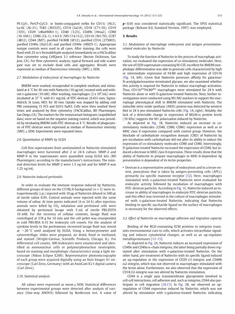
To study the function of Nattectin in the process of macrophage acti-vation, we evaluated the expression of co-stimulatory molecules. Here,the useof L929 supernatant containingM-CSF, excellent for BMDMmor-phologic differentiation was able to generate cells characterized by highor intermediate expression of F4/80 and high expression of CD11b(Fig. 1A, left). Given that Nattectin possesses affinity for galactose/N-acetylgalactosamine-terminated glycans, we also examined whetherthis activity is required for Nattectin to induce macrophage activation.Thus, CD11bposF4/80pos macrophages were stimulated for 24 h withNattectin alone or with D-galactose-treated Nattectin. Next, further in-vestigationswere conducted usingM1/M2markers to support themac-rophage phenotypical shift in BMDM stimulated with Nattectin. Theinducible nitric oxide synthase (iNOS) protein was detected by westernblot in 24 h pos-stimulated Nattectin cells (Fig. 1A, right). Notably, thelack of a detectable change in expression of RELM-α protein levels(10 kDa) suggests the M1 polarization induced by Nattectin.
As depicted in Fig. 1B, Nattectin induced an increase in co-stimulatory molecules (CD40, CD80, CD86) expression as well as inMHC class II expression compared with control group. However, theblockade of carbohydrate recognition domain (CRD) of Nattectin bypre-incubation with carbohydrate did not alter its ability to induce theexpression of co-stimulatory molecules CD80 and CD86. Interestingly,D-galactose-treated Nattectin increased the expression of CD40, but in-duced a decrease inMHC class II expression. These results show that theability of Nattectin to prepare macrophages to MHC-II-dependent Agpresentation is dependent of its lectin properties.
• Dextran is a representative antigen of endocytosis and to a lesser ex-tent, pinocytosis that is taken by antigen-presenting cells (APCs)primarily via specific mannose receptor [12]. Here, macrophagesstimulated with D-galactose-treated Nattectin were evaluated forendocytic activity followed by incubation of macrophages withFITC-dextran particles. According to Fig. 1C, Nattectin induced an in-crease in ability of macrophages to endocyte FITC-dextran particles,and this effect was reversed in the culture of macrophages stimulat-ed with D-galactose-treated Nattectin, indicating that Nattectinbinding to specific saccharide ligand on the surface of macrophagesis necessary for the observed effects.
3.2. Effect of Nattectin on macrophage adhesion and migration capacity
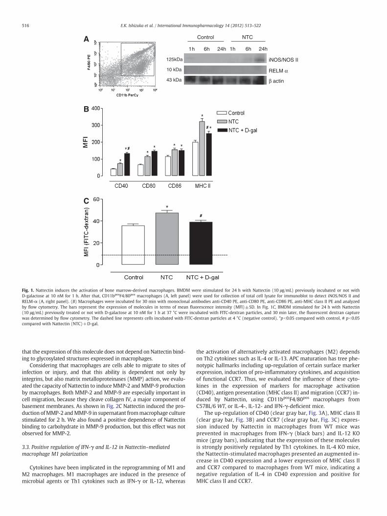
Binding of the RGD-containing ECM proteins to integrins trans-mits environmental cues to cells, which activates intracellular signal-ing and induces cytoskeletal changes, as well as an up-regulatesmetalloproteinases [13–15].
As depicted in Fig. 2A, Nattectin induces an increased expression ofCD49e and CD49aα-chain integrins, the latter being partially down reg-ulated after stimulation with D-galactose-treated Nattectin. On theother hand, pre-treatment of Nattectin with its specific ligand inducedan up-regulation in the expression of CD29 β1-integrin and CD49bα2-integrin, which was not observed in macrophages stimulated withthe lectin alone. Furthermore, we also observed that the expression ofCD18 β2-integrin was not altered by Nattectin stimulation.
CD44 is a single pass transmembrane glycoprotein involved incell-cell interactions, cell adhesion and, such as integrins, CD44 also par-ticipate in cell migration [16,17]. In Fig. 2B, we observed an up-regulation of CD44 expression induced by Nattectin, which was notaltered by stimulation with D-galactose-treated Nattectin, indicating
Control NTC
1h 6h 24h
β actin
RELM α
iNOS/NOS II125kDa
10 kDa
43 kDa
1h 6h 24h
A
B
C
Fig. 1. Nattectin induces the activation of bone marrow-derived macrophages. BMDM were stimulated for 24 h with Nattectin (10 μg/mL) previously incubated or not withD-galactose at 10 nM for 1 h. After that, CD11bposF4/80pos macrophages (A, left panel) were used for collection of total cell lysate for immunoblot to detect iNOS/NOS II andRELM-α (A, right panel). (B) Macrophages were incubated for 30 min with monoclonal antibodies anti-CD40 PE, anti-CD80 PE, anti-CD86 PE, anti-MHC class II PE and analyzedby flow cytometry. The bars represent the expression of molecules in terms of mean fluorescence intensity (MFI)±SD. In Fig. 1C, BMDM stimulated for 24 h with Nattectin(10 μg/mL) previously treated or not with D-galactose at 10 nM for 1 h at 37 °C were incubated with FITC-dextran particles, and 30 min later, the fluorescent dextran capturewas determined by flow cytometry. The dashed line represents cells incubated with FITC-dextran particles at 4 °C (negative control). *pb0.05 compared with control, # pb0.05compared with Nattectin (NTC)+D-gal.
516 E.K. Ishizuka et al. / International Immunopharmacology 14 (2012) 513–522
that the expression of this molecule does not depend on Nattectin bind-ing to glycosylated structures expressed in macrophages.
Considering that macrophages are cells able to migrate to sites ofinfection or injury, and that this ability is dependent not only byintegrins, but also matrix metalloproteinases (MMP) action, we evalu-ated the capacity of Nattectin to induce MMP-2 andMMP-9 productionby macrophages. Both MMP-2 and MMP-9 are especially important incell migration, because they cleave collagen IV, a major component ofbasement membranes. As shown in Fig. 2C Nattectin induced the pro-duction ofMMP-2 andMMP-9 in supernatant frommacrophage culturestimulated for 2 h. We also found a positive dependence of Nattectinbinding to carbohydrate in MMP-9 production, but this effect was notobserved for MMP-2.
3.3. Positive regulation of IFN-γ and IL-12 in Nattectin–mediatedmacrophage M1 polarization
Cytokines have been implicated in the reprogramming of M1 andM2 macrophages. M1 macrophages are induced in the presence ofmicrobial agents or Th1 cytokines such as IFN-γ or IL-12, whereas
the activation of alternatively activated macrophages (M2) dependson Th2 cytokines such as IL-4 or IL-13. APC maturation has tree phe-notypic hallmarks including up-regulation of certain surface markerexpression, induction of pro-inflammatory cytokines, and acquisitionof functional CCR7. Thus, we evaluated the influence of these cyto-kines in the expression of markers for macrophage activation(CD40), antigen presentation (MHC class II) and migration (CCR7) in-duced by Nattectin, using CD11bposF4/80pos macrophages fromC57BL/6 WT, or IL-4-, IL-12- and IFN-γ-deficient mice.
The up-regulation of CD40 (clear gray bar, Fig. 3A), MHC class II(clear gray bar, Fig. 3B) and CCR7 (clear gray bar, Fig. 3C) expres-sion induced by Nattectin in macrophages from WT mice wasprevented in macrophages from IFN-γ (black bars) and IL-12 KOmice (gray bars), indicating that the expression of these moleculesis strongly positively regulated by Th1 cytokines. In IL-4 KO mice,the Nattectin-stimulated macrophages presented an augmented in-crease in CD40 expression and a lower expression of MHC class IIand CCR7 compared to macrophages from WT mice, indicating anegative regulation of IL-4 in CD40 expression and positive forMHC class II and CCR7.
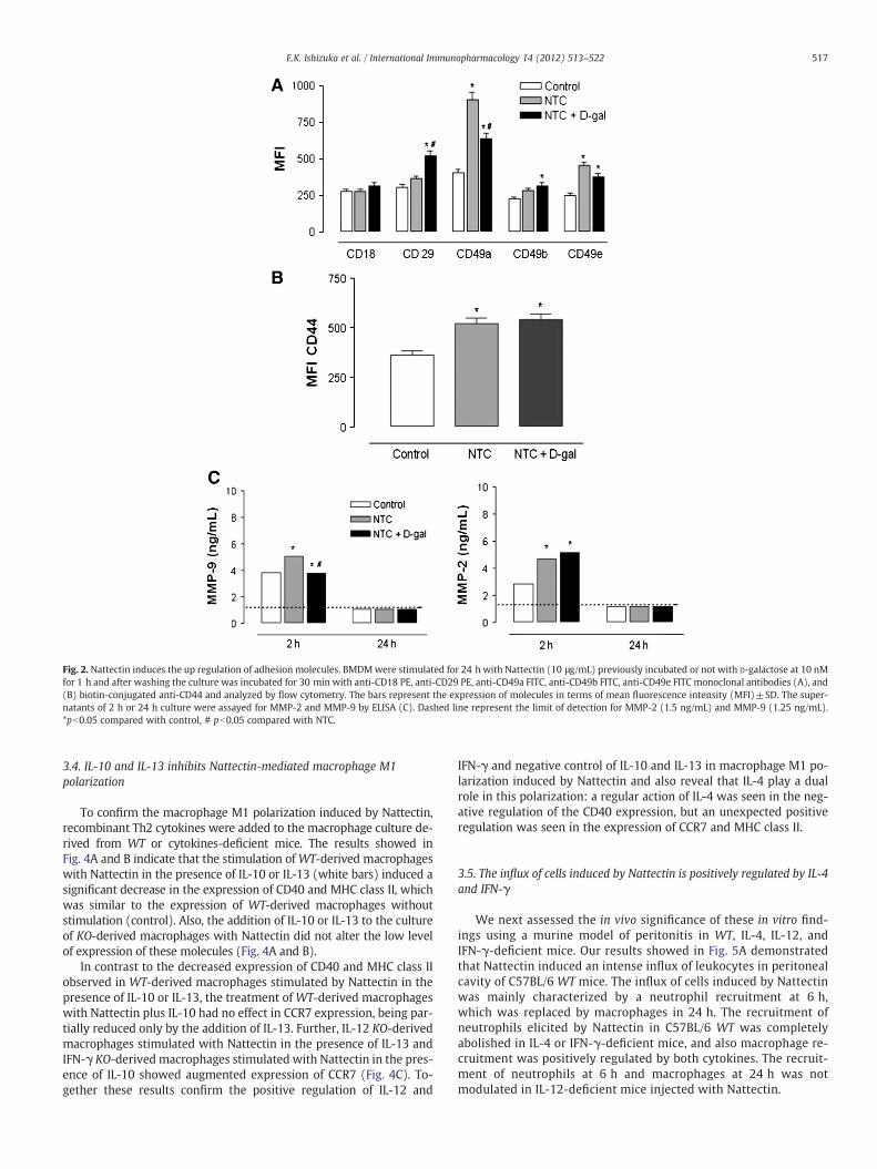
Fig. 2. Nattectin induces the up regulation of adhesion molecules. BMDMwere stimulated for 24 h with Nattectin (10 μg/mL) previously incubated or not with D-galactose at 10 nMfor 1 h and after washing the culture was incubated for 30 min with anti-CD18 PE, anti-CD29 PE, anti-CD49a FITC, anti-CD49b FITC, anti-CD49e FITC monoclonal antibodies (A), and(B) biotin-conjugated anti-CD44 and analyzed by flow cytometry. The bars represent the expression of molecules in terms of mean fluorescence intensity (MFI)±SD. The super-natants of 2 h or 24 h culture were assayed for MMP-2 and MMP-9 by ELISA (C). Dashed line represent the limit of detection for MMP-2 (1.5 ng/mL) and MMP-9 (1.25 ng/mL).*pb0.05 compared with control, # pb0.05 compared with NTC.
517E.K. Ishizuka et al. / International Immunopharmacology 14 (2012) 513–522
3.4. IL-10 and IL-13 inhibits Nattectin‐mediated macrophage M1polarization
To confirm the macrophage M1 polarization induced by Nattectin,recombinant Th2 cytokines were added to the macrophage culture de-rived from WT or cytokines-deficient mice. The results showed inFig. 4A and B indicate that the stimulation of WT-derived macrophageswith Nattectin in the presence of IL-10 or IL-13 (white bars) induced asignificant decrease in the expression of CD40 and MHC class II, whichwas similar to the expression of WT-derived macrophages withoutstimulation (control). Also, the addition of IL-10 or IL-13 to the cultureof KO-derived macrophages with Nattectin did not alter the low levelof expression of these molecules (Fig. 4A and B).
In contrast to the decreased expression of CD40 and MHC class IIobserved in WT-derived macrophages stimulated by Nattectin in thepresence of IL-10 or IL-13, the treatment of WT-derived macrophageswith Nattectin plus IL-10 had no effect in CCR7 expression, being par-tially reduced only by the addition of IL-13. Further, IL-12 KO-derivedmacrophages stimulated with Nattectin in the presence of IL-13 andIFN-γ KO-derived macrophages stimulated with Nattectin in the pres-ence of IL-10 showed augmented expression of CCR7 (Fig. 4C). To-gether these results confirm the positive regulation of IL-12 and
IFN-γ and negative control of IL-10 and IL-13 in macrophage M1 po-larization induced by Nattectin and also reveal that IL-4 play a dualrole in this polarization: a regular action of IL-4 was seen in the neg-ative regulation of the CD40 expression, but an unexpected positiveregulation was seen in the expression of CCR7 and MHC class II.
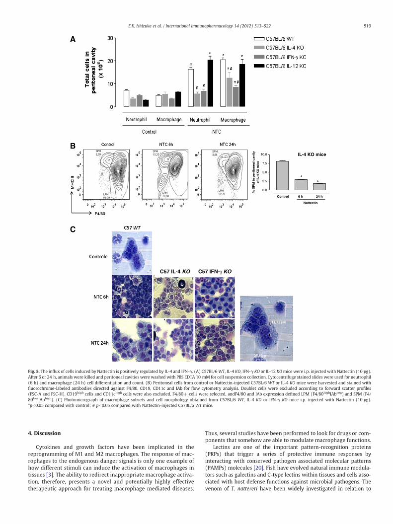
3.5. The influx of cells induced by Nattectin is positively regulated by IL-4and IFN-γ
We next assessed the in vivo significance of these in vitro find-ings using a murine model of peritonitis in WT, IL-4, IL-12, andIFN-γ-deficient mice. Our results showed in Fig. 5A demonstratedthat Nattectin induced an intense influx of leukocytes in peritonealcavity of C57BL/6 WT mice. The influx of cells induced by Nattectinwas mainly characterized by a neutrophil recruitment at 6 h,which was replaced by macrophages in 24 h. The recruitment ofneutrophils elicited by Nattectin in C57BL/6 WT was completelyabolished in IL-4 or IFN-γ-deficient mice, and also macrophage re-cruitment was positively regulated by both cytokines. The recruit-ment of neutrophils at 6 h and macrophages at 24 h was notmodulated in IL-12-deficient mice injected with Nattectin.
Fig. 3. Positive regulation of IFN-γ and IL-12 in Nattectin‐mediated M1 polarization.BMDM from C57BL/6 WT mice or deficient in IL-4, IL-12 or IFN-γ were stimulated withNattectin (10 μg/mL) for 24 h. After that, macrophages were incubated for 30 min withmonoclonal antibodies anti-CD40 PE, anti-MHC class II PE, anti-CCR7 PE-Cy5 and analyzedby flow cytometry. The bars represent the expression of (A) CD40, (B) MHC class II, and(C) CCR7 in terms of mean fluorescence intensity (MFI)±SD. *pb0.05 compared withcontrol; # pb0.05 compared with Nattectin-injected C57BL/6 WT mice. Fig. 4. IL-10 and IL-13 inhibit Nattectin‐mediated macrophage M1 polarization. BMDM
from C57BL/6 WT mice and deficient in IL-12 and IFN-γ were stimulated with Nattectin(10 μg/mL) for 24 h in the absence or presence of recombinant cytokines IL-10 or IL-13,each at a concentration of 5 ng/ml. After that, CD11bposF4/80posCD14posLy6Clow macro-phages were incubated for 30 min with monoclonal antibodies anti-CD40 PE, anti-MHCclass II PE, anti-CCR7 PE-Cy5 and analyzed by flow cytometry. The bars represent the ex-pression of (A)CD40, (B)MHC class II and (C)CCR7 in terms ofmean fluorescence intensity(MFI)±SD. *pb0.05 compared with control; # pb0.05 compared with Nattectin-injectedC57BL/6 WT mice; § pb0.05 compared with Nattectin-injected Nattectin-injected C57BL/6 IL-12 KO or IFN-γ KO mice.
518 E.K. Ishizuka et al. / International Immunopharmacology 14 (2012) 513–522
Utilizing a high-dimensional digital FACSwith 11-color combination,Ghosn et al. [18] recently demonstrated that peritoneal macrophagescomprises two distinct sub-populations, namely large (LPM) and smallperitoneal macrophage (SPM); these sub-populations have phenotypesand functions distinct from monocytes and DC. In the present study,these sub-populations were identified using a 4-color flow cytometryaccording to Cassado and coworkers [19]. In dot plot of Fig. 5B we ob-served that C57BL/6WTmice injected onlywith saline showed predom-inance of LPM (91.09%±1.82 — F4/80hiMHC class IIneg) and a lowproportion of SPM (5.99%±0.04 — F4/80lowMHC class IIhi). Nattectinpromoted a decrease in the number of LPMs (78.50%±0.63) and an in-tense recruitment of SPM (10.20%±0.12) and neutrophils at 6 h afterstimulation. The composition of peritoneal cavity after 24 h of Nattectinstimulation was similar to control group (SPM: 3.88%±1.02 and LPM:92.08%±0.82).
In IL-4-deficient mice, we observed a drastic reduction in the SPMinflux triggered by Nattectin to the peritoneal cavity at 6 h (2.5%±0.05) and 24 h (1.8%±0.09) after injection compared with KO miceinjected with saline (7.5%±0.10). Also, light micrograph of peritonealcells in Fig. 5C confirms a predominant influx of LPM at 6 h in bothIL-4 and IFN-γ KO mice injected with Nattectin, indicating that IL-4
and IFN-γ positively regulate the recent influx of SPM induced byNattectin. In addition, we also observed a replacement of neutrophilsby eosinophils in IFN-γ-deficient mice 6 h after Nattectin injection.Finally, the influx of LPM at 24 h was not altered by the functional ab-sence of both cytokines.
Among the several soluble mediators released in peritonitis, theresults depicted in Fig. 6 indicate that 1 h after i.p. injection ofNattectin, only IL-6 was released in the peritoneal cavity of C57BL/6WT mice compared to control group. However, IL-4-deficient miceshowed a more pronounced production of IL-6, indicating that its re-lease is negatively regulated by IL-4. In contrast, the absence of IFN-γor IL-12 abolished the IL-6 production induced by Nattectin, showinga positive regulation. These data suggest that in vivo IL-6-producingmacrophage M1 polarization induced by Nattectin is positively regu-lated by Th1 cytokines as IFN-γ and IL-12.
C57 IL-4 KO C57 IFN-γ KO
Control 6 h 24 h
0.0
2.5
5.0
7.5
10.0
**
Nattectin
% S
PM
in p
erit
on
eal c
avit
yo
f IL
-4 K
O m
ice
IL-4 KO mice
B
A
C
Fig. 5. The influx of cells induced by Nattectin is positively regulated by IL-4 and IFN-γ. (A) C57BL/6WT, IL-4 KO, IFN-γ KO or IL-12 KOmice were i.p. injected with Nattectin (10 μg).After 6 or 24 h, animals were killed and peritoneal cavities were washed with PBS EDTA 10 mM for cell suspension collection. Cytocentrifuge stained slides were used for neutrophil(6 h) and macrophage (24 h) cell differentiation and count. (B) Peritoneal cells from control or Nattectin-injected C57BL/6 WT or IL-4 KO mice were harvested and stained withfluorochrome-labeled antibodies directed against F4/80, CD19, CD11c and IAb for flow cytometry analysis. Doublet cells were excluded according to forward scatter profiles(FSC-A and FSC-H). CD19high cells and CD11chigh cells were also excluded. F4/80+ cells were selected, andF4/80 and IAb expression defined LPM (F4/80highIAbneg) and SPM (F4/80lowIAbhigh). (C) Photomicrograph of macrophage subsets and cell morphology obtained from C57BL/6 WT, IL-4 KO or IFN-γ KO mice i.p. injected with Nattectin (10 μg).*pb0.05 compared with control; # pb0.05 compared with Nattectin-injected C57BL/6 WT mice.
519E.K. Ishizuka et al. / International Immunopharmacology 14 (2012) 513–522
4. Discussion
Cytokines and growth factors have been implicated in thereprogramming of M1 and M2 macrophages. The response of mac-rophages to the endogenous danger signals is only one example ofhow different stimuli can induce the activation of macrophages intissues [3]. The ability to redirect inappropriate macrophage activa-tion, therefore, presents a novel and potentially highly effectivetherapeutic approach for treating macrophage-mediated diseases.
Thus, several studies have been performed to look for drugs or com-ponents that somehow are able to modulate macrophage functions.
Lectins are one of the important pattern-recognition proteins(PRPs) that trigger a series of protective immune responses byinteracting with conserved pathogen associated molecular patterns(PAMPs) molecules [20]. Fish have evolved natural immune modula-tors such as galectins and C-type lectins within tissues and cells asso-ciated with host defense functions against microbial pathogens. Thevenom of T. nattereri have been widely investigated in relation to
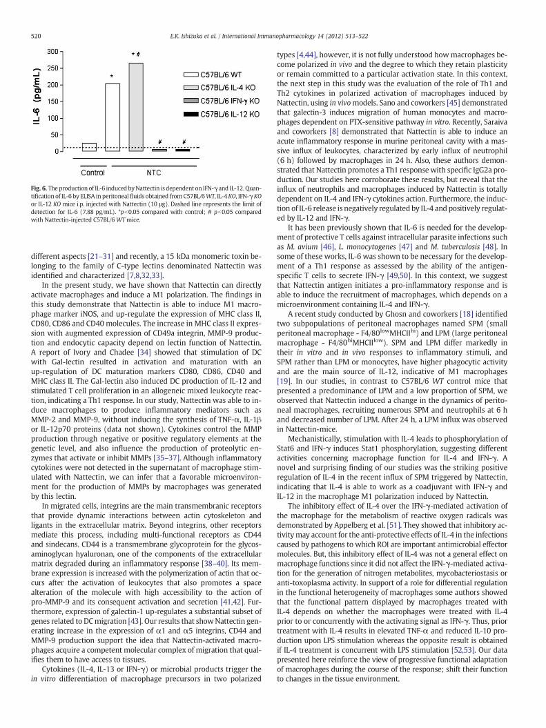
Fig. 6. The production of IL-6 induced byNattectin is dependent on IFN-γ and IL-12. Quan-tification of IL-6 by ELISA in peritoneal fluids obtained from C57BL/6WT, IL-4KO, IFN-γKOor IL-12 KO mice i.p. injected with Nattectin (10 μg). Dashed line represents the limit ofdetection for IL-6 (7.88 pg/mL). *pb0.05 compared with control; # pb0.05 comparedwith Nattectin-injected C57BL/6 WTmice.
520 E.K. Ishizuka et al. / International Immunopharmacology 14 (2012) 513–522
different aspects [21–31] and recently, a 15 kDa monomeric toxin be-longing to the family of C-type lectins denominated Nattectin wasidentified and characterized [7,8,32,33].
In the present study, we have shown that Nattectin can directlyactivate macrophages and induce a M1 polarization. The findings inthis study demonstrate that Nattectin is able to induce M1 macro-phage marker iNOS, and up-regulate the expression of MHC class II,CD80, CD86 and CD40 molecules. The increase in MHC class II expres-sion with augmented expression of CD49a integrin, MMP-9 produc-tion and endocytic capacity depend on lectin function of Nattectin.A report of Ivory and Chadee [34] showed that stimulation of DCwith Gal-lectin resulted in activation and maturation with anup-regulation of DC maturation markers CD80, CD86, CD40 andMHC class II. The Gal-lectin also induced DC production of IL-12 andstimulated T cell proliferation in an allogeneic mixed leukocyte reac-tion, indicating a Th1 response. In our study, Nattectin was able to in-duce macrophages to produce inflammatory mediators such asMMP-2 and MMP-9, without inducing the synthesis of TNF-α, IL-1βor IL-12p70 proteins (data not shown). Cytokines control the MMPproduction through negative or positive regulatory elements at thegenetic level, and also influence the production of proteolytic en-zymes that activate or inhibit MMPs [35–37]. Although inflammatorycytokines were not detected in the supernatant of macrophage stim-ulated with Nattectin, we can infer that a favorable microenviron-ment for the production of MMPs by macrophages was generatedby this lectin.
In migrated cells, integrins are the main transmembranic receptorsthat provide dynamic interactions between actin cytoskeleton andligants in the extracellular matrix. Beyond integrins, other receptorsmediate this process, including multi-functional receptors as CD44and sindecans. CD44 is a transmembrane glycoprotein for the glycos-aminoglycan hyaluronan, one of the components of the extracellularmatrix degraded during an inflammatory response [38–40]. Its mem-brane expression is increased with the polymerization of actin that oc-curs after the activation of leukocytes that also promotes a spacealteration of the molecule with high accessibility to the action ofpro-MMP-9 and its consequent activation and secretion [41,42]. Fur-thermore, expression of galectin-1 up-regulates a substantial subset ofgenes related to DCmigration [43]. Our results that showNattectin gen-erating increase in the expression of α1 and α5 integrins, CD44 andMMP-9 production support the idea that Nattectin-activated macro-phages acquire a competent molecular complex of migration that qual-ifies them to have access to tissues.
Cytokines (IL-4, IL-13 or IFN-γ) or microbial products trigger thein vitro differentiation of macrophage precursors in two polarized
types [4,44], however, it is not fully understood howmacrophages be-come polarized in vivo and the degree to which they retain plasticityor remain committed to a particular activation state. In this context,the next step in this study was the evaluation of the role of Th1 andTh2 cytokines in polarized activation of macrophages induced byNattectin, using in vivomodels. Sano and coworkers [45] demonstratedthat galectin-3 induces migration of human monocytes and macro-phages dependent on PTX-sensitive pathway in vitro. Recently, Saraivaand coworkers [8] demonstrated that Nattectin is able to induce anacute inflammatory response in murine peritoneal cavity with a mas-sive influx of leukocytes, characterized by early influx of neutrophil(6 h) followed by macrophages in 24 h. Also, these authors demon-strated that Nattectin promotes a Th1 responsewith specific IgG2a pro-duction. Our studies here corroborate these results, but reveal that theinflux of neutrophils and macrophages induced by Nattectin is totallydependent on IL-4 and IFN-γ cytokines action. Furthermore, the induc-tion of IL-6 release is negatively regulated by IL-4 and positively regulat-ed by IL-12 and IFN-γ.
It has been previously shown that IL-6 is needed for the develop-ment of protective T cells against intracellular parasite infections suchas M. avium [46], L. monocytogenes [47] and M. tuberculosis [48]. Insome of these works, IL-6 was shown to be necessary for the develop-ment of a Th1 response as assessed by the ability of the antigen-specific T cells to secrete IFN-γ [49,50]. In this context, we suggestthat Nattectin antigen initiates a pro-inflammatory response and isable to induce the recruitment of macrophages, which depends on amicroenvironment containing IL-4 and IFN-γ.
A recent study conducted by Ghosn and coworkers [18] identifiedtwo subpopulations of peritoneal macrophages named SPM (smallperitoneal macrophage - F4/80lowMHCIIhi) and LPM (large peritonealmacrophage - F4/80hiMHCIIlow). SPM and LPM differ markedly intheir in vitro and in vivo responses to inflammatory stimuli, andSPM rather than LPM or monocytes, have higher phagocytic activityand are the main source of IL-12, indicative of M1 macrophages[19]. In our studies, in contrast to C57BL/6 WT control mice thatpresented a predominance of LPM and a low proportion of SPM, weobserved that Nattectin induced a change in the dynamics of perito-neal macrophages, recruiting numerous SPM and neutrophils at 6 hand decreased number of LPM. After 24 h, a LPM influx was observedin Nattectin-mice.
Mechanistically, stimulation with IL-4 leads to phosphorylation ofStat6 and IFN-γ induces Stat1 phosphorylation, suggesting differentactivities concerning macrophage function for IL-4 and IFN-γ. Anovel and surprising finding of our studies was the striking positiveregulation of IL-4 in the recent influx of SPM triggered by Nattectin,indicating that IL-4 is able to work as a coadjuvant with IFN-γ andIL-12 in the macrophage M1 polarization induced by Nattectin.
The inhibitory effect of IL-4 over the IFN-γ-mediated activation ofthe macrophage for the metabolism of reactive oxygen radicals wasdemonstrated by Appelberg et al. [51]. They showed that inhibitory ac-tivitymay account for the anti-protective effects of IL-4 in the infectionscaused by pathogens to which ROI are important antimicrobial effectormolecules. But, this inhibitory effect of IL-4 was not a general effect onmacrophage functions since it did not affect the IFN-γ-mediated activa-tion for the generation of nitrogen metabolites, mycobacteriostasis oranti-toxoplasma activity. In support of a role for differential regulationin the functional heterogeneity of macrophages some authors showedthat the functional pattern displayed by macrophages treated withIL-4 depends on whether the macrophages were treated with IL-4prior to or concurrently with the activating signal as IFN-γ. Thus, priortreatment with IL-4 results in elevated TNF-α and reduced IL-10 pro-duction upon LPS stimulation whereas the opposite result is obtainedif IL-4 treatment is concurrent with LPS stimulation [52,53]. Our datapresented here reinforce the view of progressive functional adaptationof macrophages during the course of the response; shift their functionto changes in the tissue environment.
521E.K. Ishizuka et al. / International Immunopharmacology 14 (2012) 513–522
Together, the results obtained indicate that Nattectin is able toinduce maturation of BMDM that have increased expression of co-stimulatory and MHC class II molecules, increased endocytic capacityand greater ability to migrate through extracellular matrix. Moreover,the phenotypic changes in peritoneal and bonemarrow-derivedmacro-phages induced byNattectin are consistentwith the classicalM1 activa-tion (iNOS expression), dependent on Th1 cytokines (IL-12 and IFN-γ),and negatively regulated by Th2 cytokines (IL-4, IL-10 and IL-13). Final-ly, these findings presented here allow us to extend the M1/M2 dichot-omy, which is strongly influenced by the action of mutually inhibitorycytokines, since we have demonstrate that Nattectin generates amicro-environmentwhere the co-participation of IL-4 and IFN-γ is decisive forthe recruitment of SPM macrophages that are classically activated (invivo) and for the induction of up-regulation of MHC class II and CCR7in BMDM (in vitro). However, a negative regulatory role of IL-4 was ob-served in CD40 expression (in vitro) and IL-6 synthesis (in vivo). Thus,the results presented here allowed us to expand the knowledge aboutthe regulation of macrophage activation and polarization, as well asconfirmed the ability of Nattectin, a member of C-type lectin family, asan important immunomodulatory agent, thus opening the possibilityof using synthetic agents derived from secretions of marine animals asimmunomodulators, useful in vaccine development and for therapeuticapplication.
Acknowledgments
This work was supported by the Fundação de Amparo à Pesquisado Estado de São Paulo (FAPESP), and CNPq.
References
[1] Romagnani S. T-cell subsets (Th1 versus Th2). Ann Allergy Asthma Immunol2000;85:9–18 (quiz 18–21).
[2] Gordon S. Alternative activation of macrophages. Nat Rev Immunol 2003;3:23-35.[3] Martinez FO, Helming L, Gordon S. Alternative activation of macrophages: an
immunologic functional perspective. Annu Rev Immunol 2009;27:451-83.[4] Mosser DM, Edwards JP. Exploring the full spectrum of macrophage activation.
Nat Rev Immunol 2008;8:958-69.[5] Mantovani A, Sozzani S, Locati M, Allavena P, Sica A. Macrophage polarization:
tumor-associated macrophages as a paradigm for polarized M2 mononuclearphagocytes. Trends Immunol 2002;23:549-55.
[6] Vasta GR, Ahmed H, Du S. Henrikson, D. Galectins in teleost fish: Zebrafish (Daniorerio) as a model species to address their biological roles in development and in-nate immunity. Glycoconj J 2004;21:503-21.
[7] Lopes-FerreiraM,MagalhãesGS, Fernandez JH, Junqueira-de-Azevedo IL, Ho PL, LimaC, et al. Structural and biological characterization of Nattectin, a new C-type lectinfrom the venomous fish Thalassophryne nattereri. Biochimie 2011;93:971-80.
[8] Saraiva TC, Grund LZ, Komegae EN, Ramos AD, Conceição K, Orii NM, et al.Nattectin a fish C-type lectin drives Th1 responses in vivo: license macrophagesto differentiate into cells exhibiting typical DC function. Int Immunopharmacol2011;11:1546-56.
[9] Falk LA, Fortier AH. Isolation of murine macrophages. In: Coico R, editor. CurrentProtocols in Immunology, 3. New York: John Wiley & Sons; 1995. p. 14.
[10] Muller WA. Leukocyte - endothelial cell interactions in leukocyte transmigrationand the inflammatory response. Trends Immunol 2003;24:326-33.
[11] Bradford MM. A rapid and sensitive method for quantification of microgramquantities of protein utilizing the principle of protein dye binding. Anal Biochem1976;72:248-54.
[12] Sallusto F, Cella M, Danieli C, Lanzavecchia A. Dendritic cells use macropinocytosisand the mannose receptor to concentrate macromolecules in the major histocom-patibility complex class II compartment: downregulation by cytokines and bacte-rial products. J Exp Med 1995;182:389-400.
[13] Huveneers S, Truong H, Fassler R, Sonnenberg A, Danen EH. Binding of solublefibronectin to integrin alpha5 beta1 - link to focal adhesion redistribution andcontractile shape. J Cell Sci 2008;121:2452-62.
[14] Carson AE, Barker TH. Emerging concepts in engineering extracellular matrixvariants for directing cell phenotype. Regen Med 2009;4:593-600.
[15] Booms P, Pregla R, Ney A, Barthel F, Reinhardt DP, Pletschacher A, et al.RGD-containing fibrillin-1 fragments upregulate matrix metalloproteinase ex-pression in cell culture: a potential factor in the pathogenesis of the Marfan syn-drome. Hum Genet 2005;116:51-61.
[16] Aderem A, Underhill DM. Mechanisms of phagocytosis in macrophages. Annu RevImmunol 1999;17:593-623.
[17] Lesley J, Hyman R, Kincade PW. CD44 and its interaction with extracellular matrix.Adv Immunol 1993;54:271-335.
[18] Ghosn EE, Cassado AA, Govoni GR, Fukuhara T, Yang Y, Monack DM, et al. Twophysically, functionally, and developmentally distinct peritoneal macrophagesubsets. Proc Natl Acad Sci U S A 2010;107:2568-73.
[19] Cassado AA, de Albuquerque JA, Sardinha LR, Buzzo Cde L, Faustino L, NascimentoR, et al. Cellular renewal and improvement of local cell effector activity in perito-neal cavity in response to infectious stimuli. PLoS One 2011;6:e22141.
[20] Doyle RJ. Introduction of lectins and their interactions with microorganisms. In:Doyle RJ, Slifkin M, editors. Lectin–microorganism interactions. New York: MarcelDekker; 1994. p. 1–66.
[21] Lopes-Ferreira M, Barbaro KC, Cardoso DF, Moura-Da-Silva AM, Mota I.Thalassophryne nattereri fish venom: biological and biochemical characterizationand serum neutralization of its toxic activities. Toxicon 1998;36:405-10.
[22] Lopes-Ferreira M, Moura-Da-Silva AM, Mota I, Takehara HA. Neutralization ofThalassophryne nattereri (niquim) fish venom by an experimental antivenom.Toxicon 2000;38:1149-56.
[23] Fonseca LA, Lopes-Ferreira M. Clinical and experimental studies regarding poison-ing caused by a fish Thalassophryne nattereri (niquim). An Bras Dermatol 2000;75:435-43.
[24] Lopes-Ferreira M, Nunez J, Rucavado A, Farsky SHP, Lomonte B, Angulo Y, et al. Skel-etal muscle necrosis and regeneration after injection of Thalassophryne nattereri(niquim) fish venom in mice. J Exp Pathol 2001;82:55-64.
[25] Lopes-FerreiraM,Moura-Da-Silva AM, Piran-Soares AA, Angulo Y, Lomonte B, GutierrezJM, et al. Hemostatic effects induced by Thalassophryne nattereri fish venom: amodel ofendothelium-mediated blood flow impairment. Toxicon 2002;40:1141-7.
[26] Lima C, Bianca CP, Piran-Soares A, Tanjoni I, Moura-Da-Silva AM, Lopes-FerreiraM. Characterizations of local inflammatory response induced by Thalassophrynenattereri fish venom in a mouse model of tissue injury. Toxicon 2003;42:499-507.
[27] Lopes-Ferreira M, Emim JA, Oliveira V, Puzer L, Cezari MH, Araujo Mda S, et al.Kininogenase activity of Thalassophryne nattereri fish venom. Biochem Pharmacol2004;68:2151-7.
[28] Magalhães GS, Lopes-Ferreira M, Junqueira-De-Azevedo ILM, Spencer PJ, AraújoMS, Portaro FCV, et al. Natterins, a new class of proteins with kininogenase activ-ity characterized from Thalassophryne nattereri fish venom. Biochimie 2005;87:687-99.
[29] Grund LZ, Souza VM, Faquim-Mauro EL, Lima C, Lopes-Ferreira M. Experimentalimmunization with Thalassophryne nattereri fish venom: striking IL-5 productionand impaired of B220+ cells. Toxicon 2006;48:499-508.
[30] Piran-Soares AA, Komegae EN, Souza VM, Fonseca LA, Lima C, Lopes-Ferreira M.Neutralizing antibodies obtained in a persistent immune response are effectiveagainst deleterious effects induced by the Thalassophryne nattereri fish venom.Toxicon 2007;49:920-30.
[31] Pareja-Santos A, Saraiva TC, Costa EP, Santos MF, Zorn TT, Souza VMO, et al. De-layed local inflammatory response induced by Thalassophryne nattereri venom isrelated to extracellular matrix degradation. Int J Exp Pathol 2009;90:34-43.
[32] Magalhães GS, Junqueira-De-Azevedo ILM, Lopes-Ferreira M, Lorenzini DM, Ho PL,Moura-Da-Silva AM. Transcriptome analysis of expressed sequence tags from thevenom glands of the fish Thalassophryne nattereri. Biochimie 2006;88:693-9.
[33] Komegae EN, Ramos AD, Oliveira AK, Serrano SM, Lopes-Ferreira M, Lima C.Insights into the local pathogenesis induced by fish toxins: role of natterins andnattectin in the disruption of cell-cell and cell-extracellular matrix interactionsand modulation of cell migration. Toxicon 2011;58:509-17.
[34] Ivory C, Chadee K. Activation of dendritic cells by the Gal-lectin of Entamoebahistolytica drives Th1 responses in vitro and in vivo. Eur J Immunol 2007;37:385-94.
[35] Woessner Jr JF. Matrix metalloproteinases and their inhibitors in connective tis-sue remodeling. FASEB J 1991;5:2145-54.
[36] Galboiz Y, Shapiro S, Lahat N, Miller A. Modulation of monocytes matrixmetalloproteinase-2, MT1-MMP and TIMP-2 by interferon-gamma and -beta:implications to multiple sclerosis. J Neuroimmunol 2002;131:191-200.
[37] Ries C, Petrides PE. Cytokine regulation of matrix metalloproteinase activity andits regulatory dysfunction in disease. Biol Chem Hoppe Seyler 1995;376:345-55.
[38] Iczkowski KA. Cell adhesion molecule CD44: its functional roles in prostatecancer. Am J Transl Res 2010;3:1-7.
[39] Goodison S, Urquidi V, Tarin D. CD44 cell adhesion molecules. Mol Pathol1999;52:189-96.
[40] Rangaswami H, Bulbule A, Kundu GC. Osteopontin: role in cell signaling andcancer progression. Trends Cell Biol 2006;16:79-87.
[41] Samanna V, Ma T, Mak TW, Rogers M, Chellaiah MA. Actin polymerization modu-lates CD44 surface expression, MMP-9 activation, and osteoclast function. J CellPhysiol 2007;213:710-20.
[42] Redondo-Muñoz J, Ugarte-Berzal E, García-Marco JA, Del Cerro MH, Van DenSteen PE, Opdenakker G, et al. Alpha4beta1 integrin and 190-kDa CD44v consti-tute a cell surface docking complex for gelatinase B/MMP-9 in chronic leukemicbut not in normal B cells. Blood 2008;112:169-78.
[43] Fulcher JA, Hashimi ST, Levroney EL, Pang M, Gurney KB, Baum LG, et al.Galectin-1-matured human monocyte-derived dendritic cells have enhancedmigration through extracellular matrix. J Immunol 2006;177:216-26.
[44] Stout RD, Jiang C, Matta B, Tietzel I, Watkins SK, Suttles J. Macrophages sequentiallychange their functional phenotype in response to changes in microenvironmentalinfluences. J Immunol 2005;175:342-9.
[45] Sano H, Hsu DK, Yu L, Apgar JR, Kuwabara I, Yamanaka T, et al. Human galectin-3is a novel chemoattractant for monocytes andmacrophages. J Immunol 2000;165:2156-64.
[46] Appelberg R, Castro AG, Pedrosa J, Minoprio P. Role of interleukin-6 in the induc-tion of protective T cells during mycobacterial infections in mice. Immunology1994;82:361-4.

522 E.K. Ishizuka et al. / International Immunopharmacology 14 (2012) 513–522
[47] Liu Z, Simpson RJ, Cheers C. Role of IL-6 in activation of T cells for acquired cellularresistance to Listeria monocytogenes. J Immunol 1994;152:5375-80.
[48] Ladel CH Blum C, Dreher A, Reifenberg K, Kopf M, Kaufmann SH. Lethal tuberculosisin interleukin-6-deficient mutant mice. Infect Immun 1997;65:4843-9.
[49] Saunders BM, FrankAA, Orme IM, CooperAM. Interleukin-6 induces early gamma in-terferon production in the infected lung but is not required for generation of specificimmunity toMycobacterium tuberculosis infection. Infect Immun 2000;68:3322-6.
[50] Liu Z, Simpson RJ, Cheers C. Role of interleukin-6 in T-cell activation during primaryand secondary infectionwith Listeriamonocytogenes. Infect Immun 1995;63:2790-2.
[51] Appelberg R, Orme IM, Pinto de SousaMI, SilvaMT. In vitro effects of interleukin-4 oninterferon-gamma-induced macrophage activation. Immunology 1992;76:553-9.
[52] Kambayashi T, Jacob CO, Strassmann G. IL-4 and IL-13 modulate IL-10 release inendotoxin-stimulated murine peritoneal mononuclear phagocytes. Cell Immunol1996;171:153-8.
[53] D'Andrea A, Ma X, Aste Amezaga M, Paganin C, Trinchieri G. Stimulatory and in-hibitory effects of interleukin (IL)-4 and IL-13 on the production of cytokines byhuman peripheral blood mononuclear cells: priming for IL-12 and tumor necrosisfactor alpha production. J Exp Med 1995;181:537-46.
![Mind-Body Skills for Regulating the Autonomic Nervous System[1]](https://static.fdocument.org/doc/165x107/55d1650cbb61eb417d8b47ed/mind-body-skills-for-regulating-the-autonomic-nervous-system1.jpg)