REVIEW Open Access NF-κB pathway activators as potential ...
16
REVIEW Open Access NF-κB pathway activators as potential ageing biomarkers: targets for new therapeutic strategies Carmela R Balistreri * , Giuseppina Candore, Giulia Accardi, Giuseppina Colonna-Romano and Domenico Lio Abstract Chronic inflammation is a major biological mechanism underpinning biological ageing process and age-related diseases. Inflammation is also the key response of host defense against pathogens and tissue injury. Current opinion sustains that during evolution the host defense and ageing process have become linked together. Thus, the large array of defense factors and mechanisms linked to the NF-κB system seem to be involved in ageing process. This concept leads us in proposing inductors of NF-κB signaling pathway as potential ageing biomarkers. On the other hand, ageing biomarkers, represented by biological indicators and selected through apposite criteria, should help to characterize biological age and, since age is a major risk factor in many degenerative diseases, could be subsequently used to identify individuals at high risk of developing age-associated diseases or disabilities. In this report, some inflammatory biomarkers will be discussed for a better understanding of the concept of biological ageing, providing ideas on eventual working hypothesis about potential targets for the development of new therapeutic strategies and improving, as consequence, the quality of life of elderly population. Keywords: Biological ageing process, Inflammatory network and its effects in ageing, NF-κB signaling pathway as hub of inflammatory ageing network, Inflammatory biomarkers Introduction Ageing is a complex process, induced by an intricate interaction of genetic, epigenetic, stochastic and envir- onmental factors. They determine the lost of molecular fidelity followed by an improved entropy [1,2]. As result, loss complexity and random accumulation of damages (i.e. particularly damages to nuclear and mitochondrial DNA) at cellular, tissue, organ levels and/or of whole body arise, compatibly with the disposable soma theory of ageing [3]. Thus, it establishes a condition, which modifies both architecture and functioning of physiological processes and regulatory (immune and endocrine) systems. This de- termines a deterioration of the homeostasis. Accordingly, it becomes more easily vulnerable to internal and external stressors, frailty, disability and disease (Figure 1). On the other hand, the loss of DNA integrity, the principal ran- dom damages able in modifying cellular fidelity and indu- cing cellular and whole body senescence, determines the decline of the functionality of stress resistance and survival pathways (i.e. autophagic uptake mechanisms, chaperone systems, DNA repair mechanisms, apoptotic process, immune/inflammatory response), involved in cellular and organism defense to environmental stress and maintaining homeostasis [2]. However, a large heterogeneity in occur- rence, complications, speed, and age and gender manifest- ation of ageing process at cellular, tissue, organ levels and/ or of whole body has been observed in humans. Among human people, there are individuals at the age ≥90 years still in good mental and physical conditions, and others that at the age ≥ 60 years show cognitive difficulties, and/ or the onset of chronic inflammatory diseases, such as Alzheimer’ s disease (AD), cardiovascular disease (CVD) and type 2 diabetes mellitus (T2DM) and cancer [4]. The principal causes of the heterogeneity in human ageing rate, measured as decline of functional capacity and stress resistance, seem to be genetic and environ- mental factors. However, the overall impression is that environmental factors are the major determinants of both ageing and age-related diseases [4,5]. Thus, ageing process is not a genetically programmed process [5]. This consideration is based on studies on heritability of age-related diseases and ageing [6,7]. A similar value of heritability in lifespan and age-related diseases, such as * Correspondence: [email protected] Department of Pathobiology and Medical and Forensic Biotechnologies, University of Palermo, Corso Tukory 211, Palermo 90134, Italy IMMUNITY & AGEING © 2013 Balistreri et al.; licensee BioMed Central Ltd. This is an Open Access article distributed under the terms of the Creative Commons Attribution License (http://creativecommons.org/licenses/by/2.0), which permits unrestricted use, distribution, and reproduction in any medium, provided the original work is properly cited. Balistreri et al. Immunity & Ageing 2013, 10:24 http://www.immunityageing.com/content/10/1/24
Transcript of REVIEW Open Access NF-κB pathway activators as potential ...
IMMUNITY & AGEINGBalistreri et al. Immunity & Ageing 2013, 10:24http://www.immunityageing.com/content/10/1/24
REVIEW Open Access
NF-κB pathway activators as potential ageingbiomarkers: targets for new therapeutic strategiesCarmela R Balistreri*, Giuseppina Candore, Giulia Accardi, Giuseppina Colonna-Romano and Domenico Lio
Abstract
Chronic inflammation is a major biological mechanism underpinning biological ageing process and age-relateddiseases. Inflammation is also the key response of host defense against pathogens and tissue injury. Current opinionsustains that during evolution the host defense and ageing process have become linked together. Thus, the largearray of defense factors and mechanisms linked to the NF-κB system seem to be involved in ageing process. Thisconcept leads us in proposing inductors of NF-κB signaling pathway as potential ageing biomarkers. On the otherhand, ageing biomarkers, represented by biological indicators and selected through apposite criteria, should help tocharacterize biological age and, since age is a major risk factor in many degenerative diseases, could besubsequently used to identify individuals at high risk of developing age-associated diseases or disabilities. In thisreport, some inflammatory biomarkers will be discussed for a better understanding of the concept of biologicalageing, providing ideas on eventual working hypothesis about potential targets for the development of newtherapeutic strategies and improving, as consequence, the quality of life of elderly population.
Keywords: Biological ageing process, Inflammatory network and its effects in ageing, NF-κB signaling pathway ashub of inflammatory ageing network, Inflammatory biomarkers
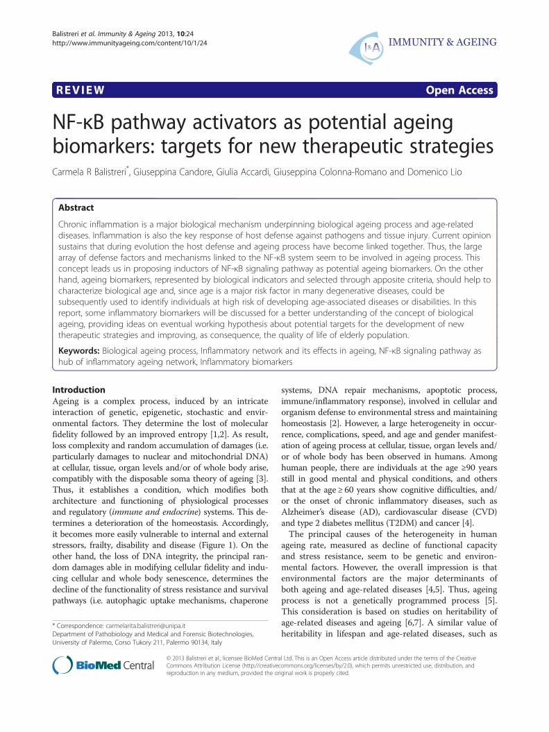
IntroductionAgeing is a complex process, induced by an intricateinteraction of genetic, epigenetic, stochastic and envir-onmental factors. They determine the lost of molecularfidelity followed by an improved entropy [1,2]. As result,loss complexity and random accumulation of damages (i.e.particularly damages to nuclear and mitochondrial DNA)at cellular, tissue, organ levels and/or of whole body arise,compatibly with the disposable soma theory of ageing [3].Thus, it establishes a condition, which modifies botharchitecture and functioning of physiological processesand regulatory (immune and endocrine) systems. This de-termines a deterioration of the homeostasis. Accordingly,it becomes more easily vulnerable to internal and externalstressors, frailty, disability and disease (Figure 1). On theother hand, the loss of DNA integrity, the principal ran-dom damages able in modifying cellular fidelity and indu-cing cellular and whole body senescence, determines thedecline of the functionality of stress resistance and survivalpathways (i.e. autophagic uptake mechanisms, chaperone
* Correspondence: [email protected] of Pathobiology and Medical and Forensic Biotechnologies,University of Palermo, Corso Tukory 211, Palermo 90134, Italy
© 2013 Balistreri et al.; licensee BioMed CentraCommons Attribution License (http://creativecreproduction in any medium, provided the or
systems, DNA repair mechanisms, apoptotic process,immune/inflammatory response), involved in cellular andorganism defense to environmental stress and maintaininghomeostasis [2]. However, a large heterogeneity in occur-rence, complications, speed, and age and gender manifest-ation of ageing process at cellular, tissue, organ levels and/or of whole body has been observed in humans. Amonghuman people, there are individuals at the age ≥90 yearsstill in good mental and physical conditions, and othersthat at the age ≥ 60 years show cognitive difficulties, and/or the onset of chronic inflammatory diseases, such asAlzheimer’s disease (AD), cardiovascular disease (CVD)and type 2 diabetes mellitus (T2DM) and cancer [4].The principal causes of the heterogeneity in human
ageing rate, measured as decline of functional capacityand stress resistance, seem to be genetic and environ-mental factors. However, the overall impression is thatenvironmental factors are the major determinants ofboth ageing and age-related diseases [4,5]. Thus, ageingprocess is not a genetically programmed process [5].This consideration is based on studies on heritability ofage-related diseases and ageing [6,7]. A similar value ofheritability in lifespan and age-related diseases, such as
l Ltd. This is an Open Access article distributed under the terms of the Creativeommons.org/licenses/by/2.0), which permits unrestricted use, distribution, andiginal work is properly cited.
Figure 1 Ageing process results by the lost of molecular fidelity followed by an improved entropy. This determines loss complexity andrandom accumulation of damages (i.e. particularly damages to nuclear and mitochondrial DNA) at cellular, tissue, organ levels and/or of wholebody. Thus, it becomes more easily vulnerable to internal and external stressors, frailty, disability and disease.
Balistreri et al. Immunity & Ageing 2013, 10:24 Page 2 of 16http://www.immunityageing.com/content/10/1/24
cancer, AD, CVD and T2DM, has been identified (35%vs. 40%, respectively) [8,9]. However, this does not implythat genetic factors have an irrelevant role in ageing andage-related diseases. For example, mutations identifiedin familial forms of AD consented understanding its mo-lecular mechanisms, such as the toxicity of amyloid βpeptide and potential therapeutic targets in more com-mon sporadic late onset AD [10]. Common suggestion isbased on both a complex contribution of genetic factorsin ageing and diseases of later life and weak effects of in-dividual genes [5]. Furthermore, diverse genetic factorsare associated with ageing and exceptional longevity.Human genome-wide genetic analyses have revealedonly few age-related loci and polymorphic longevitygenes [11-13]. Among these, current promising candi-dates are Sirtuins, Forkhead box O protein (FoxOs) andthe field of epigenetics. Functional genomics, i.e. expres-sion profiling studies, have revealed a group of geneswhich are differently expressed in ageing, such asimmune/inflammatory genes [14].From the observations described above, another crit-
ical point of ageing process emerges based on the con-cept of biological age as real expression in human ofboth ageing rate and onset of the common diseases oflater life rather than chronological age [15]. This conceptopened an important area of research focused on identi-fying of potential molecular targets as biomarkers of
human biological ageing [16]. On the one hand, it couldconsent to develop potential anti-ageing treatment strat-egies. On the other hand, probable anti-ageing treat-ments could retard or prevent age-associated diseasesresulting in widespread health, social and economicbenefit. Such treatment could include genetic engineer-ing, such as gene therapy or endogenous gene repair, orpharmacological therapies, or changes in lifestyle, i.e.physical activity, diet.In this report, many of these aspects are discussed,
giving particular emphasis in describing some bio-markers of inflammation. In particular, the data dis-cussed in this report are based on an expert opinionderived on the findings from author’s studies on ageing,age-related diseases and inflammation.
Definition and selection criteria of ageing biomarkersAs established by National Institute of Health, a bio-marker is a “feature objectively measured and evaluatedas an indicator of normal biologic processes, pathogenicprocesses, or pharmacologic responses to a therapeuticintervention” [16,17].In the case of ageing process, this definition might
concern measures related to physical changes, such asgrey hairs, reduced skin elasticity, wrinkles, reducedmuscle strength or changes in the near vision, which arethought to be the result of molecular mechanisms
Balistreri et al. Immunity & Ageing 2013, 10:24 Page 3 of 16http://www.immunityageing.com/content/10/1/24
occurring in the old age [18]. However, they reflect thechronological age rather than the biological age, whichis the most important indicator of health and potentiallifespan [15].A biomarker of real ageing should reflect a process of
biological ageing, be easily reproducible in cross-speciescomparison, be easily obtainable. Since ageing is the re-sult of the deterioration of more than one system orprocess, it would be more appropriate to consider differ-ent biomarkers. Panels of biomarkers associated withconditions, alterations or changes of a set of critical sys-tems to assess the biological age of any organism shouldbe used [16,18].The gerontologists have begun to face this problem
already in the early 1980s, with the development of alarge number of ageing biomarkers [16,18]. Despite thenumerous efforts and the support in this research fromNational Institute of Ageing, the major number of bio-markers is still to date under discussion, like inflamma-tory markers, hormones, markers of oxidative stress ortelomere shortening [16,18]. Most (perhaps all) markersare also not really proven in longitudinal studies inhumans. In addition, they have been considered for avariety of purposes, which are not distinguished suffi-ciently. Most studies used biomarkers as tools for com-paring ageing rate in several populations or cohorts of asingle population. In contrast, others considered bio-markers for person-specific predisposition, which repre-sents much more challenging, principally becauseageing, as a biological process, is not well defined at in-dividual level. In addition, the research of comparativeor predictive biomarkers have determined the attempteduse of measure panels associated with survival, healthof old age, frailty, age-related (multi) morbidity andmortality [16,18].However, none of the identified biomarkers is a “real”
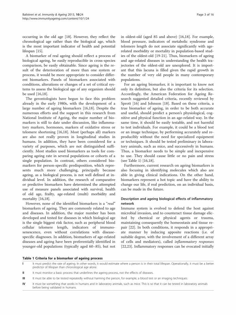
biomarkers of ageing. They are commonly related to ageand diseases. In addition, the major number has beendeveloped and tested for diseases in which biological ageis the single biggest risk factor, such as peripheral bloodcellular telomere length, indicators of immuno-senescence, even without correlations with disease-specific diagnoses. In addition, biomarkers of age-relateddiseases and ageing have been preferentially identified inyounger-old populations (typically aged 60–85), but not
Table 1 Criteria for a biomarker of ageing process
I It must predict the rate of ageing. In other words, it would estimatepredictor of lifespan than chronological age alone.
II It must monitor a basic process that underlines the ageing process, n
III It must be able to be tested repeatedly without harming the person
IV It must be something that works in humans and in laboratory animabefore being validated in humans.
in oldest-old (aged 85 and above) [16,18]. For example,blood pressure, indicators of metabolic syndrome andtelomere length do not associate significantly with age-related morbidity or mortality in population-based stud-ies of the oldest-old [19-21]. Thus, biomarkers of ageingand age-related diseases in understanding the health tra-jectories of the oldest-old are unexplored. It is import-ant that this lacuna is filled given the rapid growth inthe number of very old people in many contemporarypopulations.For an ageing biomarker, it is important to know not
only its definition, but also the criteria for its selection.Accordingly, the American Federation for Ageing Re-search suggested detailed criteria, recently reviewed bySprott [16] and Johnson [18]. Based on these criteria, atrue biomarker of ageing, in order to be both accurateand useful, should predict a person’s physiological, cog-nitive and physical function in an age-related way. In thesame time, it should be easily testable, and not harmfulto test individuals. For example, it could be a blood testor an image technique, by performing accurately and re-producibly without the need for specialized equipmentor techniques. It should be tested preliminary in labora-tory animals, such as mice, and successively in humans.Thus, a biomarker needs to be simple and inexpensiveto use. They should cause little or no pain and stress(see Table 1) [16,18].Furthermore, current research on ageing biomarkers is
also focusing in identifying molecules which also areable in giving clinical indications. On the other hand,biomarkers represent a hot topic and have the ability tochange our life, if real prediction, on an individual basis,can be made in the future.
Description and ageing biological effects of inflammatorynetworkImmune system is evolved to defend the host againstmicrobial invasion, and to counteract tissue damage elic-ited by chemical or physical agents or trauma,maintaining consequently the homeostasis and tissue re-pair [22]. In both conditions, it responds in a appropri-ate manner by inducing apposite reactions (i.e. ofsuitable degree, with the involvement of a different arrayof cells and mediators), called inflammatory responses[22,23]. Inflammatory responses can be evocated initially
where a person is in their total lifespan. Operationally, it must be a better
ot the effects of diseases.
, for example, a blood test or an imaging techniques.
ls, such as mice. This is so that it can be tested in laboratory animals

Balistreri et al. Immunity & Ageing 2013, 10:24 Page 4 of 16http://www.immunityageing.com/content/10/1/24
as localized tissue reactions and subsequently as acutephase reaction, represented by systemic cytokine-inducedreactions, including leukocytosis, fever, somnolence, an-orexia, activation of hypothalamic-pituitary-adrenal axisand increased level of glucocorticoids, and acute phasesynthesis, i.e. C reactive protein (CRP), in the liver. Acomplex network of molecules (the mediators) and cells(neutrophils, monocytes, mast cells, endothelial cells, etc.)characterize these reactions. They work together in con-cert and interact mediating the activation of different sig-naling pathways and the expression and transcriptionalregulation of hub genes. The hub genes receive and directthe activity of many other genes [22,23]. Thus, these re-sponses are induced through an inflammatory network.Recent studies on topology of this network evidence thecrucial role of some mediators in driving the different cel-lular interactions and regulating the type of inflammatoryreaction. Several mediators as pro- and anti-inflammatorymolecules are involved [24]. Their release is modulated bydifferent factors linked to well-known Nuclear Factor(NF)-κB pathway [25,26]. In addition, the magnitude oftheir production varies individually because of genetic het-erogeneity. Single nucleotide polymorphisms (SNPs) inseveral genes and epigenetic factors seem to be involved[27]. Among the inflammatory mediators, the classicalpro-inflammatory cytokines, Tumor necrosis factor α(TNF-α), Interleukin-1 (IL-1) and IL-6, play a key role.They are able in inducing both local and systemic effects[28]. When the causes of the inflammatory reaction are ofa high intensity, their production is increased. Thus, theyare released in the circulation provoking the acute phaseresponse. In contrast, the anti-inflammatory cytokines,such as IL-10 are able to regulate the activation of in-flammatory cells, by inhibiting the release of pro-inflammatory cytokines and therefore turning off theinflammatory processes [29].Whether tissue health is not restored or in response to
stable low grade irritation, inflammation becomes achronic condition provoking continuous damages in thesurrounding tissues. The collateral damage caused bythis type of inflammation usually accumulates slowly,sometimes asymptomatically for years but can eventuallylead to severe tissue deterioration [30].From the above, it emerges that the inflammatory re-
sponse is not per se a negative phenomenon. It is pro-grammed by the evolution in neutralizing infectiousagents, playing a beneficial role until the time ofreproduction and parental care. In contrast, in old age, ina period largely not foreseen by evolution, it can deter-mine a detrimental effect through chronic inflammatoryresponses (“antagonistic pleiotropy”) in several/ all tissueand organs, which are cause of both the ageing phenotypeand chronic diseases [24,30]. A low chronic grade of in-flammation, the “inflammageing”, characterized by a 2 to
4-fold increase in serum levels of inflammatory mediatorshas been identified in ageing [31]. It seems to be as opti-mal predictor of mortality and, as mentioned above, a crit-ical risk factor in the pathogenesis of several age-relatedchronic diseases as AD, CVD, T2DM, sarcopenia, frailtyand functional disability [32].Augment of age-related body fat and consequent in-
crease of visceral adiposity, age-related decline of sexhormones, oxidative and genotoxic stress, cellular andtissue damage, nutrition, alterations of physical condi-tion of gut microbiota, other organs (brain, liver) andsystems (immune and endocrine) have been associatedwith inflamm-ageing [32-34]. In addition, factors linkingto physiological stress, such a long-term smoking anddepression, seem also to contribute to inflammageing[32-34]. However, the most important factor for age-related inflammation is the long-life pathogen burden[30]. Some recent studies have, indeed, evidenced associ-ations between past infections and levels of chronic in-flammation and increased risk of heart attack, stroke,and cancer [32,34]. For instance, persistent peripheralmultibacteria infection, such as periodontitis, associatedwith gram-negative anaerobic bacteria capable ofexhibiting localized and systemic infections in the host,is considered as possible aggravating cofactor in subjectswith vascular diseases and risk factor for the onset ofother age-related diseases, such as AD [30].Of special relevance is the inflammation status in cen-
tenarian people. The literature data seem to be apparentlycontradictory. Increased levels of both inflammatory andanti-inflammatory mediators and significant frequenciesof protective genotypes have been assessed in centenariansthan the old subjects [30,35]. As consequence, identifyingof apposite biomarkers likely in long lived subjects shouldbe necessary. This might permit a preferential and se-lected development of pleiotropic therapeutic interven-tions acting concomitantly on different targets and atdifferent levels.
Inflammatory ageing biomarkers: the crucial role of NF-κBactivatorsAgeing is not a genetically programmed process, as de-scribed above [5]. In contrast, it is recognized as an en-tropic process, characterized by loss of molecular fidelityand subsequent accumulation of different products [1,2].In addition, it has been recently proposed that duringevolution the host defense and the ageing process havebecome linked together [2]. Host defense and ageingmechanisms seem to be overlapping. In particular, hostdefenses seem to be involved in ageing process, to activeinflammatory network and also to evocate the release ofso-called senescence associated secretory phenotype(SASP), represented by a myriad of factors, such as thepro-inflammatory mediators [36,37]. A large range of
Balistreri et al. Immunity & Ageing 2013, 10:24 Page 5 of 16http://www.immunityageing.com/content/10/1/24
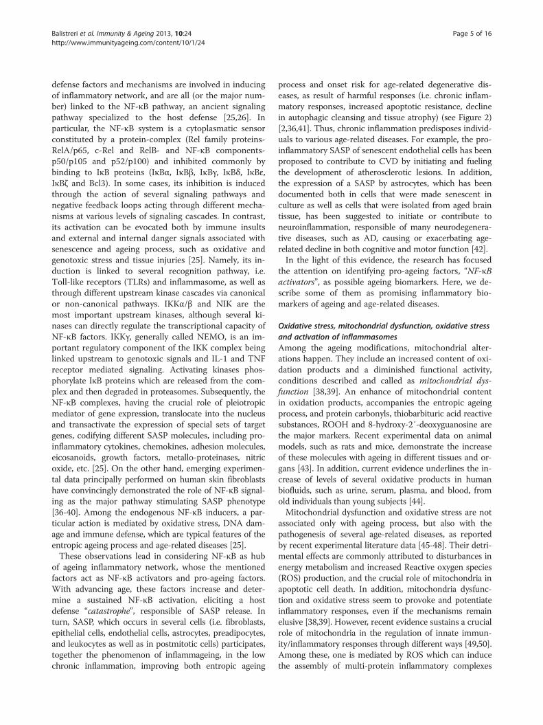
defense factors and mechanisms are involved in inducingof inflammatory network, and are all (or the major num-ber) linked to the NF-κB pathway, an ancient signalingpathway specialized to the host defense [25,26]. Inparticular, the NF-κB system is a cytoplasmatic sensorconstituted by a protein-complex (Rel family proteins-RelA/p65, c-Rel and RelB- and NF-κB components-p50/p105 and p52/p100) and inhibited commonly bybinding to IκB proteins (IκBα, IκBβ, IκBγ, IκBδ, IκBε,IκBζ and Bcl3). In some cases, its inhibition is inducedthrough the action of several signaling pathways andnegative feedback loops acting through different mecha-nisms at various levels of signaling cascades. In contrast,its activation can be evocated both by immune insultsand external and internal danger signals associated withsenescence and ageing process, such as oxidative andgenotoxic stress and tissue injuries [25]. Namely, its in-duction is linked to several recognition pathway, i.e.Toll-like receptors (TLRs) and inflammasome, as well asthrough different upstream kinase cascades via canonicalor non-canonical pathways. IKKα/β and NIK are themost important upstream kinases, although several ki-nases can directly regulate the transcriptional capacity ofNF-κB factors. IKKγ, generally called NEMO, is an im-portant regulatory component of the IKK complex beinglinked upstream to genotoxic signals and IL-1 and TNFreceptor mediated signaling. Activating kinases phos-phorylate IκB proteins which are released from the com-plex and then degraded in proteasomes. Subsequently, theNF-κB complexes, having the crucial role of pleiotropicmediator of gene expression, translocate into the nucleusand transactivate the expression of special sets of targetgenes, codifying different SASP molecules, including pro-inflammatory cytokines, chemokines, adhesion molecules,eicosanoids, growth factors, metallo-proteinases, nitricoxide, etc. [25]. On the other hand, emerging experimen-tal data principally performed on human skin fibroblastshave convincingly demonstrated the role of NF-κB signal-ing as the major pathway stimulating SASP phenotype[36-40]. Among the endogenous NF-κB inducers, a par-ticular action is mediated by oxidative stress, DNA dam-age and immune defense, which are typical features of theentropic ageing process and age-related diseases [25].These observations lead in considering NF-κB as hub
of ageing inflammatory network, whose the mentionedfactors act as NF-κB activators and pro-ageing factors.With advancing age, these factors increase and deter-mine a sustained NF-κB activation, eliciting a hostdefense “catastrophe”, responsible of SASP release. Inturn, SASP, which occurs in several cells (i.e. fibroblasts,epithelial cells, endothelial cells, astrocytes, preadipocytes,and leukocytes as well as in postmitotic cells) participates,together the phenomenon of inflammageing, in the lowchronic inflammation, improving both entropic ageing
process and onset risk for age-related degenerative dis-eases, as result of harmful responses (i.e. chronic inflam-matory responses, increased apoptotic resistance, declinein autophagic cleansing and tissue atrophy) (see Figure 2)[2,36,41]. Thus, chronic inflammation predisposes individ-uals to various age-related diseases. For example, the pro-inflammatory SASP of senescent endothelial cells has beenproposed to contribute to CVD by initiating and fuelingthe development of atherosclerotic lesions. In addition,the expression of a SASP by astrocytes, which has beendocumented both in cells that were made senescent inculture as well as cells that were isolated from aged braintissue, has been suggested to initiate or contribute toneuroinflammation, responsible of many neurodegenera-tive diseases, such as AD, causing or exacerbating age-related decline in both cognitive and motor function [42].In the light of this evidence, the research has focused
the attention on identifying pro-ageing factors, “NF-κBactivators”, as possible ageing biomarkers. Here, we de-scribe some of them as promising inflammatory bio-markers of ageing and age-related diseases.
Oxidative stress, mitochondrial dysfunction, oxidative stressand activation of inflammasomesAmong the ageing modifications, mitochondrial alter-ations happen. They include an increased content of oxi-dation products and a diminished functional activity,conditions described and called as mitochondrial dys-function [38,39]. An enhance of mitochondrial contentin oxidation products, accompanies the entropic ageingprocess, and protein carbonyls, thiobarbituric acid reactivesubstances, ROOH and 8-hydroxy-2′-deoxyguanosine arethe major markers. Recent experimental data on animalmodels, such as rats and mice, demonstrate the increaseof these molecules with ageing in different tissues and or-gans [43]. In addition, current evidence underlines the in-crease of levels of several oxidative products in humanbiofluids, such as urine, serum, plasma, and blood, fromold individuals than young subjects [44].Mitochondrial dysfunction and oxidative stress are not
associated only with ageing process, but also with thepathogenesis of several age-related diseases, as reportedby recent experimental literature data [45-48]. Their detri-mental effects are commonly attributed to disturbances inenergy metabolism and increased Reactive oxygen species(ROS) production, and the crucial role of mitochondria inapoptotic cell death. In addition, mitochondria dysfunc-tion and oxidative stress seem to provoke and potentiateinflammatory responses, even if the mechanisms remainelusive [38,39]. However, recent evidence sustains a crucialrole of mitochondria in the regulation of innate immun-ity/inflammatory responses through different ways [49,50].Among these, one is mediated by ROS which can inducethe assembly of multi-protein inflammatory complexes
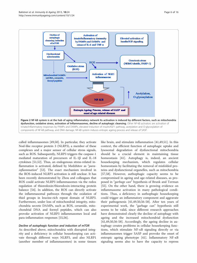
Figure 2 NF-kB system is at the hub of aging inflammatory network Its activation is induced by different factors, such as mitochondriadysfunction, oxidative stress, activation of Inflammasomes, decline of autophagic cleansing. Other NF-kB activators are activation ofinnate/inflammatory responses by PAMPs and DAMPs, elevated induction of insulin/IGF1 pathway, acetylation and O-glycosylation ofcomponents of NF-kB pathway, and DNA damage. NF-kB system induces entropic ageing process and release of SASP.
Balistreri et al. Immunity & Ageing 2013, 10:24 Page 6 of 16http://www.immunityageing.com/content/10/1/24
called inflammasomes [49,50]. In particular, they activateNod-like receptor protein 3 (NLRP3), a member of thesecomplexes and a major sensor of cellular stress signals,such as ROS. Subsequently, NLRP3 triggers the caspase-1mediated maturation of precursors of IL-1β and IL-18cytokines [51,52]. Thus, an endogenous stress-related in-flammation is activated, defined by Medzhitov as “para-inflammation” [53]. The exact mechanism involved inthe ROS-induced NLRP3 activation is still unclear. It hasbeen recently demonstrated by Zhou and colleagues thatROS could activate NLRP3 inflammasomes via the redoxregulation of thioredoxin/thioredoxin-interacting proteinbalance [54]. In addition, the ROS can directly activatethe inflammasomal pathways through the oxidation ofthiol groups in leucine-rich repeat domain of NLRP3.Furthermore, under loss of mitochondrial integrity, mito-chondria secrete DAMPs, such as ROS, ceramide, mito-chondrial DNA and formyl peptides, which can alsoprovoke activation of NLRP3 inflammasomes local andpara-inflammation responses [55,56].
Decline of autophagic function and induction of mTORAs described above, mitochondria with disrupted integ-rity and a deficiency in cellular housekeeping can acti-vate through different ways NLRP3, and also NLRP1(another member of inflammasomes) in some tissues
like brain, and stimulate inflammation [41,49,51]. In thiscontext, the efficient function of autophagic uptake andlysosomal degradation of dysfunctional mitochondriashould be a crucial element in maintaining tissuehomeostasis [41]. Autophagy is, indeed, an ancienthousekeeping mechanism, which regulates cellularhomeostasis by facilitating the removal of misfolded pro-teins and dysfunctional organelles, such as mitochondria[57,58]. However, authophagic capacity seems to becompromised in ageing and age-related diseases, as pro-posed in “garbage can” hypothesis of Brunk and Terman[55]. On the other hand, there is growing evidence oninflammasome activation in many pathological condi-tions. Thus, a deficiency in authophagic housekeepingcould trigger an inflammatory component and aggravatetheir pathogenesis [41,49,50,56-58]. After ten years ofexperimental work, the “garbage can” hypothesis stillseems to be valid, since different research approacheshave demonstrated clearly the decline of autophagy withageing and the increased mitochondrial dysfunction[41,49,50,56-58]. Accordingly, the ageing decline in au-tophagy creates problems in cellular housekeeping func-tions, which stimulate NF-κB signaling directly or viainflammasomes trigger SASP and provoke the onset ofentropic ageing phenotype [41]. Inflammatory NF-κBsignaling seems also to have the capacity to repress
Balistreri et al. Immunity & Ageing 2013, 10:24 Page 7 of 16http://www.immunityageing.com/content/10/1/24
autophagy and to induce this destructive interplay be-tween autophagy and inflammasomes [41,49,50,56-58].In particular, TNF-α can induce or repress autophagy ina NF-κB dependent manner. In presence of NF-κB signal-ing, TNF-α activates mammalian Target of Rapamycin(mTOR), a major autophagy inhibitor. On the contrary,in cells lacking of NF-κB activation, TNF-α stimulatesthe expression Beclin 1, an enhancer of autophagy[41,49,50,56-58].TOR is a highly conserved serine/threonine kinase and
a central controller of cell growth, metabolism and age-ing. mTOR is activated in response to nutrients, growthfactors and cellular energy. Deregulation of mTOR hasbeen implicated in inflammation, ageing and several age-related diseases (i.e. cancer, metabolic syndrome, neuro-logical diseases) [59]. It interacts with several proteins toform two distinct complexes named mTOR complex 1(mTORC1) and 2 (mTORC2), differentially activated bydistinct extracellular and intracellular signals. mTORC1responds to amino acids, stress, oxygen, energy, andgrowth factors and is acutely sensitive to rapamycin. Itpromotes cell growth by inducing and inhibiting ana-bolic and catabolic processes, respectively, and alsodrives cell-cycle progression. mTORC2 responds togrowth factors and regulates cell survival and metabol-ism, as well as the cytoskeleton. mTORC2 is insensitiveto acute rapamycin treatment but chronic exposure tothe drug can disrupt its structure. The activation ofmTORC1 by growth factors and nutrients inhibits au-tophagy and promotes protein synthesis. Over time, thismay promote cellular stress (protein aggregation, organ-elle dysfunction, and oxidative stress), which might leadto damage accumulation and a reduction in cell functionand thus promote the development of aging-related dis-eases. Also, mTORC1 activation induces stem cell ex-haustion, which reduces tissue repair and promotestissue dysfunction [59].
Activation of innate/inflammatory response by PAMPs andDAMPsDuring ageing, clonotypic immunity declines. This con-dition is defined immune-senescence [30]. In contrast, in-nate immunity seems to be efficiently activated and toinduce a chronic inflammatory phenotype, as mentionedabove [30]. The activation of innate immunity is medi-ated through the linking of pattern recognition receptors(PRRs), multi-ligand and evolutionarily conserved recep-tors (i.e. TLRs, NLRs and RIG-I-like receptors), with in-vading pathogen structures, called pathogen-associatedmolecular patterns (PAMPs), and endogenous dangermolecules, the DAMPs [26]. This determines the releaseof different inflammatory mediators (i.e. IL-6 and TNF-α)by NF-κB pathway [25]. Among PRRs, TLRs, and mainlyTLR4 and TLR2, recognize not only PAMPs, but also a
large number of different alarmin age type DAMPs, in-cluding high mobility group box 1 (HMGB1), S100, heatshock protein (HSP)-60 and −70, and defensins [26,34]. Inaddition, both TLR2 and −4 have a key role in the patho-genesis of several age-related diseases [34]. Accordingly,variants of genes codifying these molecules seem to mod-ify the susceptibility of age-related diseases and survival toextreme age, as recently described in our study [27]. Onthe other hand, the +896A/G (Asp299Gly; rs4986790) and+1196C/T (Thr399Ile; rs4986791) TLR4 SNPs have beenphenotypically associated with changes in the productionof pro- and anti-inflammatory cytokines, and principallythe Asp299Gly SNP seems to have a key role in AD,prostate cancer, atherosclerosis and, reciprocally, in lon-gevity [27,34].Furthermore, an intriguing and innovative hypothesis
has been recently suggested based on the crucial role ofmicroRNAs in the dysfunction of TLRs signaling and theacquisition of SASP with NF-κB activation. Thus, theseconditions can be considered as two interconnected phe-nomena [60].During ageing, proteins, DNA and lipids, long-live
macro-molecules can be targets of different age alter-ations. i.e. the Maillard reaction, a well known non-enzymatic glycosylation mechanism, induced as result ofenhance of oxidative stress and hyperglycemia [2]. Thisresults in the formation of protein glycation products,called AGEs (advanced glyaction end products), consid-ered pro-ageing factors and activating NF-κB pathwayby their linking with characteristic PRR receptors, theRAGE receptors (receptor for advanced glycation endproducts). With advancing age, AGE content increasesin tissues. AGE process also improves in diabetes, ath-erosclerosis, neurodegeneration and several inflamma-tory diseases. The major harmful AGE effect in ageingseems to be the maintenance of anti-apoptotic and pro-inflammatory phenotype. Of special note is the glycationof collagen and elastin which seem to have a key role invascular pathologies [26].
Induction of NF-κB signaling pathway by pro-inflammatorycytokines (mediated/or not by lipid rafts)Activation of innate immunity in ageing process (seeabove) determines the production and release of SASP,such as different inflammatory molecules. Among these,pro-inflammatory cytokines are mostly observed to be atelevated level in elderly people. These cytokines can alsoactivate the NF-κB pathway and this way can propagateand aggravate the inflammatory changes. IL-6 and TNF-α are clearly up-regulated with ageing, even if their exactrole in the ageing process has been difficult to establishbecause of their complex and cell-type functions [25].Recent evidence reported that NF-κB pathway activa-
tion via pro-inflammatory cytokines, and particularly via
Balistreri et al. Immunity & Ageing 2013, 10:24 Page 8 of 16http://www.immunityageing.com/content/10/1/24
TNF-α, can be mediated by lipid rafts. Precisely, thebinding of TNF-α to the TNF receptor (TNFR) results inreceptor clustering within specialized domains at the cellsurface, named as lipid rafts, which function as physicalplatforms for various molecules and are involved in avariety of biologic processes, such as molecular sorting,membrane trafficking, and signal transduction. For ex-ample, lipid rafts are important in the primary steps ofT-cell antigen receptor signaling and B-cell antigen re-ceptor signaling via triggering the phosphorylation ofadaptor proteins [61]. The dynamic recruitment ofligand-bound receptors into lipid rafts has been sug-gested to be critical for the initiation of signaling trans-duction, including the NF-κB pathway. Legler andcolleagues [62] reported that translocation of TNFR tolipid rafts is essential for TNF-α-mediated NF-κB activa-tion, and disturbances in lipid raft organization switchthe effect of TNF-α signaling from NF-κB activation toapoptosis, demonstrating that lipid rafts are crucial forthe outcome of TNF-α-activated signaling pathways. Inaddition, lipid rafts have also been demonstrated to berequired for NF-κB activation induced by IL-1, lipopoly-saccharides, CD40L, or CD3/CD28, and the disruptionof lipid rafts results in the inhibition of NF-κB activationmediated by these stimuli [63-65]. These studies indicatethat lipid rafts play important roles in the activation ofproximal NF-κB signaling.
Excessive stimulation of insulin/ insulin like growth factor(IGF) signalingAn excessive insulin/IGF signaling has been demon-strated to accompany ageing process [2]. Insulin/IGFsignaling determines detrimental age effects via NF-κBpathway evoking activation of IκB kinase α/β complex.As consequence improving of inflammatory responsesand resistance of apoptosis are induced. Given thatimpairing the signaling of insulin/IGF signaling pathwaycan activate the FOXO-dependent lifespan extension,this implies the role of NF-κB pathway in driving theageing process via insulin/IGF axis [66].
Post-translation modifications of the members of NF-κBpathwayMembers of NF-κB pathway are targets of several post-translation modifications. They influence both the activa-tion of the pathway and transcriptional efficiency of NF-κB system [2,25]. Phosphorylation and ubiquitination arethe major regulatory changes during activation. Acetyl-ation, O-glycosylation and sumoylation can also controlthe transcriptional efficiency of NF-κB system duringstress condition., i.e. inflammatory responses. In addition,increased protein acetylation can also activate cellular sen-escence. On the other hand, molecules involved in cellularsurvival, such as the Sirtuin molecules (see below), and
particularly SIRT 1 and 6, can deacetylate a NF-κB com-ponent, the p65, and repress NF-κB signaling [2,25].Glucose tolerance decline, cause of insulin resistance
and hyperglycemic disorders, determines O-glycosylation.Chronic hyperglycemia mediates glucotoxicity throughAGE formation or via the production of O-linked N-acetylglucosamine (o-GlcNac)-modified proteins. On theother hand, levels of O-glycosylated proteins increaseduring ageing. In particular, an increased O- glycosyla-tion of Ikkβ protein, able to enhance NF-κB activity, hasbeen observed during ageing. O-glycosylation can alsotarget p65 NF-κB protein and potentiate the transcrip-tional efficiency NF-κB components. This action is reg-ulated by p53 protein, which can inhibit glycolysis andsubsequently suppress the activation of Ikkβ/ NF-κBsignaling [2,25].
DNA damagesOne of the major stochastic age mechanism is genomicinstability [2,67]. DNA lesions appear during ageing inboth nuclear and mitochondrial DNA, as result of freeradicals and oxidative stress. Under genotoxic stress, themajor pathways activated are p53, NF-κB and PARP-1(poly-(ADP-ribose)-polymerase-1) [67]. In particular, ac-tivation of NF-κB signaling represents one of the princi-pal cellular features evoked by DNA damage [68]. TheDNA damage-dependent NF-κB activation cascade is de-fined NEMO shuttle, since an essential NF-κB modula-tor (NEMO; as mentioned above) under genotoxic stressforms an complex with PIDD (p53-induced protein witha death domain) and RIP1 (receptor interacting protein)kinase [69]. This complex accumulates in nuclei and anuclear matrix ligase (PIASy) can sumoylate the NEMOprotein. Sumoylation is a prerequisite to allow a Ataxiatelangiectasia mutated (ATM) kinase to phopshorylateNEMO protein. Subsequently, NEMO is desumoylatedand the NEMO/ATM complex is exported from nucleiin cytoplasm where it activates Ikk kinases, by triggeringNF-κB signaling. This consents to prevent the p53-induced apoptosis, since Ikk kinases phosphorylate p53and induce its degradation by proteasomes [67-69].Another hallmark of DNA damage is the induction of
PARP-1 pathway, an ubiquitously expressed member ofPARP family of enzymes able to modify proteins by poly(ADP-ribosyl)lation. PARP-1 is a sensor of DNA damageand maintains the genome integrity by regulating DNArepair [70]. In addition, PARP-1 is considered a novelco-activator of NF-κB signaling, which can potentiatethe NF-κB activation in genotoxic stress [25,71]. Fur-thermore, it is one of the proteins involved in the regula-tion of the length of nucleoprotein structures located atthe ends of chromosomes, the telomeres [71]. Telomeresare subject to shortening at each cycle of cell divisionand are highly sensitive to damage induced by oxidative
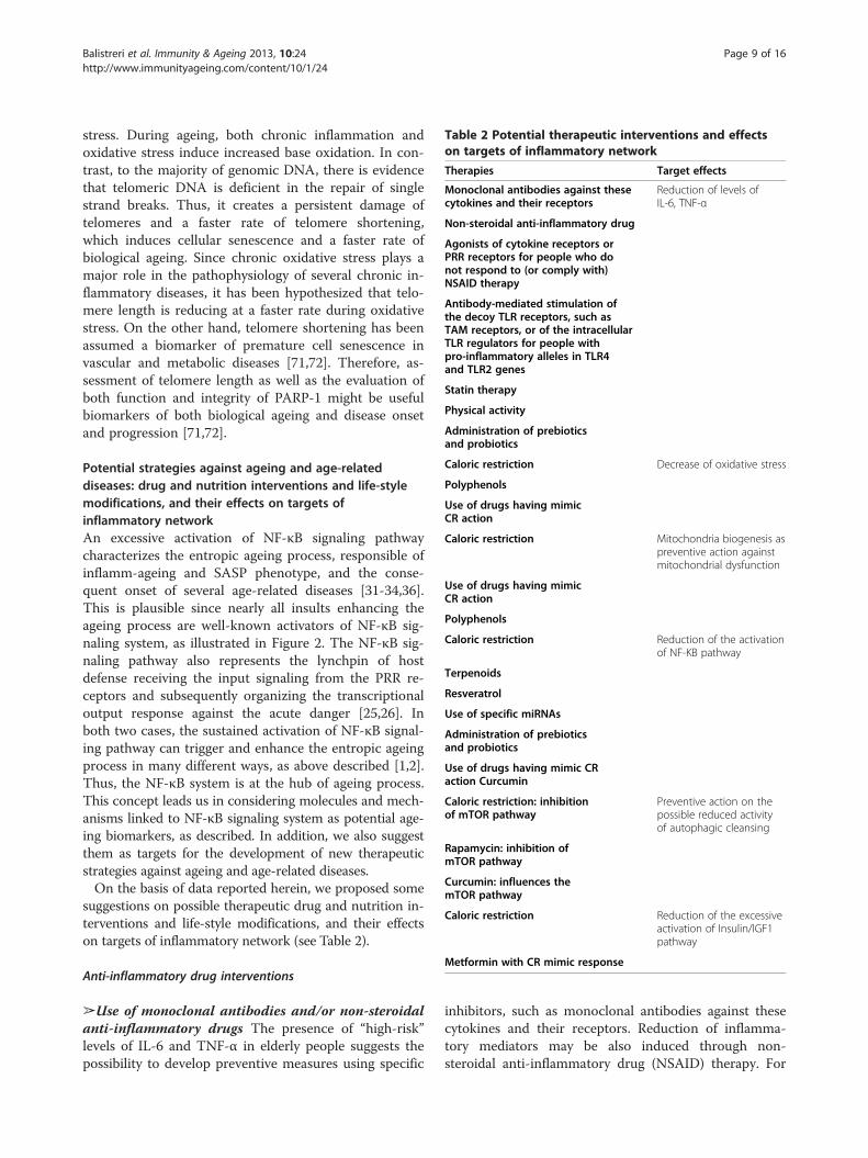
Table 2 Potential therapeutic interventions and effectson targets of inflammatory network
Therapies Target effects
Monoclonal antibodies against thesecytokines and their receptors
Reduction of levels ofIL-6, TNF-α
Non-steroidal anti-inflammatory drug
Agonists of cytokine receptors orPRR receptors for people who donot respond to (or comply with)NSAID therapy
Antibody-mediated stimulation ofthe decoy TLR receptors, such asTAM receptors, or of the intracellularTLR regulators for people withpro-inflammatory alleles in TLR4and TLR2 genes
Statin therapy
Physical activity
Administration of prebioticsand probiotics
Caloric restriction Decrease of oxidative stress
Polyphenols
Use of drugs having mimicCR action
Caloric restriction Mitochondria biogenesis aspreventive action againstmitochondrial dysfunction
Use of drugs having mimicCR action
Polyphenols
Caloric restriction Reduction of the activationof NF-KB pathway
Terpenoids
Resveratrol
Use of specific miRNAs
Administration of prebioticsand probiotics
Use of drugs having mimic CRaction Curcumin
Caloric restriction: inhibitionof mTOR pathway
Preventive action on thepossible reduced activityof autophagic cleansing
Rapamycin: inhibition ofmTOR pathway
Curcumin: influences themTOR pathway
Caloric restriction Reduction of the excessiveactivation of Insulin/IGF1pathway
Metformin with CR mimic response
Balistreri et al. Immunity & Ageing 2013, 10:24 Page 9 of 16http://www.immunityageing.com/content/10/1/24
stress. During ageing, both chronic inflammation andoxidative stress induce increased base oxidation. In con-trast, to the majority of genomic DNA, there is evidencethat telomeric DNA is deficient in the repair of singlestrand breaks. Thus, it creates a persistent damage oftelomeres and a faster rate of telomere shortening,which induces cellular senescence and a faster rate ofbiological ageing. Since chronic oxidative stress plays amajor role in the pathophysiology of several chronic in-flammatory diseases, it has been hypothesized that telo-mere length is reducing at a faster rate during oxidativestress. On the other hand, telomere shortening has beenassumed a biomarker of premature cell senescence invascular and metabolic diseases [71,72]. Therefore, as-sessment of telomere length as well as the evaluation ofboth function and integrity of PARP-1 might be usefulbiomarkers of both biological ageing and disease onsetand progression [71,72].
Potential strategies against ageing and age-relateddiseases: drug and nutrition interventions and life-stylemodifications, and their effects on targets ofinflammatory networkAn excessive activation of NF-κB signaling pathwaycharacterizes the entropic ageing process, responsible ofinflamm-ageing and SASP phenotype, and the conse-quent onset of several age-related diseases [31-34,36].This is plausible since nearly all insults enhancing theageing process are well-known activators of NF-κB sig-naling system, as illustrated in Figure 2. The NF-κB sig-naling pathway also represents the lynchpin of hostdefense receiving the input signaling from the PRR re-ceptors and subsequently organizing the transcriptionaloutput response against the acute danger [25,26]. Inboth two cases, the sustained activation of NF-κB signal-ing pathway can trigger and enhance the entropic ageingprocess in many different ways, as above described [1,2].Thus, the NF-κB system is at the hub of ageing process.This concept leads us in considering molecules and mech-anisms linked to NF-κB signaling system as potential age-ing biomarkers, as described. In addition, we also suggestthem as targets for the development of new therapeuticstrategies against ageing and age-related diseases.On the basis of data reported herein, we proposed some
suggestions on possible therapeutic drug and nutrition in-terventions and life-style modifications, and their effectson targets of inflammatory network (see Table 2).
Anti-inflammatory drug interventions
➢Use of monoclonal antibodies and/or non-steroidalanti-inflammatory drugs The presence of “high-risk”levels of IL-6 and TNF-α in elderly people suggests thepossibility to develop preventive measures using specific
inhibitors, such as monoclonal antibodies against thesecytokines and their receptors. Reduction of inflamma-tory mediators may be also induced through non-steroidal anti-inflammatory drug (NSAID) therapy. For
Balistreri et al. Immunity & Ageing 2013, 10:24 Page 10 of 16http://www.immunityageing.com/content/10/1/24
people who do not respond to (or comply with) NSAIDtherapy, other more sophisticated preventive approachesmay be possible, including the use of agonists of cyto-kine receptors or PRR receptors, i.e. TLR4 and −2, par-ticularly in subjects carriers of high inflammatoryresponder alleles [27,34]. On the other hand, the activa-tion of PRR receptors, such as TLR4 and −2, evocatedby PAMPs or DAMPs particularly upon ageing, inducethe release via NF-κB pathway of a large number ofcomponents of SASP, such as pro-inflammatory IL-6and TNF-α cytokines [26,34]. In addition, the magnitudeof cytokine production, and in general that of all pro-inflammatory mediators, has been shown to vary indi-vidually and is likely based on genetic heterogeneity.One or more functional SNPs in one or more innateimmunty genes might be responsible. Accordingly, re-cent studies have suggested the role of +896A/G TLR4SNP in cytokine production. In particular, high levels ofpro-inflammatory cytokines were observed in carriersbearing the +896A/G TLR4 SNP [27,34].Another possible therapeutic intervention in subjects
with pro-inflammatory alleles of TLR4 and TLR2 genesmight be antibody-mediated stimulation of the decoyTLR receptors, such as TAM receptors, or the intracel-lular TLR regulators (i.e. Supressor of cytokinesignaling-SOCS molecules), involved in the inhibition ofthe inflammatory response, by mediating TLR degrad-ation, or the activation of competitive or depho-sphorylation functions [73]. The sequential induction ofthese pathways, and their integration with upstreamTLR and cytokine signaling networks, may limit the in-flammatory response and maintain innate immune sys-tem homeostasis. A better understanding of theregulatory mechanisms of this cascade may have import-ant implications for therapeutic intervention in humanimmune disorders and reduce the risk development forseveral age-related diseases [27,34].
➢Statin therapy Statin therapy has been demonstratedto have benefical effects in reducing primary and sec-ondary CVD risk through the lipid-lowering, but alsoin inducing anti-ageing actions, such as inflammatorymolecule lowering, especially IL-6 and CRP. On theother hand, results from Justification Trial EvaluatingRosuvastatin (JUPITER) confirmed that statin treat-ment in apparently healthy subjects with elevatedCRP and non-elevated Low density lipoprotein choles-terol resulted in significant reduction in both thesemarkers and CVD [74].
Nutrition interventions and life-style modifications
➢Caloric restriction Another possible anti-ageing strat-egy, able to reduce the biological effects of NF-κB
signaling pathway in ageing, is the notable caloric re-striction (CR) [75]. Restricting the intake of calories hasbeen practiced as a method for increasing both thelength and quality of life for over 500 years. Experimen-tal work confirming the success of this approach in ani-mals has accumulated over the last 100 years. CR mayextend life by up to 50% in rodents, with progressivelyless impact the later in life it is started. This effect ismatched by profound impacts on age-related diseases,including reduced risk of cancer, neurodegenerative dis-orders, autoimmune disease, CVD and T2DM [75]. Thedisposable soma theory of ageing suggests that CRevolved as a somatic protection response to enable ani-mals to survive periods of food shortage [4]. The shut-down of reproductive function during CR is consistentwith this suggestion, but other features of thephenomenon are less consistent with this theory. Someresearchers have, indeed, proposed that in rodents itmay be mostly an artifact of domestication. CR inducesprofound effects on animals at all levels from the tran-scriptome to whole animal physiology and behavior. An-imals under CR lose weight which is disproportionatelycontributed to by white adipose tissue. Generally animalson CR change their activity patterns. Thus, they aremore active prior to food delivery each day, but total ac-tivity may be unchanged or reduced [75]. Considerabledebate has occurred over the effects of CR on restingmetabolic rate (RMR). Total RMR declines, but as bodymass and body composition also change it is unclearwhether metabolism at the tissue level also declines, isunchanged or even increases. Body temperature univer-sally decreases. Hunger is increased and does not seemto decline even with very long term restriction. Circulat-ing adipokines are reduced reflecting the reduction inwhite adipose tissue mass under CR [75]. There is also alarge reduction in circulating insulin and glucose levels.There are profound tissue level changes in metabolismwith a generalized shift from carbohydrate to fatmetabolism.Four pathways have been implicated in mediating the
CR effects. They are the insulin/IGF-1signaling pathway,the Sirtuin pathway, the adenosine monophosphate(AMP) activated protein kinase (AMPK) pathway andmTOR pathway [75]. These different pathways mayinteract and all play important roles mediating differentaspects of CR response. Exactly how they generate thehealth benefits remains open for debate. However, oneof the major impact of CR is the reduction of oxidativestress [76]. As described above, the major cellular sourceof ROS are the mitochondria. Isolated mitochondriafrom animals under CR show a reduced ROS produc-tion. In particular, CR results in an increase in the leveland activation of adenine nucleotide translocase and un-coupling proteins able to reduce the mitochondrial
Balistreri et al. Immunity & Ageing 2013, 10:24 Page 11 of 16http://www.immunityageing.com/content/10/1/24
membrane potential. This results in a decline in super-oxide radical (O2) production and a less damage to thelipids in the mitochondrial membrane reduced ulteriorlyby increases in the membrane lipid saturation [76]. In-creases in superoxide dismutase convert superoxide intohydrogen peroxide and increased levels of Se-dependentglutathione peroxidase and catalase convert this to waterreducing the production of the toxic hydroxyl radical(OH). Lowered levels of OH diminish the oxidative dam-age to proteins and DNA, which is further amelioratedby enhanced levels of degradation and base excision re-pair respectively [76]. Furthermore, CR induces mito-chondrial biogenesis, as evidenced by changes inmtDNA levels, and protein levels [77]. Such effects onmitochondrial biogenesis are consistent with the ideathat there may be a tissue level increase in oxygen con-sumption under CR which is accommodated in the re-duced overall energy budget by the reduced amount ofmetabolizing tissue.In addition, CR increases the levels of a member of
Sirtuin family (SIRT1 to SIRT 7), NAD + dependentdeactylases involved in the regulation of the activity ofmany proteins, energy metabolism, cell survival and lon-gevity [78,79]. In particular, CR increases the expressionof SIRT1 in multiple tissues, even if this effect does notappear to be uniform in all tissues or across differentstudies [80]. It has been demonstrated that SIRT1 inter-acts with p65/RelA protein and specifically cleaves theacetyl group form, the lysine-310 of p65 protein, in-volved in enhancing the trans-activation efficiency ofNF-κB system [81,82]. Thus, SIRT1 is a potent inhibitorof NF-κB system.An enhanced autophagy is also induced by CR via the
inhibition of mTOR or the activation of AMPK pathway.This last is an evolutionary conserved sensor for distur-bances in cellular energy balance and a major inductorof autophagy. Thus, CR acts directly or indirectly as in-hibitor of NF-κB system [75,81,82].Based on these observations, it is possible to assume
that CR has beneficial effects, i.e. the extension of theaverage and maximum life span and delaying the onsetof age-associated changes. However, this has beenproven only in animal models, such as yeast, worms,flies and some mammals (rats and mice), and somecriticisms (as above suggested) lead to consider it asan artifact of domestication, particularly in rodents[75,83-85]. In higher mammals, CR delays many diseasesassociated with aging including cancer, diabetes, athero-sclerosis, CVD and neurodegenerative diseases [86,87].The incidence of these diseases increases with age andthey contribute significantly to mortality. Therefore, CRcould increase life span by increasing the body’s generalstate of health and providing a nonspecific, resistance tochronic diseases and metabolic derangements [86,87].
However, the ultimate question, how does CR effect thehuman body, was studied in a limited number of experi-ments [88]. The study of CR effects on human longevityfaces ethical and logistical challenges since the averagelife span is close to 80 years for the population in devel-oped countries. Therefore, human studies are focusedon measuring the CR-related changes that could slowthe aging process and the progression of chronic dis-eases thus increasing life span. The most convincing evi-dence that CR could have a positive effect in humanswas provided by experiments by Fontana and coworkers,by the Comprehensive Assessment of Long-Term Effectsof Reducing Calorie Intake (CALERIE Phase 1), and bydata obtained on the members of the Caloric RestrictionSociety [89-93].Fontana and coworkers [89] assessed the effect of a 6-
year long CR diet on risk factors for atherosclerosis inadult male and female adults (age range 35–82 years)and compared them to age-matched healthy individualson typical American diets (control group). The totalserum cholesterol level and low-density lipoprotein(LDL) cholesterol levels, the ratio of total cholesterol tohigh-density lipoprotein cholesterol (HDL), triglycerides,fasting glucose, fasting insulin, CRP, platelet-derivedgrowth factor AB, and systolic and diastolic blood pres-sures were all markedly lower in the CR group. TheHDL cholesterol was higher after CR. Medical records ofindividuals in the CR group indicated that, before theybegan CR, they had serum lipid-lipoprotein and bloodpressure levels in the expected range for individuals ontypical American diets, and similar to those of the com-parison group. Thus, this study concluded that long-term CR can reduce the risk factors for atherosclerosis.The effect of fat loss induced by either (a) a long-term
20% CR or (b) a 20% increased energy expenditure (IEE)by exercise on coronary heart disease (CHD) risk factorswas detected in a one-year randomized, controlled trialon 48 non-obese male and female subjects. The CR orexercise induced reductions in body fat were quantita-tively similar and were accompanied by similar reduc-tions in most of the major CHD risk factors, includingplasma LDL-cholesterol, total cholesterol/HDL ratio,and CRP concentrations. Thus, these data evidenced thatlong-term CR or IEE of the same magnitude lead to sub-stantial and similar improvements in the major risk fac-tors for CHD in normal-weight and overweight middle-aged adults [90].The effects of a 1-year, 20% CR regime or 20% IEE by
exercise, on the oxidative damage of DNA and RNA,was evaluated by white blood cell and urine analyses innormal-to-overweight adults. Both interventions signifi-cantly reduced oxidative damage to both DNA and RNAin white blood cells compared to baseline. However,urinary levels of DNA and RNA oxidation products did
Balistreri et al. Immunity & Ageing 2013, 10:24 Page 12 of 16http://www.immunityageing.com/content/10/1/24
not differ from baseline values following either 1-yearintervention program. The conclusion of the study wasthat either CR or IEE by exercise reduce systemic oxida-tive stress which is reflected in a decreased DNA orRNA oxidative damage [91].CALERIE, a research program initiated by the Na-
tional Institute on aging and involving three researchcenters, performed in the Phase 1 three pilot studies todetermine whether long-term (6–12 months) effects of20–25% CR in free-living, non-obese humans could beinvestigated and to evaluate the adaptive responses toCR. This randomized, controlled, clinical trial concludedthat CR subjects had a lower body weight, a decreasedwhole body and visceral fat, a reduced activity energy ex-penditure, improved fasting insulin levels, improvementsin cardiovascular disease markers (LDL, total cholesterolto HDL ratio, and CRP), and no change in bone densitycompared to controls [88]. In the ongoing CALERIEPhase 2, the researchers are testing whether 2 yearssustained 25% CR of ad libitum energy intake results inbeneficial effects, similar to those observed in animalstudies [92].Members of the Caloric Restriction Society (CRS) re-
strict food intake with the expectation that this woulddelay the disease processes responsible for secondaryaging and to slow the primary aging process. Comparedto age-matched individuals eating typical American di-ets, CRS members (average age 50 ± 10 yr) had a lowerbody mass index, a reduced body fat, significantly lowervalues for total serum cholesterol, LDL cholesterol, totalcholesterol/LDL, and higher HDL cholesterol. Alsofasting plasma insulin and glucose values were signifi-cantly lower than in the age-matched control group. Leftventricular diastolic function in CRS members was similarto that of about 16 years younger individuals. Chronic in-flammation was reduced by CR and this was reflected insignificantly lower levels of plasma CRP and TNF-α [88].Aging is associated with a progressive reduction in
heart-rate-variability (HRV)—a measure of decliningautonomic function—and also a worse health outcome.The effect of a 30% CR on heart autonomic functionwas assessed by 24-hour monitoring of HRV in adultson self-imposed CR for 3 to 15 years and compared withan age-matched control eating a Western diet. The CRgroup had a significantly lower heart rate and signifi-cantly higher values for HRV. Also, HRV in the CR indi-viduals was comparable to published norms for healthyindividuals 20 years younger. The authors suggest thatCR reset the balance between the sympathetic/parasym-pathetic modulation of heart frequency in favor of theparasympathetic drive thus increasing the circadian vari-ability of heart rate [93].Thus, in humans CR could delay many diseases associ-
ated with aging including cancer, diabetes, atherosclerosis,
cardiovascular disease, and neurodegenerative diseases. Asan alternative to CR, several CR mimetics have been testedon animals and humans, as described below.
➢CR mimetic drugs: biguanides, stilbenes and drugsConsiderable effort has been directed in recent years tofind drugs that mimic the CR response. Promising can-didates are those that intersect with the critical signalingpathways identified above and include biguanides suchas metformin, capable to target insulin signaling path-way, stilbenes (e.g. resveratrol) affecting sirtuin activityand drugs such as rapamycin that interact with mTORsignaling. Whether it will ever be possible to find drugsthat capture the health benefits of CR without the nega-tive side-effects remains unclear. Moreover, even if suchdrugs are developed how the current licensing systemfor drug use in western societies would cope with themmay be a further obstacle to their use [75,88,94-96].
➢Polyphenols and resveratrol (a stilbene phytochemical)As mentioned above, several plant derived, folk medicalcompounds and extracts have been claimed to have anti-ageing effects [75,88,97-101]. However, only a small num-ber of traditional remedies has been subjected to a clinicaltrial. Recently, many promising compounds have beenidentified and scrutinized. Among these, there are poly-phenols (i.e. flavonoids and terpenoids), the major ingredi-ents of fruits, vegetables and different spices [75,88,97-101].Many of polyphenols are inhibitors of NF-κB signalingsystem, since they are potent antioxidants, and as conse-quence they can inhibit ROS production and activation ofNF-κB signaling system [97-101]. Some of them (i.e. terpe-noids) can also directly inhibit Ikk/ NF-κB signaling[97-101]. Accordingly, it has been found that low-doses ofterpenoids can trigger cellular stress response and subse-quently induce adaptive stress resistance, condition de-fined hormesis [97-101]. Stress resistance involves severalmolecular adaptations via the activation of AMPK path-way and the subsequent increase in the expression of sur-vival genes, such as Sirtuins, FOXOs and p53 [97-101]. Ofspecial note is the effect of resveratrol, a stilbene phyto-chemical. It induces activation of SIRT1 via AMPK path-way, and indirectly inhibition of NF-κB signaling systemvia the activation of survival genes [97-101].
➢Curcumin It has been postulated that a natural agent,curcumin, could influence cellular senescence [102].Curcumin has attracted the attention of researchers andclinicians as an anti-inflammatory and anti-oxidantagent with a potential use in the therapy of many dis-eases with an inflammation constituent, e.g. cancer,CVD, AD, rheumatoid arthritis and metabolic syndrome.A plethora of studies using animal and cell line modelshave been undertaken to elucidate the molecular

Balistreri et al. Immunity & Ageing 2013, 10:24 Page 13 of 16http://www.immunityageing.com/content/10/1/24
mechanisms and biological effects of curcumin andsome clinical trials are underway. Sikora and colleagues[103] proposed that curcumin might act as an anti-agingagent not only by inhibition of NF-κB, but also byindirect influence on cellular senescence via mTOR.However, they also showed that conversely curcumincan induce senescence in colon cancer cells [104]. More-over recent studies by Quitschke [105] also revealedthe prosenescence activity of curcumin. Nonetheless,curcumin has many molecular targets and evokes a bi-phasic hormetic dose–response [106]. Thus, one cannotexclude that much lower concentration of curcuminthan that used in these studies will inhibit/postpone cel-lular senescence or, at least, through NF-kB inhibitionwill reduce SASP. Interestingly, curcumin was shown toprolong life of model organisms such as Caenorhabditiselegans and Drosophila melanogaster, but did not influ-ence (similarly to resveratrol) the life span of mice [107].
➢Physical activity Promising evidence suggests a roleof physical activity in reducing the levels of inflamma-tory markers. Several speculations have been advanced[108-111]. However, the mechanisms underlying its anti-inflammatory effects seem complex and not fully eluci-dated. It has been recently considered that the decreasedproduction of proinflammatory cytokines may originatefrom a reduction of adiposity, or the release of musclederived IL-6 [108-111]. This last seems to induce severalmetabolic adaptations, i.e. hepatic glycogenolysis and lip-olysis, and the release of cytokine inhibitors (i.e. IL-1ra,sTNFR and IL-10) and cytokine with potent anabolic ef-fect, as IL-15 [108-111].
➢Probiotics and/or prebiotics Administration ofprobiotics and/or prebiotics to elderly seems to inducechanges in several inflammatory parameters (i.e. pro-inflammatory cytokine lowering, CRP reduction), demon-strating that manipulation of gut microbiota may result inmodification of functionality of an aged immune system.On the other hand, intestinal microbiota seems to play afundamental role in maintaining human health. Its sup-posed importance in human physiology has recently led tolabel human subjects as “metaorganisms” because of theirclose symbiotic relation with indigenous gut microbiota.The “metaorganisms” hypothesis evidences the use of diet-ary supplementation with probiotics and prebiotics, astherapeutic strategy to preserve human health particularlyduring that life period not foreseen by evolution- “ageing”-,that inexorably alters gut microbiota composition, stabilityand functionality [112,113].
ConclusionsFrom all observations described above, chronic inflam-mation has been proposed as the major biological
mechanism underpinning the entropic ageing processand age-related diseases [31-34,36]. Inflammation is alsothe key response of host defense against pathogens andtissue injury [25,26]. In addition, it is current opinionthat during evolution the host defense and ageingprocess have become linked together [2]. Thus, the largearray of defense factors and mechanisms linked to theNF-κB system seem to be involved in the entropic age-ing process [2,25]. This concept leads us in proposinginductors of NF-κB signaling system as potential ageingbiomarkers and promising targets for the developmentof new therapeutic strategies against ageing and age-related diseases. In this report, we describe some inflam-matory mechanisms linked to NF-κB signaling system aspotential ageing biomarkers. In addition, some sugges-tions on their role as promising targets for the develop-ment of new therapeutic strategies have been discussed.Our interest has been, particularly, focused on possibleinterventions on molecular survival and resistance stresspathways, capable to reduce or inhibit NF-κB signalingpathway. However, it is not impossible to predict,whether these possible interventions (appropriate andspecific drug therapies, lifestyle modifications, use of CRmimetics and other preventive therapeutic strategies)can very reduce or retard the onset of ageing biologicalphenotype and the onset risk of age-related diseases. Dif-ferent motivations lead us to have prudence. Firstly, themajor literature data on anti-ageing effects of thera-peutic strategies have been obtained from studies on ani-mals. Thus, potential therapy interventions on the basisof pathways identified in model organisms may be anillusion, because gains in longevity achieved in these or-ganisms seem to decline with organismal complexity ordepend on idiosyncratic physiology. Furthermore, life-span in some organisms may be less plastic than inothers. In addition, there are still enormous gaps in ourknowledge about how metabolic pathways operate andinteract. Serious side effects may constrain the effective-ness of pharmacological interventions.The best treatment might be that which consents the
repair of macromolecular damage. However, it is notclear that all toxic lesions associated with ageing processhave been identified, or whether practical and appropri-ated strategies exist to eliminate them, as those men-tioned above. Today, the researchers are becoming tospeculate the concept based on reprogramming cellularsenescence as way of organism rejuvenation or at leastto alleviate age-related diseases considering cellular sen-escence as target model [114-116]. This hope derives byresults of recent studies on progeroid mice demonstrat-ing the possibility to reverse progeroid phenotypethrough genetic manipulation. This intervention ofavoiding or reversing cellular senescence is based on in-duced pluripotent stem cell technology, which opened a
Balistreri et al. Immunity & Ageing 2013, 10:24 Page 14 of 16http://www.immunityageing.com/content/10/1/24
new avenue of autologous regenerative medicine and thepossibility to activate telomerase and change the telo-mere length [117,118]. Accordingly, other studies areneeded to confirm and extend these current data. Forexample, genomic, transcriptomic and epigenetic investi-gations may eventually lead to better understanding themolecular and cellular inflammatory mechanisms associ-ated with biological ageing. In addition, for the develop-ment of anti-ageing therapies for human, it should bemore appropriate identifying cellular and serum ageingbiomarkers and potential targets using apposite studymodel, such as centenarian offspring, healthy elderlypeople with a family history of longevity, as recently sug-gested [119]. On the other hand, the research ofbiomarkers of ageing and age-related diseases in under-standing the health trajectories of the oldest-old is unex-plored territory. It is important that this lacuna is filledgiven the rapid growth in the number of very old peoplein many contemporary populations. The goal of this re-search might guarantee improving of life quality ratherthan searching the elixir of long life.
Competing interestsThe authors declare that they have no competing interests.
Authors’ contributionsCRB wrote the drafts of this manuscript and revised the intellectual content;GC contributed to collection of literature data. CRB had the overallsupervision of the review processing. All authors edited the paper andapproved its final version.
Received: 23 December 2012 Accepted: 2 June 2013Published: 20 June 2013
References1. Hayflick L: Entropy explains aging, genetic determinism explains
longevity, and undefined terminology explains misunderstanding both.PLoS Genet 2007, 3:e220.
2. Salminen A, Kaarniranta K: Genetics vs. entropy: longevity factors suppressthe NF-kappaB-driven entropic aging process. Ageing Res Rev 2010,9:298–314.
3. Kirkwood TB, Holliday R: The evolution of ageing and longevity. Proc R SocLond B Biol Sci 1979, 205:531–546.
4. Kirkwood TBL: A systematic look at an old problem. Nature 2008,451:644–647.
5. Bostock CV, Soiza RL, Whalley LJ: Genetic determinants of ageingprocesses and diseases in later life. Maturitas 2009, 62:225–229.
6. Longo VD, Finch CE: Genetics of aging and disease. Arch Neurol 2002,59:1706–1709.
7. Finch CE, Ruvkun G: The genetics of aging. Annu Rev Genomics Hum Genet2001, 2:435–462.
8. McGue M, Vaupel JW, Holm N, Harvald B: Longevity is moderatelyheritable in a sample of Danish Twins born 1870–1880. J Geront Biol Sci1993, 348:B237–B244.
9. Herskind AM, McGue M, Holm NV, Sorensen TI, Harvard B, Vaupel JW: Theheritability of human longevity: a population-based study of 2872Danish twin pairs born 1870–1900. Hum Genet 1996, 97:319–323.
10. Mattson MP: Pathways towards and away from Alzheimer’s disease.Nature 2004, 430:631–639.
11. Puca AA, Daly MJ, Brewster SJ, Matise TC, Barrett J, Shea-Drinkwater M, KangS, Joyce E, Nicoli J, Benson E, Kunkel LM, Perls T: A genome-wide scan forlinkage to human exceptional longevity identifies a locus onchromosome 4. Proc Natl Acad Sci USA 2001, 98:10505–10508.
12. Capri M, Salvioli S, Sevini F, Valensin S, Celani L, Monti D, Pawelec G, DeBenedictis G, Gonos ES, Franceschi C: The genetics of human longevity.Ann N Y Acad Sci 2006, 1067:252–263.
13. Lunetta KL, D’Agostino RB Sr, Karasik D, Benjamin EJ, Guo CY, Govindaraju R,Kiel DP, Kelly-Hayes M, Massaro JM, Pencina MJ, Seshadri S, Murabito JM:Genetic correlates of longevity and selected age-related phenotypes: agenome-wide association study in the Framingham Study. BMC MedGenet 2007, 19:8.
14. de Magalhães JP, Curado J, Church GM: Meta-analysis of age-related geneexpression profiles identifies common signatures of aging. Bioinformatics2009, 25:875–881.
15. Troen BR: The biology of aging. Mt Sinai J Med. 2003, 70:3–22.16. Sprott RL: Biomarkers of aging and disease: introduction and definitions.
Exp Gerontol 2010, 45:2–4.17. Crimmins E, Vasunilashorn S, Kim JK, Alley D: Biomarkers related to aging
in human populations. Adv Clin Chem 2008, 46:161–216.18. Simm A, Johnson TE: Biomarkers of ageing: A challenge for the future.
Exp Gerontol 2010, 45:731–732.19. Euser SM, van Bemmel T, Schram MT, Gussekloo J, Hofman A, Westendorp RG,
Breteler MM: The effect of age on the association between blood pressureand cognitive function later in life. J Am Geriatr Soc 2009, 57:1232–1237.
20. van Bemmel T, Vinkers DJ, Macfarlane PW, Gussekloo J, Westendorp RG:Markers of autonomic tone on a standard ECG are predictive ofmortality in old age. Int J Cardiol 2006, 107:36–41.
21. Martin-Ruiz C, Dickinson HO, Keys B, Rowan E, Kenny RA, Von Zglinicki T:Telomere length predicts poststroke mortality, dementia, and cognitivedecline. Ann Neurol 2006, 60:174–180.
22. Rodríguez RM, López-Vázquez A, López-Larrea C: Immune systemsevolution. Adv Exp Med Biol 2012, 739:237–251.
23. Rock KL, Latz E, Ontiveros F, Kono H: The sterile inflammatory response.Annu Rev Immunol 2010, 28:321–342.
24. Vasto S, Candore G, Balistreri CR, Caruso M, Colonna-Romano G, GrimaldiMP, Listi F, Nuzzo D, Lio D, Caruso C: Inflammatory networks in ageing,age-related diseases and longevity. Mech Ageing Dev 2007, 128:83–91.
25. Gilmore TD, Wolenski FS: NF-κB: where did it come from and why?Immunol Rev 2012, 246:14–35.
26. Newton K, Dixit VM: Signaling in innate immunity and inflammation. ColdSpring Harb Perspect Biol 2012, 4:1–19.
27. Balistreri CR, Caruso C, Listì F, Colonna-Romano G, Lio D, Candore G:LPS-mediated production of pro/anti-inflammatory cytokines andeicosanoids in whole blood samples: biological effects of +896A/G TLR4polymorphism in a Sicilian population of healthy subjects. Mech AgeingDev 2011, 132:86–92.
28. Mitchell RN, Cotran RS: Acute and chronic inflammation, in Robbins BasicPathology. Philadelphia, USA: Saunderrs; 2003:30–56.
29. Lio D, Caruso C: IL-10, genetic polymorphism and its relevance to agerelated diseases. In Interleukin-10. Edited by Marincola FM. Georgetown, TX,USA: Eureka.com; 2006:93–106.
30. Candore G, Caruso C, Colonna-Romano G: Inflammation, geneticbackground and longevity. Biogerontology 2010, 11:565–573.
31. Franceschi C, Bonafe’ M, Valensin S, Olivieri F, De Luca M, Ottaviani E, DeBenedictis G: Inflamm-aging. An evolutionary perspective onimmunosenescence. Ann NY Acad Sci 2000, 908:244–254.
32. Licastro F, Candore G, Lio D, Porcellini E, Colonna-Romano G, Franceschi C,Caruso C: Innate immunity and inflammation in ageing: a key forunderstanding age-related diseases. Immun Ageing 2005, 2:8.
33. Balistreri CR, Caruso C, Candore G: The role of adipose tissue and adipokinesin obesity-related inflammatory diseases. Mediators Inflamm 2010, 2010:1–19.
34. Balistreri CR, Colonna-Romano G, Lio D, Candore G, Caruso C: TLR4polymorphisms and ageing: implications for the pathophysiology ofage-related diseases. J Clin Immunol 2009, 29:406–415.
35. Balistreri CR, Candore G, Accardi G, Bova M, Buffa S, Bulati M, Forte GI, ListìF, Martorana A, Palmeri M, Pellicanò M, Vaccarino L, Scola L, Lio D: Colonna-Romano G. Genetics of longevity. data from the studies on Siciliancentenarians. Immun Ageing. 2012, 9:8.
36. Salminen A, Kauppinen A, Kaarniranta K: Emerging role of NF-κB signalingin the induction of senescence-associated secretory phenotype (SASP).Cell Signal 2012, 24:835–845.
37. Tchkonia T, Zhu Y, van Deursen J, Campisi J, Kirkland JL: Cellularsenescence and the senescent secretory phenotype: therapeuticopportunities. J Clin Invest 2013, 123:966–972.
Balistreri et al. Immunity & Ageing 2013, 10:24 Page 15 of 16http://www.immunityageing.com/content/10/1/24
38. Chien Y, Scuoppo C, Wang X, Fang X, Balgley B, Bolden JE, Premsrirut P, LuoW, Chicas A, Lee CS, Kogan SC, Lowe SW: Control of the senescence-associated secretory phenotype by NF-κB promotes senescence andenhances chemosensitivity. Genes Dev 2011, 25:2125–2136.
39. Crescenzi E, Pacifico F, Lavorgna A, De Palma R, D’Aiuto E, Palumbo G,Formisano S, Leonardi A: NF-κB-dependent cytokine secretion controlsFas expression on chemotherapy-induced premature senescent tumorcells. Oncogene 2011, 30:2707–2717.
40. Rovillain E, Mansfield L, Caetano C, Alvarez-Fernandez M, Caballero OL,Medema RH, Hummerich H, Jat PS: Activation of nuclear factor-kappa Bsignalling promotes cellular senescence. Oncogene 2011, 30:2356–2366.
41. Salminen A, Kaarniranta K, Kauppinen A: Inflammaging: disturbed interplay betweenautophagy and inflammasomes. 4th edition. Albany NY: Aging; 2012:166–175.
42. Campisi J, Andersen JK, Kapahi P, Melov S: Cellular senescence: a linkbetween cancer and age-related degenerative disease? Semin Cancer Biol2011, 21:354–359.
43. Ishii T, Miyazawa M, Onouchi H, Yasuda K, Hartman PS, Ishii N: Modelanimals for the study of oxidative stress from complex II. Biochim BiophysActa 1827, 2013:588–597.
44. Jacob KD, Hooten NN, Trzeciak AR, Evans MK: Markers of oxidant stressthat are clinically relevant in aging and age-related disease. Mech AgeingDev 2013, 134:139–157.
45. Dai DF, Rabinovitch PS, Ungvari Z: Mitochondria and cardiovascular aging.Circ Res 2012, 110:1109–1124.
46. Morán M, Moreno-Lastres D, Marín-Buera L, Arenas J, Martín MA, Ugalde C:Mitochondrial respiratory chain dysfunction: implications inneurodegeneration. Free Radic Biol Med 2012, 53:595–609.
47. Ma ZA, Zhao Z, Turk J: Mitochondrial dysfunction and β-cell failure intype 2 diabetes mellitus. Exp Diabetes Res 2012, 2012:703538.
48. Weinberg F, Chandel NS: Reactive oxygen species-dependent signalingregulates cancer. Cell Mol Life Sci 2009, 66:3663–3673.
49. Salminen A, Ojala J, Kaarniranta K, Kauppinen A: Mitochondrial dysfunctionand oxidative stress activate inflammasomes: impact on the agingprocess and age-related diseases. Cell Mol Life Sci 2012, 69:2999–3013.
50. Deretic V: Autophagy as an innate immunity paradigm: expanding thescope and repertoire of pattern recognition receptors. Curr OpinionImmunol 2011, 24:1–11.
51. West AP, Shadel GS, Ghosh S: Mitochondria in innate immune responses.Nat Rev Immunol 2011, 11:389–402.
52. Martinon F, Mayor A, Tschopp J: The inflammasomes: guardians of thebody. Annu Rev Immunol 2009, 27:229–265.
53. Medzhitov R: Origin and physiological roles of inflammation. Nature 2008,454:428–435.
54. Zhou R, Yazdi AS, Menu P, Tschopp J: A role for mitochondria in NLRP3inflammasome activation. Nature 2011, 469:221–225.
55. Brunk UT, Terman A: The mitochondrial-lysosomal axis theory of aging.Accumulation of damaged mitochondria as a result of imperfectautophagocytosis. Eur J Biochem 2002, 269:1996–2002.
56. Green DR, Galluzzi L, Kroemer G: Mitochondria and the autophagy-inflammation-cell death axis in organismal aging Science. 2011,333:1109–1112.
57. Rubinsztein DC, Marino G, Kroemer G: Autophagy and aging Cell 2011,146:682–695.
58. Cianfanelli V, Cecconi F: Autophagy-dependent NFκB regulation. Cell Cycle2012, 11:436–437.
59. Laplante M, Sabatini DM: mTOR signaling in growth control and disease.Cell 2012, 149:274–293.
60. Olivieri F, Rippo MR, Prattichizzo F, Babini L, Graciotti L, Recchioni R,Procopio AD: Toll like receptor signaling in “inflammaging”: microRNA asnew players. Immun Ageing 2013, 10:11.
61. Tomoiu A, Larbi A, Fortin C, Dupuis G, Fulop T Jr: Do membrane rafts contributeto human immunosenescence? Ann N Y Acad Sci 2007, 1100:98–110.
62. Legler DF, Micheau O, Doucey MA, Tschopp J, Bron C: Recruitment of TNFreceptor 1 to lipid rafts is essential for TNFalpha-mediated NF-kappaBactivation. Immunity 2003, 18:655–664.
63. Larbi A, Muti E, Giacconi R, Mocchegiani E, Fülöp T: Role of lipid rafts inactivation-induced cell death: the fas pathway in aging. Adv Exp Med Biol2006, 584:137–155.
64. Fessler MB, Parks JS: Intracellular lipid flux and membrane microdomainsas organizing principles in inflammatory cell signaling. J Immunol 2011,187:1529–1535.
65. Tewari R, Choudhury SR, Mehta VS, Sen E: TNFα regulates the localizationof CD40 in lipid rafts of glioma cells. Mol Biol Rep 2012, 39:8695–8699.
66. Martins JO, Zanoni FL, Martins DO, Coimbra R, Krieger JE, Jancar S,Sannomiya P: Insulin regulates cytokines and intercellular adhesionmolecule-1 gene expression through nuclear factor-kappaB activation inLPS-induced acute lung injury in rats. Shock 2009, 31:404–409.
67. Schumacher B, Garinis GA, Hoeijmakers JH: Age to survive: DNA damageand aging. Trends Genet 2008, 2:77–85.
68. Wu ZH, Miyamoto S: Many faces of NF-kappaB signaling induced bygenotoxic stress. J Mol Med (Berl) 2007, 85:1187–1202.
69. Salminen A, Suuronen T, Huuskonen J, Kaarniranta K: NEMO shuttle: a linkbetween DNA damage and NF-kappaB activation in progeroidsyndromes? Biochem Biophys Res Commun 2008, 367:715–718.
70. Beneke S: Poly(ADP-ribose) polymerase activity in different pathologies—the link to inflammation and infarction. Exp Gerontol 2008, 43:605–614.
71. Campisi J: Senescent cells, tumor suppression, and organismal aging:good citizens, bad neighbors. Cell 2005, 120:513–522.
72. Balistreri CR, Pisano C, Merlo D, Fattouch K, Caruso M, Incalcaterra E,Colonna-Romano G, Candore G: Is the mean blood leukocyte telomerelength a predictor for sporadic thoracic aortic aneurysm? Data from apreliminary study Rejuvenation Res 2012, 1:170–173.
73. Lemke G, Rothlin CV: Immunobiology of the TAM receptors Nat Rev Immunol2008, 8:327–336.
74. Ridker PM: The JUPITER trial: results, controversies, and implications forprevention. Circ Cardiovasc Qual Outcomes 2009, 2:279–285.
75. Speakman JR, Mitchell SE: Caloric restriction. Mol Aspects Med 2011,32:159–221.
76. Ash CE, Merry BJ: The molecular basis by which dietary restricted feedingreduces mitochondrial reactive oxygen species generation. Mech AgeingDev 2011, 132:43–54.
77. Nisoli E, Tonetto C, Cardile A, Cozzi V, Bracale R, Tedesco L, Falcone S,Valerio A, Cantoni O, Clementi E, Moncada S, Carruba MO: Calorierestriction promotes mitochondrial biogenesis by inducing theexpression of eNOS. Science 2005, 310:314–317.
78. Sauve AA, Youn DY: Sirtuins: NAD(+)-dependent deacetylase mechanismand regulation. Curr Opin Chem Biol 2012, 16:535–543.
79. Guarente L: Sirtuins and calorie restriction. Nat Rev Mol Cell Biol 2012,13:207.
80. Geng YQ, Li TT, Liu XY, Li ZH, Fu YC: SIRT1 and SIRT5 activity expressionand behavioral responses to calorie restriction. J Cell Biochem 2011,112:3755–3761.
81. Huang W, Shang WL, Wang HD, Wu WW, Hou SX: Sirt1 overexpressionprotects murine osteoblasts against TNF-α-induced injury in vitro bysuppressing the NF-κB signaling pathway. Acta Pharmacol Sin 2012,33:668–674.
82. Jung YJ, Lee JE, Lee AS, Kang KP, Lee S, Park SK, Lee SY, Han MK, Kim DH,Kim W: SIRT1 overexpression decreases cisplatin-induced acetylation ofNF-κB p65 subunit and cytotoxicity in renal proximal tubule cells.Biochem Biophys Res Commun 2012, 419:206–210.
83. Anderson RM, Weindruch R: The caloric restriction paradigm: implicationsfor healthy human aging. Am J Hum Biol 2012, 24:101–106.
84. Kennedy BK, Steffen KK, Kaeberlein M: Ruminations on dietary restrictionand aging. Cell Mol Life Sci 2007, 64:1323–1328.
85. Piper MD, Bartke A: Diet and aging. Cell Metab 2008, 8:99–104.86. Roth GS, Ingram DK, Lane MA: Caloric restriction in primates and
relevance to humans. Ann N Y Acad Sci 2001, 928:305–315.87. Walford RL, Mock D, Verdery R, MacCallum T: Calorie restriction in
biosphere 2: alterations in physiologic, hematologic, hormonal, andbiochemical parameters in humans restricted for a 2-year period.J Gerontol A Biol Sci Med Sci 2002, 57:B211–B224.
88. Ribarič S: Diet and aging. Oxid Med Cell Longevity 2012, 2012:741468.89. Fontana L, Meyer TE, Klein S, Holloszy JO: Long-term calorie restriction is
highly effective in reducing the risk for atherosclerosis in humans. ProcNatl Acad Sci USA 2004, 101:6659–6663.
90. Fontana L, Villareal DT, Weiss EP, Racette SB, Steger-May K, Klein S, HolloszyJO, Washington University School of Medicine CALERIE Group: Calorierestriction or exercise: effects on coronary heart disease risk factors. Arandomized, controlled trial. Am J Physiol Endocrinol Metab 2007,293(1):E197–E202.
91. Holloszy JO, Fontana L: Caloric restriction in humans. Exp Gerontol2007, 42:709–712.

Balistreri et al. Immunity & Ageing 2013, 10:24 Page 16 of 16http://www.immunityageing.com/content/10/1/24
92. Rochon J, Bales CW, Ravussin E, Redman LM, Holloszy JO, Racette SB,Roberts SB, Das SK, Romashkan S, Galan KM, Hadley EC, Kraus WE, CALERIEStudy Group: Design and conduct of the CALERIE study: comprehensiveassessment of the long-term effects of reducing intake of energy.J Gerontol A Biol Sci Med Sci 2011, 66:97–108.
93. Stein PK, Soare A, Meyer TE, Cangemi R, Holloszy JO, Fontana L: Caloricrestriction may reverse age-related autonomic decline in humans. AgingCell 2012, 11:644–650.
94. Madeo F, Tavernarakis N, Kroemer G: Can autophagy promote longevity?Nat Cell Biol 2010, 12:842–846.
95. Berstein LM: Metformin in obesity, cancer and aging: addressing controversies.4th edition. Albany NY: Aging; 2012:320–329.
96. Van Meter M, Seluanov A, Gorbunova V: Forever young? Exploring the linkbetween rapamycin, longevity and cancer. Cell Cycle 2012, 11:4296–4297.
97. Pallauf K, Rimbach G: Autophagy, polyphenols and healthy ageing. AgeingRes Rev 2012, 12:237–252.
98. Zhang C, Lin G, Wan W, Li X, Zeng B, Yang B, Huang C: Resveratrol, apolyphenol phytoalexin, protects cardiomyocytes against anoxia/reoxygenation injury via the TLR4/NF-κB signaling pathway. Int J MolMed 2012, 29:557–563.
99. Relja B, Töttel E, Breig L, Henrich D, Schneider H, Marzi I, Lehnert M: Plantpolyphenols attenuate hepatic injury after hemorrhage/resuscitation byinhibition of apoptosis, oxidative stress, and inflammation via NF-kappaBin rats. Eur J Nutr 2012, 51:311–321.
100. Calabrese E, Iavicoli I, Calabrese V: Hormesis: its impact on medicine andhealth. Hum Exp Toxicol 2012, 13:215–235.
101. Chirumbolo S: Possible role of NF-κB in hormesis during ageing.Biogerontology 2012, 13:637–646.
102. Salvioli S, Sikora E, Cooper EL, Franceschi C: Curcumin in cell deathprocesses: a challenge for CAM of age-related pathologies. Evid BasedComplement Alternat Med 2007, 4:181–190.
103. Sikora E, Bielak-Zmijewska A, Mosieniak G, Piwocka K: The promise of slowdown ageing may come from curcumin. Curr Pharm Des 2010,16:884–892.
104. Mosieniak G, Adamowicz M, Alster O, Jaskowiak H, Szczepankiewicz AA,Wilczynski GM, Ciechomska IA, Sikora E: Curcumin induces permanentgrowth arrest of human colon cancer cells: link between senescenceand autophagy. Mech Ageing Dev 2012, 133:444–455.
105. Quitschke WW: Curcuminoid binding to embryonal carcinoma cells:reductive metabolism, induction of apoptosis, senescence, andinhibition of cell proliferation. PLoS One 2012, 7:e39568.
106. Ali RE, Rattan SI: Curcumin’s biphasic hormetic response on proteasomeactivity and heat-shock protein synthesis in human keratinocytes. Ann NY Acad Sci 2006, 1067:394–399.
107. Strong R, Miller RA, Astle CM, Baur JA, de Cabo R, Fernandez E, Guo W,Javors M, Kirkland JL, Nelson JF, Sinclair DA, Teter B, Williams D, Zaveri N,Nadon NL, Harrison DE: Evaluation of resveratrol, green tea extract,curcumin, oxaloacetic acid, and medium-chain triglyceride oil on lifespan of genetically heterogeneous mice. J Gerontol A Biol Sci Med Sci2013, 68:6–16.
108. Walsh NP, Gleeson M, Shephard RJ, Gleeson M, Woods JA, Bishop NC,Fleshner M, Green C, Pedersen BK, Hoffman-Goetz L, Rogers CJ, Northoff H,Abbasi A, Simon P: Position statement. Part one: immune function andexercise. Exerc Immunol Rev 2011, 17:6–63.
109. Kay SJ, Fiatarone Singh MA: The influence of physical activity onabdominal fat: a systematic review of the literature. Obes Rev 2006,7:183–200.
110. Brandt C, Pedersen BK: The role of exercise-induced myokines in musclehomeostasis and the defense against chronic diseases. J BiomedBiotechnol 2010, 2010:520258. Epub 2010 Mar 9.
111. Ambarish V, Chandrashekara S, Suresh KP: Moderate regular exercisesreduce inflammatory response for physical stress. Indian J PhysiolPharmacol 2012, 56:7–14.
112. Candore G, Balistreri CR, Colonna-Romano G, Grimaldi MP, Lio D, Listi’ F,Scola L, Vasto S, Caruso C: Immunosenescence and anti-immunosenescence therapies: the case of probiotics. Rejuvenation Res2008, 11:425–432.
113. Balistreri CR, Accardi G, Candore G: Probiotics and prebiotics: healthpromotion by immune modulation in the elderly. In Bioactive Food asDietary Interventions for Arthritis and Related Inflammatory Diseases. Edited byWatson RR, Preedy VR. San Diego: Academic Press; 2013:257–269.
114. Liu L, Rando TA: Manifestations and mechanisms of stem cell aging. J CellBiol 2011, 193:257–266.
115. Rando TA, Chang HY: Aging, rejuvenation, and epigeneticreprogramming: resetting the aging clock. Cell 2012, 148:46–57.
116. Tacutu R, Budovsky A, Yanai H, Fraifeld VE: Molecular links between cellularsenescence, longevity and age-related diseases—a systems biologyperspective. Aging (Albany NY) 2011, 3:1178–1191.
117. Takahashi K, Yamanaka S: Induction of pluripotent stem cells from mouseembryonic and adult fibroblast cultures by defined factors. Cell 2006,126:663–676.
118. Takubo K, Aida J, Izumiyama-Shimomura N, Ishikawa N, Sawabe M,Kurabayashi R, Shiraishi H, Arai T, Nakamura KN: Changes of telomerelength with aging. Geriatr Gerontol Int 2010, 10(Suppl. 1):S197–S206.
119. Balistreri CR, Accardi G, Buffa S, Bulati M, Bova M, Candore G, Colonna-Romano G, Lio D, Martorana A, Caruso C: Centenarian Offspring: a modelfor Understanding Longevity. Curr Vasc Pharm 2012. in press.
doi:10.1186/1742-4933-10-24Cite this article as: Balistreri et al.: NF-κB pathway activators as potentialageing biomarkers: targets for new therapeutic strategies. Immunity &Ageing 2013 10:24.
Submit your next manuscript to BioMed Centraland take full advantage of:
• Convenient online submission
• Thorough peer review
• No space constraints or color figure charges
• Immediate publication on acceptance
• Inclusion in PubMed, CAS, Scopus and Google Scholar
• Research which is freely available for redistribution
Submit your manuscript at www.biomedcentral.com/submit