Importance of Antibody in Virus Infection and Vaccine-Mediated ...

Varicella-Zoster Virus Inhibition of the NF-�B Pathway duringInfection of Human Dendritic Cells: Role for Open Reading Frame 61as a Modulator of NF-�B Activity
Elizabeth Sloan,a,b Rodney Henriquez,a,b Paul R. Kinchington,c Barry Slobedman,a,b and Allison Abendrotha,b
Centre for Virus Research, Westmead Millennium Institute,a and Department of Infectious Diseases and Immunology, University of Sydney,b Sydney, Australia, andDepartments of Ophthalmology and Molecular Microbiology and Genetics, University of Pittsburgh, Pittsburgh, Pennsylvania, USAc
Dendritic cells (DC) are antigen-presenting cells essential for initiating primary immune responses and therefore an ideal targetfor viral immune evasion. Varicella-zoster virus (VZV) can productively infect immature human DCs and impair their functionas immune effectors by inhibiting their maturation, as evidenced by the expression modulation of functionally important cellsurface immune molecules CD80, CD86, CD83, and major histocompatibility complex I. The NF-�B pathway largely regulatesthe expression of these immune molecules, and therefore we sought to determine whether VZV infection of DCs modulates theNF-�B pathway. Nuclear localization of NF-�B p50 and p65 indicates pathway activation; however, immunofluorescence studiesrevealed cytoplasmic retention of these NF-�B subunits in VZV-infected DCs. Western blotting revealed phosphorylation of theinhibitor of �B� (I�B�) in VZV-infected DCs, indicating that the pathway is active at this point. We conclude that VZV infec-tion of DC inhibits the NF-�B pathway following protein phosphorylation but before the translocation of NF-�B subunits intothe nucleus. An NF-�B reporter assay identified VZV open reading frame 61 (ORF61) as an inhibitor of tumor necrosis factoralpha-induced NF-�B reporter activity. Mutational analysis of ORF61 identified the E3 ubiquitin ligase domain as a region re-quired for NF-�B pathway inhibition. In summary, we provide evidence that VZV inhibits the NF-�B signaling pathway in hu-man DCs and that the E3 ubiquitin ligase domain of ORF61 is required to modulate this pathway. Thus, this work identifies amechanism by which VZV modulates host immune function.
Varicella-zoster virus (VZV) is an alphaherpesvirus causingchickenpox (varicella) during primary infection and shingles
(herpes zoster) following reactivation from a latent infection. Fol-lowing initial exposure to the virus, there is a 10- to 21-day incu-bation period before the appearance of the varicella rash. Duringthis time it has been proposed that VZV actively evades immunerecognition in this period, since the development of adaptive im-munity is delayed (reviewed in reference 1). We have postulatedthat VZV infection of dendritic cells (DCs) and/or modulation ofthe immune function of these potent antigen-presenting cellswould provide a strategy that would enhance the capacity of thevirus to be transported from the site of inoculation to the draininglymph nodes to infect T cells while also evading immune detec-tion.
We have previously shown that VZV can productively infecthuman DCs in vitro and in vivo (2, 16, 22). These studies includeddemonstration that productively infected immature monocyte-derived DCs (MDDCs) are unable to upregulate the functionallyimportant immune molecules CD80, CD83, CD86, major histo-compatibility complex I, and CCR7, which are required for DCmaturation and induction of an effective antiviral immune re-sponse (2). The expression of the immune molecules inhibited byVZV are largely regulated by the nuclear factor �B (NF-�B) signaltransduction pathway (4, 6, 12–14). The NF-�B signal transduc-tion pathway is an important regulator of innate immunity andinflammation that is triggered by a wide variety of stimuli, includ-ing virus infection, tumor necrosis factor alpha (TNF-�), andother cytokines and pathogens (26, 29). Activation of the NF-�Bpathway via pattern recognition receptors results in the phosphor-ylation of inhibitor of �B kinase complex (IKK), which in turnphosphorylates I�B, targeting it for ubiquitination and degrada-
tion, allowing NF-�B proteins (p50 and p65) to translocate intothe nucleus and bind to promoters containing NF-�B responseelements, initiating transcription of target genes (reviewed in ref-erences 26 and 29).
Herpesviruses encode multiple proteins that function in im-mune evasion, and several herpesvirus proteins target and disruptthe NF-�B pathway. Viral genes encoded by Epstein-Barr virus(19, 27, 28), cytomegalovirus (23, 34), and herpes simplex virus 1(HSV-1) (3, 9, 24) have been identified to regulate the NF-�Bpathway in a cell type-dependent manner. Jones and Arvin (17)reported that VZV inhibits the NF-�B pathway in human fibro-blasts in vitro and in vivo following the phosphorylation and ubiq-uitination of I�B� but prior to the translocation of NF-�B pro-teins into the nucleus.
In the present study, we sought to extend these studies andexamine the effect of VZV on the NF-�B pathway within VZV-infected human MDDCs. Using flow cytometry, immunofluores-cent staining, and Western blotting, we establish the point whereVZV impacts the NF-�B pathway in VZV antigen-positive DCs. Inaddition, using a transient-transfection approach and flow cytom-etry, we identified the E3 ubiquitin ligase domain of VZV ORF61as responsible for the inhibition of TNF-�-induced NF-�B re-
Received 1 October 2011 Accepted 3 November 2011
Published ahead of print 16 November 2011
Address correspondence to Allison Abendroth, [email protected]. Slobedman and A. Abendroth contributed equally to this article.
Copyright © 2012, American Society for Microbiology. All Rights Reserved.
doi:10.1128/JVI.06400-11
0022-538X/12/$12.00 Journal of Virology p. 1193–1202 jvi.asm.org 1193
on March 22, 2018 by guest
http://jvi.asm.org/
Dow
nloaded from

porter activity. In summary, we provide evidence here that VZVinhibits the NF-�� signaling pathway in human DCs and define arole for ORF61 as a modulator of this pathway.
MATERIALS AND METHODSViruses and cell culture. Peripheral blood mononuclear cells were iso-lated from healthy adult donors by Ficoll-Hypaque density gradient (Am-ersham Pharmacia Biotech, Sweden) in accordance with University ofSydney Human Ethics Approval. Monocytes were isolated by CD14 mag-netic bead separation (MACS Miltenyi Biotec, Germany) and resus-pended at 5 � 105 cells/ml in RPMI (Gibco, Gaithersburg, MD) contain-ing 10% heat-inactivated fetal bovine serum (FBS; CSL, Australia)supplemented with interleukin-4 (IL-4; Schering-Plough, Germany) at500 U/ml and granulocyte-macrophage-colony-stimulating factor(GMCSF; Schering-Plough, Germany) at 400 U/ml. Cells were culturedfor 6 days. Typically, �90% of the MDDCs were shown by flow cytometryanalysis to be CD1a�/CD14� and have an immature dendritic cell phe-notype as we have previously described (2).
Human foreskin fibroblasts (HFFs; American Type Culture Collec-tion) and human embryonic kidney cells (293FT; Invitrogen) werecultured in Dulbecco modified Eagle medium (DMEM; Gibco, Gaith-ersburg, MD) containing 10% heat-inactivated FBS and 1% PenStrep.Recombinant OKA (rOKA) VZV, kindly provided by A. M. Arvin,Stanford University, was propagated in HFFs and used for cell-associated infections when 60 to 80% of cells were infected. HSV-1strain (CW) was kindly provided by A. L. Cunningham, WestmeadMillennium Institute (20).
TNF-�-treated HFFs, MDDCs, and 293FT-transfected cells were in-cluded as controls for flow cytometry, Western blot analysis, and theNF-�B reporter assay. HFFs and MDDCs were treated with 20 nM TNF-�(R&D Systems) 5 min prior to harvest. 293FT-transfected cells weretreated with 20 nM TNF-� 24 h prior to analysis.
VZV infection of DCs. DCs were infected using VZV rOKA as previ-ously described (2). Mock-infected DCs were prepared in parallel usinguninfected HFFs as an inoculum.
Antibodies. Antibodies for human polyclonal NF-�B p50 and NF-�Bp65 were obtained from Santa Cruz Biotechnology and used for immu-nofluorescence staining. Human I�B�, phosphorylated I�B�, NF-�Bp50, and NF-�B p65 antibodies were obtained from Cell Signaling Tech-nologies and used for Western blot analysis. Monoclonal �-tubulin usedfor Western blot analysis was obtained from Millipore and topoisomeraseII (Ab-1) from Merck (Australia). Monoclonal TLR3-fluorescein isothio-cyanate (FITC)-conjugated (clone 40C1285.6), TLR8-FITC (clone44C143), and TLR9-FITC (clone 26C593.2) antibodies were obtainedfrom Imgenex. Monoclonal CD1a-allophycocyanin (APC)-conjugatedand CD14-FITC-conjugated antibodies were obtained from BD Biosci-ences (Australia). CD120a (TNF-Rec p55) (clone MR1-2) and CD120b(TNF-Rec p75) were obtained from Biodesign and Caltag, respectively.Anti-HA-Alexa Fluor 647 antibody was obtained from Cell SignalingTechnologies. Antibodies used to detect VZV antigens were VZV glyco-protein E (gE; Chemicon, Australia), VZV gB (Biodesign), and VZV-mixed epitope-FITC (Biodesign). Isotype control antibodies includedmouse monoclonal IgG1-APC (BD Biosciences), mouse monoclonalIgG2a�-PE (Pharmingen), mouse monoclonal IgG2a-FITC (Caltag), andnormal rabbit IgG (Santa Cruz Biotechnology).
Flow cytometry. MDDCs were washed twice in 1� phosphate-buffered saline (PBS) and fixed using 1% paraformaldehyde (PFA) for 15min. The cells were washed twice in 1� PBS before permeabilization using0.5% saponin (Sigma-Aldrich, Australia) in 1� PBS containing 1% FBS at105 cells/100 �l for 15 min at room temperature. Primary antibodies werediluted 1:50 for anti-human TLR3-FITC, TLR8-FITC, and TLR9-FITCand 1:200 for VZV gB in 0.5% saponin-PBS. Secondary antibody for gB,goat anti-mouse-PE was diluted 1:200 in 0.5% saponin-PBS. Primary andsecondary antibody incubations were for 20 min at room temperature inthe dark. The cells were washed twice in 0.5% saponin-PBS between each
incubation with a final resuspension in fluorescence-activated cell sorting(FACS) buffer (1� PBS containing 1% FBS and 0.2% sodium azide) be-fore acquisition using FACScanto (Becton Dickinson) and analysis usingFlowJo analysis software. The cells were incubated with the appropriateisotype control antibodies in parallel. A signal exceeding the level of 98%of the isotype control cell sample was considered to be positive for thecell-specific antibody staining.
TNF-�-stimulated 293FT cells cotransfected with hemagglutinin(HA)-tagged expression constructs and pNF�B-hrGFP reporter (Strat-agene/Agilent Technologies, Australia) were washed twice in 1� PBS andfixed using 1� PBS containing 1% PFA. Cells were washed twice in 1�PBS before permeabilization using 100% ice-cold methanol incubated at�20°C for 30 min. Cells were washed twice in 1� PBS and resuspended inFACS buffer. Anti-HA-Alexa Fluor 647 antibody was diluted 1:50 in FACSbuffer and incubated for 30 min at room temperature. The cells werewashed twice in FACS buffer and resuspended in 400 �l of FACS bufferprior to analysis with FACScanto and FlowJo software. 293FT cells notexposed to TNF-� and no-plasmid DNA transfection controls were per-formed in parallel.
FACS. Cells were washed twice in 1� PBS and resuspended in 500 �lof FACS buffer per 2.5 � 106 cells. Anti-CD1a-APC (20 �l/2.5 � 106 cells)and anti-VZV-mixed epitope-FITC (3.5 �l/2.5 � 106 cells) primary anti-bodies were incubated for 20 min at 4°C in the dark. The cells were washedthree times in FACS buffer, resuspended in FACS buffer (3 � 106 cells/ml), and sorted immediately using FACS Vantage (Becton Dickinson).Cells were sorted to positively select for CD1a� VZV� cells. To establishnonspecific background staining, the cells were incubated with appropri-ate isotype control antibodies, and a signal exceeding the level of 98% ofthe isotype control cell sample was considered to be positive for the cellsurface-specific antibody staining. Mock-infected cells were stained inparallel using CD1a-APC antibody only and sorted for CD1a� cells. Thepurity of DCs after sorting was typically �98% for mock-infected DCsand �93% for VZV-infected DCs. Sorted DCs were used for subsequentimmunofluorescence staining or Western blot analyses.
Western blot. Cells were incubated for 10 min in lysis buffer (50 mMTris [pH 7.4], 240 mM NaCl, 0.5% NP-40, 10% glycerol, and 0.1 mMEDTA) containing a protease inhibitor cocktail (Roche, Australia). Celllysates for immunoblotting with phospho-specific antibodies were lysedusing PhosphoSafe extraction reagent (Merck, Australia) according to themanufacturer’s instructions. Proteins were resolved by 12% polyacryl-amide gels (Bio-Rad, Australia), transferred to Hybond-P polyvinylidenedifluoride (PVDF) membrane (Amersham, United Kingdom), with non-specific sites blocked using 5% skim milk powder in 1� TBST (Tris-buffered saline containing 0.1% Tween 20) for at least 1 h. Primary anti-bodies were incubated for 1 h or overnight diluted in 5% skim milkpowder or 5% bovine serum albumin-TBST, respectively. Membraneswere washed in 1� TBST for three 5-min intervals, and secondary anti-body conjugated to horseradish peroxidase was incubated for 1 h, fol-lowed by three 5-min TBST washes, and exposed using the ECL Plusdetection system (GE Healthcare, Australia). Densitometry was per-formed using Kodak Image Station 4000MM (Carestream Health, Inc.) toquantify the relative protein amounts detected by Western blotting.
Immunofluorescence staining and confocal microscopy. Immuno-fluorescence staining was performed as previously described (15). At least2 � 105 cells were spotted onto glass slides. Primary antibodies wereincubated for 1 h at room temperature, and secondary antibodies wereincubated for 45 min. Slides were mounted using Prolong Slowfade Goldwith DAPI (4=,6=-diamidino-2-phenylindole; Invitrogen), and confocalanalysis was performed using an Olympus FV1000 confocal microscope.The percentages of cells with NF-�B p50 or p65 nuclear localized stainingwere determined by counting 100 cells per slide.
Plasmids and transfection. A series of plasmids was used, developedin the vector pGK2-HA (parental construct) (11), and designated pGK2-ORF61-HA, pGK2-ORF64-HA, pGK2-ORF47-HA, pGK2-ORF2-HA,pGK2-ORF21-HA, pGK2-ORF23-HA, and pGK2-ORF49-HA depending
Sloan et al.
1194 jvi.asm.org Journal of Virology
on March 22, 2018 by guest
http://jvi.asm.org/
Dow
nloaded from

on the indicated ORF. Each was generated by PCR amplification of thecomplete ORF from VZV, using primers designed with flanking restric-tion sites (either EcoRI or MfeI at the amino end and either BclI, BamHI,or BglII at the 3= end) for ligation into the pGK2-HA construct. All ORFscontain an amino-terminal HA tag for antibody recognition. PlasmidpIRES2-ORF61-DsRED2 was generated by PCR amplification of thecomplete ORF61 sequence from VZV S strain, using primers designedwith flanking EcoRI and BamHI sites to facilitate cloning into EcoRI andBamHI sites of pIRES2-DsRED2 (Clontech). Fugene HD (Promega) wasutilized for transfection of 293FT cells according to the manufacturer’sinstructions using a DNA/Fugene ratio of 2:6. The cells were seeded 24 hprior to transfection, so they were 30% confluent at time of transfection.Transfected cells were harvested at 48 h posttransfection and analyzed byflow cytometry or Western blot analysis.
Site-directed mutagenesis. QuikChange II XL site-directed mutagen-esis kit (Stratagene/Agilent Technologies) was used to construct pIRES2-ORF61 C19G (C19G; disrupt RING finger) (31), pIRES2-ORF61Q62_START (M1T, M23T, M33T, and Q62M; express following RINGfinger domain), pIRES2-ORF61 Q62_STOP (Q62Stop; express RING fin-ger domain), and pIRES2-ORF61 Q139_STOP (Q139Stop; express E3ubiquitin ligase domain) according to the manufacturer’s instructions.Primers were designed using PrimerX software, with sequences availableupon request. All mutations were confirmed by sequencing using Austra-lian Genome Research Facility. Protein expression was confirmed by flowcytometry and Western blot analysis.
NF-�B reporter assay. The day prior to transfection 293FT cells wereseeded into six-well plates so they were 30% confluent at time of transfec-tion. The cells were transfected using Fugene HD with 2 �g of pNF�B-hrGFP (Stratagene/Agilent Technologies) reporter construct and 1 �g ofDsRED2- or HA-tagged expression construct. TNF-� (20 nM) was addedat 24 h posttransfection, followed by additional culture for a further 24 hbefore the cells were harvested and analyzed by flow cytometry usingFACScanto and FlowJo software as described above. The percentages ofVZV ORF� GFP� cells were normalized to the respective parental plas-mid, pIRES2-DsRED2 or pGK2-HA.
RESULTSVZV infection prevents NF-�B p50 and p65 translocation intothe nuclei of DCs. VZV infection of immature DCs inhibits theirmaturation in vitro (2). It has also been reported that VZV activelyinhibits NF-�B signal transcription pathways within infected fi-broblasts in vitro and in vivo (17). Given the importance of NF-�Bsignal transduction pathways in regulating expression of func-tionally important immune molecules expressed by DCs, wesought to determine whether VZV modulated NF-�B signalingwithin human DCs.
We first investigated the cellular localization of the NF-�B sub-units, p50 and p65, via immunofluorescent staining and confocalmicroscopy, within VZV-infected DCs. Human MDDCs were in-fected using an established cell-associated method of infection byusing VZV rOKA-infected fibroblasts (HFFs) to infect immatureDCs at a DC/fibroblast ratio of 2:1 (2, 22). DCs were differentiatedfrom the HFF inoculum using CD1a immunostaining, a markerfor DCs (2). Since our infection protocol does not result in 100%of the DCs becoming infected (2), the cells were also stained withanti-VZV antigen antibody (VZV-mixed epitope-FITC) and sub-jected to FACS to select for VZV� CD1a� DCs. The purity ofVZV� CD1a� sorted populations was typically 93 to 98%. Mock-infected MDDCs were immunostained in parallel, and CD1a�
cells were isolated by FACS. Figure 1 shows the localization of p50,and Fig. 2 shows the distribution of the p65 subunit.
VZV-infected DCs and mock-infected DCs were spotted ontoglass microscope slides, fixed, permeabilized, and stained with
anti-NF-�B p50 or p65 antibody (red), along with an anti-VZV gEantibody (green). In parallel, this staining was also performed onTNF-�-treated uninfected HFFs and uninfected DCs (as positivecontrols for activation of the NF-�B pathway), uninfected HFFs(as a negative control for activation of the NF-�B pathway), as wellas VZV-infected HFFs, as a control for the inhibition of NF-�Bactivation (17). In parallel, duplicate cell spots were stained withisotype control antibodies to establish background fluorescence.All immunostained slides were then analyzed by confocal micros-copy.
Mock-infected DCs did not stain for VZV antigens, and themajority of the cells showed predominant cytoplasmic localiza-tion of NF-�B p50 and p65 subunits, indicating an inactive NF-�Bpathway (Fig. 1A and 2A). On average, 31 and 29% of the mock-infected DCs showed nuclear staining for NF-�B p50 and p65,respectively. NF-�B p50 and p65 localized to the nucleus in 100%of DCs treated with TNF-� (Fig. 1B and 2B), a finding consistentwith activation of the NF-�B pathway. In stark contrast, VZV�
CD1a� DCs showed an average of only 8% of cells with NF-�Bp50 localized to the nucleus (Fig. 1C). NF-�B p65 within VZV�
CD1a� DCs localized predominantly to the cytoplasm with nodetectable nuclear location (Fig. 2C). This indicates that VZV pre-vents NF-�B subunits from translocating into the nucleus of VZVantigen-positive cells. In a side-by-side comparison, the NF-�Bpathway within unstimulated HFFs was predominantly inactive,as demonstrated by cytoplasmic NF-�B p50 and NF-�B p65 stain-ing within 67 and 100% of the uninfected cells, respectively (Fig.1D and 2D), and active within TNF-�-treated HFFs, with NF-�Bp50 localized to the nucleus of 83% of cells and NF-�B p65 local-ized to the nucleus of 100% of cells within this group (Fig. 1E and2E). In VZV infection of HFFs, typically 80% of the HFFs werepositive for VZV antigen, and an average of 20% of VZV antigen-positive HFFs showed NF-�B p50 localized to the nucleus, with4% of cells with nuclear localized NF-�B p65 (Fig. 1F and 2F),suggesting that the NF-�B pathway is predominantly inactivewithin these cells. We conclude that VZV efficiently prevents nu-clear localization of NF-�B subunits in DCs.
Four replicate experiments were quantified for the percentagesof cells with nuclear localization of NF-�B proteins (Fig. 1G and2G). The VZV-infected DCs had a greatly reduced ability to acti-vate the NF-�B pathway, as indicated by the predominant cyto-plasmic localization of both NF-�B p50 and p65 subunits. Takentogether, these results establish VZV modulation of the NF-�Bpathway by sequestering NF-�B protein subunits, p50 and p65 inthe cytoplasm of infected DCs.
VZV does not downmodulate the expression of NF-�B path-way stimulatory receptors—TLR3, TLR8, TLR9, TNFR-1 orTNFR-2—within infected DCs. The NF-�B pathway was subse-quently examined for upstream events known to trigger NF-�Bp50 and p65 nuclear translocation. Immune receptors that stim-ulate the NF-�B pathway include Toll-like receptors (TLRs) andtumor necrosis factor receptors (TNFRs) (26), so mock-infectedor VZV-infected DCs at 48 h postinfection were costained forVZV antigen (VZV gB) and TLR3, TLR8, TLR9, TNFR-1, orTNFR-2 in conjunction with the DC cell surface marker CD1a.We have previously demonstrated that VZV infection of DCs doesnot affect the cell surface expression of this molecule on DCs (2).Additional controls consisted of mock-infected and VZV-infectedDCs immunostained with isotype control antibodies. The cellswere then analyzed by flow cytometry to determine the level of
Modulation of NF-�� Activity by VZV ORF61
January 2012 Volume 86 Number 2 jvi.asm.org 1195
on March 22, 2018 by guest
http://jvi.asm.org/
Dow
nloaded from

expression of each cellular marker on VZV antigen-positive DCs.The mean fluorescence intensities of immune molecule expres-sion by VZV antigen-positive DCs from three independent repli-cate experiments were averaged and normalized to mock-infectedDCs (Fig. 3). There was no significant difference in the levels ofexpression of TLR3, TLR8, TLR9, TNFR-1, or TNFR-2 when wecompared VZV-infected DCs to mock-infected DCs. These resultsdemonstrate that VZV infection does not inhibit the expression ofthe NF-�B signaling receptors TLR3, TLR8, TLR9, TNFR-1, orTNFR-2 by DCs, suggesting the NF-�B pathway may be modu-lated downstream of these signaling receptors.
VZV infection of DCs induces phosphorylation of I�B�. The
activation of the NF-�B pathway routinely involves phosphoryla-tion of I�B, leading to its ubiquitination and degradation (14, 26).To assess I�B phosphorylation, DCs were mock infected or VZVinfected and, at 48 h postinfection, were first immunostained forVZV antigen and CD1a. VZV� CD1a� DCs from infected DCcultures and CD1a� DCs from mock-infected cultures were sep-arated from any remaining HFF inoculum by FACS and thenlysed, and the protein extracts were examined by SDS-PAGE andWestern blot with antibodies against I�B� or phosphorylatedI�B� (Fig. 4, phos I�B�). The relative amounts of each proteinwere determined by densitometry with all values normalized toexpression of �-tubulin, which was included as a protein loading
FIG 1 Cellular localization of NF-�B p50 determined by immunofluorescent staining and confocal microscopy. NF-�B p50 subunit cellular localization wasdetermined within DCs (A to C) and HFFs (D to F) that were either mock infected (A and D), TNF-� (20 nM 5 min) stimulated (B and E), or VZV infected (Cand F). Cells were harvested at 48 h postinfection. DCs were fluorescence activated cell sorted for CD1a� cells within the mock-infected population or VZV�
CD1a� cells from the infected population. Sorted cells were stained with an antibody against NF-�B p50 (red), VZV gE (green), and DAPI nuclei (blue).Three-color images are shown on the left, with magnified inserts of NF-�B p50 alone in the upper right portion of the figure and three-color magnified insertsin the lower right portion. Nuclear localization of NF-�B p50 is indicative of an active NF-�B pathway. The percentages of cells with NF-�B p50 localized to thenucleus were determined by analysis of 100 cells per slide (G). Statistical significance was determined by using the Student t test (�, P � 0.05; ����, P � 0.00005).
Sloan et al.
1196 jvi.asm.org Journal of Virology
on March 22, 2018 by guest
http://jvi.asm.org/
Dow
nloaded from

control (Fig. 4). In seven independent replicate experiments, itwas observed that whereas unstimulated HFFs showed basal levelsof phosphorylation and mock CD1a� DCs demonstrated thepresence of low levels of phosphorylated I�B� (Fig. 4A), VZV�
CD1a� DCs showed the presence of high levels of phosphorylatedI�B� (Fig. 4A). The average densitometry measurements of sevenreplicate experiments, normalized to �-tubulin and then to mock-infected DC levels (Fig. 4B), showed that VZV� CD1a� DCs dem-onstrated a statistically significant increase in the expression ofboth I�B� and phosphorylated I�B� (P � 0.05). These data dem-onstrate that impairment of the NF-�B pathway in VZV-infectedDCs is not a consequence of inhibited phosphorylation of I�B�since the pathway remains active at this step.
VZV ORF61 inhibits TNF-� induced NF-�B pathway activa-tion. The inhibition of the NF-�B pathway in VZV-infected cellsargues in favor of a viral gene product that encodes this function.To identify viral gene products that modulate the NF-�B pathway,we used a transient-transfection approach to assess the ability ofindividual VZV gene products to inhibit NF-�B activity. Due tothe low transfection efficiency of DCs (30), 293FT cells werecotransfected with pNF�B-hrGFP reporter, along with a plasmidexpressing, from the complete cytomegalovirus immediate-earlypromoter, one of several HA-tagged VZV ORFs: ORF2, ORF21,ORF23, ORF47, ORF49, ORF61, or ORF64. After 24 h, cotrans-fected cells were stimulated with TNF-� to activate the NF-�Bpathway 24 h prior to harvest. The cells were also stained with
FIG 2 Cellular localization of NF-�B p65 protein subunits determined by immunofluorescence staining and confocal microscopy, as detailed in the legend toFig. 1. Cellular localization of NF-�B p65 in DCs (A to C) and HFFs (D to F), either mock infected (A and D), TNF-� treated (20 nM, 5 min) (B and E), or VZVinfected (C and F). NF-�B p65 (red), VZV gE (green), and DAPI nuclei (blue) are shown. (G) The percentages of cells with NF-�B p65 localized to the nucleuswere determined by analysis of 100 cells per slide. The statistical significance was determined by using the Student t test (�, P � 0.05).
Modulation of NF-�� Activity by VZV ORF61
January 2012 Volume 86 Number 2 jvi.asm.org 1197
on March 22, 2018 by guest
http://jvi.asm.org/
Dow
nloaded from

anti-HA antibody prior to analysis by flow cytometry. The paren-tal pGK2-HA construct without any VZV ORF expressed a level ofgreen fluorescent protein (GFP) expression indicative of NF-�Bpathway activation that was set as a value of 1. Three replicateexperiments were averaged and normalized to values from thecells transfected with the pGK2-HA control and treated withTNF-� (Fig. 5). Relative to the control, plasmids expressing VZVORFs (ORF64, ORF47, ORF2, ORF49, ORF21, and ORF23) didnot significantly alter TNF-�-stimulated NF-�B reporter activitycompared to the parental control. However, pGK2-HA VZVORF61 showed a striking reduction in TNF-�-stimulated NF-�Breporter activity, with statistical significance (P � 0.00005). Thissuggested this viral gene product is at least in part, responsible forinhibiting the NF-�B pathway.
VZV ORF61 inhibits TNF-�-induced I�B� degradation.Phosphorylation of I�B� stimulates its ubiquitin-induced degra-dation, allowing NF-�B subunits to translocate into the nucleus
FIG 3 Mean fluorescence intensity (MFI) of the pattern recognition receptorsTLR3, TLR8, or TLR9 or of surface TNFR-1 or -2 on VZV-infected DCs.VZV-infected DCs were harvested at 48 h postinfection and dual stained forintracellular TLR3, TLR8, or TLR9 or surface stained for TNFR-1 or -2, alongwith VZV gB. The MFI values were normalized to the respective mock-infectedDCs. CD1a was included as a control since its expression should remain un-changed following infection.
FIG 4 Analysis of I�B� phosphorylation following VZV infection of DCs. (A) Western blot analysis was performed on total cell lysates obtained from uninfectedHFFs, mock-infected CD1a� DCs, and VZV antigen-positive CD1a� DCs separated on a 12% polyacrylamide gel and transferred to PVDF membrane. Blots wereprobed with antibodies specific for I�B� or phosphorylated I�B�. All blots were also probed with an antibody against �-tubulin as a protein loading control. (B)The results from seven independent replicate experiments were averaged and normalized to �-tubulin by densitometry, and the statistical significance wasdetermined using the Student t test (�, P � 0.05).
Sloan et al.
1198 jvi.asm.org Journal of Virology
on March 22, 2018 by guest
http://jvi.asm.org/
Dow
nloaded from

for transcription of target genes (5). However, no decrease in I�B�was seen following VZV infection of DCs, and the phosphoryla-tion of I�B� was readily detected. With the identification ofORF61 as an inhibitor of NF-�B pathway activity, we sought to
determine whether ORF61 functions to inhibit I�B� degradation,preventing subsequent NF-�B p50 and p65 translocation intothe nucleus. 293FT cells transiently transfected with pGK2-ORF61-HA and cultured for 48 h, followed by TNF-� (20 nM, 5min) stimulation, showed that I�B� protein was detected withinall transfected 293FT cells following TNF-� stimulation (Fig. 6A).However, densitometric quantification of three replicate experi-ments, normalized to �-tubulin and then to levels in cells trans-fected with the parent construct (Fig. 6B), revealed that ORF61expression construct (pGK2-ORF61-HA) had significantly moreI�B� protein present following TNF-� stimulation compared tothe parental construct control (P � 0.05). These findings indicateeither that ORF61 protects I�B� protein from TNF-�-induceddegradation, further defining the mechanism of ORF61-mediatedNF-�B pathway inhibition, or that ORF61 upregulates the expres-sion of I�B�, which is consistent with previous cell-specific tran-scriptional activation.
The E3 ubiquitin ligase domain of ORF61 is responsible forinhibiting TNF-�-induced NF-�B pathway activation. To ex-plore the ORF61 inhibition of TNF-�-stimulated NF-�B re-porter activity further, ORF61 was expressed using a vector,pIRES2-DsRED2, in which DsRED2 is a second reporter ofactivity of expression. In the NF-�B reporter assay, TNF-�-stimulated pIRES2-DsRED2-ORF61 expressing 293FT cells re-
FIG 5 Identification of a viral gene product responsible for inhibiting TNF-�-stimulated NF-�B pathway activation. 293FT cells were cotransfected withpNF�B-hrGFP reporter construct and either pGK2-HA-ORF61, -ORF64,-ORF47, -ORF2, -ORF49, -ORF21, or -ORF23. The cells were stimulated withTNF-� (20 nM) at 24 h posttransfection and analyzed by flow cytometry at 48 hposttransfection. The level of GFP expression within HA expressing cells was thendetermined using GFP expression representing NF-�B pathway activity. Replicateexperiments were averaged and normalized to the parental control, pGK2-HA,with statistical significance obtained using the Student t test (����, P � 0.00005).
FIG 6 Impact of ORF61 on TNF-�-induced levels of I�B� protein. (A) Western blot analysis performed on total cell lysates from 293FT cells transfected withpGK2-ORF61-HA cells and stimulated with TNF-� 48 h posttransfection. The proteins were separated by SDS-PAGE, membranes were probed with an antibodyagainst I�B�. pGK2-HA (parent construct), and no DNA transfected cells were included as controls. (B) Densitometry was performed to determine the relativelevels of protein expression, with results averaged and normalized to �-tubulin, which was included as a protein loading control. The results are presented relativeto the parental control. Statistical significance was determined using the Student t test (�, P � 0.05).
Modulation of NF-�� Activity by VZV ORF61
January 2012 Volume 86 Number 2 jvi.asm.org 1199
on March 22, 2018 by guest
http://jvi.asm.org/
Dow
nloaded from
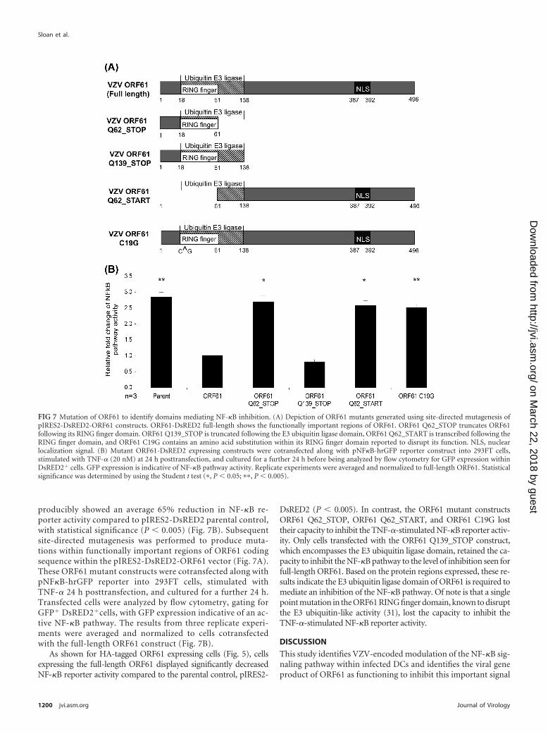
producibly showed an average 65% reduction in NF-�B re-porter activity compared to pIRES2-DsRED2 parental control,with statistical significance (P � 0.005) (Fig. 7B). Subsequentsite-directed mutagenesis was performed to produce muta-tions within functionally important regions of ORF61 codingsequence within the pIRES2-DsRED2-ORF61 vector (Fig. 7A).These ORF61 mutant constructs were cotransfected along withpNF�B-hrGFP reporter into 293FT cells, stimulated withTNF-� 24 h posttransfection, and cultured for a further 24 h.Transfected cells were analyzed by flow cytometry, gating forGFP� DsRED2�cells, with GFP expression indicative of an ac-tive NF-�B pathway. The results from three replicate experi-ments were averaged and normalized to cells cotransfectedwith the full-length ORF61 construct (Fig. 7B).
As shown for HA-tagged ORF61 expressing cells (Fig. 5), cellsexpressing the full-length ORF61 displayed significantly decreasedNF-�B reporter activity compared to the parental control, pIRES2-
DsRED2 (P � 0.005). In contrast, the ORF61 mutant constructsORF61 Q62_STOP, ORF61 Q62_START, and ORF61 C19G losttheir capacity to inhibit the TNF-�-stimulated NF-�B reporter activ-ity. Only cells transfected with the ORF61 Q139_STOP construct,which encompasses the E3 ubiquitin ligase domain, retained the ca-pacity to inhibit the NF-�B pathway to the level of inhibition seen forfull-length ORF61. Based on the protein regions expressed, these re-sults indicate the E3 ubiquitin ligase domain of ORF61 is required tomediate an inhibition of the NF-�B pathway. Of note is that a singlepoint mutation in the ORF61 RING finger domain, known to disruptthe E3 ubiquitin-like activity (31), lost the capacity to inhibit theTNF-�-stimulated NF-�B reporter activity.
DISCUSSION
This study identifies VZV-encoded modulation of the NF-�B sig-naling pathway within infected DCs and identifies the viral geneproduct of ORF61 as functioning to inhibit this important signal
FIG 7 Mutation of ORF61 to identify domains mediating NF-�B inhibition. (A) Depiction of ORF61 mutants generated using site-directed mutagenesis ofpIRES2-DsRED2-ORF61 constructs. ORF61-DsRED2 full-length shows the functionally important regions of ORF61. ORF61 Q62_STOP truncates ORF61following its RING finger domain. ORF61 Q139_STOP is truncated following the E3 ubiquitin ligase domain, ORF61 Q62_START is transcribed following theRING finger domain, and ORF61 C19G contains an amino acid substitution within its RING finger domain reported to disrupt its function. NLS, nuclearlocalization signal. (B) Mutant ORF61-DsRED2 expressing constructs were cotransfected along with pNF�B-hrGFP reporter construct into 293FT cells,stimulated with TNF-� (20 nM) at 24 h posttransfection, and cultured for a further 24 h before being analyzed by flow cytometry for GFP expression withinDsRED2� cells. GFP expression is indicative of NF-�B pathway activity. Replicate experiments were averaged and normalized to full-length ORF61. Statisticalsignificance was determined by using the Student t test (�, P � 0.05; ��, P � 0.005).
Sloan et al.
1200 jvi.asm.org Journal of Virology
on March 22, 2018 by guest
http://jvi.asm.org/
Dow
nloaded from

transduction pathway. Given that DCs are proposed as the firstimmune cells to encounter VZV following inoculation, these re-sults indicate that ORF61 plays an important role in immune eva-sion of the NF-�B signal transduction pathway. This pathwaycontrols transcription of many immune molecules required toinitiate an immune response to foreign pathogens, and so disrup-tion of this pathway is likely to suppress critical immune effectorcapacity of the host cell.
Other herpesviruses modulate the NF-�B pathway in a celltype-dependent manner (3, 9, 19, 23, 24, 27, 28, 34). Our workindicates VZV modulates the NF-�B pathway within DCs. Thecytoplasmic retention of NF-�B protein subunits p50 and p65indicates that the pathway is modulated prior to their transloca-tion into the nucleus, as shown by immunofluorescence and con-focal microscopy. These results are distinct from those reportedfor HSV-1 infection, where NF-�B proteins are translocated intothe nucleus of HSV-1-infected C33-A cells (24). The NF-�B path-way in VZV-infected DCs was not modulated by downregulationof TLR3, TLR8, TLR9, TNFR-1, or TNFR-2, which signal the ac-tivation of the NF-�B pathway, since these were unaffected byVZV infection. This finding is consistent with VZV interferingwith NF-�B signaling at a point downstream of cell surface(TNFR-1 and TNFR-2) or intracellular receptors which triggerthis pathway (TLR3, TLR8, and TLR9).
A critical component of activation of the NF-�B pathway isphosphorylation of I�B� (5). We therefore explored the phos-phorylation state of I�B� in infected DCs. The increase in phos-phorylated I�B� within VZV-infected DCs indicated that theNF-�B pathway was able to be activated at this level followinginfection. In addition, I�B� protein levels within DCs did notdecrease following infection with VZV. Thus, I�B� can be phos-phorylated and is not necessarily degraded following VZV infec-tion of DCs. The NF-�B pathway is therefore apparently inhibitedfollowing phosphorylation of I�B� but prior to translocation ofNF-�B p50 and p65 into the nucleus of VZV-infected DCs. Theidentification of this step is under further consideration.
We identified VZV ORF61 as a gene responsible for inhibitingTNF-�-stimulated NF-�B reporter expression. VZV ORF61 istranscribed within 1 h of infection; however, it is not a componentof the virion, supporting reports of UV-inactivated VZV beingunable to inhibit the NF-�B pathway within fibroblasts and that denovo gene synthesis is required (17, 18, 33). ORF61 has also beenimplicated in the modulation of other cellular pathways, such asthe mitogen-activated protein kinase (MAPK) pathways where asignificant increase in the phosphorylation of JNK/SAPK and adecrease in p38/MAPK phosphorylation were observed in MeWocells transfected with ORF61 (25). ORF61 also downmodulatesthe IRF3-mediated IFN-� pathway by its direct binding to anddegradation of IRF3 (35). We demonstrate that when ORF61 isexpressed alone, it is sufficient to inhibit TNF-�-induced I�B�degradation, further defining the role of ORF61 in NF-�B path-way inhibition. We also showed that the E3 ubiquitin ligase do-main of ORF61 is essential for NF-�B pathway inhibition, sincemutations within this domain rendered ORF61 unable to inhibitNF-�B reporter activity. HSV-1 ICP0 and VZV ORF61 share ho-mology within their RING finger domains, which are involved inE3 ubiquitin ligase activity (21). ICP0 modulates the NF-�B path-way in a cell type-dependent manner, as shown by transfection ofICP0 expression constructs into SHSY-5Y and HEK293 cells.Within HEK293 cells stably expressing either TLR2 or TLR4, ICP0
causes inhibition of TLR-mediated NF-�B signaling (9). ICP0 in-hibits the NF-�B pathway within these cells by binding and trans-porting the host cell protein USP7, a ubiquitin-specific protease,to the cytoplasm, where it deubiquitinates TRAF6 and IKK�. TheUSP7 binding site on ICP0 is within the C-terminal region; due tothe homology of ICP0 and ORF61 being restricted to the RINGfinger domain in the N terminus, it is unlikely that ORF61 encodesthis USP7 binding region and therefore inhibits the NF-�B path-way in a manner different from that of its HSV-1 homologousprotein in HEK293/TLR cells. Expression of ICP0 withinSHSY-5Y cells stimulated NF-�B reporter activity through the E3ubiquitin ligase action of ICP0, resulting in ubiquitination andsubsequent degradation of I�B� (10). Our demonstration that theE3 ubiquitin ligase domain of ORF61 is responsible for the inhi-bition of NF-�B reporter activity indicates that it may be the pro-cess of ubiquitination causing the effect; however, protein expres-sion analysis of I�B� within VZV-infected DCs suggests that I�B�is not degraded. The Vpu protein of HIV, however, blocksproteasome-dependent degradation of I�B� by binding to�TRCP in the E3 ubiquitin ligase complex that is involved in theregulated degradation of I�B� (7). This suggests that the E3 ubiq-uitin ligase activity of ORF61 may have an indirect effect onNF-�B proteins.
Although we have shown that ORF61 is a viral factor thatdownregulates NF-�B, we have not yet verified whether it is theonly viral factor able to do this. This requires the construction ofVZV that lacks the ORF61 protein, particularly in the amino-terminal ring finger domain. Such mutants have proven very dif-ficult to develop. Using a VZV bacterial artificial chromosome(BAC) system, we found that VZV BAC constructs engineered tocontain the amino acid substitution within the RING finger do-main of ORF61 (C19G) or a stop codon inserted directly after theRING finger domain or a stop codon inserted directly followingthe E3 ubiquitin ligase domain all resulted in either failure toobtain any recombinant VZV or the reversion of the point muta-tion to the wild type (M. B. Yee and P. Kinchington, unpublisheddata). This mirrors previous reports of ORF61 playing an impor-tant role in virus replication (8, 33). Thus, although ORF61 hasbeen identified as functioning to inhibit the NF-�B pathway, itremains possible that other VZV gene products may also encode asimilar function. In addition, whether the E3 ubiquitin ligase ac-tivity of ORF61 exerts a direct effect on NF-�B proteins or whetherit impacts on the cellular regulators of these transcription factorswill be an important focus of future work to delineate the mode ofaction of ORF61 in suppressing NF-�B signaling. The continuedpresence of phosphorylated I�B� within VZV-infected DCs indi-cates that it is not being degraded. Therefore, it would be useful toalso investigate ubiquitin/protease pathways that control the deg-radation of I�B� following NF-�B pathway activation.
In summary, we identify here inhibition of the NF-�B signaltransduction pathway in VZV-infected human MDDCs, whichoccurs following phosphorylation of I�B�, but prior to the trans-location of NF-�B p50 and p65 into the nucleus. We also demon-strate that the immediate-early gene ORF61 inhibits TNF-�-stimulated NF-�B reporter expression, indicating that this viralgene can inhibit NF-�B pathway activation. Furthermore, weshow that the region of ORF61 important for its inhibitory affectson TNF-�-stimulated NF-�B reporter activity is the E3 ubiquitinligase domain, indicating that it is the process of ubiquitinationthat causes this pathway inhibition. During the preparation of this
Modulation of NF-�� Activity by VZV ORF61
January 2012 Volume 86 Number 2 jvi.asm.org 1201
on March 22, 2018 by guest
http://jvi.asm.org/
Dow
nloaded from

manuscript, Wang et al. (32) presented findings that ORF61 bindsSUMO-1 via three SUMO-interacting motifs (SIMs) and thatthese SIMs were required for ORF61 binding to and disrupting ofPML nuclear bodies (32). That study also reported that ORF61can act as an inhibitor of TNF-�-induced NF-�B reporter activityin a transient-transfection assay within a melanoma cell line, sup-porting the results presented here. VZV-encoded modulation ofthe NF-�B pathway may be the mechanistic basis for the observeddownregulation of immune molecules in VZV-infected DCs, animportant immune evasion strategy of VZV.
ACKNOWLEDGMENTS
This study was supported by an Australian National Health and MedicalResearch (NHMRC) project grant awarded to A.A. and B.S. E.S. was therecipient of an Australian Postgraduate Award and Westmead Millen-nium Foundation Stipend Enhancement Award. P.K. was supported byNational Institutes of Health grants EY08098 and NS064022, by unre-stricted funds from the Eye and Ear Institute of Pittsburgh, and by Re-search to Prevent Blindness, Inc.
REFERENCES1. Abendroth A, Kinchington PR, Slobedman B. 2010. Varicella-zoster
virus immune evasion strategies. Curr. Top. Microbiol. Immunol. 342:155–171.
2. Abendroth A, Morrow G, Cunningham AL, Slobedman B. 2001.Varicella-zoster virus infection of human dendritic cells and transmissionto T cells: implications for virus dissemination in the host. J. Virol. 75:6183– 6192.
3. Amici C, et al. 2006. Herpes simplex virus disrupts NF-�B regulation byblocking its recruitment on the I�B� promoter and directing the factor onviral genes. J. Biol. Chem. 281:7110 –7117.
4. Baldwin AS, Jr. 1996. The NF-�B and I �B proteins: new discoveries andinsights. Annu. Rev. Immunol. 14:649 – 683.
5. Beg AA, Baldwin AS, Jr. 1993. The I �B proteins: multifunctional regu-lators of Rel/NF-�B transcription factors. Genes Dev. 7:2064 –2070.
6. Blackwell TS, Christman JW. 1997. The role of nuclear factor-�B incytokine gene regulation. Am. J. Respir. Cell Mol. Biol. 17:3–9.
7. Bour S, Perrin C, Akari H, Strebel K. 2001. The human immunodefi-ciency virus type 1 Vpu protein inhibits NF-�B activation by interferingwith beta TrCP-mediated degradation of I�B. J. Biol. Chem. 276:15920 –15928.
8. Cohen JI, Nguyen H. 1998. Varicella-zoster virus ORF61 deletion mu-tants replicate in cell culture, but a mutant with stop codons in ORF61reverts to wild-type virus. Virology 246:306 –316.
9. Daubeuf S, et al. 2009. HSV ICP0 recruits USP7 to modulate TLR-mediated innate response. Blood 113:3264 –3275.
10. Diao L, et al. 2005. Herpesvirus proteins ICP0 and BICP0 can activateNF-�B by catalyzing I�B� ubiquitination. Cell. Signal. 17:217–229.
11. Eisfeld AJ, Yee MB, Erazo A, Abendroth A, Kinchington PR. 2007.Downregulation of class I major histocompatibility complex surface ex-pression by varicella-zoster virus involves open reading frame 66 proteinkinase-dependent and -independent mechanisms. J. Virol. 81:9034 –9049.
12. Foo SY, Nolan GP. 1999. NF-�B to the rescue: RELs, apoptosis andcellular transformation. Trends Genet. 15:229 –235.
13. Ghosh S, Karin M. 2002. Missing pieces in the NF-�B puzzle. Cell109(Suppl):S81–S96.
14. Ghosh S, May MJ, Kopp EB. 1998. NF-�B and Rel proteins: evolution-arily conserved mediators of immune responses. Annu. Rev. Immunol.16:225–260.
15. Hood C, Cunningham AL, Slobedman B, Boadle RA, Abendroth A.2003. Varicella-zoster virus-infected human sensory neurons are resistant
to apoptosis, yet human foreskin fibroblasts are susceptible: evidence for acell-type-specific apoptotic response. J. Virol. 77:12852–12864.
16. Huch JH, et al. 2010. Impact of varicella-zoster virus on dendritic cellsubsets in human skin during natural infection. J. Virol. 84:4060 – 4072.
17. Jones JO, Arvin AM. 2006. Inhibition of the NF-�B pathway by varicella-zoster virus in vitro and in human epidermal cells in vivo. J. Virol. 80:5113–5124.
18. Kinchington PR, Vergnes JP, Turse SE. 1995. Transcriptional mappingof varicella-zoster virus regulatory proteins. Neurology 45:S33–S35.
19. Laherty CD, Hu HM, Opipari AW, Wang F, Dixit VM. 1992. TheEpstein-Barr virus LMP1 gene product induces A20 zinc finger proteinexpression by activating nuclear factor �B. J. Biol. Chem. 267:24157–24160.
20. Miranda-Saksena M, Armati P, Boadle RA, Holland DJ, CunninghamAL. 2000. Anterograde transport of herpes simplex virus type 1 in cul-tured, dissociated human and rat dorsal root ganglion neurons. J. Virol.74:1827–1839.
21. Moriuchi H, Moriuchi M, Smith HA, Straus SE, Cohen JI. 1992.Varicella-zoster virus open reading frame 61 protein is functionally ho-mologous to herpes simplex virus type 1 ICP0. J. Virol. 66:7303–7308.
22. Morrow G, Slobedman B, Cunningham AL, Abendroth A. 2003.Varicella-zoster virus productively infects mature dendritic cells and alterstheir immune function. J. Virol. 77:4950 – 4959.
23. Moutaftsi M, Brennan P, Spector SA, Tabi Z. 2004. Impaired lymphoidchemokine-mediated migration due to a block on the chemokine receptorswitch in human cytomegalovirus-infected dendritic cells. J. Virol. 78:3046 –3054.
24. Patel A, et al. 1998. Herpes simplex type 1 induction of persistent NF-�Bnuclear translocation increases the efficiency of virus replication. Virology247:212–222.
25. Rahaus M, Desloges N, Wolff MH. 2005. ORF61 protein of varicella-zoster virus influences JNK/SAPK and p38/MAPK phosphorylation. J.Med. Virol. 76:424 – 433.
26. Rahman MM, McFadden G. 2011. Modulation of NF-�B signalling bymicrobial pathogens. Nat. Rev. Microbiol. 9:291–306.
27. Stewart S, et al. 2004. Epstein-Barr virus-encoded LMP2A regulates viraland cellular gene expression by modulation of the NF-�B transcriptionfactor pathway. Proc. Natl. Acad. Sci. U. S. A. 101:15730 –15735.
28. Sylla BS, et al. 1998. Epstein-Barr virus-transforming protein latent in-fection membrane protein 1 activates transcription factor NF-�B througha pathway that includes the NF-�B-inducing kinase and the I�B kinasesIKK� and IKK�. Proc. Natl. Acad. Sci. U. S. A. 95:10106 –10111.
29. Vallabhapurapu S, Karin M. 2009. Regulation and function of NF-�Btranscription factors in the immune system. Annu. Rev. Immunol. 27:693–733.
30. Van Tendeloo VF, et al. 2001. Highly efficient gene delivery by mRNAelectroporation in human hematopoietic cells: superiority to lipofectionand passive pulsing of mRNA and to electroporation of plasmid cDNA fortumor antigen loading of dendritic cells. Blood 98:49 –56.
31. Walters MS, Kyratsous CA, Silverstein SJ. 2010. The RING finger do-main of varicella-zoster virus ORF61p has E3 ubiquitin ligase activity thatis essential for efficient auto-ubiquitination and dispersion of Sp100 con-taining nuclear bodies. J. Virol. 84:6861– 6865.
32. Wang L, et al. 2011. Disruption of PML nuclear bodies is mediated byORF61 SUMO-interacting motifs and required for varicella-zoster viruspathogenesis in skin. PLoS Pathog. 7:e1002157.
33. Wang L, Sommer M, Rajamani J, Arvin AM. 2009. Regulation of theORF61 promoter and ORF61 functions in varicella-zoster virus replica-tion and pathogenesis. J. Virol. 83:7560 –7572.
34. Yurochko AD, Kowalik TF, Huong SM, Huang ES. 1995. Humancytomegalovirus upregulates NF-�B activity by transactivating the NF-�Bp105/p50 and p65 promoters. J. Virol. 69:5391–5400.
35. Zhu H, et al. 2011. Varicella-zoster virus immediate early protein ORF61abrogates IRF3-mediated innate immune response through degradationof activated IRF3. J. Virol. 85:11079 –11089.
Sloan et al.
1202 jvi.asm.org Journal of Virology
on March 22, 2018 by guest
http://jvi.asm.org/
Dow
nloaded from